EP4166676A1 - Technique de contrôle de la qualité des cellules souches pluripotentes humaines utilisant un bouillon de culture - Google Patents
Technique de contrôle de la qualité des cellules souches pluripotentes humaines utilisant un bouillon de culture Download PDFInfo
- Publication number
- EP4166676A1 EP4166676A1 EP21821235.5A EP21821235A EP4166676A1 EP 4166676 A1 EP4166676 A1 EP 4166676A1 EP 21821235 A EP21821235 A EP 21821235A EP 4166676 A1 EP4166676 A1 EP 4166676A1
- Authority
- EP
- European Patent Office
- Prior art keywords
- fibronectin
- cells
- probe
- ssea
- pluripotent stem
- Prior art date
- Legal status (The legal status is an assumption and is not a legal conclusion. Google has not performed a legal analysis and makes no representation as to the accuracy of the status listed.)
- Granted
Links
Images


Classifications
-
- G—PHYSICS
- G01—MEASURING; TESTING
- G01N—INVESTIGATING OR ANALYSING MATERIALS BY DETERMINING THEIR CHEMICAL OR PHYSICAL PROPERTIES
- G01N33/00—Investigating or analysing materials by specific methods not covered by groups G01N1/00 - G01N31/00
- G01N33/48—Biological material, e.g. blood, urine; Haemocytometers
- G01N33/50—Chemical analysis of biological material, e.g. blood, urine; Testing involving biospecific ligand binding methods; Immunological testing
- G01N33/68—Chemical analysis of biological material, e.g. blood, urine; Testing involving biospecific ligand binding methods; Immunological testing involving proteins, peptides or amino acids
- G01N33/6887—Chemical analysis of biological material, e.g. blood, urine; Testing involving biospecific ligand binding methods; Immunological testing involving proteins, peptides or amino acids from muscle, cartilage or connective tissue
-
- G—PHYSICS
- G01—MEASURING; TESTING
- G01N—INVESTIGATING OR ANALYSING MATERIALS BY DETERMINING THEIR CHEMICAL OR PHYSICAL PROPERTIES
- G01N33/00—Investigating or analysing materials by specific methods not covered by groups G01N1/00 - G01N31/00
- G01N33/48—Biological material, e.g. blood, urine; Haemocytometers
- G01N33/50—Chemical analysis of biological material, e.g. blood, urine; Testing involving biospecific ligand binding methods; Immunological testing
- G01N33/5005—Chemical analysis of biological material, e.g. blood, urine; Testing involving biospecific ligand binding methods; Immunological testing involving human or animal cells
- G01N33/5008—Chemical analysis of biological material, e.g. blood, urine; Testing involving biospecific ligand binding methods; Immunological testing involving human or animal cells for testing or evaluating the effect of chemical or biological compounds, e.g. drugs, cosmetics
- G01N33/5044—Chemical analysis of biological material, e.g. blood, urine; Testing involving biospecific ligand binding methods; Immunological testing involving human or animal cells for testing or evaluating the effect of chemical or biological compounds, e.g. drugs, cosmetics involving specific cell types
- G01N33/5073—Stem cells
-
- C—CHEMISTRY; METALLURGY
- C07—ORGANIC CHEMISTRY
- C07K—PEPTIDES
- C07K14/00—Peptides having more than 20 amino acids; Gastrins; Somatostatins; Melanotropins; Derivatives thereof
- C07K14/435—Peptides having more than 20 amino acids; Gastrins; Somatostatins; Melanotropins; Derivatives thereof from animals; from humans
- C07K14/78—Connective tissue peptides, e.g. collagen, elastin, laminin, fibronectin, vitronectin or cold insoluble globulin [CIG]
-
- G—PHYSICS
- G01—MEASURING; TESTING
- G01N—INVESTIGATING OR ANALYSING MATERIALS BY DETERMINING THEIR CHEMICAL OR PHYSICAL PROPERTIES
- G01N33/00—Investigating or analysing materials by specific methods not covered by groups G01N1/00 - G01N31/00
- G01N33/48—Biological material, e.g. blood, urine; Haemocytometers
- G01N33/50—Chemical analysis of biological material, e.g. blood, urine; Testing involving biospecific ligand binding methods; Immunological testing
- G01N33/5005—Chemical analysis of biological material, e.g. blood, urine; Testing involving biospecific ligand binding methods; Immunological testing involving human or animal cells
- G01N33/5008—Chemical analysis of biological material, e.g. blood, urine; Testing involving biospecific ligand binding methods; Immunological testing involving human or animal cells for testing or evaluating the effect of chemical or biological compounds, e.g. drugs, cosmetics
- G01N33/502—Chemical analysis of biological material, e.g. blood, urine; Testing involving biospecific ligand binding methods; Immunological testing involving human or animal cells for testing or evaluating the effect of chemical or biological compounds, e.g. drugs, cosmetics for testing non-proliferative effects
- G01N33/5023—Chemical analysis of biological material, e.g. blood, urine; Testing involving biospecific ligand binding methods; Immunological testing involving human or animal cells for testing or evaluating the effect of chemical or biological compounds, e.g. drugs, cosmetics for testing non-proliferative effects on expression patterns
-
- G—PHYSICS
- G01—MEASURING; TESTING
- G01N—INVESTIGATING OR ANALYSING MATERIALS BY DETERMINING THEIR CHEMICAL OR PHYSICAL PROPERTIES
- G01N2333/00—Assays involving biological materials from specific organisms or of a specific nature
- G01N2333/435—Assays involving biological materials from specific organisms or of a specific nature from animals; from humans
- G01N2333/78—Connective tissue peptides, e.g. collagen, elastin, laminin, fibronectin, vitronectin, cold insoluble globulin [CIG]
-
- G—PHYSICS
- G01—MEASURING; TESTING
- G01N—INVESTIGATING OR ANALYSING MATERIALS BY DETERMINING THEIR CHEMICAL OR PHYSICAL PROPERTIES
- G01N2470/00—Immunochemical assays or immunoassays characterised by the reaction format or reaction type
- G01N2470/04—Sandwich assay format
Definitions
- the present invention relates to a method for determining or evaluating the undifferentiated state of human pluripotent stem cells using a culture supernatant, a method for controlling the quality of human pluripotent stem cells, and a kit for use in these methods.
- Regenerative medicine involves transplanting pluripotent stem cells collected from oneself or another person or cells differentiated from these cells into tissues or organs with impaired functions to compensate for such tissues or organs, thereby reconstructing the tissues or regenerating the organs.
- great expectations have been placed on regenerative medicine capable of performing fundamental repairment and/or regeneration upon treatment of diseases against which only symptomatic treatment such as drug treatment etc., is available.
- Pluripotent stem cells such as embryonic stem cells (ES cells) and induced pluripotent stem cells (iPS cells) retain the undifferentiated state and can be induced to differentiate into various cells (e.g., hepatocytes, myocardial cells, etc.).
- pluripotent stem cells are expected to be a source of cells for transplantation in regenerative medicine.
- it is important that the quality of human pluripotent stem cells being a cell source should be stable (in other words, the undifferentiated state should be maintained).
- maintaining the undifferentiated state is not easy since the properties of human pluripotent stem cells are easily changed by passage or manipulation. Therefore, it is extremely important to determine or evaluate the undifferentiated state of human pluripotent stem cells in controlling the quality.
- the undifferentiated state of human pluripotent stem cells has been determined or evaluated by finding cells with changed morphology and in an advanced differentiation state from among cells observed under a microscope.
- markers indicating the undifferentiated state of human pluripotent stem cells e.g., Oct3/4, rBC2-LCN, SSEA-4, Nanog, SOX2, c-Myc, Klf4, Lin28, TRA-1-60, TRA-1-81 etc.
- markers whose expression is observed in highly differentiated cells e.g., SSEA-1 etc.
- the presence or absence of the expression of these markers is confirmed by using a flow cytometer or immunostaining etc., and then the undifferentiated state of human pluripotent stem cells can be evaluated.
- An object of the present invention is to provide a novel method that enables solving the above problems of the conventional methods for determining or evaluating the undifferentiated state of human pluripotent stem cells, and to simply, efficiently, and non-invasively determine or evaluate the undifferentiated state of human pluripotent stem cells.
- a culture supernatant of human pluripotent stem cells including cells having a low undifferentiated level contains fibronectin or SSEA-1-positive fibronectin; and that the proportion of such cells having a low undifferentiated level correlates with the amount of fibronectin or SSEA-1-positive fibronectin in the culture supernatant.
- the present inventors have further found that through detection or measurement of fibronectin or SSEA-1-positive fibronectin contained in a culture supernatant of human pluripotent stem cells, the undifferentiated state of human pluripotent stem cells can be determined or evaluated; and that based on the determination or evaluation results, the quality of human pluripotent stem cells can be controlled.
- the present inventors have further found that through the removal of fibronectin-positive cells as a target, cells having a low undifferentiated level can be selectively removed and thus the quality of human pluripotent stem cells can be controlled.
- the present invention is based on these findings and includes the following inventions.
- the present invention is to detect or measure fibronectin or SSEA-1-positive fibronectin contained in a culture supernatant of human pluripotent stem cells as a test sample, so as to determine or evaluate the undifferentiated state of human pluripotent stem cells based on the detection or the measurement result of fibronectin or SSEA-1-positive fibronectin.
- the undifferentiated state can be simply determined or evaluated without reducing valuable human pluripotent stem cells, and, cells having a low undifferentiated level can be selectively removed, so that the quality of human pluripotent stem cells can be controlled.
- pluripotency refers to the capability of differentiating into cells of any lineage of three germ layers (endoderm, mesoderm, ectoderm) (but unable to differentiate into blastocyst). Namely, it means “pluripotency.”
- human pluripotent stem cells refers to human cells being pluripotent and potentially capable of differentiating into various tissues of a living body including three germ layers (endoderm, mesoderm, ectoderm). Examples of such cells include human embryonic stem cells (ES cells), human induced pluripotent stem cells (iPS cells), and cells having pluripotency equivalent thereto.
- ES cells human embryonic stem cells
- iPS cells human induced pluripotent stem cells
- the "human pluripotent stem cells” are human ES cells or human iPS cells, and more preferably human iPS cells.
- human ES cells refers to pluripotent stem cells that can be produced by taking out an inner cell mass from a human early embryo (blastocyst) and culturing it on a feeder cell ( Thomson JA. et al., Science. 1998 Nov 6; 282(5391):1145-7 .).
- human ES cells may be those produced from a human early embryo (blastocyst) according to a conventionally known means, or established human ES cell lines (e.g., KhES-1 to KhES-5 cell lines, HES1 to HES6 cell lines, CHB-1 to CHB-12 cell lines, RUES1 cell line, RUES2 cell line, HUES1 to HUES28 cell lines, WA01 (H1) cell line, WA07 (H7) cell line, WA09 (H9) cell line, WA13 (H13) cell line, WA14 (H14) cell line, SA002 cell line, SA181 cell line, SA611 cell line, etc. (examples thereof are not limited thereto)).
- KhES-1 to KhES-5 cell lines e.g., HES-1 to KhES-5 cell lines, HES1 to HES6 cell lines, CHB-1 to CHB-12 cell lines, RUES1 cell line, RUES2 cell line, HUES1 to HUES28 cell lines
- human iPS cells refers to induced pluripotent stem cells that can be produced by introducing specific factors known as nuclear reprogramming factors (e.g., Oct3/4, Sox2, Klf4, c-Myc, Nanog, Lin28, L-Myc, hTERT, SV40 large T, etc.) into human somatic cells or undifferentiated stem cells for reprogramming (initialization) these cells ( Takahashi K. et al., Cell (2007) 131, 861-872 ; Okita, K. et al., Nature (2007) 448, 313-317 ; Nakagawa M. et al., Nature Biotechnology, (2008)26, 101-106 ; Yu J. et al., Science (2007)318, 1917-1920 ; Park IH. et al., Nature (2007) 451, 141-146 etc.).
- nuclear reprogramming factors e.g., Oct3/4, Sox2, Klf4, c-Myc, Nanog, Lin28
- human iPS cells may be those produced from human somatic cells or undifferentiated stem cells according to conventionally known means, or established human iPS cell lines (e.g., 201B7 cell line, 253G1 cell line, 409B2 cell line, 454E2 cell line, 606A1 cell line, 610B1 cell line, 648A1 cell line, Ff-WJ-18 cell line, Ff-I01s01 cell line, Ff-I01s02 cell line, Ff-I01s04 cell line, Ff-I01s06 cell line, Ff-I14s03 cell line, Ff-I14s04 cell line, QHJI01s01 cell line, QHJI01s04 cell line, QHJI14s03 cell line, QHJI14s04 cell line, 253G1 cell line, HiPS-RIKEN-1A cell line, HiPS-RIKEN-2A cell line, HiPS-RIKEN-12A cell line, Nips-B2 cell line,
- undifferentiated state of human pluripotent stem cells refers to a state in which human pluripotent stem cells retain pluripotency and have not differentiated (also referred to as “deviated”).
- differentiation occurs in human pluripotent stem cells, pluripotency decreases or disappears, and accordingly, the level of "undifferentiated state” (or the undifferentiated level) is reduced or disappears.
- the expression of the above undifferentiation marker gene(s) and/or protein(s) is decreased or disappears as compared with human pluripotent stem cells retaining the undifferentiated state, and instead, the expression of differentiation markers, fibronectin gene and/or protein such as SSEA-1, vimentin, N-cadherin, Twist, Zeb1/2, Snail, Slug, etc., is observed.
- human pluripotent stem cells or human iPS cells having a low undifferentiated level
- human pluripotent stem cells or human iPS cells having a reduced undifferentiated level
- the undifferentiated level of human pluripotent stem cells (or human iPS cells) has been reduced refers to a population of human pluripotent stem cells (or human iPS cells) including the cells whose undifferentiated level has been reduced or disappeared.
- human pluripotent stem cells having a low undifferentiated level
- human pluripotent stem cells having a reduced undifferentiated level
- the undifferentiated level of human pluripotent stem cells (or human iPS cells) has been reduced can include a form in which human pluripotent stem cells (or human iPS cells) retaining the undifferentiated state and cells whose undifferentiated level has been reduced or disappeared coexist.
- Fibronectin is a large secretory glycoprotein known as a cell adhesion molecule, which is known to be synthesized in various cells and secreted extracellularly (Gene ID: 2335 in the NCBI database). Fibronectin is known to have many functions that support the normal vital functions of spinal functions, such as adhesion to extracellular matrix, formation and retention of connective tissue, wound healing, and formation and maintenance of tissue and organ morphologies and compartments during embryogenesis. Fibronectin (monomer) is a soluble glycoprotein having a molecular weight of 210 to 250 kDa (2,146 to 2,325 amino acid residues), and is known to have seven N-type sugar chains and one O-type sugar chain. In the present invention, examples of the "fibronectin” may include the following "SSEA-1 positive fibronectin".
- SSEA-1 positive fibronectin refers to a glycoprotein having secretory fibronectin, to which SSEA-1 is bound, as a core protein.
- SSEA-1 (Stage-specific Embryonic Antigen-1) is a sugar chain marker having Lewis X, that is, Gal ⁇ 1-4 (Fuc ⁇ 1-3) GlcNAc as a sugar chain antigen (epitope). It has been reported that SSEA-1 is not expressed in human pluripotent stem cells retaining the undifferentiated state, and its expression on the cell membrane increases with differentiation ( Thomson JA. et al., Science. 1998 Nov 6; 282 (5391): 1145-7 .). SSEA-1 is considered to be bound to the non-reducing end of one or more sugar chains of seven N-type sugar chains and one O-type sugar chain of fibronectin.
- fibronectin or SSEA-1 positive fibronectin is detected or measured using a culture supernatant of an in vitro culture of human pluripotent stem cells as a test sample.
- a culture supernatant By using a culture supernatant, the undifferentiated state can be determined or evaluated non-invasively without using valuable human pluripotent stem cells that should originally be used for transplantation therapy.
- a culture supernatant is generally replaced with a fresh culture supernatant at regular intervals. Therefore, fibronectin or SSEA-1 positive fibronectin is detected or measured after a detectable amount of fibronectin or SSEA-1 positive fibronectin is secreted into the culture supernatant after replacement.
- the time required for a detectable amount of fibronectin or SSEA-1-positive fibronectin to be secreted in a culture supernatant after replacement of the culture supernatant may vary depending on the type and amount of cells and the culture conditions. Therefore, the time required for collecting a culture supernatant to be used for detecting or measuring fibronectin or SSEA-1 positive fibronectin after replacement of the culture supernatant can be appropriately set depending on the cell type and culture conditions, but ranges from, for example, about 18 to 72 hours. Since medium exchange is usually performed about every 1 to 3 days, it is preferable to use a culture supernatant to be discarded at the time of medium exchange.
- a culture supernatant collected using a conventionally known solid-liquid separation method e.g., centrifugation, filtration, etc.
- examples of a probe that can be used as a "probe that recognizes fibronectin” include proteins, polypeptides, and compounds capable of recognizing and binding to fibronectin.
- an anti-fibronectin antibody or a fragment thereof that recognizes fibronectin as an antigen (epitope) can be used.
- the anti-fibronectin antibody can be prepared by a conventional method, and can be obtained, for example, by immunizing an animal with fibronectin or a fragment thereof as it is or with the same bound to a carrier protein such as albumin or KLH.
- the anti-fibronectin antibody may be any antibody capable of recognizing and binding to, and preferably specifically binding to fibronectin as an epitope.
- anti-fibronectin antibody examples include a polyclonal antibody, a monoclonal antibody, a chimeric antibody, a humanized antibody, and a fully human antibody.
- a fragment of the anti-fibronectin antibody can also be used as long as it is capable of recognizing and binding to, and preferably specifically binding to fibronectin as an epitope.
- antibody fragments include Fab, Fab', F (ab') 2 , Fv, scFv, dsFv, diabodies, sc (Fv) 2 , etc. Polymers of these fragments (e.g., dimers, trimmers, tetramers, polymers etc.) can also be used in the present invention.
- Known or commercially available anti-fibronectin antibodies can also be used in the present invention.
- One type of such probe that recognizes fibronectin may be used alone, or two or more, a plurality of types of probes may be used in combination.
- SSEA-1 positive fibronectin in a culture supernatant can be detected or measured using (a) a probe that recognizes SSEA-1 as an epitope and (b) a probe that recognizes the fibronectin described above.
- the use of these two types of probes enables detection of a target protein with high specificity and high sensitivity, and thus is preferable.
- examples of (a) "a probe that recognizes SSEA-1 as an epitope” that can be used herein include an SSEA-1 binding lectin that recognizes and binds to the sugar chain structure of SSEA-1 (e.g., Lotus agglutinin (Lotus Tetragolonobus lectin, LTL), DC-SIGN, lectin derived from Pseudomonas aeruginosa ( Pseudomonas aeruginosa lectin, PA-IIL), etc.
- SSEA-1 binding lectin that recognizes and binds to the sugar chain structure of SSEA-1
- DC-SIGN lectin derived from Pseudomonas aeruginosa
- PA-IIL Pseudomonas aeruginosa lectin
- the "probe that recognizes SSEA-1 as an epitope” is an anti-SSEA-1 antibody or a fragment thereof.
- the anti-SSEA-1 antibody can be prepared by a conventional method, for example, by immunizing an animal with SSEA-1 as it is or SSEA-1 bound to a carrier protein such as albumin or keyhole limpet hemocyanin (KLH).
- a carrier protein such as albumin or keyhole limpet hemocyanin (KLH).
- KLH keyhole limpet hemocyanin
- a known or commercially available anti-SSEA-1 antibody can also be used in the present invention.
- the anti-SSEA-1 antibody may be any antibody capable of recognizing and binding to, and preferably specifically binding to SSEA-1 as an epitope.
- examples of the anti-SSEA-1 antibody that can be used without particular limitation include a polyclonal antibody, a monoclonal antibody, a chimeric antibody, a humanized antibody, a fully human antibody etc.
- a fragment of the anti-SSEA-1 antibody can also be used as long as it is capable of recognizing and binding to, and preferably specifically binding to SSEA-1 as an epitope. As such an antibody fragment, those defined above can be used.
- One type of such probe that recognizes SSEA-1 as an epitope may be used alone, or two or more, a plurality of types of such probes may be used in combination.
- Fibronectin or SSEA-1 positive fibronectin in a culture supernatant can be detected or measured by a technique that is generally employed for the detection or quantification of a protein in combination with the use of each of the above probes.
- the detection or measurement can be performed using one or more of such techniques such as enzyme-linked immunosorbent assay (ELISA) method, a sandwich assay method, a competitive method, a Western blotting method, a chromatography method, immunoprecipitation etc.
- ELISA enzyme-linked immunosorbent assay
- fibronectin in a culture supernatant is detected or measured by a sandwich assay method using two or more types of probes that recognizes fibronectin, and more preferably, SSEA-1 positive fibronectin in a culture supernatant is detected or measured by the sandwich assay method using probes (a) and (b) above.
- one of the probes to be used (hereinafter referred to as "first probe") is immobilized on a substrate.
- a substrate on which the first probe is immobilized include plates (e.g., microwell plate, etc.), microarray substrates (e.g., slide glass for microarray, etc.), tubes, beads (e.g., plastic beads, magnetic beads, etc.), and carriers for chromatography (e.g., Sepharose (Trade Mark), etc.), membranes (e.g., nitrocellulose membrane, PVDF membrane, etc.), and gels (e.g., polyacrylamide gel, etc.).
- plates, beads and membranes are preferably used, and plates are most preferably used because of their ease of handling.
- the first probe can be immobilized on a substrate by immobilization using a biotin-avidin bond.
- the first probe to be immobilized is biotinylated in advance and then the biotinylated probe can be prepared in a form immobilized on a streptavidin-coated substrate (well). In this case, the detection sensitivity is improved and the background can be significantly reduced.
- immobilization methods include a physical adsorption method and a chemical bonding method.
- the physical adsorption method examples thereof include methods of activating the surface by ⁇ -ray treatment, electron beam treatment, UV treatment, plasma treatment, corona treatment, etc.
- a culture supernatant which is a test sample, is diluted or not diluted with a buffer solution, and then added to a well in which the first probe has been immobilized, for interaction. Non-specifically bound contaminants are washed off with a buffer solution.
- a buffer solution containing a probe different from the first probe (hereinafter referred to as "second probe"), which has been indirectly or directly labeled, is added for reaction.
- a label for the second probe include, but are not limited to, labeling substances such as fluorescent substances (e.g., FITC, rhodamine, Cy3, Cy5, etc.), radioactive substances (e.g., 13C, 3H, etc.), and enzymes (e.g., alkaline phosphatase, peroxidase (horseradish peroxidase, etc.), glucose oxidase, ⁇ -galactosidase, etc.).
- the second probe may be labeled with biotin and (strepto)avidin may be labeled with the above-mentioned labeling substance to utilize the binding between biotin and (strepto)avidin.
- cases of "indirectly or directly labeled probe different from the first probe” or “labeled second probe” include a case in which the probe itself is not directly labeled, but the probe is indirectly labeled with a labeled second antibody that binds to the probe for detection.
- a substrate to be used herein is o-phenylenediamine (OPD), tetramethylbenzidine (TMB), 2,2'-azinobis [3-ethylbenzothiazolin-6-sulfonic acid] (ABTS), 3,3'-diaminobenzidine (DAB), Amplex (registered trademark) Red, 3-(p-hydroxyphenyl)propion (HPPA), luminol, or the like.
- OPD o-phenylenediamine
- TMB tetramethylbenzidine
- ABTS 2,2'-azinobis [3-ethylbenzothiazolin-6-sulfonic acid]
- DABTS 3,3'-diaminobenzidine
- Amplex registered trademark
- HPPA 3-(p-hydroxyphenyl)propion
- PNPP p-nitrophenyl phosphate
- BCIP 5-bromo-4-chloro-3-indolyl phosphate
- NBT nitrogen triphosphate
- AttoPhos registered trademark
- 4-methylumbelliferyl phosphate 4-methylumbelliferyl phosphate (4-MUP)
- ECF DDAO phosphate
- dioxetane CDP-Star (trademark)
- AMPPD registered trademark
- CSPD registered trademark
- fibronectin or SSEA-1-positive fibronectin in a culture supernatant binds to the first probe immobilized on the substrate and also binds to the labeled second probe to form a complex. Subsequently, a signal generated from the labeled second probe in this complex is detected or measured, so that fibronectin or SSEA-1-positive fibronectin in the sample can be detected or measured.
- the signal can be detected or measured using an appropriate detection or measurement device depending on the labeling substance used.
- Examples of a method for detecting or measuring the signal include ELISA, immunochromatography, radioimmunoassay (RIA), fluorescence immunoassay (FIA method), chemiluminescent immunoassay, and an evanescent wave analysis method. These methods are known to those skilled in the art, and any method may be selected. In addition, these methods may be performed according to ordinary procedures, and the actual setting of reaction conditions etc., are within the technical scope normally available to those skilled in the art.
- signal detection or measurement is preferably carried out by the sandwich ELISA assay method that involves detecting or measuring a signal generated from a second probe labeled with an enzyme such as horseradish peroxidase.
- the above sandwich assay method is performed using recombinant fibronectin expressed by human cells, or, fibronectin or SSEA-1-positive fibronectin purified from a culture supernatant of cells expressing the fibronectin or SSEA-1-positive fibronectin, as a standard substance, and then the amount of the labeled signal corresponding to the amount of the standard substance can be detected and blotted to prepare a calibration curve.
- the amount of the labeled signal can be converted into the amount of the fibronectin or SSEA-1 positive fibronectin, and then the fibronectin or SSEA-1 positive fibronectin in the culture supernatant can be quantitatively measured. Furthermore, with the use of a culture supernatant of human pluripotent stem cells having a low undifferentiated level, a culture supernatant of recombinant cells into which a gene encoding fibronectin has been introduced, a culture supernatant of recombinant cells into which a gene encoding Fut9 for Lewis X synthesis and a gene encoding fibronectin have been introduced, a culture supernatant of fibronectin-expressing cells, or a culture supernatant of SSEA-1 positive fibronectin-expressing cells, or fibronectin or SSEA-1-positive fibronectin purified from such a culture supernatant, as a standard substance, the amount of the labeled signal corresponding to a predetermined number of
- the undifferentiated state of human pluripotent stem cells can be determined or evaluated based on the above results of detecting or measuring fibronectin or SSEA-1 positive fibronectin in a culture supernatant of human pluripotent stem cells.
- the detection or measurement results of fibronectin or SSEA-1 positive fibronectin in a culture supernatant of human pluripotent stem cells correlates with the undifferentiated state of human pluripotent stem cells.
- fibronectin or SSEA-1 positive fibronectin is detected in a culture supernatant, it can be determined and evaluated that the undifferentiated level of the pluripotent stem cells has been reduced, that is, cells whose undifferentiated level has been reduced or disappeared are included.
- controlling the quality of human pluripotent stem cells means controlling the undifferentiated state of human pluripotent stem cells, which can be performed according to the determination or evaluation result of the undifferentiated state of human pluripotent stem cells. For example, when fibronectin or SSEA-1-positive fibronectin is detected in a culture supernatant, or a predetermined amount of fibronectin or SSEA-1-positive fibronectin is detected in a culture supernatant by the above method for determining or evaluating the undifferentiated state of human pluripotent stem cells, the human pluripotent stem cells in the culture are determined to be human pluripotent stem cells having a low undifferentiated level.
- the culture can be discarded, the human pluripotent stem cells can be collected from the culture, or cells having a low undifferentiated level can be removed from the culture, for example.
- human pluripotent stem cells having a low undifferentiated level or human pluripotent stem cells in an undifferentiated state
- a criterion can be set for determining that "human pluripotent stem cells (or human iPS cells) having a low undifferentiated level", "human pluripotent stem cells (or human iPS cells) having a reduced undifferentiated level", or "the undifferentiated level of human pluripotent stem cells (or human iPS cells) has been reduced” means that the proportion of cells whose undifferentiated level has been reduced or disappeared accounts for 10% or more of the total number of cells. Based on this criterion, when the proportion of cells whose undifferentiated level has been reduced or disappeared accounts for less than 10% of the total number of cells, these cells can be supplied as human pluripotent stem cells having stable quality.
- the criterion can be set arbitrarily and is not limited to the above value.
- Cells having a low undifferentiated level can be removed from the culture by isolating, from the culture, or killing cells expressing fibronectin (referred to as “fibronectin-positive cells") or cells expressing SSEA-1-positive fibronectin (referred to as “SSEA-1-positive fibronectin-positive cells”).
- fibronectin-positive cells cells expressing fibronectin
- SSEA-1-positive fibronectin-positive cells cells expressing SSEA-1-positive fibronectin-positive cells
- the "fibronectin-positive cells” may include "SSEA-1-positive fibronectin-positive cells”.
- Fibronectin-positive cells or SSEA-1-positive fibronectin-positive cells can be isolated by a commonly employed technique for cell isolation or purification, for example, by one or more of chromatography, magnetic separation, centrifugation, flow cytometer, cell sorter (fluorescence activated cell sorter (FACS), etc.), etc. combined with the use of the above probes.
- FACS fluorescence activated cell sorter
- Fibronectin-positive cells or SSEA-1-positive fibronectin-positive cells can be killed using any means capable of selectively, preferably specifically, killing these cells.
- a technique, so-called photoimmunotherapy, neutron capture therapy etc. that combines the use of the above probe(s) labeled with a predetermined photosensitizer with exposure to near infrared rays (wavelength 700-2,500 nm), ultraviolet rays (wavelength 10-400 nm), or thermal neutron rays can be used.
- Examples of a photosensitizer include a light-absorbing phthalocyanine dye IRDye700Dx (IR700), boron 10 ( 10 B), cadmium selenide (CdSe), etc., that is activated by near infrared radiation.
- IR700 light-absorbing phthalocyanine dye
- 10 B boron 10
- CdSe cadmium selenide
- the probe labeled with IR700 is bound to target fibronectin-positive cells or SSEA-1-positive fibronectin-positive cells, and then exposed to near infrared rays; the probe labeled with 10 B is bound to target fibronectin-positive cells or SSEA-1-positive fibronectin-positive cells, and then exposed to thermal neutron rays; or the probe labeled with CdSe is bound to target fibronectin-positive cells or SSEA-1-positive fibronectin-positive cells, and then exposed to ultraviolet rays, so that the fibronectin-positive cells or the SSEA-1-positive fibronectin-positive cells, to which the probe has been bound, can be selectively and preferably specifically killed.
- a drug delivery system using the above probe to which an inhibitor of a predetermined factor of cells or a drug promoting apoptosis (e.g., an anticancer drug, a molecular target drug, etc.) has been bound, can be used.
- the probe, to which a predetermined inhibitor or a drug has been bound is bound to target fibronectin-positive cells or SSEA-1-positive fibronectin-positive cells, to deliver the inhibitor or drug to the cells and to cause the same to act on the cells, so that the cells can be killed selectively, and preferably killed specifically.
- the kit is used in the above method for determining or evaluating the undifferentiated state of human pluripotent stem cells, or in the above method for controlling the quality of human pluripotent stem cells.
- the kit comprises one or more probes that recognize fibronectin, which are used to detect or measure fibronectin in a culture supernatant.
- the above probes include (a) a probe that recognizes SSEA-1 as an epitope and (b) a probe that recognizes fibronectin.
- the above probes included in the kit of the present invention include the first probe immobilized on a substrate and the labeled second probe.
- the kit of the present invention comprises an anti-fibronectin monoclonal antibody or a fragment thereof immobilized on a substrate as the first probe, and a labeled anti-fibronectin polyclonal antibody or a fragment thereof as the second probe.
- the kit comprises an anti-fibronectin polyclonal antibody or a fragment thereof immobilized on a substrate, and a labeled anti-fibronectin monoclonal antibody or a fragment thereof.
- the kit of the present invention comprises (a) a probe that recognizes SSEA-1 as an epitope, which is immobilized on a substrate, as the first probe, and (b) a labeled probe that recognizes fibronectin, as the second probe, or, comprises (a) a labeled probe that recognizes SSEA-1 as an epitope and (b) a probe that recognizes fibronectin, which is immobilized on a substrate.
- the kit of the present invention can include recombinant fibronectin that can be used as a standard substance and is expressed in human cells, human pluripotent stem cells having a low undifferentiated level, and recombinant cells into which a gene encoding fibronectin has been introduced, recombinant cells into which a gene encoding Fut9 for Lewis X synthesis and a gene encoding fibronectin have been introduced, a culture supernatant of fibronectin-expressing cells, a culture supernatant of SSEA-1-positive fibronectin-expressing cells, fibronectin or SSEA-1 positive fibronectin purified from the culture supernatant, etc., which can be used to prepare a calibration curve for quantitative measurement of fibronectin or SSEA-1 positive fibronectin and cells expressing the same.
- kits of the present invention when the kit of the present invention is a kit for use in a method for controlling the quality of human pluripotent stem cells, such a kit can further include a means for removing cells having a low undifferentiated level from the culture.
- a means for removing cells having a low undifferentiated level include, but are not limited to, a chromatographic column filled with one or more probes that recognize fibronectin, which are bound to an arbitrary carrier, beads for magnetic separation, wherein the probe is bound to magnetic beads, and the probes labeled with photosensitizers, such as IR700, 10 B, and CdSe, etc.
- the "fibronectin” is an SSEA-1-positive fibronectin
- the above probes include (a) a probe that recognizes SSEA-1 as an epitope and (b) a probe that recognizes fibronectin.
- the kit of the present invention may include containers for reagents, a package, an instruction manual, etc., as well as a measuring device, and a light source for irradiation with near-infrared rays, ultraviolet rays, or thermal neutron rays, etc.
- Example 1 Preparation of human iPS cells having a reduced undifferentiated level
- Human iPS cells 201B7 (HPS0063), were obtained from RIKEN BioResource Research Center. Human iPS cells were cultured on Matrigel (Catalog No. 354230; BD Biosciences) using mTeSR1 medium (Catalog No. ST-05850; Stem Cell Technologies) under feeder-free conditions. Medium exchange was performed daily.
- Human iPS cells having a reduced undifferentiated level were prepared by the following method. Human iPS cells were subcultured at a low density (1:15 to 1:20). A supplement was incubated at 56°C for 30 minutes to inactivate it, and then added to an mTeSR1 medium. The cells were cultured at 37°C and 5% CO 2 . Medium exchange was performed every 3 to 4 days, and the cells were cultured for about 2 weeks. When the cell density reached 80% on day 8 of culture, rBC2LCN-PE38 ( Tateno et al., Stem Cell Reports 2015 ) was added at a concentration of 100 ng/mL. Note that in Examples, cells were always cultured in an environment of 37°C and 5% CO 2 .
- FIG. 1A depicts phase-contrast micrographs in the process of preparing human iPS cells having a reduced undifferentiated level. It was observed that the morphology of the cell colonies gradually changed during the culture process and the cell-cell adhesion was loosened. Specifically, on day 2 of culture, thorn-shaped portions appeared on the borders of the colonies, on day 5 of culture, the cell-cell adhesion was loosened to form gaps, and on day 10 of culture, large and flat cell populations were observed. From this result, it was determined that the human iPS cells subjected to the treatment with rBC2LCN-PE38 were cells having a low undifferentiated level.
- Human iPS cells (control) and human iPS cells having a reduced undifferentiated level were each fixed with 4% paraformaldehyde (Catalog No. 163-20145; Wako) for 15 minutes at room temperature and rinsed with PBS. Transcription factor staining was performed by immersing cell samples in PBS containing 0.4% Triton X-100 (Catalog No. T8787; Sigma) for 10 minutes. The cells were pre-hybridized in PBS containing a blocking reagent, 1% BSA and 5% serum at room temperature for 1 hour and then incubated overnight at 4°C in a primary antibody solution diluted with a blocking reagent.
- the following antibodies were used as the primary antibodies: an anti-mouse Oct3/4 antibody (IgG; monoclonal; 1 : 300 dilution; Catalog No. sc-5279; Santa Cruz Biotechnology); an anti-mouse SSEA-4 antibody (IgG; monoclonal; 1: 300 dilution; Catalog No. MAB4304; Merck); and an anti-mouse SSEA-1 antibody (IgM; monoclonal; 1: 250 dilution; Catalog No. MAB4301; Merck).
- an anti-mouse Oct3/4 antibody IgG; monoclonal; 1 : 300 dilution; Catalog No. sc-5279; Santa Cruz Biotechnology
- an anti-mouse SSEA-4 antibody IgG; monoclonal; 1: 300 dilution; Catalog No. MAB4304; Merck
- an anti-mouse SSEA-1 antibody IgM; monoclonal; 1: 250 dilution; Catalog No. MAB4301; Mer
- each cell type was washed twice with PBS and incubated at room temperature for 30 minutes with a secondary antibody solution diluted with a blocking reagent.
- the following antibodies were used as secondary antibodies: an anti-mouse IgG antibody (IgG; polyclonal; 1 : 300 dilution; Catalog No. A21202; Thermo Fisher Scientific) bound to Alexa Flour 488; and an IgG antibody (IgG; polyclonal; 1 : 300 dilution; Catalog No. A21207; Thermo Fisher Scientific) bound to anti-rabbit Alexa 594.
- DAPI 4',6-diamidino-2-phenylindole solution
- Human iPS cells and human iPS cells having a reduced undifferentiated level were fixed at room temperature for 15 minutes with 4% paraformaldehyde. After rinsing with PBS, the cells were incubated at room temperature for 1 hour with a 10 ⁇ g/mL FITC-bound rBC2LCN (Catalog No. 180-02991, Wako) solution prepared by dilution with PBS. Samples were each co-stained with a DAPI solution (1 : 1000 dilution; Catalog No. D523; Dojindo). Images were acquired using a BZ-9000 fluorescence microscope (KEYENCE CORPORATION).
- FIG. 1B depicts immunostaining images in the process of preparing human iPS cells and human iPS cells having a reduced undifferentiated level (cells on days 3 to 4 of culture).
- Human iPS cells were fluorescently stained green with the undifferentiation markers Oct3/4, rBC2-LCN, and SSEA-4, but were not stained with the SSEA-1 (Lewis X sugar chain antigen) antibody that reacts with differentiated human iPS cells.
- SSEA-1 Lewis X sugar chain antigen
- a cell culture supernatant (100 ⁇ L) was incubated at room temperature for 3 hours with 90 ⁇ L of streptavidin-coated magnetic beads (Life Technologies) to which 9 ⁇ g of rABA lectin had been immobilized. After washing 5 times with 200 ⁇ L of PBST (PBS containing 1% Triton X-100), sample that had exhibited binding was eluted with 20 ⁇ L of 0.2% SDS at 95°C for 5 minutes. The eluted sample was electrophoresed under reducing conditions of 5-20% polyacrylamide gel (DRC).
- DRC polyacrylamide gel
- the isolated protein was transferred to a polyvinylidene difluoride (PVDF) membrane and incubated with a 1 ⁇ g/mL HRP-labeled anti-mouse SSEA-1 monoclonal antibody (1 : 200, Catalog No. sc-21702; Santa Cruz Biotechnology) solution. Finally, detection was performed using Western Lighting Plus (PerkinElmer). Silver staining was performed using a Silver Staining MS kit (Catalog No. 293-77601, Wako Pure Chemical Industries, Ltd.).
- FIG. 2A depicts the results of immunoprecipitation and Western blotting performed using each culture supernatant of human iPS cells (control) and human iPS cells having a reduced undifferentiated level.
- a mucin-like glycoprotein was concentrated from each culture supernatant using rABA lectin that reacts with a mucin-like glycoprotein and then cells were stained with the SSEA-1 antibody, a > 250 kDa band (indicated by an arrow) was observed in the culture supernatant of human iPS cells having a reduced undifferentiated level.
- no > 250 kDa band was observed in the culture supernatant of human iPS cells.
- FIG. 2B depicts the results of silver staining of samples subjected to immunoprecipitation obtained using the culture supernatant of human iPS cells having a reduced undifferentiated level and that of human iPS cells.
- a > 250 kDa band (indicated by an arrow) observed in the culture supernatant of human iPS cells having a reduced undifferentiated level was used for LC-MS/MS analysis.
- an analysis region (indicated by an arrow) of the culture supernatant of human iPS cells having a reduced undifferentiated level was excised from SDS gel after silver staining.
- the corresponding region in the culture supernatant of human iPS cells was also excised from the SDS gel. Digestion in the gel was carried out according to a conventionally known method ( Shevchenko et al., Anal. Chem. 1996 ). Specifically, the gel pieces were washed with water and acetonitrile, and then each sample was alkylated with dithiothreitol (DTT) and iodoacetamide.
- DTT dithiothreitol
- LC-MS/MS analysis was performed using a paradigm MS4 liquid chromatograph (Michrom Bioresources) and an LTQ OrbiTrap XL mass spectrometer (Thermo Fisher Scientific).
- the MS/MS data acquired by LC-MS/MS analysis was subjected to sequence database search.
- Mascot (ver. 2.5) (http://www.matrixscience.com/) manufactured by Matrix Science was used as the search software.
- the sequence data set for search was constructed by adding the amino acid sequence of swine (Sus scrofa) trypsin to protein entries (20,192 in total) derived from human (Homo sapiens) output from the SwissProt (http://www.uniprot.org/) 2017_08 version.
- the search result was input to a browsing software Scaffold (Proteome Software). Proteins each identified based on two or more unique peptides with a Mascot ion score equal to or above the identity score threshold were considered significant.
- Example 3 Detection of SSEA-1-positive fibronectin in the culture supernatant of human iPS cells having a reduced undifferentiated level
- a cell culture supernatant (95 ⁇ L) was incubated at room temperature for 3 hours using 40 ⁇ L of streptavidin-coated magnetic beads (Life Technologies) on which 4 ⁇ g of a biotin-labeled fibronectin polyclonal antibody (Catalog No. AF1918; R & D) had been immobilized.
- PBST PBS containing 1% Triton X-100
- sample that had exhibited binding was eluted with 20 ⁇ L of 0.2% SDS at 95°C for 5 minutes. The eluted sample was electrophoresed under reducing conditions of 5-20% polyacrylamide gel (DRC).
- the isolated protein was transferred to a polyvinylidene difluoride (PVDF) membrane and then incubated with a 1 ⁇ g/mL HRP-labeled anti-mouse SSEA-1 monoclonal antibody (1: 200, Catalog No. sc-21702; Santa Cruz Biotechnology) solution or a fibronectin polyclonal antibody (Catalog No. AF1918; R & D).
- HRP-labeled anti-sheep IgG antibody Catalog No. 313-035-003; Jackson ImmunoResearch
- detection was performed using Western Lighting Plus-ECL (PerkinElmer).
- a cell culture supernatant (95 ⁇ L) was incubated at room temperature for 3 hours using 40 ⁇ L of streptavidin-coated magnetic beads (Life Technologies) on which 4 ⁇ g of a biotin-labeled fibronectin monoclonal antibody (Catalog No. MAB1918; R & D) had been immobilized.
- PBST PBS containing 1% Triton X-100
- sample that had exhibited binding was eluted with 20 ⁇ L of 0.2% SDS at 95°C for 5 minutes. The eluted sample was electrophoresed under reducing conditions of 5-20% polyacrylamide gel (DRC).
- the isolated protein was transferred to a polyvinylidene difluoride (PVDF) membrane and then incubated with a 1 ⁇ g/mL HRP-labeled anti-mouse SSEA-1 monoclonal antibody (1 : 200, Catalog No. sc-21702; Santa Cruz Biotechnology) solution or a fibronectin monoclonal antibody (Catalog No. MAB1918; R & D).
- HRP-labeled anti-mouse IgG antibody Catalog No. 115-035-003; Jackson ImmunoResearch
- detection was performed using Western Lighting Plus (PerkinElmer).
- Human iPS cells (control) and human iPS cells having a reduced undifferentiated level were each fixed for 15 minutes at room temperature with 4% paraformaldehyde (Catalog No. 163-20145; Wako) and then rinsed with PBS.
- the cells were pre-hybridized at room temperature for 1 hour in PBS containing a blocking reagent, 1% BSA and 5% serum and then incubated at 4°C overnight in an antibody solution diluted with a blocking reagent.
- the following antibody was used: biotin-labeled fibronectin monoclonal antibody (1 : 150; Catalog No. MAB1918; R & D).
- each cell type was washed twice with PBS and incubated at room temperature for 30 minutes with PE-labeled avidin (1 : 100 dilution; Catalog No. 016-110-084; Jackson ImmunoResearch) diluted with a blocking reagent. Samples were co-stained with a DAPI solution (1 : 1000 dilution; Catalog No. D523; Dojindo). Images were acquired using a BZ-9000 fluorescence microscope (KEYENCE CORPORATION).
- FIGS. 3A and 3B depict the results of immunoprecipitation and Western blotting performed using each culture supernatant of human iPS cells (control) and human iPS cells having a reduced undifferentiated level.
- a > 250 kDa band was detected in the culture supernatant of human iPS cells having a reduced undifferentiated level (indicated by an arrow).
- no > 250 kDa band was detected in the culture supernatant of human iPS cells. Therefore, it was found that the SSEA-1 antibody-positive > 250 Da glycoprotein was fibronectin that was secreted in the culture supernatant of human iPS cells having a reduced undifferentiated level.
- FIG. 3C depicts immunostaining images of human iPS cells and human iPS cells having a reduced undifferentiated level. Although human iPS cells were not stained for fibronectin, human iPS cells having a reduced undifferentiated level were stained for fibronectin.
- Example 4 Sandwich ELISA assay using fibronectin antibody and SSEA-1 antibody
- a biotin-labeled human fibronectin monoclonal antibody (Catalog No. FN 30-8, Takara Bio) was immobilized on the surface of an avidin-coated microplate well (Catalog No. BS-X7603, Sumitomo Bakelite). Each culture supernatant of human iPS cells (control) and human iPS cells having a reduced undifferentiated level was diluted in such a manner that the concentration was a predetermined value and reacted at room temperature for 1 hour. Subsequently, an HRP-labeled SSEA-1 monoclonal antibody (Catalog No. sc-21702; Santa Cruz Biotechnology) was reacted.
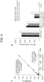
- FIG. 4A depicts the results of constructing sandwich ELISA using a fibronectin antibody and an SSEA-1 antibody and then analyzing each culture supernatant of human iPS cells and human iPS cells having a reduced undifferentiated level at each dilution stage.
- FIG. 4B is a graph showing the results of sandwich ELISA using the culture supernatants when human iPS cells having a reduced undifferentiated level were seeded at 25,000 cells/mL, 12,500 cells/mL, 6,250 cells/mL, and 0 cells/mL, respectively.
- Black indicates the actual number of cells
- gray indicates the apparent number of cells when human iPS cells having a reduced undifferentiated level were cultured alone
- white indicates the apparent number of cells when the cells were co-cultured with human iPS cells. It was found that the number of cells calculated by the sandwich ELISA method exhibited reactivity depending on the number of seeded cells and was almost the same as the actual number of cells.
- deviant cells can be detected even in the coexistence of human iPS cells. It was assumed that the reason why the slightly higher value was confirmed in the coexistence of human iPS cells was that deviant cells were mixed in the seeded human iPS cells.
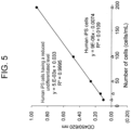
- Example 5 Sandwich ELISA assay using fibronectin monoclonal antibody and fibronectin polyclonal antibody
- a biotin-labeled human fibronectin monoclonal antibody (Catalog No. FN 30-8, Takara Bio) was immobilized on the surface of an avidin-coated microplate well (Catalog No. BS-X7603, Sumitomo Bakelite). Each culture supernatant of human iPS cells (control) and human iPS cells having a reduced undifferentiated level was diluted in such a manner that the concentration was a predetermined value and reacted at room temperature for 1 hour. Subsequently, an HRP-labeled fibronectin polyclonal antibody (Catalog No. AF1918; R & D) was reacted.
- the samples were each incubated with a TMB solution (Catalog No. 208-17371, Wako) for 30 minutes at room temperature.
- the enzyme reaction was stopped by adding 1N HCl and the enzyme activity on the microplate was measured at a main wavelength of 450 and a sub-wavelength of 620 nm (OD450/620) using a microtiter plate reader.
- FIG. 5 depicts the results of analyzing each culture supernatant of human iPS cells and human iPS cells having a reduced undifferentiated level at each dilution stage using the sandwich ELISA. As a result, it was found that although high reactivity was exhibited to the culture supernatant (black circles) of human iPS cells having a reduced undifferentiated level, no reactivity was exhibited to the culture supernatant (white triangles) of human iPS cells.
- IR700 labeling was performed using an IRDye700DX protein labeling kit (LI-COR Inc).
- a fibronectin antibody (Catalog No. AF1918; R & D) or a polyclonal antibody against sheep immunoglobulin G (Catalog No. 5-001-A; R & D) was incubated with IR700 at room temperature for 2 hours.
- the molar ratio and concentration of a protein were determined by measuring the absorbance at 280 nm (protein) and 689 nm (IR700), respectively, using a UV-1900 spectrophotometer (Shimadzu).
- Cells were incubated with the IR700-labeled fibronectin antibody at 5% CO 2 and 37°C for 1 hour or 6 hours.
- cells were further incubated with a Hoechst 33342 solution (1 : 1000 dilution, Dojindo Molecular Technologies, Inc) for 5 to 10 minutes. Images were acquired with a confocal microscope (Olympus Corporation) or a BZ-9000 fluorescence microscope (KEYENCE CORPORATION).
- FIG. 6A depicts immunostaining images of living human iPS cells (control) and living human iPS cells having a reduced undifferentiated level stained with the IR700-labeled fibronectin antibody (red) at room temperature for 1 hour. The nuclei were stained with Hoechst 33342 (blue). The scale bar indicates 100 ⁇ m.
- FIG. 6B depicts immunostaining images of living human iPS cells having a reduced undifferentiated level stained with the IR700-labeled fibronectin antibody (red) at 37°C for 1 hour (left panel) or 6 hours (right panel). The scale bar indicates 100 ⁇ m.
- Binding of the IR700-labeled fibronectin antibody to human iPS cells having a reduced undifferentiated level was confirmed, but no such binding to human iPS cells was confirmed. No difference was confirmed in the amount of binding of the human iPS cells having a reduced undifferentiated level to the IR700-labeled fibronectin antibody between the co-incubation time of 1 hour and that of 6 hours (the binding was maintained sufficiently even after 6 hours).
- NIR radiation The day before NIR radiation, cells were seeded on 96-well or 12-well plates and incubated for 24 hours. After medium exchange, the cells were incubated with an IR700-labeled fibronectin antibody or IR700-labeled sheep immunoglobulin G for 1 hour or 6 hours, and then irradiated with NIR light-emitting diodes at a power density of 10 mW/cm 2 (Ebisudenshi Co., Ltd.). Images were acquired using a BZ-9000 fluorescence microscope (KEYENCE CORPORATION).
- FIG. 7A depicts the results of subjecting human iPS cells having a reduced undifferentiated level to no treatment, treatment with the IR700-labeled fibronectin antibody, and NIR radiation treatment, and treatment with the IR700-labeled fibronectin antibody followed by NIR radiation, respectively, and then analyzing the cell viability by living cell staining (calcein-AM: green) and dead cell staining (EthD-1: red). Almost no dead cells were observed among cells subjected to no treatment, treatment with the IR700-labeled fibronectin antibody, and NIR radiation, but increases in dead cells and decreases in living cells were confirmed for cells subjected to treatment with the IR700-labeled fibronectin antibody followed by NIR radiation.
- FIG. 7B depicts the results of subjecting human iPS cells (controls) to no treatment, treatment with the IR700-labeled fibronectin antibody, NIR radiation, and treatment with the IR700-labeled fibronectin antibody followed by NIR radiation, respectively, and then analyzing the cell viability by living cell staining (calcein-AM: green) and dead cell staining (EthD-1: red). Unlike the above human iPS cells having a reduced undifferentiated level, almost no dead cells were observed among cells subjected to treatment with the IR700-labeled fibronectin antibody followed by NIR radiation.
- Cell viability was quantified using Cell-Counting Kit-8 (Dojindo Molecular Technologies, Inc). Ten (10) ⁇ L of Cell-Counting Kit-8 solution was added to cells in each well of a 96-well plate and incubated at 37°C for 2 hours. Then, the absorbance at 450 nm was measured using a microtiter plate reader (SpectraMax M3, Molecular Devices). For calculation of cell viability, an untreated medium incubated at 37°C for 2 hours was used as a negative control.
- FIG. 8A depicts each cell viability (%) of human iPS cells (control) (white circles) and human iPS cells (black circles) having a reduced undifferentiated level.
- Cell viability (%) was calculated by comparing the absorbance at 450 nm of cells subjected to treatment with the IR700-labeled fibronectin antibody followed by NIR radiation with that of untreated cells. The data are represented by the average value in triplicate measurements ⁇ standard deviation. As a result, it was confirmed that the cell viability of human iPS cells having a reduced undifferentiated level can be significantly decreased by the treatment with the IR700-labeled fibronectin antibody followed by NIR radiation.
- the effect was obtained by the use of the IR700-labeled fibronectin antibody in an amount as small as about 1 ng/mL, and when the IR700-labeled fibronectin antibody was used in an amount of about 10 ng/mL, the cells were almost killed.
- human iPS cells (control) and human iPS cells having a reduced undifferentiated level were each incubated with the 10 ng/mL IR700-labeled fibronectin antibody for 1 hour, and then exposed to NIR radiation for various times (0 to 10 minutes). After 24 hours, 10 ⁇ L of CCK-8 solution was added to the medium, further incubation was performed for 2 hours, and then the absorbance at 450 nm was measured.
- FIG. 8B depicts each cell viability (%) of human iPS cells (control) (white circles) and human iPS cells (black circles) having a reduced undifferentiated level.
- Cell viability (%) was calculated by comparing the absorbance at 450 nm of cells subjected to treatment with the IR700-labeled fibronectin antibody alone with that of cells subjected to treatment with the IR700-labeled fibronectin antibody followed by NIR radiation. The data are represented by the average value in triplicate measurements ⁇ standard deviation. As a result, it was confirmed that the cell viability of human iPS cells having a reduced undifferentiated level treated with the IR700-labeled fibronectin antibody was significantly reduced by brief NIR radiation of about 3 minutes. About 5 minutes of irradiation completely killed the cells.
- FIG. 9A depicts the results of subjecting human iPS cells (control) and human iPS cells having a reduced undifferentiated level to no treatment, treatment with an IR700-labeled fibronectin antibody, NIR radiation, and treatment with the IR700-labeled fibronectin antibody followed by NIR radiation, respectively, and then analyzing the co-culture of the human iPS cells (long elongated large flat shape) having a reduced undifferentiated level with the control human iPS cells (colony forming cells) by living cell staining (calcein-AM: green) and dead cell staining (EthD-1: red).
- living cell staining calcein-AM: green
- EthD-1 dead cell staining
- CMFDA CellTrackerGreen-5-chloromethylfluorescein diacetate
- a single cell suspension containing human iPS cells (control) and human iPS cells having a reduced undifferentiated level was prepared using a TrypLE Express recombinant enzyme solution (ThermoFisher Scientific) and then suspended in phosphate buffered saline (PBS) containing 1% bovine serum albumin.
- PBS phosphate buffered saline
- the thus obtained cell suspension was subjected to data analysis using a CytoFLEX flow cytometer (Beckman Coulter Inc.) and FlowJov10 software (BDBiosciences), and the abundance of each cell type was confirmed.
- FIG. 9B depicts the analysis results. As a result, it was confirmed that human iPS cells having a reduced undifferentiated level was selectively removed from the co-culture by treatment with the IR700-labeled fibronectin antibody followed by NIR radiation.
- the undifferentiated state of human iPS cells can be determined or evaluated non-invasively, simply and efficiently without reducing the number of cells, and only cells having a low undifferentiated level can be selectively removed. Therefore, the present invention is an extremely useful technique that is expected to contribute to the quality control of human iPS cells.
Landscapes
- Health & Medical Sciences (AREA)
- Life Sciences & Earth Sciences (AREA)
- Engineering & Computer Science (AREA)
- Biomedical Technology (AREA)
- Chemical & Material Sciences (AREA)
- Immunology (AREA)
- Molecular Biology (AREA)
- Urology & Nephrology (AREA)
- Hematology (AREA)
- General Health & Medical Sciences (AREA)
- Medicinal Chemistry (AREA)
- Biochemistry (AREA)
- Cell Biology (AREA)
- Physics & Mathematics (AREA)
- Analytical Chemistry (AREA)
- General Physics & Mathematics (AREA)
- Food Science & Technology (AREA)
- Microbiology (AREA)
- Pathology (AREA)
- Biotechnology (AREA)
- Toxicology (AREA)
- Proteomics, Peptides & Aminoacids (AREA)
- Tropical Medicine & Parasitology (AREA)
- Bioinformatics & Cheminformatics (AREA)
- Organic Chemistry (AREA)
- Zoology (AREA)
- Gastroenterology & Hepatology (AREA)
- Developmental Biology & Embryology (AREA)
- Biophysics (AREA)
- Genetics & Genomics (AREA)
- Measuring Or Testing Involving Enzymes Or Micro-Organisms (AREA)
- Micro-Organisms Or Cultivation Processes Thereof (AREA)
Applications Claiming Priority (2)
| Application Number | Priority Date | Filing Date | Title |
|---|---|---|---|
| JP2020102063 | 2020-06-12 | ||
| PCT/JP2021/023128 WO2021251507A1 (fr) | 2020-06-12 | 2021-06-11 | Technique de contrôle de la qualité des cellules souches pluripotentes humaines utilisant un bouillon de culture |
Publications (4)
| Publication Number | Publication Date |
|---|---|
| EP4166676A1 true EP4166676A1 (fr) | 2023-04-19 |
| EP4166676A4 EP4166676A4 (fr) | 2024-07-17 |
| EP4166676B1 EP4166676B1 (fr) | 2025-07-30 |
| EP4166676C0 EP4166676C0 (fr) | 2025-07-30 |
Family
ID=78846167
Family Applications (1)
| Application Number | Title | Priority Date | Filing Date |
|---|---|---|---|
| EP21821235.5A Active EP4166676B1 (fr) | 2020-06-12 | 2021-06-11 | Technique de contrôle de la qualité des cellules souches pluripotentes humaines utilisant un bouillon de culture |
Country Status (4)
| Country | Link |
|---|---|
| US (1) | US20230204600A1 (fr) |
| EP (1) | EP4166676B1 (fr) |
| JP (1) | JP7538554B2 (fr) |
| WO (1) | WO2021251507A1 (fr) |
Family Cites Families (4)
| Publication number | Priority date | Publication date | Assignee | Title |
|---|---|---|---|---|
| US11261425B2 (en) * | 2009-08-12 | 2022-03-01 | Kyoto University | Method for inducing differentiation of pluripotent stem cells into neural precursor cells |
| ES2889904T3 (es) * | 2015-08-18 | 2022-01-14 | Rakuten Medical Inc | Conjugados de colorante de ftalocianina y su almacenamiento |
| JP6733889B2 (ja) * | 2016-03-31 | 2020-08-05 | 国立研究開発法人産業技術総合研究所 | 細胞分化ポテンシャル判定法 |
| JP7171421B2 (ja) | 2018-12-25 | 2022-11-15 | エルジー ディスプレイ カンパニー リミテッド | 表示装置、インターフェースユニット及び表示システム |
-
2021
- 2021-06-11 JP JP2022529759A patent/JP7538554B2/ja active Active
- 2021-06-11 WO PCT/JP2021/023128 patent/WO2021251507A1/fr not_active Ceased
- 2021-06-11 EP EP21821235.5A patent/EP4166676B1/fr active Active
- 2021-06-11 US US18/009,632 patent/US20230204600A1/en active Pending
Also Published As
| Publication number | Publication date |
|---|---|
| JPWO2021251507A1 (fr) | 2021-12-16 |
| WO2021251507A1 (fr) | 2021-12-16 |
| EP4166676B1 (fr) | 2025-07-30 |
| US20230204600A1 (en) | 2023-06-29 |
| EP4166676A4 (fr) | 2024-07-17 |
| JP7538554B2 (ja) | 2024-08-22 |
| EP4166676C0 (fr) | 2025-07-30 |
Similar Documents
| Publication | Publication Date | Title |
|---|---|---|
| Tüshaus et al. | An optimized quantitative proteomics method establishes the cell type‐resolved mouse brain secretome | |
| Yu et al. | Large-scale production of human blastoids amenable to modeling blastocyst development and maternal-fetal cross talk | |
| Liao et al. | Single-cell detection of secreted Aβ and sAPPα from human IPSC-derived neurons and astrocytes | |
| Eapen et al. | Quantitative proteomics reveals the selectivity of ubiquitin-binding autophagy receptors in the turnover of damaged lysosomes by lysophagy | |
| Notaras et al. | Schizophrenia is defined by cell-specific neuropathology and multiple neurodevelopmental mechanisms in patient-derived cerebral organoids | |
| Lauschke et al. | A novel human pluripotent stem cell-based assay to predict developmental toxicity | |
| van Erp et al. | Age-related loss of axonal regeneration is reflected by the level of local translation | |
| Masciocchi et al. | General features, pathogenesis, and laboratory diagnostics of autoimmune encephalitis | |
| O'Brien et al. | New monoclonal antibodies to defined cell surface proteins on human pluripotent stem cells | |
| EP2460008A2 (fr) | Procédés de caractérisation de reprogrammation cellulaire et applications associées | |
| Cuddy et al. | Analysis of lysosomal hydrolase trafficking and activity in human iPSC-derived neuronal models | |
| Watzlawik et al. | Development and characterization of phospho-ubiquitin antibodies to monitor PINK1-PRKN signaling in cells and tissue | |
| US9846164B2 (en) | Detection of human somatic cell reprogramming | |
| Liang et al. | KRT18 regulates trophoblast cell migration and invasion which are essential for embryo implantation | |
| Sakai et al. | Glycolytic activity is required for the onset of neural plate folding during neural tube closure in mouse embryos | |
| Lee et al. | Proteomic analysis of germinal vesicles in the domestic cat model reveals candidate nuclear proteins involved in oocyte competence acquisition | |
| US20130178543A1 (en) | Biomarker for diagnosing cancer and method of isolating cancer cell using the same | |
| EP4166676B1 (fr) | Technique de contrôle de la qualité des cellules souches pluripotentes humaines utilisant un bouillon de culture | |
| Whitworth et al. | P300 modulates endothelial mechanotransduction of fluid shear stress | |
| Maurer et al. | Contrasting expression of keratins in mouse and human embryonic stem cells | |
| Pangrazzi et al. | Detecting Senescence in T Cells by Flow Cytometry Using the SA-β-Galactosidase Assay | |
| Li et al. | Loss of SHROOM3 affects neuroepithelial cell shape through regulating cytoskeleton proteins in cynomolgus monkey organoids | |
| Teng et al. | Non-neuronal release of gamma-aminobutyric Acid by embryonic pluripotent stem cells | |
| US20220252619A1 (en) | Detection of mediators of dopamine transmission | |
| Hirabayashi et al. | A novel probe as surface glycan marker of pluripotent stem cells: research outcomes and application to regenerative medicine |
Legal Events
| Date | Code | Title | Description |
|---|---|---|---|
| STAA | Information on the status of an ep patent application or granted ep patent |
Free format text: STATUS: THE INTERNATIONAL PUBLICATION HAS BEEN MADE |
|
| PUAI | Public reference made under article 153(3) epc to a published international application that has entered the european phase |
Free format text: ORIGINAL CODE: 0009012 |
|
| STAA | Information on the status of an ep patent application or granted ep patent |
Free format text: STATUS: REQUEST FOR EXAMINATION WAS MADE |
|
| 17P | Request for examination filed |
Effective date: 20230110 |
|
| AK | Designated contracting states |
Kind code of ref document: A1 Designated state(s): AL AT BE BG CH CY CZ DE DK EE ES FI FR GB GR HR HU IE IS IT LI LT LU LV MC MK MT NL NO PL PT RO RS SE SI SK SM TR |
|
| DAV | Request for validation of the european patent (deleted) | ||
| DAX | Request for extension of the european patent (deleted) | ||
| A4 | Supplementary search report drawn up and despatched |
Effective date: 20240617 |
|
| RIC1 | Information provided on ipc code assigned before grant |
Ipc: G01N 33/68 20060101ALI20240611BHEP Ipc: G01N 33/543 20060101ALI20240611BHEP Ipc: C12N 15/12 20060101ALI20240611BHEP Ipc: C12Q 1/04 20060101AFI20240611BHEP |
|
| GRAP | Despatch of communication of intention to grant a patent |
Free format text: ORIGINAL CODE: EPIDOSNIGR1 |
|
| STAA | Information on the status of an ep patent application or granted ep patent |
Free format text: STATUS: GRANT OF PATENT IS INTENDED |
|
| INTG | Intention to grant announced |
Effective date: 20250225 |
|
| GRAS | Grant fee paid |
Free format text: ORIGINAL CODE: EPIDOSNIGR3 |
|
| GRAA | (expected) grant |
Free format text: ORIGINAL CODE: 0009210 |
|
| STAA | Information on the status of an ep patent application or granted ep patent |
Free format text: STATUS: THE PATENT HAS BEEN GRANTED |
|
| AK | Designated contracting states |
Kind code of ref document: B1 Designated state(s): AL AT BE BG CH CY CZ DE DK EE ES FI FR GB GR HR HU IE IS IT LI LT LU LV MC MK MT NL NO PL PT RO RS SE SI SK SM TR |
|
| REG | Reference to a national code |
Ref country code: GB Ref legal event code: FG4D |
|
| REG | Reference to a national code |
Ref country code: CH Ref legal event code: EP |
|
| REG | Reference to a national code |
Ref country code: DE Ref legal event code: R096 Ref document number: 602021035269 Country of ref document: DE |
|
| REG | Reference to a national code |
Ref country code: IE Ref legal event code: FG4D |
|
| U01 | Request for unitary effect filed |
Effective date: 20250730 |
|
| U07 | Unitary effect registered |
Designated state(s): AT BE BG DE DK EE FI FR IT LT LU LV MT NL PT RO SE SI Effective date: 20250807 |
|
| PG25 | Lapsed in a contracting state [announced via postgrant information from national office to epo] |
Ref country code: IS Free format text: LAPSE BECAUSE OF FAILURE TO SUBMIT A TRANSLATION OF THE DESCRIPTION OR TO PAY THE FEE WITHIN THE PRESCRIBED TIME-LIMIT Effective date: 20251130 |
|
| PG25 | Lapsed in a contracting state [announced via postgrant information from national office to epo] |
Ref country code: NO Free format text: LAPSE BECAUSE OF FAILURE TO SUBMIT A TRANSLATION OF THE DESCRIPTION OR TO PAY THE FEE WITHIN THE PRESCRIBED TIME-LIMIT Effective date: 20251030 |
|
| PG25 | Lapsed in a contracting state [announced via postgrant information from national office to epo] |
Ref country code: HR Free format text: LAPSE BECAUSE OF FAILURE TO SUBMIT A TRANSLATION OF THE DESCRIPTION OR TO PAY THE FEE WITHIN THE PRESCRIBED TIME-LIMIT Effective date: 20250730 |
|
| PG25 | Lapsed in a contracting state [announced via postgrant information from national office to epo] |
Ref country code: GR Free format text: LAPSE BECAUSE OF FAILURE TO SUBMIT A TRANSLATION OF THE DESCRIPTION OR TO PAY THE FEE WITHIN THE PRESCRIBED TIME-LIMIT Effective date: 20251031 |
|
| PG25 | Lapsed in a contracting state [announced via postgrant information from national office to epo] |
Ref country code: PL Free format text: LAPSE BECAUSE OF FAILURE TO SUBMIT A TRANSLATION OF THE DESCRIPTION OR TO PAY THE FEE WITHIN THE PRESCRIBED TIME-LIMIT Effective date: 20250730 |
|
| PG25 | Lapsed in a contracting state [announced via postgrant information from national office to epo] |
Ref country code: RS Free format text: LAPSE BECAUSE OF FAILURE TO SUBMIT A TRANSLATION OF THE DESCRIPTION OR TO PAY THE FEE WITHIN THE PRESCRIBED TIME-LIMIT Effective date: 20251030 |
|
| PG25 | Lapsed in a contracting state [announced via postgrant information from national office to epo] |
Ref country code: ES Free format text: LAPSE BECAUSE OF FAILURE TO SUBMIT A TRANSLATION OF THE DESCRIPTION OR TO PAY THE FEE WITHIN THE PRESCRIBED TIME-LIMIT Effective date: 20250730 |
|
| PG25 | Lapsed in a contracting state [announced via postgrant information from national office to epo] |
Ref country code: SM Free format text: LAPSE BECAUSE OF FAILURE TO SUBMIT A TRANSLATION OF THE DESCRIPTION OR TO PAY THE FEE WITHIN THE PRESCRIBED TIME-LIMIT Effective date: 20250730 |
|
| PG25 | Lapsed in a contracting state [announced via postgrant information from national office to epo] |
Ref country code: CZ Free format text: LAPSE BECAUSE OF FAILURE TO SUBMIT A TRANSLATION OF THE DESCRIPTION OR TO PAY THE FEE WITHIN THE PRESCRIBED TIME-LIMIT Effective date: 20250730 |
|
| PG25 | Lapsed in a contracting state [announced via postgrant information from national office to epo] |
Ref country code: SK Free format text: LAPSE BECAUSE OF FAILURE TO SUBMIT A TRANSLATION OF THE DESCRIPTION OR TO PAY THE FEE WITHIN THE PRESCRIBED TIME-LIMIT Effective date: 20250730 |