EP4174085A1 - Antikörper gegen sars-cov-2, verfahren zum nachweis von sars-cov-2 unter verwendung eines antikörpers und kit mit antikörper - Google Patents
Antikörper gegen sars-cov-2, verfahren zum nachweis von sars-cov-2 unter verwendung eines antikörpers und kit mit antikörper Download PDFInfo
- Publication number
- EP4174085A1 EP4174085A1 EP21834296.2A EP21834296A EP4174085A1 EP 4174085 A1 EP4174085 A1 EP 4174085A1 EP 21834296 A EP21834296 A EP 21834296A EP 4174085 A1 EP4174085 A1 EP 4174085A1
- Authority
- EP
- European Patent Office
- Prior art keywords
- antibody
- sars
- cov
- fragment
- derived
- Prior art date
- Legal status (The legal status is an assumption and is not a legal conclusion. Google has not performed a legal analysis and makes no representation as to the accuracy of the status listed.)
- Withdrawn
Links
Images




Classifications
-
- C—CHEMISTRY; METALLURGY
- C07—ORGANIC CHEMISTRY
- C07K—PEPTIDES
- C07K14/00—Peptides having more than 20 amino acids; Gastrins; Somatostatins; Melanotropins; Derivatives thereof
- C07K14/005—Peptides having more than 20 amino acids; Gastrins; Somatostatins; Melanotropins; Derivatives thereof from viruses
-
- C—CHEMISTRY; METALLURGY
- C07—ORGANIC CHEMISTRY
- C07K—PEPTIDES
- C07K16/00—Immunoglobulins [IG], e.g. monoclonal or polyclonal antibodies
- C07K16/08—Immunoglobulins [IG], e.g. monoclonal or polyclonal antibodies against material from viruses
- C07K16/10—RNA viruses
- C07K16/102—Coronaviridae (F)
- C07K16/104—Severe acute respiratory syndrome coronavirus 2 [SARS‐CoV‐2]
-
- G—PHYSICS
- G01—MEASURING; TESTING
- G01N—INVESTIGATING OR ANALYSING MATERIALS BY DETERMINING THEIR CHEMICAL OR PHYSICAL PROPERTIES
- G01N33/00—Investigating or analysing materials by specific methods not covered by groups G01N1/00 - G01N31/00
- G01N33/48—Biological material, e.g. blood, urine; Haemocytometers
- G01N33/50—Chemical analysis of biological material, e.g. blood, urine; Testing involving biospecific ligand binding methods; Immunological testing
- G01N33/53—Immunoassay; Biospecific binding assay; Materials therefor
- G01N33/543—Immunoassay; Biospecific binding assay; Materials therefor with an insoluble carrier for immobilising immunochemicals
- G01N33/54366—Apparatus specially adapted for solid-phase testing
- G01N33/54386—Analytical elements
- G01N33/54387—Immunochromatographic test strips
-
- G—PHYSICS
- G01—MEASURING; TESTING
- G01N—INVESTIGATING OR ANALYSING MATERIALS BY DETERMINING THEIR CHEMICAL OR PHYSICAL PROPERTIES
- G01N33/00—Investigating or analysing materials by specific methods not covered by groups G01N1/00 - G01N31/00
- G01N33/48—Biological material, e.g. blood, urine; Haemocytometers
- G01N33/50—Chemical analysis of biological material, e.g. blood, urine; Testing involving biospecific ligand binding methods; Immunological testing
- G01N33/53—Immunoassay; Biospecific binding assay; Materials therefor
- G01N33/569—Immunoassay; Biospecific binding assay; Materials therefor for microorganisms, e.g. protozoa, bacteria, viruses
- G01N33/56983—Viruses
-
- C—CHEMISTRY; METALLURGY
- C07—ORGANIC CHEMISTRY
- C07K—PEPTIDES
- C07K2317/00—Immunoglobulins specific features
- C07K2317/30—Immunoglobulins specific features characterized by aspects of specificity or valency
-
- C—CHEMISTRY; METALLURGY
- C12—BIOCHEMISTRY; BEER; SPIRITS; WINE; VINEGAR; MICROBIOLOGY; ENZYMOLOGY; MUTATION OR GENETIC ENGINEERING
- C12N—MICROORGANISMS OR ENZYMES; COMPOSITIONS THEREOF; PROPAGATING, PRESERVING, OR MAINTAINING MICROORGANISMS; MUTATION OR GENETIC ENGINEERING; CULTURE MEDIA
- C12N2770/00—MICROORGANISMS OR ENZYMES; COMPOSITIONS THEREOF; PROPAGATING, PRESERVING, OR MAINTAINING MICROORGANISMS; MUTATION OR GENETIC ENGINEERING; CULTURE MEDIA ssRNA viruses positive-sense
- C12N2770/00011—Details
- C12N2770/20011—Coronaviridae
- C12N2770/20022—New viral proteins or individual genes, new structural or functional aspects of known viral proteins or genes
-
- G—PHYSICS
- G01—MEASURING; TESTING
- G01N—INVESTIGATING OR ANALYSING MATERIALS BY DETERMINING THEIR CHEMICAL OR PHYSICAL PROPERTIES
- G01N2333/00—Assays involving biological materials from specific organisms or of a specific nature
- G01N2333/005—Assays involving biological materials from specific organisms or of a specific nature from viruses
- G01N2333/08—RNA viruses
- G01N2333/165—Coronaviridae, e.g. avian infectious bronchitis virus
Definitions
- the present invention relates to an antibody against SARS-CoV-2, a method for detecting SARS-CoV-2 using said antibody, and a kit comprising said antibody and the like.
- SARS-CoV-2 2019 Novel Coronavirus
- SARS-CoV-2 2019 Novel Coronavirus
- Non-Patent Documents 1 and 2 Genetic tests such as real-time reverse transcription-PCR using collected nasal swab, pharyngeal swab, or sputum as specimens. By this, it is possible to determine the presence or absence of SARS-CoV-2 infection.
- Non-Patent Documents 1 and 2 Genetic tests such as real-time reverse transcription-PCR using collected nasal swab, pharyngeal swab, or sputum as specimens.
- genetic testing requires a special device that detects fluorescence in real time during PCR, and it takes several hours to obtain the results, so it remains a problem from the viewpoint of early detection in places such as infection sites, quarantine sites, and clinical sites, where the facility is not sufficient.
- immunological tests using antigen-antibody reactions against SARS-CoV-2 derived proteins are required as tests that combine accuracy, speed, and convenience.
- the spike (hereinafter referred to as "S” in some cases) protein which is present on the surface of the SARS-CoV-2 envelope and is involved in infection of host cells, is known as a protein derived from SARS-CoV-2 (Non-Patent Document 3).
- SARS-CoV-2 derived S protein is a glycoprotein, which is made up of bound many sugar chains (Non-Patent Document 3), and it is considered extremely difficult to artificially prepare a protein that exactly reproduces its structure using chemical synthesis or genetically engineered microorganisms.
- SARS-CoV-2 derived S protein is known to be easily mutated its amino acid sequence as the SARS coronavirus replicates (Non-Patent Document 4).
- SARS-CoV-2 derived S protein is considered to be easily mutated in its amino acid sequence as SARS-CoV-2 replicates.
- antibodies prepared against the SARS-CoV-2 derived S protein may not show antigen-antibody reactions against mutants of the S protein, and thus, there is a risk that immunological tests using antibodies against the SARS-CoV-2 derived S protein may produce false negative test results depending on the specimens.
- NP protein constituting SARS-CoV-2 derived nucleocapsid
- SARS-CoV-2 derived protein a protein constituting SARS-CoV-2 derived nucleocapsid
- SARS-CoV-2 derived protein The NP derived from the SARS coronavirus, which is genetically related to SARS-CoV-2, is known to be the most abundant among the various SARS coronavirus-derived proteins produced by infection (Non-Patent Document 5), and likewise, SARS-CoV-2 derived NPs are thought to be the most abundant of the various SARS-CoV-2 derived proteins produced by infection. For this reason, SARS-CoV-2 derived NPs are expected to be relatively easy to detect from specimens.
- Non-Patent Document 6 a case of a preparation of antibody against a partial peptide of SARS-CoV-2 derived NP has already been reported (Non-Patent Document 6).
- the antibodies prepared in said case shows the antigen-antibody reaction against NPs derived from MERS coronavirus and SARS coronavirus, and does not have a specificity for the SARS-CoV-2 derived NP (Non-Patent Document 6).
- the antibodies prepared in said case shows the antigen-antibody reaction against NPs derived from MERS coronavirus and SARS coronavirus, and does not have a specificity for the SARS-CoV-2 derived NP (Non-Patent Document 6).
- the present invention is aim to provide a means of detecting proteins constituting SARS-CoV-2 derived nucleocapsid.
- the present inventors analyzed the SARS-CoV-2 derived NP and other coronavirus-derived NPs. As a result, the present inventors found a region of low identity between the amino acid sequence of SARS-CoV-2 derived NP and other coronavirus-derived NPs. Then in the light of the above-mentioned problem, the inventors prepared fragments of SARS-CoV-2 derived NP corresponding to said region and came to complete the present invention by using these as immunogens and preparing antibodies according to the usual method.
- the present invention relates to the following:
- the presence or absence of SARS-CoV-2 infection, or the past history can be detected with accuracy, speed, and convenience by an antibody or fragment thereof that specifically bind to SARS-CoV-2 derived NPs, a method for detecting SARS-CoV-2 derived NPs using said antibody or fragment thereof, and a kit for detecting a protein constituting SARS-CoV-2 derived nucleocapsid comprising said antibody or fragment thereof and the like of the present invention. That is to say, by the present invention, an immunological test equipped with high practicality in the early detection in places such as infection sites, quarantine sites, and clinical sites, where the facility is not sufficient, can be provided.
- the epitope which is recognized by the antibody of the present invention or fragment thereof that specifically bind to SARS-CoV-2 derived NPs constitutes a region of low identity between the amino acid sequence of SARS-CoV-2 derived NP and other coronavirus-derived NPs and it is possible to specifically detect SARS-CoV-2 derived NPs from the specimen.
- the method of detecting SARS-CoV-2 derived NPs with the antibody of the present invention or fragment thereof that specifically bind to SARS-CoV-2 derived NPs can reduce the number of cases where false positive and false negative occur.
- An aspect of present invention relates to an antibody or fragment thereof which binds to a protein constituting a SARS-CoV-2 derived nucleocapsid, wherein an epitope which is recognized by said antibody or fragment thereof is located at positions 121 to 419 of the amino acid sequence of SEQ ID NO:1 (hereinafter referred to as "an antibody of the present invention or fragment thereof" in some cases).
- the epitope recognized by the antibody of the present invention or fragment thereof is located at positions 208 to 222, positions 332 to 351, positions 374 to 397, or positions 398 to 419 of the amino acid sequence of SEQ ID NO:1.
- the antibody of the present invention or fragment thereof specifically binds to a protein constituting a SARS-CoV-2 derived nucleocapsid.
- SARS-CoV-2 refers to a coronavirus belonging to the SARS-associated coronaviruses, which was first identified its outbreak near Wuhan, China in 2019 and used interchangeably with "2019 Novel Coronavirus”.
- SARS-CoV-2 is pathogenic to humans and causes acute respiratory disease (COVID-19).
- SARS-CoV-2 similarly to other coronaviruses, is constituted of a spike, nucleocapsid, endogenous membrane protein, envelope protein, and RNA. Among those, nucleocapsid binds to RNA to form NPs, and it is surrounded by spike, intrinsic membrane protein, and envelope protein which are bound to lipid to form the envelope.
- GenBank accession number MN908947.3 The genome sequence of SARS-CoV-2 is published under the GenBank accession number MN908947.3.
- a protein constituting of nucleocapsid refers to a protein that constitutes a complex consisting of nucleocapsid and RNA, and is used interchangeably with "NP".
- the amino acid sequence of SARS-CoV-2 derived NP is represented by SEQ ID NO:1.
- a fragment of SARS-CoV-2 derived NPs refers to a fragment of protein consisting of a region of the amino acid sequence in SARS-CoV-2 derived NPs (SEQ ID NO:1) that consists of amino acid sequence having low identity between the amino acid sequence of SARS-CoV-2 derived NP and the amino acid sequence of NPs derived from other coronaviruses (region 121 to 419 of the amino acid sequence represented by SEQ ID NO:1).
- SARS-CoV-2 derived NP fragments have 90% or more sequence identity to the amino acid sequence represented by SEQ ID NO:2.
- fragments of SARS-CoV-2 derived NPs preferably have a sequence identity of 95% or more to the amino acid sequence represented by SEQ ID NO:2, especially preferably have a sequence identity of 98% or more, even more preferably a sequence identity of 100%.
- the fragment of the SARS-CoV-2 derived NPs consists of the amino acid sequence represented by SEQ ID NO:2.
- the fragment of SARS-CoV-2 derived NPs corresponds to a region of low identity between the amino acid sequence of SARS-CoV-2 derived NP and the amino acid sequences of NPs from other coronaviruses, it is possible to prepare said fragment and to obtain the antibody of the present invention or fragment thereof by using this as an antigen according to the usual methods.
- the antibody of the present invention is, for example, a complete immunoglobulin molecule, is preferably IgM, IgD, IgE, IgA or IgG, and is more preferably IgG1, IgG2a, IgG2b, IgG3 or IgG4.
- the antibody of the present invention can be a modified and/or altered antibody, such as chimeric and humanized antibodies.
- the antibody of the present invention can be a monoclonal or polyclonal antibody, a modified or altered monoclonal or polyclonal antibody, or a recombinantly or synthetically prepared or synthesized antibody. From the viewpoint of accurate, simple, and rapid detection of SARS-CoV-2 derived NPs, it is preferred that the antibody is a monoclonal antibody.
- a fragment of the antibody of the present invention can be a portion of immunoglobulin molecules, such as a Fab fragment, VL-, VH- or CDR-regions, but the fragment of the antibody of the present invention can specifically recognize SARS-CoV-2 derived NPs to the similar extent as the antibody of the present invention.
- the fragment of the antibody of the present invention is an antibody fragment, as well as a portion of those, such as a separated light and heavy chain, Fab, Fab/c, Fv, Fab', F(ab')2.
- the fragment of the antibody of the present invention is a Fab fragment, Fab/c fragment, Fv fragment, Fab' fragment or F(ab')2 fragment.
- the antibody of the present invention or fragment thereof can be an antibody derivative such as a bifunctional antibody, or antibody construct such as single chain Fv (scFv), bispecific scFv or an antibody fusion protein.
- scFv single chain Fv
- scFv single chain Fv
- bispecific scFv bispecific scFv
- Another aspect of the present invention relates to a method for detecting a protein constituting a SARS-CoV-2 derived nucleocapsid within a sample, wherein the method comprises a step of contacting the sample with the antibody of the present invention or fragment thereof (hereinafter referred to as "a method of the present invention” in some cases).
- the sample is a biological sample.
- in the method of present invention further comprises a step of detecting a protein constituting a SARS-CoV-2 derived nucleocapsid by at least one immunological test selected from the group consisting of ELISA, immunochromatography and filter antigen assay.
- the method of the present invention comprises the step of performing an antigen-antibody reaction between the antibody of the present invention or fragment thereof and SARS-CoV-2 derived NPs. In one embodiment, the method of the present invention comprises a plurality of the steps of performing an antigen-antibody reaction between the antibody of the present invention or fragment thereof and SARS-CoV-2 derived NPs. In one embodiment, the antibody of the present invention or fragment thereof is different in its epitope at each and every time of the step. In one embodiment, the antibody of the present invention or fragment thereof is labeled with a labeling substance.
- the method of the present invention comprises the step of performing an antigen-antibody reaction between the antibody of the present invention or fragment thereof that is unlabeled and SARS-CoV-2 derived NPs, and the step of performing an antigen-antibody reaction between the antibody of the present invention or fragment thereof that is labeled by a labeled substance (that is different in its epitope from aforesaid antibody of the present invention or fragment thereof that is unlabeled) and SARS-CoV-2 derived NPs.
- a labeled substance that is different in its epitope from aforesaid antibody of the present invention or fragment thereof that is unlabeled
- the method of the present invention comprises the step of performing an antigen-antibody reaction between the antibody of the present invention or fragment thereof and SARS-CoV-2 derived NPs, and/or further comprises the step of performing an antigen-antibody reaction between the antibody of the present invention or fragment thereof and an antibody or fragment thereof that recognizes said antibody or fragment thereof.
- the antibody of the present invention or fragment thereof, and/or an antibody of fragment thereof that recognizes said antibody or fragment thereof is labeled with a labeling substance.
- a “sample” is any sample that can comprise SARS-CoV-2 derived NPs, and is not particularly limited, but for example, biological samples can be listed.
- a “sample” is used interchangeably with a “specimen”.
- a "biological sample” is any sample that can comprise SARS-CoV-2 derived NPs, such as human or animal blood, plasma, serum, urine, semen, spinal fluid, saliva, oral mucosa, nasal fluid, sweat, tears, ascites, amniotic fluid, feces, organs such as blood vessels or liver, tissues, cells, or extracts thereof.
- the method for collecting these specimens is not particularly limited, and commonly known methods can be employed. Specifically, the method using cotton swabs is often used. Animals are not particularly limited if it can be infected with SARS-CoV-2, including such as dogs and cats.
- Biological samples can be collected according to the usual methods.
- Non-Patent Document 1 1 to 2 ml of the serum after being separated is placed in a plastic tube that can be tightly stoppered, and after being covered with a lid, the tube is sealed with Parafilm.
- the tube may contain a coagulant, and when a blood collection tube containing a serum separator is used, after 1 to 2 ml of serum after centrifugation is transferred into a plastic tube (preferably a sterile tube), after that, it is covered with a lid, and then sealed with Parafilm.
- a blood collection tube containing an anticoagulant EDTA-Na or K
- 1 to 2 ml is dispensed into a plastic tube that can be tightly stoppered, and after being covered with a lid, it is sealed with Parafilm.
- blood corpuscles are separated and peripheral blood mononuclear cells are suspended in cell preservation solution and cryopreserved.
- the separation of peripheral blood mononuclear cells becomes simple by using BD Vacutainer (registered trademark) CPT (trademark) blood collection tubes for mononuclear cell separation.
- PAXgene registered trademark
- RNA collection tubes may be used for blood collection and stored frozen as is.
- lower airway-derived fluid is collected from sputum when sputum is produced, as described in Non-Patent Document 1.
- tracheal aspirate is collected using a sterilized catheter under aseptic manipulation.
- the collection of bronchoalveolar lavage fluid is also to be considered.
- the collected sputum or aspirate should be placed in a plastic tube with a screw cap, covered with a lid and sealed with Parafilm.
- nasopharyngeal swab solution for nasopharyngeal swab solution, as described in Non-Patent Document 1, a sterile cotton swab (such as a flock swab or a cotton swab containing rayon or polyester in its material. A slim one for nasal use) is inserted into the nasal cavity and the nasopharynx is thoroughly swabbed, and the swab is placed in a sterile spit tube containing 1 to 3 ml of viral transport solution (VTM/UTM), cover with a lid, and seal with Parafilm. When no viral transport solution is available, PBS or saline solution is used.
- VTM/UTM viral transport solution
- test tube e.g. Falcon tube
- Parafilm a test tube
- Non-Patent Document 1 For example, for feces, as described in Non-Patent Document 1, about 0.1 g (the size of a small pea) is collected in a plastic tube that can be tightly stoppered, and is covered with a lid, and then sealed with Parafilm.
- labeling substance refers to any substance that labels the antibody of the present invention or fragment thereof and/or the antibody or fragment thereof that recognizes said antibody or fragment thereof, and is not particularly limited, as long as those can detect antigen-antibody reactions. From the viewpoint of accurate, simple, and rapid detection of SARS-CoV-2 derived NPs, it is preferred that the labeling substance is one or more selected from enzymes, pigments, metal colloidal particles, latex particles, cellulose particles.
- an antibody of fragment thereof that recognizes the antibody of the present invention or fragment thereof refers to an antibody of fragment thereof that can specifically bind to the antibody of the present invention or fragment thereof.
- an antibody that recognizes the antibody of the present invention or fragment thereof is preferably one or more selected from a group consisting of IgG, IgM, IgA, IgD and IgE, is especially preferably IgG and/or IgM, and is even more preferably IgG.
- the antibody that recognizes the antibody of the present invention or fragment thereof can be a modified and/or altered antibody, such as chimeric and humanized antibodies.
- the antibody that recognizes the antibody of the present invention or fragment thereof can be a monoclonal or polyclonal antibody, a modified or altered monoclonal or polyclonal antibody, or a recombinantly or synthetically prepared or synthesized antibody.
- the fragment of the antibody that recognizes the antibody of the present invention or fragment thereof can be a portion of such immunoglobulin molecules, such as a Fab fragment, VL-, VH- or CDR-regions, but the fragment of the antibody that recognizes the antibody of the present invention or fragment thereof, too, can specifically recognize SARS-CoV-2 derived NPs to the similar extent as the antibody that recognizes the antibody of the present invention or fragment thereof.
- the fragment of the antibody that recognizes the antibody of the present invention or fragment thereof can be an antibody fragment, as well as a separated light and heavy chain, a portion of those such as Fab, Fab/c, Fv, Fab', F(ab')2.
- the fragment of the antibody that recognizes the fragment of the antibody of the present invention is a Fab fragment, Fab/c fragment, Fv fragment, Fab' fragment or F(ab')2 fragment.
- the fragment of the antibody that recognizes the antibody of the present invention or fragment thereof can be an antibody derivative such as a bifunctional antibody, or antibody construct such as single chain Fv (scFv), bispecific scFv or an antibody fusion protein.
- scFv single chain Fv
- scFv single chain Fv
- bispecific scFv bispecific scFv
- SARS-CoV-2 derived NPs are detected by any immunological test that can detect antigen-antibody reactions between the antibody of the invention or fragment thereof and SARS-CoV-2 derived NPs.
- the immunological tests include, for example, such as enzyme immunoassay (ELISA, EIA), fluorescence immunoassay (FIA), radioimmunoassay (RIA), luminescence immunoassay (LIA), enzyme antibody assay, fluorescence antibody assay, immunochromatography (immunochromatographic assay), filter antigen assay, immunoturbidimetry, latex turbidimetry, latex agglutination assay, erythrocyte agglutination assay, or particle agglutination assay.
- enzyme immunoassay ELISA, EIA
- FFA fluorescence immunoassay
- RIA radioimmunoassay
- LIA luminescence immunoassay
- enzyme antibody assay fluorescence antibody assay
- immunochromatography
- SARS-CoV-2-derived NPs are detected by ELISA and/or immunochromatography.
- the SARS-CoV-2-derived NPs contained in a sample can be quantified by creating a calibration curve from the antigen-antibody reaction between a predetermined amount of the antibody of the present invention or fragment thereof and the SARS-CoV-2 derived NPs, and interpolating the measured values into the calibration curve.
- kits for detecting a protein constituting a SARS-CoV-2 derived nucleocapsid comprising the antibody of the present invention or fragment thereof (hereinafter referred to as "a kit for the present invention” in some cases).
- a kit for the present invention is a form of immunochromatographic strips.
- an immunochromatographic strip is one that has the antibody of the present invention or fragment thereof and an anti-mouse immunoglobulin antibody immobilized at separate sites respectively, i.e., a test line and a control line.
- the antibody of the present invention or fragments thereof labeled with such as gold colloidal particles, latex particles, fluorescent particles, magnetic particles is immersed beforehand.
- the sample dropped onto the sample pad is expanded by capillary action, and when it reaches the test line, only the SARS-CoV-2-derived NPs in the sample react, and when it reaches the control line, only the antibody of the present invention or fragments thereof labeled with gold colloid react.
- Other compounds in the sample will migrate to the absorbent pad without reacting.
- SARS-CoV-2-derived NPs in the sample can be detected but also SARS-CoV-2 derived NPs in a sample can be quantified.
- compositions for detecting a protein constituting a SARS-CoV-2 derived nucleocapsid comprising the antibody of the present invention or fragment thereof (hereinafter referred to as "a composition of the present invention” in some cases).
- Another aspect of the present invention relates to a method for diagnosing SARS-CoV-2 infection, wherein the method comprises a step of contacting the sample with an antibody or fragment thereof (hereinafter referred to as "a method for diagnosing of the present invention” in some cases).
- the method for diagnosing of the present invention further comprises a step of detecting a protein constituting a SARS-CoV-2 derived nucleocapsid by at least one immunological test selected from the group consisting of ELISA, immunochromatography and filter antigen assay.
- the method for diagnosing of the present invention comprises the step of performing an antigen-antibody reaction between the antibody of the present invention or fragment thereof and SARS-CoV-2 derived NPs. In one embodiment, the method for diagnosing of the present invention comprises a plurality of the steps of performing an antigen-antibody reaction between the antibody of the present invention or fragment thereof and SARS-CoV-2 derived NPs. In one embodiment, the antibody of the present invention or fragment thereof is different in its epitope at each and every time of said step. In one embodiment, the antibody of the present invention or fragment thereof is labeled with a labeling substance.
- the method for diagnosing of the present invention comprises the step of performing an antigen-antibody reaction between the antibody of the present invention or fragment thereof that is unlabeled and SARS-CoV-2 derived NPs, and the step of performing an antigen-antibody reaction between the antibody of the present invention or fragment thereof that is labeled by a labeled substance (that differs from aforesaid antibody of the present invention or fragment thereof that is unlabeled in its epitope) and SARS-CoV-2 derived NPs.
- a labeled substance that differs from aforesaid antibody of the present invention or fragment thereof that is unlabeled in its epitope
- the method for diagnosing of the present invention comprises the step of performing an antigen-antibody reaction between the antibody of the present invention or fragment thereof and SARS-CoV-2 derived NPs, and/or further comprises the step of performing an antigen-antibody reaction between the antibody of the present invention or fragment thereof and an antibody or fragment thereof that recognizes said antibody or fragment thereof.
- the antibody of the present invention or fragment thereof, and/or an antibody of fragment thereof that recognizes said antibody or fragment thereof is labeled with a labeling substance.
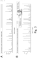
- the identities between the amino acid sequence of SARS-CoV-2 derived NP and the amino acid sequences of NPs derived from other coronaviruses were analyzed ( Figure 1 ).
- the amino acid sequence of SARS-CoV-2 derived NP had about 90% amino acid sequence identity to the amino acid sequence of SARS coronavirus derived NP, had about 47% amino acid sequence identity to the amino acid sequence of MERS coronavirus derived NP, and had about 30% or less amino acid sequence identities to the amino acid sequence of NPs derived from the other coronaviruses.
- SARS-CoV-2 derived NP consisting of the amino acid sequence represented by SEQ ID NO:2 were prepared by following methods. Furthermore, SARS-CoV-2 derived NP consists of the amino acid sequence represented by SEQ ID NO:1, and the RNA sequence corresponding to said amino acid sequence is represented by SEQ ID NO:3, and the DNA sequence corresponding to said RNA sequence is represented by SEQ ID NO:4. Furthermore, said DNA sequence is codon-optimized for protein synthesis in eukaryotic cells based on the amino acid sequence.
- a DNA sequence represented by SEQ ID NO:4 was artificially synthesized, and by PCR method using said DNA sequence as a template, the region at positions 361 to 1260 of the DNA sequence represented by SEQ ID:4, which corresponds to the region at positions 121 to 419 of the amino acid sequence represented by SEQ ID NO:1 ( Figure 1 ) was amplified.
- the DNA vector was amplified according to the method using the primers shown in SEQ ID NO:7 and SEQ ID NO:8.
- DNA vectors were prepared by linking DNA using In-Fusion ® HD Cloning Kit (Takara Bio Inc.). The obtained DNA vector was transformed into E. coli strain JM109, and the transformed E. coli was seeded on LB medium containing ampicillin. DNA vectors were prepared from E. coli after proliferation using a plasmid purification kit (Promega).
- mRNA was transcribed from obtained DNA vectors using SP6 RNA polymerase, and fragments of SARS-CoV-2 derived NP tagged with His-tag (region at positions 121 to 419 of the amino acid sequence represented by SEQ ID NO:1 ( Figure 1 )) were synthesized by cell-free protein synthesis with wheat germ extract following the user instructions of WEPRO7240 kit from Cell Free Science.
- fragments of SARS-CoV-2 derived NP were mixed with 2 ⁇ SDS sample buffer (125mM Tris-HCl, 4% SDS, 20% glycerol, 0.01% bromophenol blue, and 10% 2-mercaptoethanol), heat-treated, and subjected to polyacrylamide gels for electrophoresis. Subsequently the gels after electrophoresis were transferred to PVDF membranes. After the membranes were blocked with 2% skimmed milk solution, the membranes were allowed to react with a peroxidase-labeled anti-His tag antibody. After that, after reacting with HRP-labeled secondary antibody, peroxidase substrate solution was added to emit light, and the light emission was detected using a chemiluminescence imaging system.
- 2 ⁇ SDS sample buffer 125mM Tris-HCl, 4% SDS, 20% glycerol, 0.01% bromophenol blue, and 10% 2-mercaptoethanol
- Fragments of SARS-CoV-2 derived NP tagged with His-tag were purified from the reaction solution of cell-free protein synthesis using Ni columns. The series of steps of transcription, cell-free protein synthesis, and purification were done using an automated synthesizer (Cell Free Science). Fragments of SARS-CoV-2 derived NP tagged with His-tag were purified over 3 times, and the first to third purified fractions were denoted as E1-E3, respectively.
- E1-E3 were mixed with 2 ⁇ SDS sample buffer (125mM Tris-HCl, 4% SDS, 20% glycerol, 0.01% bromophenol blue, and 10% 2-mercaptoethanol), heat-treated, subjected to polyacrylamide gels for electrophoresis and dyed with Rapid CBB KANTO 3S (Kanto Chemical Co., Ltd.).
- 2 ⁇ SDS sample buffer 125mM Tris-HCl, 4% SDS, 20% glycerol, 0.01% bromophenol blue, and 10% 2-mercaptoethanol
- 300ug of purified fragments of SARS-CoV-2 derived NP were mixed with Freund's adjuvant, emulsified, and injected to BALB/c mice by subcutaneous injections in multiple times to immunize the mice. After 1 month B cells were collected from immunized mice, and fused with SP2/0 myeloma cells using polyethylene glycol solution. The fused hybridoma cells were seeded into 96-well microplates with selection medium.
- ELISA method firstly, fragments of SARS-CoV-2 derived NP were fixed on an ELISA plate. After blocking the plate, culture supernatant of hybridoma was added to the plate and allowed to react, and after that peroxidase-labeled goat anti-mouse immunoglobulin antibody was added and allowed to react. After that, peroxidase substrate solution was added to develop colors, and its absorbance was measured ( Figure 3A ).
- AlphaScreen method was performed following the manufacturer's recommended method. Firstly, biotin-labeled proteins of fragments of SARS-CoV-2 derived NP were synthesized by cell-free protein synthesis according to the procedure as described in Example 1. After that, after the synthesized biotin-labeled protein and culture supernatant of hybridoma were added to the plate and mixed, it was incubated at room temperature for 30 minutes. Then, 10 ⁇ l of acceptor beads (Protein G coated) and donor beads (streptavidin coated) were added. After incubating at room temperature for further 30 minutes, measurements were done in EnVision (PerkinElmer) ( Figure 3B ).
- Each region of the amino acid sequence from fragments of SARS-CoV-2 derived NP was deleted to generate three recombinant proteins (deletion mutants), respectively and the reactivities to these proteins were compared to determine the epitope which each monoclonal antibody recognized.
- each recombinant protein firstly, using a wheat cell-free expression vector pEU-E01-His-TEV-SARS-CoV-2-NP (121-419) (SEQ ID NO:9) , which was generated in Example 1 and encodes the amino acid sequence of the amino acid sequence represented by SEQ ID NO:1, as a template, expression vectors lacking the cDNA fragments encoding the amino acid sequence regions at positions 121 to 231 (d1), 232 to 340 (d2), and 341 to 419 (d3) of the amino acid sequence represented by SEQ ID NO:1 were generated respectively following the methods of PrimeSTAR ® Mutagenesis Basal Kit (Takara Bio Inc.).
- the mRNA was synthesized from the generated DNA vector, using SP6 RNA polymerase, and each recombinant protein was synthesized by cell-free protein synthesis according to the procedure as described in Example 1, following the instructions for use of WEPRO7240 kit from Cell Free Science Ltd.
- each obtained protein was mixed with 2 ⁇ SDS sample buffer (125 mM Tris-HCl, 4% SDS, 20% glycerol, 0.01% bromophenol blue, and 10% 2-mercaptoethanol), heat-treated, and subjected to a polyacrylamide gel for electrophoresis. Subsequently the electrophoresed gels were transferred to PVDF membranes. After blocking the membrane with 2% skimmed milk solution, the membrane was allowed to react with each monoclonal antibody generated in Example 2. After that, after reacting with HRP-conjugated secondary antibodies, peroxidase substrate solution was added to emit light, and the light emission was detected using a chemiluminescence imaging system.
- 2 ⁇ SDS sample buffer 125 mM Tris-HCl, 4% SDS, 20% glycerol, 0.01% bromophenol blue, and 10% 2-mercaptoethanol
- the generated antibodies were classified as having epitopes in the region at positions 121 to 231, 232 to 340, or 341 to 419 of the amino acid sequence represented by SEQ ID NO:1 ( Figure 4A ).
- DNA expression vectors lacking cDNA fragments encoding amino acids regions at positions 208 to 222 (d4), 203 to 222 (d5), 198 to 222 (d6), 190 to 222 (d7), 147 to 162 (d8), 398 to 419 (d9), 374 to 419 (d10), and 332 to 351 (d11) of the amino acid sequence represented by SEQ ID NO:1 respectively were generated and tested according to the method as above.
- #7 did not react with d4, d5, d6, nor d7 among 5 types of defective mutant (d4, d5, d6, d7, and d8), it was found that #7 recognized the region at positions 208 to 222 of the amino acid sequence represented by SEQ ID NO:1. Since 11 clones, #2, #4, #9, #13, #40, #45, #55, #97, #104, #111, and #128, did not react with d11 among 3 types of defective mutants (d9, d10, and d11), It was found that the 11 clones recognized the region at positions 332 to 351 of the amino acid sequence represented by SEQ ID NO:1.
- the generated antibodies were classified as having epitopes in the regions at positions 208 to 222, 332 to 351, 374 to 397, or 398 to 419 of the amino acid sequence represented by SEQ ID NO:1 ( Figure 4B ).
- each DNA sequence encoding each one of NPs derived from other human coronaviruses was synthesized (contracted to GeneWiz) (represented by SEQ ID NO:10, 11, 12, 13, 14, and 15, respectively). Furthermore, each DNA sequence was codon-optimized for protein synthesis in eukaryotic cells based on the amino acid sequence.
- each one of NPs derived from other human coronaviruses was synthesized by cell-free protein synthesis according to the procedure as described in Example 1, respectively.
- #7, #9, and #98 reacted with each NPs derived from other human coronaviruses (SARS, MERS, OC43, NL63, HKU1, and 229E) but these antibodies specifically recognized SARS-CoV-2 derived NPs.
- both #128 and #144 reacted with SARS-CoV-2 derived NPs and SARS coronavirus-derived NPs, and specifically recognized SARS-CoV-2 derived NPs and SARS coronavirus-derived NPs.
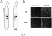
- Example 2 Whether the monoclonal antibodies (#7, #98) generated in Example 2 react with SARS-CoV-2 infected cells was considered.
- JCRB1819 cells derived from African green monkey kidney, VeroE6/TMPRSS2 (JCRB1819), were infected with SARS-CoV-2 provided by the National Institute of Infectious Diseases (NIID), at 37°C for 24 hours.
- NIID National Institute of Infectious Diseases
- SARS-CoV-2 derived NP was confirmed according to the Western blotting method as described in Example 1.
- lysate of aforesaid infected cells was mixed with 2 ⁇ SDS sample buffer (125 mM Tris-HCl, 4% SDS, 20% glycerol, 0.01% bromophenol blue, and 10% 2-mercaptoethanol), heat-treated, and subjected to polyacrylamide gels for electrophoresis. Subsequently the electrophoresed gels were transferred to PVDF membranes. After the membranes were blocked with 2% skimmed milk solution, the membranes were allowed to react with peroxidase-labeled monoclonal antibodies (#7, #98). After that, after reacting with HRP-labeled secondary antibody, peroxidase substrate solution was added to emit light, and the light emission was detected using a chemiluminescence imaging system.
- 2 ⁇ SDS sample buffer 125 mM Tris-HCl, 4% SDS, 20% glycerol, 0.01% bromophenol blue, and 10% 2-mercaptoethanol
- Example 2 After monoclonal antibodies (#7, #98) generated in Example 2 were added to the culture medium of aforesaid infected cells and allowed to react, and fluorescence labeled goat anti-mouse immunoglobulin antibody was added to immunostain aforesaid infected cells. After that, a fluorescence microscopy was used to image immunostained aforesaid infected cells. Furthermore, noninfected cells were used as negative controls.
- SARS-CoV-2 derived NPs full length were synthesized by cell-free protein synthesis based on SEQ ID NO:1 according to the procedure as described in Example 1.
- #9 was diluted to 2.5ug/mL in PBS and fixed on an ELISA plate, and it was blocked with 2% skimmed milk solution. After washing the plate with PBS-T, SARS-CoV-2 derived NPs were dispensed into wells in series dilutions at 0, 1.25, 5, and 20 ng/mL and allowed to react. After washing the plate with PBS-T, it was allowed to react with peroxidase-labeled #98. After washing the plate with PBS-T, peroxidase substrate solution (KPL) was added to develop colors, and the absorbance was measured.
- KPL peroxidase substrate solution
Landscapes
- Health & Medical Sciences (AREA)
- Life Sciences & Earth Sciences (AREA)
- Chemical & Material Sciences (AREA)
- Immunology (AREA)
- Virology (AREA)
- Molecular Biology (AREA)
- Engineering & Computer Science (AREA)
- Organic Chemistry (AREA)
- Biochemistry (AREA)
- General Health & Medical Sciences (AREA)
- Medicinal Chemistry (AREA)
- Urology & Nephrology (AREA)
- Hematology (AREA)
- Biomedical Technology (AREA)
- Biophysics (AREA)
- Proteomics, Peptides & Aminoacids (AREA)
- Genetics & Genomics (AREA)
- Microbiology (AREA)
- Cell Biology (AREA)
- Biotechnology (AREA)
- Food Science & Technology (AREA)
- Physics & Mathematics (AREA)
- Analytical Chemistry (AREA)
- General Physics & Mathematics (AREA)
- Pathology (AREA)
- Tropical Medicine & Parasitology (AREA)
- Gastroenterology & Hepatology (AREA)
- Peptides Or Proteins (AREA)
Applications Claiming Priority (2)
| Application Number | Priority Date | Filing Date | Title |
|---|---|---|---|
| JP2020111984A JP2022022607A (ja) | 2020-06-29 | 2020-06-29 | SARS-CoV-2に対する抗体、該抗体を用いてSARS-CoV-2を検出する方法および該抗体を含むキット |
| PCT/JP2021/024267 WO2022004622A1 (ja) | 2020-06-29 | 2021-06-28 | SARS-CoV-2に対する抗体、該抗体を用いてSARS-CoV-2を検出する方法および該抗体を含むキット |
Publications (2)
| Publication Number | Publication Date |
|---|---|
| EP4174085A1 true EP4174085A1 (de) | 2023-05-03 |
| EP4174085A4 EP4174085A4 (de) | 2024-11-13 |
Family
ID=79315345
Family Applications (1)
| Application Number | Title | Priority Date | Filing Date |
|---|---|---|---|
| EP21834296.2A Withdrawn EP4174085A4 (de) | 2020-06-29 | 2021-06-28 | Antikörper gegen sars-cov-2, verfahren zum nachweis von sars-cov-2 unter verwendung eines antikörpers und kit mit antikörper |
Country Status (6)
| Country | Link |
|---|---|
| US (1) | US20230235028A1 (de) |
| EP (1) | EP4174085A4 (de) |
| JP (2) | JP2022022607A (de) |
| KR (1) | KR20230028482A (de) |
| CN (1) | CN116194584A (de) |
| WO (1) | WO2022004622A1 (de) |
Cited By (1)
| Publication number | Priority date | Publication date | Assignee | Title |
|---|---|---|---|---|
| US20220373546A1 (en) * | 2021-05-11 | 2022-11-24 | Fujirebio Inc. | METHODS AND REAGENTS OF IMMUNOLOGICALLY DETECTING SARS-CoV-2 |
Families Citing this family (5)
| Publication number | Priority date | Publication date | Assignee | Title |
|---|---|---|---|---|
| JP7728100B2 (ja) * | 2021-05-11 | 2025-08-22 | 富士レビオ株式会社 | SARS-CoV-2の免疫学的検出方法および試薬 |
| JP7105970B1 (ja) * | 2021-06-16 | 2022-07-25 | 積水メディカル株式会社 | SARS-CoV-2の免疫測定方法及び免疫測定キット |
| JP7216948B1 (ja) * | 2021-06-16 | 2023-02-02 | 積水メディカル株式会社 | SARS-CoV-2の免疫測定方法及び免疫測定キット、並びにモノクローナル抗体又はその抗体断片 |
| CN117715931A (zh) * | 2021-06-30 | 2024-03-15 | 滋吾堂有限公司 | 针对SARS-CoV-2的核衣壳蛋白的抗体及其用途 |
| KR102778413B1 (ko) * | 2022-02-18 | 2025-03-11 | 한국생명공학연구원 | SARS-CoV-2 핵단백질에 특이적인 항체 및 이의 용도 |
Family Cites Families (3)
| Publication number | Priority date | Publication date | Assignee | Title |
|---|---|---|---|---|
| CN111153991A (zh) * | 2020-02-26 | 2020-05-15 | 北京博奥森生物技术有限公司 | 一种人SARS-CoV-2单克隆抗体及其制备方法和应用 |
| CN111024954A (zh) * | 2020-03-09 | 2020-04-17 | 深圳市易瑞生物技术股份有限公司 | 联合检测covid-19抗原和抗体的胶体金免疫层析装置及其使用法 |
| US11815513B2 (en) * | 2020-04-01 | 2023-11-14 | Institut Pasteur | Severe acute respiratory syndrome (SARS)-associated coronavirus diagnostics |
-
2020
- 2020-06-29 JP JP2020111984A patent/JP2022022607A/ja active Pending
-
2021
- 2021-06-28 CN CN202180046743.0A patent/CN116194584A/zh active Pending
- 2021-06-28 EP EP21834296.2A patent/EP4174085A4/de not_active Withdrawn
- 2021-06-28 US US18/013,189 patent/US20230235028A1/en active Pending
- 2021-06-28 WO PCT/JP2021/024267 patent/WO2022004622A1/ja not_active Ceased
- 2021-06-28 KR KR1020237002613A patent/KR20230028482A/ko not_active Withdrawn
-
2024
- 2024-12-11 JP JP2024216320A patent/JP2025028135A/ja active Pending
Cited By (1)
| Publication number | Priority date | Publication date | Assignee | Title |
|---|---|---|---|---|
| US20220373546A1 (en) * | 2021-05-11 | 2022-11-24 | Fujirebio Inc. | METHODS AND REAGENTS OF IMMUNOLOGICALLY DETECTING SARS-CoV-2 |
Also Published As
| Publication number | Publication date |
|---|---|
| CN116194584A (zh) | 2023-05-30 |
| JP2022022607A (ja) | 2022-02-07 |
| JP2025028135A (ja) | 2025-02-28 |
| KR20230028482A (ko) | 2023-02-28 |
| US20230235028A1 (en) | 2023-07-27 |
| WO2022004622A1 (ja) | 2022-01-06 |
| EP4174085A4 (de) | 2024-11-13 |
Similar Documents
| Publication | Publication Date | Title |
|---|---|---|
| EP4174085A1 (de) | Antikörper gegen sars-cov-2, verfahren zum nachweis von sars-cov-2 unter verwendung eines antikörpers und kit mit antikörper | |
| TW202200603A (zh) | 對抗sars-cov-2之結構蛋白質之抗體之抗原決定區、和該抗原決定區反應之抗體、使用該抗體檢測sars-cov-2之方法、含有該抗體之sars-cov-2檢測套組、檢測含有該抗原決定區之多胜肽之抗sars-cov-2抗體之方法、含有該抗原決定區之多胜肽之抗sars-cov-2抗體檢測套組、含有該抗原決定區之多胜肽之sars-cov-2用疫苗以及含有該抗體之sars-cov-2傳染病治療藥 | |
| CN116143931B (zh) | 一种抗人IgM抗体及其制备方法和用途 | |
| WO2022007304A1 (zh) | 能够特异性识别RBD蛋白的IgA抗体以及试剂盒 | |
| CN109265542B (zh) | 特异性结合诺如病毒gii.4基因型vp1蛋白或vlp的抗体及其制备方法和应用 | |
| WO2022265066A1 (ja) | SARS-CoV-2の免疫測定方法及び免疫測定キット | |
| WO2015025968A1 (ja) | マイコプラズマ・ニューモニエの免疫学的検出法およびキット | |
| CN115785262B (zh) | 一种抗登革ns1蛋白的抗体及其应用 | |
| CN119143871B (zh) | 用于人偏肺病毒检测的单克隆抗体、检测试剂及其应用 | |
| CN109180810B (zh) | 特异性结合诺如病毒gi.1基因型vp1蛋白或vlp的抗体及其制备方法和应用 | |
| WO2023078452A1 (zh) | 一种抗登革ns1蛋白的抗体及其应用 | |
| KR102450481B1 (ko) | 돼지열병 바이러스 특이적인 항체 및 이의 용도 | |
| CN115028713A (zh) | 一种抗冠状病毒n蛋白的抗体及其应用 | |
| CN104220090A (zh) | 肠道病毒71型的特异性抗体及其用途 | |
| CN102232087A (zh) | 修饰的人igf-1/e肽的抗体 | |
| EP2692735B1 (de) | Antikörper zur reaktion mit einem nativen cochlin-tomoprotein (ctp) und verfahren zur ctp-messung damit | |
| CN120623326B (zh) | 一种抗体或其抗原结合片段及其相关产品和应用 | |
| CN114058593A (zh) | SARS-CoV-2的N抗原的单克隆抗体、检测方法及其用途 | |
| EP4116324A1 (de) | Adenovirus-immuntestverfahren und adenovirus-immuntestinstrument | |
| CN118064377A (zh) | 一种分泌抗绵羊肺炎支原体单克隆抗体的杂交瘤细胞株及其单抗与应用 | |
| KR20250140089A (ko) | SARS-CoV-2의 검출 방법 및 이를 위한 키트 | |
| CN116121199A (zh) | 布鲁氏菌单克隆抗体6a5及其应用、试剂盒 | |
| CN115724957A (zh) | 抗牛病毒性腹泻病毒ns3蛋白单克隆抗体的重链和轻链可变区 | |
| WO2023167317A1 (ja) | 変異株を含む、体液中のSARS-CoV-2抗原に対する抗SARS-CoV-2抗体、該抗体を用いてSARS-CoV-2を検出する方法、および該抗体を含むキット | |
| CN109266620B (zh) | 抗人IgG单克隆抗体、其杂交瘤细胞株及应用 |
Legal Events
| Date | Code | Title | Description |
|---|---|---|---|
| STAA | Information on the status of an ep patent application or granted ep patent |
Free format text: STATUS: THE INTERNATIONAL PUBLICATION HAS BEEN MADE |
|
| PUAI | Public reference made under article 153(3) epc to a published international application that has entered the european phase |
Free format text: ORIGINAL CODE: 0009012 |
|
| STAA | Information on the status of an ep patent application or granted ep patent |
Free format text: STATUS: REQUEST FOR EXAMINATION WAS MADE |
|
| 17P | Request for examination filed |
Effective date: 20230120 |
|
| AK | Designated contracting states |
Kind code of ref document: A1 Designated state(s): AL AT BE BG CH CY CZ DE DK EE ES FI FR GB GR HR HU IE IS IT LI LT LU LV MC MK MT NL NO PL PT RO RS SE SI SK SM TR |
|
| DAV | Request for validation of the european patent (deleted) | ||
| DAX | Request for extension of the european patent (deleted) | ||
| RIC1 | Information provided on ipc code assigned before grant |
Ipc: G01N 33/543 20060101ALI20240610BHEP Ipc: G01N 33/53 20060101ALI20240610BHEP Ipc: C12N 15/50 20060101ALI20240610BHEP Ipc: C12N 15/13 20060101ALI20240610BHEP Ipc: C07K 16/10 20060101AFI20240610BHEP |
|
| A4 | Supplementary search report drawn up and despatched |
Effective date: 20241011 |
|
| RIC1 | Information provided on ipc code assigned before grant |
Ipc: G01N 33/543 20060101ALI20241007BHEP Ipc: G01N 33/53 20060101ALI20241007BHEP Ipc: C12N 15/50 20060101ALI20241007BHEP Ipc: C12N 15/13 20060101ALI20241007BHEP Ipc: C07K 16/10 20060101AFI20241007BHEP |
|
| STAA | Information on the status of an ep patent application or granted ep patent |
Free format text: STATUS: THE APPLICATION IS DEEMED TO BE WITHDRAWN |
|
| 18D | Application deemed to be withdrawn |
Effective date: 20250430 |