EP4220162A2 - Connecteur, conjugué connecteur-étiquette et marqueur pour analyser des échantillons biologiques - Google Patents
Connecteur, conjugué connecteur-étiquette et marqueur pour analyser des échantillons biologiques Download PDFInfo
- Publication number
- EP4220162A2 EP4220162A2 EP22208330.5A EP22208330A EP4220162A2 EP 4220162 A2 EP4220162 A2 EP 4220162A2 EP 22208330 A EP22208330 A EP 22208330A EP 4220162 A2 EP4220162 A2 EP 4220162A2
- Authority
- EP
- European Patent Office
- Prior art keywords
- connector
- affinity
- label
- interactor
- nucleic acid
- Prior art date
- Legal status (The legal status is an assumption and is not a legal conclusion. Google has not performed a legal analysis and makes no representation as to the accuracy of the status listed.)
- Pending
Links
- 239000003550 marker Substances 0.000 title claims abstract description 46
- 239000012472 biological sample Substances 0.000 title claims abstract description 40
- 108091034117 Oligonucleotide Proteins 0.000 claims abstract description 70
- 150000007523 nucleic acids Chemical group 0.000 claims abstract description 36
- 238000000034 method Methods 0.000 claims abstract description 25
- 230000000295 complement effect Effects 0.000 claims abstract description 15
- 239000003153 chemical reaction reagent Substances 0.000 claims description 108
- YBJHBAHKTGYVGT-ZKWXMUAHSA-N (+)-Biotin Chemical compound N1C(=O)N[C@@H]2[C@H](CCCCC(=O)O)SC[C@@H]21 YBJHBAHKTGYVGT-ZKWXMUAHSA-N 0.000 claims description 46
- 230000027455 binding Effects 0.000 claims description 35
- 239000000975 dye Substances 0.000 claims description 27
- 229960002685 biotin Drugs 0.000 claims description 23
- 235000020958 biotin Nutrition 0.000 claims description 23
- 239000011616 biotin Substances 0.000 claims description 23
- 108010090804 Streptavidin Proteins 0.000 claims description 20
- JLCPHMBAVCMARE-UHFFFAOYSA-N [3-[[3-[[3-[[3-[[3-[[3-[[3-[[3-[[3-[[3-[[3-[[5-(2-amino-6-oxo-1H-purin-9-yl)-3-[[3-[[3-[[3-[[3-[[3-[[5-(2-amino-6-oxo-1H-purin-9-yl)-3-[[5-(2-amino-6-oxo-1H-purin-9-yl)-3-hydroxyoxolan-2-yl]methoxy-hydroxyphosphoryl]oxyoxolan-2-yl]methoxy-hydroxyphosphoryl]oxy-5-(5-methyl-2,4-dioxopyrimidin-1-yl)oxolan-2-yl]methoxy-hydroxyphosphoryl]oxy-5-(6-aminopurin-9-yl)oxolan-2-yl]methoxy-hydroxyphosphoryl]oxy-5-(6-aminopurin-9-yl)oxolan-2-yl]methoxy-hydroxyphosphoryl]oxy-5-(6-aminopurin-9-yl)oxolan-2-yl]methoxy-hydroxyphosphoryl]oxy-5-(6-aminopurin-9-yl)oxolan-2-yl]methoxy-hydroxyphosphoryl]oxyoxolan-2-yl]methoxy-hydroxyphosphoryl]oxy-5-(5-methyl-2,4-dioxopyrimidin-1-yl)oxolan-2-yl]methoxy-hydroxyphosphoryl]oxy-5-(4-amino-2-oxopyrimidin-1-yl)oxolan-2-yl]methoxy-hydroxyphosphoryl]oxy-5-(5-methyl-2,4-dioxopyrimidin-1-yl)oxolan-2-yl]methoxy-hydroxyphosphoryl]oxy-5-(5-methyl-2,4-dioxopyrimidin-1-yl)oxolan-2-yl]methoxy-hydroxyphosphoryl]oxy-5-(6-aminopurin-9-yl)oxolan-2-yl]methoxy-hydroxyphosphoryl]oxy-5-(6-aminopurin-9-yl)oxolan-2-yl]methoxy-hydroxyphosphoryl]oxy-5-(4-amino-2-oxopyrimidin-1-yl)oxolan-2-yl]methoxy-hydroxyphosphoryl]oxy-5-(4-amino-2-oxopyrimidin-1-yl)oxolan-2-yl]methoxy-hydroxyphosphoryl]oxy-5-(4-amino-2-oxopyrimidin-1-yl)oxolan-2-yl]methoxy-hydroxyphosphoryl]oxy-5-(6-aminopurin-9-yl)oxolan-2-yl]methoxy-hydroxyphosphoryl]oxy-5-(4-amino-2-oxopyrimidin-1-yl)oxolan-2-yl]methyl [5-(6-aminopurin-9-yl)-2-(hydroxymethyl)oxolan-3-yl] hydrogen phosphate Polymers Cc1cn(C2CC(OP(O)(=O)OCC3OC(CC3OP(O)(=O)OCC3OC(CC3O)n3cnc4c3nc(N)[nH]c4=O)n3cnc4c3nc(N)[nH]c4=O)C(COP(O)(=O)OC3CC(OC3COP(O)(=O)OC3CC(OC3COP(O)(=O)OC3CC(OC3COP(O)(=O)OC3CC(OC3COP(O)(=O)OC3CC(OC3COP(O)(=O)OC3CC(OC3COP(O)(=O)OC3CC(OC3COP(O)(=O)OC3CC(OC3COP(O)(=O)OC3CC(OC3COP(O)(=O)OC3CC(OC3COP(O)(=O)OC3CC(OC3COP(O)(=O)OC3CC(OC3COP(O)(=O)OC3CC(OC3COP(O)(=O)OC3CC(OC3COP(O)(=O)OC3CC(OC3COP(O)(=O)OC3CC(OC3COP(O)(=O)OC3CC(OC3CO)n3cnc4c(N)ncnc34)n3ccc(N)nc3=O)n3cnc4c(N)ncnc34)n3ccc(N)nc3=O)n3ccc(N)nc3=O)n3ccc(N)nc3=O)n3cnc4c(N)ncnc34)n3cnc4c(N)ncnc34)n3cc(C)c(=O)[nH]c3=O)n3cc(C)c(=O)[nH]c3=O)n3ccc(N)nc3=O)n3cc(C)c(=O)[nH]c3=O)n3cnc4c3nc(N)[nH]c4=O)n3cnc4c(N)ncnc34)n3cnc4c(N)ncnc34)n3cnc4c(N)ncnc34)n3cnc4c(N)ncnc34)O2)c(=O)[nH]c1=O JLCPHMBAVCMARE-UHFFFAOYSA-N 0.000 claims description 16
- 239000007788 liquid Substances 0.000 claims description 15
- 239000000178 monomer Substances 0.000 claims description 3
- 239000000523 sample Substances 0.000 description 30
- 238000003776 cleavage reaction Methods 0.000 description 27
- 230000007017 scission Effects 0.000 description 27
- 108020004414 DNA Proteins 0.000 description 13
- 230000003287 optical effect Effects 0.000 description 13
- 108090000765 processed proteins & peptides Proteins 0.000 description 12
- 230000005284 excitation Effects 0.000 description 8
- 239000000758 substrate Substances 0.000 description 8
- 239000007850 fluorescent dye Substances 0.000 description 7
- 102000004169 proteins and genes Human genes 0.000 description 7
- 108090000623 proteins and genes Proteins 0.000 description 7
- 239000012634 fragment Substances 0.000 description 6
- 238000002156 mixing Methods 0.000 description 6
- 108091023037 Aptamer Proteins 0.000 description 5
- 108091005804 Peptidases Proteins 0.000 description 5
- 230000008901 benefit Effects 0.000 description 5
- 210000004027 cell Anatomy 0.000 description 5
- 238000003384 imaging method Methods 0.000 description 5
- 150000003384 small molecules Chemical class 0.000 description 5
- 102000004190 Enzymes Human genes 0.000 description 4
- 108090000790 Enzymes Proteins 0.000 description 4
- 239000004365 Protease Substances 0.000 description 4
- 108010003723 Single-Domain Antibodies Proteins 0.000 description 4
- 238000005859 coupling reaction Methods 0.000 description 4
- 230000001086 cytosolic effect Effects 0.000 description 4
- 238000004519 manufacturing process Methods 0.000 description 4
- 108020004999 messenger RNA Proteins 0.000 description 4
- 239000002773 nucleotide Substances 0.000 description 4
- 238000005406 washing Methods 0.000 description 4
- 102000035195 Peptidases Human genes 0.000 description 3
- 230000008878 coupling Effects 0.000 description 3
- 238000010168 coupling process Methods 0.000 description 3
- 238000001514 detection method Methods 0.000 description 3
- 230000002427 irreversible effect Effects 0.000 description 3
- -1 lactic acid) Chemical class 0.000 description 3
- 238000000386 microscopy Methods 0.000 description 3
- 125000003729 nucleotide group Chemical group 0.000 description 3
- 108091008146 restriction endonucleases Proteins 0.000 description 3
- 239000000243 solution Substances 0.000 description 3
- 108091032973 (ribonucleotides)n+m Proteins 0.000 description 2
- 241000743776 Brachypodium distachyon Species 0.000 description 2
- 241000252212 Danio rerio Species 0.000 description 2
- 241000255581 Drosophila <fruit fly, genus> Species 0.000 description 2
- 108010074860 Factor Xa Proteins 0.000 description 2
- 102100037486 Reverse transcriptase/ribonuclease H Human genes 0.000 description 2
- 240000004808 Saccharomyces cerevisiae Species 0.000 description 2
- 235000014680 Saccharomyces cerevisiae Nutrition 0.000 description 2
- 108010076818 TEV protease Proteins 0.000 description 2
- 239000000427 antigen Substances 0.000 description 2
- 108091007433 antigens Proteins 0.000 description 2
- 102000036639 antigens Human genes 0.000 description 2
- 238000013459 approach Methods 0.000 description 2
- 230000015572 biosynthetic process Effects 0.000 description 2
- 239000011449 brick Substances 0.000 description 2
- 239000007853 buffer solution Substances 0.000 description 2
- 238000004113 cell culture Methods 0.000 description 2
- 230000001419 dependent effect Effects 0.000 description 2
- 210000002257 embryonic structure Anatomy 0.000 description 2
- 238000002474 experimental method Methods 0.000 description 2
- 230000003993 interaction Effects 0.000 description 2
- JVTAAEKCZFNVCJ-UHFFFAOYSA-N lactic acid Chemical compound CC(O)C(O)=O JVTAAEKCZFNVCJ-UHFFFAOYSA-N 0.000 description 2
- 230000008569 process Effects 0.000 description 2
- 102000004196 processed proteins & peptides Human genes 0.000 description 2
- 230000009870 specific binding Effects 0.000 description 2
- 238000001228 spectrum Methods 0.000 description 2
- 238000003786 synthesis reaction Methods 0.000 description 2
- 210000001519 tissue Anatomy 0.000 description 2
- 238000012604 3D cell culture Methods 0.000 description 1
- 241000203069 Archaea Species 0.000 description 1
- 241000894006 Bacteria Species 0.000 description 1
- FGUUSXIOTUKUDN-IBGZPJMESA-N C1(=CC=CC=C1)N1C2=C(NC([C@H](C1)NC=1OC(=NN=1)C1=CC=CC=C1)=O)C=CC=C2 Chemical compound C1(=CC=CC=C1)N1C2=C(NC([C@H](C1)NC=1OC(=NN=1)C1=CC=CC=C1)=O)C=CC=C2 FGUUSXIOTUKUDN-IBGZPJMESA-N 0.000 description 1
- 102000011727 Caspases Human genes 0.000 description 1
- 108010076667 Caspases Proteins 0.000 description 1
- 102000053602 DNA Human genes 0.000 description 1
- 241000206602 Eukaryota Species 0.000 description 1
- SXRSQZLOMIGNAQ-UHFFFAOYSA-N Glutaraldehyde Chemical compound O=CCCCC=O SXRSQZLOMIGNAQ-UHFFFAOYSA-N 0.000 description 1
- 102000007999 Nuclear Proteins Human genes 0.000 description 1
- 108010089610 Nuclear Proteins Proteins 0.000 description 1
- 102000007056 Recombinant Fusion Proteins Human genes 0.000 description 1
- 108010008281 Recombinant Fusion Proteins Proteins 0.000 description 1
- 238000010870 STED microscopy Methods 0.000 description 1
- 241000277289 Salmo salar Species 0.000 description 1
- 241000723792 Tobacco etch virus Species 0.000 description 1
- 241000700605 Viruses Species 0.000 description 1
- 241000195615 Volvox Species 0.000 description 1
- 241000269368 Xenopus laevis Species 0.000 description 1
- 230000004913 activation Effects 0.000 description 1
- 239000012491 analyte Substances 0.000 description 1
- 238000004458 analytical method Methods 0.000 description 1
- 238000001574 biopsy Methods 0.000 description 1
- 210000001124 body fluid Anatomy 0.000 description 1
- 210000004556 brain Anatomy 0.000 description 1
- 239000000872 buffer Substances 0.000 description 1
- 230000008859 change Effects 0.000 description 1
- 238000006243 chemical reaction Methods 0.000 description 1
- 239000003795 chemical substances by application Substances 0.000 description 1
- 238000004587 chromatography analysis Methods 0.000 description 1
- 238000013500 data storage Methods 0.000 description 1
- 238000010494 dissociation reaction Methods 0.000 description 1
- 230000005593 dissociations Effects 0.000 description 1
- 229940079593 drug Drugs 0.000 description 1
- 239000003814 drug Substances 0.000 description 1
- 230000000694 effects Effects 0.000 description 1
- 238000000799 fluorescence microscopy Methods 0.000 description 1
- 238000012632 fluorescent imaging Methods 0.000 description 1
- 102000034287 fluorescent proteins Human genes 0.000 description 1
- 108091006047 fluorescent proteins Proteins 0.000 description 1
- 230000002068 genetic effect Effects 0.000 description 1
- 210000002216 heart Anatomy 0.000 description 1
- 239000005556 hormone Substances 0.000 description 1
- 229940088597 hormone Drugs 0.000 description 1
- 238000009396 hybridization Methods 0.000 description 1
- 238000005286 illumination Methods 0.000 description 1
- 238000011065 in-situ storage Methods 0.000 description 1
- 230000002779 inactivation Effects 0.000 description 1
- 210000000936 intestine Anatomy 0.000 description 1
- 238000002372 labelling Methods 0.000 description 1
- 235000014655 lactic acid Nutrition 0.000 description 1
- 239000004310 lactic acid Substances 0.000 description 1
- 238000011528 liquid biopsy Methods 0.000 description 1
- 210000004185 liver Anatomy 0.000 description 1
- 230000004807 localization Effects 0.000 description 1
- 239000006166 lysate Substances 0.000 description 1
- 210000004962 mammalian cell Anatomy 0.000 description 1
- 238000002844 melting Methods 0.000 description 1
- 230000008018 melting Effects 0.000 description 1
- 239000002207 metabolite Substances 0.000 description 1
- 229910021645 metal ion Inorganic materials 0.000 description 1
- 238000004264 monolayer culture Methods 0.000 description 1
- 125000004573 morpholin-4-yl group Chemical group N1(CCOCC1)* 0.000 description 1
- 230000000877 morphologic effect Effects 0.000 description 1
- 239000002858 neurotransmitter agent Substances 0.000 description 1
- 108020004707 nucleic acids Proteins 0.000 description 1
- 102000039446 nucleic acids Human genes 0.000 description 1
- 210000000056 organ Anatomy 0.000 description 1
- 210000002220 organoid Anatomy 0.000 description 1
- 230000004962 physiological condition Effects 0.000 description 1
- 238000011533 pre-incubation Methods 0.000 description 1
- 239000002096 quantum dot Substances 0.000 description 1
- 238000003259 recombinant expression Methods 0.000 description 1
- 230000001105 regulatory effect Effects 0.000 description 1
- 238000001338 self-assembly Methods 0.000 description 1
- 239000007787 solid Substances 0.000 description 1
- 230000002269 spontaneous effect Effects 0.000 description 1
- 238000010186 staining Methods 0.000 description 1
- 239000000725 suspension Substances 0.000 description 1
- 239000003053 toxin Substances 0.000 description 1
- 231100000765 toxin Toxicity 0.000 description 1
- 230000004304 visual acuity Effects 0.000 description 1
Images





Classifications
-
- G—PHYSICS
- G01—MEASURING; TESTING
- G01N—INVESTIGATING OR ANALYSING MATERIALS BY DETERMINING THEIR CHEMICAL OR PHYSICAL PROPERTIES
- G01N33/00—Investigating or analysing materials by specific methods not covered by groups G01N1/00 - G01N31/00
- G01N33/48—Biological material, e.g. blood, urine; Haemocytometers
- G01N33/50—Chemical analysis of biological material, e.g. blood, urine; Testing involving biospecific ligand binding methods; Immunological testing
- G01N33/53—Immunoassay; Biospecific binding assay; Materials therefor
- G01N33/531—Production of immunochemical test materials
- G01N33/532—Production of labelled immunochemicals
-
- C—CHEMISTRY; METALLURGY
- C12—BIOCHEMISTRY; BEER; SPIRITS; WINE; VINEGAR; MICROBIOLOGY; ENZYMOLOGY; MUTATION OR GENETIC ENGINEERING
- C12Q—MEASURING OR TESTING PROCESSES INVOLVING ENZYMES, NUCLEIC ACIDS OR MICROORGANISMS; COMPOSITIONS OR TEST PAPERS THEREFOR; PROCESSES OF PREPARING SUCH COMPOSITIONS; CONDITION-RESPONSIVE CONTROL IN MICROBIOLOGICAL OR ENZYMOLOGICAL PROCESSES
- C12Q1/00—Measuring or testing processes involving enzymes, nucleic acids or microorganisms; Compositions therefor; Processes of preparing such compositions
- C12Q1/68—Measuring or testing processes involving enzymes, nucleic acids or microorganisms; Compositions therefor; Processes of preparing such compositions involving nucleic acids
- C12Q1/6804—Nucleic acid analysis using immunogens
-
- C—CHEMISTRY; METALLURGY
- C40—COMBINATORIAL TECHNOLOGY
- C40B—COMBINATORIAL CHEMISTRY; LIBRARIES, e.g. CHEMICAL LIBRARIES
- C40B40/00—Libraries per se, e.g. arrays, mixtures
- C40B40/04—Libraries containing only organic compounds
- C40B40/06—Libraries containing nucleotides or polynucleotides, or derivatives thereof
-
- G—PHYSICS
- G01—MEASURING; TESTING
- G01N—INVESTIGATING OR ANALYSING MATERIALS BY DETERMINING THEIR CHEMICAL OR PHYSICAL PROPERTIES
- G01N33/00—Investigating or analysing materials by specific methods not covered by groups G01N1/00 - G01N31/00
- G01N33/48—Biological material, e.g. blood, urine; Haemocytometers
- G01N33/50—Chemical analysis of biological material, e.g. blood, urine; Testing involving biospecific ligand binding methods; Immunological testing
- G01N33/53—Immunoassay; Biospecific binding assay; Materials therefor
- G01N33/531—Production of immunochemical test materials
- G01N33/532—Production of labelled immunochemicals
- G01N33/533—Production of labelled immunochemicals with fluorescent label
-
- G—PHYSICS
- G01—MEASURING; TESTING
- G01N—INVESTIGATING OR ANALYSING MATERIALS BY DETERMINING THEIR CHEMICAL OR PHYSICAL PROPERTIES
- G01N33/00—Investigating or analysing materials by specific methods not covered by groups G01N1/00 - G01N31/00
- G01N33/48—Biological material, e.g. blood, urine; Haemocytometers
- G01N33/50—Chemical analysis of biological material, e.g. blood, urine; Testing involving biospecific ligand binding methods; Immunological testing
- G01N33/58—Chemical analysis of biological material, e.g. blood, urine; Testing involving biospecific ligand binding methods; Immunological testing involving labelled substances
- G01N33/582—Chemical analysis of biological material, e.g. blood, urine; Testing involving biospecific ligand binding methods; Immunological testing involving labelled substances with fluorescent label
-
- G—PHYSICS
- G01—MEASURING; TESTING
- G01N—INVESTIGATING OR ANALYSING MATERIALS BY DETERMINING THEIR CHEMICAL OR PHYSICAL PROPERTIES
- G01N33/00—Investigating or analysing materials by specific methods not covered by groups G01N1/00 - G01N31/00
- G01N33/48—Biological material, e.g. blood, urine; Haemocytometers
- G01N33/50—Chemical analysis of biological material, e.g. blood, urine; Testing involving biospecific ligand binding methods; Immunological testing
- G01N33/58—Chemical analysis of biological material, e.g. blood, urine; Testing involving biospecific ligand binding methods; Immunological testing involving labelled substances
- G01N33/583—Chemical analysis of biological material, e.g. blood, urine; Testing involving biospecific ligand binding methods; Immunological testing involving labelled substances with non-fluorescent dye label
Definitions
- the invention relates to a connector, a connector-label conjugate, a marker and a method and device for generating the connector-label conjugate.
- markers typically comprise an affinity reagent that only attaches to the structure in question and one or more fluorescent dyes or labels that are either directly conjugated to the affinity reagent or attached to the affinity reagent by other means, for example a secondary affinity reagent.
- Fluorescence microscopy for example allows for imaging the sample with high spatial resolution but generally involves only a low number of different fluorescent dyes, typically between 1 and 5.
- the available dyes have to be distributed to all markers that are used to identify cell types, functional markers like protein-of-interest and general morphological markers in the same experiment. This means that only a limited number of structures can be marker by unique fluorescent dyes and thus identified at the same time. This further means that different cell types can only be poorly identified in most imaging experiments. While modern approaches that allow for a much more reliable and robust identification of cell types, e.g. based on the analysis of genetic regulatory networks (GRNs), exist, they require a much higher number of different markers to be read-out from the sample.
- GPNs genetic regulatory networks
- a connector for analysing biological sample comprising: a nucleic acid backbone; at least one linker oligonucleotide comprising a first affinity interactor, the linker oligonucleotide at least partially hybridised to a complementary part of the nucleic acid backbone.
- the first affinity interactor is configured to specifically bind to a label comprising a second affinity interactor.
- the connector allows molecular building blocks to be selectively linked to each other in an efficient way, which preferably leads to linkages with practically no spontaneous dissociation.
- the connector may in particular be used to enable the rapid, cost-efficient production of large libraries of markers, wherein a marker is the conjugate of a label and an affinity reagent and wherein a label is a construct bearing one or more labeling agents, like a fluorescent dye or a combination of fluorescent dyes.
- the first affinity interactor is one of biotin and streptavidin and the second affinity interactor is the other one of biotin and streptavidin. This enables high affinity and high specificity of the affinity interactor pair and therefore a robust binding of the affinity interactors.
- the streptavidin is a tetramer with one active biotin binding site or a monomer with one active biotin binding site. This enables binding of the affinity interactor pair at a predictable stoichiometric ratio.
- the first affinity interactor is the biotin.
- the linker oligonucleotide may be a biotinylated oligonucleotide. This enables directed attachment of entities with the second affinity interactor to the backbone.
- the nucleic acid backbone comprises DNA-origami.
- DNA origami structures may range in size from a few nanometres into the micron range.
- longer DNA molecules are folded at precisely identified positions by so called staple strands.
- the DNA origami may be designed to provide a self-assembly backbone of a particular predetermined shape. This enables an easy and reproducible synthesis and assembly of the backbone. Staple strands may be position-selectively functionalised. The positional resolution in this case is limited by the size of a nucleotide, which is in the range of a nanometre or below.
- the nucleic acid backbone may comprise or consist of DNA bricks, e.g. as described in Three-Dimensional Structures Self-Assembled from DNA Bricks, Yin et al., Science 2012, November 30; Vol. 338 Issue 6111, p..1177 - 1183 .
- DNA bricks are shorter length oligonucleotides that have overlapping hybridising stretches in order to bind to each other and assemble the backbone.
- the DNA origami structure comprises at least one scaffold strand and multiple staple strands, wherein the staple strands are complementary to at least parts of the scaffold strand and configured to bring the scaffold strand into a predetermined conformation.
- the strands are oligonucleotides. This enables generating backbones with predetermined two- or three-dimensional shapes that can self-assemble. Further, this enables the site-specific placement of attachments such as the linker oligonucleotide on the backbone. Staple strands allow the spatially precise functionalisation of the DNA origami at their respective locations on the DNA origami.
- the linker oligonucleotide is a staple strand of the DNA-origami. This enables spatially precise attachment of the linker oligonucleotide to the backbone.
- the connector comprises at least two linker oligonucleotides, wherein the linker oligonucleotides are hybridised to the nucleic acid backbone at a distance from each other in a range from 1 nm to 2000 nm, preferably in a range from 200 nm to 1000 nm.
- This enables a particularly dense arrangement of the linker oligonucleotides, in particular the label, on the backbone.
- the spacing between the linker oligonucleotides may be chosen depending on the resolving power of a readout device used to optically read out the marker in a sample.
- Particularly preferable ranges may correspond to the lateral and/or axial resolution achievable with different microscopic modalities such as single molecule localization microscopy (1 nm to 25 nm), structured illumination and STED microscopy (50 nm to 100 nm), high NA (numerical aperture) light microscopy (around 200 nm), and low NA light microscopy (around 500 nm).
- the linker oligonucleotides may be distanced from each other such that the readout device can resolve the dyes linked to the respective linker oligonucleotides individually.
- the largest spatial extent of the nucleic acid backbone is in a range from 10 nm to 10000 nm, preferably in a range from 0.1 ⁇ m to 5 ⁇ m. This enables a particularly compact backbone.
- a connector-label conjugate for analysing biological samples comprising: the connector of above and a label.
- the label comprises the second affinity interactor bound to the first affinity interactor of the connector.
- the connector-label conjugate provides a building block from which a marker may be efficiently assembled.
- the label comprises at least two dyes each dye bound to the nucleic acid backbone by one linker oligonucleotide. This enables individually attaching dyes of the label to the backbone.
- the dyes are fluorophores with different fluorescent properties.
- the fluorescent properties may include excitation wavelength, emission wavelength and emission lifetime. Since the dyes may be individually attached to the backbone, dyes with particular properties may be chosen and combined as the label.
- the linker oligonucleotide of each dye hybridises to a unique complementary part of the nucleic acid backbone. This enables attaching dyes to the backbone specifically, in particular, at specific positions of the backbone.
- a marker for analysing biological samples comprising: the connector-label conjugate of above, and a first affinity reagent.
- the first affinity reagent comprises an affinity linker oligonucleotide hybridised to a complementary part of the nucleic acid backbone of the connector.
- the complementary part has a unique sequence.
- the first affinity reagent enables binding of the marker to a specific target molecule in the biological sample.
- the marker may be attached to a target analyte in a biological sample by means of the first affinity reagent, for example.
- the marker is particular efficient to generate from the individual building blocks such as the connector, the label and the first affinity reagent.
- a large variety of markers may be generated from these individual building blocks having different properties. For example, labels may be used having different optical properties in order to efficiently generate markers with these different optical properties.
- the first affinity reagent comprises an antibody.
- the affinity linker oligonucleotide is connected to a Fc portion of the antibody.
- the antibody may bind to a target molecule in a biological sample. This enables binding to a large variety of target molecules with particularly high specificity.
- the affinity linker oligonucleotide is a staple strand of the nucleic acid backbone. This enables attaching the first affinity reagent specifically to the nucleic acid backbone.
- a method for generating the connector-label conjugate of above is provided, with the following steps: combining the label with the linker oligonucleotide of the connector; and combining the linker oligonucleotide with the nucleic acid backbone of the connector.
- the first affinity reagent may be combined with the connector in order to generate the marker.
- the steps of the method described above may be carried out in any order. Therefore alternatively, the method may comprise the steps in the following order: combining the linker oligonucleotide with the nucleic acid backbone of the connector described above. In a further step, the label is combined with the linker oligonucleotide of the connector.
- a device for generating a connector-label conjugate comprising: a liquid dispensing unit; a plurality of liquid containers; and a control unit configured to direct the liquid dispensing unit to dispense liquids containing labels, linker oligonucleotides, or nucleic acid backbones into the liquid containers according to the method steps of above.
- the liquid dispensing unit is configured to dispense the liquids with acoustic droplet ejection. This enables particularly accurate dispensing of small volumes.
- another connector for analysing biological samples comprising at least one first affinity reagent configured to bind directly or indirectly to a target molecule; a backbone connected to the first affinity reagent and comprising at least one first affinity interactor; wherein the first affinity interactor is configured to specifically bind to a second affinity interactor comprising a label in order to bind the label to the backbone; wherein the backbone comprises a cleavage site for irreversibly separating the first affinity reagent and the first affinity interactor with the label.
- the label may, in particular, be optically detectable.
- the target molecule is a particular structure of the biological sample, for example, a protein of the biological sample, a small molecule (e.g.
- the biological sample may be, for example, tissue, bodily fluids, a solid biopsy, a liquid biopsy, embryos (e.g. zebrafish, Drosophila), model organisms (e.g. zebrafish larvae, Drosophila embryos, C. elegans), cells (e.g. prokaryotes, eukaryotes, archaea), multicellular organisms (e.g. Volvox), suspension cell cultures, monolayer cell cultures, 3D cell cultures (e.g. spheroids, tumoroids, organoids derived from various organs such as intestine, brain, heart, liver), a lysate of any of the aforementioned, or a virus.
- tissue e.g. zebrafish, Drosophila
- model organisms e.g. zebrafish larvae, Drosophila embryos, C. elegans
- cells e.g. prokaryotes, eukaryotes, archaea
- multicellular organisms e
- the connector comprises in order: the first affinity reagent, the backbone with the cleavage site, and the first affinity interactor.
- the binding between the first affinity reagent and the target molecule may be specific, in particular. That means, that the first affinity reagent only binds to the target molecule. However, when the binding is indirect, the first affinity reagent may only bind to a further entity, which in turn binds only to the target molecule. Thus, in this case, the first affinity reagent binds indirectly only to the target molecule.
- the first affinity interactor may only bind to the second affinity interactor. This binding may be irreversible at physiological conditions.
- the present invention enables generating connectors using monovalent (first) affinity reagents such as for example a nanobody, a single-chain antibody, an aptamer, an oligonucleotide, or a small molecule compound, which is combined with a backbone that comprises a cleavage site for dye inactivation and an affinity interactor, which is used to mediate the high affinity interaction or coupling of the connector to the label.
- first affinity reagents such as for example a nanobody, a single-chain antibody, an aptamer, an oligonucleotide, or a small molecule compound, which is combined with a backbone that comprises a cleavage site for dye inactivation and an affinity interactor, which is used to mediate the high affinity interaction or coupling of the connector to the label.
- This design has many advantages, for example it is possible to construct fully peptide-based or fully-nucleotide based connectors, which allows their synthesis in one go or their expression from
- the first affinity reagent is a nanobody, a single domain antibody, an aptamer, a small-molecule, or an oligonucleotide. This enables particularly easy assembly of the connector and binding to the target molecule with high affinity and specificity.
- the connector comprises a second affinity reagent bound to the first affinity reagent, and the second affinity reagent is configured to (directly) bind to the target molecule.
- the second affinity reagent is an antibody and the first affinity reagent is configured to bind to a fragment crystallisable region (Fc region) of the antibody.
- Fc region fragment crystallisable region
- the first affinity reagent may be a nanobody.
- the backbone comprises an oligonucleotide or a peptide.
- the backbone may be DNA-origami-based or a nanoruler. This enables easy assembly and efficient production of the backbone.
- the cleavage site is a photocleavable cleavage site. This enables easy cleaving of the cleavage site.
- the backbone is cleavable specifically at the cleavage site by a site-specific enzyme, for example TEV protease, caspase, Factor Xa or a restriction enzyme, cleavage light, for example UV light, or by a temperature change. Specifically, this causes cleaving of covalent bonds of the backbone with restriction enzyme or proteolytic enzyme, UV light; or melting/denaturing of hybridised oligonucleotide backbone. This enables efficient cleaving of the cleavage site.
- a site-specific enzyme for example TEV protease, caspase, Factor Xa or a restriction enzyme
- cleavage light for example UV light
- the first affinity interactor and the second affinity interactor are configured to form a bioconjugate.
- the first and second affinity interactor may covalently bind to each other.
- Examples of pairs of interactors that form bioconjugates are spytag/spycatcher, snooptag/snoopcatcher and dogtag/dogcatcher. This enables robust and specific binding of the first and second affinity interactor to each other.
- the first affinity interactor is one of biotin or streptavidin and the second affinity interactor is the other one of biotin or streptavidin. This enables robust and specific binding of the first and second affinity interactor to each other.
- the connector comprises a plurality of first affinity interactors for binding one second affinity interactor each.
- the label comprises at least a first fluorophore. This enables detection of the label by an optical read-out device such as a microscope.
- a marker for analysing biological samples comprising the connector and further comprising a second affinity interactor with a label.
- the second affinity interactor with the label is bound to first affinity interactor to connect the label to the connector.
- the label may in particular comprise at least one fluorophore.
- the fluorophore may, for example, be a fluorescent protein, a fluorescent molecule, or a fluorescent quantum dot.
- a method for analysing a biological sample comprising the following steps: providing, in particular, generating, at least a first set of markers, wherein the first set of markers comprises at least a first plurality of connectors configured to bind directly or indirectly to a first target molecule, and each first connector comprising a first label; introducing at least the first set of markers into the sample in order for the markers to bind to their respective target molecule in the sample; directing excitation light onto the biological sample, the excitation light being configured to visualise or excite at least the first label; and generating at least one optical readout from light emitted by at least the first label.
- the marker may comprise only the first affinity reagent or additionally the second affinity reagent, thus the first target molecule may be bound directly by the first affinity reagent or indirectly by the second affinity reagent.
- the label may be bound to the connector by the first and second affinity interactors.
- the sample may be permeabilised.
- the marker may be introduced into the sample as individual parts or as with the parts bound together prior.
- the individual parts may be introduced individually into the sample.
- the marker comprises the connector with the first and the second affinity reagent and the label
- the individual parts may be assembled or bound together prior to introduction into the sample.
- the optical readout may be generated by means of a read-out device capable of fluorescence multicolour reading or imaging.
- a readout device typically includes at least one excitation light source, a detection system including at least one detection channel and may contain filters and/or dispersive optical elements to route excitation light to the sample and/or to route emission light from the sample onto a detector.
- the read-out device may be a fluorescent microscope.
- washing steps may be included, for example, after introduction of the marker into the sample, in order to remove unbound markers.
- the first set of markers further comprises at least a second plurality of connectors configured to bind directly or indirectly to a second target molecule, and each second connector comprising a second label.
- the second target molecule is bound by the first affinity reagent of the second connector. This enables analysing a larger number of target molecules.
- the first label and the second label have different fluorescent properties.
- the label is a fluorophore.
- the fluorescent properties may include an excitation wavelength/spectrum, an emission wavelength/spectrum and an emission duration or fluorescence lifetime. This enables distinguishing between the first and the second label.
- cleavage site of the connectors of at least the first set of markers is cleaved.
- cleaving may be achieved by addition of an enzyme, or by illuminating the markers with UV light. The cleaving results in separating the label from the marker. This enables blanking of the markers, meaning the fluorescent signal is removed from the markers.
- the cleaved off second affinity interactor is removed with the label from the biological sample.
- This may be achieved by washing the sample with buffer, for example. This enables removing background fluorescence of the cleaved labels.
- the steps a) to d) are repeated with a second set of markers, wherein the second set of markers comprises at least a third plurality of connectors configured to bind directly or indirectly to a third target molecule, and each connector comprising a third label.
- the method and the marker have the same advantages as the connector described above.
- the method and the marker may be supplemented using the features of the dependent claims directed at the connector.
- Figure 1 shows a schematic view of a connector 100 for analysing biological samples.
- the connector 100 comprises a first affinity reagent 102, which is configured to specifically and directly bind to a target molecule 104.
- the first affinity reagent 102 is a nanobody, for example.
- the connector 100 comprises a backbone 106 connected to the first affinity reagent 102.
- the backbone 106 comprises a cleavage site 108 for irreversibly separating the backbone 106 into two parts 106', 106", for example, by an enzyme 109.
- the backbone comprises at least one first affinity interactor 110.
- the first affinity interactor 110 is configured to specifically bind to a second affinity interactor 112 comprising a label 114, in order to enable the binding of the label 114 to the backbone 106 of the connector 100.
- the first affinity interactor 110 and the second affinity interactor 112 can thus bind to each other specifically.
- the second affinity interactor 112 with the label 114 is connected to the connector 100.
- the binding of the first affinity interactor 110 to the second affinity interactor 112 may be substantially irreversible.
- the cleavage site 108 enables irreversibly separating the label 114 from the label from the connector 100, or rather from the first affinity reagent 102.
- the label 114 may be a dye, in particular, one or more fluorophores.
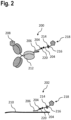
- Figures 2 and 3 show schematic views of exemplary embodiments of the connector 100.
- FIG. 2 shows a schematic view of nucleotide-based connectors 200, 202.
- the connectors 200, 202 each comprise a backbone 204, which may be an oligonucleotide, in particular a DNA or RNA oligonucleotide, and/or comprise nucleic acid analogues such as PNA, LNA, XNA, and morpholinos.
- the backbone 204 is connected to a first affinity reagent 206, which is oligonucleotide-based, similar to the backbone 204, and which comprise a specific sequence enabling the first affinity reagent 206 to directly or indirectly bind to an target molecule 208, 210.
- the first affinity reagent 206 is indirectly bound to the target molecule 208, for example, a protein, via a second affinity reagent 212, such as an antibody.
- the second affinity reagent 212 comprises, for example, at its fragment crystallisable region (Fc region), an oligonucleotide sequence complementary to the specific sequence of the first affinity reagent 206.
- the antigen-binding fragment (Fab region) of the second affinity reagent 212 may bind specifically to the target molecule 208.
- the connector 200 specifically binds to the target molecule 208 indirectly via the second affinity reagent 212.
- the target molecule 210 is an RNA or DNA molecule, for example a mRNA molecule.
- the first affinity reagent 206 binds to a specific sequence of the target molecule 210 complementary to the sequence of the first affinity reagent 206.
- the backbone 204 comprises a first affinity interactor 214 configured to specifically bind to a second affinity interactor 216.
- the first affinity interactor 214 may, for example, be an oligonucleotide-based aptamer, similar to the backbone 204.
- the second affinity interactor 216 may be a further aptamer or biotin, for example.
- the second affinity interactor 216 is connected to a label 218, such as a dye molecule.
- the backbone 204 comprises a cleavage site 220.
- the cleavage site 220 is preferably a restriction site for a restriction enzyme, in particular a rare cutter such as Notl. This enables separating the label 218 from the connector 200, 202.
- the connector 200, 202 including the first affinity reagent 206, the backbone 204 with the cleavage site 220 and the first affinity interactor 214 are all oligonucleotide-based, the connector 200, 202 may each be synthesised in a single reaction or synthesised in a single piece.
- FIG. 3 shows a schematic view of peptide-based connectors 300, 302.
- the connectors 300, 302 each comprise a backbone 304, which may be a peptide.
- the backbone 304 is connected to a first affinity reagent 306, 308, which are peptide-based, similar to the backbone 304, and which directly or indirectly bind specifically to a target molecule 310.
- the first affinity reagent 306 is indirectly bound to the target molecule 310, for example, a protein, via a second affinity reagent 312, such as an antibody.
- the first affinity reagent 306 is, for example, a nanobody or a single domain antibody, which specifically binds to the second affinity reagent 312, for example, at its fragment crystallisable region (Fc region).
- the antigen-binding fragment (Fab region) of the second affinity reagent 312 may bind specifically to the target molecule 310.
- the connector 300 specifically binds to the target molecule 310 indirectly via the second affinity reagent 312.
- the first affinity reagent 308 is, for example, a nanobody or a single domain antibody, which specifically binds directly to the target molecule 310.
- the backbone 304 comprises a first affinity interactor 314 configured to specifically bind to a second affinity interactor 316.
- the first affinity interactor 314 may be peptide-based, similar to the backbone 304, for example, it may be streptavidin.
- the second affinity interactor 316 may be biotin, for example.
- the second affinity interactor 316 is connected to a label 318, such as a dye molecule.
- Alternative pairs of first and second affinity interactors 314, 316 that bind specifically to each other may be pairs of peptides that form bioconjugates, such as SpyTag and SpyCatcher, SnoopTag and SnoopCatcher, or DogTag and DogCatcher.
- the backbone 304 comprises a cleavage site 320.
- the cleavage site 320 is preferably a specific peptide sequence at which a respective protease, for example, TEV protease from Tobacco etch virus or Factor Xa, or scUlp1 (from Saccharomyces cerevisiae with substrate scSUMO), or bdSENP1 (from Brachypodium distachyon with substrate bdSUMO), or bdNEDP1 (from Brachypodium distachyon with substrate bdNEDD8), or sNEDP1 (from Salmo salar with substrate ssNEDD8), or scAtg4 (from Saccharomyces cerevisiae with substrate Atg4p), or xlUsp2 (from Xenopus laevis with substrate xlUsp2), may cleave the peptide-based backbone 304. This enables separating the label 318 from the
- bdSENP1 or a bdSENP1 derivative is used to perform cleavage.
- This provides numerous advantages including very efficient substrate cleavage at concentrations in the nanomolar (nM) regime, i.e. 5nM at 25°C or 20-50nm at 0° for near quantitative cleavage (conditions described in the literature: Steffen Frey, Dirk Görlich, A new set of highly efficient, tag-cleaving proteases for purifying recombinant proteins, Journal of Chromatography A, Volume 1337, 2014, Pages 95-105 ), and good activity at temperatures between 0-37°C.
- the connector 300, 302 including the first affinity reagent 306, the backbone 304 with the cleavage site 320 and the first affinity interactor 314 are all oligonucleotide-based, the connector 300, 302 may each be synthesised by recombinant expression of a single DNA construct and/or synthesised as a continuous single piece.
- the first affinity reagents 102, 206, 306 may be a multimer of a nanobody or single-domain antibody, a conventional antibody, an aptamer, or a drug, small molecule or toxin.
- the cleavage sites 108, 220, 320 may be cleaved by a temperature shift, for example in case the backbone is oligonucleotide-based, in particular a DNA-origami, and the cleavage site is a hybridised part of two oligonucleotide strands, or by activation light, such as UV light, in case the cleavage site is a photocleavable linker.
- a temperature shift for example in case the backbone is oligonucleotide-based, in particular a DNA-origami, and the cleavage site is a hybridised part of two oligonucleotide strands, or by activation light, such as UV light, in case the cleavage site is a photocleavable linker.
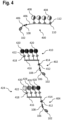
- FIG. 4 shows connector 400, 402, 404 that each have a label attached. Elements with the same structure and the same function have the same reference signs.
- the connector 400 comprises the first affinity reagent 102, as previously described, the label attached to a backbone 406 of the connector 400 comprises four fluorophores 408.
- the backbone 406 may, for example, be peptide-based.
- the backbone 406 comprises four first affinity interactors 110 that are each configured to specifically bind to four second affinity interactors 112 in order to individually attach the fluorophores 408 connected to the second affinity interactors 112.
- the fluorophores 408 may be attached to the connector 400 by mixing both, the fluorophores 408 with second affinity interactors 112 and the connector 400.
- the first and second affinity interactors 110, 112 may be streptavidin and biotin, respectively, for example.
- Connector 402 has a label 410 attached, specifically the label 410 is attached to a first affinity interactor 412 of the connector 402 via a second affinity interactor 414 that specifically binds to the first affinity interactor 412.
- the label 410 comprises a DNA-origami backbone 416 that is connected to the second affinity interactor 414 and that comprises four first auxiliary affinity interactors 418.
- the DNA-origami backbone 416 may, for example, be a nanoruler.
- Four fluorophores 420 are individually attached to backbone 416, by a second auxiliary affinity interactor 422 that specifically binds to the first auxiliary affinity interactor 418 and that is connected to each of the fluorophores 410.
- the first and second auxiliary affinity interactor are one of the biotin-streptavidin pair.
- biotinylated staple strands are allowed to bind to streptavidin-conjugated dyes or fluorophores before the staple strands are allowed to interact with the DNA-origami backbone 416 (preincubation).
- this can be leveraged to attach multiple copies of the same dye as in example 402 or multiple dyes (single or multiple copy) as in example 404 to the DNA-origami backbone 416.
- Connector 404 has a label 424 attached to the first affinity interactor 412 via a second affinity interactor 414, as described for connector 402.
- the label 424 comprises the DNA-origami backbone 416 with four first auxiliary affinity interactors 418.
- the label 424 comprises four fluorophores 420, 426, 428, 430 that each differ in their fluorescent properties. These properties may include excitation wavelength, fluorescent wavelength, and fluorescent duration.
- Each fluorophore 420, 426, 428, 430 comprises one of the second auxiliary affinity interactors 422 that enable the fluorophores 420, 426, 428, 430 to be attached to one of the first auxiliary affinity interactors 418 of the label 424.
- Figure 5 shows a schematic view of a biological sample 500 with a plurality of connectors.
- the biological sample 500 may, for example, be a cell, in particular, a mammalian cell comprising a nucleus 501.
- Several connectors 502, 504, 506, 508, 510, 512, 514 with labels are used to specifically bind to a particular target molecule in the sample 500, illustrating the use of the labelled connectors.
- the labels may differ in their fluorescent properties.
- connector 502 is indirectly bound to a cytoplasmic protein target molecule 516 via a second affinity reagent 518, here an antibody, and a nanobody first affinity reagent 519.
- Connector 504 is directly bound to a cytoplasmic protein target molecule 520 via first affinity reagent 522, here a nanobody of the connector 504.
- Connector 506 is directly bound to a mRNA target molecule 524 via an oligonucleotide-based first affinity reagent 526.
- the connector 508 is directly bound to a cytoplasmic protein target molecule 528 via a small molecule-based first affinity reagent 530.
- connector 510 is indirectly bound to a cytoplasmic protein target molecule 532, however, connector 510 is attached to a label 534 comprising a plurality of fluorophores.
- connector 512 is indirectly bound to a nuclear protein target molecule 534.
- connector 514 is directly bound to a nuclear mRNA target molecule 536.
- the individual components 516, 520, 524, 528, 532, 534, 536 of the biological sample 500 may be visualised by fluorescent imaging, for example, of the biological sample 500 with the connector 502, 504, 506, 508, 510, 512, 514 bound to their target molecules.
- the components 516, 520, 524, 528, 532, 534, 536 can be localised within the image of the biological sample 500 depending on where a respective fluorescent signal was detected.
- the combination of one of the connectors with one of the labels specifically bound to the connector via the first and second affinity interactors may also be termed a marker.
- the connector with the label is optionally specifically bound to one of the second affinity reagents.
- the connector, the label and optionally the second affinity reagents may be mixed together. This allows binding of the label to the connector, more precisely, the binding of the second affinity interactor of the label to the first affinity interactor of the connector and thereby attachment of the label to the connector.
- the second affinity reagent may be added at the same time, prior or after adding the label. Since the first affinity interactor and the second affinity interactor bind to each other specifically, as do the first and second affinity reagents, the first affinity interactor will only bind to the second affinity interactor, and the first affinity reagent will only bind to the second affinity reagent, even when mixing all components together.
- the connector and the label and/or the connector and the second affinity reagent may optionally be crosslinked, for example, with glutaraldehyde.
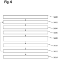
- FIG. 6 shows a flowchart for a method for analysing the biological sample 500, for example, which may be used in the context of multiplex imaging.
- the process starts in step S600.
- a set of markers is provided.
- the first set of markers may comprise a first plurality of connectors with a first label and a first affinity reagent configured to specifically bind to a first target molecule.
- the first plurality of connectors may further comprise a second affinity reagent configured to specifically bind to the first target molecule and the first affinity reagent being configured to bind to the second affinity reagent.
- the first set of markers includes at least a second plurality of connectors with a second label and a first (or second) affinity reagent configured to specifically bind to a second target molecule.
- the first and second labels have different fluorescent properties such as excitation wavelength, emission wavelength or emission duration.
- the step of providing the first set of markers may include generating the markers from the respective connector, the label and optionally the second affinity reagent. This may include mixing the respective connector, the label, and optionally the second affinity reagent, in step S602, as explained above.
- step S604 the sample is stained with the markers.
- the markers are added to the sample, which causes binding of the affinity reagents to their respective target molecules.
- the respective connector, the label and optionally the second affinity reagent may be added individually to the sample in step S604. This is particularly the case, when the marker only comprises a first affinity reagent and not a second affinity reagent.
- the marker is generated in-situ. The marker then stains the sample by binding to the respective target molecule with the first or second affinity reagent.
- step S606 markers that are not bound to their target molecules are washed out, for example, by washing the sample with a buffer solution. This is to reduce background staining by the label of unbound markers.
- an image readout is performed, for example, an image of the sample is acquired by illuminating the sample with fluorescent light and capturing the emission light. This may be done by means of a fluorescent microscope. The image reveals locations within the sample of the target molecules, which are stained by a marker with an affinity reagent configured to bind to the particular target molecule.
- step S610 the label is separated from the connector by cleaving of the cleavage site of the connector. This removes the fluorescent signal from the target molecule that the marker is bound to. This may also be called blanking.
- step S612 the cleaved off label is washed out of the sample, for example, by washing the sample with the buffer solution.
- the method ends in step S614.
- the method may proceed by repeating steps S602 to S612 with a different set of markers.
- This enables using a new set of connectors with affinities to further target molecules whilst at least partially using labels with the same fluorescent properties as the first set of markers.
- steps S602 to S612 By iterating through steps S602 to S612 repeatedly, a number of target molecules larger than the number of markers with unique fluorescent properties may be stained, imaged and localised.
- the connector, or the markers as described herein, may further be used in conjunction with methods and workflows for analysing biological samples disclosed in applications PCT/EP2021/066645 and PCT/EP2021/073819 , the contents of which are incorporated herein.
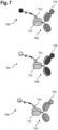
- FIG. 7 shows a schematic view of several markers 700, 702, 704.
- the markers 700, 702, 704, comprise antibodies 706, 708, 710 of the same isotype as second affinity reagents. This means, their Fc region 712 is the same.
- the first affinity reagents 714 bind to the Fc region 712 of the antibodies 706, 708, 710. This means that based on one type of connector comprising only the same first affinity reagent 714, markers may be generated that nevertheless bind specifically to different target molecules 716, 718, 720. Further, this allows using antibodies of the same isotype in a single set of markers.
- the markers 700, 702, 704 have to be prepared separately to avoid mixing the different antibodies 706, 708, 710 and labels.
- FIG 8 is a schematic view of a marker 800 comprising a connector 802 with a nucleic acid backbone 804 and four linker oligonucleotides 806 with a first affinity interactor 808.
- the first affinity interactor 808 may be biotin, for example.
- the linker oligonucleotide 806 may thus be a biotinylated oligonucleotide hybridised to a complementary part of the nucleic acid backbone 804.
- the marker 800 further comprises a label 810 with four fluorescent dyes 812, 814, 816, 818.
- the dyes 812, 814, 816, 818 preferably each have different optical properties and are each connected to the connector by a second affinity interactor 820 bound to the respective first affinity interactor 808.
- a first affinity reagent 822 of the marker 800 is bound to backbone 804 of the connector 802 by means of an affinity linker oligonucleotide 824.
- the affinity linker oligonucleotide 824 hybridises to a complementary sequence of the backbone 804.
- the first affinity reagent 822 enables binding the marker 800 to a target structure 826 in a biological sample and therefore marking the target 826.
- Figure 9 is a schematic view of several configurations 900, 902, 904, 906, 908 of streptavidin.
- the first configuration 900 of streptavidin is a native tetramer with four active biotin binding sites 910.
- the second configuration 902 of streptavidin is a tetramer with three active sites 910 and one non-functional biotin binding site 912.
- the third configuration 904 of streptavidin is a tetramer with two active sites 910 and two non-functional biotin binding sites 912.
- the fourth configuration 906 of streptavidin is a tetramer with one active site 910 and three non-functional biotin binding site 912.
- the fifth configuration 908 of streptavidin is a monomer with one active site 910.
- the number of active sites 910 By varying the number of active sites 910, the number of entities able to bind to the connectors 802 may be varied. Further, by choosing the configurations 906, 908 with only one binding site 910, the stoichiometric ratio of connectors 802 to entities binding to each other via the first and second affinity interactors 808, 820 is predictable, such that only one connector 802 binds to one entity.
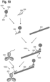
- Figure 10 is a schematic view of steps to assemble a connector-label conjugate 1000 and a marker 1002.
- the connector-label conjugate 1000 is assembled from a label 1004 comprising at least one dye 1006 and the second affinity interactor 820.
- the label 1004 is initially combined with the linker oligonucleotide 806 comprising the first affinity interactor 808.
- the first and second affinity interactor 808, 820 bind to each other specifically.
- the linker oligonucleotide 806 may be combined with the backbone 804 of the connector 802, forming the connector-label conjugate 1000.
- the linker oligonucleotide 806 has a sequence that is at least partially complementary to a sequence of the nucleic acid backbone 804.
- the linker oligonucleotide 806 may hybridise with the nucleic acid backbone 804 at a specific location 1008 and bind the label 1004 to the backbone 804.
- the nucleic acid backbone 804 may comprise several, in particular unique, sequences, that enable binding of a plurality of linker oligonucleotides 806 to the backbone 804.
- combinations of dyes 1006 may be specifically generated by combining those dyes 1006 with respective linker oligonucleotides 806 that may be specifically attached to the nucleic acid backbone 804.
- the connector-label conjugate 1000 may be formed by initially combining the linker oligonucleotide 806 with the backbone 804 of the connector 802. In a subsequent step, the dye 1006 of the label 1004 may be combined with the linker oligonucleotide 806 already attached to the backbone 804 causing the first and second affinity interactor 808, 820 to bind to each other. This forms the connector-label conjugate 1000.
- the marker 1002 may subsequently be generated by combining the connector-label conjugate 1000 with the first affinity reagent 822, for example, an antibody.
- the antibody comprises the affinity linker oligonucleotide 824 that is at least partially complementary to the nucleic acid backbone 804.
- the marker 1002 may be introduced into a biological sample in order to mark a particular biological structure such as the target 826, based on the specific affinity of the first affinity reagent 822.
- the location of the label 1004 reveals the location of the target 826 in the sample.
- the connector 802 enables efficiently assembling a variety of different connector-label conjugates 1000, in particular with labels 1004 with different optical properties. These conjugates 1000 may then be used to generate a variety of markers 1002, for example, with affinities a plurality of targets in a sample.
- a mix-and-match approach may be adopted.
- a particular label 1004 with particular optical properties may efficiently connected to a particular first affinity reagent 822 that has an affinity for a particular biological structure in the biological sample.
- a particular marker 1002 may be generated.
- a different marker may be generated by combining in a container the solutions with another label, the connector and another first affinity reagent, in particular with affinity to another biological structure in the biological sample.
- aspects have been described in the context of an apparatus, it is clear that these aspects also represent a description of the corresponding method, where a block or device corresponds to a method step or a feature of a method step. Analogously, aspects described in the context of a method step also represent a description of a corresponding block or item or feature of a corresponding apparatus.
Landscapes
- Health & Medical Sciences (AREA)
- Life Sciences & Earth Sciences (AREA)
- Immunology (AREA)
- Engineering & Computer Science (AREA)
- Chemical & Material Sciences (AREA)
- Molecular Biology (AREA)
- Biomedical Technology (AREA)
- Hematology (AREA)
- Urology & Nephrology (AREA)
- Biochemistry (AREA)
- Analytical Chemistry (AREA)
- Physics & Mathematics (AREA)
- Medicinal Chemistry (AREA)
- Microbiology (AREA)
- Biotechnology (AREA)
- General Health & Medical Sciences (AREA)
- Pathology (AREA)
- Cell Biology (AREA)
- General Physics & Mathematics (AREA)
- Food Science & Technology (AREA)
- Organic Chemistry (AREA)
- Zoology (AREA)
- Proteomics, Peptides & Aminoacids (AREA)
- Wood Science & Technology (AREA)
- Biophysics (AREA)
- Bioinformatics & Cheminformatics (AREA)
- General Engineering & Computer Science (AREA)
- Genetics & Genomics (AREA)
- Chemical Kinetics & Catalysis (AREA)
- General Chemical & Material Sciences (AREA)
- Measuring Or Testing Involving Enzymes Or Micro-Organisms (AREA)
Priority Applications (2)
| Application Number | Priority Date | Filing Date | Title |
|---|---|---|---|
| US18/510,752 US20240168031A1 (en) | 2022-02-01 | 2023-11-16 | Connector, connector-label conjugate and marker for analysing biological samples |
| CN202311546494.9A CN118056910A (zh) | 2022-02-01 | 2023-11-20 | 用于分析生物样品的连接体、连接体-标签缀合物和标记物 |
Applications Claiming Priority (1)
| Application Number | Priority Date | Filing Date | Title |
|---|---|---|---|
| EP22154515.5A EP4092414B1 (fr) | 2021-05-19 | 2022-02-01 | Connecteur, marqueur et procédé pour analyser des échantillons biologiques |
Publications (2)
| Publication Number | Publication Date |
|---|---|
| EP4220162A2 true EP4220162A2 (fr) | 2023-08-02 |
| EP4220162A3 EP4220162A3 (fr) | 2023-10-18 |
Family
ID=84331037
Family Applications (1)
| Application Number | Title | Priority Date | Filing Date |
|---|---|---|---|
| EP22208330.5A Pending EP4220162A3 (fr) | 2022-02-01 | 2022-11-18 | Connecteur, conjugué connecteur-étiquette et marqueur pour analyser des échantillons biologiques |
Country Status (3)
| Country | Link |
|---|---|
| US (1) | US20240168031A1 (fr) |
| EP (1) | EP4220162A3 (fr) |
| CN (2) | CN116539570A (fr) |
Cited By (6)
| Publication number | Priority date | Publication date | Assignee | Title |
|---|---|---|---|---|
| EP4521112A1 (fr) * | 2023-09-06 | 2025-03-12 | Leica Microsystems CMS GmbH | Marqueur, kit et procédé d'analyse d'un échantillon biologique |
| EP4556890A1 (fr) * | 2023-11-14 | 2025-05-21 | Leica Microsystems CMS GmbH | Étiquette, marqueur et kit de réactifs pour analyser un échantillon biologique |
| EP4585695A1 (fr) * | 2024-01-15 | 2025-07-16 | Leica Microsystems CMS GmbH | Marqueur et marqueur pour analyser un échantillon biologique |
| EP4596714A1 (fr) * | 2024-02-05 | 2025-08-06 | Leica Microsystems CMS GmbH | Marqueur pour l'analyse d'un échantillon biologique |
| WO2025168224A1 (fr) | 2024-02-08 | 2025-08-14 | Leica Microsystems Cms Gmbh | Marqueur et procédé d'analyse d'un échantillon biologique |
| EP4653542A1 (fr) * | 2024-05-21 | 2025-11-26 | Leica Microsystems CMS GmbH | Étiquette, marqueur et procédé d'analyse d'un échantillon biologique |
Families Citing this family (1)
| Publication number | Priority date | Publication date | Assignee | Title |
|---|---|---|---|---|
| WO2026008147A1 (fr) * | 2024-07-04 | 2026-01-08 | Leica Microsystems Cms Gmbh | Étiquette, marqueur et procédé pour l'analyse d'un échantillon biologique |
Citations (1)
| Publication number | Priority date | Publication date | Assignee | Title |
|---|---|---|---|---|
| US20140057805A1 (en) | 2012-08-22 | 2014-02-27 | Philip Tinnefeld | Dna-origami-based standard |
Family Cites Families (2)
| Publication number | Priority date | Publication date | Assignee | Title |
|---|---|---|---|---|
| WO2017015097A1 (fr) * | 2015-07-17 | 2017-01-26 | Nanostring Technologies, Inc. | Quantification simultanée d'une pluralité de protéines dans une région définie par l'utilisateur d'un tissu en coupe transversale |
| US11821023B2 (en) * | 2018-05-30 | 2023-11-21 | Generation Biotech, Llc | Method for capturing long polynucleotides for sequencing specific gnomic regions |
-
2022
- 2022-11-18 EP EP22208330.5A patent/EP4220162A3/fr active Pending
-
2023
- 2023-01-30 CN CN202310045014.4A patent/CN116539570A/zh active Pending
- 2023-11-16 US US18/510,752 patent/US20240168031A1/en active Pending
- 2023-11-20 CN CN202311546494.9A patent/CN118056910A/zh active Pending
Patent Citations (1)
| Publication number | Priority date | Publication date | Assignee | Title |
|---|---|---|---|---|
| US20140057805A1 (en) | 2012-08-22 | 2014-02-27 | Philip Tinnefeld | Dna-origami-based standard |
Non-Patent Citations (2)
| Title |
|---|
| STEFFEN FREYDIRK GORLICH: "A new set of highly efficient, tag-cleaving proteases for purifying recombinant proteins", JOURNAL OF CHROMATOGRAPHY A, vol. 1337, 2014, pages 95 - 105, XP028634158, DOI: 10.1016/j.chroma.2014.02.029 |
| YIN ET AL., SCIENCE, vol. 338, 30 November 2012 (2012-11-30), pages 1177 - 1183 |
Cited By (10)
| Publication number | Priority date | Publication date | Assignee | Title |
|---|---|---|---|---|
| EP4521112A1 (fr) * | 2023-09-06 | 2025-03-12 | Leica Microsystems CMS GmbH | Marqueur, kit et procédé d'analyse d'un échantillon biologique |
| EP4521113A1 (fr) * | 2023-09-06 | 2025-03-12 | Leica Microsystems CMS GmbH | Réactif d'affinité, marqueur et procédé d'analyse d'un échantillon biologique |
| EP4521114A1 (fr) * | 2023-09-06 | 2025-03-12 | Leica Microsystems CMS GmbH | Réactif d'affinité, marqueur et procédé d'analyse d'un échantillon biologique |
| EP4556890A1 (fr) * | 2023-11-14 | 2025-05-21 | Leica Microsystems CMS GmbH | Étiquette, marqueur et kit de réactifs pour analyser un échantillon biologique |
| EP4585695A1 (fr) * | 2024-01-15 | 2025-07-16 | Leica Microsystems CMS GmbH | Marqueur et marqueur pour analyser un échantillon biologique |
| WO2025153258A1 (fr) * | 2024-01-15 | 2025-07-24 | Leica Microsystems Cms Gmbh | Étiquette et marqueur pour l'analyse d'un échantillon biologique |
| EP4596714A1 (fr) * | 2024-02-05 | 2025-08-06 | Leica Microsystems CMS GmbH | Marqueur pour l'analyse d'un échantillon biologique |
| WO2025168224A1 (fr) | 2024-02-08 | 2025-08-14 | Leica Microsystems Cms Gmbh | Marqueur et procédé d'analyse d'un échantillon biologique |
| EP4653542A1 (fr) * | 2024-05-21 | 2025-11-26 | Leica Microsystems CMS GmbH | Étiquette, marqueur et procédé d'analyse d'un échantillon biologique |
| WO2025242752A1 (fr) * | 2024-05-21 | 2025-11-27 | Leica Microsystems Cms Gmbh | Étiquette, marqueur et procédé d'analyse d'un échantillon biologique |
Also Published As
| Publication number | Publication date |
|---|---|
| CN116539570A (zh) | 2023-08-04 |
| US20240168031A1 (en) | 2024-05-23 |
| EP4220162A3 (fr) | 2023-10-18 |
| CN118056910A (zh) | 2024-05-21 |
Similar Documents
| Publication | Publication Date | Title |
|---|---|---|
| EP4220162A2 (fr) | Connecteur, conjugué connecteur-étiquette et marqueur pour analyser des échantillons biologiques | |
| EP4200441B1 (fr) | Procédés de libération d'une sonde de capture étendue à partir d'un substrat et leurs utilisations | |
| EP4092414B1 (fr) | Connecteur, marqueur et procédé pour analyser des échantillons biologiques | |
| Olejnik et al. | Photocleavable peptide-DNA conjugates: synthesis and applications to DNA analysis using MALDI-MS | |
| Crut et al. | Detection of single DNA molecules by multicolor quantum-dot end-labeling | |
| Kodadek et al. | Synthetic molecules as antibody replacements | |
| CN118345145A (zh) | 用于使用标记的核酸成像剂进行多路复用成像的改进方法 | |
| US20230258628A1 (en) | Connector, marker and method for analysing biological samples | |
| JP4457742B2 (ja) | 生体標本への試薬の分注方法及び生体標本の解析方法 | |
| Kohman et al. | Fluorescent in situ sequencing of DNA barcoded antibodies | |
| WO2024211058A1 (fr) | Procédés et compositions de séquençage de cellule unique à résolution spatiale | |
| Zhou et al. | Expressed peptide assay for DNA detection | |
| WO2016057842A2 (fr) | Imagerie haute résolution de protéines tissulaires | |
| US20260104421A1 (en) | Connector, marker, capture construct and data storage device | |
| WO2025014746A1 (fr) | Détection moléculaire par l'utilisation de sondes à codes-barres ou de sondes de fusion à codes-barres et réaction d'hybridation en chaîne | |
| EP4067871B1 (fr) | Partie constitutive d'un marqueur | |
| Vanderschoot et al. | Spatial multiomics lipids and gene expression using MALDI ISH MSI | |
| EP4703479A1 (fr) | Marqueur et procédé d'analyse d'un échantillon biologique | |
| US20250075205A1 (en) | Affinity reagent, marker and method for analysing a biological sample | |
| EP4509615A1 (fr) | Marqueur détectable optiquement | |
| US20240353413A1 (en) | Label and marker structure for marking a target structure | |
| EP4617378A1 (fr) | Réactif d'affinité et procédé d'analyse d'un échantillon biologique | |
| EP4556890A1 (fr) | Étiquette, marqueur et kit de réactifs pour analyser un échantillon biologique | |
| US20240353327A1 (en) | Marker for analyzing a biological sample | |
| CN117625738A (zh) | 用于分析生物样品的标记物和方法 |
Legal Events
| Date | Code | Title | Description |
|---|---|---|---|
| PUAI | Public reference made under article 153(3) epc to a published international application that has entered the european phase |
Free format text: ORIGINAL CODE: 0009012 |
|
| STAA | Information on the status of an ep patent application or granted ep patent |
Free format text: STATUS: THE APPLICATION HAS BEEN PUBLISHED |
|
| AK | Designated contracting states |
Kind code of ref document: A2 Designated state(s): AL AT BE BG CH CY CZ DE DK EE ES FI FR GB GR HR HU IE IS IT LI LT LU LV MC ME MK MT NL NO PL PT RO RS SE SI SK SM TR |
|
| PUAL | Search report despatched |
Free format text: ORIGINAL CODE: 0009013 |
|
| AK | Designated contracting states |
Kind code of ref document: A3 Designated state(s): AL AT BE BG CH CY CZ DE DK EE ES FI FR GB GR HR HU IE IS IT LI LT LU LV MC ME MK MT NL NO PL PT RO RS SE SI SK SM TR |
|
| RIC1 | Information provided on ipc code assigned before grant |
Ipc: G01N 33/533 20060101ALI20230913BHEP Ipc: C40B 40/10 20060101ALI20230913BHEP Ipc: C40B 40/06 20060101ALI20230913BHEP Ipc: C12Q 1/6876 20180101ALI20230913BHEP Ipc: G01N 33/58 20060101ALI20230913BHEP Ipc: G01N 33/532 20060101AFI20230913BHEP |
|
| P01 | Opt-out of the competence of the unified patent court (upc) registered |
Effective date: 20231221 |
|
| STAA | Information on the status of an ep patent application or granted ep patent |
Free format text: STATUS: REQUEST FOR EXAMINATION WAS MADE |
|
| 17P | Request for examination filed |
Effective date: 20240412 |
|
| RBV | Designated contracting states (corrected) |
Designated state(s): AL AT BE BG CH CY CZ DE DK EE ES FI FR GB GR HR HU IE IS IT LI LT LU LV MC ME MK MT NL NO PL PT RO RS SE SI SK SM TR |
|
| STAA | Information on the status of an ep patent application or granted ep patent |
Free format text: STATUS: EXAMINATION IS IN PROGRESS |
|
| 17Q | First examination report despatched |
Effective date: 20241106 |