EP4230144A1 - Appareil et procédé pour fournir des informations d'aide au diagnostic de la démence - Google Patents
Appareil et procédé pour fournir des informations d'aide au diagnostic de la démence Download PDFInfo
- Publication number
- EP4230144A1 EP4230144A1 EP22206524.5A EP22206524A EP4230144A1 EP 4230144 A1 EP4230144 A1 EP 4230144A1 EP 22206524 A EP22206524 A EP 22206524A EP 4230144 A1 EP4230144 A1 EP 4230144A1
- Authority
- EP
- European Patent Office
- Prior art keywords
- value
- standard
- group
- standardized uptake
- diagnosis
- Prior art date
- Legal status (The legal status is an assumption and is not a legal conclusion. Google has not performed a legal analysis and makes no representation as to the accuracy of the status listed.)
- Withdrawn
Links
- 238000003745 diagnosis Methods 0.000 title claims abstract description 120
- 206010012289 Dementia Diseases 0.000 title claims abstract description 78
- 238000000034 method Methods 0.000 title claims abstract description 40
- 210000004556 brain Anatomy 0.000 claims abstract description 108
- 238000002600 positron emission tomography Methods 0.000 claims description 47
- 238000002595 magnetic resonance imaging Methods 0.000 claims description 39
- 230000001131 transforming effect Effects 0.000 claims description 21
- 230000009466 transformation Effects 0.000 claims description 18
- 238000004590 computer program Methods 0.000 claims description 5
- 238000013507 mapping Methods 0.000 claims description 5
- 208000010877 cognitive disease Diseases 0.000 description 32
- 208000027061 mild cognitive impairment Diseases 0.000 description 32
- 230000032683 aging Effects 0.000 description 24
- 238000013500 data storage Methods 0.000 description 21
- 208000024827 Alzheimer disease Diseases 0.000 description 14
- 238000010586 diagram Methods 0.000 description 9
- 102000013455 Amyloid beta-Peptides Human genes 0.000 description 4
- 108010090849 Amyloid beta-Peptides Proteins 0.000 description 4
- 238000004891 communication Methods 0.000 description 4
- 238000003384 imaging method Methods 0.000 description 4
- 230000008569 process Effects 0.000 description 4
- 208000024891 symptom Diseases 0.000 description 4
- 201000004810 Vascular dementia Diseases 0.000 description 3
- 238000013135 deep learning Methods 0.000 description 3
- 239000003814 drug Substances 0.000 description 3
- 229940079593 drug Drugs 0.000 description 3
- 230000006870 function Effects 0.000 description 3
- 238000012360 testing method Methods 0.000 description 3
- 208000009829 Lewy Body Disease Diseases 0.000 description 2
- 201000002832 Lewy body dementia Diseases 0.000 description 2
- 230000003920 cognitive function Effects 0.000 description 2
- 230000006378 damage Effects 0.000 description 2
- 238000002405 diagnostic procedure Methods 0.000 description 2
- 210000002569 neuron Anatomy 0.000 description 2
- 238000012545 processing Methods 0.000 description 2
- 230000005855 radiation Effects 0.000 description 2
- 208000000044 Amnesia Diseases 0.000 description 1
- 208000031124 Dementia Alzheimer type Diseases 0.000 description 1
- 208000026139 Memory disease Diseases 0.000 description 1
- 238000012879 PET imaging Methods 0.000 description 1
- 206010039966 Senile dementia Diseases 0.000 description 1
- 230000008859 change Effects 0.000 description 1
- 208000035850 clinical syndrome Diseases 0.000 description 1
- 230000001149 cognitive effect Effects 0.000 description 1
- 150000001875 compounds Chemical class 0.000 description 1
- 201000010099 disease Diseases 0.000 description 1
- 208000037265 diseases, disorders, signs and symptoms Diseases 0.000 description 1
- 238000005516 engineering process Methods 0.000 description 1
- YNDIAUKFXKEXSV-CRYLGTRXSA-N florbetapir F-18 Chemical compound C1=CC(NC)=CC=C1\C=C\C1=CC=C(OCCOCCOCC[18F])N=C1 YNDIAUKFXKEXSV-CRYLGTRXSA-N 0.000 description 1
- 230000036541 health Effects 0.000 description 1
- 208000011977 language disease Diseases 0.000 description 1
- 230000006984 memory degeneration Effects 0.000 description 1
- 208000023060 memory loss Diseases 0.000 description 1
- 238000010295 mobile communication Methods 0.000 description 1
- 238000012986 modification Methods 0.000 description 1
- 230000004048 modification Effects 0.000 description 1
- 238000002610 neuroimaging Methods 0.000 description 1
- 230000003287 optical effect Effects 0.000 description 1
- 230000008520 organization Effects 0.000 description 1
- 230000037081 physical activity Effects 0.000 description 1
- 239000000700 radioactive tracer Substances 0.000 description 1
- 238000011160 research Methods 0.000 description 1
- 239000007787 solid Substances 0.000 description 1
- 230000009747 swallowing Effects 0.000 description 1
Images





Classifications
-
- A—HUMAN NECESSITIES
- A61—MEDICAL OR VETERINARY SCIENCE; HYGIENE
- A61B—DIAGNOSIS; SURGERY; IDENTIFICATION
- A61B5/00—Measuring for diagnostic purposes; Identification of persons
- A61B5/0033—Features or image-related aspects of imaging apparatus, e.g. for MRI, optical tomography or impedance tomography apparatus; Arrangements of imaging apparatus in a room
- A61B5/0035—Features or image-related aspects of imaging apparatus, e.g. for MRI, optical tomography or impedance tomography apparatus; Arrangements of imaging apparatus in a room adapted for acquisition of images from more than one imaging mode, e.g. combining MRI and optical tomography
-
- A—HUMAN NECESSITIES
- A61—MEDICAL OR VETERINARY SCIENCE; HYGIENE
- A61B—DIAGNOSIS; SURGERY; IDENTIFICATION
- A61B6/00—Apparatus or devices for radiation diagnosis; Apparatus or devices for radiation diagnosis combined with radiation therapy equipment
- A61B6/02—Arrangements for diagnosis sequentially in different planes; Stereoscopic radiation diagnosis
- A61B6/03—Computed tomography [CT]
- A61B6/037—Emission tomography
-
- A—HUMAN NECESSITIES
- A61—MEDICAL OR VETERINARY SCIENCE; HYGIENE
- A61B—DIAGNOSIS; SURGERY; IDENTIFICATION
- A61B5/00—Measuring for diagnostic purposes; Identification of persons
- A61B5/05—Detecting, measuring or recording for diagnosis by means of electric currents or magnetic fields; Measuring using microwaves or radio waves
- A61B5/055—Detecting, measuring or recording for diagnosis by means of electric currents or magnetic fields; Measuring using microwaves or radio waves involving electronic [EMR] or nuclear [NMR] magnetic resonance, e.g. magnetic resonance imaging
-
- A—HUMAN NECESSITIES
- A61—MEDICAL OR VETERINARY SCIENCE; HYGIENE
- A61B—DIAGNOSIS; SURGERY; IDENTIFICATION
- A61B5/00—Measuring for diagnostic purposes; Identification of persons
- A61B5/40—Detecting, measuring or recording for evaluating the nervous system
- A61B5/4076—Diagnosing or monitoring particular conditions of the nervous system
- A61B5/4088—Diagnosing of monitoring cognitive diseases, e.g. Alzheimer, prion diseases or dementia
-
- A—HUMAN NECESSITIES
- A61—MEDICAL OR VETERINARY SCIENCE; HYGIENE
- A61B—DIAGNOSIS; SURGERY; IDENTIFICATION
- A61B5/00—Measuring for diagnostic purposes; Identification of persons
- A61B5/72—Signal processing specially adapted for physiological signals or for diagnostic purposes
- A61B5/7235—Details of waveform analysis
- A61B5/7264—Classification of physiological signals or data, e.g. using neural networks, statistical classifiers, expert systems or fuzzy systems
-
- A—HUMAN NECESSITIES
- A61—MEDICAL OR VETERINARY SCIENCE; HYGIENE
- A61B—DIAGNOSIS; SURGERY; IDENTIFICATION
- A61B5/00—Measuring for diagnostic purposes; Identification of persons
- A61B5/74—Details of notification to user or communication with user or patient; User input means
- A61B5/742—Details of notification to user or communication with user or patient; User input means using visual displays
- A61B5/743—Displaying an image simultaneously with additional graphical information, e.g. symbols, charts, function plots
-
- A—HUMAN NECESSITIES
- A61—MEDICAL OR VETERINARY SCIENCE; HYGIENE
- A61B—DIAGNOSIS; SURGERY; IDENTIFICATION
- A61B6/00—Apparatus or devices for radiation diagnosis; Apparatus or devices for radiation diagnosis combined with radiation therapy equipment
- A61B6/50—Apparatus or devices for radiation diagnosis; Apparatus or devices for radiation diagnosis combined with radiation therapy equipment specially adapted for specific body parts; specially adapted for specific clinical applications
- A61B6/501—Apparatus or devices for radiation diagnosis; Apparatus or devices for radiation diagnosis combined with radiation therapy equipment specially adapted for specific body parts; specially adapted for specific clinical applications for diagnosis of the head, e.g. neuroimaging or craniography
-
- A—HUMAN NECESSITIES
- A61—MEDICAL OR VETERINARY SCIENCE; HYGIENE
- A61B—DIAGNOSIS; SURGERY; IDENTIFICATION
- A61B6/00—Apparatus or devices for radiation diagnosis; Apparatus or devices for radiation diagnosis combined with radiation therapy equipment
- A61B6/52—Devices using data or image processing specially adapted for radiation diagnosis
- A61B6/5211—Devices using data or image processing specially adapted for radiation diagnosis involving processing of medical diagnostic data
- A61B6/5217—Devices using data or image processing specially adapted for radiation diagnosis involving processing of medical diagnostic data extracting a diagnostic or physiological parameter from medical diagnostic data
-
- A—HUMAN NECESSITIES
- A61—MEDICAL OR VETERINARY SCIENCE; HYGIENE
- A61B—DIAGNOSIS; SURGERY; IDENTIFICATION
- A61B6/00—Apparatus or devices for radiation diagnosis; Apparatus or devices for radiation diagnosis combined with radiation therapy equipment
- A61B6/52—Devices using data or image processing specially adapted for radiation diagnosis
- A61B6/5211—Devices using data or image processing specially adapted for radiation diagnosis involving processing of medical diagnostic data
- A61B6/5229—Devices using data or image processing specially adapted for radiation diagnosis involving processing of medical diagnostic data combining image data of a patient, e.g. combining a functional image with an anatomical image
- A61B6/5235—Devices using data or image processing specially adapted for radiation diagnosis involving processing of medical diagnostic data combining image data of a patient, e.g. combining a functional image with an anatomical image combining images from the same or different ionising radiation imaging techniques, e.g. PET and CT
-
- G—PHYSICS
- G16—INFORMATION AND COMMUNICATION TECHNOLOGY [ICT] SPECIALLY ADAPTED FOR SPECIFIC APPLICATION FIELDS
- G16H—HEALTHCARE INFORMATICS, i.e. INFORMATION AND COMMUNICATION TECHNOLOGY [ICT] SPECIALLY ADAPTED FOR THE HANDLING OR PROCESSING OF MEDICAL OR HEALTHCARE DATA
- G16H10/00—ICT specially adapted for the handling or processing of patient-related medical or healthcare data
- G16H10/60—ICT specially adapted for the handling or processing of patient-related medical or healthcare data for patient-specific data, e.g. for electronic patient records
-
- G—PHYSICS
- G16—INFORMATION AND COMMUNICATION TECHNOLOGY [ICT] SPECIALLY ADAPTED FOR SPECIFIC APPLICATION FIELDS
- G16H—HEALTHCARE INFORMATICS, i.e. INFORMATION AND COMMUNICATION TECHNOLOGY [ICT] SPECIALLY ADAPTED FOR THE HANDLING OR PROCESSING OF MEDICAL OR HEALTHCARE DATA
- G16H50/00—ICT specially adapted for medical diagnosis, medical simulation or medical data mining; ICT specially adapted for detecting, monitoring or modelling epidemics or pandemics
- G16H50/20—ICT specially adapted for medical diagnosis, medical simulation or medical data mining; ICT specially adapted for detecting, monitoring or modelling epidemics or pandemics for computer-aided diagnosis, e.g. based on medical expert systems
-
- G—PHYSICS
- G16—INFORMATION AND COMMUNICATION TECHNOLOGY [ICT] SPECIALLY ADAPTED FOR SPECIFIC APPLICATION FIELDS
- G16H—HEALTHCARE INFORMATICS, i.e. INFORMATION AND COMMUNICATION TECHNOLOGY [ICT] SPECIALLY ADAPTED FOR THE HANDLING OR PROCESSING OF MEDICAL OR HEALTHCARE DATA
- G16H50/00—ICT specially adapted for medical diagnosis, medical simulation or medical data mining; ICT specially adapted for detecting, monitoring or modelling epidemics or pandemics
- G16H50/30—ICT specially adapted for medical diagnosis, medical simulation or medical data mining; ICT specially adapted for detecting, monitoring or modelling epidemics or pandemics for calculating health indices; for individual health risk assessment
Definitions
- the present invention disclosed herein relates to an apparatus and method for providing dementia diagnosis assistance information, and more particularly, to an apparatus and method for providing dementia diagnosis assistance information by acquiring a standardized uptake value ratio for a brain image of a subject for diagnosis.
- Dementia refers to a clinical syndrome in which the cognitive functions of various regions, such as memory, language, and judgment, are reduced and daily life is unable to be properly performed.
- a typical dementia is senile dementia called Alzheimer's disease, and vascular dementia caused by stroke, and in addition, there is dementia due to various causes, but 50% to 70% of dementia patients are dementia due to Alzheimer's disease.
- Alzheimer's disease develops long before symptoms appear, but changes in the brain are not easily noticed until symptoms appear.
- the time when conspicuous symptoms such as memory loss and language disorder appear is after some degree of brain change has already occurred.
- the symptoms are due to damage or destruction of nerve cells in the brain involved in thinking, learning, and memory (cognitive function).
- As the disease progresses other neurons in the brain are damaged and destroyed, eventually affecting basic physical activities such as walking and swallowing food. Therefore, prompt and accurate diagnosis of Alzheimer's disease is very important.
- the PET stands for a positron emission tomography and is method of photographing the inside of the body by injecting a test drug that emits radiation to a patient and detecting the radiation emitted by the drug from the outside.
- the test drug only amyloid beta may be labeled by administering the compound that is easily attached to amyloid beta.
- a typical diagnostic method using amyloid PET is a method using amyloid standardized uptake value ratio (SUVR).
- Amyloid standardized uptake value ratio represents a ratio of amyloid standardized uptake value (SUV) from two different regions (target and reference region) within the same amyloid PET image, and the amyloid standardized uptake value (SUV) may represent the ratio of the concentration of radioactivity of the image to the concentration of radioactivity of the whole injected body.
- the amyloid standardized uptake value ratio (SUVR) is being used as the main data for determining the progression of Alzheimer's disease.
- ADNI Alzheimer's Disease Neuroimaging Initiative
- the ADNI suggests an SUVR value of approximately 1.10 based on a florbetapir tracer, as a criterion for quantitatively classifying beta amyloid positive and negative.
- the value of 1.10 represents the confidence limit value for the top 5% of the distribution of the amyloid standardized uptake value ratio (SUVR) obtained from a control group of 21 people under the age of 55 years. That is, the ADNI suggests determining whether amyloid is positive or negative based on 1.10 of the amyloid standardized uptake value ratio (SUVR) through the analysis of amyloid PET images.
- the present invention provides an apparatus and method for providing dementia diagnosis assistance information that provides various assistance information needed for dementia diagnosis to medical staff performing dementia diagnosis by using a PET images, in addition to simply providing a standardized uptake value ratio (SUVR), by generating a standard value indicating the degree to which the standardized uptake value ratio (SUVR) of the subject for diagnosis is close to the average value of SUVR of the normal group and the average value of SUVR of the Alzheimer's group and displaying the standard value as a graph of comparison with a control group.
- SUVR standardized uptake value ratio
- a method for providing dementia diagnosis assistance information that is performed in an apparatus for providing dementia diagnosis assistance information, the apparatus including a processor and a memory for storing predetermined instructions, the method including: (a) calculating a standardized uptake value ratio (SUVR) for each of regions (divided regions) obtained by dividing a brain region of a brain image of a subject for diagnosis into a plurality of predefined regions; (b) obtaining a standard value by mapping the standardized uptake value ratio of each divided region to a standard interval defined in advance for each divided region; and (c) outputting the standard value of each divided region together with statistical information about an average standard value of a control group for each divided region.
- SUVR standardized uptake value ratio
- the step (a) may include (a1) dividing the brain region included in a magnetic resonance imaging (MRI) image of the subject for diagnosis into a plurality of predefined divided regions and registering the image of the brain region with a positron emission tomography (PET) image of the subject for diagnosis, and (a2) generating a standardized uptake value ratio (SUVR) for each of the divided regions in the registered image.
- MRI magnetic resonance imaging
- PET positron emission tomography
- SUVR standardized uptake value ratio
- the standard value may represent a degree of proximity of the standardized uptake value ratio to an average value of standardized uptake value ratios of a group of Alzheimer's patients and an average value of standardized uptake value ratios of a normal group.
- the standard interval may be defined by setting, as both ends of a regular interval, the average value of the standardized uptake value ratios of the group of the Alzheimer's patients and the average value of the standardized uptake value ratios of the normal group for a region corresponding to each of the plurality of divided regions.
- the standard interval may be an interval generated by transforming the average value of the standardized uptake value ratios of the normal group to correspond to 0 according to a transformation formula, and transforming the average value of the standardized uptake value ratios of the group of the Alzheimer's patients to correspond to a predetermined number greater than 0 according to a predetermined transformation formula, and the standard value may be obtained by transforming the standardized uptake value ratio according to the transformation formula.
- the step (c) may include outputting a pair of values as large as a predefined value and small as a predefined value from the average standard value of the control group together with the standard value as a graph, for each divided region.
- the step (c) may include outputting a pair of values that differ by a standard deviation from the average standard value of the control group together with the standard value, for each divided region.
- control group may include at least one of an Alzheimer's patient group, a mild cognitive impairment group, a normal aging group, and a normal group.
- the step (c) may include displaying a proportion of divided regions where the standard value is located between a pair of values of the control group or a proportion of divided regions where the standard value deviates from the pair of values of the control group together with the graph.
- the step (c) may include estimating that the subject for diagnosis belongs to the control group when the proportion of divided regions where the standard value is located between the pair of values of the control group is equal to or greater than a predefined threshold value, and outputting an estimation result together with the graph.
- the step (c) may include outputting a degree of agreement between a graph obtained by connecting the standard values and a graph obtained by connecting the average standard values of the control group together with the graph, for the divided regions.
- a computer program that is stored in a non-transitory storage medium and executed in a computer including a processor, the computer program performing the method for providing dementia diagnosis assistance information.
- an apparatus for providing dementia diagnosis assistance information including: a processor; and a memory for storing predetermined instructions, in which the processor is configured to, when executing the instructions stored in the memory: (a) calculate a standardized uptake value ratio (SUVR) for each of regions (divided regions) obtained by dividing a brain region of a brain image of a subject for diagnosis into a plurality of predefined regions; (b) obtain a standard value by mapping the standardized uptake value ratio of each divided region to a standard interval defined in advance for each divided region; and (c) output the standard value of each divided region together with statistical information about an average standard value of a control group for each divided region.
- SUVR standardized uptake value ratio
- the processor in the step (a), is further configured to: (a1) divide the brain region included in a magnetic resonance imaging (MRI) image of the subject for diagnosis into a plurality of predefined divided regions and register the image of the brain region with a positron emission tomography (PET) image of the subject for diagnosis; and (a2) generate a standardized uptake value ratio (SUVR) for each of the divided regions in the registered image.
- MRI magnetic resonance imaging
- PET positron emission tomography
- the standard value may represent a degree of proximity of the standardized uptake value ratio to an average value of standardized uptake value ratios of a group of Alzheimer's patients and an average value of standardized uptake value ratios of a normal group.
- the standard interval may be defined by setting, as both ends of a regular interval, the average value of the standardized uptake value ratios of the group of the Alzheimer's patients and the average value of the standardized uptake value ratios of the normal group for a region corresponding to each of the plurality of divided regions.
- the standard interval may be an interval generated by transforming the average value of the standardized uptake value ratios of the normal group to correspond to 0 according to a predetermined transformation formula, and transforming the average value of the standardized uptake value ratios of the group of the Alzheimer's patients to correspond to a predetermined number greater than 0 according to the transformation formula, and the standard value may be obtained by transforming the standardized uptake value ratio according to the transformation formula.
- the processor in the step (c), is further configured to output a pair of values as large as a predefined value and small as a predefined value from the average standard value of the control group together with the standard value as a graph, for each divided region.
- the processor in the step (c), is further configured to output a pair of values that differ by a standard deviation from the average standard value of the control group together with the standard value, for each divided region.
- control group may include at least one of an Alzheimer's patient group, a mild cognitive impairment group, a normal aging group, and a normal group.
- the processor in the step (c), is further configured to display a proportion of divided regions where the standard value is located between a pair of values of the control group or a proportion of divided regions where the standard value deviates from the pair of values of the control group together with the graph.
- the processor in the step (c), is further configured to estimate that the subject for diagnosis belongs to the control group when the proportion of divided regions where the standard value is located between the pair of values of the control group is equal to or greater than a predefined threshold value, and output an estimation result together with the graph.
- the processor in the step (c), is further configured to output a degree of agreement between a graph obtained by connecting the standard values and a graph obtained by connecting the average standard values of the control group together with the graph, for the divided regions.
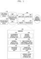
- FIG. 1 is a diagram illustrating an apparatus 200 for providing dementia diagnosis assistance information in accordance with a preferred embodiment of the present invention.
- the apparatus 200 for providing dementia diagnosis assistance information in accordance with an embodiment of the present invention is connected to a first image acquisition device 110 and a second image acquisition device 120 through a wired or wireless communication network, and receives an MRI image and a PET image from the first image acquisition device 110 and the second image acquisition device 120, respectively.
- the wired or wireless communication network may be implemented as any one of a mobile phone mobile communication network, a local area network (LAN), a wireless personal area network (WPAN), and a wireless local area network (WLAN), or may be implemented as a combination of two or more of them.
- LAN local area network
- WPAN wireless personal area network
- WLAN wireless local area network
- the first image acquisition device 110 may be implemented as an MRI imaging device that outputs an MRI brain image by imaging a brain region of a subject for diagnosis
- the second image acquisition device 120 may be implemented as a PET imaging device that outputs a PET brain image by imaging the brain region of a subject for diagnosis
- the apparatus 200 for providing dementia diagnosis assistance information may receive the MRI brain image and the PET brain image in real time at the same time as imaging.
- the first image acquisition device 110 and the second image acquisition device 120 may capture the MRI brain image and the PET brain image and store the images therein, and when the apparatus 200 for providing dementia diagnosis assistance information requests the MRI brain image and PET brain image of the subject for diagnosis, the first image acquisition device 110 and the second image acquisition device 120 may provide the MRI brain image and PET brain image of the subject for diagnosis to the apparatus 200 for providing dementia diagnosis assistance information.
- first image acquisition device 110 and the second image acquisition device 120 may be implemented as a database server that stores MRI brain images and PET brain images of a plurality of subjects for diagnosis that have been generated in advance.
- the MRI brain image is preferably a T1 weighted MRI image, but is not limited thereto, and the PET brain image may be an amyloid PET image or a Tau PET image.
- the apparatus 200 for providing dementia diagnosis assistance information in accordance with a preferred embodiment of the present invention includes a processor 240, a memory 230, an input unit 210, and an output unit 220.
- the memory 230 in accordance with a preferred embodiment of the present invention may store instructions executable by the processor 240 and programs executed by the processor 240, and store input/output data.
- Examples of the memory 230 include a hard disk drive (HDD), a solid state drive (SSD), a flash memory, a read-only memory (ROM), a random access memory (RAM), and the like.
- the memory 230 may be replaced by operating a web storage or a cloud server performing a function of a storage medium on the Internet.
- the memory 230 When the memory 230 of the present invention is conceptually partitioned according to the data stored therein, the memory 230 includes a program storage unit 233, an Alzheimer's group data storage unit 231, and a normal group data storage unit 232, a region division algorithm 234, a mild cognitive impairment group data storage unit 235, an operation data storage unit 236, and a normal aging group data storage unit 237.
- the program storage unit 233 stores a program including instructions for being loaded into the processor 240 and performing a method for providing dementia diagnosis assistance information, which will be described later with reference to FIG. 3 .
- the Alzheimer's group data storage unit 231 may store a registered image obtained by registering MRI brain images and PET brain images of members of the Alzheimer's group prepared in advance, and may store images obtained by dividing the registered image into a predefined number of regions and SUVR data generated for each of the divided regions. In addition, the Alzheimer's group data storage unit 231 may calculate the average value of the SUVRs of the corresponding region of all members of the group of the Alzheimer's patients for each of the divided brain regions and store the average value.
- FIG. 2 is a table showing an example of Alzheimer's group data, normal group data, normal aging group data, and mild cognitive impairment group data that are stored in a memory of the present invention.
- data for M members A 1 to A M are stored in the Alzheimer's group, and for each member, N SUVR values V A1R1 to V A1RN , ..., V AMR1 to V AMRN for N regions R 1 to R N obtained by dividing a brain image region of the member are stored.
- average values AV AR1 to AV ARN and standard deviations ⁇ AR1 to ⁇ ARN of the SUVRs of the corresponding region for the respective members are stored.
- the Alzheimer's group data storage unit 231 stores N average values AV AR1 to AV ARN and standard deviations ⁇ AR1 to ⁇ ARN of the SUVRs for each of N regions corresponding to the number of divided regions.
- the brain image region is divided into 97 regions according to a predefined brain map and the Alzheimer's group data storage unit 231 stores the average values and standard deviations of the SUVRs of each of the 97 regions; however, the number of regions is not limited to 97.
- the Alzheimer's group data storage unit 231 may store only average values AV AR1 to AV ARN and standard deviations ⁇ AR1 to ⁇ ARN of a predefined number of divided regions without storing the brain images of Alzheimer's members.
- the normal group data storage unit 232 may use the same method as the Alzheimer's group data, as shown in FIG. 2 to store a registered image obtained by registering MRI brain images and PET brain images of members of the normal group and an image obtained by dividing the registered image into a predefined number of regions.
- the normal group data storage unit 232 may store SUVR values V B1R1 to V B1RN , ..., V BMR1 to V BMRN calculated for each of the divided regions R 1 to R N of all normal members B 1 to B M , and may store average values AV BR1 to AV BRN and standard deviations ⁇ BR1 to ⁇ BRN of the SUVRs of the corresponding region of all members of the normal group for each divided brain region.
- the brain image region is divided into 97 regions according to a predefined brain map and the average values and standard deviations of the SUVRs of each of the 97 regions are stored; however, the number of divided regions is not limited to 97.
- the normal group data storage unit 232 may store only the average values AV BR1 to AV BRN and standard deviations ⁇ BR1 to ⁇ BRN of the SUVRs of a predefined number of divided regions without storing brain images of normal members.
- the mild cognitive impairment group data storage unit 235 may use the same method as the normal group data, as shown in FIG. 2 , to store a registered image obtained by registering MRI brain images and PET brain images of members of the mild cognitive impairment group and an image obtained by dividing the registered images into a predefined number of regions.
- the mild cognitive impairment group data storage unit 235 may store SUVR values V C1R1 to V C1RN , ..., V CMR1 to V CMRN calculated for each of the divided regions R 1 to R N of all mild cognitive impairment members C 1 to C M , and may store average values AV CR1 to AV CRN and standard deviations ⁇ CR1 to ⁇ CRN of the SUVRs of the corresponding region of all members of the mild cognitive impairment group for each of the divided brain regions.
- the brain image region is divided into 97 regions according to a predefined brain map and the average values and the standard deviations of the SUVRs of each of the 97 regions are stored; however, the number of divided regions is not limited to 97.
- the mild cognitive impairment group data storage unit 235 may store only the average values AV CR1 to AV CRN and standard deviations ⁇ CR1 to ⁇ CRN of the SUVRs of a predefined number of divided regions without storing brain images of the mild cognitive impairment members.
- the normal aging group data storage unit 237 stores data for the elderly who have been determined to be normal among the elderly in a predefined age range.
- the normal aging group data storage unit 237 may use the same method as the normal group data, as shown in FIG. 2 , to store a registered image obtained by registering MRI brain images and PET brain images of members of the normal aging group and an image obtained by dividing the registered images into a predefined number of regions.
- the normal aging group data storage unit 237 may store SUVR values V D1R1 to V D1RN , ..., V DMR1 to V DMRN calculated for each of the divided regions R 1 to R N of all normal aging group members D 1 to D M , and may store average values AV DR1 to AV DRN and standard deviations ⁇ DR1 to ⁇ DRN of the SUVRs of the corresponding region of all members of the normal aging group for each of the divided brain regions.
- the brain image region is divided into 97 regions according to a predefined brain map and the average values and the standard deviations of the SUVRs of each of the 97 regions are stored; however, the number of divided regions is not limited to 97.
- the normal aging group data storage unit 237 may store only the average values AV DR1 to AV DRN and the standard deviations ⁇ DR1 to ⁇ DR4 of the SUVRs of a predefined number of divided regions without storing brain images of normal aging group members.
- the region division algorithm 234 is trained by a deep learning method and stored as an algorithm trained to divide an MRI brain image into a predefined number of divided regions.
- the input unit 210 of the apparatus 200 for providing dementia diagnosis assistance information may be implemented as a typical input means such as a mouse and a keyboard, and may receive setting information, selection information, or the like, from the user and output received information to the processor 240.
- the input unit 210 may include a communication module (not shown) therein to receive MRI brain images and PET brain images from the first image acquisition device 110 and the second image acquisition device 120 through a wired/wireless communication network.
- the output unit 220 may be implemented as a typical output means such as a monitor and a printer to display the data and dementia diagnosis assistance information generated by the processor 240 to the user.
- the processor 240 in accordance with a preferred embodiment of the present invention may be implemented as a central processing unit (CPU) or a similar device, and performs each step of the method for providing dementia diagnosis assistance information, which will be described later with reference to FIG. 3 , by executing instructions stored in the memory 230.
- CPU central processing unit
- the processor 240 in accordance with a preferred embodiment of the present invention may be implemented as a central processing unit (CPU) or a similar device, and performs each step of the method for providing dementia diagnosis assistance information, which will be described later with reference to FIG. 3 , by executing instructions stored in the memory 230.
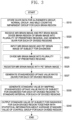
- FIG. 3 is a flowchart illustrating a method for providing dementia diagnosis assistance information in accordance with a preferred embodiment of the present invention.
- a processing operation of the processor 240 of the present invention and a method for providing dementia diagnosis assistance information performed by the processor 240 in accordance with a preferred embodiment of the present invention will be described.
- SUVR data for each of the brain regions of the Alzheimer's group, the normal group, and the mild cognitive impairment group are stored in the memory 230 of the present invention (S310).
- An example of SUVR data for each of the brain regions of the Alzheimer's group, the normal group, and the mild cognitive impairment group stored in the memory 230 is as described above with reference to FIG. 2 .
- a detailed configuration of the step S310 will be described later with reference to FIG. 9 .
- the processor 240 of the apparatus 200 for providing dementia diagnosis assistance information receives an MRI brain image and a PET brain image of a subject for diagnosis with dementia, divides the brain region of the brain image into a plurality of predefined regions, and generates the SUVR for each divided region (S320).
- the processor 240 receives the MRI brain image of the subject for diagnosis from the first image acquisition device 110, and receives the PET brain image of the subject for diagnosis from the second image acquisition device 120 (S321).
- the MRI brain image is preferably a T1-weighted MRI brain image
- the PET brain image may be an amyloid PET image or a tau PET image as needed.
- the processor 240 divides the MRI brain image into a plurality of predefined regions (refer to FIG. 5 ) by using a deep learning-based region division algorithm stored in the memory 230 (S323). As described above, in the preferred embodiment of the present invention, the processor 240 divides the MRI brain image into 97 regions of interest according to the brain map; however, the division method and the number of divided regions are not limited thereto.
- the processor 240 performs image registration on the MRI brain image and the PET brain image so that the two images are located in the same space through rigid transform (S325).
- FIG. 6 illustrates an example of an image in which an MRI brain image and a PET brain image are matched.
- the processor 240 may identify regions corresponding to the divided regions in the PET brain image through the divided regions of the MRI brain image.
- the processor 240 generates a standardized uptake value ratio (SUVR) for each divided region (S327).
- SUVR standardized uptake value ratio
- the standardized uptake value ratio represents a ratio of amyloid (or tau) standardized uptake value (SUV) from two different regions (target and reference region) within the same amyloid (or tau) PET image, and the amyloid (or tau) standardized uptake value (SUV) may represent the ratio of the concentration of radioactivity of the image to the concentration of radioactivity of the whole injected body.
- the method for obtaining the standardized uptake value ratio (SUVR) is a known technique in the technical field of the present invention, and thus a detailed description thereof will be omitted.
- the standardized uptake value ratio (SUVR) of the subject for diagnosis is obtained for each of the divided regions R 1 to R N in step S327
- the respective standardized uptake value ratios V SR1 to V SRN for the divided regions R 1 to R N are stored in the memory 230.
- the processor 240 generates a standard value by mapping the standardized uptake value ratio V SR1 to V SRN of the subject for diagnosis for each of the divided regions to a standard interval for each of the divided regions (S330).
- the standard value represents a degree of proximity of the standardized uptake value ratio of the subject for diagnosis for each of divided regions to an average value of standardized uptake value ratios of a group of Alzheimer's patients and an average value of standardized uptake value ratios of a normal group.
- the standard interval to which the standardized uptake value ratio of the subject for diagnosis for each of the divided regions is to be mapped is defined by setting, as both ends of a regular interval, the average value of the standardized uptake value ratios of the group of the Alzheimer's patients and the average value of the standardized uptake value ratios of the normal group for each of the plurality of divided regions.
- the standard interval is an interval generated by transforming the average value of the standardized uptake value ratios of the group of the Alzheimer's patients to correspond to 100 according to a predetermined transformation formula and transforming the average value of the standardized uptake value ratios of the normal group to correspond to 0 according to the transformation formula, and the standard value of the standardized uptake value ratio of the subject for diagnosis for each of the regions is a value obtained by transforming the standardized uptake value ratio according to the same transformation formula.
- the transformation formula may be defined as in Equation 1 below.
- A represents the average value of the standardized uptake value ratios of the group of the Alzheimer's patients
- B represents the average value of the standardized uptake value ratios of the normal group
- x represents the SUVR of the subject for diagnosis, which is a parameter to be transformed.
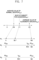
- FIG. 7 is a diagram for describing a standard interval obtained from average values of standardized uptake value ratios of a group of Alzheimer's patients and a normal group for each region in accordance with a preferred embodiment of the present invention, and a standard value of a subject for diagnosis mapped for each region accordingly.
- the average value AV AR1 of the group of Alzheimer's patients in a first divided region R 1 is 2.0
- the average value AV BR1 of the normal group is 0.6
- the SUVR of the subject for diagnosis is 1.1
- the average value AV BR1 of the normal group of 0.6 corresponds to 0
- the average value AV AR1 of the group of Alzheimer's patients of 2.0 corresponds to 100
- the SUVR of the subject for diagnosis of 1.1 corresponds to 35.7 according to Equation 1 above.
- the SUVR of the divided region R 1 of the subject for diagnosis is 1.1, which is at a borderline for determining whether PET is positive and negative, but when transformed to a standard interval, the standard value does not exceed 50, which is closer to the normal group, which makes it possible to provide useful information in determining whether PET is positive and negative.
- the processor 240 For each of the remaining divided regions R 2 to R N , the processor 240 generates a standard interval by transforming the average value of the normal group to 0 and transforming the average value of the group of Alzheimer's patients to 100, and generates a standard value to be mapped to the standard interval by transforming the SUVR of the subject for diagnosis of dementia in the same way.
- the standard interval was set to 0 to 100, but according to another embodiment, by modifying Equation 1 above to transform the average value of the SUVRs of the Alzheimer's group to 1, the standard interval may be set to 0 to 1.
- the processor 240 outputs the standard value of each divided region of the subject for diagnosis as the dementia diagnosis assistance information, together with statistical information about the average standard value of the control group for each divided region (S340).
- control group may include the group of Alzheimer's patients, the normal group, the normal aging group, and the mild cognitive impairment group stored in the memory 230.
- statistical information about the average standard value of the control group may include a standard value obtained by transforming the average value of the control group for the corresponding divided region according to Equation 1 above and a standard deviation corresponding thereto.
- FIG. 8 is a diagram illustrating an example of dementia diagnosis assistance information output in step S340 in accordance with a preferred embodiment of the present invention.
- the processor 240 may output a pair of values greater by a predefined value and less by the predefined value, than the average standard value of the control group, together with the standard value of the subject for diagnosis of dementia as a graph, where the predefined value may be a standard deviation.
- the predefined value may be a standard deviation.
- (a) shows the average standard value and standard deviation for each of the divided regions of the Alzheimer's group, and also shows the standard value for each of the divided regions of the subject for diagnosis of dementia.
- standard values (average standard values) of the average values of the Alzheimer's group are located on a horizontal line 801c having a constant value of 100 according to Equation 1 above, and when the values are connected, a horizontal line having a value of 100 is obtained.
- there is a difference in the standard deviations for the regions and thus they are arranged to correspond to each other around 100, and when the values obtained for each of the divided regions are connected, two graphs 801a and 801b of standard deviations are drawn.
- a line graph 800 is displayed together.
- the graph of the SUVR standard values of the subject for diagnosis of dementia is located inside graphs 801a and 801b of the standard deviations of the group of Alzheimer's patients in all divided regions, it is possible to determine that the subject for diagnosis of dementia is highly likely to have dementia since the SUVR characteristic of the subject for diagnosis of dementia shows the same pattern as the Alzheimer's patients, and to output, to the subject for diagnosis of dementia, a diagnosis result of Alzheimer's dementia.
- the graph of the SUVR standard values of the subject for diagnosis of dementia is located inside the graphs 801a and 801b of the standard deviations of the group of Alzheimer's patients only in some divided regions, it is possible to determine that the subject for diagnosis of dementia has a low likeliness of Alzheimer's dementia since the SUVR characteristic of the subject for diagnosis does not show the same pattern as the Alzheimer's patients. Accordingly, it is possible to output a diagnosis result indicating that the subject for diagnosis of dementia does not have Alzheimer's dementia, but may have dementia caused by other factors such as vascular dementia or Lewy body dementia.
- the processor 240 displays, for each divided region, the average standard values calculated by transforming the average values of the mild cognitive impairment group for each divided region according to Equation 1 above, or the average standard values calculated in advance and stored in the memory 230, and displays a line graph 802c obtained by connecting the average standard values.
- the processor 240 displays standard deviations centered on the average standard values of the mild cognitive impairment group for each of the divided regions, and displays graphs 802a and 802b of the standard deviations by connecting the standard deviations to each other.
- the processor 240 draws points corresponding to the SUVR standard values of the subject for diagnosis of dementia for each of the divided regions, and displays a line graph 800 obtained by connecting the points to each other together.
- the graph 800 of the SUVR standard values of the subject for diagnosis of dementia is included in the graphs 802a and 802b of the standard deviations of the mild cognitive impairment group, and thus it can be seen that the subject for diagnosis of dementia shows the same pattern as the mild cognitive impairment group. Accordingly, a diagnosis result indicating that the subject for diagnosis has mild cognitive impairment may be output.
- the processor 240 transforms the average values of the normal aging group for each of the divided regions according to Equation 1 above, and displays a graph obtained by connecting the transformed values to each other.
- the processor 240 displays graphs 803a and 803b of the standard deviations obtained by connecting the standard deviations of the normal aging group together.
- the processor 240 draws points corresponding to the SUVR standard values of the subject for diagnosis of dementia for each of the divided regions, and displays the line graph 800 obtained by connecting the points to each other together.
- the graph 800 of the SUVR standard value of the subject for diagnosis of dementia is only partially included in graphs 803c and 803b of the standard deviations of the normal aging group, and thus it can be seen that the subject for diagnosis of dementia does not belong to the normal aging group.
- the processor 240 transforms the average values of the normal group for each of the divided regions according to Equation 1 above, and displays a graph obtained by connecting the transformed values to each other.
- the processor 240 displays graphs 804a and 804b of the standard deviations obtained by connecting the standard deviations of the normal group together.
- graphs 804a and 804b of the standard deviations of the normal group are displayed as a pair arranged to face each other with respect to a horizontal line passing through the origin.
- the processor 240 draws points corresponding to the SUVR standard values of the subject for diagnosis of dementia for each of divided regions and displays the line graph 800 obtained by connecting the points to each other together.
- the graph 800 of the SUVR standard values of the subject for diagnosis of dementia does not have a region included between the graphs 804a and 804b of the standard deviations of the normal group, and thus it can be seen that the subject for diagnosis of dementia shows a significantly different pattern from the normal group.
- the processor 240 may calculate a proportion of divided regions where the standard value of the subject for diagnosis of dementia for each divided region is located between a pair of values (standard deviations) of the control group or a proportion of divided regions where the standard value for each divided region is located outside the pair of values (standard deviations) of the control group, and may display the calculated proportion as statistical information together with the graphs.
- the processor 240 may count the number of divided regions for which the standard values are included between the graphs of the standard deviations among all the divided regions, and display the proportion of the counted divided regions in the total divided regions together with a graph.
- the processor 240 may set a threshold value for the proportion of the regions for which the SUVR standard values of the subject for diagnosis of dementia are included between the graphs of the standard deviations, estimate that the subject for diagnosis of dementia belongs to the control group when the proportion is greater than or equal to the threshold value, and output an estimation result together with a graph. For example, assuming that the threshold value is set to 90%, the proportion of the standard values of the subject for diagnosis of dementia included between the graphs 802a and 802b of the standard deviations of the mild cognitive impairment group exceeds the threshold value of 90%, and thus, the processor 240 may output that the subject for diagnosis of dementia has mild cognitive impairment with a graph.
- the processor 240 may also output a degree of agreement between the graph obtained by connecting the SUVR standard values of the subject for diagnosis of dementia and the graph obtained by connecting the average standard values of the control group, for the divided regions. For example, the closer the sum of the absolute values obtained by subtracting the average standard values of the control group from the SUVR standard values of the subject for diagnosis of dementia to 0, the higher the degree of agreement between the graphs. Therefore, the processor 240 may calculate
- FIG. 9 is a flowchart for describing a process of obtaining average values of the SUVRs of the Alzheimer's group, the normal group, the mild cognitive impairment group, and the normal aging group for each divided region and storing the average values of the SUVRs in the memory 230, in step S310 shown in FIG. 3 , in accordance with a preferred embodiment of the present invention.
- the process of calculating the average values of the SUVRs of the Alzheimer's group, the normal group, the mild cognitive impairment group, and the normal aging group for each of the divided regions is the same as the above-described step S320, except that the subjects are members of the group of Alzheimer's patients, members of the normal group, members of the mild cognitive impairment group, and members of the normal aging group.
- the processor 240 receives MRI brain images of Alzheimer's patients, normal people, people who have been diagnosed with mild cognitive impairment, and the elderly who have been diagnosed with normal from the first image acquisition device 110, and receives PET brain images of Alzheimer's patients, the normal people, the people who have been diagnosed with mild cognitive impairment, and the elderly who have been diagnosed with normal from the second image acquisition device 120 (S311).
- the MRI brain image is preferably a T1-weighted MRI brain image
- the PET brain image may be an amyloid PET image or a tau PET image as needed.
- the processor 240 divides the MRI brain image into a plurality of predefined regions by using a deep learning-based region division algorithm stored in the memory 230 (S313). As described above, in the preferred embodiment of the present invention, the processor 240 divides the MRI brain image into 97 regions of interest according to the brain map; however, the division method and the number of divided regions are not limited thereto.
- the processor 240 performs image registration on the MRI brain image and the PET brain image so that the two images are located in the same space through rigid transform (S315).
- the processor 240 generates standardized uptake value ratios (SUVR) of the Alzheimer's patients, the normal people, the people who have been diagnosed with mild cognitive impairment, and the elderly who have been diagnosed with normal for each divided region and store them as shown in FIG. 2 (S317).
- SUVR standardized uptake value ratios
- the processor 240 calculates the average values AV AR1 to AV ARN , AV BR1 to AV BRN , AV CR1 to AV CRN , and AV DR1 to AV DRN and the standard deviations ⁇ AR1 to ⁇ ARN , ⁇ BR1 to ⁇ BR N , ⁇ CR1 to ⁇ CRN , ⁇ DR1 to ⁇ DRN of the standardized uptake value ratios of the group of Alzheimer's patients, the normal group, the mild cognitive impairment group, and the normal aging group for each of the divided regions R 1 to R N and stores them in the memory 230 (S319).
- the method for providing dementia diagnosis assistance information in accordance with a preferred embodiment of the present invention described above may be implemented as a computer program stored in a non-transitory storage medium by being implemented as computer-executable instructions.
- an MRI brain image and a PET brain image of a subject for diagnosis are received, the MRI brain image is divided into a plurality of regions, and then the MRI brain image is matched with the PET brain image. Then, a standardized uptake value ratio of each divided region is obtained from the registered image, and for each divided region, a standard value indicating the degree of proximity of a standardized uptake value ratio of the subject for diagnosis to the average value of the standardized uptake value ratios of the group of the Alzheimer's patients and the average value of the standardized uptake value ratios of the normal group, which are stored in advance, is obtained.
- the present invention enables health care providers to select an appropriate treatment plan and treatment method by diagnosing the subject for diagnosis as Alzheimer's dementia with high probability when the standard values of the standardized uptake value ratios of the subject for diagnosis is within the range of the standard deviation of the group of Alzheimer's patients and providing the fact, and by providing information that the subject for diagnosis does not fall under Alzheimer's dementia, but may fall under other factors, such as vascular dementia or Lewy body dementia, when the standard values of the standardized uptake value ratios of the subject for diagnosis is not within the range of the standard deviation of the group of Alzheimer's patients.
- the storage medium includes all kinds of recording devices in which data that may be read by a computer system is stored.
- Examples of computer-readable storage media include read only memory (ROM), random access memory (RAM), compact disc ROMs (CD-ROM), magnetic tapes, floppy disks, and optical data storage devices.
- ROM read only memory
- RAM random access memory
- CD-ROM compact disc ROMs
- magnetic tapes magnetic tapes
- floppy disks and optical data storage devices.
- optical data storage devices optical data storage devices.
- the computer-readable storage medium may be distributed in a computer system connected through a network, so that the computer-readable code may be stored and executed in a distributed manner.
Landscapes
- Health & Medical Sciences (AREA)
- Life Sciences & Earth Sciences (AREA)
- Engineering & Computer Science (AREA)
- Medical Informatics (AREA)
- Public Health (AREA)
- General Health & Medical Sciences (AREA)
- Biomedical Technology (AREA)
- Physics & Mathematics (AREA)
- Pathology (AREA)
- Veterinary Medicine (AREA)
- Nuclear Medicine, Radiotherapy & Molecular Imaging (AREA)
- Molecular Biology (AREA)
- Surgery (AREA)
- Animal Behavior & Ethology (AREA)
- Heart & Thoracic Surgery (AREA)
- Biophysics (AREA)
- Radiology & Medical Imaging (AREA)
- High Energy & Nuclear Physics (AREA)
- Neurology (AREA)
- Optics & Photonics (AREA)
- Epidemiology (AREA)
- Primary Health Care (AREA)
- Physiology (AREA)
- Computer Vision & Pattern Recognition (AREA)
- Databases & Information Systems (AREA)
- Data Mining & Analysis (AREA)
- Psychiatry (AREA)
- Artificial Intelligence (AREA)
- Neurosurgery (AREA)
- Child & Adolescent Psychology (AREA)
- Developmental Disabilities (AREA)
- Evolutionary Computation (AREA)
- Fuzzy Systems (AREA)
- Mathematical Physics (AREA)
- Hospice & Palliative Care (AREA)
- Signal Processing (AREA)
- Psychology (AREA)
- Dentistry (AREA)
- Oral & Maxillofacial Surgery (AREA)
- Nuclear Medicine (AREA)
Applications Claiming Priority (1)
| Application Number | Priority Date | Filing Date | Title |
|---|---|---|---|
| KR1020220023103A KR102444082B1 (ko) | 2022-02-22 | 2022-02-22 | 치매 진단 보조 정보 제공 장치 및 방법 |
Publications (1)
| Publication Number | Publication Date |
|---|---|
| EP4230144A1 true EP4230144A1 (fr) | 2023-08-23 |
Family
ID=83445190
Family Applications (1)
| Application Number | Title | Priority Date | Filing Date |
|---|---|---|---|
| EP22206524.5A Withdrawn EP4230144A1 (fr) | 2022-02-22 | 2022-11-10 | Appareil et procédé pour fournir des informations d'aide au diagnostic de la démence |
Country Status (4)
| Country | Link |
|---|---|
| US (1) | US11730368B1 (fr) |
| EP (1) | EP4230144A1 (fr) |
| JP (1) | JP7477906B2 (fr) |
| KR (1) | KR102444082B1 (fr) |
Families Citing this family (2)
| Publication number | Priority date | Publication date | Assignee | Title |
|---|---|---|---|---|
| CN116245833B (zh) * | 2023-02-13 | 2024-12-27 | 复旦大学 | 一种大脑多模态pet影像suvr的预测方法 |
| KR102911012B1 (ko) * | 2023-08-16 | 2026-01-13 | (주)에코인사이트글로벌 | Mri와 fdg pet 영상을 이용한 알츠하이머 루이소체 및 혼합형 치매 진단 의료기기와 진단 방법 |
Family Cites Families (5)
| Publication number | Priority date | Publication date | Assignee | Title |
|---|---|---|---|---|
| WO2012173976A2 (fr) * | 2011-06-17 | 2012-12-20 | Carroll Robert G | Procédés et appareil pour évaluer l'activité d'un organe et utilisations de ceux-ci |
| US11373311B2 (en) * | 2015-07-15 | 2022-06-28 | Adm Diagnostics, Inc. | System and methods for determining a brain condition of a patient subject to multiple disease states |
| CN112702947A (zh) | 2018-09-12 | 2021-04-23 | 株式会社Splink | 诊断辅助系统以及方法 |
| KR102351283B1 (ko) * | 2020-05-22 | 2022-01-14 | 동아대학교 산학협력단 | 딥러닝을 이용한 짧은 시간 촬영한 뇌 단층 촬영 이미지 복원 장치 |
| KR102414974B1 (ko) * | 2020-08-03 | 2022-06-30 | 사회복지법인 삼성생명공익재단 | Pet-ct 영상을 이용하는 알츠하이머 질환의 진단 장치 및 이의 동작 방법 |
-
2022
- 2022-02-22 KR KR1020220023103A patent/KR102444082B1/ko active Active
- 2022-11-04 JP JP2022177366A patent/JP7477906B2/ja active Active
- 2022-11-09 US US18/054,072 patent/US11730368B1/en active Active
- 2022-11-10 EP EP22206524.5A patent/EP4230144A1/fr not_active Withdrawn
Non-Patent Citations (2)
| Title |
|---|
| ROWE CHRISTOPHER C ET AL: "18F-Florbetaben PET beta-amyloid binding expressed in Centiloids", EUROPEAN JOURNAL OF NUCLEAR MEDICINE AND MOLECULAR IMAGING, SPRINGER BERLIN HEIDELBERG, BERLIN/HEIDELBERG, vol. 44, no. 12, 22 June 2017 (2017-06-22), pages 2053 - 2059, XP036342876, ISSN: 1619-7070, [retrieved on 20170622], DOI: 10.1007/S00259-017-3749-6 * |
| YAMAO TENSHO ET AL: "Centiloid scale analysis for 18F-THK5351 PET imaging in Alzheimer's disease", PHYSICA MEDICA, ACTA MEDICA EDIZIONI E CONGRESSI, ROME, IT, vol. 82, 1 February 2021 (2021-02-01), pages 249 - 254, XP086530442, ISSN: 1120-1797, [retrieved on 20210304], DOI: 10.1016/J.EJMP.2021.02.017 * |
Also Published As
| Publication number | Publication date |
|---|---|
| JP2023122529A (ja) | 2023-09-01 |
| US20230263394A1 (en) | 2023-08-24 |
| KR102444082B1 (ko) | 2022-09-16 |
| JP7477906B2 (ja) | 2024-05-02 |
| US11730368B1 (en) | 2023-08-22 |
Similar Documents
| Publication | Publication Date | Title |
|---|---|---|
| US12198809B2 (en) | Machine learning algorithms for detecting medical conditions, related systems, and related methods | |
| EP4231311A1 (fr) | Procede et appareil permettant de fournir des informations necessaires au diagnostic de la demence | |
| Pham et al. | Effectiveness of consumer-grade contactless vital signs monitors: a systematic review and meta-analysis | |
| US9123101B2 (en) | Automatic quantification of asymmetry | |
| Son et al. | Evaluating fMRI-based estimation of eye gaze during naturalistic viewing | |
| ES2960027T3 (es) | Detección de anomalías en las señales de ECG | |
| CN112702947A (zh) | 诊断辅助系统以及方法 | |
| KR20090024808A (ko) | 치매 및 치매성 장애의 평가 | |
| EP4230144A1 (fr) | Appareil et procédé pour fournir des informations d'aide au diagnostic de la démence | |
| WO2010005656A2 (fr) | Evaluation d’une affection cérébrale | |
| US10832410B2 (en) | Computer system, method, and program for diagnosing subject | |
| US11848075B2 (en) | Biomarker detection method, disease assessment method, biomarker detection device, and computer readable medium | |
| US11342077B2 (en) | Medical information processing apparatus and medical information processing method | |
| JP5732015B2 (ja) | グラフ作成装置およびグラフ作成方法並びにグラフ作成プログラム | |
| US20090016579A1 (en) | Method and system for performing quality control of medical images in a clinical trial | |
| JP5348998B2 (ja) | 画像検索装置及びその方法 | |
| JP6943853B2 (ja) | イメージングワークフローの品質管理 | |
| US11328414B2 (en) | Priority judgement device, method, and program | |
| Min et al. | Enhanced multistage deep learning for diagnosing anterior disc displacement in the temporomandibular joint using MRI | |
| CN114266738B (zh) | 轻度脑损伤磁共振影像数据的纵向分析方法及系统 | |
| US20250104240A1 (en) | Personalized profiling of future brain trajectories and future disease evolution using generative artificial intelligence | |
| CN120514407A (zh) | 儿童脑代谢模板建立方法、处理装置、系统、设备及介质 | |
| JP6998760B2 (ja) | 脳画像解析装置、脳画像解析方法、及び脳画像解析プログラム | |
| JP2024003782A (ja) | 統合失調スペクトラム障害に関する情報を提供するための診断支援システム、診断支援装置、診断支援方法、及び支援プログラム | |
| CN117522770A (zh) | 基于图像的特征性眼睛移动的检测 |
Legal Events
| Date | Code | Title | Description |
|---|---|---|---|
| PUAI | Public reference made under article 153(3) epc to a published international application that has entered the european phase |
Free format text: ORIGINAL CODE: 0009012 |
|
| STAA | Information on the status of an ep patent application or granted ep patent |
Free format text: STATUS: REQUEST FOR EXAMINATION WAS MADE |
|
| 17P | Request for examination filed |
Effective date: 20221110 |
|
| AK | Designated contracting states |
Kind code of ref document: A1 Designated state(s): AL AT BE BG CH CY CZ DE DK EE ES FI FR GB GR HR HU IE IS IT LI LT LU LV MC ME MK MT NL NO PL PT RO RS SE SI SK SM TR |
|
| RBV | Designated contracting states (corrected) |
Designated state(s): AL AT BE BG CH CY CZ DE DK EE ES FI FR GB GR HR HU IE IS IT LI LT LU LV MC ME MK MT NL NO PL PT RO RS SE SI SK SM TR |
|
| STAA | Information on the status of an ep patent application or granted ep patent |
Free format text: STATUS: EXAMINATION IS IN PROGRESS |
|
| 17Q | First examination report despatched |
Effective date: 20240603 |
|
| STAA | Information on the status of an ep patent application or granted ep patent |
Free format text: STATUS: THE APPLICATION IS DEEMED TO BE WITHDRAWN |
|
| 18D | Application deemed to be withdrawn |
Effective date: 20241005 |