EP4247408B1 - Inhibitoren des gewebefaktor-protease-aktivierten rezeptor 2 (tf-par2)-signalweges zur verwendung bei der behandlung oder prävention von herzinsuffizienz (hf) - Google Patents
Inhibitoren des gewebefaktor-protease-aktivierten rezeptor 2 (tf-par2)-signalweges zur verwendung bei der behandlung oder prävention von herzinsuffizienz (hf) Download PDFInfo
- Publication number
- EP4247408B1 EP4247408B1 EP21820471.7A EP21820471A EP4247408B1 EP 4247408 B1 EP4247408 B1 EP 4247408B1 EP 21820471 A EP21820471 A EP 21820471A EP 4247408 B1 EP4247408 B1 EP 4247408B1
- Authority
- EP
- European Patent Office
- Prior art keywords
- inhibitor
- par2
- signaling pathway
- tgf
- activation
- Prior art date
- Legal status (The legal status is an assumption and is not a legal conclusion. Google has not performed a legal analysis and makes no representation as to the accuracy of the status listed.)
- Active
Links
Images







Classifications
-
- A—HUMAN NECESSITIES
- A61—MEDICAL OR VETERINARY SCIENCE; HYGIENE
- A61K—PREPARATIONS FOR MEDICAL, DENTAL OR TOILETRY PURPOSES
- A61K38/00—Medicinal preparations containing peptides
- A61K38/16—Peptides having more than 20 amino acids; Gastrins; Somatostatins; Melanotropins; Derivatives thereof
- A61K38/17—Peptides having more than 20 amino acids; Gastrins; Somatostatins; Melanotropins; Derivatives thereof from animals; from humans
- A61K38/1767—Peptides having more than 20 amino acids; Gastrins; Somatostatins; Melanotropins; Derivatives thereof from animals; from humans from invertebrates
-
- A—HUMAN NECESSITIES
- A61—MEDICAL OR VETERINARY SCIENCE; HYGIENE
- A61K—PREPARATIONS FOR MEDICAL, DENTAL OR TOILETRY PURPOSES
- A61K45/00—Medicinal preparations containing active ingredients not provided for in groups A61K31/00 - A61K41/00
- A61K45/06—Mixtures of active ingredients without chemical characterisation, e.g. antiphlogistics and cardiaca
-
- A—HUMAN NECESSITIES
- A61—MEDICAL OR VETERINARY SCIENCE; HYGIENE
- A61P—SPECIFIC THERAPEUTIC ACTIVITY OF CHEMICAL COMPOUNDS OR MEDICINAL PREPARATIONS
- A61P9/00—Drugs for disorders of the cardiovascular system
- A61P9/04—Inotropic agents, i.e. stimulants of cardiac contraction; Drugs for heart failure
-
- G—PHYSICS
- G01—MEASURING; TESTING
- G01N—INVESTIGATING OR ANALYSING MATERIALS BY DETERMINING THEIR CHEMICAL OR PHYSICAL PROPERTIES
- G01N33/00—Investigating or analysing materials by specific methods not covered by groups G01N1/00 - G01N31/00
- G01N33/48—Biological material, e.g. blood, urine; Haemocytometers
- G01N33/50—Chemical analysis of biological material, e.g. blood, urine; Testing involving biospecific ligand binding methods; Immunological testing
- G01N33/68—Chemical analysis of biological material, e.g. blood, urine; Testing involving biospecific ligand binding methods; Immunological testing involving proteins, peptides or amino acids
- G01N33/6893—Chemical analysis of biological material, e.g. blood, urine; Testing involving biospecific ligand binding methods; Immunological testing involving proteins, peptides or amino acids related to diseases not provided for elsewhere
-
- G—PHYSICS
- G01—MEASURING; TESTING
- G01N—INVESTIGATING OR ANALYSING MATERIALS BY DETERMINING THEIR CHEMICAL OR PHYSICAL PROPERTIES
- G01N2333/00—Assays involving biological materials from specific organisms or of a specific nature
- G01N2333/435—Assays involving biological materials from specific organisms or of a specific nature from animals; from humans
- G01N2333/475—Assays involving growth factors
- G01N2333/495—Transforming growth factor [TGF]
-
- G—PHYSICS
- G01—MEASURING; TESTING
- G01N—INVESTIGATING OR ANALYSING MATERIALS BY DETERMINING THEIR CHEMICAL OR PHYSICAL PROPERTIES
- G01N2333/00—Assays involving biological materials from specific organisms or of a specific nature
- G01N2333/435—Assays involving biological materials from specific organisms or of a specific nature from animals; from humans
- G01N2333/745—Assays involving non-enzymic blood coagulation factors
- G01N2333/7454—Tissue factor (tissue thromboplastin, Factor III)
-
- G—PHYSICS
- G01—MEASURING; TESTING
- G01N—INVESTIGATING OR ANALYSING MATERIALS BY DETERMINING THEIR CHEMICAL OR PHYSICAL PROPERTIES
- G01N2440/00—Post-translational modifications [PTMs] in chemical analysis of biological material
- G01N2440/14—Post-translational modifications [PTMs] in chemical analysis of biological material phosphorylation
-
- G—PHYSICS
- G01—MEASURING; TESTING
- G01N—INVESTIGATING OR ANALYSING MATERIALS BY DETERMINING THEIR CHEMICAL OR PHYSICAL PROPERTIES
- G01N2800/00—Detection or diagnosis of diseases
- G01N2800/32—Cardiovascular disorders
- G01N2800/325—Heart failure or cardiac arrest, e.g. cardiomyopathy, congestive heart failure
-
- G—PHYSICS
- G01—MEASURING; TESTING
- G01N—INVESTIGATING OR ANALYSING MATERIALS BY DETERMINING THEIR CHEMICAL OR PHYSICAL PROPERTIES
- G01N2800/00—Detection or diagnosis of diseases
- G01N2800/50—Determining the risk of developing a disease
Definitions
- the present invention relates to inhibitors of the Tissue Factor-Protease Activated Receptor 2 (TF-PAR2) signaling pathway for use in the treatment or prevention of heart failure (HF), and associated or resulting diseases such as ischemic heart failure (IHF), myocardial infarction (MI), heart failure with reduced ejection fraction (HFrEF), or heart failure with preserved ejection fraction (HFpEF).
- HF heart failure
- IHF ischemic heart failure
- MI myocardial infarction
- HFrEF heart failure with reduced ejection fraction
- HFpEF heart failure with preserved ejection fraction
- TF is also known as thromboplastin or CD142 relating to a glycosylated transmembrane protein consisting of a single polypeptide chain having a molecular weight of 45,000 (7).
- the extracellular part of TF is made up of two fibronectin type III domains, and membrane anchoring of TF has been demonstrated to be essential to support full proteolytic activity of FVlla (8).
- FVII is rapidly converted to its activated form (FVlla) via limited proteolysis (9, 10).
- US 2004/0102402 A1 describes inhibitors of TF and methods of treating an animal having a disease or condition associated with TF.
- nucleic acid compounds are described, consisting of 8 to 80 nucleobases in length targeted to a nucleic acid molecule encoding TF.
- WO 97/20939 A describes a fusion protein that is capable of inhibiting or neutralizing TF from inducing blood coagulation through the extrinsic coagulation pathway.
- the inhibitor of TF-PAR2 signaling refers to any biological or chemical compound resulting in a blockage of TF signaling in human or animal cells.
- the inhibitor can be any compound that modulates TF signaling leading to (i) a hyper-activation of mitogen-activated protein kinase 1 (MAPK1), (ii) an extracellular-signal-regulated kinase 1/2 (ERK1/2) phosphorylation, and (iii) TGF- ⁇ 1 activation.
- MAPK1 mitogen-activated protein kinase 1
- ERK1/2 extracellular-signal-regulated kinase 1/2
- the inhibitor of the TF-PAR2 signaling pathway is an inhibitor of the recruitment of NOX2-positive myeloid cells, preferably an inhibitor of NOX-2-positive monocytes as the recruitment of these cells is crucial for TGF- ⁇ 1 activation.
- the inhibitor of the TF-PAR2 signaling pathway is an inhibitor of TGF- ⁇ 1 activation.
- a preferred inhibitor of the TF-PAR2 signaling pathway that fulfills these requirements is nematode anticoagulant protein C2 (NAPc2).
- the invention also relates to inhibitory compounds that comprise fusion proteins, mutants, variants of fragments thereof that retain the capability for a reduced TGF- ⁇ 1 activation and/or cardiac or myelomonocytic cell NOX2 expression and/or the recruitment of NOX2-positive myeloid cells, preferably NOX2-positive monocytes.
- the invention further includes nucleic acid molecules, such as DNA, cDNA, RNA, that encode an inhibitor of TF-PAR2 signaling according to the present invention.
- the TF-PAR2 signaling inhibitor is selected from a protein, a peptide, an antibody, an antigen-binding fragment thereof, an enzyme, an enzyme inhibitor, an aptamer or a small molecule.
- Small molecules as used in the context of the present invention, refer to low molecular weight molecules having a molecular weight ⁇ 900 Da. The upper molecular weight limit allows a rapid crossing of said membranes so that the smaller molecule can reach its intracellular sites of action.
- Factor Vlla/Tissue Factor or “FVlla/TF” or “FVlla/TF” are synonymous and are commonly known to refer to a catalytically active complex of the serine protease coagulation factor VIIa (FVlla) and the non-enzymatic protein Tissue Factor (TF), wherein the complex is assembled on the surface of a physiologically relevant phospholipid membrane of defined composition.
- FVlla serine protease coagulation factor VIIa
- TF non-enzymatic protein Tissue Factor
- NAPc2 nematode anticoagulant protein C2
- NAPc2 describes a single-chain, non-glycosylated 85 amino acid protein having a molecular weight of 9732 Da. NAPc2 exerts its effects by binding to FXa and has an inhibitory mechanism resembling that of TFPI. The antithrombotic effect of NAPc2 has been demonstrated in a dose-finding study on the prevention of venous thromboembolism in patients undergoing total knee replacement (27).
- NAPc2 treatment improved cardiac function, attenuated cardiac infiltration of myeloid cells, and also diminished ERK1/2 phosphorylation, NOX2 expression, TGF- ⁇ 1 activation and up-regulation of the downstream target ⁇ -SMA in the infarcted heart.
- a short term NAPc2 treatment up to 7 days post MI significantly reduced cardiac fibrosis and improved cardiac function in chronic Ml.
- the NAPc2 and the NAPc2/proline variant comprise an amino acid sequence as shown in Table 1: Table 1 - NAPc2 and NAPc2/proline variant amino acid sequences Protein SEQ ID NO Sequence NAPc2 1 NAPc2/ proline 2
- the invention also comprises variants, mutants or homologs of those proteins, such as NAPc2 variants exhibiting at least 80%, 85%, 90%, 95%, 96%, 97%, 98, or 99% sequence homology with any one of the amino acid sequences set forth in SEQ ID NO:1 or SEQ ID NO:2, wherein such a variant has the capability of inhibiting the TF-PAR2 signaling pathway.
- NAPc2 variants exhibiting at least 80%, 85%, 90%, 95%, 96%, 97%, 98, or 99% sequence homology with any one of the amino acid sequences set forth in SEQ ID NO:1 or SEQ ID NO:2, wherein such a variant has the capability of inhibiting the TF-PAR2 signaling pathway.
- An example of a non-limiting range for a therapeutically effective dose of a TF-PAR2 signaling pathway inhibitor of the present invention is preferably between 0.1-100 ⁇ g/kg, more preferred between 0.5-20 ⁇ g/kg, preferably between 1 and 10 ⁇ g/kg.
- the invention also includes any range or single values within said ranges, such as an amount of about 5 ⁇ g/kg, about 7 ⁇ g/kg, or about 10 ⁇ g/kg.
- the administration of the inhibitor can be achieved in a single dose or in multiple doses per day, or given on alternating days or other dosing schemes. A physician or veterinarian having ordinary skill in the art may readily determine and prescribe the effective amount of the pharmaceutical composition required.
- a suitable daily dose of a composition of the present invention will be that amount of the compound which is the lowest dose effective to produce a therapeutic effect.
- Such an effective dose will generally depend upon the mode of administrations such as intravenous, intramuscular, intraperitoneal, or subcutaneous administration.
- the effective daily dose of a pharmaceutical composition may be administered as two, three, four, five, six or more sub-doses administered separately at appropriate intervals throughout the day, optionally, in unit dosage forms.
- NAPc2 or any of its variants, such as NAPc2/proline is preferably provided at a dose of between about 5 ⁇ g/kg and 10 ⁇ g/kg, wherein all intermediate values and ranges are included within the scope of the present invention. Similar to other inhibitors comprised by the invention, the administration of NAPc2 or any of its variants, such as NAPc2/proline, can be achieved in a single dose or in multiple doses per day, or given on alternating days or other dosing schemes.
- NAPc2 or any of its variants, such as NAPc2/proline is provided at a dose of about 5 ⁇ g/kg, preferably at a dose of about 7.5 ⁇ g/kg, or preferably at a dose of about 10 ⁇ g/kg. In some embodiments, NAPc2 or any of its variants, such as NAPc2/proline, is provided at a dose of about 7.5 ⁇ g/kg on a first day, a dose of about 5 ⁇ g/kg on a third day, and a dose of about 5 ⁇ g/kg on a fifth day.
- compositions of the present invention can be formulated by methods known to those skilled in the art.
- such pharmaceutical compositions can be used parenterally, as injections which are sterile solutions or suspensions including the compositions along with water or another pharmaceutically acceptable liquid.
- such compositions may be formulated as unit doses that meet the requirements for the preparation of pharmaceuticals by appropriately combining the compositions with pharmaceutically acceptable carriers, diluents, adjuvants or excipients, specifically with sterile water, physiological saline, a vegetable oil, emulsifier, suspension, surfactant, stabilizer, flavoring agent, excipient, vehicle, preservative, binder or such.
- the amount of active ingredient is adjusted such that an appropriate dose that falls within a pre-determined range can be obtained.
- the inventions also relates to a method for the treatment or prevention of heart failure (HF), and associated or resulting diseases such as ischemic heart failure (IHF), myocardial infarction (MI), heart failure with reduced ejection fraction (HFrEF), or heart failure with preserved ejection fraction (HFpEF) using a herein described inhibitor of the Tissue Factor-Protease Activated Receptor 2 (TF-PAR2) signaling pathway, or a pharmaceutical composition comprising such an inhibitor.
- HF heart failure
- IHF ischemic heart failure
- MI myocardial infarction
- HFrEF heart failure with reduced ejection fraction
- HFpEF heart failure with preserved ejection fraction
- the biological sample of the subject is preferably obtained from a heart biopsy, liquid biopsy, blood, serum, or plasma.
- a normal, healthy subject is a subject, for example a human or an animal that has no condition of MI or HF.
- the determination of TF phosphorylation can be carried out qualitatively or quantitatively.
- a quantitative analysis can be carried out, for example, for flow cytometry analysis or confocal microscopy of monocytes isolated from the subjects.
- a qualitative analysis can be carried out, for example, by Western blot or enzyme linked immunosorbent assay (ELISA) analysis of monocytic protein expression for TF cytoplasmic domain phosphorylation (4G6) and TF (10H10).
- the method can also utilize a scoring method to determine if a patient is at risk of developing MI or HF. Preferably, a desired score is integrated for the biomarker.
- the total score is then quantified and compared with a predetermined total score. In a following step, it is determined whether the subject has a risk for developing MI or HF based on the total score determined above. Increased levels of phosphorylation of the TF cytoplasmic domain and active TGF- ⁇ 1 will be indicative for an increased risk of developing IHF or adverse remodeling following MI.
- the method of the present invention therefore further comprises determining whether there is an upregulation of IL6, CCL2 and/or CCR. These markers can also be integrated in the evaluation and scoring. Further markers that can be included are myeloid cell recruitment, increased TGF- ⁇ 1 activation and/or downstream phosphorylation of SMAD2. In a further preferred embodiment, the method further comprises an analysis of the numbers of CD45+ cells within said biological sample, wherein an increased number of CD45+ cells as compared to the numbers in samples collected from normal, healthy test subjects are indicative for IHF.
- PBMCs as used in the context of the present invention refer to a mixed population of myeloid and lymphoid cells.
- Myeloid cells include monocytes, dendritic cells and macrophages. Monocytes circulate through the blood to different tissues, thereby differentiating into tissue-resident macrophages and dendritic cells.
- the invention also concerns the use of a hyper-phosphorylated TF as biomarker to identify a subject at risk for developing IHF or MI, in particular adversary modeling following MI and to identify drug therapies that prevent IHF or adverse remodeling.
- the invention provides a more detailed insight into the pathophysiology of coagulation in IHF and uncovers a crucial function of coagulation-related signaling in adverse remodeling in the ischemic myocardium.
- Treatment of arterial thrombosis and vascular occlusion is central to the therapy of acute coronary syndromes.
- the invention provides novel insights into a non-canonical mechanism of coagulation, TF-PAR2 signaling leading to TGF- ⁇ 1 activation in the infarcted myocardium and the precise role of myeloid cells in this context.
- the inventors Based on unbiased (phospho)proteomics in human IHF as well as genetic and pharmacologic interventions in mouse models, the inventors establish a link between hyper-activation of the MAPK pathway and TGF- ⁇ 1 mediated cardiac remodeling driven by inflammatory pro-oxidant myeloid cells infiltrating the heart in permanent Ml.
- the inventors localize ERK1/2 activation specific to myeloid cells infiltrating the ischemic heart and show that preclinical intervention with the MEK inhibitor trametinib significantly diminished the CCR2 dependent (29) Ly-6C high monocyte recruitment into the infarcted myocardium.
- Activation of TGF- ⁇ 1 is essential for its biological functions (29), specifically for activating myofibroblasts and disease progression in MI (30).
- the inventors directly show with isolated cells in vitro that cytokine primed monocytes exposed to hypoxia activate latent TGF- ⁇ 1 dependent on Nox2 and MEK1/2 signaling. Furthermore, experiments with isolated monocytes from NOX2 -/- and PAR2 -/- in combination with in vivo analysis of myeloid cell-specific PAR2 and TF deletion confirmed that myeloid cell derived TF-PAR2 signaling is the driver for ERK1/2-TGF- ⁇ 1 activation in chronic MI.
- Shear stress induced platelet derived TGF- ⁇ 1 activation depends on reduced protein-disulfide isomerase (PDI) (31) which also plays a critical role in TF decryption (32, 33).
- TF decryption is favoured by complement activation (34, 35), which was a prominent feature of the ischemic myocardium identified by the inventor's proteomic screen.
- the TF-FVlla complex furthermore interacts with integrin ⁇ 1 independent of coagulation activation (36) and regulates ROS production by endosomal NOX2 trafficking which depends on the TF cytoplasmic tail (37).
- ROS can regulate fibroblast proliferation and collagen synthesis in MI (38), but major cellular sources of ROS and underlying mechanisms of how they mediate cardiac remodeling remained elusive.
- the inventor's present data show that myeloid cell TF-PAR2 linked through the TF cytoplasmic tail is required for increased phagocyte type NADPH oxidase derived ROS production and TGF- ⁇ 1 activation to promote cardiac remodeling in chronic Ml.
- sub-acute MI patients with coronary no-reflow and/or delayed presentation after onset of symptoms
- thrombo-inflammation shows signs of thrombo-inflammation and are marked by a worse clinical outcome (39-41).
- biomarkers other than Q-waves or T-wave inversion in electrocardiography (42) are currently not established to predict sub-acute MI or poor outcomes of MI.
- the inventor's data indicate that TF phosphorylation of circulating monocytes may serve as a marker for patients at increased risk of developing IHF and adverse remodeling following MI.
- the inventor's proof-of-principle experiments delineate potential highly specific avenues to target this pathway.
- the putative clinical benefit is exemplified by the TF inhibitor NAPc2 with dual antithrombotic action and signaling disruption, resulting in diminished fibrosis post experimental MI.
- the identified biomarker applicable to liquid biopsies of patients with MI may facilitate clinical development of strategies to specifically target coagulation TF-PAR2 signaling for myeloid cell reprogramming. This strategy has the potential to prevent TGF- ⁇ 1 activation leading to excess cardiac damage and to avert the development of IHF after Ml.
- the invention is based on the surprising discovery that myeloid cell TF-PAR2-signaling is a crucial driver for TGF- ⁇ 1 activation and the target for the treatment or prevention of IHF or MI.
- the following examples are given by way of illustration only, and various changes and modifications that are within the spirit of the present invention will become apparent to the person in the art.
- MAPK1 Activation of MAPK1 in clinical and preclinical setting of MI and IHF.
- the inventors evaluated the contributions of the MAPK pathway to IHF by blocking the ERK1/2 activator mitogen-activated extracellular signal-regulated kinase (MEK) with the MEK 1 ⁇ 2 inhibitor trametinib in the preclinical mouse model of IHF induced by permanent ligation of the left anterior descending coronary artery (LAD, Figure 2A ).
- Long-term high-dose trametinib regimens in cancer therapy may have cardiotoxic side effects 17.
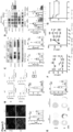
- the inventors observed that treatment with trametinib (1mg/kg/d) initiated 1 day and continued for 6 days after MI attenuated deterioration of cardiac function ( Figure 2B ) and prevented activation of the ERK pathway in cardiac tissue ( Figure 2C ).
- Trametinib therapy had no significant effects on cardiac function in sham-operated mice ( Figure 2B ) and did not significantly interfere with pJNK/SAK or p38 MAPK pathways ( Figure 9A ).
- TGF- ⁇ 1 has been implicated in adverse LV remodeling and development of heart failure after MI (4, 18).
- Trametinib significantly reduced activation of TGF- ⁇ 1 ( Figure 2C ) and the mRNA expression of COLO1A1 and COLO3A 1 coding for collagen type I and III alpha 1 chain as well as Posn and ACTA2 , encoding for the fibrosis markers periostin and ⁇ smooth muscle actin ( Figure 9B ) in the infarcted myocardium.
- Infiltration of Ly6G+ neutrophils and Ly6Chigh monocytes 3 and subsequent expansion of Ly6Clow monocytes and macrophages (19) orchestrate the inflammatory reaction within the infarcted myocardium.
- Trametinib did not reduce mRNA expression of the inflammatory cytokines and chemokines interleukin-6 ( Il6 ), tumor necrosis factor alpha (Tnf) and C-C chemokine ligand 2 ( Ccl 2) , implying that the local inflammatory response post M! was unaltered. Importantly, trametinib significantly reduced the mRNA expression of the CCL2 receptor and monocyte marker C-C chemokine receptor 2 (Ccr2, Figure 2D ).
- Myeloid cell PAR signaling is upstream of a pro-fibrotic MEK1/2-TGF- ⁇ 1 pathway
- Immunofluorescence staining supported a pivotal role of infiltrating CD45+ immune cells as the main source for increased ERK1/2 activation in the infarcted myocardium, compared to CD31 + endothelial cells, ⁇ -SMA+ myofibroblasts or cTNT+ cardiomyocytes ( Figure 3A ).
- the inventors therefore investigated the role of the MAPK pathway in monocytes and asked whether ERK1/2 activation might be linked to a major driver of adverse remodeling, i.e. TGF- ⁇ 1 activation.
- the inventors exposed isolated murine monocytes to both hypoxia and the inflammatory cytokines IL-6, TNF- ⁇ and of CCL2 which were detected in the ischemic heart ( Fig. 2D ).
- the inventors evaluated the role of monocyte-expressed PAR2 by subjecting PARfl/fl LysMcre mice to permanent LAD ligation for 7 days.
- Myeloid cell PAR2 deficient mice had reduced TGF- ⁇ 1 activation and phosphorylation of small mothers against decapentaplegic homolog 2 (SMAD2, Figure 3E ) in the myocardium and were protected from cardiac dysfunction 7d post MI ( Figure 3F).
- SAD2, Figure 3E decapentaplegic homolog 2
- Figure 10 immune cell recruitment into the infarcted myocardium was not diminished in myeloid cell PAR2-deficient mice. This indicates that a primary function of myeloid 135 cell PAR2 is to support local TGF- ⁇ 1 and drive cardiac remodeling post MI.
- Myeloid cell TF cytoplasmic domain signaling is linked to NOX2/ERK-dependent TGF- ⁇ 1 activation in permanent MI.
- TF makes additional signaling contributions to pro-fibrotic TGF- ⁇ 1 activation.
- Ligation of TF by FVlla activates rac and p38 dependent on the TF cytoplasmic domain (43).
- the TF cytoplasmic tail binds the regulatory subunit of PI3 kinase and rac adaptor p85 (44) and recruits the NADPH oxidase for endosomal translocation and reactive oxygen species (ROS) production (37).
- ROS reactive oxygen species
- TFfl/fl LysMcre mice with permanent LAD ligation had significantly reduced cardiac NOX2 expression compared to controls and that monocytes isolated from PAR2-/-mice had reduced NOX2 expression when exposed to hypoxia plus cytokines in vitro.
- superoxide anion (02 •-) formation in the ischemic myocardium of PARfl/fl LysMcre mice was reduced in comparison to PARfl/fl littermate controls ( Figures 11A-C ).
- cytoplasmic tail deficiency mice TF ⁇ CT mice exposed to permanent MI had markedly reduced CD45+/NOX2+ immune cell infiltration ( Figure 5A ) and 02 •- formation ( Figure 5B ) in the infarcted myocardium.
- TGF- ⁇ 1 and reactive oxygen species (ROS) can act as a feed-forward mechanism for fibrosis (45) and the phagocyte type NADPH oxidase NOX2 significantly contributes to oxidative stress and cardiac remodeling after MI (46).
- NOX2-/- monocytes exposed to cytokine mix and hypoxia were impaired in TGF- ⁇ 1 activation and had significantly decreased ERK1/2 phosphorylation, indicating a central role for NADPH oxidase derived ROS ( Figure 5D ).
- infarcted myocardium of TF ⁇ CT mice showed markedly reduced ERK1/2 phosphorylation, decreased TGF- ⁇ 1 activation, and reduced TGF- ⁇ 1 signaling, evidenced by phosphorylation of SMAD2 and by pro-fibrotic ⁇ -SMA induction ( Figure 5C , Figure 11E ) compared to strain-matched wild-type (WT).
- the phosphorylation of the alternative TF signaling target p38 MAPK 25 ( Figure 11F ) were not altered in TF ⁇ CT mice, underscoring the specificity for the ERK pathway.
- TGF- ⁇ 1+Ly6C+ inflammatory cells were drastically increased in infarcted hearts of control mice, but significantly less in TF ⁇ CT mice ( Figure 6A ).
- BM bone marrow
- IHF2 IHF LVAD f 49 10 148 IHF3 IHF TAH m 73 ⁇ 10 330 IHF4 IHF TAH m 40 49 ⁇ 50 IHF5 IHF TAH m 45 15 162 IHF6 IHF TAH m 58 20 179 IHF7 IHF TAH m 48 20 138 IHF8 IHF TAH m 45 10 48.9 IHF9 IHF TAH m 47 15 127 IHF10 IHF TAH m 63 15 160 Donor 1 NI EXPL f 40 - - Donor 2 NI EXPL m 61 - - Donor 3 NI EXPL f 64 - - Donor 4 NI EXPL m 44 - - Donor 5 NI EXPL f 54 - - -
- TF phosphorylation in liquid biopsies can be used for identifying patients at risk for IHF.
- Mice with permanent LAD ligation revealed a significant up-regulation of TF on circulating PBMCs ( Figure 7A-B ).
- Patients with sub-acute, prolonged MI have a twofold higher risk of death and of developing heart failure compared to MI patients presenting early for interventional therapy (25).
- the inventors focused on sub-acute MI compared to stable coronary artery disease (CAD) patients admitted for percutaneous coronary intervention (PCI, table 2 ).
- CAD coronary artery disease
- TF cytoplasmic domain phosphorylation in circulating monocytes and plasma levels of active TGF- ⁇ 1 were significantly increased in sub-acute MI compared to stable CAD patients ( Figure 7C-E ). These data indicate that myeloid cell TF phosphorylation may serve as a marker for patients at increased risk of developing IHF and adverse remodeling following MI.
- MICAT registry Patient characteristics (MICAT registry). The inventors included patients that were subjected to percutaneous coronary intervention with either stable coronary artery disease (CAD) or patients with subacute MI. All participants had been enrolled in the MICAT registry (Mainz Intracoronary database, ClinicalTrials.gov Identifier: NCT02180178). ⁇ , 100% had 3-vessel CAD; ⁇ , 50% had 3-vessel-4 CAD, 50% had 2-vessel-CAD, 0% had 1-vessel CAD, n.s. Chi-square test. BMI, body mass index; WBC, white blood cell count; CRP, C-reactive protein; n.s., not significant. CAD + Subacute MI++ n 6 6 n.s.
- n.s. History of smoking, n (%) 4 (66%) 1 (16%) p 0.0357 BMI (kg/m 2 ) 28.6 ⁇ 1.1 26.6 ⁇ 2.52 n.s. Heart Rate, bpm 67.2 ⁇ 5.1 79.8 ⁇ 7.6 n.s. Alcohol Consumption, n (%) 0 % 0 % n.s. History of Diabetes mellitus, n(%) 1 (16%) 0 (0%) n.s. History of Peripheral Artery Disease, n(%) 0 (0%) 0 (0%) n.s.
- WBC (cells/ ⁇ l) 9866 ⁇ 985 8033 ⁇ 844 n.s.
- NAPc2 Nematode anticoagulant protein C2
- Phase 2 clinical trials have documented safety of this drug in percutaneous interventions for coronary heart disease (16).
- Recapitulating the findings in mouse monocytes isolated human monocytes exposed to inflammatory cytokines and hypoxia had increased NOX2 expression and TGF- ⁇ 1 activation, which were suppressed in the presence of NAPc2, along with reduced ERK1/2 phosphorylation ( Figure 8A ).
- NAPc2 improved cardiac function ( Figure 8D ), attenuated cardiac infiltration of CD11b+ myeloid cells ( Figure 14A ) and diminished ERK1/2 phosphorylation, NOX2 expression, TGF- ⁇ 1 activation and up-regulation of the downstream target ⁇ -SMA in the infarcted heart ( Figure 8E , Figure 14B ).
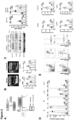
- FIG. 1 Activation of MAPK1 or ERK2 mediates myocardial ischemia in clinical setting of MI.
- A) Heat maps showing the quantity profile for 208 proteins and 685 phosphopeptides significantly differing (p ⁇ 0.05, fold-change > 2 or ⁇ 0.5 and identified in more than 60% of all LC-MS/MS replicates in either control or infarct group), log10 transformed and row-wise normalized using z-score and sorted by descending fold-change.
- B) Reactome pathway enrichment analysis of differentially abundant proteins.
- C) Differentially regulated (p ⁇ 0.05) non phosphorylated and phosphorylated proteins were analyzed for their previously known protein-protein interactions between each other by Cytoscape STRING DB search and sorted by the number of interactions in a circular layout.
- MAPK1 shows the most interactions followed by mTOR, TTN CDK13, PI4KA and TAOK2.
- D) Volcano plot depicts results of the significantly differing integrative inferred kinase activity (InKA) scores between infarct versus control group.
- InKA integrative inferred kinase activity
- FIG. 2 Inhibition of ERK1/2 activation attenuates myocardial remodeling and inflammation in preclinical setting of MI.
- A Design of experimental mouse model. Wild type (WT) mice were subjected to permanent LAD ligation versus sham surgery and administered with vehicle or trametinib (1 mg/kg/d) or vehicle treatment once daily via oral gavage from day 1 through 7.
- B High-frequency ultrasound echocardiography obtained in parasternal long axis (PLAX) with measurement of left ventricular ejection fraction (LVEF, %) and left ventricular end diastolic volume (LVEDV, ⁇ l) on day 7 after operation.
- PDAX parasternal long axis
- LVEF left ventricular ejection fraction
- LVEDV left ventricular end diastolic volume
- CD45+ leukocytes CD45+/CD90.2-/B220-/NK1.1-/CD11b+ myelomonocytic cells
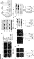
- FIG. 3 A pro-fibrotic MEK1/2-TGF- ⁇ 1 pathway is linked to PAR-2 mediated ROS signaling in monocytes.
- B-C Protein expression analysis of monocytes isolated from WT mice and pre-treated with trametinib (10 ⁇ M) for 1h (B); and from PAR2-/- mice (C).
- (D) Western blot analysis of activated TGF- ⁇ 1 (normalized to GAPDH) and p-SMAD2 (normalized to total SMAD2) in the infarcted myocardium obtained from PAR2 fl/fl LysMCre and PAR2 fl/fl littermates. Representative blots and quantification of biological replicates.
- FIG. 4 Myeloid cell derived TF-PAR2 complex is required for TGF- ⁇ 1 activation.
- C Western blot analysis of activated TGF- ⁇ 1 (normalized to GAPDH) and p-SMAD2 (normalized to total SMAD2) in the infarcted myocardium obtained from TF fl/fl LysMCre and TF fl/fl littermates. Representative blots and quantification of biological replicates.
- FIG. 5 TF cytoplasmic tail deletion attenuates ROS production and ERK1/2-TGF- ⁇ 1 signaling-dependent cardiac fibrosis and improves cardiac function.
- WT and TF ⁇ CT mice were subjected to permanent LAD ligation versus sham surgery and investigated after 7 days and 4 weeks.
- B Assessment of superoxide formation in infarcted myocardium by DHE-HPLC analysis.
- D Protein expression analysis of monocytes isolated from NOX2-/- animals and stimulated with an inflammatory cytokine cocktail containing IL-6, TNF- ⁇ and CCL2 at a concentration of 20 ng/ml with and without hypoxia for 4 hrs.
- Ordinary one-way ANOVA, Sidak's multiple comparison test n 4 replicates per each group (2-3 mice were pooled for each sample).
- E Sirius red staining and de-convoluted images of fibrotic area on paraffin embedded heart sections 4 weeks post M! versus sham surgery. Representative images and 827 quantification of fibrotic areas normalized to surface area.
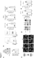
- FIG. 6 Myeloid cell TF cytoplasmic domain phosphorylation mediates ERK1/2-TGF- ⁇ 1 dependent cardiac remodeling in pre-clinical and clinical setting of MI.
- Bone marrow (BM) transplanted mice were subjected to permanent LAD ligation versus sham surgery and investigated 7 days later.
- BM Bone marrow
- Figure 7 Phosphorylation of TF cytoplasmic domain on circulating monocytes in pre-clinical and 851 clinical setting of MI.
- C Confocal microscopy of monocytes isolated from the patients described in table 1 stained for p-TF (red), TF (red) and DAPI (blue).
- D Western blot analysis of monocytic protein expression for TF cytoplasmic domain phosphorylation (4G6) and TF (10H10)
- FIG. 8 Pharmacological targeting of TF-FVlla improves cardiac function by preventing TGF- ⁇ 1 activation.
- A Protein expression analysis of pERK1/2 (normalized to total ERK1/2), NOX2 and TGF- ⁇ 1 (normalized to GAPDH) on isolated human monocytes exposed to hypoxia in the presence of cytokine cocktail mix (20 ng/ml) with and without NAPc2 (200 ng/ml). Representative blots and quantification.
- B Western blot analysis of NOX2 and TGF- ⁇ 1 (normalized to GAPDH) obtained from PBMCs of the experimental animals 7 days after MI. Representative images and quantification of replicates.
- mice were injected with NAPc2 (1mg/kg/d) once daily by i.p. injection from day 1 through day 7.
- D High-frequency echocardiography obtained in parasternal long axis (PLAX) with measurement of LVEF, LVEDV on day 7 after LAD ligation.
- E Representative blots for protein expression analysis of pERK1/2, NOX2, TGF- ⁇ 1 and ⁇ -SMA in infarcted myocardium obtained from vehicle or NAPc2 treated mice.
- Figure 11 Analysis of WT (C57BL/6J) and TF ⁇ CT mice subjected to permanent 912 LAD ligation versus sham surgery after 7 days.
- Figure 12 Analysis of bone marrow (BM) transplanted mice subjected to permanent LAD ligation versus sham surgery after 7 days.
- A Scheme of the BM transplantation experiment.
- B Representative pie chart showing the percentage of peripheral blood CD45.1+ and CD45.2+ cells to quantify donor chimerism.
- Figure 13 Analysis of bone marrow (BM) transplanted mice subjected to permanent LAD ligation versus sham surgery after 7 days.
- A Western blot analysis of pSMAD2 (normalized to SMAD2) and ⁇ -SMA (normalized to GAPDH) in infarcted myocardium obtained from BM transplanted mice. Representative blots and quantification of biological replicates.
- Ordinary one-way ANOVA, Sidak's multiple comparison test, n 5-6 animals per group.
- B LVEF (%) and LVEDV ( ⁇ l) in PLAX M-mode after 6 weeks post MI.
- Ordinary one-way ANOVA, Sidak's multiple comparison test n 6-10 animals per each group.
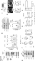
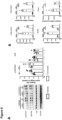
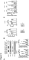
- Figure 15 Deficiency of coagulation factor VII in CX 3 CR 1 + myeloid cells attenuates the development of ischemic heart failure independent of affecting the number of infiltrated immune cells.
- FVII fl/fl CX3CR1 Cre and FVII fl/fl littermates were subjected to permanent LAD ligation versus sham surgery and investigated after 7 days.
- A) High-frequency ultrasound echocardiography obtained from FVII fl/fl CX3CR1 Cre and FVII fl/fl littermates. Mann-Whitney test, n 5 animals per group.
- B) Western blot analysis of activated TGF- ⁇ 1 (normalized to GAPDH) and p-SMAD2 (normalized to total SMAD2) in the infarcted myocardium obtained from integrin ⁇ 1 fl/fl LysM Cre and integrin ⁇ 1 fl/fl littermates. Representative blots and quantification of biological replicates. Mann-Whitney test, n 5 animals per group.
- Peripheral blood cells from the whole blood were isolated by HistopaqueR (CAT #11191, 1077, Sigma-Aldrich, Germany) gradient cell separation. Peripheral blood collected from the patients was added to the 1:1 ratio of HistopaqueR-1119 and HistopaqueR-1077. After centrifugation at 700 x g for 30 min at room temperature, collected mononuclear cell layer was washed with PBS for further platelets elimination.
- HistopaqueR HistopaqueR
- Monocytes were isolated from peripheral blood mononuclear cells (PBMCs) by negative selection using Monocytes Isolation Kit II (human: CAT # 130-117-337, Miltenyi Biotech, Germany) according to the manufacturer's instructions. Enriched monocytes were homogenized in appropriate RIPA buffer for further western blot analysis. In addition to the protein expression analysis, cells were plated and fixed on the cover slip for further confocal imaging.
- PBMCs peripheral blood mononuclear cells
- Monocytes Isolation Kit II human: CAT # 130-117-337, Miltenyi Biotech, Germany
- Ischemic heart samples were obtained from the left ventricular wall of explanted hearts following cardiac transplantation or from heart tissue obtained during implantation of left ventricular assist device.
- Donor hearts were acquired from organ donors whose hearts were not used for transplantation. All subjects provided written informed consent for tissue donation and analyses with ethical approval by the Herz-und Diabetestechnik NRW (HDZ-NRW), Erich & Hanna Klessmann-Institute.
- Acquired heart samples were divided into at least 3 pieces, snap frozen in liquid nitrogen, and stored at -80° C by HDZ-NRW. Upon receipt, samples were used for analysis of protein by western blotting, RNA quantification and cryo-sectioning. Obtained samples were embedded in the O.C.T. to obtain transverse sections of the myocardial tissue.
- Heart tissue biopsy samples from five healthy donors and five patients with ischemic heart failure (IHF) were prepared for whole proteome and phosphoproteome analysis.
- the tissues were lysed in buffer (7 M Urea / 2 M Thiourea / 1% Phosphatase Inhibitor Cocktail 3 (Sigma, Darmstadt, Germany) / 100 mM NH4HCO3) by sonication for 15 min (30 s on / off cycles) at 4 °C with high power in a Bioruptor device (Diagenode, Basil, Belgium).
- the protein concentration was determined using the Pierce 660 335 nm protein assay (Thermo Fisher Scientific, Waltham MA, USA) according to the manufacturer's protocol.
- FASP Filter Aided Sample Preparation
- 700 ⁇ g protein was aliquoted for in-solution digest followed by phosphopeptide enrichment.
- the 20 ⁇ g aliquot of proteins were transferred onto Nanosep molecular weight cutoff (MWCO) spin filter columns with 30 kDa MWCO (Pall, Port Washington NY, USA) and washed with 8 M urea following the previously published protocol (50, 51).
- MWCO Nanosep molecular weight cutoff
- DTT and IAA was used for reduction and alkylation, excess IAA was quenched with DTT and the membrane was washed with 50 mM Ammoniumbicarbonate (AMBIC).
- AMBIC Ammoniumbicarbonate
- the proteins were digested overnight at 37°C with trypsin (Trypsin Gold, Promega, Fitchburg Wl, USA) using an enzyme-to-protein ratio of 1:50 (w/w). After digestion, peptides were eluted by centrifugation and two washes with 50 mM AMBIC.
- the pooled flow-throughs were acidified with trifluoroacetic acid (TFA) to a final concentration of 1 % (v/v) TFA and lyophilized.
- TFA trifluoroacetic acid
- peptides were reconstituted in 0.1 % (v/v) formic acid (FA) for LC-MS analysis.
- 200 ng tryptic peptides from FASP preparation were separated on a nanoAcquity LC (Waters Corporation, Milford MA, USA) at 300 nL/min by a reversed phase C18 column (HSS-T3 C18 1.8 ⁇ m, 75 ⁇ m ⁇ 250 mm, Waters Corporation) at 55 °C using a 90 min linear gradient from 5 % Eluent A (0.1 % TFA / 3 % DMSO / Water) to 40 % Eluent B (0.1 % TFA / 3 % DMSO / ACN) (52).
- Eluting peptides were analyzed in positive mode ESI-MS by ion-mobility separation (IMS) enhanced data independent acquisition (DIA) UDMSE on a Synapt G2-S HDMS mass spectrometer (Waters Corporation) mode as described before [2,4].
- IMS ion-mobility separation
- DIA enhanced data independent acquisition
- Acquired MS data were post-acquisition lock mass corrected using [Glu1]-Fibrinopeptide B, which was sampled every 30 s into the mass spectrometer via the reference sprayer of the NanoLockSpray source at a concentration of 250 fmol/ ⁇ L.
- LC-MS DIA raw data processing was performed with ProteinLynx Global SERVER (PLGS) (version 3.02 build 5, Waters Corporation).
- the human reference proteomes (entries: 20,365) obtained from UniProtKB/SwissProt containing common contaminants was used for peptide identification with allowed missed cleavages of two, carbamidomethylation as fixed modification as well as oxidation on methionine as variable modification.
- the false discovery rate (FDR) for peptide and protein identification was assessed searching a reversed decoy database and set to a 1 % threshold for database search in PLGS. Label-free quantification analysis was performed using ISOQuant as described before (53).
- Phosphopeptide analysis The 700 ⁇ g protein aliquot was digested in-solution by first diluting the lysis buffer 1:4.44 with 50 mM NH4HCO3. After reduction with 1 h incubation with 10 mM DTT at 32 °C and alkylation with 45 min incubation of 25 mM IAA at room temperature in the dark, 367 the proteins were digested over night with trypsin (TPCK-treated, Sigma-Aldrich) at 32 °C using an enzyme-to-protein ratio of 1:25 (w/w). After acidification by addition of 0.5 % TFA the samples were desalted on 500 mg Sep-Pak tC18 columns (Waters Corporation) and lyophilized.
- Phosphopeptide enrichment was performed using preloaded TiO2 spin-tips (3 mg TiO2 / 200 ⁇ L tips, GL Sciences, Tokyo, Japan). The tips were conditioned by centrifugation (3000x g / 2 min / room temperature (RT) ) of 20 ⁇ L washing buffer (80 % CAN / 0.4 % TFA) followed by centrifugation of 20 ⁇ L loading buffer with same settings. The peptides were resuspended in 150 ⁇ L loading buffer (57 %ACN / 0.3 %TFA / 40 % lactic acid), loaded onto the spin-tips and centrifuged (1000x g / 10 min / RT).
- the flow-through was re-applied and centrifuged with same settings.
- the bound phosphopeptides were washed with 20 ⁇ L loading buffer and centrifuged (3000x g /2 min / RT) followed by three times centrifugations with 20 ⁇ L washing buffer.
- the purified phosphopeptides were eluted by centrifugation (1000x g / 10 min / RT) of first 50 ⁇ L of 1.5 % NH3 and second 50 ⁇ L of 5 % Pyrrolidine into one collection tube. After acidification by adding 100 ⁇ L 2.5 % TFA, the phosphopeptides were desalted using Pierce graphite spin-columns (Thermo Scientific) following the manufacturers protocol.
- the phosphopeptides were reconstituted in20 ⁇ L of 0.1 % (v/v) formic acid (FA) for LC-MS analysis 2 ⁇ L of the reconstituted phosphopeptides were separated on an Ultimate 3000 nanoUPLC (Thermo Scientific) with 300 nL/min by a reversed phase C18 column (HSS-T3 C18 1.8 ⁇ m, 75 ⁇ m ⁇ 250 mm, Waters Corporation) at 55 °C using a 90 min linear gradient from 5 % Eluent A (0.1 % TFA / 3 % DMSO / Water) to 35 % Eluent B (0.1 % TFA / 3 % DMSO /ACN) followed by ionization using a Nanospray Flex electrospray ionization source (Thermo Scientific).
- the resulting fragment ion m/z ratios were measured as MS2 spectra over a automatically selected m/z range with a mass resolution of 15 000 @ 200 m/z, AGC target was set to "Standard" and maximum injection time to "Auto”.
- the mass spectrometry proteomics data have been deposited to the ProteomeXchange Consortium via the PRIDE [6] partner repository with the dataset identifier PXD024727.
- Raw data processing for discovery phosphoproteomics analysis was performed in Proteome Discoverer V2.4 (Thermo Scientific) using Sequest HT Search Engine in the processing workflow.
- UniProtKB/SwissProt entries of the human reference proteomes were used as database 400 for peptide and protein identification with maximum allowed missed cleavages of two, maximum precursor and fragment ion mass tolerance of 10 ppm and 0.02 Da respectively.
- Carbamidomethylation on cysteine (+57.021 Da) was set as only fixed modification.
- Oxidation on methionine (+15.995 Da) as well as phosphorylation on serine, threonine and tyrosine (+79.966 Da) were set as dynamic modifications while allowing up to 4 dynamic modifications per peptide.
- N-terminal acetylation of proteins was enabled as variable modification.
- the resulting p-values were corrected for multiple testing using the Benjamini-Hochberg (FDR) method.
- the resulting proteins and phosphosites were then filtered for FCs > 2 and ⁇ 0.5 and an adjusted p-value of ⁇ 0.05.
- After sorting for descending fold change, the protein and phosphopeptide abundances were log10 transformed, normalized by z-score and plotted as heatmap.
- Protein-Protein-Interaction 425 (PPI) networks were generated using Cytoscape V3.7.2 including the stringAPP, NetworkAnalyzer plugins and ClueGO app (56, 57-59). Changes in InKA scores were identified by calculating FC and performing t-test applying the same settings as for proteome and phosphoproteome data.
- the log2 of the FC and -log10 of the adjusted p-value was calculated and displayed in a volcano plot.
- TF ⁇ CT mice 9 to 12 weeks old male mice lacking 21 amino acid of the cytoplasmic tail of TF
- PAR2-/- mice on a C57BL/6N background were used along with strain matched controls (37).
- PAR2fl/fl mice were crossed to LysMCre mice to generate conditional knockout of PAR2 on myeloid cell compartment (PAR2fl/fl LysMCre+ mice) 13, as controls Cre negative PAR2fl/f 432 I littermates were used.
- Generation of TFfl/fILysMCre+/- mice has been previously described56. All animals were bred and housed in the Translational Animal Research Center (TARC) of the Johannes Gutenberg University, Mainz, Germany. All animal experiments were carried out in accordance with the "Guide for the care and use of Laboratory animals" and approved by the "Landesuntersuchungments Rheinland-Pfalz” and ethics committee of the University Medical Centre of Johannes Gutenberg University, Mainz.
- MI myocardial infarction
- LAD left anterior descending artery
- mice received buprenorphine (0.075 mg/kg s.c.) twice daily for 2d, starting on the day of surgery.
- HF was defined in this study by a reduction of the LVEF below 35% and/or visual infarction of the LV. Animals which did not fulfil these criteria were excluded from the study.
- LAD ligated animals were further randomly divided into vehicle treated group (Ml+vehicle) and MI+trametinib/MI+NAPC2 group.
- Trametenib (GSK1120212) was purchased from Selleck Chemicals and reconstituted in 200 ⁇ l vehicle (methocel/polysorbate buffer) and orally administrated to the mice (1 mg/kg/day) once a day starting from day 1 after MI throughout day 7.
- mice were treated with intraperitoneal injections (1 mg/kg/d) of NAPc2 reconstituted in NaCl. Dosing was performed once a day staring from 1 day after MI until sacrifice at day 7.
- Bone marrow transplantation TF ⁇ CT, C57Ly5.1 and C57BL/6J mice aged 8-11 weeks old were irradiated with a lethal dose of 9 Gy. Briefly, donor bone marrow (BM) was harvested in 2% PBS/ FCS, filtered through a 70 ⁇ m cell strainer. Collected BM cells from the donor mice were washed in fresh 2% PBS/ FCS and then re-suspended at final concentration of 4 x 10⁸ cells/ml. At 24h after irradiation, approximately 200 ⁇ l was injected into the recipient mice via the tail vein. Chimeric animals were allowed to recover for 9-10 weeks, followed by the LAD ligation. Donor vs. host composition in the infarcted myocardium was determined by the flow cytometry analysis in the infarcted myocardium after 7 days post Ml.
- Echocardiography Transthoracic echocardiography was performed by using a VEVO-3100 and VEVO-770 (FUJIFILM VisualSonics Inc. Toronto, Canada) High-Frequency Ultrasound System (HFUS). Equipped with a 38MHz (MZ400) linear array transducer, images were acquired at a frame rate consistently above 200 frames. Electrocardiogram (ECG) and breathing rate were monitored, body temperature was kept at 37 ⁇ C using a heating system within the handling platform.
- ECG Electrocardiogram
- breathing rate were monitored, body temperature was kept at 37 ⁇ C using a heating system within the handling platform.
- mice were examined by longitudinal analysis (from 1 day up to 4 weeks post MI) to measure left ventricular (LV) end-diastolic volume (LVEDV), internal diameter in diastole and systole (LVID,d and LVID,s), posterior wall thickness in diastole (LVPW,d) and interventricular septum thickness in diastole (IVS,d) analyzed in the parasternal long axis (PLAX) by means of M-mode images, which are linked to 2D B-mode images. Post-acquisition analysis was performed with the VevoLab Software. LVID,d and LVID,s were applied to calculate LV end-diastolic volume (LVEDV) and LV ejection fraction (LVEF).
- LVEDV left ventricular
- LVID,d and LVID,s internal diameter in diastole and systole
- LVPW,d posterior wall thickness in diastole
- IVS,d interventricular septum thickness
- Flow cytometry analysis of immune cells isolated from heart Infiltration of immune cells into the infarcted myocardium was analyzed by flow cytometry. Collected infarcted myocardium from the experimental animals was enzymatically digested by using collagenase II (1 mg/ml) / DNase I (50 ⁇ g/ml) for 30 min at 37 °C. The lysates were passed through a 70 ⁇ m cell strainer and washed with 2% PBS / FCS. Cell staining: After washing, cells were pelleted by centrifugation for 5 min at 300 x g at 4°C and unspecific antibody binding was blocked by using FC blocking solution (anti-CD16/CD32).
- CD45 APC-eFluor 780 eBioscience, clone 30-F11
- CD45.1 APC-eFluor 780 eBioscience, clone A20
- CD45.2 FITC eBioscience, clone 104
- B220 FITC eBioscience, clone RA3-6B2
- CD11b FITC BD Bioscience, clone M1/70
- PerCP-Cy5.5 eBioscience, clone M1/70
- CD90.2 SuperBright 645 eBioscience, clone 53-2.1
- NK1.1 PE-Cy7 eBioscience, clone PK136
- Ly6G PE BD Bioscience, clone 1A8
- Ly6C Pacific Blue eBioscience, clone HK1.4
- F4/80 APC eBioscience, clone BM8
- TF PE R&D systems
- circulating mononuclear cells were isolated from the whole blood and further analyzed for living CD45+/TF+ leukocytes and CD45+/TF+/CD11b+/CD115+/Ly6Chigh inflammatory monocytes after 7d post MI.
- DHE-HPLC Mouse myocardial tissue collected from the experimental animals were cut into small pieces and incubated with 50 ⁇ mol/L of dihydroethidium (DHE) at 37 °C for 30 min. After incubation, hearts were dried from adhering DHE buffer and washed with PBS. Heart weights were determined and homogenized by using glass/glass homogenizer in 400 ⁇ l of PBS / acetonitrile (1:1) for extraction of DHE oxidation products. Samples were freed from denatured protein by centrifugation for 10 min at 20,000 x rpm and supernatants were analyzed by high performance liquid chromatography (HPLC) 496 for superoxide (O2•-) specific oxidation product 2-hydroxyethidium.
- HPLC high performance liquid chromatography
- the system consisted of a control unit, two pumps, a mixer, detectors, a column oven, a degasser, an autosampler (AS-2057 plus) from Jasco (Gros-Umstadt, Germany), and a C18-Nucleosil 100-3 (125 x 4) column from Macherey & Nagel (Duren, Germany).
- a high pressure gradient was employed with acetonitrile/water (90/10(v/v)%) and 50 mM citrate buffer pH 2 as mobile phases with the following percentages of the organic solvent: 0 min, 41%; 7 min, 45%; 8-9 min, 100%; 10 min, 41%.
- the flow was 1 ml/min and DHE was detected by its absorption at 355 nm, whereas 2-hydroxyethidium and ethidium were detected by fluorescence (Ex. 480 nm/Em. 580 nm).
- Monocytes isolation and in vitro culture Bone marrow derived cell suspensions were isolated by flushing femurs and tibias of 8-12 weeks old mice. Cell aggregates were removed by gentle pipetting and the cell lysates were passed through a 70 ⁇ m nylon strainer to remove the cell debris. Furthermore, to enrich monocytes population, mouse monocyte isolation Kit (STEMCELL Technologies Inc., Vancouver, BC, Canada) was used according to standard protocols provided by the manufacturer.
- OGD oxygen-glucose-deprivation
- monocytes from healthy individuals were treated with NAPc2 in the presence of OGD along with the cytokine cocktail mix followed by the protein expression analysis of pERK1/2, NOX2 and TGF- ⁇ 1.
- anti-pERK1/2 (#4370S, Cell signaling Technology) (14-9109-82, Thermo Fisher) co-stained for ant-CD31 (SC-18916, Santa Cruz), anti-CD45 (30-F11, BioLegend), anti- ⁇ SMA (A2547, Sigmaaldrich) and anti-cardiac troponin T (ab92546, Abcam) were used.
- anti-NOX2 (611414, BD Biosciences, Germany)
- anti-CD68 (ab955, Abcam) and TGF- ⁇ 1, (NBP2-22114, Novus Biologicals) were used to monitor NOX2 and TGF- ⁇ 1 localization.
- PBMCs peripheral blood mononuclear cells
- monocytes Either isolated peripheral blood mononuclear cells (PBMCs), myocardium or monocytes were homogenized in lysis buffer (1% Triton X-100, 20 mM Tris pH 7.4-7.6, 150 mM NaCl, 50 mM NaF, 1 mM EDTA, 1 mM EGTA, 1 mM glycerolphosphatase, 1% SDS, 100 mM PMSF, and 0.1% protease phosphatase inhibitor cocktail) for 20 min on ice. Lysates were cleared by centrifuging at 11,000 x g for 15 min at 4 oC.
- lysis buffer 1% Triton X-100, 20 mM Tris pH 7.4-7.6, 150 mM NaCl, 50 mM NaF, 1 mM EDTA, 1 mM EGTA, 1 mM glycerolphosphatase, 1% SDS, 100
- Total protein concentration was estimated using Lowry Assay (DC Protein Assay, Biorad) and an equal protein amount in all samples was mixed in 6x Laemmli sample buffer, heated to 99°C for 10 min, separated according to their molecular weight on a SDS PAGE gel (4-15%) and probed with respective primary antibodies, pERK1/2, (#4370S, Cell signaling Technology), ERK1/2 (#4695, Cell Signaling Technology), pP38 (#4511S, Cell Signaling Technology), p38 (#9219, Cell Signaling Technology), TGF- ⁇ 1, (NBP2-22114, Novus Biologicals), pSMAD2, SMAD2, (#12747T, Cell Signaling Technology), p67phox (610912 BD Biosciences), NOX 2 (611414, BD Biosciences) and anti-alpha smooth muscle actin (ab7817, abcamR).
- DC Protein Assay DC Protein Assay, Biorad
- phospho-specific mouse anti-human TF antibody (4G6) and mouse anti-human TF antibodies (10H10) were used.
- PVDF membranes were incubated with secondary antibodies for 2 h (goat anti-rabbit HRP (#7074, Cell Signaling Technology) and anti-mouse HRP (#7076, Cell signaling Technology) and developed using Fusion FX (PEQLAB Biotechnologie GmbH, Germany) ECL's Western blotting ECL (Thermo Scientific Technologies) chemiluminescent reagents. Relative densitometry was performed with appropriate software and the ratios were used for statistical analysis.
- RNA from pulverized heart samples from the infarcted part of LAD-ligated mice or non-infarcted myocardium from SHAM-operated mice was extracted by guanidine isothiocyanate phenol chloroform extraction.
- Relative mRNA expression analysis of chemokines and cytokines were performed by quantitative real-time reverse-transcription polymerase chain reaction. 0.05 ⁇ g of total RNA was used for qRT-PCR examination with the QuantiTect TM Probe RT-PCR kit (Qiagen, Hilden, Germany). For cDNA synthesis, briefly 1 ⁇ g of RNA was used.
- the qPCR buffer in each well composed of 10 ⁇ l 2x master mix (Applied Biosystems, Foster city, CA, USA), 5 ⁇ l RNase, DNase, Protease-free purified water, 1 ⁇ l primer of the gene being investigated and 5 ⁇ l of the cDNA sample.
- TaqManR Gene Expression assays (Applied Biosystems, Foster City, CA, USA) for TATA-box binding protein ( tbp ; Mm00446973_m1), Ccr2 (Mm00438270_m1), Il6 (Mm00446190_m1), Ccl2 (Mm00441242_m1), Tnf (Mm00443260_g1) were used.
- the relative mRNA expression level quantification was carried out according to the ⁇ Ct method and normalized to the reference gene (TBP).
- IL-6 For human heart samples, IL-6, CCR2, and CCL2 specific primers were used. GAPDH was used for normalization of the data.
- test groups were evaluated by a Mann-Whitney test. For more than two experimental groups, a Kruskal-Wallis test was performed, followed by Dunn's test for multiple comparisons. Asterisks were used as follows: *, p ⁇ 0.05; **, p ⁇ 0.01; ***, p ⁇ 0.001.
Landscapes
- Health & Medical Sciences (AREA)
- Life Sciences & Earth Sciences (AREA)
- Engineering & Computer Science (AREA)
- Chemical & Material Sciences (AREA)
- Immunology (AREA)
- Medicinal Chemistry (AREA)
- General Health & Medical Sciences (AREA)
- Urology & Nephrology (AREA)
- Molecular Biology (AREA)
- Hematology (AREA)
- Biomedical Technology (AREA)
- Proteomics, Peptides & Aminoacids (AREA)
- Bioinformatics & Cheminformatics (AREA)
- Pharmacology & Pharmacy (AREA)
- Animal Behavior & Ethology (AREA)
- Public Health (AREA)
- Veterinary Medicine (AREA)
- Epidemiology (AREA)
- Physics & Mathematics (AREA)
- Analytical Chemistry (AREA)
- Gastroenterology & Hepatology (AREA)
- Biotechnology (AREA)
- Cell Biology (AREA)
- Zoology (AREA)
- Pathology (AREA)
- Microbiology (AREA)
- General Physics & Mathematics (AREA)
- Biochemistry (AREA)
- Food Science & Technology (AREA)
- Cardiology (AREA)
- General Chemical & Material Sciences (AREA)
- Chemical Kinetics & Catalysis (AREA)
- Nuclear Medicine, Radiotherapy & Molecular Imaging (AREA)
- Hospice & Palliative Care (AREA)
- Organic Chemistry (AREA)
- Heart & Thoracic Surgery (AREA)
- Medicines That Contain Protein Lipid Enzymes And Other Medicines (AREA)
- Medicines Containing Antibodies Or Antigens For Use As Internal Diagnostic Agents (AREA)
- Pharmaceuticals Containing Other Organic And Inorganic Compounds (AREA)
Claims (13)
- Inhibitor des Gewebefaktor-Protease-aktivierten Rezeptor 2 (TF-PAR2)-Signalwegs zur Verwendung bei der Behandlung oder Prävention von Herzinsuffizienz (HF).
- Inhibitor des TF-PAR2-Signalwegs zur Verwendung nach Anspruch 1, wobei die Herzinsuffizienz (HF) mit ischämischer Herzinsuffizienz (IHF), Myokardinfarkt (MI), IHF aufgrund akutem und/oder anhaltendem Mi, Herzinsuffizienz mit reduzierter Ejektionsfraktion (HFrEF) oder Herzinsuffizienz mit erhaltener Ejektionsfraktion (HFpEF) in Verbindung steht oder synonym ist.
- Inhibitor des TF-PAR2-Signalwegs zur Verwendung nach Anspruch 1, wobei der TF-PAR2-Signalweg gekennzeichnet ist durch (i) eine Hyperaktivierung der Mitogenaktivierten Proteinkinase 1 (MAPK1), (ii) eine Phosphorylierung der extrazellulär-Signalregulierten Kinase 1/2 (ERK1/2) und (iii) eine TGF-β1-Aktivierung.
- Inhibitor des TF-PAR2-Signalwegs zur Verwendung nach Anspruch 1, wobei der Inhibitor des TF-PAR2-Signalwegs eine chemische oder biologische Verbindung ist, die mit reduzierter ERK1/2-Phosphorylierung, verringerter TGF-β1-Aktivierung und reduzierter NOX2-Expression in kardialen oder myelomonozytischen Zellen in Verbindung steht.
- Inhibitor des TF-PAR2-Signalwegs zur Verwendung nach Anspruch 1, wobei der Inhibitor des TF-PAR2-Signalwegs ein Inhibitor der Rekrutierung von NOX2-positiven Myeloidzellen, vorzugsweise ein Inhibitor von NOX2-positiven Monozyten, ist.
- Inhibitor des TF-PAR2-Signalwegs zur Verwendung nach Anspruch 1, wobei der Inhibitor des TF-PAR2-Signalwegs ein Inhibitor der TGF-β1-Aktivierung ist.
- Inhibitor des TF-PAR2-Signalwegs zur Verwendung nach einem der Ansprüche 1 bis 6, wobei der Inhibitor ein TF/FVlla-Inhibitor ist.
- Inhibitor des TF-PAR2-Signalwegs zur Verwendung nach Anspruch 7, wobei der TF/FVlla-Inhibitor ausgewählt ist aus der Gruppe bestehend aus Anti-TF-Antikörpern, kleinen Molekülen, TF-Pathway Inhibitor (TFPI), humanem rekombinantem FVIIa-Inhibitor (rFVIIai), chimärem Protein XK1 und PAR2-Antagonisten.
- Inhibitor des TF-PAR2-Signalwegs zur Verwendung nach Anspruch 8, wobei der Antikörper ausgewählt ist aus AP-1, ALT836, Tisotumab, ICON-2.
- Inhibitor des TF-PAR2-Signalwegs zur Verwendung nach Anspruch 1, wobei der Inhibitor des TF-PAR2-Signalwegs das gerinnungshemmende Nematodenprotein c2 (NAPc2) ist.
- Inhibitor des TF-PAR2-Signalwegs zur Verwendung nach Anspruch 10, wobei der NAPc2 eine modifizierte NAPc2- oder rekombinante NAPc2-Variante ist.
- Inhibitor des TF-PAR2-Signalwegs zur Verwendung nach Anspruch 11, wobei die NAPc2-Variante NAPc2/Prolin ist, welches die Aminosäuresequenz von SEQ ID NO:2 oder Mutanten oder Homologe davon umfasst.
- Inhibitor des TF-PAR2-Signalwegs zur Verwendung nach einem der Ansprüche 1 bis 12, wobei der Inhibitor des TF-PAR2-Signalwegs ein Oligonukleotid-Inhibitor, ausgewählt aus Antisense-Oligonukleotid, siRNA, shRNA, Antisense-Oligonukleotid, das auf eine mRNA, nicht-kodierende RNA (ncRNA), miRNA und lange nicht-kodierende RNA (IncRNA) abzielt, oder ein Protein- oder ein Nukleinsäure-Aptamer ist.
Applications Claiming Priority (3)
| Application Number | Priority Date | Filing Date | Title |
|---|---|---|---|
| EP20208835 | 2020-11-20 | ||
| EP21194468 | 2021-09-02 | ||
| PCT/EP2021/082355 WO2022106648A1 (en) | 2020-11-20 | 2021-11-19 | Inhibitors of the tissue factor-protease activated receptor 2 (tf-par2) signaling pathway for use in the treatment or prevention of heart failure (hf) and associated or resulting diseases |
Publications (3)
| Publication Number | Publication Date |
|---|---|
| EP4247408A1 EP4247408A1 (de) | 2023-09-27 |
| EP4247408C0 EP4247408C0 (de) | 2024-12-18 |
| EP4247408B1 true EP4247408B1 (de) | 2024-12-18 |
Family
ID=78824838
Family Applications (1)
| Application Number | Title | Priority Date | Filing Date |
|---|---|---|---|
| EP21820471.7A Active EP4247408B1 (de) | 2020-11-20 | 2021-11-19 | Inhibitoren des gewebefaktor-protease-aktivierten rezeptor 2 (tf-par2)-signalweges zur verwendung bei der behandlung oder prävention von herzinsuffizienz (hf) |
Country Status (5)
| Country | Link |
|---|---|
| US (1) | US20240108688A1 (de) |
| EP (1) | EP4247408B1 (de) |
| JP (1) | JP2023550046A (de) |
| CA (1) | CA3202095A1 (de) |
| WO (1) | WO2022106648A1 (de) |
Families Citing this family (2)
| Publication number | Priority date | Publication date | Assignee | Title |
|---|---|---|---|---|
| JP7532682B2 (ja) | 2021-03-10 | 2024-08-13 | バンジー, インコーポレイテッド | マークアップフリーレッジグラブ |
| CN118252938B (zh) * | 2024-01-24 | 2025-02-11 | 中国医学科学院阜外医院 | 抑制或敲除Jun基因表达的试剂用于治疗射血分数保留的心衰的用途 |
Family Cites Families (10)
| Publication number | Priority date | Publication date | Assignee | Title |
|---|---|---|---|---|
| EP0866870B1 (de) | 1995-12-01 | 2003-03-12 | Genentech, Inc. | Fusionsproteine bestehend aus gewebefaktor-kunitzdomäne als inhibitoren des faktors viia |
| JP2003530819A (ja) | 1999-06-14 | 2003-10-21 | ノボ ノルディスク アクティーゼルスカブ | FVIIa/TF活性阻害化合物 |
| EP1420807A4 (de) * | 2001-08-09 | 2005-08-17 | Cornell Res Foundation Inc | Thrombozyten-wachstumsfaktor zum schutz des kardialen myokards |
| AU2003215189A1 (en) * | 2002-02-15 | 2003-09-09 | Cornell Research Foundation, Inc. | Endothelial precursor cells for enhancing and restoring vascular function |
| US20040102402A1 (en) | 2002-11-22 | 2004-05-27 | Isis Pharmaceuticals Inc. | Modulation of tissue factor expression |
| JP2006335709A (ja) * | 2005-06-03 | 2006-12-14 | Taiyo Kagaku Co Ltd | 組織因子阻害組成物、及びそれを含有する飲食品、飼料、医薬部外品、医薬品 |
| WO2007092607A2 (en) | 2006-02-09 | 2007-08-16 | Genetech, Inc. | Treatment of hemorrhagic viral infections using a tissue factor inhibitor |
| JP6436654B2 (ja) * | 2014-06-02 | 2018-12-12 | 学校法人北里研究所 | リン脂質ヒドロペルオキシド依存性細胞死の検出方法及びキット、リン脂質ヒドロペルオキシド依存性細胞死の抑制剤、並びにリン脂質ヒドロペルオキシド依存性細胞死が関連する疾患の予防又は治療剤 |
| JP6622102B2 (ja) | 2016-02-04 | 2019-12-18 | 松森 昭 | 急性心筋梗塞の判定方法 |
| WO2018170134A1 (en) | 2017-03-14 | 2018-09-20 | Ohio State Innovation Foundation | Methods and compositions related to a tissue factor-targeting igg3 immunoconjugates |
-
2021
- 2021-11-19 EP EP21820471.7A patent/EP4247408B1/de active Active
- 2021-11-19 US US18/252,642 patent/US20240108688A1/en active Pending
- 2021-11-19 WO PCT/EP2021/082355 patent/WO2022106648A1/en not_active Ceased
- 2021-11-19 CA CA3202095A patent/CA3202095A1/en active Pending
- 2021-11-19 JP JP2023528217A patent/JP2023550046A/ja active Pending
Also Published As
| Publication number | Publication date |
|---|---|
| CA3202095A1 (en) | 2022-05-27 |
| EP4247408C0 (de) | 2024-12-18 |
| JP2023550046A (ja) | 2023-11-30 |
| WO2022106648A1 (en) | 2022-05-27 |
| EP4247408A1 (de) | 2023-09-27 |
| US20240108688A1 (en) | 2024-04-04 |
Similar Documents
| Publication | Publication Date | Title |
|---|---|---|
| Garlapati et al. | Targeting myeloid cell coagulation signaling blocks MAP kinase/TGF-β1–driven fibrotic remodeling in ischemic heart failure | |
| d’Alessandro et al. | Thrombo-inflammation in cardiovascular disease: an expert consensus document from the Third Maastricht Consensus Conference on Thrombosis | |
| US20130316337A1 (en) | Methods of Diagnosing and Treating an Inflammatory Response | |
| EP2170366B1 (de) | Verwendung eines tlr-2 antagonisten zur behandlung von reperfusionsverletzungen und gewebeschäden | |
| Yang et al. | Neutrophil‐derived advanced glycation end products‐Nε‐(carboxymethyl) lysine promotes RIP3‐mediated myocardial necroptosis via RAGE and exacerbates myocardial ischemia/reperfusion injury | |
| CN1997385B (zh) | 膜联蛋白v用于预防动脉血栓和斑块破裂 | |
| EP4247408B1 (de) | Inhibitoren des gewebefaktor-protease-aktivierten rezeptor 2 (tf-par2)-signalweges zur verwendung bei der behandlung oder prävention von herzinsuffizienz (hf) | |
| EP2941270A1 (de) | Verfahren zur behandlung von entzündungen | |
| ES2941693T3 (es) | Métodos y composiciones farmacéuticas para el tratamiento de lesiones tisulares | |
| JP2017508806A (ja) | 出血性疾患の治療における止血を改善するためのヒトプロトロンビン及び活性化第x因子の組成物 | |
| Jiang et al. | Dual specificity phosphatase 3 (DUSP3) knockdown alleviates acute myocardial infarction damage via inhibiting apoptosis and inflammation | |
| CN108076629A (zh) | 一种用于治疗、预防或改善主动脉病的gm-csf负调节剂 | |
| US20210052701A1 (en) | Methods of Treating Vascular Leakage Using CXCL12 Peptides | |
| US20230293649A1 (en) | Cst6, cells expressing cst6 and methods of use | |
| Redgrave et al. | Exogenous TGFβ1 and its mimic HpTGM attenuate the heart’s inflammatory response to ischaemic injury and improve long term cardiac outcomes | |
| EP4259181A1 (de) | Antagonist des interleukin-17b-rezeptors (il-17rb) und verwendung davon | |
| Smargiassi et al. | Pharmacologic Treatment of IPF | |
| Nandave | in High-Risk Patients as Compared | |
| Nowak | Experimental investigations on the role of the immunoproteasome in lung fibrogenesis | |
| Shahzad | Different DOACs Control Inflammation in Cardiac Ischemia-Reperfusion Differently | |
| ZENG | IMMUNE CELLS EXPRESS ELEVATED COAGULATION FACTOR VIII IN PROTHROMBOTIC PONATINIB-TREATED MICE | |
| Falcione | Thrombin generation in ischemic stroke | |
| WO2008047486A1 (fr) | Agent de lutte contre les lésions myocardiques | |
| EP4203959A1 (de) | Zusammensetzung mit ttf-ngr zur verwendung bei der behandlung von weichgewebesarkom | |
| EA049199B1 (ru) | КОМПОЗИЦИЯ, СОДЕРЖАЩАЯ tTF-NGR, ДЛЯ ПРИМЕНЕНИЯ ПРИ ЛЕЧЕНИИ САРКОМЫ МЯГКИХ ТКАНЕЙ |
Legal Events
| Date | Code | Title | Description |
|---|---|---|---|
| STAA | Information on the status of an ep patent application or granted ep patent |
Free format text: STATUS: UNKNOWN |
|
| STAA | Information on the status of an ep patent application or granted ep patent |
Free format text: STATUS: THE INTERNATIONAL PUBLICATION HAS BEEN MADE |
|
| PUAI | Public reference made under article 153(3) epc to a published international application that has entered the european phase |
Free format text: ORIGINAL CODE: 0009012 |
|
| STAA | Information on the status of an ep patent application or granted ep patent |
Free format text: STATUS: REQUEST FOR EXAMINATION WAS MADE |
|
| 17P | Request for examination filed |
Effective date: 20230613 |
|
| AK | Designated contracting states |
Kind code of ref document: A1 Designated state(s): AL AT BE BG CH CY CZ DE DK EE ES FI FR GB GR HR HU IE IS IT LI LT LU LV MC MK MT NL NO PL PT RO RS SE SI SK SM TR |
|
| RAP3 | Party data changed (applicant data changed or rights of an application transferred) |
Owner name: UNIVERSITAETSMEDIZIN DER JOHANNES GUTENBERG-UNIVERSITAET MAINZ |
|
| DAV | Request for validation of the european patent (deleted) | ||
| DAX | Request for extension of the european patent (deleted) | ||
| STAA | Information on the status of an ep patent application or granted ep patent |
Free format text: STATUS: EXAMINATION IS IN PROGRESS |
|
| 17Q | First examination report despatched |
Effective date: 20240417 |
|
| REG | Reference to a national code |
Ref legal event code: R079 Ref country code: DE Ref legal event code: R079 Ref document number: 602021023674 Country of ref document: DE Free format text: PREVIOUS MAIN CLASS: A61K0038170000 Ipc: A61P0009040000 |
|
| GRAP | Despatch of communication of intention to grant a patent |
Free format text: ORIGINAL CODE: EPIDOSNIGR1 |
|
| STAA | Information on the status of an ep patent application or granted ep patent |
Free format text: STATUS: GRANT OF PATENT IS INTENDED |
|
| RIC1 | Information provided on ipc code assigned before grant |
Ipc: G01N 33/50 20060101ALI20240627BHEP Ipc: C07K 16/00 20060101ALI20240627BHEP Ipc: A61K 45/06 20060101ALI20240627BHEP Ipc: A61K 38/17 20060101ALI20240627BHEP Ipc: A61P 9/04 20060101AFI20240627BHEP |
|
| INTG | Intention to grant announced |
Effective date: 20240717 |
|
| GRAS | Grant fee paid |
Free format text: ORIGINAL CODE: EPIDOSNIGR3 |
|
| GRAA | (expected) grant |
Free format text: ORIGINAL CODE: 0009210 |
|
| STAA | Information on the status of an ep patent application or granted ep patent |
Free format text: STATUS: THE PATENT HAS BEEN GRANTED |
|
| AK | Designated contracting states |
Kind code of ref document: B1 Designated state(s): AL AT BE BG CH CY CZ DE DK EE ES FI FR GB GR HR HU IE IS IT LI LT LU LV MC MK MT NL NO PL PT RO RS SE SI SK SM TR |
|
| REG | Reference to a national code |
Ref country code: CH Ref legal event code: EP |
|
| REG | Reference to a national code |
Ref country code: DE Ref legal event code: R081 Ref document number: 602021023674 Country of ref document: DE Owner name: UNIVERSITAETSMEDIZIN DER JOHANNES GUTENBERG-UN, DE Free format text: FORMER OWNER: ANMELDERANGABEN UNKLAR / UNVOLLSTAENDIG, 80297 MUENCHEN, DE |
|
| REG | Reference to a national code |
Ref country code: IE Ref legal event code: FG4D |
|
| REG | Reference to a national code |
Ref country code: DE Ref legal event code: R096 Ref document number: 602021023674 Country of ref document: DE |
|
| U01 | Request for unitary effect filed |
Effective date: 20250115 |
|
| U07 | Unitary effect registered |
Designated state(s): AT BE BG DE DK EE FI FR IT LT LU LV MT NL PT RO SE SI Effective date: 20250121 |
|
| PG25 | Lapsed in a contracting state [announced via postgrant information from national office to epo] |
Ref country code: HR Free format text: LAPSE BECAUSE OF FAILURE TO SUBMIT A TRANSLATION OF THE DESCRIPTION OR TO PAY THE FEE WITHIN THE PRESCRIBED TIME-LIMIT Effective date: 20241218 |
|
| PG25 | Lapsed in a contracting state [announced via postgrant information from national office to epo] |
Ref country code: NO Free format text: LAPSE BECAUSE OF FAILURE TO SUBMIT A TRANSLATION OF THE DESCRIPTION OR TO PAY THE FEE WITHIN THE PRESCRIBED TIME-LIMIT Effective date: 20250318 |
|
| PG25 | Lapsed in a contracting state [announced via postgrant information from national office to epo] |
Ref country code: GR Free format text: LAPSE BECAUSE OF FAILURE TO SUBMIT A TRANSLATION OF THE DESCRIPTION OR TO PAY THE FEE WITHIN THE PRESCRIBED TIME-LIMIT Effective date: 20250319 |
|
| PG25 | Lapsed in a contracting state [announced via postgrant information from national office to epo] |
Ref country code: RS Free format text: LAPSE BECAUSE OF FAILURE TO SUBMIT A TRANSLATION OF THE DESCRIPTION OR TO PAY THE FEE WITHIN THE PRESCRIBED TIME-LIMIT Effective date: 20250318 |
|
| PG25 | Lapsed in a contracting state [announced via postgrant information from national office to epo] |
Ref country code: SM Free format text: LAPSE BECAUSE OF FAILURE TO SUBMIT A TRANSLATION OF THE DESCRIPTION OR TO PAY THE FEE WITHIN THE PRESCRIBED TIME-LIMIT Effective date: 20241218 |
|
| PG25 | Lapsed in a contracting state [announced via postgrant information from national office to epo] |
Ref country code: PL Free format text: LAPSE BECAUSE OF FAILURE TO SUBMIT A TRANSLATION OF THE DESCRIPTION OR TO PAY THE FEE WITHIN THE PRESCRIBED TIME-LIMIT Effective date: 20241218 |
|
| PG25 | Lapsed in a contracting state [announced via postgrant information from national office to epo] |
Ref country code: ES Free format text: LAPSE BECAUSE OF FAILURE TO SUBMIT A TRANSLATION OF THE DESCRIPTION OR TO PAY THE FEE WITHIN THE PRESCRIBED TIME-LIMIT Effective date: 20241218 |
|
| PG25 | Lapsed in a contracting state [announced via postgrant information from national office to epo] |
Ref country code: IS Free format text: LAPSE BECAUSE OF FAILURE TO SUBMIT A TRANSLATION OF THE DESCRIPTION OR TO PAY THE FEE WITHIN THE PRESCRIBED TIME-LIMIT Effective date: 20250418 |
|
| PG25 | Lapsed in a contracting state [announced via postgrant information from national office to epo] |
Ref country code: SK Free format text: LAPSE BECAUSE OF FAILURE TO SUBMIT A TRANSLATION OF THE DESCRIPTION OR TO PAY THE FEE WITHIN THE PRESCRIBED TIME-LIMIT Effective date: 20241218 |
|
| PG25 | Lapsed in a contracting state [announced via postgrant information from national office to epo] |
Ref country code: CZ Free format text: LAPSE BECAUSE OF FAILURE TO SUBMIT A TRANSLATION OF THE DESCRIPTION OR TO PAY THE FEE WITHIN THE PRESCRIBED TIME-LIMIT Effective date: 20241218 |
|
| PLBE | No opposition filed within time limit |
Free format text: ORIGINAL CODE: 0009261 |
|
| STAA | Information on the status of an ep patent application or granted ep patent |
Free format text: STATUS: NO OPPOSITION FILED WITHIN TIME LIMIT |
|
| U20 | Renewal fee for the european patent with unitary effect paid |
Year of fee payment: 5 Effective date: 20251016 |
|
| 26N | No opposition filed |
Effective date: 20250919 |
|
| PGFP | Annual fee paid to national office [announced via postgrant information from national office to epo] |
Ref country code: GB Payment date: 20251120 Year of fee payment: 5 |