EP4249889B1 - Verfahren zum analysieren von biopsien und biologischen proben - Google Patents
Verfahren zum analysieren von biopsien und biologischen proben Download PDFInfo
- Publication number
- EP4249889B1 EP4249889B1 EP22163930.5A EP22163930A EP4249889B1 EP 4249889 B1 EP4249889 B1 EP 4249889B1 EP 22163930 A EP22163930 A EP 22163930A EP 4249889 B1 EP4249889 B1 EP 4249889B1
- Authority
- EP
- European Patent Office
- Prior art keywords
- sample
- cells
- cell
- liquid
- imaging
- Prior art date
- Legal status (The legal status is an assumption and is not a legal conclusion. Google has not performed a legal analysis and makes no representation as to the accuracy of the status listed.)
- Active
Links
Images


Classifications
-
- G—PHYSICS
- G01—MEASURING; TESTING
- G01N—INVESTIGATING OR ANALYSING MATERIALS BY DETERMINING THEIR CHEMICAL OR PHYSICAL PROPERTIES
- G01N1/00—Sampling; Preparing specimens for investigation
- G01N1/28—Preparing specimens for investigation including physical details of (bio-)chemical methods covered elsewhere, e.g. G01N33/50, C12Q
- G01N1/2813—Producing thin layers of samples on a substrate, e.g. smearing, spinning-on
-
- G—PHYSICS
- G01—MEASURING; TESTING
- G01N—INVESTIGATING OR ANALYSING MATERIALS BY DETERMINING THEIR CHEMICAL OR PHYSICAL PROPERTIES
- G01N21/00—Investigating or analysing materials by the use of optical means, i.e. using sub-millimetre waves, infrared, visible or ultraviolet light
- G01N21/62—Systems in which the material investigated is excited whereby it emits light or causes a change in wavelength of the incident light
- G01N21/63—Systems in which the material investigated is excited whereby it emits light or causes a change in wavelength of the incident light optically excited
- G01N21/64—Fluorescence; Phosphorescence
- G01N21/645—Specially adapted constructive features of fluorimeters
- G01N21/6456—Spatial resolved fluorescence measurements; Imaging
- G01N21/6458—Fluorescence microscopy
-
- G—PHYSICS
- G01—MEASURING; TESTING
- G01N—INVESTIGATING OR ANALYSING MATERIALS BY DETERMINING THEIR CHEMICAL OR PHYSICAL PROPERTIES
- G01N15/00—Investigating characteristics of particles; Investigating permeability, pore-volume or surface-area of porous materials
- G01N15/10—Investigating individual particles
- G01N15/14—Optical investigation techniques, e.g. flow cytometry
- G01N15/1468—Optical investigation techniques, e.g. flow cytometry with spatial resolution of the texture or inner structure of the particle
-
- G—PHYSICS
- G01—MEASURING; TESTING
- G01N—INVESTIGATING OR ANALYSING MATERIALS BY DETERMINING THEIR CHEMICAL OR PHYSICAL PROPERTIES
- G01N15/00—Investigating characteristics of particles; Investigating permeability, pore-volume or surface-area of porous materials
- G01N15/10—Investigating individual particles
- G01N2015/1006—Investigating individual particles for cytology
-
- G—PHYSICS
- G01—MEASURING; TESTING
- G01N—INVESTIGATING OR ANALYSING MATERIALS BY DETERMINING THEIR CHEMICAL OR PHYSICAL PROPERTIES
- G01N15/00—Investigating characteristics of particles; Investigating permeability, pore-volume or surface-area of porous materials
- G01N15/10—Investigating individual particles
- G01N15/14—Optical investigation techniques, e.g. flow cytometry
- G01N2015/1493—Particle size
-
- G—PHYSICS
- G01—MEASURING; TESTING
- G01N—INVESTIGATING OR ANALYSING MATERIALS BY DETERMINING THEIR CHEMICAL OR PHYSICAL PROPERTIES
- G01N15/00—Investigating characteristics of particles; Investigating permeability, pore-volume or surface-area of porous materials
- G01N15/10—Investigating individual particles
- G01N15/14—Optical investigation techniques, e.g. flow cytometry
- G01N2015/1497—Particle shape
-
- G—PHYSICS
- G01—MEASURING; TESTING
- G01N—INVESTIGATING OR ANALYSING MATERIALS BY DETERMINING THEIR CHEMICAL OR PHYSICAL PROPERTIES
- G01N21/00—Investigating or analysing materials by the use of optical means, i.e. using sub-millimetre waves, infrared, visible or ultraviolet light
- G01N21/62—Systems in which the material investigated is excited whereby it emits light or causes a change in wavelength of the incident light
- G01N21/63—Systems in which the material investigated is excited whereby it emits light or causes a change in wavelength of the incident light optically excited
- G01N21/64—Fluorescence; Phosphorescence
- G01N21/6428—Measuring fluorescence of fluorescent products of reactions or of fluorochrome labelled reactive substances, e.g. measuring quenching effects, using measuring "optrodes"
- G01N2021/6439—Measuring fluorescence of fluorescent products of reactions or of fluorochrome labelled reactive substances, e.g. measuring quenching effects, using measuring "optrodes" with indicators, stains, dyes, tags, labels, marks
Definitions
- the present invention relates to matrix-assisted methods for analyzing biological specimens, particularly liquid biopsies, and other liquid samples, by microscopy, including fluorescence microscopy.
- Liquid biopsies are typically obtained from bodily fluids, such as peripheral blood, bone marrow, cerebrospinal fluid, urine, saliva, sputum, tears, seminal fluid, or other tissue sources. Biomarkers, or components, in the liquid samples can be evaluated or measured for various diagnostic applications, such as disease screening, detection, staging, and surveillance.
- the biomarkers in a liquid biopsy can include cellular and extracellular components, the selection of which can depend on multiple factors, such as the underlying medical condition or treatment status.
- biomarkers can correspond to antigens or other attributes that distinguish rare circulating cells, such as circulating tumor cells (CTCs) and CTC clusters derived from solid tumors or metastases, as well as circulating endothelial cells (CEC) associated with cardiovascular and other conditions.
- CTCs circulating tumor cells
- CEC circulating endothelial cells
- Biomarkers can also identify extracellular components, such as circulating tumor-derived factors, secreted proteins, released vesicles and exosomes, and cell-free nucleic acids.
- Cell-free nucleic acids include cell-free tumor DNA (ctDNA), which has applications in cancer monitoring, as well as cell-free fetal DNA (cffDNA), which is found in maternal blood and has applications in non-invasive prenatal testing. Campos et al. 2018, Cancer J. 24, 93-103 ; Sifakis et al. 2014, Mol. Med. Rep. 11, 2367-2372 . Subsequent genomic and protein processing can allow further analysis of extracellular biomarkers.
- Typical biopsy methods can encompass several approaches. See, e.g., Harouaka et al., 2014, Pharmacol. Ther. 141, 209-221
- immunoaffinity such as microfluidics and microchip-based methods, which allow detection of antibodies bound to cellular and extracellular targets. These methods rely on antibody-antigen binding between floating cells and antibody-coated surfaces, and are therefore limited to antigens present on the target cell surface, such as membrane proteins.
- the efficacy of these methods depends on sufficient levels of cell surface antigens to allow efficient and specific recognition by the antibodies, (e.g., low EpCAM expression on a cell surface may result in poor cell to chip surface binding). These methods generally have low cell flow rates that preclude effective analysis of complex samples. Accordingly, the scope, sensitivity, specificity, and throughput of such methods, as well as other immunoaffinity methods (such as ones based on magnetic beads) is limited.
- cytology directly examining the cells under a microscope.
- this approach is limited to examining only a small number of cells at a time (e.g., as a single layer on a glass slide) due to the light-scattering nature of cells; it is therefore not practical - and not economically feasible - to use this approach to detect rare targets in millions of cells that may be present in even 2 c.c. of blood.
- flow cytometry thousands of cells per second pass one by one through one or more laser beams, where they can give rise to different patterns of light scattering (depending, for example, on cell size and granularity) and fluorescence emission (depending on what fluorescent probes are bound to the cells). See, e.g., Flow cytometry: retrospective, fundamentals and recent instrumentation, Cytotechnology, 2012 Mar; 64(2): 109-130 .
- flow cytometry methods do not provide high resolution and confidence when cells of interest are rare. For example, they do not offer direct visual inspection of cells to confirm their potential morphological or functional properties.
- gating issues can arise based on fluorescence signal intensity and the pixels captured by the detector (which does not provide, information on labeling quality or morphological details of the analyzed sample). For example, only small gating changes or perturbations can lead to the exclusion of CTC cells and other rare cells (e.g., CECs) that are small in size or have a faint fluorescent signal.
- CTC cells and other rare cells e.g., CECs
- the current invention addresses these and other needs in the art by providing methods and compositions that label, disperse, and capture biopsy components in a 3-dimensional gel or other non-liquid form.
- discrete and rare biomarkers can be detected with high resolution and sensitivity by rapid imaging methods, such as light sheet fluorescent microscopy and other methods. More generally, these methods are applicable to components in any sample - biological or non-biological - whose resolution can be improved by dispersal in a liquid and subsequent capture and imaging in a non-liquid state.
- US 2021/0018441 A1 describes "a quantitative liquid biopsy diagnostic system and methods for performing diagnostic assays.
- the system offers a liquid biopsy method using circulating tumor cells (CTCs) or White Blood Cells (WBC) subpopulations for precision cancer diagnosis, early detection of disease evolution, and cancer patient management.
- CTCs circulating tumor cells
- WBC White Blood Cells
- the [method] utilizes selective plane illumination microscopy (SPIM) to deliver high sensitivity and specificity for the detection and isolation of individual CTCs, superseding the efficacy of existing methodologies for early cancer detection. Isolated CTCs can be analyzed for their molecular fingerprint, which can lead to matching genetic abnormalities with specific drug treatments. ".
- the present invention provides a matrix-assisted method, such as those based on gel formation, to prepare and analyze components in biological and non-biological samples, including liquid biological specimens, as described further herein.
- the liquid specimen can originate from any source, including humans and animals. In embodiments, it can derive from a liquid biopsy obtained from peripheral blood, bone marrow, cerebrospinal fluid, and other tissue sources, all of which may be further processed. In embodiments, it can derive from a liquid dispersal of materials obtained from other sources, including solid sources, such as from a solid tissue biopsy.
- the method of the present invention is defined in claim 1.
- the method of the invention comprises adding a solidifying agent (e.g., a gelling agent) to a biological specimen comprising biological materials; generating a solidified sample (e.g., a gelled sample) comprising dispersed biological materials; and imaging the solidified sample to identify one or more components in the biological materials.
- a solidifying agent e.g., a gelling agent
- the biological materials can include any biomolecules, including nucleic acids, proteins, and small molecules, which in embodiments, can serve as biomarkers of medical conditions or disease states.
- the biomolecules can serve as biomarkers of rare circulating cells in the blood, such as circulating tumor cells, circulating endothelial cells, and other cells and cell clusters that may be present in the biological specimen.
- the biological materials in the specimen can be enriched, for example by concentrating cells from a large amount of blood or other sample source.
- the presently disclosed method does not require any preselection or pre-screening of the cells; instead, the instant method allows for unbiased analysis of a sample of unselected or unscreened cells by detecting biomarker labels, such as antibodies or nucleic acid probes, bound to biological materials in the specimen, for example by detecting labeled antibodies identifying specific cell surface markers in the specimen.
- the solidifying agent comprises a mixture of chemical monomers and cross linkers to create a synthetic chemical gel having a chemically cross-linked polymer network.
- the step of adding a solidifying agent in any of the methods can comprise adding hydrogel precursors, to the sample; adding the agent directly to a sample under conditions that allow formation of a solid, such as a gel; forming a hydrogel, or other methods, as disclosed herein.
- the methods Prior to adding the solidifying agent, can comprise subjecting components of biological specimen to a fixation procedure, as described herein, and they can also comprise labeling one or more biomolecules, such as a protein or nucleic acid, with a molecular probe.
- Molecular probes include antibodies, dyes, and nucleic acid probes known in the art, including those as described herein.
- the probes can be used to identify circulating tumor cells and clusters, as well as cell-free tumor or fetal-derived DNA, and genetic and structural changes in cell nuclei, such as DNA and chromosomal abnormalities, amplifications, deletions, and translocations.
- the step of adding a solidifying agent to a liquid specimen comprises mixing a sample containing biological materials (e.g., a pellet comprising biological materials) with a mixture or solution comprising one or more hydrogel precursors, and altering the conditions of the mixture to induce solidification (e.g., gelation).
- a sample containing biological materials e.g., a pellet comprising biological materials
- a mixture or solution comprising one or more hydrogel precursors e.g., gelation
- a hydrogel is known in the art, and can be accomplished by a variety of methods according to the subject disclosure.
- a second agent e.g., a Ca+ ion or crosslinker
- a second agent e.g., a Ca+ ion or crosslinker
- the specimen can be processed as described herein, including with steps in which the sample is centrifuged to obtain a pellet, and resuspended in alginate-hydrogel precursor solution and mixed with an adequate amount of CaCl 2 solution (e.g., 0.2M) to initiate gelling.
- the gel would form within a short period of time (e.g., about 15 minutes) which can then be mounted for refractive index matching (if necessary) and imaging.
- the step of generating a solidified sample comprising dispersed biological materials in any of the methods can comprise transferring the sample to a sample holder after adding the solidifying agent and allowing solidification to occur.
- the sample can be prepared directly in a sample holder, to which the solidifying agent is added, therefore consolidating the pre-gelling and solidification steps in a single tube.
- the sample can be stirred, shaken, vibrated or otherwise agitated in the sample holder prior to solidification to ensure dispersal of the materials.
- the solidified sample has a shape suitable for imaging, such as a block, cylindrical shape, or any other form that is compatible with the desired imaging system.
- the matrix-assisted methods according to the invention comprise introducing a refractive index matching material to the solidified sample.
- the solidified sample comprising the dispersed biological materials is transferred to a clearing (or equilibration) solution to attain refractive index matching.
- a clearing (or equilibration) solution to attain refractive index matching.
- solidification includes mixing the biological sample with a refractive index-matching material (e.g., a refractive index-matching solution), thereby preventing the need for separate processing with a refractive index-matching material.
- the step of imaging the solidified sample can comprise, in embodiments, imaging a suitably shaped solid sample, such as a block, by a variety of microscopic techniques, including fluorescence microscopy, and more particularly, fluorescence lightsheet microscopy.
- imaging allows single cell identification in the solidified sample, and more particular, for example, can include detecting one or more cancer cells, such as circulating tumor cells, or cancer markers.
- the size of the solidified sample can vary, depending on the application, sensitivity and biomolecules being assayed.
- imaging can be used to analyze cell-cell interactions, as well as morphological and structural features of cells, such as size, shape, and nucleus-to-cytoplasm ratio, and features of organelles.
- the methods are useful in numerous applications, which include, but are not limited to, evaluating, diagnosing, or monitoring a disease, for example by microscopically analyzing a liquid and/or tissue biopsy; screening candidate therapeutic agents for their effect on a sample (e.g., a blood or tissue sample) in a disease state; and assessing the expression of a panel of biomarkers in sample.
- a sample e.g., a blood or tissue sample
- the disclosure provides matrix-assisted methods and compositions to analyze a liquid sample, such as a liquid biopsy, for the presence of one or more biomarkers.
- the methods and compositions are used to solidify dispersed materials in the sample to capture and immobilize them in a three-dimensional state. Subsequently, biomarkers of interest, such as rare disease markers, that may be present in the materials can be detected and resolved with high sensitivity and specificity.
- the matrix assisted methods include the use of a solidifying agent, such as hydrogel precursors, to transform the liquid sample into a solid sample with dispersed components.
- the disclosure provides a three-dimensional imaging approach to detect biomarkers, including rare molecules, such as cancer cell markers, using matrix-assisted methods and compositions for analyzing liquid biopsies and other liquid samples by microscopy, including fluorescence microscopy.
- the biomarkers are indicative of cellular components, such as components of rare circulating cells, such as circulating tumor cells and circulating endothelial cells.
- the biomarkers may include a cell-surface protein, morphological marker, or nucleic acid sequence that can identify such tumor cells.
- the biomarkers are indicative of extracellular components, such as extracellular DNA, proteins, or vesicles, that indicate particular diseases or health states.
- the biomarkers can be labeled by known techniques, as described herein, and they can be detected - even in complex samples - by a broad arrange of imaging methods, and more particularly, by fluorescence microscopy, including light sheet fluorescence microscopy and other microscopy methods.
- the term “about” or “approximately” means a range of values including the specified value, which a person of ordinary skill in the art would consider reasonably similar to the specified value. In embodiments, “about” means within a standard deviation using measurements generally acceptable in the art. In embodiments, “about” means a range extending to +/- 10% of the specified value. In embodiments, “about” means the specified value.
- concentrations provided as percentages or weight (wt) percentage refer to weight/volume (w/v) concentrations. For example, 2% or 2 wt% of a component in a 100 ml solution corresponds to 2 grams of that component.
- solidified sample refers to a sample in a non-liquid form, e.g., a solid or gel form, wherein the solid or gel material provides a supporting matrix to capture, i.e., immobilize the biological materials in a dispersed state in a 3-dimensional sample. The dispersed materials in the sample can subsequently be efficiently identified and imaged in 3-dimensions.
- solidifying agent includes gelling agents capable of forming a gel, as well as epoxies and other agents which, for example, may be desirable when supporting dispersed biological materials at high densities.
- biological sample and “biological specimen” (and depending on the context, “sample” or “specimen”) refers to any biological material that comprises or is believed to comprise a biomolecule, such as a nucleic acid or protein.
- Samples that can be manipulated with the compositions and methods provided herein can be obtained from in vivo or in vitro sources and therefore include specimens, such as cells, tissues, viruses, and organs, dissected from a subject, such as a rodent model, as well as specimens, such as cells, tissues, and mini-organs, grown in vitro.
- Exemplary biological specimens include solid tissues and organs, including, but not limited to, liver, spleen, kidney, lung, intestine, thymus, colon, tonsil, testis, skin, brain, heart, muscle and pancreas tissues and organs.
- the samples are whole organs obtained from an animal, including mice, rats, and other animals.
- the biological specimen is a brain tissue or a whole brain, such as from a rodent, and more particularly, a mouse.
- Other biological samples include cells, viruses, and other microbes.
- the biological sample is derived from a human, animal, or plant.
- samples are derived from humans, companion animals such as dogs or cats, agricultural animals such as cows, sheep and pigs, rodents such as rats or mice, zoo animals, primates such as monkeys, and the like.
- Exemplary biological samples include, but are not limited to, materials derived from biopsies, bone marrow samples, organ samples, skin fragments, organisms, and materials obtained from clinical or forensic settings.
- the biological sample is a tissue sample, preferably an organ sample.
- the sample can be obtained from an animal or human subject affected by disease or other pathology or suspected of same (normal or diseased), or considered normal or healthy.
- Specimens, such as organ and tissues sample may be collected and processed using the methods described herein and subjected to microscopic analysis immediately following processing, or may be preserved and subjected to microscopic analysis at a future time, e.g., after storage for an extended period of time.
- the methods described herein can be used to analyze living cells, and in other embodiments, the methods describe herein can be used to analyze fixed cells.
- the biological sample is a liquid biopsy obtained from a bodily fluid, such as peripheral blood, bone marrow, cerebrospinal fluid, urine, saliva, sputum, tears, seminal fluid, or other tissue sources.
- a bodily fluid such as peripheral blood, bone marrow, cerebrospinal fluid, urine, saliva, sputum, tears, seminal fluid, or other tissue sources.
- biomolecule is interchangeable with molecule” and refers to a molecule present in a biological sample or specimen.
- the biomolecule is an endogenous biomolecule.
- the biomolecule is an exogenous biomolecule.
- Non-limiting examples of an exogenous biomolecule include an artificially implanted biomolecule, e.g., one transferred or expressed by a virus or a plasmid.
- Biomolecules include, but are not limited to, proteins, nucleic acids, lipids, carbohydrates, steroids, metabolites, and other sub-cellular structures or components within a cell, tissue, or organ.
- proteins include enzymes, membrane proteins, transcription factors, synaptic proteins, and neuronal markers.
- the biomolecule is selected from a subunit of a macromolecule, a receptor, a receptor subunit, a membrane protein, an intermediate filament protein, a membrane pump, a transcription factor, and combinations thereof.
- the biomolecule is Olig2 (Oligodendrocyte transcription factor), NeuN (Neuronal Nuclear Antigen), NKCC2 (Na+K+Cl- Cotransporter 2).
- the biomolecule comprises an RNA.
- the biomolecule comprises a DNA molecule.
- the biomolecule is located on a structure, examples of which include flagella, cilia, synapse, synaptic spines, extracellular matrix (ECM), cell wall, cell envelope, membrane, cytoplasm, Golgi Network, mitochondria, endoplasmic reticulum (ER) (e.g., rough ER or smooth ER), nucleus, centrioles, ribosomes, polyribosomes, lysosomes, liposomes, cytoskeletal component, vesicles, granules, peroxisome, vacuoles, protoplast, tonoplast, plasmodesmata plastid, chloroplast, pseudopodia a vascular-associated structure of the brain, dense astrocytic network of the brain, or combinations thereof.
- the biomolecule is a cell marker, such as a protein expressed on the surface of a cancer cell.
- a "biomolecule” is in a liquid biopsy sample and is evaluated or measured for diagnostic applications, such as screening, detection, staging, or surveilling (monitoring) a disease condition, such as cancer, or a medical condition, such as a metabolic disorder.
- a biomolecule is evaluated or measured in a non-invasive prenatal screening or diagnostic test.
- labeling refers to any technique and reagent that is now known or discovered in the future that can provide a signal-based indication of the presence or absence of a particular target moiety within a sample of the disclosures.
- a labeling agent include a small molecule, a dye, an antibody, an enzyme, a nanoparticle, a nucleic acid probe, or a combination thereof.
- the labeling agent comprises a label, for example, a chromogenic label, a fluorescent label, a radionuclide-conjugated label, or a combination thereof.
- One aspect of the present disclosure provides a method of analyzing a liquid sample that includes biological materials for one or more target components.
- the method includes adding a solidifying agent to a specimen obtained from the liquid sample that includes the biological materials, generating a solidified sample comprising dispersed biological materials, and imaging the solidified sample to identify the one or more target components in the dispersed biological materials.
- the method of analyzing a liquid sample according to the subject disclosure further includes labelling the specimen obtained from the liquid sample with one or more probes for the one or more target components prior to adding the solidifying agent; and/or labelling the solidified sample with one or more probes for the one or more target components.
- the method includes labelling the specimen obtained from the liquid sample with one or more probes for the one or more target components prior to adding the solidifying agent.
- the method further includes introducing a refractive index matching material to the solidified sample.
- the liquid sample is a liquid blood sample.
- the specimen obtained from the liquid blood sample can be processed to remove red blood cells and platelets from the liquid blood sample, and/or can be a specimen includes peripheral blood mononuclear cells (PBMC) cells isolated from the liquid blood sample.
- PBMC peripheral blood mononuclear cells
- red blood cells, or other components of the liquid sample can be isolated.
- the specimen can be obtained from other biological liquids and fluids obtained from a mammal (e.g., a human or rat) besides blood.
- the one or more target components includes a nucleic acid, a protein, a virus, or a vesicle. In certain embodiments, the one or more target components includes an extracellular target. In certain embodiments, the one or more target components includes a cellular or intracellular target.
- labelling includes contacting the specimen obtained from the liquid sample with a molecular probe; and/or contacting the solidified sample from step (b) with a molecular probe.
- the molecular probes can individually be, for example, an antibody, a fluorescent dye or a nucleic acid probe.
- the method of analyzing a liquid sample according to the subject disclosure further includes transferring the specimen to a sample holder.
- the method can further include, in exemplary embodiments, shaking or vibrating the sample in the sample holder.
- the solidified sample is a solid or gel block suitable for imaging.
- the method of analyzing a liquid sample according to the subject disclosure further includes performing a fixation procedure on the specimen, such as by incubating the specimen (if performed prior to solidification), or the solidified sample in a fixative solution.
- the fixative solution comprises glutaraldehyde, formaldehyde, an epoxy, or a mixture of any two or more of the foregoing.
- Imaging as described in any of the above embodiments, can be accomplished, for example, using microscopy and camera technology known to those of ordinary skill in the art.
- the imaging is carried out by fluorescence microscopy, such as light sheet fluorescence microscopy.
- the imaging identifies the presence or absence of a specific cell type in the solidified sample.
- the method includes labelling a specimen comprising isolated peripheral blood mononuclear cells (PBMC) obtained from the liquid blood sample with one or more probes for the rare circulating cells; adding a solidifying agent to the labelled specimen comprising peripheral blood mononuclear cells (PBMC); generating a solidified sample comprising dispersed peripheral blood mononuclear cells (PBMC); introducing a refractive index matching material to the solidified sample to provide an optically cleared solidified sample having a refractive index suitable for imaging; and imaging the solidified sample or optically cleared solidified sample to determine the presence of the one or more probes, thereby determining the presence of rare circulating cells in the liquid blood sample.
- the labelling can further include adding a probe for white blood cells, which can serve, for example, as a control.
- the one or more probes recognizes a cancer specific antigen or a tumor-specific DNA or RNA sequence.
- the one or more probes can be selected from an antibody or nucleic acid probe.
- the one or more probes confers detection of one or more of EpCAM, HER2, CDX2, CK20, CK19, PD/PDL-1 and EGFR antigen or corresponding nucleic acid sequence.
- the method of analyzing a liquid sample according to any of the above embodiments further includes introducing a refractive index matching material to the solidified sample.
- the optically cleared gelled sample is introduced to a sample holder that is immersed in a solution that includes a refractive index matching material.
- the solidifying agent includes a hydrogel precursor.
- the step of generating the solidified sample comprises adding an agent to a hydrogel precursor to induce gelation.
- the solidified sample can further include a probe for one or more target components in the liquid biopsy.
- the biologic materials include peripheral blood mononuclear cells (PBMC) and a rare circulating cells, such as circulating tumor cells, circulating epithelial cells, and circulating endothelial cells.
- PBMC peripheral blood mononuclear cells
- rare circulating cells such as circulating tumor cells, circulating epithelial cells, and circulating endothelial cells.
- the labeling agent comprises a small molecule that is capable of binding to a particular target moiety within the tissue.
- small molecule dyes include DAPI, propidium iodide, lectin, phalloidin, and any other small molecule that can bind to a target moiety within the tissue.
- the small molecule inherently produces a signal, such as a fluorescence signal produced by DAPI, propidium iodide, or acridine orange.
- the small molecule is conjugated to an indicator to produce a signal, such a fluorescence signal producing indicator, e.g., in the case of a lectin dye, or a non-fluorescent signal producing indicator, e.g., a colorimetric indicator (e.g., horseradish peroxidase (HRP) or 3,3'-diaminobenzidine tetrahydrochloride (DAB)).
- a fluorescence signal producing indicator e.g., in the case of a lectin dye
- a non-fluorescent signal producing indicator e.g., a colorimetric indicator (e.g., horseradish peroxidase (HRP) or 3,3'-diaminobenzidine tetrahydrochloride (DAB)
- HRP horseradish peroxidase
- DAB 3,3'-diaminobenzidine tetrahydrochloride
- the staining agent comprises an antibody, as described further herein.
- staining comprises in situ hybridization such that the stain comprises a nucleotide-based probe capable of hybridizing to a predetermined sequence of nucleic acids within the tissue.
- the nucleotide-based probe comprises a label (e.g., one or more of the labels provided above) to enable signal production and detection of the nucleotide-based probe.
- the nucleotide-based probe comprises a fluorescent label, as in fluorescent in situ hybridization (FISH).
- the biological sample such as a cell, tissue, organ, organism, or organ substructure, provides an endogenous signal, e.g., an endogenously fluorescent molecule.
- an endogenously fluorescent molecule include a fluorescent protein reporter (e.g., green fluorescent protein (GFP) or red fluorescent protein (RFP)).
- the sample is derived from a transgenic model, and the fluorescent molecules are expressed by a constitutive or an inducible promoter.
- the organism is infected with a recombinant virus or transfected with a plasmid encoding the fluorescent protein.
- Exemplary fluorescent protein reporters include: green fluorescent protein (GFP), EGFP (enhanced GFP), BFP (Blue fluorescent protein), CFP (cyan), red fluorescent protein (RFP), wtGFP (White GFP), YFP (yellow fluorescent protein), dsRed, mCherry, mVenus, mCitrine, tdTomato, Luciferase, mTurquoise2, etc.
- the liquid specimen can be labeled with a molecular probe, such as an antibody.
- the antibody is a primary antibody comprising a label that directly or indirectly produces a signal, such as a biotin label, a fluorescent label (fluorophore), an enzyme label (e.g., HRP or DAB), a coenzyme label, a chemiluminescent label, or a radioactive isotope label.
- the primary antibody is applied as the single stain (e.g., with or without additional reagents, such as a labeled streptavidin or an enzyme/coenzyme substrate to provide a signal).
- the primary antibody does not comprise a label and is instead detected by secondary antibody conjugated to a label.
- Example of fluorophores that can be attached to primary or secondary antibody include: Alexa Fluor 350, Alexa Fluor 405, Alexa Fluor 488, Alexa Fluor 532, Alexa Fluor 546, Alexa Fluor 555, Alexa Fluor 568, Alexa Fluor 594, Alexa Fluor 647, Alexa Fluor 680, or Alexa Fluor 750.
- fluorophores include BODIPY FL, Coumarin, Cy3, Cy5, Fluorescein (FITC), Oregon Green, Pacific Blue, Pacific Green, Pacific Orange, Tetramethylrhodamine (TRITC), Texas Red, APC-eFluor 780, eFluor 450, eFluor 506, eFluor 660, PE-eFluor 610, PerCP-eFluor 710, Super Bright 436, Super Bright 645, Super Bright 702, Super Bright 780, Super Bright 600, Qdot 525, Qdot 565, Qdot 605, Qdot 655, Qdot 705, Qdot 800, R-phycoerythrin (R-PE), and Allophycocyanin (APC).
- FITC Fluorescein
- TRITC Tetramethylrhodamine
- the solidified sample can be imaged by any microscopy-based application, and the disclosed subject matter is thus, in certain embodiments, not limited to the particular imaging technique employed.
- the microscopy-based application include, but are not limited to, immunofluorescence, confocal microscopy, two-photon microscopy, super-resolution microscopy, light-sheet microscopy, as well as x-ray microscopy, etc.
- the term "detectable agent” or “detectable label” refers to a molecule that can be used for the direct or indirect detection of a biomarker. A wide variety of detectable agents are known in the art and can be readily identified and used by a person skilled in the art.
- Suitable detectable agents include, but are not limited to, fluorescent dyes (e.g., fluorescein, fluorescein isothiocyanate (FITC), Oregon Green TM , rhodamine, Texas Red, tetrarhodamine isothiocynate (TRITC), Cy3, Cy5, Alexa Fluor ® 647, Alexa Fluor ® 555, Alexa Fluor ® 488), fluorescent protein markers (e.g., green fluorescent protein (GFP), phycoerythrin, etc.), enzymes (e.g., luciferase, horseradish peroxidase, alkaline phosphatase, etc.), nanoparticles, biotin, digoxigenin, metals, and the like.
- fluorescent dyes e.g., fluorescein, fluorescein isothiocyanate (FITC), Oregon Green TM , rhodamine, Texas Red, tetrarhodamine isothiocy
- immunofluorescent marker refers to a detectable agent that is an antibody or functional fragment thereof that targets a fluorescent dye to a specific molecule within or on a cell.
- An immunofluorescent marker can be used in methods that employ a fluorescent light microscope to produce immunostaining for a desired sample.
- An immunofluorescent marker can also be employed in immunocytochemistry (ICC) or immunohistochemistry (IHC) methods described herein.
- ICC immunocytochemistry
- IHC immunohistochemistry
- an immunofluorescent marker can be used to detect a rare circulating cell (e.g., CTC or CTC mimic) as described herein.
- antibody refers to any immunoglobulin or derivative thereof, whether natural or wholly or partially synthetically produced. All antibody derivatives which maintain specific binding ability can also be used in the disclosed methods.
- the antibodies of this disclosure can bind specifically to a biomarker.
- the antibodies can bind specifically to a single biomarker (e.g., chondroitin sulfate proteoglycan 4 (CSPG4)). Additionally, the antibodies can be pan-specific.
- CSPG4 chondroitin sulfate proteoglycan 4
- pan-specific antibodies of this disclosure can bind specifically to one or more members of a biomarker family (e.g., one or more members of the chondroitin sulfate proteoglycan family, including chondroitin sulfate proteoglycan 1, 2, 3, 4, 5, 6, 7 and 8).
- the antibody can have a binding domain that is homologous or largely homologous to an immunoglobulin binding domain and can be derived from natural sources, or partly or wholly synthetically produced.
- the antibody can be a monoclonal or polyclonal antibody.
- the antibody is a single-chain antibody.
- the antibody includes a single-chain antibody fragment.
- the antibody can be an antibody fragment including, but not limited to, Fab, Fab, F(ab)2, scFv, Fv, dsFv diabody, and Fd fragments. Due to their smaller size antibody fragments can offer advantages over intact antibodies in certain applications.
- the antibody can comprise multiple chains which are linked together, for example, by disulfide linkages, and any functional fragments obtained from such molecules, wherein such fragments retain specific-binding properties of the parent antibody molecule.
- the antibody can be provided in any of a variety of forms including, for example, humanized, partially humanized, chimeric, chimeric humanized, etc.
- the antibody can be prepared using any suitable methods known in the art. For example, the antibody can be enzymatically or chemically produced by fragmentation of an intact antibody or it can be recombinantly produced from a gene encoding the partial antibody sequence.
- biomarker refers to a biological molecule, or a fragment of a biological molecule, the change and/or the detection of which can be correlated with a particular physical condition or state of a rare circulating cell (e.g., CTC, CTC mimic, or CEC) or other target component.
- a rare circulating cell e.g., CTC, CTC mimic, or CEC
- the terms “marker” and “biomarker” are used interchangeably throughout the disclosure.
- biomarkers include, but are not limited to, biological molecules comprising nucleotides, nucleic acids, nucleosides, amino acids, sugars, fatty acids, steroids, metabolites, peptides, polypeptides, proteins, carbohydrates, lipids, hormones, antibodies, regions of interest that serve as surrogates for biological macromolecules and combinations thereof (e.g., glycoproteins, ribonucleoproteins, lipoproteins).
- the term also encompasses portions or fragments of a biological molecule, for example, peptide fragment of a protein or polypeptide.
- exemplary biomarkers for CTCs such as circulating melanoma cells (CMCs) include chondroitin sulfate proteoglycan 4 (CSPG4), premelanosome protein (Pmel17) and S100 calcium-binding protein A1 (S100A1).
- CMCs circulating melanoma cells
- CSPG4 chondroitin sulfate proteoglycan 4
- Pmel17 premelanosome protein
- S100A1 S100 calcium-binding protein A1
- the disclosure provides for analyzing materials in a liquid sample, which can comprise biological or non-biological components.
- a liquid sample can comprise biological or non-biological components.
- the methods can be used to analyze liquid biopsies, as well as other samples with biological components.
- Biological components can include cellular materials, as well as extracellular materials, such as vesicles and cell-free DNA, secreted proteins, and other cell-free biomolecules.
- the methods include the step of solidifying the liquid sample, thereby capturing (immobilizing) dispersed materials in a form that can be subsequently imaged by a microscopic application (or other imaging method).
- the resulting scanned image allows detection of components that are immobilized and spatially separated in 3-dimensions in the solid sample, allowing high resolution and sensitivity.
- Selectively labeled components such as those labeled with a fluorescent-tagged antibody, can then be sensitively and rapidly identified, such as by fluorescent microscopy to detect labeled targets of interest.
- compositions and methods described therefore provide, in effect, a 3D scan, or image, of a liquid biopsy.
- Rare biomarkers such as, but not limited to, rare circulating cells such as circulating tumor cells (and even intact circulating tumor cell clusters), or cell-free nucleic acids, can appear as discrete signals in the resulting scanned sample. This is in contrast to other methods, such as microfluidic-based applications in which such biomarkers are indirectly detected, or conventional cytology methods based on examining a small sample of overlapping cells on a standard slide.
- the present methods offer the additional advantage of not imposing any morphological or cell-size cutoffs on the components being analyzed.
- existing CTC detection technologies based on specific enrichment methods may inadvertently miss rare biomarkers.
- multiple processing steps underlying existing CTC detection methods may induce changes in cellular biomarkers that can further reduce the sensitivity and accuracy of detection methods.
- embodiments of the methods described in present disclosure do not require any specific selection criteria with respect to biological materials being analyzed. Instead, they allow capture of a complex array of cellular and extracellular materials, regardless of shape and morphology, to be captured, preserved, and spatially separated in a three-dimensional solidified sample.
- the presently disclosed methods allow for discrete resolution and identification of individual cells in the solidified sample, due to their spatial dispersal (separation) in the matrix of the solidified sample. Still further, the presently disclosed methods allow for analysis of specific details of the identified cell itself (e.g., morphological details of the cell) and the identified cells' spatial relationship to other cells within the sample. For example, in exemplary embodiments, the presently disclosed methods allow for analysis of a cluster of cells, in which one of cells within the cluster is the particular cell of interest. The morphology of the particular cell of interest can also be compared in its unbound state versus the cells' morphology in a cluster in order to ascertain, for example, a clinical progression of the disease state in the subject. In other exemplary embodiments, fragments of cells (e.g., cell fragments in white blood cells), or biomolecules secreted by cells can be analyzed according to the presently disclosed methods.
- fragments of cells e.g., cell fragments in white blood cells
- the disclosure provides, in part, matrix-assisted methods for processing and analyzing a liquid sample, such as a biopsy sample that may contain rare biomarkers, such as circulating tumor cells or cell-free tumor DNA.
- the steps in the methods are used to generate an image of the dispersed components of the sample in a 3D spatial configuration.
- the methods include solidifying a liquid sample comprising dispersed biological materials in a form allowing rapid imaging, such as by lightsheet microscopy or other microscopic techniques.
- the methods comprise: (a) adding a solidifying agent to a liquid specimen comprising biological materials; (b) generating a solidified sample comprising the biological materials; and (c) imaging the solidified sample to identify one or more components in the dispersed biological materials.
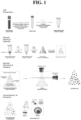
- a liquid sample comprises biological materials and undergoes multiple processing steps, which may include, but are not limited to, those illustrated in the stages depicted in FIG. 1 , which are described in detail below.
- the methods comprise preparing or procuring a liquid sample with biological materials, which can be dispersed and subsequently captured in solid form, thereby allowing high resolution detection.
- liquid samples can be derived from multiple sources, for example, bone marrow, cerebrospinal fluid, urine, saliva, sputum, tears, seminal fluid, or other fluid sources. It is further noted that while exemplary disclosure refers to CTC cells, it can be equally applied to other biomarkers that can be present in liquid biopsy samples.
- materials in the liquid sample are obtained from a processed blood sample, such as that obtained in a liquid biopsy.
- Processing can involve one or more steps.
- processing can include red blood cells removal and isolation (collection) of PBMC cells.
- the blood sample may be subjected to centrifugation to remove erythrocytes (red blood cells) and platelets.
- processing may also involve fractionating the blood sample into different components by well-known separation techniques, such as density gradient centrifugation.
- separation techniques can include red blood cell (RBC) removal and peripheral blood mononuclear (PBMC) collection or isolation.
- RBC red blood cell
- PBMC peripheral blood mononuclear
- blood samples can be provided in a tube or vessel that includes anticoagulants, such as an evacuated blood collection tube with EDTA or heparin.
- the blood sample can be treated with cell separation medium, such as Lymphoprep TM or Ficoll-Paque TM , and centrifuged, and the supernatant removed.
- cell separation medium such as Lymphoprep TM or Ficoll-Paque TM
- PBMC isolation tubes can be employed (e.g., SepMate TM Tubes available from Stemcell TM Technologies).
- the blood sample can also be subjected to RBC lysis, if desired, according to known techniques, such as application of commercially available or synthesized ammonium chloride solution (e.g., ammonium chloride solutions available from Stemcell TM Technologies) based on protocols known to those of ordinary skill in the art, which can be performed before or after centrifugation.
- RBC lysis is not employed as trace amounts of RBC do not affect imaging and analysis of the sample.
- a pellet e.g., a pellet containing isolated PBMC
- the volume of the pellet can be at least 5 ⁇ l, or at least 10 ⁇ l, or at least 15 ⁇ l. In other exemplary embodiments, lower volumes of pellet are processed. In any event, the entire pellet is encapsulated in a gel, and has a size orders of magnitude larger than typical working volumes encountered in microfluidic processes known in the art.
- the dispersed materials in the liquid sample can originate from other tissue sources.
- they may be derived from a biopsy obtained from bodily fluids other than blood, such as bone marrow, cerebrospinal fluid, urine, saliva, sputum, tears, seminal fluid, or other fluid sources.
- the dispersed materials in the liquid sample may reflect liquid dispersal of materials from a solid biopsy, such as a tissue sample obtained from a tumor or other solid samples derived from a structure or organ of interest.
- the dispersed materials can originate from non-tissue sources.
- the biological materials in the specimen can be enriched by concentrating a large amount of sample, for example, from collecting and pelleting cells from a larger volume of blood, e.g., 2 ml, 4 ml, 8 ml, or more, or other sample source.
- the biological materials in the specimen can be enriched by concentrating from a relatively large amount of sample, for example, from centrifuging and collecting a cell pellet from a large volume of blood (e.g., 0.5 cc or 1 cc or more) or other tissue source.
- a relatively large amount of sample for example, from centrifuging and collecting a cell pellet from a large volume of blood (e.g., 0.5 cc or 1 cc or more) or other tissue source.
- the materials Prior to dispersal in the liquid sample, the materials may undergo additional processing steps.
- the biological materials may undergo a fixation step prior to their dispersal in a liquid sample, as discussed further herein.
- fixation may occur after the biological materials have been collected in the liquid sample. Fixation may also occur after cells are labeled.
- the particular fixatives that can find use according to the present disclosure are not limited and include those known to those of ordinary skill in the art.
- the fixative is a solution that includes one or more of a glutaraldehyde, a formaldehyde, an epoxy, or a cross-linked product of one or more of the foregoing.
- liquid biopsy sample can be gathered serially and collected into a single tube.
- 2 or more ml of blood can be collected and processed and the cell pellets pooled and resuspended in a liquid sample buffer, e.g., PBS.
- a liquid sample buffer e.g., PBS.
- the biological materials include circulating tumor cells (CTCs), circulating tumor cell fragments, circulating tumor cell mimics, circulating epithelial cells (CECs), and similar rare circulating cells.
- CTCs circulating tumor cells
- CECs circulating epithelial cells
- the instant methods further comprise labeling one or more targets of interest and adding a solidifying agent that will allow the dispersed labeled materials to be captured in a solid form that includes a three-dimensional cross-linked network.
- the solidifying agent provides a solid matrix to support 3D visualization of sample components.
- Labeling can involve any method known in the art for identifying a biomolecules, including immunological and molecular means.
- protein targets of interest - whether on a cell surface, intracellular, or extracellular - can be labeled with antibodies (or related immunological regents) that are detected directly, e.g., with a fluorescent conjugated antibody, or indirectly, e.g., with immunohistochemistry or primary antibodies and conjugated secondary antibodies.
- Labeling e.g., chemical and immunolabelling
- the labelling is a multi-step process.
- multiple antibodies from different host species can be introduced at or about the same time, or at different times.
- primary antibodies can be conjugated (or pre-labeled) with a tag, such as a fluorescent dye or enzyme, e.g., a fluorophore or corresponding secondary antibodies can be introduced in the mixture in one step.
- a tag such as a fluorescent dye or enzyme, e.g., a fluorophore or corresponding secondary antibodies.
- One-step labelling is preferred over multi-step labelling since labeling steps generally require washing afterwards, and thus additional centrifugation and supernatant removal steps could result in cell loss or cell damage and potentially decrease signal sensitivity.
- biomolecule targets such as DNA or RNA
- nucleic acid probes that are detected directly or indirectly, such as for fluorescence in situ hybridization (FISH).
- FISH fluorescence in situ hybridization
- the signal can be further amplified by available technologies, such as systems based on biotin-streptavidin binding and the polymerase chain reaction.
- the probes can identify other disease biomarkers, such as cell-free tumor or fetal-derived DNA, or can visualize genetic and structural changes in cell nuclei, such as DNA and chromosomal abnormalities, amplifications, deletions, and translocations.
- Samples can also be labelled by other methods knowns in the art, for example with various dyes directed to cellular components, including fluorescent dyes such as DAPI and PI (that bind to nuclear components) and DiD or DiL (that bind to membrane components).
- fluorescent dyes such as DAPI and PI (that bind to nuclear components) and DiD or DiL (that bind to membrane components).
- Labeling may also involve other steps, such as cell permeabilization or fixation, as appropriate and known in the art, to allow efficient and specific binding of the immunological or molecular reagents to the target (biomolecule) of interest.
- the labeling is applied prior to sample gelling and clearing.
- the labelling is preferably applied before obtaining a pellet, or otherwise separating components from the sample, e.g., while the biological materials are dispersed in a liquid (e.g., blood) sample.
- Labeling can also be performed after cells are fixed in the gel state.
- cells collected from the liquid sample e.g., PBMCs obtained from blood
- a gel can be formed directly, before labelling.
- the gel sample can then be labeled with probes either passively or by other active immunolabeling approaches, such as those methods involving electrophoresis or pressure-based approaches.
- probes either passively or by other active immunolabeling approaches, such as those methods involving electrophoresis or pressure-based approaches.
- a processed gel sample after being solidified can be immunolabeled (i.e., labelled for the first time or to apply further labelling).
- this approach of labelling after gelation may take a longer processing time, it may also enhance preservation of the target cell number.
- a fixation procedure can be performed on the solidified sample after gel formation and subsequent to labelling.
- delipidation can be performed on a solidified (e.g., gelled) cell sample, where there is a need or desire to enhance sample transparency for imaging.
- solidified samples can be further crosslinked with a hydrogel precursor or epoxy and be delipidated following the CLARITY approach. See, e.g., " Advances in CLARITY-based tissue clearing and imaging," Exp Ther. Med. 2008; 16(3): 1567-1576 . It should be noted, however, that due to the small stack of cells in the sample that is dispersed in the solidified sample, delipidation steps are generally not required according to most embodiments.
- solidifying a sample includes introducing a solidifying agent (e.g., a gelling agent) that will allow the biological materials to be captured in a solid form, such as a gel form, that is compatible with subsequent imaging and detection of labeled biomolecules of interest.
- Solidifying agents can include, but are not limited to polyacrylamide precursors. In embodiments, such agents are based on polysaccharides or proteins. See, e.g., Kar et al. 2019, Current developments in excipient science: in Fundamentals of Drug Delivery, 29-83 .
- the solidifying agent can consist, or consist essentially of, a refractive index-matching solution itself, modified as necessary to provide the proper viscosity, to provide a rigid physical gel.
- the solidifying agent comprises a mixture of chemical monomers and cross-linkers to create a synthetic chemical gel, i.e., having a chemically cross-linked polymer network.
- Solidifying can comprise, in exemplary embodiments, dispersing or resuspending the biological materials in a gelling material that is in a liquid form.
- the sample remains in a fluid or molten state and the materials can be maintained in a dispersed state prior to transfer to an imaging holders in Stage 3.
- the solidifying agent can be introduced to a labeled specimen (e.g., a labeled PBMC pellet as shown in FIG. 1 ) and the mixture is mixed, such as with a pipet or via a vortex mixer.
- the gelling agent in certain embodiments is a reversible gelling agent, in that the gelling agent (and sample) can be heated to achieve the fluid or molten state, if desired.
- dispersing or re-suspending the biological sample in a mixture and forming a gel generally allows the components in the sample to be stabilized in a spatial distribution with sufficient properties, such as transparency, to allow subsequent imaging.
- refractive index matching materials having a refractive index e.g., having a refractive index between 1.3-1.6 or 1.33-1.5, or a RI approximately equivalent to water
- RI matching materials having other refractive indices can find use in other embodiments.
- refractive index matching materials are capable of penetrating into the tissue/cell to achieve tissue/cell transparency and include, but are not limited to CUBIC-R+, RapidClear, RIMS, or ScaleView. See Neuropathol Appl Neurobiol, 2016 Oct;42(6):573-87 .
- a refractive index matching material is not required.
- laser light can still penetrate through the solidified sample and excite labeled cells or other target components. That is, refractive index differences in the solidified sample are not noticeable when a laser does not have to travel too deep to cause refraction of light. With, for example, a two-photon laser, which has more power, the laser can penetrate even deeper without being bent.
- refractive index matching material such as a refractive index matching solution
- pre-gelling includes dispersing or resuspending the biological materials in a gelling material comprising one or more hydrogel precursors, such as polymerizable materials, monomers or oligomers, including monomers selected from the group consisting of water-soluble groups containing a polymerizable ethylenically unsaturated group.
- hydrogel precursors such as polymerizable materials, monomers or oligomers, including monomers selected from the group consisting of water-soluble groups containing a polymerizable ethylenically unsaturated group.
- Monomers or oligomers can comprise one or more substituted or unsubstituted methacrylates, acrylates, acrylamides, methacrylamides, vinylalcohols, vinylamines, allylamines, allylalcohols.
- Precursors can also include polymerization initiators, cross-linkers, and other components, as are known in the art, described, for example, in WO2019023214 and WO/2020
- the methods comprise transferring the pre-gelling mixture, while in a liquid or molten state, to sample wells in a holder.
- the wells in the holder allow formation of a solid sample that is suitable for imaging.
- the solid samples can be in the shape of a block or other form that is customized for imaging by fluorescent microscopy.
- the pre-gelling step can be carried out in a combination tube-sample holder, therefore allowing solidification (e.g., gelling) to occur in the same tube, eliminating the need for subsequent transfer of the liquid solution to a separate sample holder.
- the sample can be processed by additional steps prior to imaging.
- the sample can be equilibrated in a refractive index material so that its appropriately matched (e.g., cleared) for imaging in step 3 (RI-matching).
- the RI-matching material is added at the same time as the solidifying agent.
- RI-matching material is added with the solidifying agent, and then a second round of RI-matching material is added to the solidified sample to provide a final, desired transparency of the sample.
- the RI-matching material has a RI of from about 1.39 to about 1.65, or from about 1.49 to about 1.55 (e.g., about 1.52)
- the RI-matching material can be those obtained from Table 3, Neuropathology and Applied Neurobiology, November 2015, "Bringing CLARITY to the human brain: Visualization of Lewy pathology in three dimensions," Liu et al., available at ⁇ https://www.researchgate.net/figure/Comparison-of-different-refractive-index-matching-solutions-Abbreviations-BABB_tbl3_283493188> .
- This step entails, in exemplary embodiments, mounting the sample on an image holder and imaging the solid sample by an appropriate imaging means, such as fluorescent microscopy.
- the methods can include, prior to imaging, mounting the sample on an image holder, such as a 3D printed sample holder as shown in FIG. 1 , and imaging the sample, for example, in a water chamber or RI-matching solution.
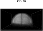
- the sample holder can be customized in accordance with the particular sample preparation. In embodiments, it may comprise a base and two side-walls. In embodiments, such as that use for a sample prepared in a viscous medium, the holder may have four wall that surround and preserve the spatial configuration of the dispersed components in the sample.
- imaging can be carried out in a sample cuvette with a standard epi-fluorescence microscope.
- the 3D gel sample can be mounted on the sample holder with the assistance of a mounting gel (e.g., agarose, poly-L-lysine, superglue).
- a mounting gel e.g., agarose, poly-L-lysine, superglue. See, e.g., Asano et al., Expansion Microscopy: Protocols for Imaging Proteins and RNA in Cells and Tissue, Current Protocols in cell biology (2016), particularly pages 34-36 - "Sample mounting”.
- Fluorescence microscopy approaches include, but are not limited to, conventional confocal microscopy, resonance scanning confocal microscopy, spinning-disk microscopy, and lightsheet microscopy as depicted in FIG. 1 .
- a gel sample is rapidly imaged by lightsheet microscopy, such as Light-Sheet Fluorescence Microscopy (LSFM) using a detection lens and an illumination lens, as shown in FIG. 1 .
- LSFM Light-Sheet Fluorescence Microscopy
- Gaussian beams can be used, or alternatively specialized Beam profiles such as non-diffracting Bessel beam can find use, as depicted in FIG. 1 .
- Such imaging allows materials in the 3D sample block to be scanned and evaluated for the discrete presence of a labeled biomarker.
- a processor can be in communication with the microscope to receive output therefrom and combine the sequentially imaged adjacent object areas (i.e., "stitching"). See, e.g., the techniques and apparatus disclosed in U.S. Patent Nos. 10,746,981 , 10,876,870 and U.S. Published Patent Application No 2016/0041099 and International Published Patent Application No. WO 2017/031249 , which may find use in accordance with the presently disclosed subject matter.
- light microscopy can be used to examine visible dye-stained cells, such as those stained with H&E (Hematoxylin and eosin) or by immunohistochemistry (IHC).
- IHC immunohistochemistry
- X-ray microscopy can be used to detect liquid components that have been labeled with appropriate metal tags.
- Imaging techniques can find use, including images acquired by high resolution cameras that are included in commercially-available smartphones (e.g., iPhone ® or Android ® -based smartphones) or related light-detection systems.
- the instant methods can also be used to evaluate, diagnose, or monitor a disease.
- a liquid biopsy e.g., whole blood, processed as described above
- a rare cell therein e.g., whole blood, processed as described above
- the instantly disclosed methods can be detected, for example, colorectal adenocarcinoma cells, leukemia cells, the type of cancer, the extent to which cancer has developed, whether the cancer will be responsive to therapeutic intervention, etc.
- the disclosed methods can be used to detect cellular biomarkers associated with other disease and disorders, such as inflammatory, metabolic, gastrointestinal, endocrine, immunological, musculoskeletal, cardiovascular, cardiopulmonary, genitourinary, hepatological, respiratory, viral, and neurological diseases and disorders, in accordance with embodiments of the disclosure.
- imaging can be used to analyze cell morphology and structure, including analysis of the change in cell morphology and structure as compared to their native or healthy state.
- the imaging methods can be used to analyze the outward appearance of cells, such as their size, shape, or other external characteristics.
- the imaging methods can be used to analyze the form and structure of inner cell components, such as the nucleus, endoplasm reticulum, golgi apparatus, mitochondria, or other organelles.
- a biopsy may be prepared by liquid dispersal of a sample of a diseased tissue, such from kidney, heart, stomach, liver, pancreas, intestines, brain, etc., to determine the condition of the tissue, the extent to which the disease has developed, the likelihood that tissue will be successful, etc.
- a diseased tissue such from kidney, heart, stomach, liver, pancreas, intestines, brain, etc.
- the methods herein, through the use of dyes targeting membrane or cellular components for example, can be used here to assess morphological changes in cell population that may be indicative of a disease state.
- a liquid biological sample can be used to screen candidate therapeutic agents for their effect on a tissue or disease.
- a liquid sample obtained from subject such a mouse, rat, dog, primate, human, etc.
- a candidate agent can be prepared by the methods disclosed herein and microscopically analyzed for one or more cellular or tissue parameters, i.e., attributes or characteristics of subcellular components that can be measured.
- the methods can also be used to visualize the distribution of genetically encoded markers in a liquid sample prepared by dispersing materials from a tissue.
- markers may include, for example, chromosomal abnormalities (inversions, duplications, translocations), loss of genetic heterozygosity, the presence of genetic markers indicating a predisposition towards a disease state or healthy state.
- detection may be useful, for example, in diagnosing and monitoring disease, such as in personalized medicine, studying paternity, or other applications.
- the methods disclosed herein are used to detect CTCs by immunological or molecular means for diagnostic purposes and to address their clinical significance.
- CTCs are rare cells that circulate in the blood or other fluids along with millions of other circulating cells.
- the present methods capture such rare cells in a solidified 3D form of a complex liquid sample or liquid biopsy.
- any CTCs can be detected as discrete signals in the sample block.
- the methods allow such CTC detection without reducing their biological heterogeneity. They also can include fewer interventions than those required for processing by microfluidics or traditional cytology that may disrupt morphology or impede sensitivity.
- CTCs are rare cells that circulate in the blood or other fluids along with millions of other circulating cells that belong, for example, in the hematopoietic compartment, and do not adhere spontaneously. These poor adhesive properties hinder existing methods in the art for detecting CTCs, such as the use of a solid support to isolate and immobilize CTCs.
- the presently disclosed methods allow such rare cells, if present, to be detecting in a complex 3D representation of a liquid sample, without limiting their biological heterogeneity and reducing interventions that could disrupt morphology.
- methods based on modified supports and matrices coated with anti-adhesion molecules (or other binding proteins) may elicit biological responses that alter CTC morphology, leading to inaccurate analyses.
- Immuno-immobilization relies upon strong affinity between coated antibodies and cell membrane proteins. Low expression of target surface protein or low antibody affinity can lead to low capturing rate of target cells. Similarly, cytocentrifugation of cells to a support such as a microscope slide, followed by fixation and subsequent labeling, can rupture cells or disrupt their morphology, hindering diagnostic assessment. In contrast, a microscope slide approach requires cells to be coated in a single layer manner to allow imaging, which greatly reduce the throughput, or number of cells, being imaged and analyzed.
- Detection methods relying on microfluidics, nanostructures, and channels can suffer from similar limitations. See, e.g., WO2012016136 ; WO2013049636 Such methods can induce flow stress on cellular components in the liquid sample, compromising their morphology and other characteristics. More generally, such methods typically select a homogeneous population of cells based on size or expression of superficial membrane proteins, limiting the biological heterogeneity that is otherwise amenable to diagnostics analysis of a clinical or biological sample.
- CTCs in peripheral blood can be viewed as an extension of a tumor.
- a tumor is typically heterogeneous, meaning that through mutations, a tumor can develop several different cell types within. Each cell type can have its own characteristics, which can range from mild to aggressive.
- CTCs are rare cancer cells released from tumors into the bloodstream that are thought to have a key role in cancer metastasis. See, e.g., Harouaka et al., 2014, Pharmacol. Ther. 141, 209-221 .
- the present disclosure provides, in certain embodiments, a method of analyzing individual cells (e.g., from a liquid biological sample) that have been dispersed and captured in a three-dimension solidified sample. No enrichment or selection of the cells is required, and all cells can be labeled and visually screened by imaging the sample.
- each individual cell or cell cluster can be visualized and analyzed separate from each other in a three-dimensional array, which in turn can provide comprehensive cell information such as cell size, morphology, biomarker distribution, nuclear-cytoplasm ratio, and more.
- CTCs multiple types can be identified according to the subject disclosure, such as lung, liver, and colon cancers, as well as other cancers that can be detected as CTCs in a liquid biopsy.
- a liquid biopsy e.g., from a blood sample
- EpCAM is an epithelial marker indicative of invasive cancer cells that went through epithelial mesenchymal transition (EMT), which is a main cause for cancer invasion. Cancer invasion usually begins with EMT from a small population of tumor cells that will stimulate blood vessel growth, providing passage for the cells to invade the bloodstream as CTCs.
- CTCs with multiple cancer markers together, such as, but not limited to, HER2 (breast), CDX2 (colon), CK20 (colorectal, transitional cell carcinomas and Merkel cell carcinoma), CK19 (breast), PD/PDL-1 (several cancers, including NSCLC, melanoma and renal cell) and EGFR (lung) according to the subject disclosure.
- Identification of the different CTC cell subtypes within the blood sample can provide information about the origin of the cancer. In turn, this information can guide more-detailed follow-up studies, such as high-resolution analysis by MRI to identify the location of lesions and tissue biopsy for pathological examination.
- Cell heterogeneity in blood can also be applied to healthy cells such as leukocytes.
- CTCs have escaped from a primary tumor into the bloodstream, they are generally highly invasive. Such invasive cells can make cancer difficult to treat and cure due to their ability to metastasize, as well as their greater likelihood of mutations, which may increase the chance of resistance to chemotherapy and other therapeutic interventions. This underscore the importance and value in identifying the molecular characteristics of such invasive cells to determine the proper treatment plan. For example, if the CTC present in blood shows high levels of PDL-1, immunotherapy will likely be a more effective approach. PDL-1 levels can be determined, for example, with a PDL-1 antibody probe to quantify the number of PDL-1+ CTCs over all CTCs in a sample.
- CTC number can indicate the treatment effectiveness.
- CTC number in 1cc of blood may range from to tens of thousands for advanced cases to several hundred for less advanced cases. After 3 months of chemotherapy or other therapeutic intervention in some advanced stage III cases, the CTC number can drop to 5 thousand or even close to zero after 12 months. However, in some cases, the CTC number, might only decrease to about 2 thousand after 6 months of chemo and climb back to tens of thousands 6 months later. This indicates drug resistance of cancer cells to the therapeutic intervention. Chemotherapy, for example, may have killed all the drug sensitive CTCs, but other subtypes that were resistant will not be affected.
- the investigations here address the above-described limitations by describing formulations and methods to visualize a biopsy sample in three dimensions by capturing the dispersed components in the sample in a solidified state.
- This approach has numerous advantages, such as increasing the sensitivity of the analysis by separating the individual components, increasing the speed of the analysis by allowing the use of rapid imaging techniques, such as light sheet fluorescent microscopy, and enhancing the specificity of the analysis by reducing the number of processing steps that might otherwise disrupt the morphology and integrity of sample components, such as cellular markers.
- a blood sample (e.g., 2 cc, 8 cc or 10 cc) is collected from a subject and the red blood cells removed by density gradient centrifugation using standard methods, such as, for example, centrifugation at 1000 rpm for five minutes at room temperature.
- standard methods such as, for example, centrifugation at 1000 rpm for five minutes at room temperature.
- the supernatant is removed, transferred to a separate tube, and is recentrifuged to collect the remaining cells, including any CTCs, and other biological components.
- the pellet is optionally re-suspended in a fixative solution, such as a 4% paraformaldehyde solution, shaken for several minutes, centrifuged, washed in phosphate buffered saline (PBS), and centrifuged again.
- PBS phosphate buffered saline
- the pellet is re-suspended in a blocking buffer and shaken for a couple of minutes, and centrifuged again.
- the fixed and washed pellet is resuspended in blocking & permeabilizing buffer, placed on a shaker for several minutes, centrifuged, and resuspended in premixed labeling solution for 30 to 60 minutes.
- the pellet can be resuspended and incubated in labeling mixture for longer times as needed (e.g., up to 10 or 20 hours).
- Protein biomarkers of interest can be labeled with antibodies (or related fragments or derivatives) that are detected directly, e.g., with a fluorescent conjugated antibody, or indirectly, e.g., with immunohistochemistry or primary antibodies and conjugated secondary antibodies.
- nucleic acids of interest can be labeled with molecular probes that are detected directly or indirectly.
- the signal can be further amplified by available technologies, such as systems based on biotin-streptavidin binding and the polymerase chain reaction.
- the sample can be centrifuged, and the pellet washed one or more times.
- a labeling solution of 200 ⁇ L of a PBST blocking buffer is mixed with propidium iodide (PI) (1:2000) for nuclear staining, EpCAM antibody (1:250 for antibody concentration greater than 0.1mg/ml) for cancer cell detection, CD45 antibody (1:250 antibody concentration) for immune cell detection, and secondary antibodies matching primary antibody hosts (2 times weight to primary), using Fab from Jackson ImmunoResearch Laboratories, Inc. (https://www.jacksonimmuno.com/catalog/31).
- PI propidium iodide
- sequential labeling can be performed, starting with primary antibody incubation for 60 minutes or more and a wash, then secondary antibody and wash, and then staining with PI in PBS solution for 5 minutes, followed by a wash.
- PI in PBS solution for 5 minutes
- Sequential labeling with a secondary antibody or direct conjugate of a fluorescence molecule can also be employed.
- a biotinylated antibody can also be used with a boosted signal based on binding affinity to, for example, streptavidin, as known in the art.
- the pellet can optionally be re-suspended in 4% PFA for 10 minutes at room temperature to fix all the labelling materials on the cells so they do not get washed away during the solidification step.
- the sample is optionally washed one or more times, and then resuspended in a solution containing a hydrogel precursor that can be polymerized upon addition of sufficient amounts of a crosslinker and an ion-containing component (e.g., a component containing Ca 2+ ion) to induce gelation.
- a crosslinker and an ion-containing component e.g., a component containing Ca 2+ ion
- the amount of cross-linker that is introduced can be adjusted depending on whether a gel-type solid is desired, or increased to provide a higher density solidified sample.
- the gel solution containing the dispersed materials is transferred to a desired imaging holder at 25-37°C , such as a holder comprising sample wells having a block shape (or other desired shape), and the holder may be shaken or vibrated to ensure dispersal of materials in each well.
- the holder containing the sample solutions is transferred to a lower temperature that allows gelling in the wells to occur, for example by incubating a sample dispersed in LMP agarose at 4°C for about 15-30 minutes.
- the original liquid blood sample will have millions of non-cancerous cells that will obscure CTCs, which are rare and present at extremely low levels. Gelling allows one to disperse and separate the cells in 3D space for imaging (described below).
- One crucial point is that millions of cells are condensed from high volume (e.g., 10 cc of blood) to 20 ⁇ l of gel block (2.71 mm 3 ), which then is allowed to be imaged in an acceptable imaging time.
- the volume can be decreased even more to allow quick high-resolution imaging (e.g., up to 20x or 40x).
- the cell pellet is reduced to give the smallest volume possible.
- the close distance between cells may require attention to gel clearing due to the high density of lipid-bilayers (cell membrane), but can be resolved, for example, with optimized RI-matching solution or a higher power laser such as a two-photon microscope.
- the gel block (with dispersed contents) has formed, it is cleared by immersion in refractive index (RI) matching solution for about 30 minutes to make it transparent for imaging.
- RI refractive index
- the RI-matched gel sample can then be imaged to detect the presence of on more biomarkers, which are not discretely segregated in the gel block.
- Such imaging can occur by microscopy, including by efficient fluorescent imaging methods, such as by light sheet microscopy.
- the sample can be rapidly imaged by Light-Sheet Fluorescence Microscopy (LSFM) using a non-diffracting Bessel beam.
- LSFM Light-Sheet Fluorescence Microscopy
- FOV field-of-view
- each cell with an average cell diameter of 15 micron would get imaged about 10 to 15 times from top to bottom.
- FOV field-of-view
- the acquired LFSM data from the imaged sample reveals individual cells that can be visually inspected for antibody labeling validity, intensity, distribution, etc.,.
- the methods described herein allow PBMCs to be immediately fixed after RBC removal, allowing capture of their distribution and association in their original state.
- this allows direct visualization of sample characteristics such as cell-cell interactions, cell clusters (e.g., circulating tumor microemboli) and other properties that may indicate disease state or provide other prognostic or diagnostic applications.
- Anticoagulated blood collected from a human in a EDTA coated tube was transferred to a 15 ml tube, cell separation medium (e.g., Ficoll ® ) was added, and the mixture was subjected to density gradient centrifugation (1200 RPM) for five minutes. After centrifugation, the buffy coat (fraction containing white blood cells and platelets) was transferred to a 2 ml test tube without collecting any separation medium. The resulting mixture was again centrifuged at 500g for 5 minutes to allow the cell pellet to form to the bottom of the tube. The supernatant was carefully removed without disturbing the cell pellet.
- cell separation medium e.g., Ficoll ®
- the pellet was then re-suspended in 200 ⁇ l of labelling solution (1: 1000 PI in 4% PFA in PBS) to label the cell nucleus.
- labelling solution was added at 4°C for 20 minutes.
- the mixture was then centrifuged at 500g for 5 min, the supernatant carefully collected and discarded.
- 1 ml of 4% PFA in PBS was added to fix the chemical dye at 4°C for 20 minutes, and the mixture was centrifuged again at 500 g for 5 min, and the supernatant again discarded.
- a solidifying solution mixture that includes about 0.5 wt% LM agarose was prepared.
- the solidifying solution was heated to above its gelling point in a microwave to melting and then allowed to cool at room temperature, where it is be provided in a liquid state until it again reaches its gelling point.
- 20 ⁇ l of cooled solidifying solution was slowly added to the pellet to re-suspend the pellet, the solidifying solution being added slowly to avoid bubble formation.
- the mixture is brought to 4°C and maintained for a period of time to allow gel formation (less than about 30 minutes).
- the gel was carefully removed from the tube with a pipet by poking the side of the gel, to provide a 20 ⁇ l volume gel.
- the gel was then mounted onto a sample holder at room temperature, in this case a 3D gel rod 2 mm in diameter (same composition as the solidifying solution pre-made into a holding shape mounted to a 3D-printed plastic surface), and cooled at 4°C for 10 minutes to allow it to gel.
- the sample gel on the holder was immersed in RI-matching solution for 5-30 min to prepare it for imaging.
- the sample was transferred to a lightsheet imaging chamber and imaged (10X objective).
- 10X objective For a 20 ⁇ l gel sample, it took roughly 3 min to image single channel at 10X resolution with z-step of 4 ⁇ m, in which imaging time can be reduced by decreasing gel volume (increasing cell density), reducing imaging resolution (lower magnification objective or higher z-step), or by optimizing microscope settings such as z-steps, and other means known those of ordinary skill in the art.
- FIGS. 2A-2C depict images obtained from the lightsheet imaging chamber.
- FIG. 2A the image represents the entire 20 ⁇ l gel sample from the top-down view.
- FIG. 2A is a 100 ⁇ m stacked and reduced image of 25 x 4 ⁇ m images.
- FIG. 2B the image represents the entire 20 ⁇ l gel sample from the side view.
- FIG. 2C the image is magnified to capture the individual leukocytes in closer detail, in which the small horizontal line in the lower-left hand corner equals 20 ⁇ m in scale.
- CACO2 cells were cultured and collected through trypsinization.
- HL60 is a floating cell line that does not require trypsinization.
- CACO2 and HL60 cells were each collected, washed with PBS, and centrifuged to collect the cell pellet.
- the CACO2 cells were stained with Trypan Blue and counted using a cell counter. An appropriate number of cells was resuspended in 100 ⁇ l PBS. EpCAM(Dako) antibody was added to the cell solution at 1: 100 ratio and secondary antibody was added at 1:2 primary to secondary antibody molar ratio.
- the mixture was shaken at room temperature for 30 minutes. The mixture was centrifuged at 500g for 5 min, and the supernatant carefully removed. The cell pellet was washed by resuspension in PBS, and centrifuged again at 500 g for 5 min, and the supernatant was discarded. The cells were then resuspended and incubated at 4°C for 20 minutes in 1 ml of PI solution in 4% PFA (1:1000) to fix the bound antibody and label the cell nucleus, the mixture was centrifuged again at 500 g for 5 min, and the supernatant was discarded.
- the HL60 cells were stained with Trypan Blue and counted using a cell counter. An appropriate number of cells was collected in 100 ⁇ l PBS. CD45 (Dako) antibody was added to the cell solution at 1:100 ratio and secondary antibody was added at 1:2 primary to secondary antibody molar ratio.
- the mixture was shaken at room temperature for 30 minutes.
- the mixture was centrifuged at 500g for 5 min, the supernatant carefully removed, and 1 ml of PBS was added to wash, and the mixture was centrifuged again at 500 g for 5 min, and the supernatant again carefully collected and discarded.
- the cells were then resuspended and incubated at 4°C for 20 minutes in 1 ml of PI solution in 4% PFA(1:1000) to fix the bound antibody and label the cell nucleus at 4°C for 20 minutes, and the mixture was centrifuged again at 500g for 5 min, and the supernatant again carefully collected and discarded.
- a solidifying solution that contains about 1 wt% LM agarose was prepared.
- the solidifying solution was heated in a microwave to melting and then allowed to cool to room temperature, where it will eventually be provided in a gel state.
- 20 ⁇ l of cooled solidifying solution was slowly added to the pellet to re-suspend the pellet, the solidifying solution being added slowly to avoid bubble formation.
- the mixture is brought to 4°C and maintained for a period of time to allow gel formation (less than about 20 minutes).
- FIG. 3A depict CD45/PI-stained HL60 cells, as captured by light sheet microscopy.
- FIG. 3A is digitally enlarged as indicated by the scale bar in the lower left-hand corner, in which the white bar corresponds to 50 ⁇ m. The yellow is indicative of PI staining labelling the nucleus and the green is indicative of CD45 immunolabelling.
- FIGS. 3B-3C depict EpCAM/PI-stained CACO2 cells, as captured by light sheet microscopy. Images were taken with a 10x objective.
- FIG. 3B is the presentation of the entire gel volume.
- FIG. 3C is digitally enlarged as indicated by the scale bar. That is, in FIG. 3B the scale bar represents 300 ⁇ m and in FIG. 3C the scale bar represents 50 ⁇ m.
- the study is designed to assess the rare cell detection efficiency of 3D liquid biopsy method and the cell loss percentage from the cell preparation process.
- a sample of CACO2 cells was trypsinized, counted, and seeded at appropriate volume in freshly collected peripheral blood sample to make a cancer cell spiked blood sample.
- Peripheral blood mononuclear cells PBMC
- Anticoagulated blood collected from a human in a EDTA coated tube was transferred to a 15 ml tube.
- 8 c.c. of ammonia chloride solution was added to lyse the red blood cells. The lysing process was performed on ice for 15 minutes, centrifuged, supernatant removed, and the collected PBMC cell pellet including spiked cancer cells was resuspended in 100 ⁇ L PBS awaiting further process.
- EpCAM(Dako) and CD45(Dako) antibodies was added to the cell solution at 1:100 and matching secondary antibody was added at 1:2 primary to secondary antibodies molar ratio to achieve a one-step labeling process.
- Cell sample was incubated at room temperature for 1 hour to allow antibody binding. Next, the sample was centrifuged at 500g for 5 minutes to collect the pellet. The pellet was washed with 1ml PBS and centrifuged again and supernatant was removed. Next, 4% PFA with 1:1000 PI was added to the pellet to fixed the labeled antibody and stain the nucleus for 30 minutes. Next, the cells were centrifuged again to collect the pellet.
- FIG. 4A depicts the entire 3D gel data of multiplex labeling of CACO2 in PBMC, in which the EpCAM marker is shown in magenta and the PI is shown in blue.
- FIG. 4B depicts a zoomed-in, stacked image of CACO2-EpCAM (magenta), leukocyte-CD45 (green) and PI (blue).
- FIGS. 4C and 4D depict images of identified CTC with and without EpCAM (magenta signal) for confirmation.
- EpCAM labeling for cancer cell detection is a viable approach.
- CACO2 shows high EpCAM expression with no CD45 expression and PBMC(leukocytes) show variable CD45 expression but no EpCAM expression.
- a 3-dimension gel was formed and imaged via light sheet microscopy as described in Example 1, 2, and 3. Each image was denoised and featured-enhanced for all immunolabeled signals for cell detection.
- Other parameters such as cell or nucleus circularity, cell/nucleus volume, and nucleus-cytoplasm ratio (N:C ratio) can be quantified for positive cancer cells with use of MATLAB-based or Python-based cell detection algorithm.
- FIG. 4A depicts output from the 3d images of the three repeat sets of gel sample with 2000 spiked CACO2 cells.
- EpCAM(color) labelled CACO2 cells can be easily spotted visually by digitally zooming in onto the dataset.
- EpCAM positive cells are screened for negative CD45 signal and positive PI signal.
- the cell detection algorithm screens all cells with those parameters and output identified CTC images with separated and combined signals as shown in FIG. 4B . These images will be stored in a single file per sample, and all the detected cell images will be further confirmed by human eye or a signal detection algorithm.
- Total cell number in whole gel can be quantified with total PI positive signal. Dividing total positive cancer cell number with total PI signal number will output a ratio. Multiply the ratio with one million gives number of cancer cells in one million of PBMC.
- An IRB study was performed to detect CTC presence in colon cancer patients' blood.
- the study is designed to assess the rare cell detection efficiency of 3D liquid biopsy method.
- Enrolled patients included healthy and disease subjects.
- the patients with stage II to stage IV colon cancer that has metastasis to liver or lung are shown.
- PBMC Peripheral blood mononuclear cells
- EpCAM(Dako) and CD45(Dako) antibodies were added to the cell solution at 1:100 and matching secondary antibody was added at 1:2 primary to secondary antibodies molar ratio to achieve a one-step labeling process.
- Cell sample was incubated at room temperature for 1 hour to allow antibody binding. Next, the sample was centrifuged at 500g for 5 minutes to collect the pellet. The pellet was washed with 1ml PBS and centrifuged again, and supernatant was removed. Next, 4% PFA with 1: 1000 PI was added to the pellet to fix the labeled antibody and stain the nucleus for 30 minutes. Next, the cells were centrifuged again to collect the pellet.
- FIG. 5A depicts a 3D gel data (single stack block) with resolution of 0.42 ⁇ m x 0.42 ⁇ m x 1 ⁇ m, in which the EpCAM marker is shown in magenta, CD45 marker is shown in green, and the PI is shown in blue.
- FIG. 5B depicts zoomed-in image of EpCAM-positive, CD45-negative, and PI-positive cell targets.
- FIG. 6A depict 2 continuous stacks of 3D gel data (600 ⁇ m stack from 1200 ⁇ m stack) with resolution of 0.42 ⁇ m x 0.42 ⁇ m x 1 ⁇ m, in which the CK20 marker is shown in yellow, EpCAM marker is shown in magenta, and the PI is shown in blue.
- FIG. 6B depicts zoomed-in single cell image of CK20-positive, EpCAM-positive, and PI-positive cell targets.
- methods here do not require clearing steps that remove lipids or disrupt other cellular information to achieve tissue transparency for 3D imaging. Instead, methods here combine gelling the dispersed cell sample in three-dimensional space and refractive index matching to achieve complete cell sorting and 3D visualization of complete cellular information within the liquid sample.
Landscapes
- Health & Medical Sciences (AREA)
- Biochemistry (AREA)
- Physics & Mathematics (AREA)
- Life Sciences & Earth Sciences (AREA)
- Chemical & Material Sciences (AREA)
- Analytical Chemistry (AREA)
- General Health & Medical Sciences (AREA)
- General Physics & Mathematics (AREA)
- Immunology (AREA)
- Pathology (AREA)
- Nuclear Medicine, Radiotherapy & Molecular Imaging (AREA)
- Measuring Or Testing Involving Enzymes Or Micro-Organisms (AREA)
- Investigating Or Analysing Biological Materials (AREA)
Claims (16)
- Matrix-unterstütztes Verfahren zum Analysieren einer flüssigen Probe, die biologische Materialien enthält, hinsichtlich einer oder mehrerer Zielkomponenten, welches umfasst:(a) Zugabe eines Verfestigungsmittels zu einer aus der flüssigen Probe, die die biologischen Materialien enthält, erhaltenen Prüfprobe;(b) Erzeugen einer verfestigten Probe, die dispergierte biologische Materialien umfasst, um die dispergierten biologischen Materialien in einem dreidimensionalen Zustand einzufangen und zu immobilisieren;(c) Einbringen eines Brechungsindexanpassungs-Materials in die verfestigte Probe; und(d) Bildgebung der verfestigten Probe zum Identifizieren der einen oder der mehreren Zielkomponenten in den dispergierten biologischen Materialien;wobei das Verfestigungsmittel ein Gemisch aus chemischen Monomeren und Vernetzern umfasst, um ein synthetisches chemisches Gel mit einem chemisch vernetzten Polymernetzwerk zu erzeugen.
- Verfahren nach Anspruch 1, ferner umfassend (i) Markieren der in Schritt (a) aus der flüssigen Probe erhaltenen Prüfprobe mit einer oder mehreren Sonden für die eine oder die mehreren Zielkomponenten vor Zugabe des Verfestigungsmittels und/oder (ii) Markieren der verfestigten Probe aus Schritt (b) mit einer oder mehreren Sonden für die eine oder die mehreren Zielkomponenten.
- Verfahren nach Anspruch 2, wobei das Markieren das Inkontaktbringen der in Schritt (a) aus der flüssigen Probe erhaltenen Prüfprobe mit einer molekularen Sonde; und/oder das Inkontaktbringen der verfestigten Probe aus Schritt (b) mit einer molekularen Sonde umfasst, wobei die molekulare Sonde ein Antikörper, ein Fluoreszenzfarbstoff oder eine Nukleinsäuresonde ist.
- Verfahren nach einem der Ansprüche 1 bis 3, wobei die eine oder die mehreren Zielkomponenten ein zelluläres Ziel oder ein intrazelluläres Ziel umfassen.
- Verfahren nach Anspruch 1, ferner umfassend das Einführen der optisch geklärten gelierten festen Probe aus Schritt (c) in einen Probenhalter, der in einer Lösung eingetaucht ist, die ein Brechungsindexanpassungs-Material enthält.
- Verfahren nach Anspruch 1, wobei die flüssige Probe eine flüssige Blutprobe ist.
- Verfahren nach Anspruch 6, wobei die Prüfprobe aus der flüssigen Blutprobe durch ein Verfahren erhalten wird, das die Entfernung von roten Blutkörperchen und Blutplättchen aus der flüssigen Blutprobe umfasst, und wobei die Prüfprobe mononukleäre Zellen des peripheren Blutes (PBMCs) enthält, die aus der flüssigen Blutprobe isoliert wurden.
- Verfahren nach Anspruch 7, ferner umfassend:(i) Markieren der Prüfprobe, die die aus der flüssigen Blutprobe erhaltenen isolierten PBMCs enthält, mit einer oder mehreren Sonden für eine oder mehrere seltene zirkulierende Zellen vor Zugabe des Verfestigungsmittels; und/oder Markieren der Matrixgestützten Probe aus Schritt (b) mit einer oder mehreren Sonden für die eine oder die mehreren seltenen zirkulierenden Zellen; und(ii) Bildgebung der verfestigten Probe aus Schritt (c) oder der optisch geklärten verfestigten Probe aus Schritt (d), um das Vorhandensein der einen oder der mehreren Sonden zu bestimmen, wodurch das Vorhandensein seltener zirkulierender Zellen in der flüssigen Blutprobe bestimmt wird.
- Verfahren nach Anspruch 8, wobei das Markieren ferner die Zugabe einer Sonde für weiße Blutkörperchen umfasst.
- Verfahren nach Anspruch 8 oder 9, wobei die eine oder die mehreren Sonden ein krebsspezifisches Antigen oder eine tumorspezifische DNA- oder RNA-Sequenz erkennen.
- Verfahren nach einem der Ansprüche 8 bis 10, wobei die eine oder die mehreren Sonden aus einem Antikörper oder einer Nukleinsäuresonde ausgewählt sind, und insbesondere wobei die eine oder die mehreren Sonden den Nachweis eines oder mehrerer von EpCAM-, HER2-, CDX2-, CK20-, CK19-, PD/PDL-1- und EGFR-Antigen oder der entsprechenden Nukleinsäuresequenz ermöglichen.
- Verfahren nach einem der Ansprüche 8 bis 11, wobei die eine oder die mehreren seltenen zirkulierenden Zellen eine zirkulierende Tumorzelle umfassen.
- Verfahren nach Anspruch 8 oder 9, wobei die eine oder die mehreren seltenen zirkulierenden Zellen eine zirkulierende Epithelzelle oder zirkulierende Endothelzelle umfassen.
- Verfahren nach einem der vorhergehenden Ansprüche, wobei vor Schritt (a) die biologische Prüfprobe einem Fixierungsverfahren unterzogen wird, wobei das Fixierungsverfahren das Inkubieren der biologischen Prüfprobe in einer Fixiermittellösung umfasst.
- Verfahren nach einem der vorhergehenden Ansprüche, wobei die Bildgebung mittels Fluoreszenzmikroskopie, wahlweise mittels Lichtblatt-Fluoreszenzmikroskopie durchgeführt wird.
- Verfahren nach Anspruch 1, das ferner die Durchführung einer Delipidierung an der verfestigten Probe umfasst.
Priority Applications (2)
| Application Number | Priority Date | Filing Date | Title |
|---|---|---|---|
| EP22163930.5A EP4249889B1 (de) | 2022-03-23 | 2022-03-23 | Verfahren zum analysieren von biopsien und biologischen proben |
| ES22163930T ES3010089T3 (en) | 2022-03-23 | 2022-03-23 | Methods for analyzing biopsies and biological samples |
Applications Claiming Priority (1)
| Application Number | Priority Date | Filing Date | Title |
|---|---|---|---|
| EP22163930.5A EP4249889B1 (de) | 2022-03-23 | 2022-03-23 | Verfahren zum analysieren von biopsien und biologischen proben |
Publications (3)
| Publication Number | Publication Date |
|---|---|
| EP4249889A1 EP4249889A1 (de) | 2023-09-27 |
| EP4249889B1 true EP4249889B1 (de) | 2024-11-27 |
| EP4249889C0 EP4249889C0 (de) | 2024-11-27 |
Family
ID=81346460
Family Applications (1)
| Application Number | Title | Priority Date | Filing Date |
|---|---|---|---|
| EP22163930.5A Active EP4249889B1 (de) | 2022-03-23 | 2022-03-23 | Verfahren zum analysieren von biopsien und biologischen proben |
Country Status (2)
| Country | Link |
|---|---|
| EP (1) | EP4249889B1 (de) |
| ES (1) | ES3010089T3 (de) |
Family Cites Families (11)
| Publication number | Priority date | Publication date | Assignee | Title |
|---|---|---|---|---|
| GB0221467D0 (en) * | 2002-09-16 | 2002-10-23 | Univ Southampton | Novel process and apparatus |
| WO2012016136A2 (en) | 2010-07-30 | 2012-02-02 | The General Hospital Corporation | Microscale and nanoscale structures for manipulating particles |
| US9645149B2 (en) | 2011-09-30 | 2017-05-09 | The Regents Of The University Of Michigan | System for detecting rare cells |
| CN106164736A (zh) | 2014-05-30 | 2016-11-23 | 斯坦福大学托管董事会 | 用于对大的完整组织样品进行成像的方法及装置 |
| US20160041099A1 (en) | 2014-08-06 | 2016-02-11 | University Of Oregon | Light sheet fluorescence and differential interference contrast microscope |
| WO2017031249A1 (en) | 2015-08-17 | 2017-02-23 | California Institute Of Technology | Whole-body tissue stabilization and selective extractions via tissue-hydrogel hybrids for high resolution intact circuit mapping and phenotyping |
| KR20180091024A (ko) | 2015-12-02 | 2018-08-14 | 클리어라이트 다이어그노스틱스 엘엘씨 | 암의 검출 및 모니터링을 위해 종양 조직 시료를 준비하고 분석하는 방법 |
| EP3646010A4 (de) * | 2017-06-27 | 2020-11-11 | Qcdx, Llc | Quantitatives flüssigbiopsiediagnosesystem und -verfahren |
| US11180804B2 (en) | 2017-07-25 | 2021-11-23 | Massachusetts Institute Of Technology | In situ ATAC sequencing |
| JP6956014B2 (ja) | 2018-01-09 | 2021-10-27 | 東京エレクトロン株式会社 | ガスの流量を求める方法 |
| WO2020013833A1 (en) | 2018-07-13 | 2020-01-16 | Massachusetts Institute Of Technology | Dimethylacrylamide (dmaa) hydrogel for expansion microscopy (exm) |
-
2022
- 2022-03-23 EP EP22163930.5A patent/EP4249889B1/de active Active
- 2022-03-23 ES ES22163930T patent/ES3010089T3/es active Active
Also Published As
| Publication number | Publication date |
|---|---|
| EP4249889A1 (de) | 2023-09-27 |
| ES3010089T3 (en) | 2025-04-01 |
| EP4249889C0 (de) | 2024-11-27 |
Similar Documents
| Publication | Publication Date | Title |
|---|---|---|
| US11866791B2 (en) | Identifying candidate cells using image analysis with intensity levels | |
| JP6109464B2 (ja) | 生体サンプル中の希有な事象の分析のための高感度マルチパラメータ法 | |
| CN102575990B (zh) | 使用荧光图像产生明场图像的系统和方法 | |
| JP6254951B2 (ja) | 哺乳類の対象における5t4陽性循環腫瘍細胞を検出するための方法および5t4陽性がんの診断の方法 | |
| US20220389511A1 (en) | Systems and methods for artifical intelligence based cell analysis | |
| JP6639906B2 (ja) | 生物試料検出方法 | |
| US20230305013A1 (en) | Methods for preparing and analyzing biopsies and biological samples | |
| WO2004077021A2 (en) | Standardized evaluation of therapeutic efficacy based on cellular biomarkers | |
| Bocsi et al. | Scanning fluorescent microscopy analysis is applicable for absolute and relative cell frequency determinations | |
| KR20240002307A (ko) | 생검 및 생물학적 샘플의 준비 및 분석 방법 | |
| EP4249889B1 (de) | Verfahren zum analysieren von biopsien und biologischen proben | |
| AU2022203989A1 (en) | Methods for preparing and analyzing biopsies and biological samples | |
| CA3162474A1 (en) | Methods for preparing and analyzing biopsies and biological samples | |
| JP2024000599A5 (de) | ||
| JP2024000599A (ja) | 生検試料および生物学的試料を調製および分析するための方法 | |
| CN117405908A (zh) | 用于预备和分析活检和生物样本的方法 | |
| EP1354210B1 (de) | Krebsdiagnoseverfahren | |
| Grattarola et al. | Flow and Static Cytometries |
Legal Events
| Date | Code | Title | Description |
|---|---|---|---|
| PUAI | Public reference made under article 153(3) epc to a published international application that has entered the european phase |
Free format text: ORIGINAL CODE: 0009012 |
|
| STAA | Information on the status of an ep patent application or granted ep patent |
Free format text: STATUS: REQUEST FOR EXAMINATION WAS MADE |
|
| 17P | Request for examination filed |
Effective date: 20230817 |
|
| AK | Designated contracting states |
Kind code of ref document: A1 Designated state(s): AL AT BE BG CH CY CZ DE DK EE ES FI FR GB GR HR HU IE IS IT LI LT LU LV MC MK MT NL NO PL PT RO RS SE SI SK SM TR |
|
| RIC1 | Information provided on ipc code assigned before grant |
Ipc: G02B 21/16 20060101ALI20231115BHEP Ipc: G02B 21/14 20060101ALI20231115BHEP Ipc: G01N 15/10 20060101ALI20231115BHEP Ipc: G01N 21/64 20060101ALI20231115BHEP Ipc: G01N 15/14 20060101ALI20231115BHEP Ipc: G01N 1/28 20060101AFI20231115BHEP |
|
| GRAP | Despatch of communication of intention to grant a patent |
Free format text: ORIGINAL CODE: EPIDOSNIGR1 |
|
| STAA | Information on the status of an ep patent application or granted ep patent |
Free format text: STATUS: GRANT OF PATENT IS INTENDED |
|
| INTG | Intention to grant announced |
Effective date: 20240116 |
|
| GRAJ | Information related to disapproval of communication of intention to grant by the applicant or resumption of examination proceedings by the epo deleted |
Free format text: ORIGINAL CODE: EPIDOSDIGR1 |
|
| STAA | Information on the status of an ep patent application or granted ep patent |
Free format text: STATUS: REQUEST FOR EXAMINATION WAS MADE |
|
| GRAP | Despatch of communication of intention to grant a patent |
Free format text: ORIGINAL CODE: EPIDOSNIGR1 |
|
| STAA | Information on the status of an ep patent application or granted ep patent |
Free format text: STATUS: GRANT OF PATENT IS INTENDED |
|
| INTC | Intention to grant announced (deleted) | ||
| INTG | Intention to grant announced |
Effective date: 20240621 |
|
| GRAS | Grant fee paid |
Free format text: ORIGINAL CODE: EPIDOSNIGR3 |
|
| GRAA | (expected) grant |
Free format text: ORIGINAL CODE: 0009210 |
|
| STAA | Information on the status of an ep patent application or granted ep patent |
Free format text: STATUS: THE PATENT HAS BEEN GRANTED |
|
| AK | Designated contracting states |
Kind code of ref document: B1 Designated state(s): AL AT BE BG CH CY CZ DE DK EE ES FI FR GB GR HR HU IE IS IT LI LT LU LV MC MK MT NL NO PL PT RO RS SE SI SK SM TR |
|
| REG | Reference to a national code |
Ref country code: GB Ref legal event code: FG4D |
|
| REG | Reference to a national code |
Ref country code: CH Ref legal event code: EP |
|
| REG | Reference to a national code |
Ref country code: IE Ref legal event code: FG4D |
|
| REG | Reference to a national code |
Ref country code: DE Ref legal event code: R096 Ref document number: 602022008054 Country of ref document: DE |
|
| U01 | Request for unitary effect filed |
Effective date: 20241211 |
|
| U07 | Unitary effect registered |
Designated state(s): AT BE BG DE DK EE FI FR IT LT LU LV MT NL PT RO SE SI Effective date: 20241220 |
|
| RAP4 | Party data changed (patent owner data changed or rights of a patent transferred) |
Owner name: NEBULUM TECHNOLOGIES CO., LTD. |
|
| U1H | Name or address of the proprietor changed after the registration of the unitary effect |
Owner name: NEBULUM TECHNOLOGIES CO., LTD.; TW |
|
| REG | Reference to a national code |
Ref country code: ES Ref legal event code: FG2A Ref document number: 3010089 Country of ref document: ES Kind code of ref document: T3 Effective date: 20250401 |
|
| PG25 | Lapsed in a contracting state [announced via postgrant information from national office to epo] |
Ref country code: HR Free format text: LAPSE BECAUSE OF FAILURE TO SUBMIT A TRANSLATION OF THE DESCRIPTION OR TO PAY THE FEE WITHIN THE PRESCRIBED TIME-LIMIT Effective date: 20241127 Ref country code: IS Free format text: LAPSE BECAUSE OF FAILURE TO SUBMIT A TRANSLATION OF THE DESCRIPTION OR TO PAY THE FEE WITHIN THE PRESCRIBED TIME-LIMIT Effective date: 20250327 |
|
| PG25 | Lapsed in a contracting state [announced via postgrant information from national office to epo] |
Ref country code: NO Free format text: LAPSE BECAUSE OF FAILURE TO SUBMIT A TRANSLATION OF THE DESCRIPTION OR TO PAY THE FEE WITHIN THE PRESCRIBED TIME-LIMIT Effective date: 20250227 |
|
| PG25 | Lapsed in a contracting state [announced via postgrant information from national office to epo] |
Ref country code: GR Free format text: LAPSE BECAUSE OF FAILURE TO SUBMIT A TRANSLATION OF THE DESCRIPTION OR TO PAY THE FEE WITHIN THE PRESCRIBED TIME-LIMIT Effective date: 20250228 |
|
| PG25 | Lapsed in a contracting state [announced via postgrant information from national office to epo] |
Ref country code: PL Free format text: LAPSE BECAUSE OF FAILURE TO SUBMIT A TRANSLATION OF THE DESCRIPTION OR TO PAY THE FEE WITHIN THE PRESCRIBED TIME-LIMIT Effective date: 20241127 |
|
| PG25 | Lapsed in a contracting state [announced via postgrant information from national office to epo] |
Ref country code: RS Free format text: LAPSE BECAUSE OF FAILURE TO SUBMIT A TRANSLATION OF THE DESCRIPTION OR TO PAY THE FEE WITHIN THE PRESCRIBED TIME-LIMIT Effective date: 20250227 |
|
| U20 | Renewal fee for the european patent with unitary effect paid |
Year of fee payment: 4 Effective date: 20250327 |
|
| PG25 | Lapsed in a contracting state [announced via postgrant information from national office to epo] |
Ref country code: SM Free format text: LAPSE BECAUSE OF FAILURE TO SUBMIT A TRANSLATION OF THE DESCRIPTION OR TO PAY THE FEE WITHIN THE PRESCRIBED TIME-LIMIT Effective date: 20241127 |
|
| PGFP | Annual fee paid to national office [announced via postgrant information from national office to epo] |
Ref country code: ES Payment date: 20250401 Year of fee payment: 4 |
|
| PG25 | Lapsed in a contracting state [announced via postgrant information from national office to epo] |
Ref country code: SK Free format text: LAPSE BECAUSE OF FAILURE TO SUBMIT A TRANSLATION OF THE DESCRIPTION OR TO PAY THE FEE WITHIN THE PRESCRIBED TIME-LIMIT Effective date: 20241127 |
|
| PG25 | Lapsed in a contracting state [announced via postgrant information from national office to epo] |
Ref country code: CZ Free format text: LAPSE BECAUSE OF FAILURE TO SUBMIT A TRANSLATION OF THE DESCRIPTION OR TO PAY THE FEE WITHIN THE PRESCRIBED TIME-LIMIT Effective date: 20241127 |
|
| PLBE | No opposition filed within time limit |
Free format text: ORIGINAL CODE: 0009261 |
|
| STAA | Information on the status of an ep patent application or granted ep patent |
Free format text: STATUS: NO OPPOSITION FILED WITHIN TIME LIMIT |
|
| REG | Reference to a national code |
Ref country code: CH Ref legal event code: L10 Free format text: ST27 STATUS EVENT CODE: U-0-0-L10-L00 (AS PROVIDED BY THE NATIONAL OFFICE) Effective date: 20251008 |
|
| PG25 | Lapsed in a contracting state [announced via postgrant information from national office to epo] |
Ref country code: MC Free format text: LAPSE BECAUSE OF FAILURE TO SUBMIT A TRANSLATION OF THE DESCRIPTION OR TO PAY THE FEE WITHIN THE PRESCRIBED TIME-LIMIT Effective date: 20241127 |
|
| REG | Reference to a national code |
Ref country code: CH Ref legal event code: H13 Free format text: ST27 STATUS EVENT CODE: U-0-0-H10-H13 (AS PROVIDED BY THE NATIONAL OFFICE) Effective date: 20251023 |
|
| 26N | No opposition filed |
Effective date: 20250828 |
|
| PG25 | Lapsed in a contracting state [announced via postgrant information from national office to epo] |
Ref country code: CH Free format text: LAPSE BECAUSE OF NON-PAYMENT OF DUE FEES Effective date: 20250331 |
|
| PG25 | Lapsed in a contracting state [announced via postgrant information from national office to epo] |
Ref country code: IE Free format text: LAPSE BECAUSE OF NON-PAYMENT OF DUE FEES Effective date: 20250323 |
|
| PGFP | Annual fee paid to national office [announced via postgrant information from national office to epo] |
Ref country code: GB Payment date: 20260327 Year of fee payment: 5 |