EP4264308B1 - Procédés permettant d'entraîner un cnn et de traiter une séquence de perfusion entrée en utilisant ledit cnn - Google Patents
Procédés permettant d'entraîner un cnn et de traiter une séquence de perfusion entrée en utilisant ledit cnn Download PDFInfo
- Publication number
- EP4264308B1 EP4264308B1 EP21835335.7A EP21835335A EP4264308B1 EP 4264308 B1 EP4264308 B1 EP 4264308B1 EP 21835335 A EP21835335 A EP 21835335A EP 4264308 B1 EP4264308 B1 EP 4264308B1
- Authority
- EP
- European Patent Office
- Prior art keywords
- perfusion
- map
- initial
- dimensional feature
- sequence
- Prior art date
- Legal status (The legal status is an assumption and is not a legal conclusion. Google has not performed a legal analysis and makes no representation as to the accuracy of the status listed.)
- Active
Links
Images


Classifications
-
- A—HUMAN NECESSITIES
- A61—MEDICAL OR VETERINARY SCIENCE; HYGIENE
- A61B—DIAGNOSIS; SURGERY; IDENTIFICATION
- A61B5/00—Measuring for diagnostic purposes; Identification of persons
- A61B5/0033—Features or image-related aspects of imaging apparatus, e.g. for MRI, optical tomography or impedance tomography apparatus; Arrangements of imaging apparatus in a room
- A61B5/004—Features or image-related aspects of imaging apparatus, e.g. for MRI, optical tomography or impedance tomography apparatus; Arrangements of imaging apparatus in a room adapted for image acquisition of a particular organ or body part
- A61B5/0042—Features or image-related aspects of imaging apparatus, e.g. for MRI, optical tomography or impedance tomography apparatus; Arrangements of imaging apparatus in a room adapted for image acquisition of a particular organ or body part for the brain
-
- A—HUMAN NECESSITIES
- A61—MEDICAL OR VETERINARY SCIENCE; HYGIENE
- A61B—DIAGNOSIS; SURGERY; IDENTIFICATION
- A61B5/00—Measuring for diagnostic purposes; Identification of persons
- A61B5/02—Detecting, measuring or recording for evaluating the cardiovascular system, e.g. pulse, heart rate, blood pressure or blood flow
- A61B5/026—Measuring blood flow
-
- A—HUMAN NECESSITIES
- A61—MEDICAL OR VETERINARY SCIENCE; HYGIENE
- A61B—DIAGNOSIS; SURGERY; IDENTIFICATION
- A61B5/00—Measuring for diagnostic purposes; Identification of persons
- A61B5/05—Detecting, measuring or recording for diagnosis by means of electric currents or magnetic fields; Measuring using microwaves or radio waves
- A61B5/055—Detecting, measuring or recording for diagnosis by means of electric currents or magnetic fields; Measuring using microwaves or radio waves involving electronic [EMR] or nuclear [NMR] magnetic resonance, e.g. magnetic resonance imaging
-
- A—HUMAN NECESSITIES
- A61—MEDICAL OR VETERINARY SCIENCE; HYGIENE
- A61B—DIAGNOSIS; SURGERY; IDENTIFICATION
- A61B6/00—Apparatus or devices for radiation diagnosis; Apparatus or devices for radiation diagnosis combined with radiation therapy equipment
- A61B6/48—Diagnostic techniques
- A61B6/481—Diagnostic techniques involving the use of contrast agents
-
- A—HUMAN NECESSITIES
- A61—MEDICAL OR VETERINARY SCIENCE; HYGIENE
- A61B—DIAGNOSIS; SURGERY; IDENTIFICATION
- A61B6/00—Apparatus or devices for radiation diagnosis; Apparatus or devices for radiation diagnosis combined with radiation therapy equipment
- A61B6/50—Apparatus or devices for radiation diagnosis; Apparatus or devices for radiation diagnosis combined with radiation therapy equipment specially adapted for specific body parts; specially adapted for specific clinical applications
-
- A—HUMAN NECESSITIES
- A61—MEDICAL OR VETERINARY SCIENCE; HYGIENE
- A61B—DIAGNOSIS; SURGERY; IDENTIFICATION
- A61B6/00—Apparatus or devices for radiation diagnosis; Apparatus or devices for radiation diagnosis combined with radiation therapy equipment
- A61B6/50—Apparatus or devices for radiation diagnosis; Apparatus or devices for radiation diagnosis combined with radiation therapy equipment specially adapted for specific body parts; specially adapted for specific clinical applications
- A61B6/507—Apparatus or devices for radiation diagnosis; Apparatus or devices for radiation diagnosis combined with radiation therapy equipment specially adapted for specific body parts; specially adapted for specific clinical applications for determination of haemodynamic parameters, e.g. perfusion CT
-
- A—HUMAN NECESSITIES
- A61—MEDICAL OR VETERINARY SCIENCE; HYGIENE
- A61B—DIAGNOSIS; SURGERY; IDENTIFICATION
- A61B6/00—Apparatus or devices for radiation diagnosis; Apparatus or devices for radiation diagnosis combined with radiation therapy equipment
- A61B6/52—Devices using data or image processing specially adapted for radiation diagnosis
- A61B6/5211—Devices using data or image processing specially adapted for radiation diagnosis involving processing of medical diagnostic data
-
- G—PHYSICS
- G01—MEASURING; TESTING
- G01R—MEASURING ELECTRIC VARIABLES; MEASURING MAGNETIC VARIABLES
- G01R33/00—Arrangements or instruments for measuring magnetic variables
- G01R33/20—Arrangements or instruments for measuring magnetic variables involving magnetic resonance
- G01R33/44—Arrangements or instruments for measuring magnetic variables involving magnetic resonance using nuclear magnetic resonance [NMR]
- G01R33/48—NMR imaging systems
- G01R33/54—Signal processing systems, e.g. using pulse sequences ; Generation or control of pulse sequences; Operator console
- G01R33/56—Image enhancement or correction, e.g. subtraction or averaging techniques, e.g. improvement of signal-to-noise ratio and resolution
- G01R33/5601—Image enhancement or correction, e.g. subtraction or averaging techniques, e.g. improvement of signal-to-noise ratio and resolution involving use of a contrast agent for contrast manipulation, e.g. a paramagnetic, super-paramagnetic, ferromagnetic or hyperpolarised contrast agent
-
- G—PHYSICS
- G01—MEASURING; TESTING
- G01R—MEASURING ELECTRIC VARIABLES; MEASURING MAGNETIC VARIABLES
- G01R33/00—Arrangements or instruments for measuring magnetic variables
- G01R33/20—Arrangements or instruments for measuring magnetic variables involving magnetic resonance
- G01R33/44—Arrangements or instruments for measuring magnetic variables involving magnetic resonance using nuclear magnetic resonance [NMR]
- G01R33/48—NMR imaging systems
- G01R33/54—Signal processing systems, e.g. using pulse sequences ; Generation or control of pulse sequences; Operator console
- G01R33/56—Image enhancement or correction, e.g. subtraction or averaging techniques, e.g. improvement of signal-to-noise ratio and resolution
- G01R33/5608—Data processing and visualization specially adapted for MR, e.g. for feature analysis and pattern recognition on the basis of measured MR data, segmentation of measured MR data, edge contour detection on the basis of measured MR data, for enhancing measured MR data in terms of signal-to-noise ratio by means of noise filtering or apodization, for enhancing measured MR data in terms of resolution by means for deblurring, windowing, zero filling, or generation of gray-scaled images, colour-coded images or images displaying vectors instead of pixels
-
- G—PHYSICS
- G01—MEASURING; TESTING
- G01R—MEASURING ELECTRIC VARIABLES; MEASURING MAGNETIC VARIABLES
- G01R33/00—Arrangements or instruments for measuring magnetic variables
- G01R33/20—Arrangements or instruments for measuring magnetic variables involving magnetic resonance
- G01R33/44—Arrangements or instruments for measuring magnetic variables involving magnetic resonance using nuclear magnetic resonance [NMR]
- G01R33/48—NMR imaging systems
- G01R33/54—Signal processing systems, e.g. using pulse sequences ; Generation or control of pulse sequences; Operator console
- G01R33/56—Image enhancement or correction, e.g. subtraction or averaging techniques, e.g. improvement of signal-to-noise ratio and resolution
- G01R33/563—Image enhancement or correction, e.g. subtraction or averaging techniques, e.g. improvement of signal-to-noise ratio and resolution of moving material, e.g. flow contrast angiography
- G01R33/56366—Perfusion imaging
-
- G—PHYSICS
- G06—COMPUTING OR CALCULATING; COUNTING
- G06N—COMPUTING ARRANGEMENTS BASED ON SPECIFIC COMPUTATIONAL MODELS
- G06N3/00—Computing arrangements based on biological models
- G06N3/02—Neural networks
- G06N3/04—Architecture, e.g. interconnection topology
- G06N3/045—Combinations of networks
-
- G—PHYSICS
- G06—COMPUTING OR CALCULATING; COUNTING
- G06N—COMPUTING ARRANGEMENTS BASED ON SPECIFIC COMPUTATIONAL MODELS
- G06N3/00—Computing arrangements based on biological models
- G06N3/02—Neural networks
- G06N3/04—Architecture, e.g. interconnection topology
- G06N3/045—Combinations of networks
- G06N3/0455—Auto-encoder networks; Encoder-decoder networks
-
- G—PHYSICS
- G06—COMPUTING OR CALCULATING; COUNTING
- G06N—COMPUTING ARRANGEMENTS BASED ON SPECIFIC COMPUTATIONAL MODELS
- G06N3/00—Computing arrangements based on biological models
- G06N3/02—Neural networks
- G06N3/04—Architecture, e.g. interconnection topology
- G06N3/0464—Convolutional networks [CNN, ConvNet]
-
- G—PHYSICS
- G06—COMPUTING OR CALCULATING; COUNTING
- G06N—COMPUTING ARRANGEMENTS BASED ON SPECIFIC COMPUTATIONAL MODELS
- G06N3/00—Computing arrangements based on biological models
- G06N3/02—Neural networks
- G06N3/04—Architecture, e.g. interconnection topology
- G06N3/0475—Generative networks
-
- G—PHYSICS
- G06—COMPUTING OR CALCULATING; COUNTING
- G06N—COMPUTING ARRANGEMENTS BASED ON SPECIFIC COMPUTATIONAL MODELS
- G06N3/00—Computing arrangements based on biological models
- G06N3/02—Neural networks
- G06N3/04—Architecture, e.g. interconnection topology
- G06N3/049—Temporal neural networks, e.g. delay elements, oscillating neurons or pulsed inputs
-
- G—PHYSICS
- G06—COMPUTING OR CALCULATING; COUNTING
- G06N—COMPUTING ARRANGEMENTS BASED ON SPECIFIC COMPUTATIONAL MODELS
- G06N3/00—Computing arrangements based on biological models
- G06N3/02—Neural networks
- G06N3/08—Learning methods
-
- G—PHYSICS
- G06—COMPUTING OR CALCULATING; COUNTING
- G06N—COMPUTING ARRANGEMENTS BASED ON SPECIFIC COMPUTATIONAL MODELS
- G06N3/00—Computing arrangements based on biological models
- G06N3/02—Neural networks
- G06N3/08—Learning methods
- G06N3/09—Supervised learning
-
- A—HUMAN NECESSITIES
- A61—MEDICAL OR VETERINARY SCIENCE; HYGIENE
- A61B—DIAGNOSIS; SURGERY; IDENTIFICATION
- A61B6/00—Apparatus or devices for radiation diagnosis; Apparatus or devices for radiation diagnosis combined with radiation therapy equipment
- A61B6/02—Arrangements for diagnosis sequentially in different planes; Stereoscopic radiation diagnosis
- A61B6/03—Computed tomography [CT]
- A61B6/032—Transmission computed tomography [CT]
-
- A—HUMAN NECESSITIES
- A61—MEDICAL OR VETERINARY SCIENCE; HYGIENE
- A61B—DIAGNOSIS; SURGERY; IDENTIFICATION
- A61B6/00—Apparatus or devices for radiation diagnosis; Apparatus or devices for radiation diagnosis combined with radiation therapy equipment
- A61B6/02—Arrangements for diagnosis sequentially in different planes; Stereoscopic radiation diagnosis
- A61B6/03—Computed tomography [CT]
- A61B6/037—Emission tomography
Definitions
- the invention relates to methods for training a convolutional neural network for processing perfusion sequences, and for using such convolutional neural network, in particular for generating quantitative maps of perfusion parameters.
- Perfusion imaging and in particular perfusion MRI, is an advanced biomedical imaging technique that allows to visualize and quantify the blood supply of an organ, such as the brain or the heart.
- Perfusion MRI is widely used in clinical practice, notably in neuroimaging for the initial diagnosis and treatment planning of stroke and glioma.
- CBV cerebral blood volume
- CBF cerebral blood flow
- the present invention provides according to a first aspect a method for processing an inputted perfusion sequence, by means of a convolutional neural network, CNN, the method being characterized in that it comprises the implementation, by a data processor of a second server, of steps of:
- Preferred but non limiting features of the present invention are as it follows: For each enriched n-dimensional feature map, an initial n-dimensional feature map of the same scale is provided from the encoder branch to the decoder branch - via a dedicated skip connection.
- the smallest-scale enriched n-dimensional feature map is generated from the smallest-scale initial n+1-dimensional feature map, and each other enriched n-dimensional feature map is generated from the initial n-dimensional feature map of the same scale and a smaller-scale enriched n-dimensional feature map.
- the method comprises a previous step (a) of obtaining the perfusion sequence by stacking a plurality of successive images of a perfusion.
- Said successive images are acquired by a medical imaging device connected to the second server;
- Said medical imaging device is a Magnetic Resonance Imaging, MRI, scanner, and the perfusion sequence is Dynamic susceptibility Contrast, DSC, or a Dynamic Contrast Enhanced, DCE, perfusion sequence;
- said quantitative map being preferably a map of parameter chosen among a cerebral blood volume, CBV, a cerebral blood flow, CBF, a mean transit time, MTT, and a k-trans.
- Step (a) comprises pre-processing said successive images so as to improve quality of the perfusion sequence.
- Step (a) comprises extracting patches of a predetermined size from the perfusion sequence, steps (b) to (d) being performed for each extracted patch.
- Said CNN is fully convolutional.
- the encoder branch of the CNN comprises n-dimensional convolution layers applying n-dimensional filters (in particular 3D convolution layers applying tridimensional filters), and the decoder branch of the CNN comprises n-1-dimensional convolution layers applying n-1-dimensional filters (in particular standard convolution layers applying bidimensional filters).
- the perfusion sequence presents at least two spatial dimensions and one temporal dimension; the at least one quantitative map only presents said spatial dimensions; the initial n+1-dimensional feature maps present said spatial and temporal dimensional dimensions and as n+1-th dimension a semantic depth; and said initial and enriched n-dimensional feature maps present said spatial dimensions and as n-th dimension said semantic depth.
- Said skip connections perform a temporal pooling operation.
- the invention provides a method for training a convolution neural network, CNN, for processing an inputted perfusion sequence; the method being characterized in that it comprises the implementation, by a data processor of a first server, for each of a plurality of training perfusion sequence from a base of training perfusion sequences each associated to an expected quantitative map of the perfusion, of steps of:
- the method previously comprises generating at least one degraded version of at least one original training perfusion sequence of the training base, associating to said degraded version the expected quantitative map of the perfusion associated with the original training perfusion sequence, and enriching the training base by adding said degraded version.
- Said original training perfusion sequence is associated to a contrast product dose, said degraded version of the original training perfusion sequence simulating a lower contrast product dose.
- the invention provides a computer program product comprising code instructions to execute a method according to the first aspect for training a convolution neural network CNN, or according to the second aspect for processing an inputted perfusion sequence.
- perfusion sequence it is meant a stack of a plurality of images successively (the sequence is a “temporal” sequence) depicting "perfusion", i.e. passage of fluid through the lymphatic system or blood vessels to an organ or a tissue, for a person or an animal.
- the images constituting said perfusion sequence are images of a given body part to be monitored, depicting passage of a fluid within said body part.
- the images could be either bidimensional or tridimensional.
- the images of the perfusion sequence are either directly acquired, or derived from images directly acquired, by a medical imaging device of the scanner type.
- the sequence of images as acquired will be referred to as "raw” perfusion sequence, and the present method can take as input the raw perfusion sequence or an "improved” perfusion sequence having underwent a "pre-processing" in order to improve the quality of the sequence and/or to reduce its dimensionality, see below.
- Said perfusion may be:
- the acquisition of a said images may involve the injection of a contrast agent such as gadolinium (CBCA) for MRI or radioactive isotopes (for instance for the DSC/DCE perfusion).
- a contrast agent such as gadolinium (CBCA) for MRI or radioactive isotopes (for instance for the DSC/DCE perfusion).
- the present invention will not be limited to any type of input (as long as the images of the sequence represent the evolution of perfusion of a body part over time) and any origin (the perfusion sequence may simply be obtained from a database).
- the raw perfusion sequences are actually often quadridimensional objects, having three spatial dimensions (said "images” of the sequences are actually 3D tomograms, i.e. volumes constituted of stacks of bidimensional images as "slices" of the tomogram according to a third spatial dimension - in other words, we have 2+1 spatial dimensions) and one temporal dimension (according to which the tomograms are further stacked).
- convolutional neural networks usually preferably deal with bidimensional images, so that a quadridimensional raw perfusion sequence can be processed slice by slice, i.e. as a collection of tridimensional inputted perfusion sequences as temporal sequences of all the bidimensional images of a given cross section in the tomograph.
- a batch penta/quadridimensional raw perfusion sequence can be processed independent sequence by independent sequence, i.e. as a collection of quadri/tridimensional inputted perfusion sequences, and possibly collections of collections of tridimensional inputted perfusion sequences.
- the images are numbered from 1 to T, T being for example equal to 120, for example one image per second for a duration of 2 minutes.
- T a generic image of a perfusion sequence will be designated by a temporal coordinate t, with t ⁇ 1 T , of the perfusion sequence.
- Each image is a matrix of pixels/voxels of a given size.
- processing a perfusion sequence it is in particular meant the generation of at least one quantitative map of the perfusion, i.e. an image illustrating the value of a relevant parameter (i.e. a parameter characterizing said perfusion) over the body part to be monitored.
- a relevant parameter i.e. a parameter characterizing said perfusion
- said relevant parameter could be a cerebral blood volume (CBV), a cerebral blood flow (CBF) or a Mean transit time (MTT - calculated by dividing CBV by CBF), and the quantitative map is a CBV/CBF/MTT map of the brain.
- CBV cerebral blood volume
- CBF cerebral blood flow
- MTT Mean transit time
- the most commonly relevant parameter is k-trans (capillarity permeability), but other parameters such as the fractional volume of extravascular-extracellular space or the fractional volume of the plasma space can be chosen.
- the CNN is applied independently to each slice of the perfusion sequence, are generated as many quantitative maps slices that can be assembled so as to reconstitute complete tridimensional quantitative maps.
- the tridimensional quantitative map is obtained slice by slice (as a bidimensional quantitative map is obtained for each tridimensional inputted perfusion sequence corresponding to a slice).
- perfusion sequences are "patched", i.e. patches are extracted from the tridimensional perfusion sequences, meaning they are divided into smaller and calibrated spatial pieces, for example "strips" of size 32x256, and the CNN is applied independently to each perfusion sequence patch so to generate as many quantitative maps patches (i.e. the input of the CNN may be a 3D patch of size 32x256x120 and the output may be a 2D patch of size 32x256) that can be assembled so as to reconstitute complete bidimensional quantitative maps (that may be assembled so as to reconstitute complete tridimensional quantitative maps).
- Extracting patches allows a lighter CNN and allows operating on any size of perfusion sequence, but the invention is not limited to this embodiment, and the whole perfusion sequence could be processed.
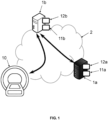
- the above-mentioned methods are implemented within an architecture such as illustrated in Figure 1 , by means of a first and/or second server 1a, 1b.
- the first server 1a is the training server (implementing the training method) and the second server 1b is a processing server (implementing the processing method). It is fully possible that these two servers may be merged.
- Each of these servers 1a, 1b is typically remote computer equipment connected to an extended network 2 such as the Internet for data exchange.
- Each one comprises data processing means 11a, 11b of processor type (in particular the data processor 11a of the first server 1a have strong computing power, since learning is long and complex compared with ordinary use of the trained CNN), and optionally storage means 12a, 12b such as a computer memory e.g. a hard disk.
- the second server 1b may be connected to one or more medical imaging devices 10 as client equipment, for providing new perfusion sequences to be processed.
- the memory 12a of the first server 1a stores a training database i.e. a set of already processed perfusion sequences referred to as training perfusion sequences (as opposed to so-called inputted perfusion sequence that precisely is sought to be processed in order to obtain the corresponding quantitative maps).
- a training database i.e. a set of already processed perfusion sequences referred to as training perfusion sequences (as opposed to so-called inputted perfusion sequence that precisely is sought to be processed in order to obtain the corresponding quantitative maps).
- the data is here constituted of perfusion sequences (in particular patched), the training examples being perfusion sequences associated with the corresponding quantitative maps as "expected results" typically previously computed with a reference perfusion sequence processing algorithm (software or machine learning approach).
- the method for processing an inputted perfusions sequence starts with a step (a) of obtaining said perfusion sequence to be processed.
- step (a) typically comprises stacking the plurality of successive images of a perfusion so as to generate the inputted perfusion sequence as a n-dimensional object, possibly after obtaining said images from a medical imaging device 10 (which may involve actually acquiring the images using the medical imaging device 10).
- the inputted perfusion sequence may also be directly retrieved from a database.
- Step (a) may also comprise pre-processing the perfusion sequence, in particular so as to generate an "improved" perfusion sequence (possibly with reduced dimensionality) from a "raw" prefusion sequence (which is the stack of the images as acquired from the medical imaging device 10). Note that the images may be pre-processed individually then stacked, or the opposite.
- step (a) may also comprise extracting patches from the raw/improved perfusion sequences thanks to a "image-to-patch” generator that maps the whole image surface with the multiple patches and which is able to work as a "patch-to-image” generator to reconstitute an image.
- patches are still 3D objects.
- this step (a) may be implemented by the data processor 11b of the second server 1b, or directly by the medical imaging device 10 if it has such computing capabilities.
- U-Net is a neural network of the encoder-decoder type: it comprises an encoder branch (or “contracting path”) that maps the inputted image into a high-level representation and then a decoder branch (or “expanding path”) generating the output image (the segmentation mask) from the high-level representation.
- U-Net further comprises skip (or “lateral”) connections between the encoder branch and decoder branch.
- the encoder branch acts as a backbone, and can be seen as a conventional feature extraction network that can be of many types, and in particular a conventional CNN, preferably a fully convolutional neural network (direct succession of blocks of convolution layers and non-linear layers such as ReLU).
- the encoder branch extracts from the input image a plurality of initial feature maps E1, E2, E3, E4, E5 representative of the input image at different scales.
- the backbone consists of a plurality of successive convolution blocks, such that the first block produces a first initial feature map E2 from the input image, then the second block produces a second initial feature map E2 from the first initial feature map E1, etc.
- a pooling layer is placed between two blocks to decrease the scale by a factor of 2 (typically 2x2 max pooling operation with stride 2 for down sampling), and from one block to another the number of filters of the convolution layers used (generally 3x3 convolutions) is increased (and preferably doubled).
- the feature maps E1, E2, E3, E4, E5 obtained by the encoder branch are said to be initial because they will be reprocessed by the decoder branch. Indeed, as explained, "low-level" maps have a higher spatial resolution but a shallow semantic depth.
- the decoder branch aims to increase their semantic depth by incorporating the information from the "high-level” maps.
- said decoder branch of the CNN has the symmetrical architecture of the encoder branch, as it generates, from the initial feature maps E1, E2, E3, E4, E5, a plurality of enriched feature maps D1, D2, D3, D4, D5 that are again representative of the input image at different scales, but they incorporate the information from the initial feature maps of smaller or equal scale while reducing the number of channels.
- the decoder branch also consists of a plurality of successive convolution blocks but in opposite order, such that the first block produces the first enriched feature map D1 (from which the output image may be directly generated) from the second enriched feature map D2 and the first initial feature map E1, after the second block produces the second enriched feature map D2 from the third enriched feature map D3 and the second initial feature map E2, etc.
- the decoder branch is also preferably a fully convolutional CNN (direct succession of blocks of convolution layers and non-linear layers such as ReLU),
- each i-th enriched map Di has the scale of the corresponding i-th initial map Ei (i.e. sensibly same spatial size) but incorporates the information of all j-th maps Di, for each j ⁇ i.
- each i-th enriched map Di is generated according to the corresponding i-th initial map Ei and/or the next (i+1-th) enriched map Di+1, hence the "contracting and expansive" nature of the branches (i.e. "U" shaped): the initial maps are obtained in ascending order and then the enriched maps are obtained in descending order.
- the maximum semantic level is obtained at the "smallest-scale” map, and from there each map is enriched on the way back down again with the information of the already enriched maps.
- the skip connections between the encoder branch and the decoder branch provide the decoder branch with the various initial maps.
- the generation of an enriched map Di based on the corresponding initial map Ei and the smaller-scale enriched map Di+1 comprises rescaling of the enriched map Di+1, typically doubling the scale (if there has been halving of scale in the encoder branch), i.e. up sampling of the enriched feature map with by a 2x2 convolution ("up-convolution") that halves the number of feature channels, then concatenation with the corresponding initial map Ei (cropped of necessary, both maps are now sensibly the same scale) to double again the number of channels, and from one block to another the number of filters of the convolution layers used (generally 3x3 convolutions) is again decreased (and preferably further halved).
- up-convolution 2x2 convolution
- the stU-Net type CNN used in the methods according to the invention still have an encoder branch, a decoder branch, and skip connections, but it notably differs from U-Net or other known CNN in that the encoder and the decoder branches do not have the same number of dimensions, leading to an asymmetrical architecture.
- a m-dimensional (preferably 2D) decoder branch with a m+1-dimensional (preferably 3D) encoder branch able to exploit the intrinsic spatio-temporal regularities of perfusion data using convolution filters that jointly act on the spatial and temporal domains (it can be seen in the case of tridimensional inputted perfusion sequences as a "2+1D" encoder).
- V-Net Fully convolutional neural networks for volumetric medical image segmentation
- Steps (b) to (d), implemented by the data processor 11b of the second server 1b, correspond to the application of the stU-Net type CNN to the inputted perfusion sequence.
- the present CNN still successively comprises an encoder branch, a decoder branch, and skip connections between the encoder branch and the decoder branch.
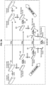
- the encoder branch of the CNN extracts a plurality of initial n+1-dimensional features maps E1, E2, E3, E4 (at least two, advantageously at least three, more advantageously less than five, optimally four) representative of the inputted perfusion sequence at different scales.
- the corresponding feature maps are at least m+2-dimensional, and m+3-dimensional if there is a batch dimension.
- the perfusion sequence presents n dimensions including m spatial dimensions (dimensions of each image) one temporal sequence (dimension according to which the images are stacked) and possibly one batch dimension, therefore the initial n+1-dimensional feature maps advantageously present as first n dimension said spatial, temporal and batch dimensions, and as n+1-th dimension a semantic depth (number of channels).
- the encoder branch can be simply fed with the n-dimensional inputted perfusion sequence patch by considering it as a "mono-channel" n+1-dimensional object, see below.
- such encoder branch advantageously consists of a plurality of successive convolution blocks, such that the first block produces a first initial feature map E2 from the input image, then the second block produces a second initial feature map E2 from the first initial feature map E1, etc., but with n-dimensional convolution layers applying n-dimensional filters in particular at least 3D convolution layers applying tridimensional filters, for instance spatio-temporal convolutions, using anisotropic kernels of size 3x3x7 (or 3x3x1 for the last block, see below) with stride 1.
- the scale may be reduced, halved, or even divided by four for instance by "down-convolution" using size 2x2x4 with stride 4 for down sampling (both spatial sizes are halved, and temporal size is divided by four, while depth is doubled), but pooling operation may alternatively be used.
- the decoder branch In a following step (c), the decoder branch generates a plurality of enriched n-dimensional feature maps D1, D2, D3, D4 also representative of the inputted perfusion sequence at different scales.
- said enriched n-dimensional feature maps only present the m spatial dimensions, the possible batch dimension, and as n-th dimension said semantic depth, but not the temporal dimension.
- each enriched n-dimensional feature map D1, D2, D3, D4 incorporating the information from the initial n+1-dimensional feature maps E1, E2, E3, E4 of smaller or equal scale.
- the present invention astutely proposes to have the skip connection projecting the initial n+1-dimensional features maps E1, E2, E3, E4 into initial n-dimensional feature maps E'1, E'2, E'3, E'4, instead of simply providing the initial n+1-dimensional features maps E1, E2, E3, E4 to the decoder branch.
- the decoder is fed with initial feature maps with a reduced number of dimensions, and typically for each enriched n-dimensional feature map D1, D2, D3, D4, an initial n-dimensional feature map E'1, E'2, E'3, E'4 of the same scale is provided from the encoder branch to the decoder branch of via a dedicated skip connection.
- the decoder only handles dimensionally-consistent n-dimensional feature maps (both the initial and enriched n-dimensional feature maps E'1, E'2, E'3, E'4, D1, D2, D3, D4 present the m spatial dimensions, possibly a batch dimension, and as n-thdimension said semantic depth), and it can only comprise standard convolution layers applying n-1-dimensional filters (bidimensional filters in the case of inputted tridimensional perfusion sequences).
- n-1-dimensional filters bidimensional filters in the case of inputted tridimensional perfusion sequences.
- each enriched n-dimensional feature map D1, D2, D3, D4 incorporating the information from the initial n-dimensional feature maps of smaller or equal scale E'1, E'2, E'3, E'4.
- said skip connections may perform a temporal pooling operation (preferably a temporal max-pooling operation denoted maxt on the figure 3b , but other pooling such as average-pooling are possible).
- a temporal pooling operation preferably a temporal max-pooling operation denoted maxt on the figure 3b , but other pooling such as average-pooling are possible.
- n+1-dimensional features map E4 has already only one temporal channel, so that it is virtually already a n-dimensional feature map and the temporal dimension may be simply dropped, but alternatively any temporal pooling operation can still be performed (the result would be the same).
- the decoder branch also consists of a plurality of successive convolution blocks but in opposite order, such that the first block produces the first enriched n-dimensional feature map D1 (from which the output image may be directly generated) from the second enriched n-dimensional feature map D2 and the first initial n-dimensional feature map E'1, after the second block produces the second enriched n-dimensional feature map D2 from the third enriched n-dimensional feature map D3 and the second initial n-dimensional feature map E'2, etc.
- the decoder branch is also preferably a fully convolutional CNN (direct succession of blocks of convolution layers and non-linear layers such as ReLU).
- the generation of an enriched n-dimensional map Di based on the corresponding initial n-dimensional map E'i and the smaller-scale enriched tridimensional map Di+1 comprises rescaling of the enriched n-dimensional map Di+1, typically doubling the scale (if there has been halving of scale in the encoder branch), i.e.
- up-convolution up sampling of the enriched n-dimensional feature map for example with by a 2x2 convolution (“up-convolution") that halves or even divides by four the number of feature channels, then concatenation with the corresponding initial n-dimensional map E'i to keep or double again the number of channels, and from one block to another the number of filters of the convolution layers used (generally 5x5 convolutions) is again decreased (and preferably further halved).
- a final step (d) at least one quantitative map of the inputted perfusion is generated from the largest-scale enriched n-dimensional feature map D1.
- said quantitative maps can be directly extracted as channels of the largest-scale enriched n-dimensional feature map D1.
- the at least one quantitative map only presents the two spatial dimensions.
- said largest-scale enriched n-dimensional feature map D1 has exactly the spatial size as the inputted patches (32x256) and one channel, i.e. it is directly a 32x256 quantitative map of a relevant parameter.
- the final 1x1 convolution of the last convolution block acts as a calculator of the relevant parameter(s).
- step (d) may comprise rescaling (if the output spatial size does not perfectly match the input spatial size) and/or reconstruction of a complete image from the patches and/or the slices.
- a training method implemented by the data processor 11a of the first server 1a. Said method trains the stU-Net type CNN, for processing inputted perfusion sequence.
- training it is meant the determination of the optimal values of parameters and weights or the CNN.
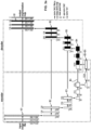
- the CNN used in the processing method is preferably trained according to the present training method, hence referred to at step (a0) in the figure 2 .
- the CNN may be directly taken “off the shelf” with preset values of parameters and weights.
- Said training method is similar to the previously described processing method, but is iteratively performed on training perfusion sequences of the training database, i.e. the training perfusion sequence are associated with an expected quantitative map of the perfusion, for instance obtained using a reference algorithm (such as a known software, for example Verbena in the case of DSC-perfusion).
- a reference algorithm such as a known software, for example Verbena in the case of DSC-perfusion.
- the training method comprises, for each of the plurality of training perfusion sequences from the base of training perfusion sequences stored on the storage means 12a of the first server, an optional step (A) and then steps (B) et (D), respectively corresponding to steps (a) to (d) of the processing method.
- the training perfusion sequence may be obtained (i.e. the expected quantitative maps generated) and possibly pre-processed (quality enhancement, patching, etc.).
- the encoder branch extracts a plurality of initial n+1-dimensional features maps representative of the training perfusion sequence at different scales.
- the decoder branch In the step (C), the decoder branch generates a plurality of enriched n-dimensional feature maps also representative of the training perfusion sequence at different scales, each enriched n-dimensional feature map incorporating the information from the initial n-dimensional feature maps of smaller or equal scale.
- At least one candidate quantitative map of the perfusion is generated from the largest-scale enriched n-dimensional feature map, so as to minimize a distance between said candidate quantitative map and the expected quantitative map of the perfusion.
- the CNN is trained with the Adam optimizer, with or without a scheduler for setting the learning rate, with default hyper-parameters to minimize the L2 loss between the quantitative maps (or just the patches).
- patches can be sampled regularly from the original images with a stride of 8.
- patches can be sampled with a stride of 1 and all the predictions be assembled by averaging.
- Data augmentation is known to play a central role in CNN training, and said training method preferably comprises generating at least one degraded version of at least one original training perfusion sequence of the training base, associating to said degraded version the expected quantitative map of the perfusion associated with the original training perfusion sequence, and enriching the training base with said degraded version (by adding it to the base).
- the degraded version associated with the original expected quantitative map is considered as a new training perfusion sequence in the training base. Note that there might be several degraded versions generated for the same original training perfusion sequence and/or degraded versions generated for several original training perfusion sequence.
- the original training perfusion sequence is associated to a contrast product dose (in particular a gadolinium-based contrast-agent), referred to as original dose (the dose used when the perfusion sequence had been acquired), and said degraded version of the original training perfusion sequence simulates a lower contrast product dose (than said dose associated to the original training perfusion sequence), referred to as degraded dose.
- degraded dose is 50% of the original dose.
- Such data augmentation is really advantageous to improve the performances of the CNN at low-doses (it is reminded that in first-pass perfusion methods such as DSC, the observed signal is proportional to the injected quantity of GBCA, thus a solution to have "less degraded” results is to increase this injected quantity), and therefore it ultimately allows a reduced use of contrast-product such as gadolinium, which is consistent with recent clinical guidelines that suggest, based on precautionary considerations, the use of the minimum dosage that achieves a sufficient contrast enhancement.
- the temporal signal S(t) in each voxel of the perfusion sequence varies linearly with the dose of contrast-product this voxel contains C(t) at any time t.
- this voxel contains C(t) at any time t.
- S t S 0 + ⁇ . C t + ⁇ t , where ⁇ t ⁇ N 0 ⁇ ⁇ 2
- the degraded version of the original training perfusion sequence simulating a lower contrast product dose can advantageously be generated by calculating the degraded temporal signal S d ( t ), for each voxel of the original perfusion sequence, only as a function of the temporal signal S ( t ) of said voxel and the dose reduction factor d .
- This synthetic signal S d can thus be easily computed for any reference signal S or artificial dose reduction factor d.
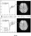
- Figure 4a displays an example of the proposed approach, which effectively allows the manipulation of the SNR of reference perfusion sequences in a realistic and theoretically-consistent manner: Based on an original perfusion sequence (top right image), a synthetic 50%-dose sequence is simulated, i.e. the "degraded version" (bottom right image). The temporal signal contained in each voxel S(t) (solid line on both left figures) is transformed into a degraded temporal signal S d (t) (dotted --- line on the bottom left figure). The ⁇ and - ⁇ - ⁇ dotted lines on the bottom left figure respectively correspond to a local average S(t) and the baseline signal S(0).
- Figure 4b represent on top two expected quantitative maps (CBV and rCBF) for the original perfusion sequence of figure 4a (which are thus also expected quantitative maps that are to be associated to the artificially degraded version of the original perfusion sequence of figure 4a ), and on bottom the two quantitative maps that would have been obtained for said degraded version of figure 4a if this degraded version was directly used as a training perfusion sequence: we see that many information would have been lost, especially for the rCBF (note the anomaly on the front part of the brain that is barely visible on the bottom map).
- CBV and rCBF expected quantitative maps
- the predictive performance of the stU-Net can be evaluated and compared to known solutions on a public data set containing 49 patients with glioma (see the document KM Schmainda, MA Prah, JM Connelly, and SD Rand, "Glioma dsc-mri perfusion data with standard imaging and rois," The Cancer Imaging Archive. http://doi.org/10.7937 K, vol. 9, 2016 ) .
- Raw brain DSC-perfusion sequences are readily available for all subjects, along with individual segmentation maps of a large vessel from which the AIF can be estimated.
- Expected quantitative CBV maps are generated using the SVD pipeline of Verbena v4.0, freely distributed as part of the FSL v6.0 software suite.
- Corresponding pairs of DSC perfusion sequences and CBV maps are individually cropped on square prisms tightly fitted on the brain voxels, and each slice is then resampled to a 256x256 size.
- the DSC and CBV signals are then standardized using the brain content statistics, and extreme values are clipped. All DSC frames are normalized similarly, according to the signal statistics of the first frame only.
- Table 1 reports quantitative results, in terms of the L1, tumor-averaged L1 (t-L1), L2, tumor-averaged L2 (t-L2) and structural similarity (SSIM) performance metrics (means and standard deviations) achieved in the full-dose, half-dose and quarter-dose scenarios by the SVD pipeline of the Verbena software, the state-of-the art method of Hess, and the present stU-Net approach.
- Verbena generated the reference full-dose CBV maps, and therefore cannot be evaluated in this scenario (indicated as n/a in the Table).
- Metrics are only computed on the test cases. The best metric of each row is indicated in bold, the second best is underlined.
- the invention provides a computer program product comprising code instructions to execute a method (particularly on the data processor 11a, 11b of the first or second server 1a, 1b) according to the second aspect of the invention for training a convolution neural network CNN, or a method according to the first aspect of the invention for processing an inputted perfusion sequence.
Landscapes
- Health & Medical Sciences (AREA)
- Engineering & Computer Science (AREA)
- Physics & Mathematics (AREA)
- Life Sciences & Earth Sciences (AREA)
- General Health & Medical Sciences (AREA)
- Nuclear Medicine, Radiotherapy & Molecular Imaging (AREA)
- Theoretical Computer Science (AREA)
- Molecular Biology (AREA)
- Biomedical Technology (AREA)
- Biophysics (AREA)
- General Physics & Mathematics (AREA)
- Radiology & Medical Imaging (AREA)
- Artificial Intelligence (AREA)
- High Energy & Nuclear Physics (AREA)
- Medical Informatics (AREA)
- General Engineering & Computer Science (AREA)
- Software Systems (AREA)
- Data Mining & Analysis (AREA)
- Computational Linguistics (AREA)
- Computing Systems (AREA)
- Evolutionary Computation (AREA)
- Mathematical Physics (AREA)
- Veterinary Medicine (AREA)
- Surgery (AREA)
- Public Health (AREA)
- Pathology (AREA)
- Heart & Thoracic Surgery (AREA)
- Animal Behavior & Ethology (AREA)
- Condensed Matter Physics & Semiconductors (AREA)
- Signal Processing (AREA)
- Optics & Photonics (AREA)
- Computer Vision & Pattern Recognition (AREA)
- Vascular Medicine (AREA)
- Dentistry (AREA)
- Oral & Maxillofacial Surgery (AREA)
- Cardiology (AREA)
- Physiology (AREA)
- Neurology (AREA)
- Hematology (AREA)
- Image Analysis (AREA)
Claims (15)
- Procédé de traitement d'une séquence de perfusion fournie en entrée représentant un empilement d'une pluralité d'images illustrant successivement le passage d'un fluide à l'intérieur d'une partie corporelle au cours du temps et présentant n≥3 dimensions dont au moins deux dimensions spatiales et une dimension temporelle, au moyen d'un réseau de neurones convolutif, CNN, le procédé comprenant la mise en oeuvre, par un processeur de données (11b) d'un second serveur (1b), des étapes consistant à :(b) extraire, au moyen d'une branche codeuse du CNN, une pluralité de cartes initiales de caractéristiques n+1-dimensionnelles représentant la séquence de perfusion fournie en entrée à différentes échelles, ledit CNN comprenant en outre une branche décodeuse et des connexions de saut entre la branche codeuse et la branche décodeuse, chaque connexion de saut projetant l'une des cartes initiales de caractéristiques n+1-dimensionnelles vers l'une d'une pluralité de cartes initiales de caractéristiques n-dimensionnelles ;(c) générer, au moyen de ladite branche décodeuse du CNN, une pluralité de cartes enrichies de caractéristiques n-dimensionnelles représentant également la séquence de perfusion fournie en entrée à différentes échelles, chaque carte enrichie de caractéristiques n-dimensionnelles incorporant les informations provenant des cartes initiales de caractéristiques n-dimensionnelles d'échelle inférieure ou égale ;(d) générer au moins une carte quantitative d'un paramètre caractérisant la perfusion sur la partie corporelle à partir des cartes enrichies de caractéristiques n-dimensionnelles à l'échelle la plus grande.
- Procédé selon la revendication 1, dans lequel, pour chaque carte enrichie de caractéristiques n-dimensionnelles, une carte initiale de caractéristiques n-dimensionnelles à la même échelle est fournie de la branche codeuse à la branche décodeuse - par le biais d'une connexion de saut dédiée.
- Procédé selon l'une des revendications 1 et 2, dans lequel, à l'étape (c), la carte enrichie de caractéristiques n-dimensionnelles à la plus petite échelle est générée à partir de la carte initiale de caractéristiques n+1-dimensionnelles à la plus petite échelle, et chaque autre carte enrichie de caractéristiques n-dimensionnelles est générée à partir de la carte initiale de caractéristiques n-dimensionnelles de même échelle et d'une carte enrichie de caractéristiques n-dimensionnelles d'échelle inférieure.
- Procédé selon l'une des revendications 1 à 3, comprenant une étape préalable (a) consistant à obtenir la séquence de perfusion par empilement d'une pluralité d'images successives d'une perfusion.
- Procédé selon la revendication 4, dans lequel lesdites images successives sont acquises par un dispositif d'imagerie médicale (10) connecté au second serveur (1a).
- Procédé selon la revendication 5, dans lequel ledit dispositif d'imagerie médicale est un dispositif de balayage d'imagerie par Résonance Magnétique, IRM, et la séquence de perfusion est une séquence de perfusion à Contraste de susceptibilité Dynamique, DSC, ou une séquence de perfusion à Amélioration de Contraste Dynamique, DCE ; ladite carte quantitative étant de préférence une carte de paramètres choisis parmi un volume sanguin cérébral, CBV, un débit sanguin cérébral, CBF, un temps de transit moyen, MTT, et un k-trans.
- Procédé selon l'une des revendications 4 à 6, dans lequel l'étape (a) consiste à extraire des plages de taille prédéterminée à partir de la séquence de perfusion, les étapes (b) à (d) étant mises en œuvre pour chaque plage extraite.
- Procédé selon l'une des revendications 1 à 7, dans lequel ledit CNN est entièrement convolutif.
- Procédé selon l'une des revendications 1 à 8, dans lequel l'au moins une carte quantitative présente uniquement les dimensions spatiales de la séquence de perfusion fournie en entrée ; les cartes initiales de caractéristiques n+1-dimensionnelles présentent lesdites dimensions spatiales et temporelles et, en tant que n+1-ième dimension, une profondeur sémantique ; et lesdites cartes initiales de caractéristiques n-dimensionnelles et enrichies présentent lesdites dimensions spatiales et, en tant que n-ième dimension, ladite profondeur sémantique ; le nombre desdites dimensions spatiales étant en particulier égal à n-1, et de préférence n=3.
- Procédé selon la revendication 9, dans lequel lesdites connexions de saut mettent en œuvre une opération de regroupement temporel.
- Procédé d'entraînement d'un réseau de neurones convolutif, CNN, pour le traitement d'une séquence de perfusion fournie en entrée représentant un empilement d'une pluralité d'images illustrant successivement le passage d'un fluide à l'intérieur d'une partie corporelle au cours du temps et présentant n≥3 dimensions dont au moins deux dimensions spatiales et une dimension temporelle ;
le procédé comprenant la mise en oeuvre, par un processeur de données (11a) d'un premier serveur (1a), pour chacune d'une pluralité de séquence de perfusion d'apprentissage provenant d'une base de séquences de perfusion d'apprentissage associées chacune à une carte quantitative attendue d'un paramètre caractérisant la perfusion sur la partie corporelle, le procédé comprenant les étapes consistant à :(B) extraire, au moyen d'une branche codeuse du CNN, une pluralité de cartes initiales de caractéristiques n+1-dimensionnelles représentant la séquence de perfusion d'apprentissage à différentes échelles, n≥3, ledit CNN comprenant en outre une branche décodeuse et des connexions de saut entre la branche codeuse et la branche décodeuse, chaque connexion de saut projetant l'une des cartes initiales de caractéristiques n+1-dimensionnelles vers l'une d'une pluralité de cartes initiales de caractéristiques n-dimensionnelles ;(C) générer, au moyen de ladite branche décodeuse du CNN, une pluralité de cartes enrichies de caractéristiques n-dimensionnelles représentant également la séquence de perfusion d'apprentissage à différentes échelles, chaque carte enrichie de caractéristiques n-dimensionnelles incorporant les informations provenant des cartes initiales de caractéristiques n-dimensionnelles d'échelle inférieure ou égale ;(D) générer au moins une carte quantitative candidate du paramètre caractérisant la perfusion sur la partie corporelle à partir de la carte enrichie de caractéristiques n-dimensionnelles à l'échelle la plus grande, et réduire au minimum une distance par rapport à la carte quantitative attendue. - Procédé selon la revendication 11, consistant préalablement à générer au moins une version dégradée d'au moins une séquence de perfusion d'apprentissage originale de la base d'apprentissage, associer à ladite version dégradée la carte quantitative attendue de la perfusion associée à la séquence de perfusion d'apprentissage originale, et enrichir la base d'apprentissage par ajout de ladite version dégradée.
- Procédé selon la revendication 12, dans lequel ladite séquence de perfusion d'apprentissage originale est associée à une dose de produit de contraste, ladite version dégradée de la séquence de perfusion d'apprentissage originale simulant une dose inférieure de produit de contraste.
- Procédé selon la revendication 13, dans lequel la version dégradée de la séquence de perfusion d'apprentissage originale simulant une dose inférieure de produit de contraste est générée par le calcul, pour chaque voxel de la séquence de perfusion d'apprentissage originale, à partir du signal temporel S(t) dudit voxel, un signal temporel dégradé Sd (t) au moyen de la formule Sd (t) = S(t) - (1 - d) - [
S(t) - S(0)], oùS(t) est une moyenne locale du signal temporel S(t), et d est un facteur de réduction de la dose. - Produit programme d'ordinateur comprenant des instructions de code permettant d'exécuter un procédé selon l'une des revendications 1 à 14 pour l'apprentissage d'un réseau de neurones convolutif, CNN, ou pour le traitement d'une séquence de perfusion fournie en entrée, lorsque ledit programme est exécuté sur un ordinateur.
Applications Claiming Priority (2)
| Application Number | Priority Date | Filing Date | Title |
|---|---|---|---|
| EP20306625.3A EP4016107A1 (fr) | 2020-12-18 | 2020-12-18 | Procédés permettant d'entraîner un cnn et de traiter une séquence de perfusion entrée en utilisant ledit cnn |
| PCT/EP2021/086794 WO2022129633A1 (fr) | 2020-12-18 | 2021-12-20 | Procédés d'entraînement d'un cnn et de traitement d'une séquence de perfusion entrée à l'aide dudit cnn |
Publications (2)
| Publication Number | Publication Date |
|---|---|
| EP4264308A1 EP4264308A1 (fr) | 2023-10-25 |
| EP4264308B1 true EP4264308B1 (fr) | 2025-02-05 |
Family
ID=74184377
Family Applications (2)
| Application Number | Title | Priority Date | Filing Date |
|---|---|---|---|
| EP20306625.3A Withdrawn EP4016107A1 (fr) | 2020-12-18 | 2020-12-18 | Procédés permettant d'entraîner un cnn et de traiter une séquence de perfusion entrée en utilisant ledit cnn |
| EP21835335.7A Active EP4264308B1 (fr) | 2020-12-18 | 2021-12-20 | Procédés permettant d'entraîner un cnn et de traiter une séquence de perfusion entrée en utilisant ledit cnn |
Family Applications Before (1)
| Application Number | Title | Priority Date | Filing Date |
|---|---|---|---|
| EP20306625.3A Withdrawn EP4016107A1 (fr) | 2020-12-18 | 2020-12-18 | Procédés permettant d'entraîner un cnn et de traiter une séquence de perfusion entrée en utilisant ledit cnn |
Country Status (5)
| Country | Link |
|---|---|
| US (1) | US20240062061A1 (fr) |
| EP (2) | EP4016107A1 (fr) |
| CN (1) | CN116802512A (fr) |
| ES (1) | ES3022836T3 (fr) |
| WO (1) | WO2022129633A1 (fr) |
Families Citing this family (6)
| Publication number | Priority date | Publication date | Assignee | Title |
|---|---|---|---|---|
| US20250139738A1 (en) * | 2021-01-14 | 2025-05-01 | Sakila S. Jayaweera | High resolution human imaging using neural network |
| WO2023062196A1 (fr) | 2021-10-15 | 2023-04-20 | Bracco Imaging S.P.A. | Entraînement d'un modèle d'apprentissage machine pour simuler des images à une dose supérieure d'agent de contraste dans des applications d'imagerie médicale |
| FR3137827B1 (fr) * | 2022-07-13 | 2024-06-28 | Olea Medical | Procédé de post-traitement d’une séquence d’acquisition de perfusion par un appareil d’imagerie médicale |
| US20250221670A1 (en) * | 2024-01-09 | 2025-07-10 | GE Precision Healthcare LLC | Method and system to compute hemodynamic parameters |
| CN119338698B (zh) * | 2024-10-10 | 2025-11-21 | 中国船舶集团有限公司第七〇九研究所 | 一种面向多场景的图像修复方法和装置 |
| CN119006615B (zh) * | 2024-10-24 | 2025-01-21 | 武汉光谷信息技术股份有限公司 | 一种地理空间业务标定方法及系统 |
Family Cites Families (6)
| Publication number | Priority date | Publication date | Assignee | Title |
|---|---|---|---|---|
| JP6209520B2 (ja) * | 2011-10-24 | 2017-10-04 | コーニンクレッカ フィリップス エヌ ヴェKoninklijke Philips N.V. | 灌流イメージング |
| WO2017106469A1 (fr) * | 2015-12-15 | 2017-06-22 | The Regents Of The University Of California | Systèmes et procédés pour analyser une imagerie médicale pondérée par perfusion au moyen de réseaux neuronaux profonds |
| US20190150764A1 (en) * | 2016-05-02 | 2019-05-23 | The Regents Of The University Of California | System and Method for Estimating Perfusion Parameters Using Medical Imaging |
| DE102018214325A1 (de) * | 2018-08-24 | 2020-02-27 | Siemens Healthcare Gmbh | Verfahren und Bereitstellungseinheit zum Bereitstellen eines virtuellen tomographischen Schlaganfall-Nachfolgeuntersuchungsbildes |
| US11341647B2 (en) * | 2018-09-14 | 2022-05-24 | The Board Of Regents Of The University Of Texas System | Systems and methods for evaluating the brain after onset of a stroke using computed tomography angiography |
| CN110223285B (zh) * | 2019-06-13 | 2021-02-02 | 中南大学湘雅二医院 | 基于动物迁移和神经网络模型的影像结果预测方法及系统 |
-
2020
- 2020-12-18 EP EP20306625.3A patent/EP4016107A1/fr not_active Withdrawn
-
2021
- 2021-12-20 EP EP21835335.7A patent/EP4264308B1/fr active Active
- 2021-12-20 WO PCT/EP2021/086794 patent/WO2022129633A1/fr not_active Ceased
- 2021-12-20 ES ES21835335T patent/ES3022836T3/es active Active
- 2021-12-20 US US18/267,884 patent/US20240062061A1/en active Pending
- 2021-12-20 CN CN202180092684.0A patent/CN116802512A/zh active Pending
Non-Patent Citations (1)
| Title |
|---|
| SANDFORT VEIT ET AL: "Reliable segmentation of 2D cardiac magnetic resonance perfusion image sequences using time as the 3rd dimension", EUROPEAN RADIOLOGY, SPRINGER BERLIN HEIDELBERG, BERLIN/HEIDELBERG, vol. 31, no. 6, 27 November 2020 (2020-11-27), pages 3941 - 3950, XP037454995, ISSN: 0938-7994, [retrieved on 20201127], DOI: 10.1007/S00330-020-07474-5 * |
Also Published As
| Publication number | Publication date |
|---|---|
| CN116802512A (zh) | 2023-09-22 |
| ES3022836T3 (en) | 2025-05-29 |
| EP4264308A1 (fr) | 2023-10-25 |
| WO2022129633A1 (fr) | 2022-06-23 |
| US20240062061A1 (en) | 2024-02-22 |
| EP4016107A1 (fr) | 2022-06-22 |
Similar Documents
| Publication | Publication Date | Title |
|---|---|---|
| EP4016107A1 (fr) | Procédés permettant d'entraîner un cnn et de traiter une séquence de perfusion entrée en utilisant ledit cnn | |
| Aubert-Broche et al. | A new improved version of the realistic digital brain phantom | |
| JP2021509953A (ja) | 深層学習を使用した低線量petイメージングからの全線量pet画像の推定 | |
| EP1747477B1 (fr) | Procede d'imagerie par resonance magnetique | |
| US8326400B2 (en) | Method of MR imaging | |
| Stute et al. | Analytical simulations of dynamic PET scans with realistic count rates properties | |
| JP2003210456A (ja) | 時系列画像の処理装置 | |
| JP2024507766A (ja) | 機械学習によるコントラスト強調 | |
| CN118941718A (zh) | 一种基于扩散多尺度生成对抗网络的三维pet图像重建方法、系统及存储介质 | |
| Ayaz et al. | Brain MR image simulation for deep learning based medical image analysis networks | |
| Kovacs et al. | Robustness analysis of denoising neural networks for bone scintigraphy | |
| Morell-Ortega et al. | Robust deep MRI contrast synthesis using a prior-based and task-oriented 3D network | |
| EP4224420B1 (fr) | Procédé mis en uvre par ordinateur pour déterminer la segmentation des cicatrices | |
| WO2023118044A1 (fr) | Procédé de traitement d'au moins une image de pré-contraste et d'une image de contraste représentant respectivement une partie du corps avant et après une injection d'une première dose d'un agent de contraste | |
| Merickel et al. | Image analysis and quantification of atherosclerosis using MRI | |
| Bai et al. | Dual-domain unsupervised network for removing motion artifact related to Gadoxetic acid-enhanced MRI | |
| Hou et al. | NC2C-TransCycleGAN: Non-contrast to contrast-enhanced CT image synthesis using transformer CycleGAN | |
| EP3771405A1 (fr) | Procédé et système d'acquisition dynamique automatisée d'images médicales | |
| CN113052840A (zh) | 一种基于低信噪比pet图像的处理方法 | |
| US20240377492A1 (en) | Apparatus and method for generating a perfusion image, and method for training an artificial neural network therefor | |
| EP2601886B1 (fr) | Analyseur de compartiment, procédé d'analyse compartimentée, programme et support d'enregistrement | |
| Claus et al. | Circular tomosynthesis for neuro perfusion imaging on an interventional C-arm | |
| Abbas et al. | VIRTUAL GADOLINIUM: DEEP LEARNING-BASED VIRTUAL CONTRAST MRI SYNTHESIS AND BRAIN TUMOR SEGMENTATION | |
| Liu et al. | Multiscale deep-learning network based reconstruction of PET images from sinogram domain | |
| Qian et al. | Physics-informed Deep Diffusion MRI Reconstruction with Synthetic Data: Break Training Data Bottleneck in Artificial Intelligence |
Legal Events
| Date | Code | Title | Description |
|---|---|---|---|
| STAA | Information on the status of an ep patent application or granted ep patent |
Free format text: STATUS: UNKNOWN |
|
| STAA | Information on the status of an ep patent application or granted ep patent |
Free format text: STATUS: THE INTERNATIONAL PUBLICATION HAS BEEN MADE |
|
| PUAI | Public reference made under article 153(3) epc to a published international application that has entered the european phase |
Free format text: ORIGINAL CODE: 0009012 |
|
| STAA | Information on the status of an ep patent application or granted ep patent |
Free format text: STATUS: REQUEST FOR EXAMINATION WAS MADE |
|
| 17P | Request for examination filed |
Effective date: 20230629 |
|
| AK | Designated contracting states |
Kind code of ref document: A1 Designated state(s): AL AT BE BG CH CY CZ DE DK EE ES FI FR GB GR HR HU IE IS IT LI LT LU LV MC MK MT NL NO PL PT RO RS SE SI SK SM TR |
|
| DAV | Request for validation of the european patent (deleted) | ||
| DAX | Request for extension of the european patent (deleted) | ||
| GRAP | Despatch of communication of intention to grant a patent |
Free format text: ORIGINAL CODE: EPIDOSNIGR1 |
|
| STAA | Information on the status of an ep patent application or granted ep patent |
Free format text: STATUS: GRANT OF PATENT IS INTENDED |
|
| INTG | Intention to grant announced |
Effective date: 20240715 |
|
| GRAS | Grant fee paid |
Free format text: ORIGINAL CODE: EPIDOSNIGR3 |
|
| GRAA | (expected) grant |
Free format text: ORIGINAL CODE: 0009210 |
|
| STAA | Information on the status of an ep patent application or granted ep patent |
Free format text: STATUS: THE PATENT HAS BEEN GRANTED |
|
| AK | Designated contracting states |
Kind code of ref document: B1 Designated state(s): AL AT BE BG CH CY CZ DE DK EE ES FI FR GB GR HR HU IE IS IT LI LT LU LV MC MK MT NL NO PL PT RO RS SE SI SK SM TR |
|
| REG | Reference to a national code |
Ref country code: GB Ref legal event code: FG4D |
|
| REG | Reference to a national code |
Ref country code: CH Ref legal event code: EP |
|
| REG | Reference to a national code |
Ref country code: DE Ref legal event code: R096 Ref document number: 602021025788 Country of ref document: DE |
|
| REG | Reference to a national code |
Ref country code: IE Ref legal event code: FG4D |
|
| P01 | Opt-out of the competence of the unified patent court (upc) registered |
Free format text: CASE NUMBER: APP_13721/2025 Effective date: 20250319 |
|
| REG | Reference to a national code |
Ref country code: ES Ref legal event code: FG2A Ref document number: 3022836 Country of ref document: ES Kind code of ref document: T3 Effective date: 20250529 |
|
| REG | Reference to a national code |
Ref country code: NL Ref legal event code: MP Effective date: 20250205 |
|
| PG25 | Lapsed in a contracting state [announced via postgrant information from national office to epo] |
Ref country code: RS Free format text: LAPSE BECAUSE OF FAILURE TO SUBMIT A TRANSLATION OF THE DESCRIPTION OR TO PAY THE FEE WITHIN THE PRESCRIBED TIME-LIMIT Effective date: 20250505 |
|
| PG25 | Lapsed in a contracting state [announced via postgrant information from national office to epo] |
Ref country code: FI Free format text: LAPSE BECAUSE OF FAILURE TO SUBMIT A TRANSLATION OF THE DESCRIPTION OR TO PAY THE FEE WITHIN THE PRESCRIBED TIME-LIMIT Effective date: 20250205 |
|
| PG25 | Lapsed in a contracting state [announced via postgrant information from national office to epo] |
Ref country code: PL Free format text: LAPSE BECAUSE OF FAILURE TO SUBMIT A TRANSLATION OF THE DESCRIPTION OR TO PAY THE FEE WITHIN THE PRESCRIBED TIME-LIMIT Effective date: 20250205 |
|
| REG | Reference to a national code |
Ref country code: LT Ref legal event code: MG9D |
|
| PG25 | Lapsed in a contracting state [announced via postgrant information from national office to epo] |
Ref country code: IS Free format text: LAPSE BECAUSE OF FAILURE TO SUBMIT A TRANSLATION OF THE DESCRIPTION OR TO PAY THE FEE WITHIN THE PRESCRIBED TIME-LIMIT Effective date: 20250605 Ref country code: NO Free format text: LAPSE BECAUSE OF FAILURE TO SUBMIT A TRANSLATION OF THE DESCRIPTION OR TO PAY THE FEE WITHIN THE PRESCRIBED TIME-LIMIT Effective date: 20250505 |
|
| PG25 | Lapsed in a contracting state [announced via postgrant information from national office to epo] |
Ref country code: NL Free format text: LAPSE BECAUSE OF FAILURE TO SUBMIT A TRANSLATION OF THE DESCRIPTION OR TO PAY THE FEE WITHIN THE PRESCRIBED TIME-LIMIT Effective date: 20250205 |
|
| PG25 | Lapsed in a contracting state [announced via postgrant information from national office to epo] |
Ref country code: HR Free format text: LAPSE BECAUSE OF FAILURE TO SUBMIT A TRANSLATION OF THE DESCRIPTION OR TO PAY THE FEE WITHIN THE PRESCRIBED TIME-LIMIT Effective date: 20250205 |
|
| PG25 | Lapsed in a contracting state [announced via postgrant information from national office to epo] |
Ref country code: PT Free format text: LAPSE BECAUSE OF FAILURE TO SUBMIT A TRANSLATION OF THE DESCRIPTION OR TO PAY THE FEE WITHIN THE PRESCRIBED TIME-LIMIT Effective date: 20250605 Ref country code: LV Free format text: LAPSE BECAUSE OF FAILURE TO SUBMIT A TRANSLATION OF THE DESCRIPTION OR TO PAY THE FEE WITHIN THE PRESCRIBED TIME-LIMIT Effective date: 20250205 |
|
| PG25 | Lapsed in a contracting state [announced via postgrant information from national office to epo] |
Ref country code: BG Free format text: LAPSE BECAUSE OF FAILURE TO SUBMIT A TRANSLATION OF THE DESCRIPTION OR TO PAY THE FEE WITHIN THE PRESCRIBED TIME-LIMIT Effective date: 20250205 Ref country code: GR Free format text: LAPSE BECAUSE OF FAILURE TO SUBMIT A TRANSLATION OF THE DESCRIPTION OR TO PAY THE FEE WITHIN THE PRESCRIBED TIME-LIMIT Effective date: 20250506 |
|
| REG | Reference to a national code |
Ref country code: AT Ref legal event code: MK05 Ref document number: 1764977 Country of ref document: AT Kind code of ref document: T Effective date: 20250205 |
|
| PG25 | Lapsed in a contracting state [announced via postgrant information from national office to epo] |
Ref country code: SE Free format text: LAPSE BECAUSE OF FAILURE TO SUBMIT A TRANSLATION OF THE DESCRIPTION OR TO PAY THE FEE WITHIN THE PRESCRIBED TIME-LIMIT Effective date: 20250205 |
|
| PG25 | Lapsed in a contracting state [announced via postgrant information from national office to epo] |
Ref country code: SM Free format text: LAPSE BECAUSE OF FAILURE TO SUBMIT A TRANSLATION OF THE DESCRIPTION OR TO PAY THE FEE WITHIN THE PRESCRIBED TIME-LIMIT Effective date: 20250205 |
|
| PG25 | Lapsed in a contracting state [announced via postgrant information from national office to epo] |
Ref country code: DK Free format text: LAPSE BECAUSE OF FAILURE TO SUBMIT A TRANSLATION OF THE DESCRIPTION OR TO PAY THE FEE WITHIN THE PRESCRIBED TIME-LIMIT Effective date: 20250205 |
|
| PG25 | Lapsed in a contracting state [announced via postgrant information from national office to epo] |
Ref country code: AT Free format text: LAPSE BECAUSE OF FAILURE TO SUBMIT A TRANSLATION OF THE DESCRIPTION OR TO PAY THE FEE WITHIN THE PRESCRIBED TIME-LIMIT Effective date: 20250205 |
|
| PG25 | Lapsed in a contracting state [announced via postgrant information from national office to epo] |
Ref country code: EE Free format text: LAPSE BECAUSE OF FAILURE TO SUBMIT A TRANSLATION OF THE DESCRIPTION OR TO PAY THE FEE WITHIN THE PRESCRIBED TIME-LIMIT Effective date: 20250205 Ref country code: CZ Free format text: LAPSE BECAUSE OF FAILURE TO SUBMIT A TRANSLATION OF THE DESCRIPTION OR TO PAY THE FEE WITHIN THE PRESCRIBED TIME-LIMIT Effective date: 20250205 |
|
| PG25 | Lapsed in a contracting state [announced via postgrant information from national office to epo] |
Ref country code: RO Free format text: LAPSE BECAUSE OF FAILURE TO SUBMIT A TRANSLATION OF THE DESCRIPTION OR TO PAY THE FEE WITHIN THE PRESCRIBED TIME-LIMIT Effective date: 20250205 |
|
| PG25 | Lapsed in a contracting state [announced via postgrant information from national office to epo] |
Ref country code: SK Free format text: LAPSE BECAUSE OF FAILURE TO SUBMIT A TRANSLATION OF THE DESCRIPTION OR TO PAY THE FEE WITHIN THE PRESCRIBED TIME-LIMIT Effective date: 20250205 |
|
| REG | Reference to a national code |
Ref country code: DE Ref legal event code: R097 Ref document number: 602021025788 Country of ref document: DE |
|
| PLBE | No opposition filed within time limit |
Free format text: ORIGINAL CODE: 0009261 |
|
| STAA | Information on the status of an ep patent application or granted ep patent |
Free format text: STATUS: NO OPPOSITION FILED WITHIN TIME LIMIT |
|
| PGFP | Annual fee paid to national office [announced via postgrant information from national office to epo] |
Ref country code: GB Payment date: 20251229 Year of fee payment: 5 |
|
| 26N | No opposition filed |
Effective date: 20251106 |
|
| PGFP | Annual fee paid to national office [announced via postgrant information from national office to epo] |
Ref country code: IT Payment date: 20251219 Year of fee payment: 5 |
|
| PGFP | Annual fee paid to national office [announced via postgrant information from national office to epo] |
Ref country code: FR Payment date: 20251226 Year of fee payment: 5 |
|
| PGFP | Annual fee paid to national office [announced via postgrant information from national office to epo] |
Ref country code: ES Payment date: 20260102 Year of fee payment: 5 |
|
| PGFP | Annual fee paid to national office [announced via postgrant information from national office to epo] |
Ref country code: DE Payment date: 20251229 Year of fee payment: 5 |