EP4282882A2 - Domaines de liaison dirigés contre des complexes gpcr: protéine g et leurs utilisations - Google Patents
Domaines de liaison dirigés contre des complexes gpcr: protéine g et leurs utilisations Download PDFInfo
- Publication number
- EP4282882A2 EP4282882A2 EP23193788.9A EP23193788A EP4282882A2 EP 4282882 A2 EP4282882 A2 EP 4282882A2 EP 23193788 A EP23193788 A EP 23193788A EP 4282882 A2 EP4282882 A2 EP 4282882A2
- Authority
- EP
- European Patent Office
- Prior art keywords
- protein
- complex
- gpcr
- binding
- receptor
- Prior art date
- Legal status (The legal status is an assumption and is not a legal conclusion. Google has not performed a legal analysis and makes no representation as to the accuracy of the status listed.)
- Pending
Links
Images
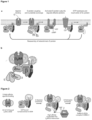


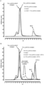
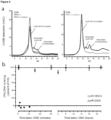


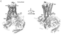


Classifications
-
- C—CHEMISTRY; METALLURGY
- C07—ORGANIC CHEMISTRY
- C07K—PEPTIDES
- C07K14/00—Peptides having more than 20 amino acids; Gastrins; Somatostatins; Melanotropins; Derivatives thereof
- C07K14/435—Peptides having more than 20 amino acids; Gastrins; Somatostatins; Melanotropins; Derivatives thereof from animals; from humans
- C07K14/46—Peptides having more than 20 amino acids; Gastrins; Somatostatins; Melanotropins; Derivatives thereof from animals; from humans from vertebrates
- C07K14/47—Peptides having more than 20 amino acids; Gastrins; Somatostatins; Melanotropins; Derivatives thereof from animals; from humans from vertebrates from mammals
- C07K14/4701—Peptides having more than 20 amino acids; Gastrins; Somatostatins; Melanotropins; Derivatives thereof from animals; from humans from vertebrates from mammals not used
- C07K14/4722—G-proteins
-
- C—CHEMISTRY; METALLURGY
- C07—ORGANIC CHEMISTRY
- C07K—PEPTIDES
- C07K16/00—Immunoglobulins [IG], e.g. monoclonal or polyclonal antibodies
- C07K16/18—Immunoglobulins [IG], e.g. monoclonal or polyclonal antibodies against material from animals or humans
- C07K16/28—Immunoglobulins [IG], e.g. monoclonal or polyclonal antibodies against material from animals or humans against receptors, cell surface antigens or cell surface determinants
-
- C—CHEMISTRY; METALLURGY
- C07—ORGANIC CHEMISTRY
- C07K—PEPTIDES
- C07K14/00—Peptides having more than 20 amino acids; Gastrins; Somatostatins; Melanotropins; Derivatives thereof
- C07K14/435—Peptides having more than 20 amino acids; Gastrins; Somatostatins; Melanotropins; Derivatives thereof from animals; from humans
- C07K14/705—Receptors; Cell surface antigens; Cell surface determinants
- C07K14/70571—Receptors; Cell surface antigens; Cell surface determinants for neuromediators, e.g. serotonin receptor, dopamine receptor
-
- C—CHEMISTRY; METALLURGY
- C07—ORGANIC CHEMISTRY
- C07K—PEPTIDES
- C07K14/00—Peptides having more than 20 amino acids; Gastrins; Somatostatins; Melanotropins; Derivatives thereof
- C07K14/435—Peptides having more than 20 amino acids; Gastrins; Somatostatins; Melanotropins; Derivatives thereof from animals; from humans
-
- C—CHEMISTRY; METALLURGY
- C07—ORGANIC CHEMISTRY
- C07K—PEPTIDES
- C07K14/00—Peptides having more than 20 amino acids; Gastrins; Somatostatins; Melanotropins; Derivatives thereof
- C07K14/435—Peptides having more than 20 amino acids; Gastrins; Somatostatins; Melanotropins; Derivatives thereof from animals; from humans
- C07K14/705—Receptors; Cell surface antigens; Cell surface determinants
-
- G—PHYSICS
- G01—MEASURING; TESTING
- G01N—INVESTIGATING OR ANALYSING MATERIALS BY DETERMINING THEIR CHEMICAL OR PHYSICAL PROPERTIES
- G01N33/00—Investigating or analysing materials by specific methods not covered by groups G01N1/00 - G01N31/00
- G01N33/48—Biological material, e.g. blood, urine; Haemocytometers
- G01N33/50—Chemical analysis of biological material, e.g. blood, urine; Testing involving biospecific ligand binding methods; Immunological testing
- G01N33/74—Chemical analysis of biological material, e.g. blood, urine; Testing involving biospecific ligand binding methods; Immunological testing involving hormones or other non-cytokine intercellular protein regulatory factors such as growth factors, including receptors to hormones and growth factors
-
- C—CHEMISTRY; METALLURGY
- C07—ORGANIC CHEMISTRY
- C07K—PEPTIDES
- C07K2317/00—Immunoglobulins specific features
- C07K2317/20—Immunoglobulins specific features characterized by taxonomic origin
- C07K2317/22—Immunoglobulins specific features characterized by taxonomic origin from camelids, e.g. camel, llama or dromedary
-
- C—CHEMISTRY; METALLURGY
- C07—ORGANIC CHEMISTRY
- C07K—PEPTIDES
- C07K2317/00—Immunoglobulins specific features
- C07K2317/50—Immunoglobulins specific features characterized by immunoglobulin fragments
- C07K2317/56—Immunoglobulins specific features characterized by immunoglobulin fragments variable (Fv) region, i.e. VH and/or VL
- C07K2317/569—Single domain, e.g. dAb, sdAb, VHH, VNAR or nanobody®
-
- C—CHEMISTRY; METALLURGY
- C12—BIOCHEMISTRY; BEER; SPIRITS; WINE; VINEGAR; MICROBIOLOGY; ENZYMOLOGY; MUTATION OR GENETIC ENGINEERING
- C12N—MICROORGANISMS OR ENZYMES; COMPOSITIONS THEREOF; PROPAGATING, PRESERVING, OR MAINTAINING MICROORGANISMS; MUTATION OR GENETIC ENGINEERING; CULTURE MEDIA
- C12N2799/00—Uses of viruses
- C12N2799/02—Uses of viruses as vector
- C12N2799/021—Uses of viruses as vector for the expression of a heterologous nucleic acid
- C12N2799/026—Uses of viruses as vector for the expression of a heterologous nucleic acid where the vector is derived from a baculovirus
-
- G—PHYSICS
- G01—MEASURING; TESTING
- G01N—INVESTIGATING OR ANALYSING MATERIALS BY DETERMINING THEIR CHEMICAL OR PHYSICAL PROPERTIES
- G01N2333/00—Assays involving biological materials from specific organisms or of a specific nature
- G01N2333/435—Assays involving biological materials from specific organisms or of a specific nature from animals; from humans
- G01N2333/705—Assays involving receptors, cell surface antigens or cell surface determinants
- G01N2333/72—Assays involving receptors, cell surface antigens or cell surface determinants for hormones
- G01N2333/726—G protein coupled receptor, e.g. TSHR-thyrotropin-receptor, LH/hCG receptor, FSH
-
- G—PHYSICS
- G01—MEASURING; TESTING
- G01N—INVESTIGATING OR ANALYSING MATERIALS BY DETERMINING THEIR CHEMICAL OR PHYSICAL PROPERTIES
- G01N2500/00—Screening for compounds of potential therapeutic value
- G01N2500/02—Screening involving studying the effect of compounds C on the interaction between interacting molecules A and B (e.g. A = enzyme and B = substrate for A, or A = receptor and B = ligand for the receptor)
Definitions
- the present invention relates to the field of G protein coupled receptor (GPCR) structural biology and signaling.
- GPCR G protein coupled receptor
- the present invention relates to binding domains directed against and/or specifically binding to GPCR:G protein complexes.
- nucleic acid sequences encoding such binding domains and cells expressing or capable of expressing such binding domains.
- the binding domains of the present invention can be used as universal tools for the structural and functional characterization of G-protein coupled receptors in complex with downstream G proteins and bound to various natural or synthetic ligands, for investigating the dynamic features of G protein activation, as well as for screening and drug discovery efforts that make use of GPCR:G protein complexes.
- TMRs Seven-transmembrane receptors
- GPCRs G protein-coupled receptors
- GPCRs once activated, convey their signals in a GTP dependent manner via a complex of three proteins known as heterotrimeric G proteins, or G ⁇ . Binding of extracellular ligands to GPCRs modulates their capacity to catalyse GDP-GTP exchange in Ga ⁇ , thereby regulating the intracellular level of secondary messengers.
- the inactive G ⁇ heterotrimer is composed of two principal elements, G ⁇ GDP and the G ⁇ heterodimer. G ⁇ sequesters the switch II element on G ⁇ such that it is unable to interact with second messenger systems, such as those involving cAMP, diacylglycerol and calcium.
- Activated GPCRs catalyze the release of GDP from G ⁇ , allowing GTP to bind and liberate the activated G ⁇ -GTP subunit.
- switch II forms a helix stabilized by the ⁇ -phosphate of GTP allowing it to interact with effectors such as adenylyl cyclase.
- the inventors identified tools that stabilize complexes of GPCRs and G proteins, which allowed them to capture and purify such complexes, and finally to crystallize such complexes. This will facilitate the identification of ligands or drug compounds by, for example, structure based virtual screening or design, high-throughput screening or fragment-based drug discovery (see e.g. Example 10). More specifically, the inventors have identified binding domains, in particular immunoglobulin single variable domains, suitable for the structural and functional analysis of an active state complex composed of an agonist, GPCR and G protein (see e.g. Example 4-7). Interestingly, it was demonstrated that some of these GPCR:G protein complex-selective binding domains are specifically directed against the G protein, and not against the GPCR.
- binding domains were identified that bind Gs at the interface of G ⁇ s and G ⁇ , without making contact with the beta-adrenergic receptor (see e.g; Example 3), and will thus be useful to capture and stabilize other Gs coupled receptors, as was demonstrated for the arginine vasopressin receptor 2 (V2R) (see e.g. Example 9).
- V2R arginine vasopressin receptor 2
- binding domains are directed against the G protein of a GPCR:G protein complex, since such a binding domain can be used as generic tool to stabilize and capture active state complexes of the range of GPCRs that interact with that particular G protein.
- the invention relates to a binding domain that is directed against and/or specifically binds to a complex comprising a GPCR and a G protein. More specifically, the binding domain as described herein binds with higher affinity to the GPCR:G protein complex as compared with binding to the G protein alone and/or to the GPCR alone, respectively. Also, the binding domain as described herein enhances the affinity of a G protein for a GPCR.
- the present invention provides for binding domains directed against and/or specifically binding to a conformational epitope of a complex comprising a GPCR and a G protein, and stabilizes or locks the complex in a particular conformational state, more specifically an active conformational state.
- the binding domain as described herein may bind to any conformational epitope that is made available or accessible by a complex of a GPCR and a G protein. These conformational epitopes may be represented by the individual proteins comprised in the complex, and/or may only be represented upon formation of the complex. Further, these conformational epitopes may or may not be represented by the individual proteins alone. According to one particular embodiment, the binding domain as described herein specifically binds to the G protein comprised in the complex, and not to the GPCR.
- G proteins are in a nucleotide-bound form. More specifically, G proteins (or at least the ⁇ subunit) are bound to either GTP or GDP depending on the activation status of a particular GPCR, as described further herein. Agonist binding to a GPCR promotes interactions with the GDP-bound G ⁇ heterotrimer leading to exchange of GDP for GTP on G ⁇ , and the functional dissociation of the G protein into G ⁇ -GTP and G ⁇ subunits, which is needed for further intracellular signaling.
- the binding domain as described herein specifically bind to and stabilize a GPCR:G protein complex in the absence of nucleotides, more specifically the binding domains bind to and stabilize a GPCR:G protein complex wherein the G protein is in a nucleotide free from.
- the binding domain as described herein will specifically bind to a conformational epitope at the interface between the alpha and beta-gamma subunit of said G protein, and as such blocks the GDP/GTP binding site and interferes with GDP/GTP binding.
- the binding domain as described herein prevents or inhibits the dissociation of the GPCR:G protein complex in the presence of nucleotides, in particular guanine nucleotides or analogs thereof, such as GTPyS. Also, the binding domain as described herein prevents or inhibits binding of nucleotides to the G protein.
- the binding domain as described herein is directed against and/or specifically binds to a complex comprising a GPCR, a G protein, and one or more receptor ligands.
- the receptor ligand will be an agonist or a positive allosteric modulator, or a combination thereof.
- the binding domain as described herein is directed against and/or specifically binds to a complex comprising a Gs protein coupled receptor and a Gs protein; or to a complex comprising a Gi protein coupled receptor and a Gi protein; or to complex comprising a Gt protein coupled receptor and a Gt protein; or to a complex comprising a Ggust protein coupled receptor and a Ggust protein; or to a complex comprising a Gz protein coupled receptor and a Gz protein; or to a complex comprising a Golf protein coupled receptor and a Golf protein; or to a complex comprising a Gq protein coupled receptor and a Gq protein; or to a complex comprising a G12 coupled receptor and a G12 protein; or to a complex comprising a G13 coupled receptor and a G13 protein.
- the GPCR and/or the G protein as comprised in the complex may be from the same or different species, in particular from a mammalian species.
- the GPCR is a human protein.
- a binding domain of the invention can be any non-naturally occurring molecule, or part thereof, that is able to specifically bind to a GPCR:G protein complex.
- the binding domain as described herein is an immunoglobulin single variable domain comprising an amino acid sequence that comprises 4 framework regions (FR1 to FR4) and 3 complementary determining regions (CDR1 to CDR3), according to the following formula (1): FR1-CDR1-FR2-CDR2-FR3-CDR3-FR4 (1).
- the binding domain as described herein is an immunoglobulin single variable domain derived from a Camelidae species, and particularly is a nanobody or V H H.
- the binding domain as described herein is an immunoglobulin single variable domain comprising an amino acid sequence that comprises 4 framework regions (FR1 to FR4) and 3 complementary determining regions (CDR1 to CDR3), according to the following formula (1): FR1-CDR1-FR2-CDR2-FR3-CDR3-FR4 (1);
- the present invention provides for an immunoglobulin single variable domain comprising an amino acid sequence that comprises 4 framework regions (FR1 to FR4) and 3 complementary determining regions (CDR1 to CDR3), according to the following formula (1): FR1-CDR1-FR2-CDR2-FR3-CDR3-FR4 (1); wherein CDR1 is SEQ ID NO: 13; wherein CDR2 is SEQ ID NO: 25; and wherein CDR3 is SEQ ID NO: 37.
- FR1-CDR1-FR2-CDR2-FR3-CDR3-FR4 (1) wherein CDR1 is SEQ ID NO: 13; wherein CDR2 is SEQ ID NO: 25; and wherein CDR3 is SEQ ID NO: 37.
- the binding domain in particular the immunoglobulin single variable domain, has an amino acid sequence selected from the group consisting of consisting of SEQ ID NOs: 1 to 6.
- binding domains as described herein may also be comprised in a polypeptide. Also, the binding domains may be immobilized on a solid support.
- the present invention relates to a binding domain that is directed against and/or specifically binds to a G protein.
- Another aspect of the invention envisages a complex comprising a binding domain as described herein.
- the complex comprises a GPCR, a G protein, and optionally a receptor ligand.
- the complex as described herein is crystalline.
- the invention also provides for nucleic acid sequences, in particular a nucleic acid sequence encoding any amino acid sequence of any of the binding domains as described herein, as well as recombinant vectors comprising any of the nucleic acid sequences as described herein.
- Particularly preferred aspects of the invention are cells comprising any of the vectors or nucleic acids as described herein, and as such, that can express or are capable of expressing a GPCR and/or a G protein.
- Cell cultures of cells according to the invention as well as membrane preparations derived thereof are also within the scope of the present invention.
- binding domains, complexes and cells may be useful in a variety of contexts and applications.
- one aspect of the invention relates to the use of a binding domain as described herein to stabilize a complex comprising a GPCR and a G protein, and optionally a receptor ligand, in a functional conformational state, more specifically in an active conformational state.
- the binding domains as described herein can be used to prevent dissociation of the complex in the presence of nucleotides, in particular guanine nucleotides or analogs thereof, such as GTPyS. Binding domains as tools to stabilize GPCR:G protein complexes and block the GPCR in a functional conformational state, preferably an active conformational state, are thus very useful for a range of applications, as outlined hereafter.
- binding domain as described herein or a cell or a membrane preparation derived thereof, as described herein, to screen for compounds that modulate the signaling activity of the GPCR.
- binding domains as described herein may be used to to capture one or more interacting proteins, in particular proteins that interact with the G protein and/or with the GPCR.
- the present invention provides for a method of capturing and/or purifying a complex comprising a GPCR and a G protein, the method comprising the steps of:
- the present invention relates to a method of determining the crystal structure of a complex comprising a GPCR and a G protein, the method comprising the steps of:
- binding domains as described herein may have therapeutic utility.
- polypeptide refers to a polymeric form of amino acids of any length, which can include coded and non-coded amino acids, chemically or biochemically modified or derivatized amino acids, and polypeptides having modified peptide backbones.
- protein complex refers to a group of two or more associated polypeptide chains. Proteins in a protein complex are linked by non-covalent protein-protein interactions.
- the "quaternary structure” is the structural arrangement of the associated folded proteins in the protein complex. It will be understood that the complex may be a multimeric complex comprising two, three, four, five, six or more polypeptides. Also, the complex may additionally comprise a non-proteinaceous molecule.
- nucleic acid molecule As used herein, the terms “nucleic acid molecule”, “polynucleotide”, “polynucleic acid”, “nucleic acid” are used interchangeably and refer to a polymeric form of nucleotides of any length, either deoxyribonucleotides or ribonucleotides, or analogs thereof. Polynucleotides may have any three-dimensional structure, and may perform any function, known or unknown.
- ligand or "receptor ligand” means a molecule that specifically binds to a GPCR, either intracellularly or extracellularly.
- a ligand may be, without the purpose of being limitative, a protein, a (poly)peptide, a lipid, a small molecule, a protein scaffold, an antibody, an antibody fragment, a nucleic acid, a carbohydrate.
- a ligand may be synthetic or naturally occurring.
- the term “ligand” includes a "native ligand” which is a ligand that is an endogenous, natural ligand for a native GPCR.
- a "specific conformation” or “specific conformational state” is any subset of the range of conformations or conformational states that a protein may adopt.
- a “functional conformation” or a “functional conformational state” refers to the fact that proteins possess different conformational states having a dynamic range of activity, in particular ranging from no activity to maximal activity.
- Examples of functional conformational states include active conformations and inactive conformations.
- a functional conformational state is meant to cover any conformational state of a GPCR, having any activity, including no activity; and is not meant to cover the denatured states of proteins.
- a particular class of functional conformations that is envisaged here is a "drugable conformation” and generally refers to a unique therapeutically relevant conformational state of a target protein.
- the active conformation of the ⁇ 2 adrenergic receptor corresponds to the drugable conformation of this receptor for the treatment of astma. It will thus be understood that drugability is confined to particular conformations depending on the therapeutic indication.
- a binding domain that specifically or selectively binds to a specific conformation or conformational state of a protein refers to a binding domain that binds with a higher affinity to a protein in a subset of conformations or conformational states than to other conformations or conformational states that the protein may assume.
- binding domains that specifically or selectively bind to a specific conformation or conformational state of a protein will stabilize this specific conformation or conformational state.
- affinity refers to the degree to which a binding domain, in particular an immunoglobulin, such as an antibody, or an immunoglobulin fragment, such as a nanobody, binds to an antigen so as to shift the equilibrium of antigen and binding domain toward the presence of a complex formed by their binding.
- an antibody (fragment) of high affinity will bind to the available antigen so as to shift the equilibrium toward high concentration of the resulting complex.
- the dissociation constant ( K d ) is commonly used to describe the affinity between the protein binding domain and the antigenic target.
- the dissociation constant is lower than 10 -5 M.
- the dissociation constant is lower than 10 -6 M, more preferably, lower than 10 -7 M.
- the dissociation constant is lower than 10 -8 M.
- a “deletion” is defined here as a change in either amino acid or nucleotide sequence in which one or more amino acid or nucleotide residues, respectively, are absent as compared to an amino acid sequence or nucleotide sequence of a parental polypeptide or nucleic acid.
- a deletion can involve deletion of about 2, about 5, about 10, up to about 20, up to about 30 or up to about 50 or more amino acids.
- a protein or a fragment thereof may contain more than one deletion.
- a deletion may in particular be a loop deletion, or an N- and/or C-terminal deletion.
- an “insertion” or “addition” is that change in an amino acid or nucleotide sequence which has resulted in the addition of one or more amino acid or nucleotide residues, respectively, as compared to an amino acid sequence or nucleotide sequence of a parental protein.
- "insertion” generally refers to addition to one or more amino acid residues within an amino acid sequence of a polypeptide, while “addition” can be an insertion or refer to amino acid residues added at an N- or C-terminus, or both termini.
- an insertion or addition is usually of about 1, about 3, about 5, about 10, up to about 20, up to about 30 or up to about 50 or more amino acids.
- a protein or fragment thereof may contain more than one insertion.
- substitution results from the replacement of one or more amino acids or nucleotides by different amino acids or nucleotides, respectively as compared to an amino acid sequence or nucleotide sequence of a parental protein or a fragment thereof. It is understood that a protein or a fragment thereof may have conservative amino acid substitutions which have substantially no effect on the protein's activity. By conservative substitutions is intended combinations such as gly, ala; val, ile, leu, met; asp, glu; asn, gln; ser, thr; lys, arg; cys, met; and phe, tyr, trp.
- sequence identity refers to the extent that sequences are identical on a nucleotide-by-nucleotide basis or an amino acid-by-amino acid basis over a window of comparison.
- a “percentage of sequence identity” is calculated by comparing two optimally aligned sequences over the window of comparison, determining the number of positions at which the identical nucleic acid base (e.g., A, T, C, G, I) or the identical amino acid residue (e.g., Ala, Pro, Ser, Thr, Gly, Val, Leu, Ile, Phe, Tyr, Trp, Lys, Arg, His, Asp, Glu, Asn, Gln, Cys and Met) occurs in both sequences to yield the number of matched positions, dividing the number of matched positions by the total number of positions in the window of comparison (i.e., the window size), and multiplying the result by 100 to yield the percentage of sequence identity. Determining the percentage of sequence identity
- Crystal or “crystalline structure”, as used herein, refers to a solid material, whose constituent atoms, molecules, or ions are arranged in an orderly repeating pattern extending in all three spatial dimensions.
- the process of forming a crystalline structure from a fluid or from materials dissolved in the fluid is often referred to as “crystallization” or “crystallogenesis”. Protein crystals are almost always grown in solution. The most common approach is to lower the solubility of its component molecules gradually. Crystal growth in solution is characterized by two steps: nucleation of a microscopic crystallite (possibly having only 100 molecules), followed by growth of that crystallite, ideally to a diffraction-quality crystal.
- X-ray crystallography is a method of determining the arrangement of atoms within a crystal, in which a beam of X-rays strikes a crystal and diffracts into many specific directions. From the angles and intensities of these diffracted beams, a crystallographer can produce a three-dimensional picture of the density of electrons within the crystal. From this electron density, the mean positions of the atoms in the crystal can be determined, as well as their chemical bonds, their disorder and various other information, as will be known by those skilled in the art.
- atomic coordinates refers to a set of three-dimensional co-ordinates for atoms within a molecular structure.
- atomic-co-ordinates are obtained using X-ray crystallography according to methods well-known to those of ordinarily skill in the art of biophysics. Briefly described, X-ray diffraction patterns can be obtained by diffracting X-rays off a crystal. The diffraction data are used to calculate an electron density map of the unit cell comprising the crystal; said maps are used to establish the positions of the atoms (i.e., the atomic co-ordinates) within the unit cell.
- atomic co-ordinates can be obtained using other experimental biophysical structure determination methods that can include electron diffraction (also known as electron crystallography) and nuclear magnetic resonance (NMR) methods.
- atomic coordinates can be obtained using molecular modeling tools which can be based on one or more of ab initio protein folding algorithms, energy minimization, and homology-based modeling. These techniques are well known to persons of ordinary skill in the biophysical and bioinformatic arts.
- Solving the structure refers to determining the arrangement of atoms or the atomic coordinates of a protein, and is often done by a biophysical method, such as X-ray crystallography.
- test compound or “test compound” or “candidate compound” or “drug candidate compound” as used herein describes any molecule, either naturally occurring or synthetic that is tested in an assay, such as a screening assay or drug discovery assay.
- these compounds comprise organic and inorganic compounds.
- the compounds include polynucleotides, lipids or hormone analogs that are characterized by low molecular weights.
- Other biopolymeric organic test compounds include small peptides or peptide-like molecules (peptidomimetics) comprising from about 2 to about 40 amino acids and larger polypeptides comprising from about 40 to about 500 amino acids, such as antibodies, antibody fragments or antibody conjugates.
- Test compounds can also be protein scaffolds.
- test compound libraries may be used, such as combinatorial or randomized libraries that provide a sufficient range of diversity. Examples include, but are not limited to, natural compound libraries, allosteric compound libraries, peptide libraries, antibody fragment libraries, synthetic compound libraries, fragment-based libraries, phage-display libraries, and the like. A more detailed description can be found further in the specification.
- determining As used herein, the terms “determining”, “measuring”, “assessing”, “monitoring” and “assaying” are used interchangeably and include both quantitative and qualitative determinations.
- biologically active refers to a GPCR having a biochemical function (e.g. a binding function, a signal transduction function, or an ability to change conformation as a result of ligand binding) of a naturally occurring GPCR.
- biochemical function e.g. a binding function, a signal transduction function, or an ability to change conformation as a result of ligand binding
- terapéuticaally effective amount means the amount needed to achieve the desired result or results.
- pharmaceutically acceptable means a material that is not biologically or otherwise undesirable, i.e., the material may be administered to an individual along with the compound without causing any undesirable biological effects or interacting in a deleterious manner with any of the other components of the pharmaceutical composition in which it is contained.
- binding domains were identified that specifically bind to GPCR:G protein complexes and are capable of stabilizing or locking the complex in a functional conformational state, in particular an active conformational state.
- the binding domains are generic tools for stabilization and capturing of a GPCR of choice in its G protein-bound state, which is generally assumed to represent an active state of a GPCR (as defined herein).
- a first aspect of the invention relates to a binding domain that is directed against and/or specifically binds to a complex comprising a GPCR and a G protein.
- the binding domain of the present invention can be any non-naturally occurring molecule or part thereof (as defined hereinbefore) that is capable of specifically binding to a complex comprising a GPCR and a G protein.
- the binding domains as described herein are protein scaffolds.
- the term "protein scaffold” refers generally to folding units that form structures, particularly protein or peptide structures, that comprise frameworks for the binding of another molecule, for instance a protein (See, e.g., Skerra (2000), for review).
- a binding domain can be derived from a naturally occurring molecule, e.g. from components of the innate or adaptive immune system, or it can be entirely artificially designed.
- a binding domain can be immunoglobulin-based or it can be based on domains present in proteins, including but limited to microbial proteins, protease inhibitors, toxins, fibronectin, lipocalins, single chain antiparallel coiled coil proteins or repeat motif proteins.
- binding domains which are known in the art include, but are not limited to: antibodies, heavy chain antibodies (hcAb), single domain antibodies (sdAb), minibodies, the variable domain derived from camelid heavy chain antibodies (VHH or nanobodies), the variable domain of the new antigen receptors derived from shark antibodies (VNAR), alphabodies, protein A, protein G, designed ankyrin-repeat domains (DARPins), fibronectin type III repeats, anticalins, knottins, engineered CH2 domains (nanoantibodies), peptides and proteins, lipopeptides (e.g.
- pepducins DNA, and RNA (see, e.g., Gebauer & Skerra, 2009; Skerra, 2000; Starovasnik et al., 1997; Binz et al., 2004; Koide et al., 1998; Dimitrov, 2009; Nygren et al. 2008; WO2010066740 ).
- combinatorial libraries comprising a consensus or framework sequence containing randomized potential interaction residues are used to screen for binding to a molecule of interest, such as a protein.
- the binding domain of the present invention may be directed against and/or specifically bind any GPCR:G protein complex of choice.
- Preferred target complexes are complexes of a GPCR and a G protein that occur in nature or, alternatively, for example in case of non-naturally occurring variants (as described further herein) of GPCRs and G protein, complexes wherein the GPCR and the G protein will associate under the appropriate physiological conditions. It will be understood by the person skilled in the art that the structural relationship between GPCR and G protein determines whether a particular GPCR:G protein complex can be formed, which will be detailed further below for members of the G protein family and members of the GPCR family.
- G proteins are meant the family of guanine nucleotide-binding proteins involved in transmitting chemical signals outside the cell, and causing changes inside the cell. G proteins are key molecular components in the intracellular signal transduction following ligand binding to the extracellular domain of a GPCR. They are also referred to as “heterotrimeric G proteins", or "large G proteins”. G proteins consist of three subunits: alpha ( ⁇ ), beta ( ⁇ ), and gamma (y) and their classification is largely based on the identity of their distinct ⁇ subunits, and the nature of the subsequent transduction event. Further classification of G proteins has come from cDNA sequence homology analysis.
- G proteins bind either guanosine diphosphate (GDP) or guanosine triphosphate (GTP), and possess highly homologous guanine nucleotide binding domains and distinct domains for interactions with receptors and effectors.
- GDP guanosine diphosphate
- GTP guanosine triphosphate
- Different subclasses of G ⁇ proteins such as Gas, Gai, Gaq and G ⁇ 12, amongst others, signal through distinct pathways involving second messenger molecules such as cAMP, inositol triphosphate (IP3), diacylglycerol, intracellular Ca 2+ and RhoA GTPases.
- the ⁇ subunit (39 - 46 kDa) contains the guanine nucleotide binding site and possesses GTPase activity; the ⁇ (37 kDa) and ⁇ (8 kDa) subunits are tightly associated and function as a ⁇ heterodimer.
- G proteins are in a nucleotide-bound form. More specifically, G proteins (or at least the ⁇ subunit) are bound to either GTP or GDP depending on the activation status of a particular GPCR. Agonist binding to a GPCR promotes interactions with the GDP-bound G ⁇ heterotrimer leading to the exchange of GDP for GTP on G ⁇ , and the functional dissociation of the G protein into G ⁇ -GTP and G ⁇ subunits.
- the separate G ⁇ -GTP and G ⁇ subunits can modulate, either independently or in parallel, downstream cellular effectors (channels, kinases or other enzymes, see Table 1).
- G proteins serve as regulated molecular switches capable of eliciting bifurcating signals through ⁇ and ⁇ subunit effects.
- the switch is turned on by the receptor and it turns itself off within a few seconds, a time sufficient for considerable amplification of signal transduction.
- G-protein coupled receptors are polypeptides that share a common structural motif, having seven regions of between 22 to 24 hydrophobic amino acids that form seven alpha helices, each of which spans the membrane. Each span is identified by number, i.e., transmembrane-1 (TM1), transmembrane-2 (TM2), etc.
- transmembrane helices are joined by regions of amino acids between transmembrane-2 and transmembrane-3, transmembrane-4 and transmembrane-5, and transmembrane-6 and transmembrane-7 on the exterior, or "extracellular" side, of the cell membrane, referred to as "extracellular" regions 1 , 2 and 3 (EC1 , EC2 and EC3), respectively.
- GPCRs can be grouped on the basis of sequence homology into several distinct families. Although all GPCRs have a similar architecture of seven membrane-spanning ⁇ - helices, the different families within this receptor class show no sequence homology to one another, thus suggesting that the similarity of their transmembrane domain structure might define common functional requirements.
- Rhodopsin a representative of this family, is the first GPCR for which the structure has been solved (Palczewski et al., 2000).
- ⁇ 2-AR the first receptor interacting with a diffusible ligand for which the structure has been solved (Rosenbaum et al, 2007) also belongs to this family.
- class B GPCRs or Class 2 (Foord et al, 2005) receptors have recently been subdivided into two families: adhesion and secretin (Fredriksson et al., 2003).
- Adhesion and secretin receptors are characterized by a relatively long amino terminal extracellular domain involved in ligand-binding. Little is known about the orientation of the transmembrane domains, but it is probably quite different from that of rhodopsin.
- Ligands for these GPCRs are hormones, such as glucagon, secretin, gonadotropin-releasing hormone and parathyroid hormone.
- the Glutamate family receptors (Class C or Class 3 receptors) also have a large extracellular domain, which functions like a "Venus fly trap" since it can open and close with the agonist bound inside.
- Family members are the metabotropic glutamate, the Ca 2+ -sensing and the ⁇ - aminobutyric acid (GABA)-B receptors.
- GPCRs include, without limitation, serotonin olfactory receptors, glycoprotein hormone receptors, chemokine receptors, adenosine receptors, biogenic amine receptors, melanocortin receptors, neuropeptide receptors, chemotactic receptors, somatostatin receptors, opioid receptors, melatonin receptors, calcitonin receptors, PTH/PTHrP receptors, glucagon receptors, secretin receptors, latrotoxin receptors, metabotropic glutamate receptors, calcium receptors, GABA-B receptors, pheromone receptors, the protease-activated receptors, the rhodopsins and other G-protein coupled seven transmembrane segment receptors.
- GPCRs also include these GPCR receptors associated with each other as homomeric or heteromeric dimers or as higher-order oligomers.
- the amino acid sequences (and the nucleotide sequences of the cDNAs which encode them) of GPCRs are readily available, for example by reference to GenBank (http://www.ncbi.nlm.nih.gov/entrez).
- the present invention provides for binding domains that are directed against and/or specifically bind GPCR:G protein complexes wherein the G protein is selected from the group consisting of Gs, Gi, Go, Gt, Ggust, Gz, Golf, Gq, G12 and G13.
- the G protein is Gs.
- the G protein is Gi.
- the G protein is Gt, more specifically transducin.
- the GPCR comprised within the complex is selected from the group consisting of a Gs coupled receptor, a Gi coupled receptor, a Go coupled receptor, a Gt coupled receptor, a Ggust coupled receptor, a Golf coupled receptor, a Gq coupled receptor, a G12 coupled receptor and a G13 coupled receptor.
- the GPCR is a Gs coupled receptor.
- the GPCR is a Gi coupled receptor.
- the GPCR is a Gt coupled receptor, more specifically rhodopsin. Particular non-limiting examples are provided in Table 1. Table 1. Non-limiting examples of the relationship of G protein-coupled receptors and signaling pathways.
- binding domains of the invention will at least bind to those forms of GPCR:G protein complexes that are most relevant from a biological and/or therapeutic point of view, as will be clear to the skilled person. It will thus be understood that, depending on the purpose and application, the GPCR and G protein comprised in the target complex may be naturally occurring or non-naturally occurring (i.e., altered by man).
- naturally-occurring means a GPCR or a G protein that are naturally produced.
- wild type polymorphic variants and isoforms of GPCRs and G proteins are examples of naturally occurring proteins, and are found for example, and without limitation, in a mammal, more specifically in a human, or in a virus, or in a plant, or in an insect, amongst others).
- GPCRs or G proteins are found in nature.
- non-naturally occurring means a GPCR or a G protein that is not naturally-occurring. In certain circumstances, it may be advantageous that the GPCR and/or G protein comprised in the complex are non-naturally occurring proteins.
- Non-limiting examples of non-naturally occurring GPCRs include, without limitation, GPCRs that have been made constitutively active through mutation, GPCRs with a loop deletion, GPCRs with an N- and/or C-terminal deletion, GPCRs with a substitution, an insertion or addition, or any combination thereof, in relation to their amino acid or nucleotide sequence, or other variants of naturally-occurring GPCRs.
- non-limiting examples of non-naturally occurring G proteins include, without limitation, G proteins with an N- and/or C-terminal deletion, G proteins with a substitution, an insertion or addition, or any combination thereof, in relation to their amino acid or nucleotide sequence, or other variants of naturally-occurring G proteins.
- target GPCR:G protein complexes comprising a chimeric or hybrid GPCR, for example a chimeric GPCR with an N- and/or C-terminus from one GPCR and loops of a second GPCR, or comprising a GPCR fused to a moiety, such as T4 lysozyme (see also Example section).
- a non-naturally occurring GPCR or G protein as comprised in the GPCR:G protein complex, may have an amino acid sequence that is at least 80% identical to, at least 90% identical to, at least 95% identical to, at least 97% identical to, or at least 99% identical to, a naturally-occurring GPCR or G protein.
- a "human G ⁇ s” or a “human G ⁇ i” or a “human G ⁇ t” has an amino acid sequence that is at least 80% identical to, at least 90% identical to, at least 95% identical to, at least 97% identical to, or at least 99% identical to the naturally occurring "human G ⁇ s” or "human G ⁇ i” or "human G ⁇ t” of Genbank accession number P63092, P63096 and P11488, respectively.
- G protein subunits include for example isoforms of Gs and Gi proteins (G ⁇ s: GNAS; G ⁇ 0: GNAO1; G ⁇ i: GNAI1 or GNAI2 or GNAI3; G ⁇ : GNB1 or GNB2 or GNB3 or GNB4 or GNB5 or GNB1L or GNB2L ; Gy: GNGT1 or GNGT2 or GNG2 or GNG3 or GNG4 or GNG5 or GNG7 or GNG8 or GNG10 or GNG11 or GNG12 or GNG13; according to HGNC standardized nomenclature to human genes; accession numbers of different isoforms from different organisms are available from www.uniprot.org ).
- binding domains of the invention will generally be capable of binding to GPCR:G protein complexes comprising all naturally occurring or synthetic analogs, variants, mutants, alleles, parts, fragments, and isoforms of a particular GPCR and/or G protein comprised in the complex; or at least to those analogs, variants, mutants, alleles, parts, fragments, and isoforms of a particular GPCR and/or G protein comprised in the complex that contain one or more antigenic determinants or epitopes that are essentially the same as the antigenic determinant(s) or epitope(s) to which the binding domains of the invention bind to a particular GPCR and/or G protein comprised in the complex.
- GPCRs As ⁇ -arrestins turn off G protein signals, they can simultaneously initiate a second, parallel set of signal cascades, such as the MAPK pathway.
- GPCRs also associate with various proteins outside the families of general GPCR-interacting proteins (G proteins, GRKs, arrestins and other receptors). These GPCR-selective partners can mediate GPCR signalling, organize GPCR signalling through G proteins, direct GPCR trafficking, anchor GPCRs in particular subcellular areas and/or influence GPCR pharmacology (Ritter and Hall 2009).
- antigen-binding fragments may be antigen-binding antibody fragments that include, but are not limited to, Fab, Fab' and F(ab')2, Fd, single-chain Fvs (scFv), single-chain antibodies, disulfide-linked Fvs (dsFv) and fragments comprising or consisting of either a VL or VH domain, and any combination of those or any other functional portion of an immunoglobulin peptide capable of binding to the target antigen.
- the term "antibodies” is also meant to include heavy chain antibodies, or functional fragments thereof, such as single domain antibodies, more specifically, immunoglobulin single variable domains such as VHHs or nanobodies, as defined further herein.
- the binding domain of the invention is an immunoglobulin single variable domain. More preferably, said said binding domain is an immunoglobulin single variable domain that comprises an amino acid sequence comprising 4 framework regions (FR1 to FR4) and 3 complementary determining regions (CDR1 to CDR3), preferably according to the following formula (1): FR1-CDR1-FR2-CDR2-FR3-CDR3-FR4 (1), or any suitable fragment thereof (which will then usually contain at least some of the amino acid residues that form at least one of the complementary determining regions).
- FR1-CDR1-FR2-CDR2-FR3-CDR3-FR4 (1) or any suitable fragment thereof (which will then usually contain at least some of the amino acid residues that form at least one of the complementary determining regions).
- the immunoglobulin single variable domains are derived from camelid antibodies, preferably from heavy chain camelid antibodies, devoid of light chains, and are known as V H H domain sequences or nanobodies (as described further herein).
- Nb refers to the smallest antigen binding fragment or single variable domain (V H H) derived from naturally occurring heavy chain antibody and is known to the person skilled in the art. They are derived from heavy chain only antibodies, seen in camelids (Hamers-Casterman et al. 1993; Desmyter et al. 1996). In the family of "camelids” immunoglobulins devoid of light polypeptide chains are found.
- Xaperone TM is a trademark of VIB and VUB (Belgium).
- Major advantages for the use of camelid antibody fragments as crystallization aid are that Xaperones (1) bind cryptic epitopes and lock proteins in unique native conformations, (2) increase the stability of soluble proteins and solubilized membrane proteins, (3) reduce the conformational complexity of soluble proteins and solubilized membrane proteins, (4) increase the polar surface enabling the growth of diffracting crystals, (5) sequester aggregative or polymerizing surfaces, (6) allow to affinity-trap active protein.
- the immunoglobulin single variable domains in particular the nanobodes, can in particular be characterized by the presence of one or more Camelidae hallmark residues in one or more of the framework sequences (according to Kabat numbering), as described for example in WO 08/020079 , on page 75, Table A-3, incorporated herein by reference).
- the humanizing substitutions should be chosen such that the resulting humanized immunoglobulin single variable domains still retain the favourable properties of immunoglobulin single variable domains as defined herein.
- the skilled person will be able to select humanizing substitutions or suitable combinations of humanizing substitutions which optimize or achieve a desired or suitable balance between the favourable properties provided by the humanizing substitutions on the one hand and the favourable properties of naturally occurring V H H domains on the other hand.
- the VH sequence that is used as a starting material or starting point for generating or designing the camelized nanobody is preferably a VH sequence from a mammal, more preferably the VH sequence of a human being, such as a VH3 sequence.
- camelized immunoglobulin single variable domains of the invention can be obtained in any suitable manner known per se (i.e. as indicated under points (1) - (8) above) and thus are not strictly limited to polypeptides that have been obtained using a polypeptide that comprises a naturally occurring VH domain as a starting material.
- the amino acid sequence of the desired humanized or camelized immunoglobulin single variable domains of the invention can be designed and then synthesized de novo using techniques for peptide synthesis known per se.
- a substitution may for example be a conservative substitution (as described herein) and/or an amino acid residue may be replaced by another amino acid residue that naturally occurs at the same position in another V H H domain.
- any one or more substitutions, deletions or insertions, or any combination thereof, that either improve the properties of the immunoglobulin single variable domains of the invention or that at least do not detract too much from the desired properties or from the balance or combination of desired properties of the nanobody of the invention are included within the scope of the invention.
- a skilled person will generally be able to determine and select suitable substitutions, deletions or insertions, or suitable combinations of thereof, based on the disclosure herein and optionally after a limited degree of routine experimentation, which may for example involve introducing a limited number of possible substitutions and determining their influence on the properties of the immunoglobulin single variable domains thus obtained.
- variants of the immunoglobulin single variable domains, in particular the nanobodies, of the present invention may have a substitution, deletion or insertion, of 1, 2 or 3 amino acids in either one, two or three of the CDRs, more specifically a substitution, deletion or insertion of 1, 2 or 3 amino acids (i) in CDR1 or CDR2 or CDR3; (ii) in CDR1 and CDR2, or, in CDR1 and CDR3, or, in CDR2 and CDR3; (iii) in CDR1 and CDR2 and CDR3, of which the amino acid sequences of CDRs are as listed in Table 3.
- the present invention provides for an immunoglobulin single variable domain comprising an amino acid sequence that comprises 4 framework regions (FR1 to FR4) and 3 complementary determining regions (CDR1 to CDR3), according to the following formula (1): FR1-CDR1-FR2-CDR2-FR3-CDR3-FR4 (1); wherein CDR1 is SEQ ID NO: 13; wherein CDR2 is SEQ ID NO: 25; and wherein CDR3 is SEQ ID NO: 37.
- FR1-CDR1-FR2-CDR2-FR3-CDR3-FR4 (1) wherein CDR1 is SEQ ID NO: 13; wherein CDR2 is SEQ ID NO: 25; and wherein CDR3 is SEQ ID NO: 37.
- Such functional groups can generally comprise all functional groups and techniques mentioned in the art as well as the functional groups and techniques known per se for the modification of pharmaceutical proteins, and in particular for the modification of antibodies or antibody fragments (including ScFv's and single domain antibodies), for which reference is for example made to Remington's Pharmaceutical Sciences, 16th ed., Mack Publishing Co., Easton, PA (1980 ).
- Such functional groups may for example be linked directly (for example covalently) to the binding domain, in particular the immunoglobulin single variable domain of the invention, or optionally via a suitable linker or spacer, as will again be clear to the skilled person.
- Suitable labels will be clear to the skilled person, and for example include moieties that can be detected using NMR or ESR spectroscopy.
- Such labelled binding domains of the invention may for example be used for in vitro, in vivo or in situ assays (including immunoassays known per se such as ELISA, RIA, EIA and other "sandwich assays", etc.) as well as in vivo diagnostic and imaging purposes, depending on the choice of the specific label.
- another modification may involve the introduction of a chelating group, for example to chelate one of the metals or metallic cations referred to above.
- Suitable chelating groups for example include, without limitation, diethyl- enetriaminepentaacetic acid (DTPA) or ethylenediaminetetraacetic acid (EDTA).
- DTPA diethyl- enetriaminepentaacetic acid
- EDTA ethylenediaminetetraacetic acid
- Yet another modification may comprise the introduction of a functional group that is one part of a specific binding pair, such as the biotin-(strept)avidin binding pair.
- a functional group may be used to link the binding domain of the invention to another protein, polypeptide or chemical compound that is bound to the other half of the binding pair, i.e. through formation of the binding pair.
- a immunoglobulin single variable domain of the invention may be conjugated to biotin, and linked to another protein, polypeptide, compound or carrier conjugated to avidin or streptavidin.
- such a conjugated immunoglobulin single variable domain may be used as a reporter, for example in a diagnostic system where a detectable signal-producing agent is conjugated to avidin or streptavidin.
- binding pairs may for example also be used to bind the immunoglobulin single variable domain of the invention to a carrier, including carriers suitable for pharmaceutical purposes.
- a carrier including carriers suitable for pharmaceutical purposes.
- One non-limiting example are the liposomal formulations described by Cao and Suresh, Journal of Drug Targetting, 8, 4, 257 (2000 ).
- Such binding pairs may also be used to link a therapeutically active agent to the binding domain of the invention.
- any multivalent or multispecific (as defined herein) immunoglobulin single variable domain of the invention may be suitably directed against two or more different epitopes on the same antigen, for example against two or more different parts of the G protein as comprised in the GPCR:G protein complex; or may be directed against two or more different antigens, for example against an epitope of the GPCR and an epitope of the G protein.
- a monovalent immunoglobulin single variable domain of the invention is such that it will bind to the target GPCR:G protein complex (as described herein) with an affinity less than 500 nM, preferably less than 200 nM, more preferably less than 10 nM, such as less than 500 pM.
- Multivalent or multispecific immunoglobulin single variable domains of the invention may also have (or be engineered and/or selected for) increased avidity and/or improved selectivity for the desired GPCR:G protein complex, and/or for any other desired property or combination of desired properties that may be obtained by the use of such multivalent or multispecific immunoglobulin single variable domains.
- binding domain usually contains amino acids that are discontinuous in the linear sequence of the protein (complex). In other words, binding of such a binding domain to a polypeptide is dependent upon the polypeptide being folded into a particular three dimensional conformation.
- conformational epitopes selectively recognized by the binding domains of the invention can be GPCR specific epitopes, or G protein specific epitopes, or otherwise GPCR:G protein complex-specific epitopes, which are only formed upon association of the constituting proteins, and thus by combining amino acid residues of said GPCR and said G protein.
- the binding domain of the invention in particular the immunoglobulin single variable domain, specifically binds to any conformational epitope of any desired G protein or parts thereof.
- said conformational epitope can be part of an intracellular or extracellular region, or an intramembraneous region, or a domain or loop structure of any desired GPCR. It is clear that some of these conformational epitopes will be accessible in the GPCR and/or the G protein in the non-associated form, while others will only be accessible once the complex is formed.
- the binding domain of the invention in particular the immunoglobulin single variable domain, specifically binds a conformational epitope at the interface between the ⁇ and ⁇ subunit of a G protein, as is specified as a non-limiting example further herein (see Example section).
- the binding domain binds to a GPCR:G protein complex wherein the G protein is in its nucleotide free from.
- the binding domain of the invention in particular the immunoglobulin single variable domain, inhibits or prevents the dissociation of the GPCR:G protein in the presence of nucleotides, in particular guanine nucleotides, such as GDP or GTP, or analogs thereof, such as nonhydrolyzable GTP analogs, such as GTPyS, or GDP in combination with aluminum or beryllium fluoride species, or minimal nucleotide analogs such as pyrophosphate or forcarnet.
- nucleotides in particular guanine nucleotides, such as GDP or GTP, or analogs thereof, such as nonhydrolyzable GTP analogs, such as GTPyS, or GDP in combination with aluminum or beryllium fluoride species, or minimal nucleotide analogs such as pyrophosphate or forcarnet.
- the invention also provides for a binding domain that is directed against or specifically binds to a G protein (i.e. a G protein alone, thus not in complex with a GPCR).
- a G protein i.e. a G protein alone, thus not in complex with a GPCR.
- the binding domain as described herein is directed against and/or specifically binds to a Gs protein.
- the binding domain of the invention prevents or inhibits the binding of nucleotides, in particular guanine nucleotides or analogs (as described hereinbefore), to the G protein.
- the binding domain of the invention is capable of displacing a guanine nucleotide or analog from the G protein.
- Non-limiting examples of assays to determine the degree of inhibition of binding or displacement of guanine nucleotides to a protein are provided in the Example section, for example in Example 3 and 8. Further, it will be appreciated that all particular embodiments related to the binding domains of the invention, as described hereinbefore, also apply for this particular aspect of the invention.
- the functional versatility of GPCRs is inherently coupled to the flexibility of these proteins resulting in a spectrum of conformations.
- the conformational energy landscape is intrinsically coupled to such factors as the presence of bound ligands (effector molecules, agonists, antagonists, inverse agonists, ...), the lipid environment or the binding of interacting proteins.
- the binding domains of the invention in particular the immunoglobulin single variable domains increase the stability of the complex comprising a GPCR and a G protein upon binding of said binding domain.
- the binding domain is capable of increasing the affinity of the G protein for the GPCR and/or the affinity of the agonist for the GPCR:G protein complex at least twofold, at least fivefold, and more preferably at least tenfold as measured by a decrease in K d .
- the binding domain is capable of inducing a shift in EC 50 or IC 50 by at least twofold, at least fivefold, and more preferably at least tenfold in any assay set-up comparing the interaction strength of the receptor and the G protein in presence of the complex stabilizing binding domain verus a condition where this binding domain is absent or any other measure of affinity or potency known to one of skill in the art.
- a functional conformational state refers to the fact that proteins, in particular membrane proteins such as GPCRs, possess many different conformational states having a dynamic range of activity, in particular ranging from no activity to maximal activity (reviewed in Kobilka and Deupi, 2007). It should be clear that "a functional conformational state” is not meant to cover the denatured states of proteins.
- a basal conformational state can be defined as a low energy state of the receptor in the absence of a ligand. The probability that a protein will undergo a transition to another conformational state is a function of the energy difference between the two states and the height of the energy barrier between the two states.
- a further aspect of the present invention relates to a complex comprising a binding domain of the invention. More specifically, a complex is provided comprising a binding domain of the invention, a GPCR, a G protein, and optionally a receptor ligand.
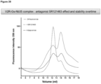
- a stable complex may be purified by gel filtration, as was done for example for the quaternary complex containing a nanobody, a GPCR, a G protein, and a receptor ligand (see Example section).
- the complex can be crystalline. Accordingly, a crystal of the complex is also provided, as well as methods of making said crystal, which are described in more detail further herein.
- nucleic acid sequence encoding an amino acid sequence of any of the binding domains of the invention, in particular immunoglobulin single variable domains is also part of the present invention and non-limiting examples are provided in Table 4.
- the invention relates to nucleic acid sequences of binding domains of the invention, in particular immunoglobulin single variable domains, in which the sequences have more than 80%, preferably more than 90%, more preferably more than 95%, such as 99% or more sequence identity (as defined herein) with the sequences of at least one of the nucleic acid sequences of the binding domains of SEQ ID NO:s 49-54 (see Table 4).
- sequences of tags e.g. His tag or EPEA tag
- the nucleic acid sequences as described herein may be comprised in a nucleic acid sequence.
- the present invention encompasses a cell or a culture of cells expressing a binding domain of the invention, in particular an immunoglobulin single variable domain that is directed against and/or capable of specifically binds to a complex comprising a GPCR and a G protein.
- the cells according to the present invention can be of any prokaryotic or eukaryotic organism.
- cells are eukaryotic cells, for example yeast cells, or insect cells, or cultured cell lines, for example mammalian cell lines, preferably human cell lines, that endogenously or recombinantly express a GPCR and/or G protein of interest.
- Eukaryotic cell or cell lines for protein production are well known in the art, including cell lines with modified glycosylation pathways, and non-limiting examples will be provided hereafter.
- Animal or mammalian host cells suitable for harboring, expressing, and producing proteins for subsequent isolation and/or purification include Chinese hamster ovary cells (CHO), such as CHO-K1 (ATCC CCL-61), DG44 ( Chasin et al., 1986, Som. Cell Molec.
- CHO-K1 Tet-On cell line (Clontech), CHO designated ECACC 85050302 (CAMR, Salisbury, Wiltshire, UK), CHO clone 13 (GEIMG, Genova, IT), CHO clone B (GEIMG, Genova, IT), CHO-K1/SF designated ECACC 93061607 (CAMR, Salisbury, Wiltshire, UK), RR-CHOK1 designated ECACC 92052129 (CAMR, Salisbury, Wiltshire, UK), dihydrofolate reductase negative CHO cells (CHO/-DHFR, Urlaub and Chasin, 1980, Proc.
- human cervical carcinoma cells HELA, ATCC CCL-2
- canine kidney cells MDCK, ATCC CCL-34
- human lung cells W138, ATCC CCL-75
- human hepatoma cells HEP-G2, HB 8065
- mouse mammary tumor cells MMT 060562, ATCC CCL-51
- buffalo rat liver cells BRL 3A, ATCC CRL-1442
- TRI cells Mather, 1982, Annals NYAcad. Sci., 383:44-68
- MCR 5 cells FS4 cells.
- the cells are mammalian cells selected from Hek293 cells or COS cells.
- Exemplary non-mammalian cell lines include, but are not limited to, Sf9 cells, baculovirus-insect cell systems (e.g. review Jarvis, Virology Volume 310, Issue 1, 25 May 2003, Pages 1-7 ), plant cells such as tobacco cells, tomato cells, maize cells, algae cells, or yeasts such as Saccharomyces species, Schizosaccharomyces species, Hansenula species, Yarrowia species or Pichia species.
- the eukaryotic cells are yeast cells from a Saccharomyces species (e.g. Saccharomyces cerevisiae ) , Schizosaccharomyces sp.
- the eukaryotic cells are Pichia cells, and in a most particular embodiment Pichia pastoris cells.
- Target cells e.g. mammalian cells
- target cells e.g. mammalian cells
- viral transduction can also be performed using reagents such as adenoviral vectors. Selection of the appropriate viral vector system, regulatory regions and host cell is common knowledge within the level of ordinary skill in the art. The resulting transfected cells are maintained in culture or frozen for later use according to standard practices.
- Another aspect of the invention relates to a method for producing a binding domain according to the invention, the method comprising at least the steps of:
- binding domains, complexes, cells or cell lines can be can be used in a variety of contexts and applications, for example, and without limitation, for capturing and/or purification of GPCR:G protein complexes, and in crystallization studies and high-resolution structural analysis of GPCR:G protein complexes. It is thus one of the aims of the invention to use the binding domain according to the invention, in particular immunoglobulin single variable domains, such as nanobodies, as tools to stabilize GPCR:G protein complexes and further to use these binding domains as co-crystallization aids for GPCRs in complex with a G protein, or in other words to facilitate crystallogenesis of GPCR:G protein complexes. Additionally, and/or alternatively, the binding domains and preferably cellular systems expressing the binding domains, as described herein, can be useful for other applications such as ligand screening, drug discovery, immunization, all of which will be described into further detail below.
- the invention relates to the use of a binding domain as described hereinbefore to stabilize a complex comprising a GPCR and a G protein.
- the complex that is stabilized further comprises a receptor ligand, more specifically an agonist.
- the term “stabilize”, “stabilizing” or “increasing the stability”, as used herein, refers to increasing the stability of a GPCR:G protein complex with respect to the structure (conformational state) and/or particular biological activity (intracellular signaling activity) of one or both of the constituting proteins of the complex, in particular the GPCR and/or the G protein.
- thermostabilize refers to the functional rather than to the thermodynamic properties of a GPCR:G protein complex and to the constituting protein's resistance to irreversible denaturation induced by thermal and/or chemical approaches including but not limited to heating, cooling, freezing, chemical denaturants, pH, detergents, salts, additives, proteases or temperature. Irreversible denaturation leads to the irreversible unfolding of the functional conformations of the protein, loss of biological activity and aggregation of the denaturated protein.
- thermostabilize means that stabilizes lipid particles or lipid layers (for example, lipid monolayers, lipid bilayers, and the like) and to GPCR:G protein complexes that have been solubilized in detergent.
- the binding domain is capable of increasing the stability as measured by an increase in the thermal stability of a GPCR:G protein complex with at least 2°C, at least 5°C, at least 8°C, and more preferably at least 10°C or 15°C or 20°C.
- the binding domain is capable of increasing the thermal stability of a GPCR:G protein complex with a receptor ligand, more specifically an agonist or positive allosteric modulator of the GPCR dependent signaling pathway.
- the binding domain according to the invention is capable of increasing the stability of a GPCR:G protein complex in the presence of a detergent or a chaotrope.
- the binding domain is capable of increasing the stability of a GPCR:G protein complex to denaturation induced by thermal or chemical approaches.
- a detergent or to a chaotrope typically the GPCR:G protein is incubated for a defined time in the presence of a test detergent or a test chaotropic agent and the stability is determined using, for example, ligand binding or a spectroscoptic method, optionally at increasing temperatures as discussed above.
- the binding domain according to the invention is capable of increasing the stability to extreme pH of a functional conformational state of a GPCR.
- the binding domain is capable of increasing the stability of a GPCR:G protein complex to extreme pH.
- a typical test pH would be chosen for example in the range 6 to 8, the range 5.5 to 8.5, the range 5 to 9, the range 4.5 to 9.5, more specifically in the range 4.5 to 5.5 (low pH) or in the range 8.5 to 9.5 (high pH).
- the ability of forming a stable GPCR:G protein complex is particularly useful for capturing and/or purifying a GPCR:G protein complex, which will allow subsequent crystallization, ligand characterization and compound screening, immunizations, amongst others.
- the binding domains of the invention can be useful generic tools that may be applicable for a range of GPCR:G protein complexes.
- the present invention also envisages a method of capturing and/or purifying a complex comprising a GPCR and a G protein, the method comprising the steps of:
- the invention provides for a method of capturing a complex comprising a GPCR and a G protein comprising the steps of:
- the present invention also envisages a method of purifying a complex comprising a GPCR and a G protein, the method comprising the steps of:
- the binding domain as described herein can also be used to capture a target GPCR:G protein complex further comprising a receptor ligand and/or one or more other interacting proteins.
- the above methods for capturing/purifying target GPCR:G protein complexes include, without limitation, affinity-based methods such as affinity chromatography, affinity purification, immunoprecipitation, protein detection, immunochemistry, surface-display, amongst others, and are all well-known in the art.
- binding domains according to the invention may increase the stability of detergent solubilized GPCR:G protein complexes, protecting them from proteolytic degradation and/or aggregation and facilitating the purification and concentration of homogenous samples of correctly folded proteins. Persons of ordinary skill in the art will recognize that such samples are the preferred starting point for the generation of diffracting crystals.
- Crystallization is another major bottleneck in the process of macromolecular structure determination by X-ray crystallography. Successful crystallization requires the formation of nuclei and their subsequent growth to crystals of suitable size. Crystal growth generally occurs spontaneously in a supersaturated solution as a result of homogenous nucleation. Proteins may be crystallized in a typical sparse matrix screening experiment, in which precipitants, additives and protein concentration are sampled extensively, and supersaturation conditions suitable for nucleation and crystal growth can be identified for a particular protein.
- binding domains of the present invention can be used as tools to increase the probability of obtaining well-ordered crystals by minimizing the conformational heterogeneity in the target GPCR:G protein complex by binding to a specific conformation of G protein.
- GPCR-detergent complexes form three-dimensional crystals in which contacts between adjacent protein molecules are made by the polar surfaces of the protein protruding from the detergent micelle (Day et al. 2007). Obviously, the detergent micelle requires space in the crystal lattice. Although attractive interactions between the micelles might stabilise the crystal packing (Rasmussen et al. 2007; Dunn et al. 1997), these interactions do not lead to rigid crystal contacts. Because many membrane proteins, including GPCRs contain relatively small or highly flexible hydrophilic domains, a strategy to increase the probability of getting well-ordered crystals is to enlarge the polar surface of the protein and/or to reduce their flexibility.
- binding domains of the present invention can be used to enlarge the polar surfaces of the GPCRs through binding of a G protein, supplementing the amount of protein surface that can facilitate primary contacts between molecules in the crystal lattice with the polar surfaces of the G protein and the nanobody. Binding domains of the present invention can also reduce the flexibility of its extracellular regions to grow well-ordered crystals.
- Immunoglobulin single variable domains are especially suited for this purpose because they bind conformational epitopes and are composed of one single rigid globular domain, devoid of flexible linker regions unlike conventional antibodies or fragments derived such as Fab's.
- the present invention provides for binding domains useful as tools to crystallize a complex comprising a GPCR and a G protein, and eventually to solve the structure.
- the complex which is crystallized by making using of a binding domain of the present invention further comprises a receptor ligand, more specifically an agonist.
- the GPCR comprised in the complex is in an active state or conformation.
- the binding domain in complex with the GPCR:G protein complex and optionally receptor ligand may be crystallized using any of a variety of specialized crystallization methods for membrane proteins, many of which are reviewed in Caffrey (2003 & 2009).
- the methods are lipid-based methods that include adding lipid to the complex prior to crystallization. Such methods have previously been used to crystallize other membrane proteins. Many of these methods, including the lipidic cubic phase crystallization method and the bicelle crystallization method, exploit the spontaneous self-assembling properties of lipids and detergent as vesicles (vesicle-fusion method), discoidal micelles (bicelle method), and liquid crystals or mesophases (in meso or cubic-phase method).
- Lipidic cubic phases crystallization methods are described in, for example: Landau et al. 1996; Gouaux 1998; Rummel et al. 1998; Nollert et al. 2004, Rasmussen et al. 2011, which publications are incorporated by reference for disclosure of those methods.
- Bicelle crystallization methods are described in, for example: Faham et al. 2005; Faham et al. 2002, which publications are incorporated by reference for disclosure of those methods.
- the diffraction data when properly assembled gives the amplitude of the 3D Fourier transform of the molecule's electron density in the unit cell. If the phases are known, the electron density can be simply obtained by Fourier synthesis.
- MR molecular replacement
- the success to derive phase information from molecular replacement (MR) alone is questionable when the fraction of proteins with a known structure (the search models) is low (less than 50% of the amino acid content) and/or when the crystals exhibit limited diffraction quality. While the combination of multiple isomorphous replacement (MIR) and MR phasing has proven successful for protein complexes (e.g. Ostermeier et al. 1995; Li et al. 1997; Hunte et al.
- one specific embodiment relates to the use of a binding domain according to the invention for the phasing of GPCR:G complexes by MR or MAD.
- immunoglobulin single variable domains, including nanobodies generally express robustly and are suitable for SeMet incorporation.
- GPCR targets for example to help guide GPCR drug discovery
- methods for acquiring structures of receptors bound to different classes of ligands including agonists, antagonists, allosteric regulators and/or G proteins are needed.
- the present invention particularly provides general tools to obtain crystals of GPCR:G protein complexes.
- agonist-bound GPCR:G protein complex crystals may provide three-dimensional representations of the active states of GPCRs. These structures will help clarify the conformational changes connecting the ligand-binding and G protein-interaction sites, and lead to more precise mechanistic hypotheses and eventually new therapeutics.
- the present invention encompasses a method of determining the crystal structure of a complex comprising a GPCR and a G protein, the method comprising the steps of:
- binding domains of the present invention can be useful in screening assays.
- Screening assays for drug discovery can be solid phase or solution phase assays, e.g. a binding assay, such as radioligand binding assays.
- a binding assay such as radioligand binding assays.
- high throughput screening of test compounds is preferred and that the methods as described above may be used as a "library screening" method, a term well known to those skilled in the art.
- the test compound may be a library of test compounds.
- high-throughput screening assays for therapeutic compounds such as agonists, antagonists or inverse agonists and/or modulators form part of the invention.
- silica porous amorphous silica
- amorphous silica i. e. the FLASH series of cartridges containing 60A irregular silica (32-63 um or 35-70 um) supplied by Biotage (a division of Dyax Corp.)
- agarose or polyacrylamide supports for example the Sepharose range of products supplied by Amersham Pharmacia Biotech, or the Affi-Gel supports supplied by Bio-Rad.
- macroporous polymers such as the pressure-stable Affi-Prep supports as supplied by Bio-Rad.
- the mutation of a particular amino acid (in a protein with known or inferred structure) to a lysine or cysteine (or other desired amino acid) can provide a specific site for covalent coupling, for example. It is also possible to reengineer a specific protein to alter the distribution of surface available amino acids involved in the chemical coupling (Kallwass et al, 1993), in effect controlling the orientation of the coupled protein. A similar approach can be applied to the binding domains according to the invention, as well as to the conformationally-stabilized GPCR:G protein complexes, so providing a means of oriented immobilisation without the addition of other peptide tails or domains containing either natural or unnatural amino acids. In case of an antibody or an antibody fragment, such as a nanobody, introduction of mutations in the framework region is preferred, minimising disruption to the antigen-binding activity of the antibody (fragment).
- the immobilised proteins may be used in immunoadsorption processes such as immunoassays, for example ELISA, or immunoaffinity purification processes by contacting the immobilised proteins according to the invention with a test sample (i.e. comprising the test compound, amongst other) according to standard methods conventional in the art.
- a test sample i.e. comprising the test compound, amongst other
- the immobilized proteins can be arrayed or otherwise multiplexed.
- the immobilised proteins according to the invention are used for the screening and selection of compounds that specifically bind to a conformationally-stabilized GPCR:G protein complex, wherein in particular the GPCR is in an active conformational state.
- the binding domain or the (conformationally stabilized) GPCR:G protein complex, or its constituting proteins may be immobilized, depending on the type of application or the type of screening that needs to be done.
- the choice of the GPCR:G protein stabilizing binding domain targeting a particular conformational epitope of the GPCR:G protein complex
- Various methods may be used to determine binding between the stabilized GPCR:G protein complex and a test compound, including for example, enzyme linked immunosorbent assays (ELISA), surface Plasmon resonance assays, chip-based assays, immunocytofluorescence, yeast two-hybrid technology and phage display which are common practice in the art, for example, in Sambrook et al. (2001), Molecular Cloning, A Laboratory Manual. Third Edition. Cold Spring Harbor Laboratory Press, Cold Spring Harbor, NY .
- Other methods of detecting binding between a test compound and a GPCR include ultrafiltration with ion spray mass spectroscopy/HPLC methods or other (bio)physical and analytical methods.
- the binding domains according to the invention can be used to screen for compounds that modulate (increase or decrease) the biological activity of the GPCR:G protein complex, or its constituents, being the GPCR or the G protein.
- the desired modulation in biological activity will depend on the GPCR of choice.
- the compounds may bind to the target GPCR:G protein complex, in particular to one or both of its constituents, resulting in the modulation (activation or inhibition) of downstream receptor signaling.
- the compounds may bind to the target complex comprising a GPCR bound to a G protein or its constituents so as to activate or increase receptor signaling; or alternatively so as to decrease or inhibit receptor signaling.
- the compounds may also bind to the target complex in such a way that they block off the constitutive activity of the GPCR.
- the compounds may also bind to the target complex in such a way that they mediate allosteric modulation (e.g. bind to the GPCR or G protein at an allosteric site). In this way, the compounds may modulate the receptor function by binding to different regions in the GPCR;G protein complex (e.g. at allosteric sites).
- cell-based assays are critical for assessing the mechanism of action of new biological targets and biological activity of chemical compounds.
- Current cell-based assays for GPCRs include measures of pathway activation (Ca 2+ release, cAMP generation or transcriptional activity); measurements of protein trafficking by tagging GPCRs and downstream elements with GFP; and direct measures of interactions between proteins using Forster resonance energy transfer (FRET), bioluminescence resonance energy transfer (BRET) or yeast two-hybrid approaches.
- FRET Forster resonance energy transfer
- BRET bioluminescence resonance energy transfer
- yeast two-hybrid approaches Introducing the binding domains of the present invention, inside the cell to the relevant compartment of the cell (intra- or extracellularly) by any means well known and commonly used in the art, may lead to new or better cell-based assays.
- the test compound is provided as a biological sample.
- the sample can be any suitable sample taken from an individual.
- the sample may be a body fluid sample such as blood, serum, plasma, spinal fluid.
- the sample is tissue or cell extract.
- the binding domains of the present invention can also be used to modulate GPCR signaling, in particular G protein-mediated GPCR signaling, including abolishing G protein-mediated GPCR signaling.
- modulating means an increase or decrease in activity of a protein or a protein complex, in particular a GPCR:G protein complex.
- the binding domains of the present invention can be allosteric modulators or allosteric inhibitors.
- allosteric modulator or “allosteric inhibitor” in the context of the present invention refer to noncompetitive modulators or inhibitors, which exert their effect by binding to a site other than the active site of the receptor, and modulate the activity of the receptor or render the receptor ineffective in terms of signal transduction.
- binding domains may have therapeutic utility and may be administered to a subject having a condition in order to treat the subject for the condition.
- the therapeutic utility for a binding domain is determined by the target GPCR:G protein complex to which the binding domain binds in that signaling via that GPCR is linked to the condition.
- the GPCR may be activated in the condition by binding to a ligand.
- the GPCR may be mutated to make it constitutively active, for example.
- a subject binding domain may be employed for the treatment of a GPCR-mediated condition such as schizophrenia, migraine headache, reflux, asthma, bronchospasm, prostatic hypertrophy, ulcers, epilepsy, angina, allergy, rhinitis, cancer e.g.
- one or more compounds that decrease the activity of the GPCR may be administered, whereas when an increase in activity of a certain GPCR is desired, one or more compounds that increase the activity of the GPCR activity may be administered.
- the invention also relates to a pharmaceutical composition
- a pharmaceutical composition comprising a therapeutically effective amount of the binding domains of the invention and at least one pharmaceutically acceptable carrier, adjuvant or diluent.
- Carriers or adjuvants may be, as a non limiting example, Ringer's solution, dextrose solution or Hank's solution. Non aqueous solutions such as fixed oils and ethyl oleate may also be used.
- a preferred excipient is 5% dextrose in saline. The excipient may contain minor amounts of additives such as substances that enhance isotonicity and chemical stability, including buffers and preservatives.
- a binding domain as described herein or a pharmaceutically acceptable salt thereof may be by way of oral, inhaled or parenteral administration.
- the nanobody is delivered through intrathecal or intracerebroventricular administration.
- the active compound may be administered alone or preferably formulated as a pharmaceutical composition.
- An amount effective to treat a certain disease or disorder that express the antigen recognized by the binding domain depends on the usual factors such as the nature and severity of the disorder being treated and the weight of the mammal. However, a unit dose will normally be in the range of 0.01 to 50 mg, for example 0.01 to 10 mg, or 0.05 to 2 mg of binding domain or a pharmaceutically acceptable salt thereof.
- Unit doses will normally be administered once or more than once a day, for example 2, 3, or 4 times a day, more usually 1 to 3 times a day, such that the total daily dose is normally in the range of 0.0001 to 1 mg/kg; thus a suitable total daily dose for a 70 kg adult is 0.01 to 50 mg, for example 0.01 to 10 mg or more usually 0.05 to 10 mg. It is greatly preferred that the compound or a pharmaceutically acceptable salt thereof is administered in the form of a unit-dose composition, such as a unit dose oral, parenteral, or inhaled composition.
- Oral liquid preparations may be in the form of, for example, aqueous or oily suspensions, solutions, emulsions, syrups, or elixirs, or may be presented as a dry product for reconstitution with water or other suitable vehicle before use.
- fluid unit dose forms are prepared containing a compound of the present invention and a sterile vehicle.
- the active compound depending on the vehicle and the concentration, can be either suspended or dissolved.
- Parenteral solutions are normally prepared by dissolving the compound in a vehicle and filter sterilising before filling into a suitable vial or ampoule and sealing.
- adjuvants such as a local anaesthetic, preservatives and buffering agents are also dissolved in the vehicle.
- the composition can be frozen after filling into the vial and the water removed under vacuum.
- Parenteral suspensions are prepared in substantially the same manner except that the compound is suspended in the vehicle instead of being dissolved and sterilised by exposure to ethylene oxide before suspending in the sterile vehicle.
- a surfactant or wetting agent is included in the composition to facilitate uniform distribution of the active compound.
- small amounts of bronchodilators for example sympathomimetic amines such as isoprenaline, isoetharine, salbutamol, phenylephrine and ephedrine; xanthine derivatives such as theophylline and aminophylline and corticosteroids such as prednisolone and adrenal stimulants such as ACTH may be included.
- the compositions will usually be accompanied by written or printed directions for use in the medical treatment concerned.
- binding domains in particular immunoglobulin single variable domains
- Delivery of binding domains, in particular immunoglobulin single variable domains, into cells may be performed as described for peptides, polypeptides and proteins. If the antigen is extracellular or an extracellular domain, the binding domain may exert its function by binding to this domain, without need for intracellular delivery.
- the binding domains of the present invention as described herein may target intracellular conformational epitopes of GPCR:G proteins of interest. To use these binding domains as effective and safe therapeutics inside a cell, intracellular delivery may be enhanced by protein transduction or delivery systems know in the art. Protein transduction domains (PTDs) have attracted considerable interest in the drug delivery field for their ability to translocate across biological membranes.
- the PTDs are relatively short (1 1-35 amino acid) sequences that confer this apparent translocation activity to proteins and other macromolecular cargo to which they are conjugated, complexed or fused (Sawant and Torchilin 2010).
- the HIV-derived TAT peptide (YGRKKRRQRRR), for example, has been used widely for intracellular delivery of various agents ranging from small molecules to proteins, peptides, range of pharmaceutical nanocarriers and imaging agents.
- receptor-mediated endocytic mechanisms can also be used for intracellular drug delivery.
- the transferrin receptor-mediated internalization pathway is an efficient cellular uptake pathway that has been exploited for site-specific delivery of drugs and proteins (Qian et al. 2002).
- transferrin This is achieved either chemically by conjugation of transferrin with therapeutic drugs or proteins or genetically by infusion of therapeutic peptides or proteins into the structure of transferrin.
- Naturally existing proteins such as the iron-binding protein transferrin
- these proteins are biodegradable, nontoxic, and non-immunogenic.
- they can achieve site-specific targeting due to the high amounts of their receptors present on the cell surface.
- Still other delivery systems include, without the purpose of being limitative, polymer- and liposome-based delivery systems.
- the efficacy of the binding domains of the invention, and of compositions comprising the same, can be tested using any suitable in vitro assay, cell-based assay, in vivo assay and/or animal model known per se, or any combination thereof, depending on the specific disease or disorder involved.
- the invention also encompasses a method of screening for binding domains directed against and/or specifically binding to a complex comprising a GPCR and a G protein, comprising the steps of:
- binding domains are generated and screened for their specific binding to a complex comprising a GPCR and a G protein and optionally a receptor ligand.
- the binding domains may also be generated and screened for their specific binding to a G protein.
- binding domains can be generated in many ways. In the case of immunoglobulin single variable domains, such as nanobodies, typically, immunization of an animal will be done with a target complex comprising a GPCR bound to a G protein and a receptor ligand, as described hereinbefore (e.g. for V H H sequences, as a non-limiting example) and also exemplified further herein.
- the proteins of the target complex may be produced and purified using conventional methods that may employ expressing a recombinant form of said proteins in a host cell, and purifying the proteins using affinity chromatography and/or antibody-based methods.
- the bactulovirus/Sf-9 system may be employed for expression, although other expression systems (e.g., bacterial, yeast or mammalian cell systems) may also be used.
- a functional GPCR:G protein complex may also be reconstituted by using purified receptor (e.g. ⁇ 2-AR or MOR) reconstituted into recombinant HDL particles with a stoichiometric excess of the G protein (Gs or Gi) as described in Whorton et al. (2009) for ⁇ 2-AR:Gs or in Kuszaket al. (2009) for MOR:Gi.
- purified receptor e.g. ⁇ 2-AR or MOR
- a GPCR may also be reconstituted in phospholipid vesicles and loaded with a stoichiometric excess of the G protein.
- methods for reconstituting an active GPCR in phospholipid vesicles are known, and are described in: Luca et al (2003), Mansoor et al (2006), Niu et al. (2005), Shimada et al. (2002), and Eroglu et al. (2003), among others.
- the GPCR and phospholipids may be reconstituted at high density (e.g., 1 mg receptor per mg of phospholipid).
- a GPCR may be present in the phospholipid vesicle in both orientations (in the normal orientation, and in the "upside down" orientation in which the intracellular loops are on the outside of the vesicle).
- Other immunization methods with a GPCR include, without limitation, the use of complete cells expressing a GPCR and/or a G protein or membranes derived thereof.
- the animal is immunized with a target complex that is crosslinked with a bifunctional crosslinker (see also Example section).
- Chemical crosslinking can be done using standard techniques that are well-known by the skilled person in the art (see e.g. Hermanson, G.T. (2008) Bioconjugate Techniques, 2nd ed., Elsevier Inc., 1202 pages ).
- the screening for binding domains specifically binding to a conformational epitope of a target complex may for example be performed by screening a set, collection or library of cells that express the binding domains on their surface (e.g. B-cells obtained from a suitably immunized Camelid), by screening of a (naive or immune) library of binding domains, or by screening of a (naive or immune) library of nucleic acid sequences that encode amino acid sequences of the binding domains, which may all be performed in a manner known per se, and which method may optionally further comprise one or more other suitable steps, such as, for example and without limitation, a step of affinity maturation, a step of expressing the desired amino acid sequence, a step of screening for binding and/or for activity against the desired antigen, a step of determining the desired amino acid sequence or nucleotide sequence, a step of introducing one or more humanizing substitutions, a step of formatting in a suitable multivalent and/or multispecific format, a step of screening for the
- kits comprising a binding domain according to the invention.
- the kit may further comprise a combination of reagents such as buffers, molecular tags, vector constructs, reference sample material, as well as a suitable solid supports, cells, nucleic acids, and the like.
- reagents such as buffers, molecular tags, vector constructs, reference sample material, as well as a suitable solid supports, cells, nucleic acids, and the like.
- Such a kit may be useful for any of the applications of the present invention as described herein.
- the kit may comprise (a library of) test compounds useful for compound screening applications.
- a last aspect of the invention is the use of any binding domain according to the invention to isolate amino acid sequences that are responsible for specific binding to a conformational epitope of a GPCR:G protein complex and to construct artificial binding domains based on said amino acid sequences.
- the framework regions and the complementary determining regions are known, and the study of derivatives of the binding domain, binding to the same conformational epitope of a GPCR:G protein complex, will allow deducing the essential amino acids involved in binding said conformational epitope.
- This knowledge can be used to construct a minimal binding domain and to create derivatives thereof, which can routinely be done by techniques known by the skilled in the art.
- a nanobody (V H H domain) that is directed against and/or specifically binds to a complex comprising a GPCR and a G protein and optionally a receptor ligand, wherein said nanobody specifically binds to the G protein and not to the GPCR in the complex, and wherein said nanobody comprises an amino acid sequence consisting of 4 framework regions (FR1 to FR4) and 3 complementary determining regions (CDR1 to CDR3), according to the following formula: FR1-CDR1-FR2-CDR2-FR3-CDR3-FR4, wherein CDR1 is SEQ ID NO:16, CDR2 is SEQ ID NO:28 and CDR3 is SEQ ID NO:40; or CDR1 is SEQ ID NO:15, CDR2 is SEQ ID NO:27 and CDR3 is SEQ ID NO:39; or CDR1 is SEQ ID NO:14, CDR2 is SEQ ID NO:26 and CDR3 is SEQ ID NO:38; or CDR
- the nanobody according to the first embodiment of the first preferred aspect wherein said nanobody has an amino acid sequence that has at least 80% amino acid identity with the amino acid sequence of SEQ ID NO:4; or an amino acid sequence that has at least 80% amino acid identity with the amino acid sequence of SEQ ID NO:3; or an amino acid sequence that has at least 80% amino acid identity with the amino acid sequence of SEQ ID NO:2; or an amino acid sequence that has at least 80% amino acid identity with the amino acid sequence of SEQ ID NO:5; or an amino acid sequence that has at least 80% amino acid identity with the amino acid sequence of SEQ ID NO:6.
- a complex comprising a nanobody according to the first or second embodiment of the first preferred aspect, a GPCR, and a G protein.
- the complex according to the first embodiment of the second preferred aspect which further comprises a receptor ligand.
- a nanobody according to the first embodiment of the first preferred aspect to stabilize in vitro a complex comprising a GPCR and a G protein and optionally a receptor ligand in a functional conformational state.
- guanine nucleotides such as GDP or GTP, or analogs thereof, such as GTPyS.
- a nanobody according to the first embodiment of the first preferred aspect to crystallize and/or to solve the structure of a complex comprising a GPCR and a G protein and optionally a receptor ligand.
- a nanobody according to the first embodiment of the first preferred aspect to screen for compounds that modulate the signaling activity of the GPCR.
- a sixth preferred aspect is disclosed the use according to any embodiment according to the third, fourth or fifth aspect, wherein the GPCR is in an active conformation.
- a method of determining the crystal structure of a complex comprising a GPCR and a G protein comprising the steps of: a) Providing a nanobody according to the first embodiment of the first preferred aspect, and b) Allowing the nanobody to bind to a complex comprising a GPCR and a G protein and optionally a receptor ligand, and c) Crystallizing the complex formed in step b)
- a method of identifying compounds capable of selectively binding to a complex comprising a GPCR and a G protein comprising the steps of: (i) Providing a complex comprising a GPCR and a G protein; (ii) Contacting the complex with a nanobody according to the first embodiment of the first preferred aspect and allowing the nanobody to bind to the complex; (iii) Providing a test compound; (iv) Evaluating whether the test compound binds to the complex; and (v) Selecting a compound that selectively binds to the complex.
- a method of screening for nanobodies (V H H domains) that specifically bind to a complex comprising a GPCR and a G protein comprising the steps of: a) Providing a plurality of binding domains, and b) Screening said plurality of binding domains for binding domains that bind to a complex comprising a GPCR and a G protein, c) Isolating the binding domains that bind to the complex, and d) Screening the isolated binding domains for binding domains that specifically bind to the G protein of the GPCR/G protein complex.
- the complex further comprises a ligand of the GPCR, preferably an agonist of the GPCR.
- a third embodiment of the ninth preferred aspect is disclosed the method according to the first or the second embodiment of the ningth preferred aspect, further comprising: screening the isolated binding domains for binding domains that prevent the GPCR/G protein complex from dissociation by guanine nucleotides, such as GDP or GTP, or analogs thereof, such as GTPgS.
- a fourth embodiment of the ninth preferred aspect is disclosed the method according to the first or the second or the third embodiment of the ningth preferred aspect, further comprising: screening the isolated binding domains for binding domains that prevent or inhibit the binding of guanine nucleotides, such as GDP or GTP, or analogs thereof, such as GTPgS to the G protein.
- guanine nucleotides such as GDP or GTP, or analogs thereof, such as GTPgS to the G protein.
- Example 1 Formation and purification of a stable agonist- ⁇ 2AR-Gs ternary complex
- Formation of a stable complex was accomplished by mixing Gs heterotrimer at approximately 100 ⁇ M concentration with BI-167107 bound T4L- ⁇ 2 AR (or ⁇ 2 AR-365) in molar excess (approximately 130 ⁇ M) in 2 ml buffer (10 mM HEPES, pH 7.5, 100 mM NaCl, 0.1 % DDM, 1 mM EDTA, 3 mM MgCl 2 , 10 ⁇ M BI-167107) and incubating for 3 hrs at room temperature.
- BI-167107 which was identified from screening and characterizing approximately 50 different ⁇ 2 AR agonists, has a dissociation half-time of approximately 30 hrs providing higher degree of stabilization to the active G protein-bound receptor than other full agonists such as isoproterenol (Rasmussen et al., 2011).
- apyrase 25 mU/ml, NEB was added after 90 min to hydrolyze residual GDP released from G ⁇ s upon binding to the receptor.
- GMP resulting from hydrolysis of GDP by apyrase has very poor affinity for the G protein in the complex. Rebinding of GDP can cause dissociation of the R:G complex ( figure 3a ).
- the R:G complex in DDM shows significant dissociation after 48 hours at 4oC ( figure 4a ).
- the complex was exchanged into MNG-3 by adding the R:G mixture (2 ml) to 8 ml buffer (20 mM HEPES, pH 7.5, 100 mM NaCl, 10 ⁇ M BI-167107) containing 1% MNG-3 for 1 hr at room temperature.
- the final size exclusion chromatography procedure to separate excess free receptor from the R:G complex ( figure 5b ) was performed on a Superdex 200 10/300 GL column (GE Healthcare) equilibrated with buffer containing 0.02 % MNG-3, 10 mM HEPES pH 7.5, 100 mM NaCl, 10 ⁇ M BI-167107, and 100 ⁇ M TCEP. Peak fractions were pooled ( figure 5b ) and concentrated to approximately 90 mg ml -1 with a 100 kDa MWCO Viva-spin concentrator and analyzed by SDS-PAGE/Coomassie brilliant blue staining ( figure 5a ) and gel filtration ( figure 5c ). To confirm a pure, homogeneous, and dephosphorylated preparation, the R:G complex was routinely analyzed by ion exchange chromatography ( figure 5d ).
- Example 2 Generation of Nanobodies binding to the agonist: ⁇ 2AR:Gs ternary complex
- Nanobodies are single domain antibodies, derived from heavy chain only antibodies from Ilama's (Muyldermans, 2001).
- To identify Nanobodies that bind the (agonist loaded) receptor coupled Gs-protein we immunized two llamas (Llama glama) with the bis(sulfosuccinimidyl)glutarate (BS2G, Pierce) cross-linked ⁇ 2AR:Gs:BI167107 ternary complex.
- VHHs were cloned as Pst1-BstEll fragments into the phage display vector pMESy4.
- pMESy4 phage display vector
- R:G complex specific nanobodies were enriched by two rounds of biopanning on i) the ⁇ 2AR:Gs:BI167107 ternary complex embedded in ApoL biotinylated high-density lipoprotein particles (rHDL, Whorton et al.
- biotinylated rHDL particles containing the ⁇ 2AR:Gs:BI167107 ternary complex were immobilized on a neutravidin coated Maxisorp plate (Nunc) at 1 ⁇ g/well in 20mM Hepes (pH 8.0), 100mM NaCl, 1mM EDTA, 100 ⁇ M TCEP, and 100nM BI167107.
- the BS2G cross-linked ⁇ 2AR:Gs:BI167107 ternary complex was solid phase coated on a Maxisorp plate at 1 ⁇ g/well.
- phage was added to immobilized antigens and incubated for one to two hours.
- non-bound phage was removed from the antigen containing wells and the wells were washed 14 times with 20mM HEPES, 100mM NaCl, pH8 and finally incubated for 10 minutes with 200 ⁇ L of 20mM Hepes (pH 8.0), 100mM NaCl, 1mM EDTA, 100 ⁇ M TCEP, and 100nM BI167107 to remove aspecific phages.
- the wells were treated with trypsin, phage was recovered and used to infect exponentially growing TG1 cells (OD600 ⁇ 0.5).
- Nanobodies enriched on biotinylated rHDL particles containing the ⁇ 2AR:Gs:BI167107 ternary complex were analysed by comparative ELISA on the same complex immobilized on neutravidin coated Maxisorb plates versus empty rHDL particles.
- Nanobodies enriched on solid phase coated BS2G cross-linked ⁇ 2AR:Gs:BI167107 ternary complex were analysed by comparative ELISA on the same solid phase coated complex versus non-coated wells. From colonies that scored positive in comparative ELISA, single clones were prepared, DNA was extracted and the sequences of the encoded nanobody genes were analyzed using routine methods (amino acid sequences shown in Tables 2-3). For Nb35, Nb36 and Nb37, binding to the ⁇ 2AR:Gs:BI167107 ternary complex was further confirmed by analytical gelfiltration ( Figures 3d , 3e , 3f, 3g ).
- Example 3 Nb35, Nb36 and Nb37 bind to Gs and prevent dissociation of the complex by GTP ⁇ S
- Nanobodies raised against the ⁇ 2AR:Gs:BI167107 ternary complex (Table 2) bind to the receptor or to Gs, we next monitored binding of these nanobodies in ELISA on purified receptor alone. All nanobodies from Table 2 scored negative in solid phase coated (Maxisorb, Nunc) agonist-bound ⁇ 2AR-356 reconstituted at high density into phospholipid vesicles (Rasmussen et al., 2011). Nb80, a ⁇ 2AR specific nanobody (Rasmussen et al. 2011) scored positive in this ELISA.
- G protein-coupled receptors are responsible for the majority of cellular responses to hormones and neurotransmitters as well as the senses of sight, olfaction and taste.
- the paradigm of GPCR signaling is the activation of a heterotrimeric GTP binding protein (G protein) by an agonist-occupied receptor.
- Gas consists of two domains, the ras-like GTPase domain (GasRas), which interacts with the ⁇ 2 AR and the G ⁇ subunit, and the ⁇ -helical domain (GasAH) (Sprang et al. 1997).
- the interface of the two Gas subdomains forms the nucleotide-binding pocket ( Figure 1 ), and EM 2D averages and 3D reconstructions suggest that in the absence of guanine nucleotide, GasAH has a variable position relative to the complex of T4L- ⁇ 2 AR-G ⁇ sRAS-G ⁇ ( Fig. 1b ).
- Crystallization leads were initially identified using in-house screens partly based on reagents from the StockOptions Salt kit (Hampton Research). Crystals for data collection were grown in 18 to 22% PEG 400, 100 mM MES pH 6.5 (figure 1c), 350 to 450 mM potassium nitrate, 10 mM foscarnet ( figure 3b ), 1 mM TCEP, and 10 ⁇ M BI167107. Crystals reached full size within 3-4 days at 20°C ( Figure 8 ) and were picked from a sponge-like mesophase and flash-frozen without additional cryo-protectant in liquid nitrogen.
- ⁇ 2 AR-Nb80 complex exhibits the same high affinity for the agonist isoproterenol as does the ⁇ 2 AR:Gs complex (Rasmussen et al, 2011), consistent with high structural homology around the ligand binding pocket.
- the electron density maps for the ⁇ 2 AR-Nb80 crystals provide a more reliable view of the conformational rearrangements of amino acids around the ligand-binding pocket and between the ligand-binding pocket and the Gs-coupling interface (Rasmussen et al, 2011).
- Figure 12c shows the position of the highly conserved sequence motifs including D/ERY and NPxxY in the ⁇ 2 AR:Gs complex compared with the ⁇ 2 AR-Nb80 complex (see also Fig. 13 ).
- These conserved sequences have been proposed to be important for activation or for maintaining the receptor in the inactive state (Hofmann et al, 2009).
- the positions of these amino acids are essentially identical in these two structures demonstrating that Nb80 is a very good G protein surrogate.
- Only Arg131 differs between these two structures. In the ⁇ 2 AR-Nb80 structure Arg131 interacts with Nb80, whereas in the ⁇ 2 AR:Gs structure Arg131 packs against Tyr391 of Gas ( Fig. 13 ).
- Phe139 which is located at the beginning of the ICL2 helix and sits in a hydrophobic pocket formed by Gas His41 at the beginning of the ⁇ 1-strand, Va!213 at the start of the ⁇ 3-strand and Phe376, Arg380 and Ile383 in the as-helix ( Fig 14c ).
- the ⁇ 2 AR mutant F139A displays severely impaired coupling to Gs (Moro et al, 1993).
- the residue corresponding to Phe139 is a Phe or Leu on almost all Gs coupled receptors, but is more variable in GPCRs known to couple to other G proteins.
- Example 8 Nb35 and Nb37 bind different epitopes on Gs and inhibit nucleotide binding
- the Gs alpha subunit (or Gs protein) is a heterotrimeric G protein subunit that activates the cAMP-dependent pathway by activating adenylate cyclase.
- the G protein-coupled receptors that couple to Gs include: 5-HT receptors types 5-HT4 and 5-HT7, ACTH receptor, Adenosine receptor types A2a and A2b, Arginine vasopressin receptor 2, ⁇ -adrenergic receptors types ⁇ 1, ⁇ 2 and ⁇ 3, Calcitonin receptor, Calcitonin gene-related peptide receptor, Corticotropin-releasing hormone receptor, Dopamine receptors D1-like family (D1 and D5), FSH-receptor, Gastric inhibitory polypeptide receptor, Glucagon receptor, Glucagon-like peptide 1 receptor (GLP1-R), Histamine H2 receptor, Luteinizing hormone/choriogonadotropin receptor, Melanocortin receptor, Parathyroid hormone receptor 1, Prostaglan
- Nb35 stabilizes the complex between the activated ⁇ 2AR and the G protein by binding the interface of G ⁇ s and G ⁇ subunits.
- An example of a screening assay to identify ligands that selectively interact with the low abundant active state conformer of ⁇ 2AR can be a radioligand assay using Nb35 to shift ⁇ 2AR population more towards its active state.
- Such radioligand assay can be executed similarly as the one described by Seifert and co-workers (1998) with minor modifications.
- the dose dependent effect of these compounds in a cellular ⁇ 2AR signalling assay will be evaluated.
- One example of such assay relies on the detection of secondary messenger molecules such as cAMP after Gas mediated signaling (e.g. HitHunter cAMP assay technology, DiscoverX).
- the radioligand assay could be performed on a ⁇ 2AR expressing cell line co-transfected with Nb35 as an intrabody (or derived membranes) in order to shift the ⁇ 2AR population to its active state.
- recombinant G protein and ⁇ 2AR can be used to stabilize via Nb35 the active state of the ⁇ 2 AR.
- the frozen membranes were thawed and diluted to a total protein concentration of 5 mg/ml with fresh wash buffer.
- Sodium cholate detergent was added to the suspension at a final concentration of 1.0%
- MgCl 2 was added to a final concentration of 5 mM
- .05 mg of purified protein phosphatase 5 was added per liter of culture volume. The sample was stirred on ice for 40 minutes, and then centrifuged at 180,000 ⁇ g for 40 minutes to remove insoluble derbies.
- Ni-NTA wash buffer 1 Ni-NTA wash buffer 2 (20 mM HEPES pH 8.0, 50 mM NaCl, 1 mM MgCl 2 , 10 mM imidazole, 0.15% Anzergent 3-12, 0.1% DDM, 1x PTT, 1x LS, 5 mM ⁇ -ME, 10 ⁇ M GDP), and Ni-NTA wash buffer 3 (20 mM HEPES pH 8.0, 50 mM NaCl, 1 mM MgCl 2 , 5 mM imidazole, 0.1% DDM, 1x PTT, 1x LS, 5 mM ⁇ -ME, 10 ⁇ M GDP).
- the elute was passed through a 0.22 ⁇ m filter and loaded directly onto a MonoQ HR 16/10 column (GE Healthcare) equilibrated in MonoQ buffer A (20 mM HEPES pH 8.0, 50 mM NaCl, 100 ⁇ M MgCl 2 , 0.1% DDM, 5 mM ⁇ -ME, 1x PTT).
- MonoQ buffer A (20 mM HEPES pH 8.0, 50 mM NaCl, 100 ⁇ M MgCl 2 , 0.1% DDM, 5 mM ⁇ -ME, 1x PTT).
- the column was washed with 150 ml buffer A at 5 ml/min and bound proteins were eluted over 350 ml with a linear gradient up to 28% MonoQ buffer B (same as buffer A except with 1 M NaCl). Fractions were collected in tubes spotted with enough GDP to make a final concentration of 10 ⁇ M.
- Nanobody-35 (SEQ ID NO: 1) was expressed in the periplasm of Escherichia coli strain WK6, extracted, and purified by nickel affinity chromatography according to previously described methods (Rasmussen et al. 2011) followed by ion-exchange chromatography ( figure 7a ) using a Mono S 10/100 GL column (GE Healthcare). Selected Nb35 fractions were dialysis against buffer (10 mM HEPES pH7.5, 100 mM NaCl) and concentrated to approximately 65 mg ml -1 with a 10 kDa MWCO Millipore concentrator.
Landscapes
- Health & Medical Sciences (AREA)
- Chemical & Material Sciences (AREA)
- Life Sciences & Earth Sciences (AREA)
- Organic Chemistry (AREA)
- Molecular Biology (AREA)
- Immunology (AREA)
- General Health & Medical Sciences (AREA)
- Biochemistry (AREA)
- Medicinal Chemistry (AREA)
- Biophysics (AREA)
- Genetics & Genomics (AREA)
- Proteomics, Peptides & Aminoacids (AREA)
- Engineering & Computer Science (AREA)
- Zoology (AREA)
- Toxicology (AREA)
- Gastroenterology & Hepatology (AREA)
- Biomedical Technology (AREA)
- Cell Biology (AREA)
- Hematology (AREA)
- Urology & Nephrology (AREA)
- Endocrinology (AREA)
- Pathology (AREA)
- Biotechnology (AREA)
- Food Science & Technology (AREA)
- Physics & Mathematics (AREA)
- Analytical Chemistry (AREA)
- General Physics & Mathematics (AREA)
- Microbiology (AREA)
- Neurology (AREA)
- Peptides Or Proteins (AREA)
- Preparation Of Compounds By Using Micro-Organisms (AREA)
- Micro-Organisms Or Cultivation Processes Thereof (AREA)
- Investigating Or Analysing Biological Materials (AREA)
- Measuring Or Testing Involving Enzymes Or Micro-Organisms (AREA)
- Medicines Containing Material From Animals Or Micro-Organisms (AREA)
Applications Claiming Priority (5)
| Application Number | Priority Date | Filing Date | Title |
|---|---|---|---|
| US201161571159P | 2011-06-21 | 2011-06-21 | |
| EP11181357 | 2011-09-15 | ||
| EP17209368.4A EP3345919B1 (fr) | 2011-06-21 | 2012-06-21 | Domaines de liaison dirigés contre des complexes gpcr: protéine g et leurs utilisations |
| PCT/EP2012/062036 WO2012175643A2 (fr) | 2011-06-21 | 2012-06-21 | Domaines de liaison dirigés contre des complexes gpcr:protéine g et leurs utilisations |
| EP12730478.0A EP2723764B1 (fr) | 2011-06-21 | 2012-06-21 | Domaines de liaison dirigés contre des complexes gpcr: protéine g et leurs utilisations |
Related Parent Applications (3)
| Application Number | Title | Priority Date | Filing Date |
|---|---|---|---|
| EP17209368.4A Division EP3345919B1 (fr) | 2011-06-21 | 2012-06-21 | Domaines de liaison dirigés contre des complexes gpcr: protéine g et leurs utilisations |
| EP17209368.4A Division-Into EP3345919B1 (fr) | 2011-06-21 | 2012-06-21 | Domaines de liaison dirigés contre des complexes gpcr: protéine g et leurs utilisations |
| EP12730478.0A Division EP2723764B1 (fr) | 2011-06-21 | 2012-06-21 | Domaines de liaison dirigés contre des complexes gpcr: protéine g et leurs utilisations |
Publications (2)
| Publication Number | Publication Date |
|---|---|
| EP4282882A2 true EP4282882A2 (fr) | 2023-11-29 |
| EP4282882A3 EP4282882A3 (fr) | 2024-03-06 |
Family
ID=47423019
Family Applications (3)
| Application Number | Title | Priority Date | Filing Date |
|---|---|---|---|
| EP23193788.9A Pending EP4282882A3 (fr) | 2011-06-21 | 2012-06-21 | Domaines de liaison dirigés contre des complexes gpcr: protéine g et leurs utilisations |
| EP17209368.4A Active EP3345919B1 (fr) | 2011-06-21 | 2012-06-21 | Domaines de liaison dirigés contre des complexes gpcr: protéine g et leurs utilisations |
| EP12730478.0A Active EP2723764B1 (fr) | 2011-06-21 | 2012-06-21 | Domaines de liaison dirigés contre des complexes gpcr: protéine g et leurs utilisations |
Family Applications After (2)
| Application Number | Title | Priority Date | Filing Date |
|---|---|---|---|
| EP17209368.4A Active EP3345919B1 (fr) | 2011-06-21 | 2012-06-21 | Domaines de liaison dirigés contre des complexes gpcr: protéine g et leurs utilisations |
| EP12730478.0A Active EP2723764B1 (fr) | 2011-06-21 | 2012-06-21 | Domaines de liaison dirigés contre des complexes gpcr: protéine g et leurs utilisations |
Country Status (14)
| Country | Link |
|---|---|
| US (4) | US9695227B2 (fr) |
| EP (3) | EP4282882A3 (fr) |
| JP (2) | JP6164535B2 (fr) |
| CN (2) | CN104053667B (fr) |
| AU (1) | AU2012273987B2 (fr) |
| CA (1) | CA2839834A1 (fr) |
| DK (1) | DK2723764T3 (fr) |
| ES (1) | ES2662372T3 (fr) |
| HR (1) | HRP20180443T1 (fr) |
| HU (1) | HUE036982T2 (fr) |
| NO (1) | NO2723764T3 (fr) |
| PL (1) | PL2723764T3 (fr) |
| PT (1) | PT2723764T (fr) |
| WO (1) | WO2012175643A2 (fr) |
Families Citing this family (55)
| Publication number | Priority date | Publication date | Assignee | Title |
|---|---|---|---|---|
| GB201014715D0 (en) | 2010-09-06 | 2010-10-20 | Vib Vzw | Nanobodies stabilizing functional conformational states of GPCRS |
| US9695227B2 (en) | 2011-06-21 | 2017-07-04 | Vrije Universiteit Brussel | Binding domains directed against GPCR:G protein complexes and uses derived thereof |
| US10048253B2 (en) | 2012-06-28 | 2018-08-14 | Ucb Biopharma Sprl | Method for identifying compounds of therapeutic interest |
| KR102160389B1 (ko) | 2013-08-05 | 2020-09-28 | 트위스트 바이오사이언스 코포레이션 | 드 노보 합성된 유전자 라이브러리 |
| JP2017522906A (ja) | 2014-07-04 | 2017-08-17 | アンスティチュ ナショナル ドゥ ラ サンテ エ ドゥ ラ ルシェルシュ メディカル | 抗mGluR2構造的単一ドメイン抗体及びその使用 |
| EP3839061B1 (fr) | 2014-10-27 | 2022-12-07 | King Abdullah University Of Science And Technology | Méthodes et systèmes d'identification de sites de liaison ligand-protéine |
| CA2975852A1 (fr) | 2015-02-04 | 2016-08-11 | Twist Bioscience Corporation | Procedes et dispositifs pour assemblage de novo d'acide oligonucleique |
| WO2016172377A1 (fr) | 2015-04-21 | 2016-10-27 | Twist Bioscience Corporation | Dispositifs et procédés pour la synthèse de banques d'acides oligonucléiques |
| KR20180050411A (ko) | 2015-09-18 | 2018-05-14 | 트위스트 바이오사이언스 코포레이션 | 올리고핵산 변이체 라이브러리 및 그의 합성 |
| WO2017053450A1 (fr) | 2015-09-22 | 2017-03-30 | Twist Bioscience Corporation | Substrats flexibles pour synthèse d'acide nucléique |
| EP3384077A4 (fr) | 2015-12-01 | 2019-05-08 | Twist Bioscience Corporation | Surfaces fonctionnalisées et leur préparation |
| CN105802970A (zh) * | 2016-05-30 | 2016-07-27 | 东北师范大学 | 靶向沉默Gβ1的shRNA |
| WO2018038772A1 (fr) | 2016-08-22 | 2018-03-01 | Twist Bioscience Corporation | Banques d'acides nucléiques synthétisés de novo |
| EP4664304A3 (fr) | 2016-09-21 | 2026-01-21 | Atlas Data Storage, Inc. | Stockage de données à base d'acide nucléique |
| CN107037116B (zh) * | 2016-12-01 | 2019-04-23 | 白波 | 检测gpcr之间相互作用及其交界面的方法 |
| GB2573069A (en) | 2016-12-16 | 2019-10-23 | Twist Bioscience Corp | Variant libraries of the immunological synapse and synthesis thereof |
| WO2018156792A1 (fr) | 2017-02-22 | 2018-08-30 | Twist Bioscience Corporation | Stockage de données reposant sur un acide nucléique |
| CA3056388A1 (fr) | 2017-03-15 | 2018-09-20 | Twist Bioscience Corporation | Banques de variants de la synapse immunologique et leur synthese |
| IL271205B2 (en) | 2017-06-12 | 2025-02-01 | Twist Bioscience Corp | Methods for assembling continuous nucleic acids |
| WO2018231864A1 (fr) | 2017-06-12 | 2018-12-20 | Twist Bioscience Corporation | Méthodes d'assemblage d'acides nucléiques continus |
| CN111566125A (zh) * | 2017-09-11 | 2020-08-21 | 特韦斯特生物科学公司 | Gpcr结合蛋白及其合成 |
| CN111565834B (zh) | 2017-10-20 | 2022-08-26 | 特韦斯特生物科学公司 | 用于多核苷酸合成的加热的纳米孔 |
| EP4635987A3 (fr) | 2017-10-31 | 2026-01-14 | Vib Vzw | Nouvelles protéines chimériques de liaison à l'antigène et leurs procédés et utilisations |
| EP3658236A1 (fr) * | 2017-12-21 | 2020-06-03 | Firmenich SA | Procédé d'identification de modulateurs allostériques positifs pour des récepteurs odorants |
| IL312616B2 (en) | 2018-01-04 | 2025-08-01 | Twist Bioscience Corp | Dna-based digital information storage |
| WO2019155041A1 (fr) * | 2018-02-12 | 2019-08-15 | Vib Vzw | ANTICORPS COMPLEXES Gβγ ET LEURS UTILISATIONS |
| CN108586620B (zh) * | 2018-05-10 | 2021-11-23 | 山东大学 | 筛选昆虫性信息素受体的配体的传感器分子以及筛选方法 |
| EP3814497A4 (fr) | 2018-05-18 | 2022-03-02 | Twist Bioscience Corporation | Polynucléotides, réactifs, et procédés d'hybridation d'acides nucléiques |
| FR3084365B1 (fr) | 2018-07-27 | 2020-10-23 | Cisbio Bioassays | Anticorps a domaine unique qui se lient a la proteine g alpha |
| WO2020127983A1 (fr) * | 2018-12-21 | 2020-06-25 | Vib Vzw | Protéines de fusion comprenant une cytokine et une protéine d'échafaudage |
| EP3902816A4 (fr) | 2018-12-26 | 2022-09-14 | Twist Bioscience Corporation | Synthèse de novo polynucléotidique hautement précise |
| FR3092172B1 (fr) * | 2019-01-30 | 2021-02-12 | Cisbio Bioassays | Méthode pour mesurer la modulation de l’activation d’un récepteur couplé à une protéine G avec des analogues du GTP |
| CA3131689A1 (fr) | 2019-02-26 | 2020-09-03 | Twist Bioscience Corporation | Banques de variants d'acides nucleiques pour le recepteur glp1 |
| KR20210144698A (ko) | 2019-02-26 | 2021-11-30 | 트위스트 바이오사이언스 코포레이션 | 항체 최적화를 위한 변이 핵산 라이브러리 |
| CA3314168A1 (en) | 2019-04-29 | 2026-08-05 | Confo Therapeutics N.V. | Screening methods and assays for use with transmembrane proteins, in particular with gpcrs |
| US12123814B1 (en) * | 2019-06-14 | 2024-10-22 | Pierce Biotechnology, Inc. | G- protein coupled receptor extraction formulations |
| WO2020257405A1 (fr) * | 2019-06-18 | 2020-12-24 | Musc Foundation For Research Development | Compositions et méthodes ciblant l'état non associé à des nucléotides de ras pour bloquer les voies de signalisation et la transformation oncogéniques |
| EP3987019A4 (fr) | 2019-06-21 | 2023-04-19 | Twist Bioscience Corporation | Assemblage de séquences d'acide nucléique basé sur des code-barres |
| CN115023440B (zh) | 2019-09-23 | 2025-09-12 | 特韦斯特生物科学公司 | 结合CD3ε的抗体 |
| US12091777B2 (en) | 2019-09-23 | 2024-09-17 | Twist Bioscience Corporation | Variant nucleic acid libraries for CRTH2 |
| AU2020400030B2 (en) | 2019-12-09 | 2026-05-21 | Twist Bioscience Corporation | Variant nucleic acid libraries for adenosine receptors |
| FR3106830B1 (fr) | 2020-01-30 | 2022-02-11 | Cisbio Bioassays | Anticorps anti-protéine G alpha |
| CA3190667A1 (fr) | 2020-08-26 | 2022-03-03 | Aaron Sato | Methodes et compositions se rapportant a des variants glp1r |
| EP4298443A4 (fr) * | 2021-02-26 | 2025-01-15 | The Regents Of The University Of Michigan | Biocapteurs fluorescents et procédés d'utilisation de ces biocapteurs dans la détection d'événements de signalisation cellulaire |
| WO2023154533A2 (fr) * | 2022-02-14 | 2023-08-17 | Twist Bioscience Corporation | Ensemble d'adn combinatoire pour anticorps multispecifiques |
| US20250164508A1 (en) * | 2022-02-17 | 2025-05-22 | The Regents Of The University Of Michigan | G-protein coupled receptor assay |
| CN114702570B (zh) * | 2022-03-23 | 2022-12-06 | 山东大学 | aGPCR拮抗剂 |
| US20260103529A1 (en) | 2022-10-18 | 2026-04-16 | Confo Therapeutics N.V. | Amino Acid Sequences Directed Against the Melanocortin 4 Receptor and Polypeptides Comprising the Same for the Treatment of MC4R-Related Diseases and Disorders |
| US11862295B1 (en) | 2022-11-08 | 2024-01-02 | United Arab Emirates University | Method of classifying conformers |
| CN116425888B (zh) * | 2023-04-24 | 2024-11-05 | 徐州医科大学 | 一种多肽tat-v2r1c及其应用 |
| US12176075B1 (en) | 2023-11-14 | 2024-12-24 | United Arab Emirates University | Method of matching conformers of different molecules |
| WO2025129064A1 (fr) * | 2023-12-15 | 2025-06-19 | Eve Bio, Llc | Codes-barres d'arn épissé pour le criblage multiplexe à haut débit utilisant des cellules |
| WO2025149667A1 (fr) | 2024-01-12 | 2025-07-17 | Pheon Therapeutics Ltd | Conjugués anticorps-médicament et leurs utilisations |
| EP4620523A1 (fr) * | 2024-03-22 | 2025-09-24 | Genome Research Limited | Mutants du récepteur bêta2-adrénergique à activité modifiée et leur utilisation dans le traitement de maladies. |
| JP2025167890A (ja) * | 2024-04-26 | 2025-11-07 | 国立大学法人 東京大学 | 標的タンパク質に付加されるためのタンパク質 |
Citations (13)
| Publication number | Priority date | Publication date | Assignee | Title |
|---|---|---|---|---|
| WO1994004678A1 (fr) | 1992-08-21 | 1994-03-03 | Casterman Cecile | Immunoglobulines exemptes de chaines legeres |
| US5721121A (en) | 1995-06-06 | 1998-02-24 | Genentech, Inc. | Mammalian cell culture process for producing a tumor necrosis factor receptor immunoglobulin chimeric protein |
| WO1999037681A2 (fr) | 1998-01-26 | 1999-07-29 | Unilever Plc | Procede servant a preparer des fragments d'anticorps |
| WO2000043507A1 (fr) | 1999-01-19 | 2000-07-27 | Unilever Plc | Procede de production de fragments d'anticorps |
| WO2001090190A2 (fr) | 2000-05-26 | 2001-11-29 | National Research Council Of Canada | Fragments d'anticorps de fixation d'antigenes monodomaines, derives d'anticorps de lamas |
| WO2002085945A2 (fr) | 2001-04-24 | 2002-10-31 | Erasmus Universiteit Rotterdam | Immunoglobuline 1 |
| WO2003025020A1 (fr) | 2001-09-13 | 2003-03-27 | Institute For Antibodies Co., Ltd. | Procede pour creer une banque d'anticorps de chameaux |
| WO2003035694A2 (fr) | 2001-10-24 | 2003-05-01 | Vlaams Interuniversitair Instituut Voor Biotechnologie Vzw | Anticorps fonctionnels a chaine lourde, fragments de ces derniers, bibliotheque de ces derniers et procedes de production |
| WO2004049794A2 (fr) | 2002-12-03 | 2004-06-17 | The Babraham Institute | Anticorps simple chaine |
| WO2004060965A2 (fr) | 2002-12-31 | 2004-07-22 | Nektar Therapeutics Al, Corporation | Polymeres a terminaison maleimide hydrolytiquement stables |
| WO2008020079A1 (fr) | 2006-08-18 | 2008-02-21 | Ablynx N.V. | Séquences d'acides aminés dirigées contre l'il-6r et polypeptides les contenant utilisés pour le traitement de maladies et de troubles associés au signal médié par il-6 |
| WO2010066740A1 (fr) | 2008-12-08 | 2010-06-17 | Complix Nv | Protéines surenroulées antiparallèles à chaîne unique |
| WO2012007593A1 (fr) | 2010-07-16 | 2012-01-19 | Vib Vzw | Domaines de liaison à des protéines stabilisant les états conformationnels fonctionnels de gpcr et utilisations de ceux-ci |
Family Cites Families (10)
| Publication number | Priority date | Publication date | Assignee | Title |
|---|---|---|---|---|
| US7294472B2 (en) * | 2001-03-14 | 2007-11-13 | Caden Biosciences | Method for identifying modulators of G protein coupled receptor signaling |
| US7208279B2 (en) * | 2001-03-14 | 2007-04-24 | Caden Biosciences, Inc. | Method for identifying inhibitors of G protein coupled receptor signaling |
| WO2004035614A1 (fr) * | 2001-07-11 | 2004-04-29 | Karo Bio Ab | Peptides synthetiques ou partiellement purifies pouvant se lier a des sous-unites specifiques de proteines g, et leurs utilisations |
| WO2006086883A1 (fr) * | 2005-02-16 | 2006-08-24 | Universite De Montreal | Biocapteurs permettant de surveiller l'activation de la proteine g induite par un recepteur |
| US7947807B2 (en) | 2007-10-15 | 2011-05-24 | The Board Of Trustees Of The Leland Stanford Junior University | Method for obtaining G protein-coupled receptor (GPCR) diffraction-quality crystals employing a monoclonal antibody that binds to the third intracellular loop (IL3) |
| US9212226B2 (en) * | 2008-05-16 | 2015-12-15 | Ablynx N.V. | Amino acid sequences directed against CXCR4 and other GPCRs and compounds comprising the same |
| WO2009147196A1 (fr) | 2008-06-04 | 2009-12-10 | Vib Vzw | Nanocorps inhibant la multimérisation de la protéine rev du vih |
| WO2010043650A2 (fr) | 2008-10-14 | 2010-04-22 | Ablynx Nv | Séquences d'acides aminés dirigées contre des récepteurs cellulaires pour des virus et des bactéries |
| WO2012148586A1 (fr) | 2011-03-15 | 2012-11-01 | The Board Of Trustees Of The Leland Stanford Junior University | Protéine de fusion gpcr contenant un domaine stable à pliage autonome n-terminal, et cristaux de celle-ci |
| US9695227B2 (en) | 2011-06-21 | 2017-07-04 | Vrije Universiteit Brussel | Binding domains directed against GPCR:G protein complexes and uses derived thereof |
-
2012
- 2012-06-21 US US14/129,100 patent/US9695227B2/en active Active
- 2012-06-21 JP JP2014516358A patent/JP6164535B2/ja active Active
- 2012-06-21 PL PL12730478T patent/PL2723764T3/pl unknown
- 2012-06-21 HU HUE12730478A patent/HUE036982T2/hu unknown
- 2012-06-21 PT PT127304780T patent/PT2723764T/pt unknown
- 2012-06-21 NO NO12730478A patent/NO2723764T3/no unknown
- 2012-06-21 WO PCT/EP2012/062036 patent/WO2012175643A2/fr not_active Ceased
- 2012-06-21 ES ES12730478.0T patent/ES2662372T3/es active Active
- 2012-06-21 CN CN201280040585.9A patent/CN104053667B/zh active Active
- 2012-06-21 EP EP23193788.9A patent/EP4282882A3/fr active Pending
- 2012-06-21 DK DK12730478.0T patent/DK2723764T3/en active
- 2012-06-21 CA CA2839834A patent/CA2839834A1/fr active Pending
- 2012-06-21 EP EP17209368.4A patent/EP3345919B1/fr active Active
- 2012-06-21 HR HRP20180443TT patent/HRP20180443T1/hr unknown
- 2012-06-21 CN CN201710728087.8A patent/CN107857810A/zh active Pending
- 2012-06-21 EP EP12730478.0A patent/EP2723764B1/fr active Active
- 2012-06-21 AU AU2012273987A patent/AU2012273987B2/en active Active
-
2017
- 2017-02-16 JP JP2017026632A patent/JP2017127306A/ja active Pending
- 2017-05-01 US US15/583,540 patent/US10626154B2/en active Active
-
2020
- 2020-04-17 US US16/852,235 patent/US12252521B2/en active Active
-
2024
- 2024-11-26 US US18/960,784 patent/US20250171510A1/en active Pending
Patent Citations (13)
| Publication number | Priority date | Publication date | Assignee | Title |
|---|---|---|---|---|
| WO1994004678A1 (fr) | 1992-08-21 | 1994-03-03 | Casterman Cecile | Immunoglobulines exemptes de chaines legeres |
| US5721121A (en) | 1995-06-06 | 1998-02-24 | Genentech, Inc. | Mammalian cell culture process for producing a tumor necrosis factor receptor immunoglobulin chimeric protein |
| WO1999037681A2 (fr) | 1998-01-26 | 1999-07-29 | Unilever Plc | Procede servant a preparer des fragments d'anticorps |
| WO2000043507A1 (fr) | 1999-01-19 | 2000-07-27 | Unilever Plc | Procede de production de fragments d'anticorps |
| WO2001090190A2 (fr) | 2000-05-26 | 2001-11-29 | National Research Council Of Canada | Fragments d'anticorps de fixation d'antigenes monodomaines, derives d'anticorps de lamas |
| WO2002085945A2 (fr) | 2001-04-24 | 2002-10-31 | Erasmus Universiteit Rotterdam | Immunoglobuline 1 |
| WO2003025020A1 (fr) | 2001-09-13 | 2003-03-27 | Institute For Antibodies Co., Ltd. | Procede pour creer une banque d'anticorps de chameaux |
| WO2003035694A2 (fr) | 2001-10-24 | 2003-05-01 | Vlaams Interuniversitair Instituut Voor Biotechnologie Vzw | Anticorps fonctionnels a chaine lourde, fragments de ces derniers, bibliotheque de ces derniers et procedes de production |
| WO2004049794A2 (fr) | 2002-12-03 | 2004-06-17 | The Babraham Institute | Anticorps simple chaine |
| WO2004060965A2 (fr) | 2002-12-31 | 2004-07-22 | Nektar Therapeutics Al, Corporation | Polymeres a terminaison maleimide hydrolytiquement stables |
| WO2008020079A1 (fr) | 2006-08-18 | 2008-02-21 | Ablynx N.V. | Séquences d'acides aminés dirigées contre l'il-6r et polypeptides les contenant utilisés pour le traitement de maladies et de troubles associés au signal médié par il-6 |
| WO2010066740A1 (fr) | 2008-12-08 | 2010-06-17 | Complix Nv | Protéines surenroulées antiparallèles à chaîne unique |
| WO2012007593A1 (fr) | 2010-07-16 | 2012-01-19 | Vib Vzw | Domaines de liaison à des protéines stabilisant les états conformationnels fonctionnels de gpcr et utilisations de ceux-ci |
Non-Patent Citations (103)
| Title |
|---|
| "G Protein-Coupled Receptors", 24 September 1999, CRC PRESS |
| "GenBank", Database accession no. NM 007420 |
| "Genbank", Database accession no. P63092 |
| "Protein Immobilisation", 1991, MARCEL DEKKER, INC. |
| "Structure-Function Analysis of G Protein- Coupled Receptors", 15 October 1999, WILEY-LISS |
| AFONINE, P. V.GROSSE-KUNSTLEVE, R. W.ADAMS, P. D.: "A robust bulk-solvent correction and anisotropic scaling procedure", ACTA CRYSTALLOGRAPHICA. SECTION D, BIOLOGICAL CRYSTALLOGRAPHY, vol. 61, 2005, pages 850 - 5 |
| BALTENSPERGER, K. ET AL.: "The beta-adrenergic receptor is a substrate for the insulin receptor tyrosine kinase", J BIOL CHEM, vol. 271, 1996, pages 1061 - 1064 |
| BINZ ET AL., NATURE BIOTECH., vol. 22, 2004, pages 575 - 582 |
| BLANC, E.ROVERSI, P.VONRHEIN, C.FLENSBURG, C.LEA, S. M.BRICOGNE, G. ET AL.: "Refinement of severely incomplete structures with maximum likelihood in BUSTER-TNT", ACTA CRYSTALLOGRAPHICA. SECTION D, BIOLOGICAL CRYSTALLOGRAPHY, vol. 60, 2004, pages 2210 - 21 |
| CAFFREY, M.CHEREZOV, V.: "Crystallizing membrane proteins using lipidic mesophases", NAT PROTOC, vol. 4, 2009, pages 706 - 731 |
| CAFFREY: "Membrane protein crystallization", J STRUCT. BIOL., vol. 142, 2003, pages 108 - 32, XP002563389, DOI: 10.1016/S1047-8477(03)00043-1 |
| CANTORSCHIMMEL: "Macromolecules", 1980, W.H. FREEMAN AND COMPANY, article "Biophysical Chemistry, Part I: The Conformation of Biological" |
| CAOSURESH, JOURNAL OF DRUG TARGETTING, vol. 8, no. 4, 2000, pages 257 |
| CHAE, P. S. ET AL.: "Maltose-neopentyl glycol (MNG) amphiphiles for solubilization, stabilization and crystallization of membrane proteins", NAT METHODS, vol. 7, 2010, pages 1003 - 1008, XP055377701, DOI: 10.1038/nmeth.1526 |
| CHAPMAN, NAT. BIOTECHNOL., vol. 54, 2002, pages 531 - 545 |
| CHASIN ET AL., SOM. CELL MOLEC. GENET., vol. 12, 1986, pages 555 - 556 |
| CHELIKANI ET AL., PROTEIN SCI., vol. 15, 2006, pages 1433 - 40 |
| CHINI, B.,PARENTI, M.: "G-protein-coupled receptors, cholesterol and palmitoylation:facts about fats", JOURNAL OF MOLECULAR ENDOCRINOLOGY, vol. 42, no. 5, 2009, pages 371 - 9 |
| CHOMCZYNSKI, P.SACCHI, N.: "Single step method of RNA isolation by acid guanidium thiocyanate-phenol-chloroform extraction", ANAL. BIOCHEM., vol. 162, 1987, pages 156, XP025650376, DOI: 10.1016/0003-2697(87)90021-2 |
| CONRATH K. E.M. LAUWEREYSM. GALLENI ET AL., ANTIMICROB AGENTS CHEMOTHER, vol. 45, no. 10, 2001, pages 2807 |
| CONRATH KPEREIRA ASMARTINS CETIM6TEO CGTAVARES PSPINELLI SKINNE JFLAUDROPS CCAMBILLAU CMUYLDERMANS S: "Camelid nanobodies raised against an integral membrane enzyme, nitric oxide reductase", PROTEIN SCI., vol. 18, no. 3, March 2009 (2009-03-01), pages 619 - 28, XP009123721, DOI: 10.1002/pro.69 |
| COSTANZI SVILAR S: "In Silico screening for agonists and blockers of the beta(2) adrenergic receptor: Implications of inactive and activated state structures", JOURNAL OF COMPUTATIONAL, vol. 33, 2011, pages 561 - 572 |
| DAY P.W.RASMUSSEN S.G.PARNOT C.FUNG J.J.MASOOD A.KOBILKA T.S.YAO X.J.CHOI H.J.WEIS W.I.ROHRER D.K. ET AL.: "A monoclonal antibody for G protein-coupled receptor crystallography", NAT METHODS, vol. 4, 2007, pages 927 - 929, XP002613886, DOI: 10.1038/nmeth1112 |
| DELEAN, A.J. M. STADEL ET AL.: "A TERNARY COMPLEX MODEL EXPLAINS THE AGONIST-SPECIFIC BINDING-PROPERTIES OF THE ADENYLATE CYCLASE-COUPLED BETA-ADRENERGIC-RECEPTOR", JOURNAL OF BIOLOGICAL CHEMISTRY, vol. 255, no. 15, 1980, pages 7108 - 7117 |
| DEREWENDA Z. S.: "Rational protein crystallization by mutational surface engineering", STRUCTURE (CAMB, vol. 12, 2004, pages 529 - 535, XP025941549, DOI: 10.1016/j.str.2004.03.008 |
| DOMANSKA, K. ET AL.: "Atomic structure of a nanobody-trapped domain-swapped dimer of an amyloidogenic beta2-microglobulin variant", PROC NATL ACAD SCI U S A, vol. 108, 2011, pages 1314 - 1319, XP055213162, DOI: 10.1073/pnas.1008560108 |
| EMSLEY, P.COWTAN, K.: "Coot: model-building tools for molecular graphics", ACTA CRYSTALLOGR D, vol. 60, 2004, pages 2126 - 2132 |
| EROGLU ET AL., EMBO, vol. 3, 2002, pages 491 - 96 |
| EROGLU ET AL., PROC. NATL. ACAD. SCI., vol. 100, 2003, pages 10219 - 10224 |
| FAHAM ET AL.: "Bicelle crystallization: a new method for crystallizing membrane proteins yields a monomeric bacteriorhodopsin structure", J MOL BIOL., vol. 316, no. 1, 8 February 2002 (2002-02-08), pages 1 - 6, XP004469323, DOI: 10.1006/jmbi.2001.5295 |
| FAHAM ET AL.: "Crystallization of bacteriorhodopsin from bicelle formulations at room temperature", PROTEIN SCI., vol. 14, 2005, pages 836 - 40 |
| FOORD, S. M.T. I. BONNER ET AL.: "International Union of Pharmacology. XLVI. G protein-coupled receptor list", PHARMACOLOGICAL REVIEWS, vol. 57, no. 2, 2005, pages 279 - 288, XP055023690, DOI: 10.1124/pr.57.2.5 |
| FREDRIKSSON, R., M. C. LAGERSTROM: "The G-protein-coupled receptors in the human genome form five main families. Phylogenetic analysis, paralogon groups, and fingerprints", MOLECULAR PHARMACOLOGY, vol. 63, no. 6, 2003, pages 1256 - 1272, XP055029878, DOI: 10.1124/mol.63.6.1256 |
| GEBAUERSKERRA, CURRENT OPINION IN CHEMICAL BIOLOGY, vol. 13, 2009, pages 245 - 255 |
| GEORGE ET AL., NAT REV DRUG DISCOV, vol. 1, 2002, pages 808 - 820 |
| GOUAUX: "It's not just a phase: crystallization and X-ray structure determination of bacteriorhodopsin in lipidic cubic phases", STRUCTURE, vol. 6, 1998, pages 5 - 10 |
| GRAHAM ET AL., J. GEN. VIROL., vol. 36, 1977, pages 59 |
| HAMERS-CASTERMAN, C.T. ATARHOUCHS. MUYLDERMANS ET AL.: "Naturally occurring antibodies devoid of light chains", NATURE, vol. 363, 1993, pages 446 - 448, XP002535892, DOI: 10.1038/363446a0 |
| HARRISCHESS, NAT. REV. DRUG. DISCOV., vol. 2, 2003 |
| HENDRICKSON WA: "Determination of macromolecular structures from anomalous diffraction of synchrotron radiation", SCIENCE, vol. 254, no. 5028, 4 October 1991 (1991-10-04), pages 51 - 8, XP002041017, DOI: 10.1126/science.1925561 |
| HOFMANN K. P.P. SCHEERERP. W. HILDEBRAND ET AL., TRENDS BIOCHEM SCI, vol. 34, no. 11, 2009, pages 540 |
| HUNTE C.,MICHEL H.: "Crystallisation of membrane proteins mediated by antibody fragments", CURR OPIN STRUCT BIOL, vol. 12, 2002, pages 503 - 508, XP086093029, DOI: 10.1016/S0959-440X(02)00354-8 |
| JAMESON, E. E. ET AL.: "Real-time detection of basal and stimulated G protein GTPase activity using fluorescent GTP analogues", J BIOL CHEM, vol. 280, 2005, pages 7712 - 7719 |
| JARVIS, VIROLOGY, vol. 310, no. 1, 25 May 2003 (2003-05-25), pages 1 - 7 |
| KENAKIN, TRENDS PHARMACOL SCI, vol. 25, 2002, pages 186 - 192 |
| KOBILKA ET AL., TRENDS IN PHARMACOLOGICAL SCIENCES, vol. 28, 2007, pages 397 - 406 |
| KOBILKA, B. K.: "Amino and carboxyl terminal modifications to facilitate the production and purification of a G protein-coupled receptor", ANAL BIOCHEM, vol. 231, 1995, pages 269 - 271, XP002954867, DOI: 10.1006/abio.1995.1533 |
| KOIDE ET AL., J. MOL BIOL, vol. 284, 1998, pages 1141 - 1151 |
| KOLAKOWSKI, L. F.: "GCRDB - A G-PROTEIN-COUPLED RECEPTOR DATABASE", RECEPTORS &, vol. 2, no. 1, 1994, pages 1 - 7 |
| KOLB, P.D. M. ROSENBAUM ET AL.: "Structure-based discovery of beta(2)-adrenergic receptor ligands", PROCEEDINGS OF THE NATIONAL ACADEMY OF SCIENCES OF THE UNITED STATES OF AMERICA, vol. 106, no. 16, 2009, pages 6843 - 6848 |
| KOLKEKAR ET AL., BIOCHEMISTRY, vol. 36, 1997, pages 10901 - 10909 |
| KUSZAK, A. J.S. PITCHIAYA ET AL.: "Purification and functional reconstitution of monomeric mu-opioid receptors: allosteric modulation of agonist binding by Gi2", THE JOURNAL OF BIOLOGICAL, vol. 284, 2009, pages 26732 - 26741 |
| LAGERSTROM, M. C.H. B. SCHI6TH: "Structural diversity of G protein-coupled receptors and significance for drug discovery", NATURE REVIEWS. DRUG DISCOVERY, vol. 7, 2008, pages 339 - 357, XP002596659, DOI: 10.1038/NRD2518 |
| LANDAU ET AL.: "Lipidic cubic phases: a novel concept for the crystallization of membrane proteins", PROC. NATL. ACAD. SCI., vol. 93, 1996, pages 14532 - 5, XP003004935, DOI: 10.1073/pnas.93.25.14532 |
| LEE GMCRAIK CS: "Trapping moving targets with small molecules", SCIENCE, vol. 324, no. 5924, 10 April 2009 (2009-04-10), pages 213 - 5 |
| LEE, A. G.: "How lipids affect the activities of integral membrane proteins", BIOCHIMICA ET BIOPHYSICA ACTA, vol. 1666, no. 1-2, 2004, pages 62 - 87, XP004617556, DOI: 10.1016/j.bbamem.2004.05.012 |
| LEFRANC, M. P.C. POMMIE ET AL.: "IMGT unique numbering for immunoglobulin and T cell receptor variable domains and Ig superfamily V-like domains", DEVELOPMENTAL AND COMPARATIVE, vol. 27, no. 1, 2003, pages 55 - 77, XP055585227, DOI: 10.1016/S0145-305X(02)00039-3 |
| LI H.DUNN J.J.LUFT B.J.LAWSON C.L.: "Crystal structure of Lyme disease antigen outer surface protein A complexed with an Fab", PROC NATL ACAD SCI U S A, vol. 94, 1997, pages 3584 - 3589, XP002456095, DOI: 10.1073/pnas.94.8.3584 |
| MANSOOR ET AL., PROC. NATL. ACAD. SCI., vol. 103, 2006, pages 3060 - 3065 |
| MARCHESE ET AL., GENOMICS, vol. 23, 1994, pages 609 - 618 |
| MATHER, ANNALS NYACAD. SCI., vol. 383, 1982, pages 44 - 68 |
| MATHER, BIOL. REPROD., vol. 23, 1980, pages 243 - 251 |
| MCCOY, A. J., GROSSE-KUNSTLEVE, R. W., ADAMS, P. D., WINN, M. D., STORONI, L. C., READ, R. J.: "Phaser crystallographic software", JOURNAL OF APPLIED CRYSTALLOGRAPHY, vol. 40, no. 4, 2007, pages 658 - 674 |
| MCCOY, A. J.: "Solving structures of protein complexes by molecular replacement with Phaser", CRYSTALLOGR D BIOL CRYSTALLOGR, vol. 63, 2007, pages 32 - 41 |
| MCEWEN, D. P., GEE, K. R., KANG, H. C.,NEUBIG, R. R.: "real-time measurement of nucleotide binding to G proteins", ANAL BIOCHEM, vol. 291, 2001, pages 109 - 117, XP027472796, DOI: 10.1006/abio.2001.5011 |
| MISQUITTA, L. V. ET AL.: "Membrane protein crystallization in lipidic mesophases with tailored bilayers", STRUCTURE, vol. 12, 2004, pages 2113 - 2124, XP025939345, DOI: 10.1016/j.str.2004.09.020 |
| MORO, O.LAMEH, J.HOGGER, P.SADEE, W.: "Hydrophobic amino acid in the i2 loop plays a key role in receptor-G protein coupling", J BIOL CHEM, vol. 268, 1993, pages 22273 - 22276, XP002109857 |
| NIU ET AL., BIOPHYS J., vol. 89, 2005, pages 1833 - 1840 |
| NOLLERT ET AL., LIPIDIC CUBIC PHASES AS MATRICES FOR MEMBRANE PROTEIN CRYSTALLIZATION METHODS, vol. 34, 2004, pages 348 - 53 |
| OSTERMEIER C.IWATA S.LUDWIG B.MICHEL H.: "Fv fragment-mediated crystallization of the membrane protein bacterial cytochrome c oxidase", NAT STRUCT BIOL, vol. 2, 1995, pages 842 - 846, XP008027288, DOI: 10.1038/nsb1095-842 |
| OTWINOWSKI, Z.MINOR, W.: "Processing of X-ray diffraction data collected in oscillation mode", METHODS IN ENZYMOLOGY, vol. 276, 1997, pages 307 - 325 |
| PALCZEWSKI, K. ET AL.: "Crystal structure of rhodopsin: A G protein-coupled receptor [see comments", SCIENCE, vol. 289, 2000, pages 739 - 745 |
| PROBST ET AL., DNA CELL BIOL., vol. 11, 1992, pages 1 - 20 |
| QIAN ZMLI HSUN HHO K: "Targeted drug delivery via the transferring receptor-mediated endocytosis pathway", PHARMACOL REV, vol. 54, 2002, pages 561 - 587 |
| RASMUSSEN S.G.CHOI H.J.ROSENBAUM D.M.KOBILKA T.S.THIAN F.S.EDWARDS P.C.BURGHAMMER M.RATNALA V.R.SANISHVILI R.FISCHETTI R.F. ET AL.: "Crystal structure of the human beta2 adrenergic G-protein-coupled receptor", NATURE, vol. 450, 2007, pages 383 - 387, XP037798617, DOI: 10.1038/nature06325 |
| REEVES ET AL., PNAS, vol. 99, 2002, pages 13419 |
| RIECHMANNMUYLDERMANS, J. IMMUNOL. METHODS, vol. 240, 2000, pages 185 - 195 |
| RIOS ET AL., PHARMACOL THER, vol. 92, 2001, pages 71 - 87 |
| RITTER, S. L.HALL, R. A.: "Nature reviews. Molecular cell biology", vol. 10, 2009, NATURE PUBLISHING GROUP, article "Fine-tuning of GPCR activity by receptor-interacting proteins", pages: 819 - 30 |
| ROSENBAUM D. M.S. G. RASMUSSENB. K. KOBILKA, NATURE, vol. 459, no. 7245, 2009, pages 356 |
| ROSENBAUM, D. M. ET AL.: "Structure and function of an irreversible agonist-beta(2) adrenoceptor complex", NATURE, vol. 469, 2011, pages 236 - 240 |
| ROSENBAUM, D. M.V. CHEREZOV ET AL.: "GPCR engineering yields high-resolution structural insights into beta2-adrenergic receptor function", SCIENCE, vol. 318, 2007, pages 1266 - 1273 |
| RUMMEL ET AL.: "Lipidic Cubic Phases: New Matrices for the Three-Dimensional Crystallization of Membrane Proteins", J. STRUCT. BIOL., vol. 121, 1998, pages 82 - 91, XP055001363, DOI: 10.1006/jsbi.1997.3952 |
| SAMBROOK ET AL.: "Molecular Cloning: A Laboratory Manual", 1989, COLD SPRING HARBOR LABORATORY PRESS |
| SAWANT RTORCHILIN V.: "Intracellular transduction using cell-penetrating peptides", MOL BIOSYST., vol. 6, no. 4, 21 December 2009 (2009-12-21), pages 628 - 40 |
| SEIFERT ET AL.: "Reconstitution of beta2-adrenoceptor-GTP-binding-protein interaction in Sf9 cells--high coupling efficiency in a beta2-adrenoceptor-G(s alpha) fusion protein", EUR. J., vol. 255, 1998, pages 369 - 382 |
| SHIMADA ET AL., J. BIOL. CHEM., vol. 277, 2002, pages 31774 - 80 |
| SKERRA, J., MOLECULAR RECOGNITION, vol. 13, 2000, pages 167 - 187 |
| SPRANG, S. R.: "G protein mechanisms: insights from structural analysis", ANNU REV BIOCHEM, vol. 66, 1997, pages 639 - 678 |
| STAROVASNIK ET AL., PROC. NATL. ACAD. SD. USA, vol. 94, 1997, pages 10080 - 10085 |
| STEYAERT JKOBILKA BK: "Nanobody stabilization of G protein-coupled receptor conformational states", CURR OPIN STRUCT BIOL., vol. 21, no. 4, August 2011 (2011-08-01), pages 567 - 72, XP028276954, DOI: 10.1016/j.sbi.2011.06.011 |
| STRONG, M. ET AL.: "Toward the structural genomics of complexes: crystal structure of a PE/PPE protein complex from Mycobacterium tuberculosis", PROC NATL ACAD SCI U S A, vol. 103, 2006, pages 8060 - 8065 |
| SUNAHARA, R. K.TESMER, J. J.GILMAN, A. G.SPRANG, S. R.: "Crystal structure of the adenylyl cyclase activator Gsalpha [see comments", SCIENCE, vol. 278, 1997, pages 1943 - 1947 |
| SWAMINATH, G.STEENHUIS, J.KOBILKA, B.LEE, T. W.: "Allosteric modulation of beta2-adrenergic receptor by Zn(2+", MOL PHARMACOL, vol. 61, 2002, pages 65 - 72 |
| URLAUBCHASIN, PROC. NATL. ACAD. SCI. USA, vol. 77, 1980, pages 4216 |
| VAN EPS, N. ET AL.: "Interaction of a G protein with an activated receptor opens the interdomain interface in the alpha subunit", PROC NATL ACAD SCI U S A, 2011 |
| VERONESEHARRIS, ADV. DRUG DELIV. REV., vol. 54, 2003, pages 453 - 456 |
| WALL, M. A. ET AL.: "The structure of the G protein heterotrimer Gia1 b1 g 2", CELL, vol. 83, 1995, pages 1047 - 1058, XP055508178 |
| WARNE, T. ET AL.: "Structure of a betal-adrenergic G-protein-coupled receptor", NATURE, vol. 454, 2008, pages 486 - 491 |
| WESOLOWSKI ET AL., MED. MICROBIOL. IMMUNOL., vol. 198, 2009, pages 157 |
| WHORTON, M. R. ET AL.: "A monomeric G protein-coupled receptor isolated in a high-density lipoprotein particle efficiently activates its G protein", PROC NATL ACAD SCI U S A, vol. 104, 2007, pages 7682 - 7687 |
| WHORTON, M. R.S. G. F. RASMUSSEN ET AL.: "The effect of ligand efficacy on the formation and stability of a GPCR-G protein complex", PNAS, vol. 106, 2009, pages 1 - 6 |
| YANG ET AL., PROTEIN ENGINEERING, vol. 16, no. 10, 2003, pages 761 - 770 |
Also Published As
| Publication number | Publication date |
|---|---|
| ES2662372T3 (es) | 2018-04-06 |
| AU2012273987A1 (en) | 2014-01-30 |
| JP6164535B2 (ja) | 2017-07-19 |
| EP2723764B1 (fr) | 2017-12-27 |
| JP2017127306A (ja) | 2017-07-27 |
| HRP20180443T1 (hr) | 2018-04-20 |
| WO2012175643A3 (fr) | 2013-06-13 |
| AU2012273987B2 (en) | 2017-01-05 |
| US20200239534A1 (en) | 2020-07-30 |
| CN107857810A (zh) | 2018-03-30 |
| EP4282882A3 (fr) | 2024-03-06 |
| PL2723764T3 (pl) | 2018-05-30 |
| US20250171510A1 (en) | 2025-05-29 |
| EP2723764A2 (fr) | 2014-04-30 |
| HUE036982T2 (hu) | 2018-08-28 |
| CA2839834A1 (fr) | 2012-12-27 |
| US20140275487A1 (en) | 2014-09-18 |
| NO2723764T3 (fr) | 2018-05-26 |
| US12252521B2 (en) | 2025-03-18 |
| PT2723764T (pt) | 2018-03-09 |
| DK2723764T3 (en) | 2018-03-12 |
| CN104053667B (zh) | 2017-09-15 |
| US9695227B2 (en) | 2017-07-04 |
| JP2014525735A (ja) | 2014-10-02 |
| EP3345919A1 (fr) | 2018-07-11 |
| US10626154B2 (en) | 2020-04-21 |
| BR112013032451A2 (pt) | 2016-11-22 |
| WO2012175643A2 (fr) | 2012-12-27 |
| CN104053667A (zh) | 2014-09-17 |
| US20170253644A1 (en) | 2017-09-07 |
| EP3345919B1 (fr) | 2026-04-01 |
Similar Documents
| Publication | Publication Date | Title |
|---|---|---|
| US20250171510A1 (en) | Binding Domains Directed Against GPCR:G Protein Complexes and Uses Derived Thereof | |
| US12092646B2 (en) | Protein binding domains stabilizing functional conformational states of GPCRS and uses thereof | |
| CN121494996A (zh) | 嵌合蛋白和筛选与gpcr结合的化合物和配体的方法 | |
| BR112013032451B1 (pt) | Domínio variável simples de imunoglobulina, usos e métodos de produção do mesmo, complexo, sequência de ácidos nucleicos, vetor recombinante, bem como método de captura e/ou purificação de um complexo compreendendo um gpcr e uma proteína g |
Legal Events
| Date | Code | Title | Description |
|---|---|---|---|
| PUAI | Public reference made under article 153(3) epc to a published international application that has entered the european phase |
Free format text: ORIGINAL CODE: 0009012 |
|
| STAA | Information on the status of an ep patent application or granted ep patent |
Free format text: STATUS: REQUEST FOR EXAMINATION WAS MADE |
|
| 17P | Request for examination filed |
Effective date: 20230829 |
|
| AC | Divisional application: reference to earlier application |
Ref document number: 2723764 Country of ref document: EP Kind code of ref document: P Ref document number: 3345919 Country of ref document: EP Kind code of ref document: P |
|
| AK | Designated contracting states |
Kind code of ref document: A2 Designated state(s): AL AT BE BG CH CY CZ DE DK EE ES FI FR GB GR HR HU IE IS IT LI LT LU LV MC MK MT NL NO PL PT RO RS SE SI SK SM TR |
|
| PUAL | Search report despatched |
Free format text: ORIGINAL CODE: 0009013 |
|
| RAP3 | Party data changed (applicant data changed or rights of an application transferred) |
Owner name: THE REGENTS OF THE UNIVERSITY OF MICHIGAN Owner name: THE BOARD OF TRUSTEES OF THE LELAND STANFORD JUNIOR UNIVERSITY Owner name: VRIJE UNIVERSITEIT BRUSSEL Owner name: VIB VZW |
|
| AK | Designated contracting states |
Kind code of ref document: A3 Designated state(s): AL AT BE BG CH CY CZ DE DK EE ES FI FR GB GR HR HU IE IS IT LI LT LU LV MC MK MT NL NO PL PT RO RS SE SI SK SM TR |
|
| RIC1 | Information provided on ipc code assigned before grant |
Ipc: C07K 16/28 20060101AFI20240130BHEP |
|
| STAA | Information on the status of an ep patent application or granted ep patent |
Free format text: STATUS: EXAMINATION IS IN PROGRESS |
|
| 17Q | First examination report despatched |
Effective date: 20241211 |