EP4300418A1 - Système d'assistance d'un utilisateur dans la détection basée sur l'image d'une dégustation tissulaire - Google Patents
Système d'assistance d'un utilisateur dans la détection basée sur l'image d'une dégustation tissulaire Download PDFInfo
- Publication number
- EP4300418A1 EP4300418A1 EP23182491.3A EP23182491A EP4300418A1 EP 4300418 A1 EP4300418 A1 EP 4300418A1 EP 23182491 A EP23182491 A EP 23182491A EP 4300418 A1 EP4300418 A1 EP 4300418A1
- Authority
- EP
- European Patent Office
- Prior art keywords
- tissue
- image
- training
- image areas
- sectional
- Prior art date
- Legal status (The legal status is an assumption and is not a legal conclusion. Google has not performed a legal analysis and makes no representation as to the accuracy of the status listed.)
- Pending
Links
Images




Classifications
-
- G—PHYSICS
- G06—COMPUTING OR CALCULATING; COUNTING
- G06T—IMAGE DATA PROCESSING OR GENERATION, IN GENERAL
- G06T7/00—Image analysis
- G06T7/0002—Inspection of images, e.g. flaw detection
- G06T7/0012—Biomedical image inspection
-
- G—PHYSICS
- G06—COMPUTING OR CALCULATING; COUNTING
- G06T—IMAGE DATA PROCESSING OR GENERATION, IN GENERAL
- G06T2207/00—Indexing scheme for image analysis or image enhancement
- G06T2207/20—Special algorithmic details
- G06T2207/20081—Training; Learning
-
- G—PHYSICS
- G06—COMPUTING OR CALCULATING; COUNTING
- G06T—IMAGE DATA PROCESSING OR GENERATION, IN GENERAL
- G06T2207/00—Indexing scheme for image analysis or image enhancement
- G06T2207/20—Special algorithmic details
- G06T2207/20084—Artificial neural networks [ANN]
-
- G—PHYSICS
- G06—COMPUTING OR CALCULATING; COUNTING
- G06T—IMAGE DATA PROCESSING OR GENERATION, IN GENERAL
- G06T2207/00—Indexing scheme for image analysis or image enhancement
- G06T2207/30—Subject of image; Context of image processing
- G06T2207/30004—Biomedical image processing
- G06T2207/30024—Cell structures in vitro; Tissue sections in vitro
-
- G—PHYSICS
- G06—COMPUTING OR CALCULATING; COUNTING
- G06T—IMAGE DATA PROCESSING OR GENERATION, IN GENERAL
- G06T2207/00—Indexing scheme for image analysis or image enhancement
- G06T2207/30—Subject of image; Context of image processing
- G06T2207/30004—Biomedical image processing
- G06T2207/30096—Tumor; Lesion
Definitions
- the invention relates to a system and a method for supporting a user in the image-based detection of tissue degeneration.
- the invention also relates to systems and methods for generating training data for an artificial neural network.
- the invention also relates to a system and a method for training an artificial neural network.
- the invention also relates to computer programs corresponding to the systems and methods.
- a system to support a user in the image-based detection of tissue degeneration, the system comprising an image provision unit which is set up to provide one of a plurality of cross-sectional images of a tissue sample, and also a classification unit which is set up using a trained artificial neural network to classify image areas of the cross-sectional image according to their probability of showing degenerated tissue.
- the system further comprises a display unit which is set up to display the sectional image in an analysis area of a display in such a way that image areas of the sectional image which show degenerated tissue according to the classification of the classification unit are marked in the sectional image displayed in the analysis area.
- the system includes a detection unit that is set up to detect tissue degeneration in the tissue sample based on a user input, which is indicative of a confirmation by the user that one of the marked image areas shows degenerated tissue.
- a detection unit that is set up to detect tissue degeneration in the tissue sample based on a user input, which is indicative of a confirmation by the user that one of the marked image areas shows degenerated tissue.
- the system for supporting a user in the image-based detection of tissue degeneration will also be referred to below as a support system.
- tissue samples can be checked safely and efficiently for tissue degeneration.
- the user can not only quickly find image areas that are likely to show degenerated tissue through the marking. Rather, he only has to confirm that one of the marked image areas shows degenerated tissue in order to arrive at a diagnosis for the tissue sample, which can mean considerable labor savings. It has been shown that the labor savings achieved are not accompanied by any impairment of the safety with which tissue degeneration can be detected. Rather, can Through the combination of classification using the neural network and confirmation by the user, tissue degeneration can be detected particularly reliably. The image-based detection of tissue degeneration can therefore be made safe and efficient using the proposed system.
- the image provision unit can be set up to first receive the plurality of cross-sectional images of the tissue sample, for example from an imaging system, and then provide them.
- the image provision unit can also be a memory in which previously recorded sectional images have been stored and from which the images can be retrieved in order to provide them.
- the image provision unit can also be part of the imaging system, correspond to it or include it.
- the cross-sectional images provided may correspond to virtual slices or slices of a three-dimensional image of the tissue sample that may have been captured by the imaging system, in which case the tissue sample is to be understood more generally and does not have to have actually been taken from the surrounding tissue.
- the sectional images provided preferably correspond to images of physical layers that were previously physically removed from the tissue sample, i.e. H.
- the layers can, for example, first be colored and then imaged under a microscope and thus correspond in particular to histological images.
- the term “layer” in this context is not necessarily to be understood as a “coherent layer”. Rather, the tissue parts removed from the tissue sample can, for example, form a layer on the slide that is disjointed with regard to the presence of tissue when prepared on a slide suitable for microscopic imaging.
- the process of creating the cross-sectional images can also be understood as digitization.
- the cut images created are preferably colored images, for example in RGB format. If the cross-sectional images were created microscopically, they can be images of the entire slide, so they could also be referred to as a whole-slide image. In this case the microscope can be a whole slide scanner.
- the cross-sectional images can also be microscopic images of only local areas, ie limited areas, of tissue prepared on a microscope slide.
- the microscope can, for example, be a manually controllable microscope with optics through which, on the one hand, a user can view tissue areas, and on the other hand, which also provides an image that appears to the user for digitization.
- local tissue areas that can be controlled by the user can be imaged simultaneously on the user's eye and on a digital image sensor, which has an interface with a storage and/or display device for storing and/or displaying the data transmitted by the user via the optics of the microscope perceived images.
- the image sensor can be integrated into the microscope or part of a separate digital camera that can be connected to an optical output of the microscope.
- Such an optical output can be provided on the microscope, in particular separately from an eyepiece of the microscope, via which the user can view tissue areas.
- digitization could also be carried out via the eyepiece, for example by connecting a digital camera to one of two optical outputs on the eyepiece.
- the cross-sectional images provided can also be cross-sectional images of more than one tissue sample.
- the tissue samples are preferably assigned to the same medical “case”, i.e. in particular to the same patient.
- the tissue samples may have been taken from different parts of the same organ of a patient, or more generally from different parts of the body but from similar tissue.
- the tissue samples may have been taken from different lymph nodes. Even one and the same cross-sectional image can show tissue from different tissue samples.
- the display unit can be set up to cause the display to display the sectional images together in a release representation.
- the release representation may be the first representation displayed to the user for a given tissue sample.
- the sectional images can, for example, be in low resolution, i.e. H. are represented as pictograms.
- the release display preferably has one or more buttons, in particular a button for each slice image displayed, via which the user can release the slice images for all further processing steps. For example, cross-sectional images with conspicuous artifacts can be initially sorted out, which can reduce the time required for subsequent processing.
- the sectional images can be divided into the image areas by the image provision unit or only by the classification unit.
- the sectional images can also be processed, for example as part of further preprocessing, by one of the image provision unit and the classification unit different preprocessing unit of the system are divided into the image areas.
- An image area of a sectional image can basically have any shape. However, it can be advantageous for the sectional images to be divided into image areas of the same shape and/or size, that is, for example, into tile-shaped image areas of the same size, the size of which can be, for example, 512 ⁇ 512 pixels. Likewise, the image areas can preferably be selected so that they do not overlap and/or that the sum of the image areas completely covers the respective entire sectional image.
- the artificial neural network may have been trained with correspondingly shaped image areas.
- the classification unit can correspond to the trained artificial neural network, include it or simply access it.
- the artificial neural network which may be referred to as a "tissue classifier”
- the trained artificial neural network is therefore able to classify the image areas, the classification corresponding to a probability that the image areas show degenerated tissue.
- the classification can therefore correspond, for example, to probability values, or to a binary classification, which corresponds to a decision as to whether the respective image area shows degenerated tissue or not. Such a decision may be made based on a previously determined probability, such as by comparison to a predetermined probability threshold.
- the time required by the tissue classifier to decide for an image region whether it contains degenerated tissue or not can be in the range of milliseconds. For example, a time of less than two milliseconds was measured for an image area of 512 ⁇ 512 pixels. It is understood that the analysis times required by the tissue classifier may depend on the specific implementation.
- the structure of the artificial neural network can correspond to a known artificial neural network or a combination, in particular a series, of known artificial neural networks.
- the artificial neural network can have a predetermined convolutional neural network (CNN) or generative adversarial network (GAN) as a "core network", which is set up to receive the image areas as input data and then extract features of the image areas and use them as output data to spend.
- CNN convolutional neural network
- GAN generative adversarial network
- the artificial neural network can also have a plurality hidden layers, for example hidden dense layers, which are set up to receive the output data corresponding to the extracted features as input data, to process these in accordance with a recombination of the extracted features and to process the processed data corresponding to the recombined features via an output layer of the artificial neural network as output data.
- the output layer can have two neurons that correspond to a binary classification of the respectively processed image area into “shows degenerated tissue” and “does not show degenerated tissue”.
- the output layer neurons may be configured to apply a sigmoid activation function. This allows the output data output by the output layer to be interpreted as the probability that the image area processed in each case falls into the respective class, ie "shows degenerated tissue” or "does not show degenerated tissue”.
- Tissue degeneration may particularly refer to a metastasis of a colorectal carcinoma in a lymph node.
- the degenerated tissue can therefore in particular be tissue from a metastasis of a colorectal carcinoma in a lymph node.
- the artificial neural network was preferably trained using cross-sectional images of lymph nodes partially affected by metastases from colorectal carcinoma.
- the display comprising the analysis area may be a screen or a window displayed by a screen.
- the marking of the image areas of the sectional image displayed in the analysis area of the display, which show degenerated tissue according to the classification of the classification unit, can correspond to a marking that is visually perceptible to the user.
- the determination unit can receive the user's confirmation that one of the marked image areas shows degenerated tissue via a user interface, wherein the user interface can be part of the system or also part of the display if the display is, for example, a touchscreen.
- the determination of tissue degeneration can be understood as a confirmation of the classification result achieved by the trained artificial neural network.
- the provision unit is set up to provide the plurality of cross-sectional images of the tissue sample, with the classification unit being set up in each of the cross-sectional images of the tissue sample by means of the trained artificial neural network
- the display unit being set up to display selection elements corresponding to one of the plurality of sectional images in a selection area of the display and to select as the sectional image to be displayed in the analysis area the one selected by the user Selection element corresponds.
- the user can therefore use the selection elements to select a cross-sectional image to be displayed in the analysis area from various cross-sectional images of the tissue sample. This can increase the certainty with which tissue degeneration is detected, as the user can select a cross-sectional image in which they are confident that they can detect degenerative tissue.
- the entirety of all cross-sectional images for a given tissue sample, including the classification unit's classification of the image areas contained in the cross-sectional images, can also be understood as a case, so that the selection area can be understood as an ordered overview of a case.
- the display unit is preferably set up to display the selection elements corresponding to the sectional images in the selection area in an orderly manner according to a relevance assigned to the respective sectional image, the relevance of a sectional image being measured using a relevance parameter which is indicative of a proportion of the image areas in the respective sectional image, which according to the classification of the classification unit show degenerated tissue. It has been shown that the certainty with which degenerated tissue can be recognized in a cross-sectional image depends on how much degenerated tissue is actually shown in the cross-sectional image. By displaying the cross-sectional images in the selection area in an orderly manner, the user can easily find those cross-sectional images in which he is more likely to be able to reliably identify degenerated tissue. This further increases the efficiency with which tissue samples can be checked for tissue degeneration.
- the cross-sectional images associated with a given tissue sample can be displayed in the selection area of the display in the form of pictograms arranged in a list-like manner, with the relevance parameter determining the position of the pictograms assigned to the respective cross-sectional images in the list.
- a numerical or area ratio between the image areas that show degenerated tissue according to the classification of the classification unit and all image areas that show tissue can serve as a relevance parameter.
- a pictogram is understood to be an image with a particularly low resolution. The resolution can, for example, be selected such that the image requires storage space below a predetermined threshold.
- the system i.e. H. the support system can have an identification unit which is set up to determine a value of one or more predetermined identification parameters for cross-sectional images and/or classification results of the classification unit for cross-sectional images, wherein the display is set up to display selection elements corresponding to the respective cross-sectional images in the selection area of the sample representation for the To visibly mark users based on the specific value of the marking parameter(s).
- the identification parameters can refer to the respective cross-sectional image or to the tissue sample as a whole.
- the values of the identification parameters can also be referred to as characteristic values.
- the selection elements corresponding to the sectional images which can be displayed in the selection area of the display, can therefore, for example in addition to the pictograms corresponding to the sectional images, include overlays of characteristic values that have been determined for the respective sectional images.
- the characteristic values displayed in the selection elements can, for example, be a) a measure of the total tissue area shown in the respective cross-sectional image, b) the relevance parameter, for example a numerical or area-related ratio between the image areas of the respective cross-sectional image, which is degenerated according to the classification of the classification unit show tissue, and the total amount of image areas of the respective cross-sectional image that show tissue, c) a total number of cross-sectional images that have been provided for the given tissue sample, i.e.
- histograms can also be displayed in the selection elements, that is, graphical representations of the frequency distribution of classification results over the image areas of the respective cross-sectional image, whereby the classification results shown can correspond, for example, to the probabilities with which the image areas have degenerated tissue according to the classification of the classification unit show.
- selected characteristics of the histograms can also be displayed in the form of additional characteristic values.
- those selection elements can be marked which, according to the classification of the classification unit, correspond to cross-sectional images that show tissue degeneration.
- the marking can be colored, for example.
- the display unit is preferably set up to display the sectional image, that is to say the sectional image selected by the user via an associated selection element, in the analysis area in such a way that contiguous areas of image areas which show degenerated tissue according to the classification of the classification unit are shown via the marking of the image areas are marked in themselves if the size of the respective contiguous area falls below a minimum size.
- the user can quickly find even relatively small areas of likely degenerative tissue, which can further increase the efficiency with which tissue samples can be checked for tissue degeneration.
- the cross-sectional image can be displayed in the analysis area with no or only a relatively low degree of magnification, so that, for example, the entire tissue shown in the cross-sectional image becomes visible to the user, without relatively small areas becoming practically invisible or due to a limited resolution of the display disappear completely.
- the user is therefore able to obtain an overview of the spatial distribution of the areas of likely degenerated tissue across the entire cross-sectional image.
- the size of a contiguous area of image areas showing degenerated tissue according to the classification of the classification unit may refer to a number of image areas in the respective contiguous area or to a size visible to the user in the display. If the size refers to a size in the display that is visible to the user, it can depend on a user-adjustable degree of enlargement of the sectional image displayed in the analysis area. The minimum size can be adjustable by the user.
- the display unit can be set up, when the user selects a marked contiguous area of image areas showing degenerated tissue according to the classification of the classification unit, to display the sectional image in the analysis area centered on the selected area and with a predetermined degree of magnification.
- the marking of the image areas that show degenerated tissue according to the classification of the classification unit can, for example, refer to a marking by flags which are displayed at a position which corresponds to a position of the respective area.
- the display unit is set up, based on a corresponding user input, to cause the display to switch from a sample representation which has the analysis area and the selection area to a sectional image representation belonging to one of the plurality of sectional images, the sectional image representation also having an analysis area and a selection area has, wherein the display unit is set up to display selection elements in the selection area of the cross-sectional image representation, which each correspond to an area of the cross-sectional image belonging to the cross-sectional image representation, which shows degenerated tissue according to the classification of the classification unit, and to display in the analysis area of the cross-sectional image representation that of these areas that corresponds to a selection element selected by the user in the selection area of the sectional image representation.
- the user is able to efficiently look through various areas of a cross-sectional image that are prone to degeneration.
- the selection area of the sample representation which includes the selection elements corresponding to the cross-sectional images, could also be understood as a cross-sectional image selection area, while the selection area of the cross-sectional image representation, which includes the areas of the respective cross-sectional image showing degenerated tissue according to the classification of the classification unit, could also be understood as a region selection area.
- the analysis area of the sample representation can correspond to the analysis area of the sectional image representation insofar as the respective sectional image is displayed in both analysis areas, which can also extend over spatially identical areas of the display. However, it may be advantageous for the sectional image to be displayed in the analysis area of the sample representation with a lower degree of magnification than in the analysis area of the sectional image representation.
- the user is enabled to gain an overview of the spatial distribution of the image areas classified as showing degenerate tissue through the analysis area of the sample representation, while the user is able to obtain a detailed analysis of the local tissue structure classified as showing degenerate tissue through the analysis area of the cross-sectional image representation Image areas of the respective sectional image is made possible.
- the sectional image representation can have a first and a second analysis area
- the display unit can be set up to cause the display to display a first and a second analysis area in the sectional image representation, the first analysis area of the sectional image representation being a section of the respective sectional image with a first Magnification level indicating degenerated tissue according to the classification of the classification unit, and wherein the second analysis area shows a section of the section shown in the first analysis area at a second level of magnification, preferably aligned with the center of the tissue classified as degenerated.
- the display unit can be set up, based on a corresponding user input, to cause the display to switch in the cross-sectional image representation between different image areas of the respective cross-sectional image that are classified as showing degenerate tissue.
- the display unit can be set up to cause the display to also display the selection elements in the selection area of the sectional image display in an orderly manner.
- the selection elements of the selection area of the cross-sectional image representation can be displayed in an orderly manner according to a size of the tissue areas corresponding to them. This can help the user quickly find areas of tissue that are likely to be degenerated.
- image areas that have been classified as showing degenerated tissue can be displayed marked both in the analysis area of the sample display and in the analysis area of the cross-sectional image display.
- the display unit can be set up to cause the display to center the respective sectional image in the respective analysis area on an image area that can be selected by the user. For example, the user can select marked image areas by clicking a mouse or touching a display in the form of a touchscreen, whereupon the sectional image is centered on the selected image area in the respective analysis area.
- the cross-sectional image display can have display elements in a further image area through which the user can determine whether, from his perspective, the area of the cross-sectional image displayed actually contains degenerated tissue.
- These display elements can be, for example, buttons, each of which corresponds to a corresponding classification by the user.
- a first button can be used to confirm the classification determined by the classification unit and a second button can be used to revoke the classification determined by the classification unit. Actuation of the first or second button can then effectively correspond to a determination as to whether the area of the cross-sectional image displayed in each case actually contains degenerated tissue or not.
- a button can be provided as a further display element in the sectional image display, which allows the user to mark a displayed area as "unclear", which can mean that the user has viewed the area but has not yet made any determination about it whether the area contains degenerated tissue or not.
- a button can be provided which allows the user to complete the analysis process, which can also be understood as a review of the classification by the classification unit, early, that is, for example, after just one confirmation of a classification determined by the classification unit, the confirmation being may refer to any of the areas classified by the classification unit as containing degenerate tissue.
- Further buttons, which can be provided as display elements of the cross-sectional image display can serve the user to identify artifacts or areas that are not within the tissue to be analyzed. For example, if the tissue to be analyzed is the tissue of one or more lymph nodes, the user can use an optional additional button to mark areas that are not located within a lymph node and instead correspond to connective tissue, for example.
- the cross-sectional images can, for example, be digital scans of an entire slide, but also images from a manually controllable microscope, through whose optics a user can, on the one hand, view local tissue areas, but on the other hand can also provide an image that appears to the user for digitization .
- the system can be characterized in particular in that a) the image provision unit is set up to provide sectional images of a sectional image sequence recorded using a manually controllable microscope while the sectional image sequence is recorded, b) the classification unit is set up to provide image areas of the sectional images provided using the trained neural network according to their probability of showing degenerated tissue while the cross-sectional image sequence is being recorded, and c) the display unit is set up to display the provided cross-sectional images one after the other in the analysis area of the display in such a way that image areas of the respectively displayed cross-sectional image are in accordance with the classification of the classification unit show degenerated tissue, are marked in the cross-sectional image displayed in the analysis area.
- the system can then in particular further have a signal generation unit which is set up to generate an acoustic and/or haptic signal that can be perceived by a user controlling the microscope when the display unit displays an image area marked while the sectional image sequence is recorded.
- the classification unit can therefore be used in particular to classify sections of an image sequence or tissue areas contained in the sections, while the image sequence is recorded by the user using the microscope, the user acoustically and/or when detecting probably degenerated tissue in a respective section it is haptically indicated.
- the image sequence can correspond to a video recording of what can also be observed by the user via the optics of the microscope while he controls through different tissue areas prepared on a respective slide. Because the cues are of an acoustic and/or haptic nature, a user looking through an eyepiece of the microscope can be made aware that he or she is looking at probably degenerated tissue.
- a "live analysis" is therefore not only made possible with - typically more expensive - remote-controllable and / or automated digital microscope systems, in which the viewing of the recorded tissue areas can be limited to their digital display, so that markings in the display are sufficient, but with conventional ones manual microscopes, as long as they allow image digitization through an integrated digital image sensor or a connection to a digital camera.
- the display unit and the signal generation unit can be connected “in parallel” to the classification unit.
- the classification unit can inform the display unit and the signal generation unit in the same way about a classification result, whereupon the display unit can determine a visual marking for display in the display and the signal generation unit can determine an acoustic and / or haptic signal for output via a corresponding output unit.
- the acoustic and/or haptic signals can be selected in terms of a frequency, a rhythm and/or an intensity depending on a respective result of the classification by the classification unit.
- a correspondingly trained user can recognize the class of a tissue area currently being viewed, which is determined by the classification unit, based on the acoustic and/or haptic signal. For example, he does not have to first turn his gaze from the eyepiece to a display such as a monitor placed near the microscope in order to perceive the linguistic indications of a corresponding tissue class, which would disrupt the microscopy process.
- the system does not necessarily have a detection unit.
- the selection areas of the sample or sectional image representation described above and also the sample and sectional image representation themselves can take a different form than for a subsequent analysis.
- a slice image sequence transmitted from the microscope to the display can be displayed in a representation that has a selection area in the form of a timeline, by means of which a user can display a slice image of the slice image sequence belonging to a respective point in time.
- a further selection area of the display is conceivable, via which the user can access previous sectional image sequences that were recorded for previous slides. If cross-sectional image sequences of several samples have already been recorded, these could also be displayed via the additional selection area.
- a representation of the sectional image sequences of this type can be understood as an alternative sectional image representation and/or sample representation.
- sectional image sequences provided "live” can also be understood as sectional image area image sequences or tissue area image sequences, since they correspond to sequences of images that only cover part of a microscope slide or the tissue prepared thereon.
- a representation of the sectional image sequences of the type mentioned, with a selection area for selecting individual sectional images or sectional image areas or tissue areas of the sequence and a selection area for selecting previous sequences, can also then be understood as an alternative sectional image representation and/or sample representation.
- the display unit can also be set up to display a protocol area which contains excerpts from a current and/or a previous sectional image sequence, to which the classification unit and/or the user can assign one of a predetermined plurality of classes via a corresponding user input assigned.
- the protocol area can, for example, contain pictograms of the corresponding sections, ie individual cross-sectional images or controlled tissue areas of a corresponding sequence.
- the respective excerpts can be displayed in the log area together with the associated time stamp, a name of the respective classification and/or an identification number.
- Identification numbers can be used for each slice image sequence and sorted by medical case or sample, tissue section or slide and/or session, i.e. H. a temporal connection between tomogram sequences.
- Corresponding information can be obtained, for example, via barcodes attached to the slides and automatically generated time stamps.
- the log area can also indicate whether tissue that is likely to have degenerated has already been identified in the cross-sectional image sequences recorded so far for a sample. Qualitative information about which classification results have been achieved so far can also be displayed in the log area, especially for non-binary classifications.
- the log area also allows the user to mark, annotate, delete and/or save individual recordings, for example in the form of a digital "case folder" containing several slides, which can then be viewed again later .
- the system can be set up to communicate via an interface with a control device via which a microscope user can make part of the user input.
- the control device can be understood as an independent aspect of the present invention.
- the control device can, for example, be attached to a microscope and can take the form of a joystick. It can have controls such as buttons that the user can use to take pictures independently while he is using the microscope. In parallel to a sectional image sequence that is already transmitted to the image provision unit, the user can use the control elements to take photographs of the tissue areas he is viewing.
- the user can take individual images themselves if they consider an area of tissue they are currently viewing to be possibly degenerated or at least questionable or in need of further diagnosis.
- the recordings made by the user via the control device can be saved separately from the tomogram sequence, or in the form of appropriately marked individual tomograms of the tomogram sequence, which are then stored approximately above those described above Protocol area of the display can be accessible and / or can be highlighted in the selection area, for example in a timeline of a video recording running parallel to the microscopy process.
- the user can also take individual recordings if he wants to make a tissue area currently being viewed accessible to training of the classification unit, as described in more detail below.
- a separate control element can be provided on the control device, when activated, a corresponding recording is saved so that it can be called up for training the classification unit.
- an assignment of the control elements of the control device to specific functions can also be configurable by the user.
- the classification unit is preferably set up to extract image areas showing tissue from the cross-sectional images and, by means of the trained artificial neural network, only image areas within the extracted, tissue-showing image areas of the cross-sectional images according to their probability of showing degenerated tissue. Since the artificial neural network only has to classify image areas that are possible for degenerated tissue, the efficiency of testing tissue samples for tissue degeneration can be significantly increased. In particular, the speed of the test can be increased without this affecting the precision of the test, since image areas that do not show any tissue will also not show any degenerated tissue.
- the classification unit can have a tissue recognition unit which is set up to recognize whether image areas show tissue or not, and to make only the image areas recognized as showing tissue available for classification according to whether they show degenerated tissue.
- the tissue recognition unit can recognize tissue shown in an image area by comparing one or more image values of the image area with a predetermined threshold value.
- the image values used for comparison can be individual pixel values or image values determined from them, such as average values.
- the classification unit can also have a tissue recognition unit which is set up to use a specially trained, further artificial neural network to classify image areas of cross-sectional images according to their probability of showing tissue, and only the image areas classified as showing tissue are then available for classification to see whether they show degenerated tissue.
- the tissue recognition unit can be set up to segment the tissue shown in the cross-sectional images and to make only the segmented tissue available for classification according to whether it is degenerated or not.
- the tissue recognition unit can be set up, for example, to first segment the tissue shown in a respective cross-sectional image, then to divide the cross-sectional image into tile-shaped image areas and then to make only those image areas available for further processing that overlap with, i.e. contain, the segmented tissue .
- sectional images into tile-shaped image areas can, for example, correspond to a Cartesian coordinate grid. It goes without saying that the additional artificial neural network that is used to recognize tissue, i.e. to segment it, must be trained specifically. It can be advantageous that cross-sectional images that show partially degenerated tissue must also be used for this training.
- the image areas showing tissue can also be filtered out by the tissue recognition unit using other segmentation techniques.
- the further artificial neural network can also fundamentally correspond structurally to a known artificial neural network.
- it can be constructed similarly or identically to the artificial neural network used to detect degenerated tissue, but trained differently, namely not only to detect degenerated tissue, but to detect any tissue.
- the cross-sectional images are histological images, wherein the artificial neural network, i.e. the artificial neural network that classifies image areas according to their probability of showing degenerated tissue, has been trained to classify image areas of histological images according to their probability of doing so that they show degenerated tissue.
- the images can be images of layers that have been cut off from the respective tissue sample. The layers may then have been stained using hematoxylin-eosin (HE).
- HE hematoxylin-eosin
- a histological, i.e. particularly microscopic, examination of a minimum number of lymph nodes is practically essential.
- cytokeratin staining could also be used, for example.
- limiting to HE staining can have cost and efficiency advantages.
- the system described develops its advantages in particular in cases in which the tissue sample was taken from a lymph node of a patient and the degenerated tissue to be detected is tissue of a possible metastasis of a colorectal carcinoma, whereby the artificial neural network, which classifies image areas according to their probability of this to show degenerated tissue was trained using cross-sectional images of lymph node tissue samples, some of which were affected by metastases from colorectal carcinoma.
- the system it is also possible to advantageously use the system to detect other metastases in other anatomical regions as well as to detect other tumors.
- a system for generating training data for an artificial neural network comprising a training data provision unit which is set up to provide first training data which are structured in pairs of first training input data and associated first training output data, wherein the first training input data correspond to first image areas of first cross-sectional images of a plurality of tissue samples, some of which have tissue degeneration, and the first training output data correspond to determinations made with respect to the first image areas as to whether these show degenerated tissue.
- the system has a network provision unit that is set up to provide an artificial neural network that has been trained with the first training data to determine image areas showing degenerate tissue, and a training data generation unit that is set up to generate second training data , which are structured in pairs of second training input data and associated second training output data, wherein the second training input data correspond to second image areas of the first and / or second cross-sectional images of the plurality of tissue samples.
- the system for generating training data for an artificial neural network will also be referred to below as a training data generation system.
- the training data generation unit is set up to select as second image areas those image areas of the first cross-sectional images which overlap with such first image areas which show degenerated tissue according to the first training output data, wherein the training data generation unit can be set up additionally or alternatively by means of the trained artificial neural Network in the second cross-sectional images to determine image areas that show degenerated tissue, and to select as second image areas those image areas of the second cross-sectional images that overlap with such image areas of the second cross-sectional images for which it has been determined by means of the trained artificial neural network that they have degenerated tissue show.
- the training data generation unit is set up to transfer the second image areas as input data to the provided artificial neural network and to determine the second training output data based on the output data then output by the provided artificial neural network and a user input, which is indicative of a confirmation of the Trained artificial neural network for the second image areas output data is output by the user.
- the artificial neural network is initially provisionally trained using first training data, with further training data then being generated using the trained artificial neural network, which can be used for further training of the artificial neural network.
- large amounts of training data can easily be generated, which enables extremely extensive training of the artificial neural network.
- the quality with which the resulting artificial neural network can classify image areas of cross-sectional images according to their probability of showing degenerated tissue can be improved in this way.
- a high-quality classification means that the user has to invest less effort in image-based detection of tissue degeneration in order to verify the results of the artificial neural network. For example, he has to look through fewer marked areas of the image to find an area of the image where he is confident that he can detect degenerated tissue.
- the system for generating training data also achieves increased efficiency in the image-based detection of tissue degeneration, so that this system also solves the task mentioned at the beginning.
- a control device such as a joystick with controls which allows a user to view tissue prepared on a slide through an eyepiece of a microscope, whose images are digitized at the same time, to save individual images of image sequences obtained in this way for training for the classification unit.
- first training data of the type mentioned above can be generated by means of the control device, whereby the first training input data could correspond to the individual images recorded by the user themselves.
- the control elements of the control device are configured in a classification-specific manner, the first training output data can also be generated by actuating the control device.
- the first training output data can then only be used as part of a subsequent “labeling” of the images previously saved for this purpose using the control device.
- the control device can also be viewed as a system for generating training data for an artificial neural network.
- the system would then generally comprise a microscope with an eyepiece for viewing various tissue areas of a tissue section prepared on a slide by a user and a camera for recording cross-sectional images corresponding to the tissue areas being viewed, and further a storage device for storing the cross-sectional images recorded by the camera as well a control device with at least one control element, which is set up, when the at least one control element is actuated by the user, to cause the storage device to store a sectional image recorded by the camera, which corresponds to a tissue area viewed by the user, in a part of the storage device intended for training data save.
- the training data provision unit can be set up to provide the first training data at least partially based on user inputs from a user in relation to one or more sectional image sequences that were recorded by the user using the microscope, the user inputs each being indicative of a section of the one or more Sectional image sequences are and were carried out via the control device, preferably arranged within reach of the microscope.
- a further aspect relates to a system for training an artificial neural network, the system comprising a training data provision unit which is set up to provide first training data in pairs of first training input data and associated first training output data are structured, wherein the first training input data correspond to first image areas of first cross-sectional images of a plurality of tissue samples, some of which have tissue degeneration, and the first training output data correspond to determinations made with respect to the first image areas as to whether these show degenerated tissue.
- the system further comprises a network provision unit which is set up to provide the artificial neural network to be trained, a training unit which is set up to provisionally train the artificial neural network to be trained in such a way that the provisionally trained artificial neural network, if it is the receives first training input data as input data, outputs output data which correspond to the first training output data belonging to the received first training input data.
- the system has a training data generation unit which is set up to generate second training data which are structured in pairs of second training input data and associated second training output data, wherein the second training input data correspond to second image areas of the first and/or second cross-sectional images of the plurality of tissue samples .
- the training data generation unit is set up to select as second image areas those image areas of the first cross-sectional images which overlap with such first image areas which show degenerated tissue according to the first training output data, and/or to determine image areas in the second cross-sectional images by means of the provisionally trained artificial neural network , which show degenerated tissue, and to select as second image areas those image areas of the second cross-sectional images which overlap with such image areas of the second cross-sectional images for which it has been determined by means of the provisionally trained artificial neural network that they show degenerated tissue.
- the training data generation unit is further set up to transfer the second image areas as input data to the provisionally trained artificial neural network and to determine the second training output data based on the output data then output by the provisionally trained artificial neural network and a user input, which is indicative of a confirmation of the Output data output by the user from the provisionally trained artificial neural network for the second image areas is, the training unit being set up to additionally train the provisionally trained artificial neural network in such a way that the additionally trained artificial neural network, if it is the first and/or -the second training input data is received as input data, outputs output data which correspond to the first and/or second training output data belonging to the received first and/or second training input data.
- the training data generation units of the system for generating training data for an artificial neural The network and the system for training an artificial neural network correspond.
- the system for training an artificial neural network will also be referred to below as a training system.
- the training data provision unit is further set up to provide first validation data which are structured in pairs of first validation input data and associated first validation output data, wherein a) the first validation data corresponds to a part of the training data that is not used by the training unit for training, or where b) the first validation input data correspond to third image areas of cross-sectional images of a large number of tissue samples, some of which have tissue degeneration, and the first validation output data correspond to determinations made with regard to the third image areas as to whether these show degenerated tissue.
- the training unit is preferably set up to carry out the preliminary training until the artificial neural network, when it receives the first validation input data as input data, outputs output data which has a predetermined accuracy with respect to the first validation output data.
- the accuracy can be determined, for example, using a validation loss function, whereby the preliminary training can be stopped as soon as the validation loss function is saturated.
- the accuracy is advantageously measured using an accuracy measure that is defined over all validation input data, the accuracy measure assigning a higher weight to those pairs of validation data for which the findings made indicate that the respective image area shows degenerated tissue than to those pairs of validation data for which the findings made indicate that the respective image area does not show any degenerated tissue.
- the additional training of the previously trained artificial neural network can also be controlled using validation data.
- This can be the first validation data already used for preliminary training or further validation data.
- the validation data used for supplementary training can be referred to as second validation data.
- the training unit can therefore also be set up to carry out the additional training of the previously trained artificial neural network until the artificial neural network, when it receives the second validation input data as input data, outputs output data which has a predetermined accuracy in relation to the second validation output data.
- the accuracy with respect to the second validation data predetermined for the supplementary training may be the accuracy with respect to the predetermined training for the preliminary training correspond to the first validation data and/or are determined or measured in the same way.
- the additional training is preferably carried out iteratively.
- the supplementary training can initially be carried out with a limited amount of second training data and can then be repeated with the proviso that the previously supplementarily trained artificial neural network is viewed as a provisionally trained artificial neural network.
- further training data is generated from the first and/or second training data by means of the previously supplementarily trained artificial neural network, which is viewed as second training data in the repeated supplementary training.
- This iteration, i.e. H. the repeatedly supplementary training can be carried out until the repeatedly supplementarily trained artificial neural network, when it receives the second validation input data as input data, outputs output data which has a predetermined, in particular sufficient, accuracy with respect to the second validation output data.
- Another aspect relates to a method for supporting a user in the image-based detection of tissue degeneration, wherein according to the method one of a plurality of cross-sectional images of a tissue sample is provided and image areas of the cross-sectional image are classified according to their probability of being degenerated using a trained artificial neural network show tissue.
- the slice image is displayed in an analysis area of a display such that image areas of the slice image that show degenerated tissue according to the classification are marked in the slice image displayed in the analysis area.
- tissue degeneration in the tissue sample is determined based on user input, which is indicative of a confirmation by the user that one of the marked image areas shows degenerated tissue.
- the method for supporting a user in the image-based detection of tissue degeneration will also be referred to below as a support method.
- first training data is provided which is structured in pairs of first training input data and associated first training output data, the first training input data corresponding to first image areas of first cross-sectional images of a plurality of tissue samples, which partially have tissue degeneration, and the first training output data correspond to determinations made regarding the first image areas as to whether these show degenerated tissue.
- an artificial neural network is provided which has been trained with the first training data to determine image areas showing degenerate tissue, and second training data is generated which is structured in pairs of second training input data and associated second training output data, the second training input data being second image areas correspond to the first and/or second cross-sectional images of the large number of tissue samples.
- first image areas of the first cross-sectional images those image areas are selected which overlap with such first image areas which show degenerated tissue according to the first training output data. Additionally or alternatively, image areas which show degenerated tissue are determined in the second cross-sectional images by means of the trained artificial neural network, and those image areas of the second cross-sectional images are selected as second image areas which overlap with such image areas of the second cross-sectional images, for which the trained artificial neural network is used have been determined to show degenerated tissue.
- the second image areas are transferred to the artificial neural network provided as input data and the second training output data is determined based on the output data then output by the artificial neural network provided and a user input, which is indicative of a confirmation of the artificial neural network previously trained for the second Image areas output data output by the user is.
- the method for generating training data for an artificial neural network will also be referred to below as the training data generation method.
- a method for training an artificial neural network in which (i) first training data is provided, which is structured in pairs of first training input data and associated first training output data, wherein the first training input data corresponds to first image areas of first cross-sectional images of a plurality correspond to tissue samples that partially have tissue degeneration, and the first training output data correspond to determinations made with regard to the first image areas as to whether these show degenerated tissue, whereby (ii) the artificial neural network to be trained is also provided and this (iii) is provisionally trained in such a way that the provisionally trained artificial neural network, when it receives the first training input data as input data, outputs output data which correspond to the first training output data belonging to the received first training input data.
- second training data generated which are structured in pairs of second training input data and associated second training output data, the second training input data corresponding to second image areas of the first or second cross-sectional images of the plurality of tissue samples.
- those image areas which overlap with such first image areas which show degenerated tissue according to the first training output data are in turn selected as second image areas of the first cross-sectional images, with image areas which are degenerate being additionally or alternatively determined in the second cross-sectional images by means of the provisionally trained artificial neural network Show tissue
- those image areas of the second cross-sectional images are selected as second image areas which overlap with those image areas of the second cross-sectional images for which it has been determined by means of the provisionally trained artificial neural network that they show degenerated tissue.
- step (iv) the second image areas are passed to the provisionally trained artificial neural network as input data, and the second training output data is determined based on the output data then output by the provisionally trained artificial neural network and a user input which is indicative of a confirmation the output data output by the user from the provisionally trained artificial neural network for the second image areas.
- the provisionally trained artificial neural network is trained in a further step (v) such that the additionally trained artificial neural network, when it receives the first and / or second training input data as input data, outputs output data that corresponds to the received first and / or correspond to first and/or second training output data belonging to the second training input data.
- the method for training an artificial neural network will also be referred to below as the training method.
- the supplementary training can take place iteratively.
- steps (ii) to (v) can be repeated, with the artificial neural network previously additionally trained in step (v) being used as the artificial neural network to be trained in step (ii). is provided and in step (iv) further training data is generated from the first and / or second training data of the previous run of steps (ii) to (v), which is temporarily considered as new "second" training data.
- This iteration, ie the repeated supplementary training can be carried out until the repeatedly supplementarily trained artificial neural network, when it receives validation input data specified for this purpose as input data, outputs output data which has a predetermined, in particular sufficient, accuracy in relation to the validation output data .
- a computer program for assisting a user in image-based detection of tissue degeneration, the computer program comprising commands which, when the computer program is executed on the support system, cause the system to carry out the support method.
- a computer program for generating training data for an artificial neural network comprising instructions that, when the computer program is executed on the training data generation system, cause the system to execute the training data generation method.
- one aspect relates to a computer program for training an artificial neural network, the computer program comprising instructions that, when the computer program is executed on the training system, cause the system to carry out the training procedure.
- Fig. 1 shows, by way of example, a system 100 for supporting a user in the image-based detection of tissue degeneration.
- the tissue degenerations that can be identified correspond to lymph node metastases from a colorectal carcinoma.
- the system 100 includes an image provision unit 101, which is set up to provide one of a plurality of cross-sectional images 10, 10 'of a tissue sample.
- a cross-sectional image 10 is shown as an example, which was obtained by a histological method.
- the image provision unit 101 can, for example, successively provide all the cross-sectional images 10, 10' that have been created for a given tissue sample.
- the system 100 also has a classification unit 102, which is set up to use a trained neural network to classify image areas 20 of the respective cross-sectional image 10, 10 ', which has been provided by the image provision unit 101, according to their probability of having degenerated tissue 30 show.
- the classification unit 102 can be set up to divide the sectional image 10 into square image areas and to carry out the classification for each of the square image areas.
- the number of image areas per slice image can be around 20,000.
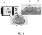
- FIG. 2 shows the cross-sectional image 10 in full size, a first section of the sectional image 10 showing tissue with a first degree of magnification and a second section of the first section showing degenerated tissue with a second level of magnification that is greater than the first level of magnification.
- the enlargement between the full-size sectional image 10 and the first section and between the first section and the second section is each graphically illustrated.
- the system 100 also has a display unit 103, which is set up to display the respective cross-sectional image 10, 10 'in an analysis area 40 of a display 50 in such a way that image areas 20 of the cross-sectional image 10, 10', which are degenerated tissue according to the classification of the classification unit 102 30 show, in the sectional image 10, 10 'displayed in the analysis area 40 are marked.
- a display unit 103 which is set up to display the respective cross-sectional image 10, 10 'in an analysis area 40 of a display 50 in such a way that image areas 20 of the cross-sectional image 10, 10', which are degenerated tissue according to the classification of the classification unit 102 30 show, in the sectional image 10, 10 'displayed in the analysis area 40 are marked.
- the display unit 103 can optionally cause the display 50 to display all sectional images belonging to a tissue sample together in the form of pictograms in a release representation 79, in particular before the sectional images are handed over to the classification unit 102.
- the user can use the release display to make a preselection as to which Cutting images should be released for further processing steps.
- cross-sectional images that are obviously affected by artifacts can be easily sorted out.
- Fig. 3 Only a pictogram for the sectional image 10' is actually shown, while the further display locations 792 for the further sectional images are only shown schematically. In the case shown, the cross-sectional images correspond to whole-slide scans, so the display locations are numbered by slide.
- the system 100 has a detection unit 104, which is set up to detect tissue degeneration in the tissue sample based on user input, which is indicative of a confirmation by the user that one of the marked image areas 20 shows degenerated tissue 30.
- the image provision unit 101 can be set up to provide the plurality of cross-sectional images 10 of the tissue sample, wherein the classification unit 102 can be set up to assign image areas 20 in each of the cross-sectional images 10, 10' of the tissue sample by means of the trained neural network according to their probability of doing so classify that they show degenerated tissue.
- the display 50 shown selection elements 70 are displayed in a selection area 60, each of which corresponds to one of the plurality of sectional images 10, 10 '. In the case shown, the user has selected the topmost of the selection elements 70 shown, so that a sectional image 10 'corresponding to this selection element is displayed in the analysis area 40 of the display.
- the user can scroll through the selection elements corresponding to the various sectional images by operating a scroll bar located on the right edge of the selection area.
- the selection elements each have a pictogram of the associated sectional images, which is shown in Fig. 4 is only shown for the selected selection element.
- the selection elements 70 have an area in which selected characteristic values are displayed, which characterize the properties of the respective cross-sectional images.
- the selection elements, as shown, can also be displayed with a colored border, for example, which is indicative of whether degenerated tissue is shown in the associated sectional image according to the classification of the classification unit 102.
- Fig. 4 corresponds to the area of the cross-sectional image 10 'represented in the analysis area 40 by the user that is displayed as a pictogram in the associated selection element 70.
- the user can freely determine the area of the sectional image 10' shown in the analysis area 40 of the sample representation 80, for example by moving or enlarging the area.
- the selection elements 70 are displayed in an orderly manner in the selection area 60, the order, which could also be referred to as a sequence, being based on a relevance assigned to the associated sectional images 10, 10 '.
- the relevance is measured using a relevance parameter which is indicative of a proportion of those image areas 20 in the respective associated cross-sectional image 10, 10' which show degenerated tissue according to the classification of the classification unit 102.
- a relevance parameter which is indicative of a proportion of those image areas 20 in the respective associated cross-sectional image 10, 10' which show degenerated tissue according to the classification of the classification unit 102.
- Fig. 4 This means, for example, that the sectional image 10 ', which corresponds to the topmost of the displayed selection elements 70, has a larger proportion of image areas showing degenerated tissue according to the classification of the classification unit 102 than the sectional images that correspond to the selection elements 70 arranged further down.
- the relevance-based arrangement of the selection elements 70 in the sample representation 80 allows the user to easily find those cross-sectional images 10, 10' of a given tissue sample in which a particularly large number of areas prone to degeneration can be seen, which can lead to a quicker assessment of whether the tissue sample actually shows degenerated tissue or not.
- the respectively selected sectional image 10, 10' can be displayed in the analysis area 40 of the sample representation 80 in such a way that contiguous areas of image areas 20, which show degenerated tissue according to the classification of the classification unit 102, are marked beyond the marking of the image areas 20 themselves, if the size of the respective contiguous area falls below a minimum size.
- contiguous areas of image areas 20 in the cross-sectional image 10 ', which show degenerated tissue according to the classification of the classification unit 102 are marked with flags, it can be seen that such a marking has the advantage that even with a relatively low level of magnification of the cross-sectional image 10 ⁇ in the analysis area 40 the probably degenerated tissue areas can be easily localized. In this way, the user can easily arrive at an assessment of the spatial distribution of the likely degenerated tissue areas.
- the marking of the contiguous areas of image areas 20 which show degenerated tissue according to the classification of the classification unit 102 is not necessarily an additional marking in addition to the marking of the image areas 20 which show the degenerated tissue according to the classification of the classification unit 102, in particular not necessarily an additional one Marking within one and the same representation of the display 50.
- the marking of the contiguous areas of image areas 20 that have degenerated according to the classification of the classification unit 102 Show tissue can also be provided as the only marking, in particular as the only marking within one and the same representation of the display 50.
- a user input regarding a marking of a contiguous area of image areas 20, which show degenerated tissue according to the classification of the classification unit 102, can lead to the cross-sectional image 10, 10 'displayed in the analysis area 40 of the sample representation 80 being included in the analysis area 40 is displayed centered with increased magnification and/or with respect to the area belonging to the marking.
- Such a user input can be, for example, a mouse click on a flag used for marking.
- the sample representation 80 can also have an input mask, via which the user can, on the one hand, enter the total number of lymph nodes he has identified in this sectional image for the selected sectional image 10, 10 ⁇ and, on the other hand, the number of them according to his Analysis of lymph nodes containing degenerated tissue, ie in this case affected by metastases, can be entered.
- the user has, for example, recognized two lymph nodes in the selected cross-sectional image 10', assessing both of them to be actually affected.
- the display unit 103 can cause the display 50 to move from the sample representation 80 to the one in Fig. 5A or 5B exemplary and schematically illustrated sectional view 90 to change.
- a separate button can be provided in the sample display 80.
- the sectional image 10, 10' previously selected in the sample display 80, i.e. the last active sectional image 10, 10', can then be displayed in the sectional image representation 90 for more detailed analysis by the user.
- the sectional image representation 90 shown has its own selection area 61, in which its own selection elements 71 are displayed, each of which corresponds to an area of the sectional image belonging to the sectional image representation 90, which shows degenerated tissue according to the classification of the classification unit 102.
- the sectional image representation 90 in the in Fig. 5A there are two separate analysis areas 41a, 41b, in which that area of the sectional image 10, 10' that probably contains degenerated tissue is displayed, which corresponds to a selection element 71 selected by the user from the selection area 61 of the sectional image representation 90.
- first analysis area 41a a section of the sectional image 10, 10' is shown with a first degree of magnification is displayed, which corresponds to the selected area, and in the second analysis area 41b a section of the section shown in the first analysis area 41a is displayed, in this case aligned with the center of the tissue classified as degenerate in the selected area.
- the degree of magnification of the first analysis area 41a is smaller than the degree of magnification of the second analysis area 41b, so that the user is able to view the tissue classified as degenerate in the second analysis area in detail and at the same time keep an eye on the position of the tissue area using the first analysis area 41a .
- the selection elements 71 of the selection area 61 of the sectional image representation 90 can also contain pictograms, in which case the pictograms show the area of the sectional image corresponding to the respective selection elements 71 that is marked as degenerate.
- the image areas of the cross-sectional image classified as showing degenerate tissue are marked, on the one hand, by borders of a different color, that is to say in comparison to the image areas not classified as showing degenerate tissue, and on the other hand by the fact that they are compared to those not shown as degenerate tissue Showing classified image areas can be displayed with increased contrast and increased brightness.
- the last-mentioned type of marking could also be understood to mean that the image areas of the cross-sectional image that are classified as not showing any degenerated tissue are displayed grayed out or masked. This naturally directs the user's focus to the relevant image areas of the cutaway image.
- the selection area 61 of the sectional image representation 90 also has a scroll bar on the right edge with which the user can scroll through the selection elements 71 corresponding to the different areas.
- the sectional image representation 90 in a display area 66 - in which Fig. 5A In the case shown below the second analysis area 41b - buttons that the user can press to confirm or revoke the classification determined by the classification unit 102.
- the user also has the option of using one of the buttons to indicate that they are unsure about whether the area displayed contains degenerated tissue.
- the sectional image display has a further button, which is in Fig. 5A to the right of the buttons intended for classification by the user, and serves to give the user the opportunity to close a case early.
- the prerequisite for early closure of a case is that at least one area of at least one of the cross-sectional images of the respective tissue sample that is classified by the classification unit 102 as showing degenerated tissue has been confirmed by the user.
- the determination unit 104 can be set up to check the existence of this requirement upon a user input that corresponds to early completion, that is, for example, to pressing a corresponding button, and if the result of this check is positive, the areas that have not yet been classified by the user as "unseen " and to store the classification determined by the classification unit 102 for this.
- the determination unit 104 can be set up to subsequently accept a positive finding for the case or at least the tissue sample belonging to the area confirmed by the user. The sooner the user completes the case processing, the greater the proportion of the classification results that will have been achieved solely by the classification unit 102, that is, without confirmation or revocation by the user. Regardless of whether the user completed the diagnosis process early or not, he or she can have the diagnosis result displayed in a final overview after the diagnosis of a case has been completed.
- the sectional image representation 90' shown is an exemplary alternative to the sectional image representation 90 Fig. 5A to understand.
- the difference between the sectional image representation 90 and the sectional image representation 90' is only that in the Cross-sectional image display 90 'instead of a single button ("Tumor") to confirm that the displayed area of the cross-sectional image contains degenerated tissue, two buttons ("Extracapsular Metastasis", "Intracapsular Metastasis”) are provided, which enable the user to do this to mark each displayed area according to whether it corresponds to degenerated tissue inside or outside a lymph node.
- the user can modify the display area 66 of the sectional image representation 90 Fig. 5A corresponds to a display area 66 ', thus further refining the classification of the classification unit.
- lymph node status of a case When assessing the lymph node status of a case, it can be crucial how many of the examined lymph nodes (LNs) are specifically affected. The user can also determine this manually (e.g. by taking notes with pen and paper) during the diagnosis. However, it is preferably intended that the user has the opportunity for each OT to enter the number of imaged and affected LK and to derive the relevant representation of the key figures for the entire case.
- further interactive elements can be displayed for the user in the sample representation 80 and/or the cross-sectional image representation 90, via which user input can be made regarding the number of lymph nodes shown in a cross-sectional image and the lymph nodes affected by metastases.
- Fig. 4 These are shown by way of example and schematically in the display area 65.
- the efficiency of image-based detection of tissue degeneration can also be increased through more resource-efficient machine performance.
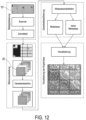
- it has been shown that it can make sense to design the classification unit 102 in two parts, namely in such a way that those image areas that show tissue at all are first recognized in the cross-sectional images, and Only then are those image areas that show degenerated tissue within the image areas that show tissue at all recognized. This is particularly beneficial for detecting degenerated tissue in images such as those in the Fig. 2 to 5 shown histological images, since in such images, due to the preparation of the tissue sample layers on microscope slides suitable for microscopy, the majority of the image area does not show any tissue.
- the classification unit 102 is therefore preferably set up to extract image areas 20 showing tissue from the sectional images 10, 10' and, by means of the trained artificial neural network, to assign only image areas 20 within the extracted, tissue-showing image areas 20 of the sectional images 10, 10' according to their probability of doing so classify that they show degenerated tissue 30.
- the wording “within the” is to be understood here to mean that the image areas passed to the trained artificial neural network for classification correspond to the image areas previously extracted as showing tissue or form a subset of these.
- the extraction of the image areas showing tissue can be achieved by the classification unit 102 using a specially trained, i.e. additional, artificial neural network for this task, which can be referred to as a “tissue detector”.
- the sectional images 10, 10 ' which are in the Fig. 2 to 5 What can be seen are histological images created using hematoxylin-eosin staining. Accordingly, both of the artificial neural networks used were trained using histological images also created by hematoxylin-eosin staining. In addition, they show in the Fig. 2 to 5 Sectional images 10, 10' shown on microscope slides are parts of a tissue sample that was taken from a patient's lymph node, the marked image areas 20 in the sectional images 10, 10' of Fig. 2 to 5 Contains metastatic tissue from a colorectal carcinoma. Accordingly, both artificial neural networks used were trained using cross-sectional images 10, 10' of lymph node tissue samples, some of which were affected by metastases from colorectal carcinoma.
- Fig. 6 shows, by way of example and schematically, a system 200 for generating training data for an artificial neural network, such as the artificial neural network of the classification unit 102 of the system 100.
- the system 200 includes a training data provision unit 201, which is set up to provide first training data.
- the first training data are structured in pairs of first training input data and associated first training output data, the first training input data corresponding to first image areas 21 of first cross-sectional images 10, 10 'of a plurality of tissue samples, some of which have tissue degeneration, and the first training output data relating to the first image areas 21 Findings correspond to whether these show degenerated tissue 30.
- the first training data can, for example, correspond to 500,000 pairs of first training input data and associated first training output data, wherein the first training data can have been generated manually. This means that personnel trained to generate the first training data may have looked through the image areas 21 used for training and made a determination for each of the image areas 21 as to whether it shows degenerated tissue 30 or not. Making these determinations, i.e. generating the training output data, can also be understood as “labeling”.
- the system 200 further has a network provision unit 202, which is set up to provide an artificial neural network that has been trained with the first training data to determine image areas showing degenerated tissue 30.
- the artificial neural network can, for example, have initially been provided as a prototype and then trained using the manually generated first training data.
- the system 200 further has a training data generation unit 203, which is set up to generate second training data that is structured in pairs of second training input data and associated second training output data.
- the second training input data correspond to second image areas 22 of either the first slice images 10, 10', that is to say the slice images 10, 10' of the first training data used to train the artificial neural network, and/or second, that is to say not yet used slice images 10, 10' the large number of tissue samples.
- the second cross-sectional images 10, 10' can be images of tissue samples that have already been used for training or images of tissue samples that have not yet been used for training, that is, from which no first training input data has been obtained. It is understood that the second sectional images were also previously provided by a unit of the system 200 intended for this purpose.
- the training data generation unit 203 of the system 200 is set up to select as second image areas 22 those image areas of the first cross-sectional images 10, 10' which overlap with such first image areas 21 which show degenerated tissue 30 according to the first training output data.
- the training data generation unit 203 is set up to use the trained artificial neural network to determine image areas 21 in the second cross-sectional images 10, 10 'that show degenerated tissue 30, and such image areas of the second cross-sectional images 10, 10' as second image areas 22 to select which overlap with those image areas 21 of the second cross-sectional images 10, 10' for which it has been determined by means of the trained artificial neural network that they show degenerated tissue 30.
- the second training output data is generated by the training data generation unit 203 by passing the second image areas 22 as input data to the artificial neural network provided, that is to say trained based on the first training data, the second training output data being based on the data then output by the artificial neural network Output data and additionally a user input are determined, which is indicative of a confirmation by the user of the output data output by the artificial neural network for the second image areas 22.
- the generation of the second training data which can be understood as a semi-automated process due to the involvement of the user, is in Fig. 7 shown as an example and schematically.
- the artificial neural network of the training data generation unit 203 trained on the basis of the first training data which could still be referred to as a prototype, has degenerated tissue 30 in one of the image areas in the cross-sectional image 10, 10 ', which is divided into 4 ⁇ 4 tile-shaped first image areas 21 recognized that in Fig. 7 is symbolized by a star.
- the slice image 10, 10' can be a (first) slice image that has already been used as part of the first training input data for training the artificial neural network, so that the star symbol could be interpreted as a label.
- the cross-sectional image 10, 10' could just as well correspond to a (second) cross-sectional image of a tissue sample that has not yet been used for training, in which the artificial neural network trained on the basis of the first training data has recognized the degenerated tissue 30. It is understood that a recognition of a tissue degeneration 30 by the artificial neural network is here only understood as a recognition with a certain probability, that is, without complete certainty.
- a training data generation area is determined which corresponds to an area of the cross-sectional image 10, 10 'that includes the image area 21 in which degenerated tissue 30 has been recognized, but above this image area goes beyond.
- the training data generation area can consist of all image areas 21 adjacent to this image area 21 in addition to the image area 21 in which degenerated tissue 30 has been detected.
- the training data generation area which is in Fig. 7 shown hatched can also be referred to as a region of interest.
- second image areas 22 are selected from the training data generation area for use as second training input data.
- the second image areas 22 can, as in Fig. 7 is shown, have the same shape as the first image areas 21, that is, for example, a tile-like shape.
- the training data generation unit 203 can be set up to move a virtual extraction window within the training data generation area, in particular over the entire training data generation area, and at different positions of the extraction window in each case from the extraction window to extract second image areas 22 corresponding to the framed image area.
- the training data generation area, the extraction window and the positions at which the second image areas 22 are extracted are selected so that the second image areas 22 overlap with the image area 21 in which the artificial neural network has detected degenerated tissue 30.
- the training data generation unit 203 can, for example, be set up to select the second image areas 22 so that the image areas 21 in which the artificial neural network has recognized degenerated tissue 30 are completely divided into the second image areas 22.
- the fact that the second image areas 22 overlap with image areas 21 for which the presence of degenerated tissue has been determined either by means of manual determinations or by means of the trained artificial neural network ensures that the second training input data at least partially correspond to image areas that have degenerated tissue 30 show.
- second image areas 22 necessarily show degenerated tissue, since in most cases the image areas 21 for which the presence of degenerated tissue has been determined will not be completely filled by the degenerated tissue, so that there is the possibility that in the overlap areas no degenerated tissue is shown in the second image areas 22.
- Not all second training input data necessarily correspond Image areas 22 that show degenerated tissue 30. This enables the generation of balanced training data that is therefore suitable for high-quality training.
- the selected second image areas 22, that is, for example, those in Fig. 7 Tiles of the sectional image 10, 10 'shown schematically on the far right are passed to the trained artificial neural network as input data by the training data generation unit 203 in a further step, whereupon the neural network outputs output data which corresponds to a probability determined by the neural network or to a probability determined by the neural network correspond to such a probability-based decision as to whether the respective image areas 22 show degenerated tissue 30 or not.
- the sectional image 10, 10' can be displayed to the user, for example in the display 50, in such a way that the image areas 22 are marked according to the output data output by the neural network.
- the user can then confirm or revoke the markings, and thus the output data output by the artificial neural network, via a corresponding user input, such as a mouse click or a touch on a touchscreen used for the display.
- a corresponding user input such as a mouse click or a touch on a touchscreen used for the display.
- the second training output data belonging to the second image areas 22 corresponding to the second training input data are generated.
- the output data output by the artificial neural network for the second image areas 22 that the user has confirmed can be accepted as the second training output data. It is therefore not absolutely necessary for the user to make user input regarding all second image areas of the sectional image 10, 10'.
- the second training data is added to the training data set containing the first training data.
- Fig. 8 shows by way of example and schematically a system 300 for training an artificial neural network such as the artificial neural network of the classification unit 102 of the system 100.
- the system 300 comprises a training data provision unit 301, which is set up to provide first training data in pairs of first training input data and associated first training output data are structured, wherein the first training input data correspond to first image areas 21 of first cross-sectional images 10, 10 'of a large number of tissue samples, some of which have tissue degeneration, and the first training output data correspond to determinations made with respect to the first image areas 21 as to whether these degenerate tissue 30 show.
- the training data provision unit 301 of the system 300 can correspond to the training data provision unit 201 of the system 200.
- the system 300 further includes a network provision unit 302, which is set up to provide the artificial neural network to be trained.
- the artificial neural network provided by the network provision unit 302 can therefore in particular be an artificial neural network that has not yet been trained.
- the network provision unit 302 of the system 300 can differ from the network provision unit 202.
- the system 300 further includes a training unit 303, which is set up to provisionally train the artificial neural network to be trained in such a way that the provisionally trained artificial neural network, when it receives the first training input data as input data, outputs output data that corresponds to the first one received Training input data correspond to the first training output data.
- the preliminarily trained artificial neural network may correspond to the artificial neural network provided by the network provisioning unit 202 of the system 200.
- the system 300 also includes a training data generation unit 304, which is set up to generate second training data that is structured in pairs of second training input data and associated second training output data, the second training input data being second image areas 22 of the first and/or second sectional images 10, 10 ⁇ correspond to the large number of tissue samples.
- a training data generation unit 304 which is set up to generate second training data that is structured in pairs of second training input data and associated second training output data, the second training input data being second image areas 22 of the first and/or second sectional images 10, 10 ⁇ correspond to the large number of tissue samples.
- the training data generation unit 304 is set up to select, as second image areas 22, those image areas of the first cross-sectional images 10, 10' which overlap with such first image areas 21 which show degenerated tissue 30 according to the first training output data, and/or by means of the provisionally trained artificial neuronal Network in the second cross-sectional images 10, 10 'to determine image areas 21 which show degenerated tissue 30, and to select as second image areas 22 those image areas of the second cross-sectional images 10, 10' which overlap with such image areas 21 of the second cross-sectional images, for which by means of the Preliminarily trained artificial neural network has been determined to show degenerated tissue 30.
- the training data generation unit 304 is set up to transfer the second image areas 22 as input data to the provisionally trained artificial neural network and to determine the second training output data based on the output data then output by the provisionally trained artificial neural network and also a user input that is indicative of is a confirmation by the user of the output data output by the provisionally trained artificial neural network for the second image areas 22.
- the training data generation unit 304 of the system 300 can therefore be the training data generation unit 203 of the system 200.
- the generation of the second training data by the system 300 can also be understood as a semi-automated process, since both the provisionally trained neural network is used and confirmation from the user is used.
- the training unit 303 of the system 300 is set up to additionally train the previously trained artificial neural network in such a way that the additionally trained artificial neural network, when it receives the first and / or second training input data as input data, outputs output data that corresponds to the first one received and/or second training input data corresponding to first and/or second training output data.
- the artificial neural network that has already been provisionally trained using the first training data is therefore additionally trained using the first and/or second training data.
- the steps of generating second training data and the additional training of the artificial neural network based on the training data set supplemented by the second training data can be repeated until the artificial neural network detects degenerated tissue with a satisfactory level of certainty, which can correspond to a predetermined accuracy.
- the advantage of the described semi-automated generation of second training data is that the training data set can be increased many times over in a relatively short time without affecting the quality of the training data.
- a larger training data set enables the training of larger artificial neural networks, which in turn can better represent the complex image morphological relationships in images of tissue samples, such as those found in particular in histological images of lymph node metastases from colorectal carcinomas.
- Both the preliminary and the supplementary training can be carried out in the form of supervised learning, in which artificial neural network pairs of respective image areas are given corresponding training input data and respective training output data, which can also be understood as labels. While manual labeling is required for the preliminary training, as already stated, labeling can be used for the supplementary training, which is generated semi-automatically, namely on the one hand by analysis results of the provisionally trained neural network and on the other hand by confirmation of these results a user.
- the image areas can be passed on to the artificial neural network in a pre-processed manner at any stage of training.
- image augmentation for example in the form of online augmentation, can be used.
- the image areas can be mirrored, rotated, changed in contrast, changed in brightness or changed in quality before they are handed over to the artificial neural network.
- the training result i.e. the achieved accuracy of the artificial neural network trained to this extent
- the training data set is enlarged in order to further train the artificial neural network using the enlarged training data set. This successive increase in the amount of first training data during preliminary training can be repeated until a predetermined accuracy is achieved.
- the training data provision unit 304 can be set up to provide first validation data that is structured in pairs of first validation input data and associated first validation output data, wherein a) the first validation data corresponds to a part of the first training data that is not used by the training unit 303 for training, or where b) the first validation input data correspond to third image areas of cross-sectional images of a large number of tissue samples, some of which have tissue degeneration, and the first validation output data correspond to determinations made with respect to the third image areas as to whether these show degenerated tissue 30.
- the determinations may be made manually, semi-automatically through manual confirmation of machine-obtained classification results, or fully automated using an artificial neural network known to achieve classification accuracy above a predetermined threshold. It is understood that the system 300 may have a unit for providing the third image areas.
- the training unit 303 may be configured to perform the preliminary training until the artificial neural network, upon receiving the first validation input data as input data, outputs output data having a predetermined accuracy with respect to the first validation output data.
- the predetermined accuracy can be measured using an accuracy measure that is defined over all validation input data, wherein the accuracy measure can assign a higher weight to those pairs of validation data for which the findings made indicate that the respective image area shows degenerated tissue 30 than to those pairs of validation data for which the findings made indicate that the respective image area does not show any degenerated tissue 30.
- the accuracy measure can assign a higher weight to those validation data pairs for which a determination has been made that indicates that the respective image area contains an image artifact than to those validation data pairs for which the determinations made indicate that the respective image area does not contain degenerated tissue 30 shows.
- the weights that the accuracy measure may assign to pairs of validation data for which the determinations made indicate that the respective image area shows a) degenerated tissue and b) an image artifact may be the same or different.
- Image artifacts can include, among others: staining artifacts (e.g. too much or too little color), compression of the tissue, scanning artifacts (e.g. blurred image), file errors (e.g. break lines in the image due to incorrect data), bleed and image of non-tissue (e.g. thread residue, cellulose ), clumps of color and tissue, superposition of several colored clusters of cells.
- staining artifacts e.g. too much or too little color
- compression of the tissue e.g. blurred image
- file errors e.g. break lines in the image due to incorrect data
- bleed and image of non-tissue e.g. thread residue, cellulose
- clumps of color and tissue e.g. thread residue, cellulose
- Fig. 9 shows, by way of example and schematically, a method 1100 for supporting a user in the image-based detection of tissue degeneration, the method 1100 comprising a step 1101 of providing 1101 of a plurality of cross-sectional images 10, 10 'of a tissue sample, as well as a step 1102 of classifying 1102 of Image areas 20 of the cross-sectional image 10, 10 'according to their probability of showing degenerated tissue 30, using a trained artificial neural network.
- a step 1103 of the method 1100 the cross-sectional image 10, 10' is displayed in an analysis area 40 of a display 50 in such a way that image areas 20 of the cross-sectional image 10, 10', which show degenerated tissue 30 according to the classification, in the cross-sectional image displayed in the analysis area 40 10, 10' are marked.
- tissue degeneration in the tissue sample is determined based on a user input, which is indicative of a confirmation by the user that one of the marked image areas 20 shows degenerated tissue 30.
- the method can be carried out by the system 100, for example.
- Fig. 10 shows, by way of example and schematically, a method 1200 for generating training data for an artificial neural network, wherein according to the method 1200, in a step 1201, first training data is provided, which is structured in pairs of first training input data and associated first training output data, the first training input data being in first image areas 21 of first cross-sectional images 10, 10 'correspond to a large number of tissue samples, some of which have tissue degeneration, and the first training output data correspond to determinations made with regard to the first image areas 21 as to whether these show degenerated tissue 30.
- a step 1202 of the method 1220 an artificial neural network that has been trained with the first training data to determine image areas showing degenerate tissue 30 is provided, with second training data being generated in a step 1203 of the method, which is in pairs of second training input data and associated second training output data are structured, wherein the second training input data correspond to second image areas 22 of the first and / or second cross-sectional images 10, 10 'of the plurality of tissue samples.
- the second image areas 22 selected are those image areas of the first cross-sectional images 10, 10' which overlap with those first image areas 21 which show degenerated tissue 30 according to the first training output data, alternatively or additionally using the trained artificial neural network in the second cross-sectional images 10 , 10 'image areas 21 are determined, the degenerate tissue 30 show, and as second image areas 22 Such image areas of the second section images 10, 10' are selected, which overlap with such image areas 21 of the second section images, for which the trained artificial neuronal network have been shown to show degenerated tissue 30.
- the second image areas 22 are transferred to the provided artificial neural network as input data, the second training output data being determined based on the output data then output by the provided artificial neural network and a user input that is indicative of a confirmation by the user of the artificial neural network that has been preliminarily trained Network output data for the second image areas is.
- Fig. 11 shows by way of example and schematically a method 1300 for training an artificial neural network, the method 1300 comprising a step 1301 of providing first training data, which are structured in pairs of first training input data and associated first training output data, the first training input data first image areas 21 of first sectional images 10, 10 'correspond to a large number of tissue samples, some of which have tissue degeneration, and the first training output data determines the first image areas 21 correspond to whether these show degenerate tissue 30, further a step 1302 of providing the artificial neural network to be trained, and a step 1303 of provisionally training the artificial neural network to be trained in such a way that the provisionally trained artificial neural network, if it receives first training input data as input data, outputs output data which correspond to the first training output data belonging to the received first training input data.
- the method 1300 includes generating second training data, which are structured in pairs of second training input data and associated second training output data, the second training input data corresponding to second image areas 22 of the first or second cross-sectional images 10, 10' of the plurality of tissue samples.
- the second image areas 22 selected are those image areas of the first cross-sectional images 10, 10' which overlap with those first image areas 21 which show degenerated tissue 30 according to the first training output data, and/or are converted into the second one using the previously trained artificial neural network Sectional images 10, 10' image areas 21 are determined which show degenerated tissue 30, and those image areas of the second sectional images 10, 10' are selected as second image areas 22 which overlap with such image areas 21 of the second sectional images, for which the previously trained artificial neural network is used have been determined to show degenerated tissue 30.
- the second image areas 22 are transferred to the provisionally trained artificial neural network as input data, the second training output data being determined based on the output data then output by the provisionally trained artificial neural network and a user input which is indicative of a confirmation by the user of the provisionally trained one artificial neural network for the second image areas 22 output data is.
- the provisionally trained artificial neural network is additionally trained in a further step 1305 of the method 1300, in such a way that the additionally trained artificial neural network, when it receives the first and / or second training input data as input data, outputs output data that corresponds to the correspond to first and/or second training output data belonging to the first and/or second training input data obtained.
- Fig. 12 is an exemplary and schematic flowchart for a clear representation of the overall process that leads to the display of sectional images 10, 10 ', in this case already from Fig. 4 and 5 known sectional image 10 ', with marking of image areas 20 classified by the classification unit 102 as showing degenerated tissue 30.
- the overall process begins with the creation of a respective sectional image 10, 10 'and its digitization, for example using a whole slide scanner, through which or on which the image is made available for diagnosis.
- any pre-processing of the cross-sectional image 10, 10' which in the case shown also includes its division into image areas 20, image areas 20 showing tissue are detected using the artificial neural network (tissue detector) trained specifically for this purpose and the number of image areas 20 is reduced to these.
- the tissue shown in the remaining image areas is classified into degenerated and non-degenerated tissue by the specially trained artificial neural network (tissue classifier), whereby the tissue degenerations in the case shown correspond to tumor metastases.
- tissue classifier tissue classifier
- the result of the classification is visualized for the user in such a way that he can easily find those tissue areas in the cross-sectional image 10, 10' that are highly likely to have tissue degeneration. For example, in order to determine a positive result, the user then only has to confirm the classification of these areas.
- Fig. 13 shows by way of example and schematically an embodiment for the "live analysis" of cross-sectional images 10", which are obtained by a user using a manually controlled light microscope 410 and a digital camera 412 connected to it.
- the microscope 412 has a support surface 413 on which microscope slides are arranged can, on each of which a tissue section has been prepared, the support surface 401 being movable vertically and horizontally by the user via a control device 430 attached to the microscope 412 and connected to a drive unit of the microscope 412.
- the control device 430 can be used to move the support surface 401 For example, have a joystick.
- an eyepiece 411 of the microscope 412 the user can view tissue areas focused by an optic of the microscope 412.
- the camera 412 While the user navigates with the microscope 410 through various tissue areas prepared on the slide, the camera 412 records a corresponding cross-sectional image sequence, the individual cross-sectional images 10 "correspond to what was also visible to the user at a given time via the eyepiece 411.
- the recorded sectional image sequence is passed on to the system 100, in particular provided by the image provision unit 101.
- the display unit 103 is set up to display the provided sectional image sequence in an analysis area 42 of a display 50.
- the transmission of the sectional image sequence, which can also be understood as a video, to the display 50 can take place essentially without any time delay, so that essentially what can also be seen via the eyepiece 411 of the microscope 410 can be displayed on the display 50.
- the user can also have the option of otherwise to stop the continuous display of the slice sequence or to jump back and forth within it.
- the display unit 103 can be set up to place a timeline with corresponding buttons over the analysis area 42.
- Such a timeline can also be understood as a selection area 62, since the user can use it to select individual slice images 10" of the displayed slice image sequence, that is, to display them.
- the classification unit 102 is also set up in this embodiment to classify image areas of the provided cross-sectional images 10", i.e. the individual "frames" of the cross-sectional image sequence, using an appropriately trained neural network according to their probability of showing degenerated tissue 30. As degenerated tissue 30 In this embodiment, classified image areas can then also be displayed marked accordingly in the analysis area 42 of the display 50. Furthermore, in this embodiment, the system 100 has a signal generation unit 105, which is set up to cause the control device 430 to produce an acoustic and/or haptic signal To generate a signal that can be perceived by the user if, while navigating through different tissue areas using the microscope 410, a tissue area is encountered in which the classification unit 102 has recognized degenerated tissue 30.
- the degenerated tissue can be marked in parallel with an acoustic/or haptic indication for the user. This has the advantage that the user does not have to look at the display 50 in order to be alerted to areas of tissue that are likely to be degenerated.
- control device 430 In addition to an operating element such as a joystick for controlling the microscope 410, the control device 430 preferably has further operating elements such as buttons, by means of which the user can separately save individual sectional images of the recorded sectional image sequence during the ongoing microscopy process.
- a sectional image saved by actuating a control element can correspond precisely to the view that the user has at the time the control element is actuated through the eyepiece 411.
- the user can use an acoustic and/or haptic signal that he perceives as an opportunity to linger on a tissue area that is currently visible to him via the eyepiece 411 and to examine it more closely, for example through appropriate optical magnification via the microscope 410, in order to then to decide whether he agrees or disagrees with the classification of the area conveyed via the signal by the classification unit. If he agrees, he can save a view of the tissue area in question that he prefers.
- the control device 430 can therefore also be understood as a determination unit 104, which is set up to determine tissue degeneration in a tissue sample based on a user input, which is indicative of a confirmation by the user that an image area marked based on a classification by the classification unit 102 is displayed degenerate tissue 30 shows
- Different operating elements of the control device 430 can be provided for storage in correspondingly different parts of a file system intended for this purpose or with correspondingly different annotations.
- the actuation of a control element of the control device 430 can also be understood as user input, and the sectional images 10" saved thereby as sections of the respective sectional image sequence.
- a separate storage device 420 can be provided for storing the sectional images 10" recorded by the user using the control device 430, which is part of the system 100 in this embodiment.
- Those cross-sectional images 10" of the recorded sequences in which the classification unit 102 has recognized degenerated tissue areas, and/or the cross-sectional images 10" specifically saved by the user using the control device 430 can be summarized in a digital "case folder", which is also referred to as a protocol could and can be stored in the storage device 420.
- These sectional images 10" can be displayed by the display unit 103 in a selection area 63 of the display 50. The user can use the selection area 63, when he turns his gaze to the display 50, to jump to previously saved sectional images 10", which then take place temporarily of the “live recording” can be displayed in the analysis area 42.
- the control device 430 can in particular have an operating element, the actuation of which by the user causes the storage device 420 to store a sectional image 10 ", which corresponds to a tissue area viewed by the user at the time of actuation, in a part of the storage device 420 intended for training data.
- the The first training data which is provided by the training data provision unit 201, 301 for training the neural network, can be stored in this part of the storage device.
- the microscope 410, the storage device 420 and the control device 430 can therefore be viewed together as a system 400 for generating training data for an artificial neural network.
- the user has the option of manually navigating through a tissue sample and selectively selecting views of the tissue for training the neural network, without having to look between the eyepiece 411 and a separate display, such as one arranged next to the microscope 410 Turn the monitor back and forth.
- the user is supported by the system 100 in recognizing potentially degenerated tissue areas by not only receiving visual signals through corresponding markings in the display 50, but also acoustic and/or haptic signals via the control device 430.
- the systems and procedures described also allow for standardization of the diagnostic process, which means quality assurance.
- Increasing efficiency can be understood, among other things, as making better use of attention and time.
- the most important tissue areas can be presented to the user, who may be an assessor, in a focused and reliable manner.
- the areas that are not essential for the assessment no longer have to be searched manually. This allows attention and time to be focused on the most important areas, so that more slides can be examined in the same time while maintaining the same load (or, alternatively, the load on the interpreter can be reduced).
- Quality assurance and standardization rely on consistent machine classification performance. In particular, the possibility of the examiner "closing" a slide prematurely before he/she has evaluated all suspicious areas can speed up the diagnosis in clear cases.
- the tissue samples of a case can first be evaluated macroscopically by a medical specialist, cut to size and placed in cassettes intended for this purpose for fixation.
- a marker can be activated in a digital case file that describes the characteristics essential for the system, i.e. a sample is characterized, for example, by attributes such as "colon” and "lymph node” (a selection of predefined markers/flags is possible). This marking can be used later in the analysis to automatically carry out corresponding processing steps.
- tissue samples or parts thereof can be embedded in paraffin and chemically fixed.
- the paraffin-embedded preparations can be removed in thin layers using appropriate cutting devices and applied to microscope slides. These can subsequently be stained using hematoxylin-eosin staining (HE), after which they can be made available for microscopic diagnosis.
- HE hematoxylin-eosin staining
- the preparations marked using flags can be selected and digitized in a corresponding scanner. New files created on a local storage medium with the scans can be checked for the presence of flags.
- the downstream analysis system can be informed about new files using a “notify” function. According to the registered flag marking, the analysis system provided can be started and the file(s) can be transferred.
- the image areas can be registered in a dedicated folder.
- the image areas are loaded from the folder into a volatile memory in batches and/or in parallel and are thus kept ready for analysis by the tissue classifier. To do this, they are linked as pairs of image files and the associated label or flag, for example a string variable.
- the bundles can be passed to the tissue classifier, optionally parallelized.
- a single unit or device can perform the functions of several elements listed in the claims.
- the fact that individual functions and elements are listed in different dependent claims does not mean that a combination of these functions or elements could not also be used advantageously.
- a computer program may be stored or distributed on a suitable medium, such as an optical storage medium or solid state medium (SSD), which may be provided together with or as part of other hardware, or by other means, such as via the Internet or other wired means or wireless telecommunications systems.
- a suitable medium such as an optical storage medium or solid state medium (SSD)
- SSD solid state medium
- the invention relates to a system for supporting a user in the image-based detection of tissue degeneration.
- the system includes an image provision unit that is set up to provide one of a plurality of cross-sectional images of a tissue sample, and a classification unit that is set up to assign image areas of the cross-sectional image according to their probability of doing so using a trained artificial neural network classify that they show degenerated tissue.
- the system comprises a display unit which is set up to display the sectional image in an analysis area of a display in such a way that image areas of the sectional image which show degenerated tissue according to the classification of the classification unit are marked in the sectional image displayed in the analysis area, and a determination unit which is set up to determine tissue degeneration in the tissue sample based on a user input, which is indicative of a confirmation by the user that one of the marked image areas shows degenerated tissue.
Landscapes
- Engineering & Computer Science (AREA)
- Health & Medical Sciences (AREA)
- General Health & Medical Sciences (AREA)
- Medical Informatics (AREA)
- Nuclear Medicine, Radiotherapy & Molecular Imaging (AREA)
- Radiology & Medical Imaging (AREA)
- Quality & Reliability (AREA)
- Computer Vision & Pattern Recognition (AREA)
- Physics & Mathematics (AREA)
- General Physics & Mathematics (AREA)
- Theoretical Computer Science (AREA)
- Image Analysis (AREA)
Applications Claiming Priority (1)
| Application Number | Priority Date | Filing Date | Title |
|---|---|---|---|
| DE102022116407.1A DE102022116407A1 (de) | 2022-06-30 | 2022-06-30 | System zur Unterstützung eines Nutzers bei der bildbasierten Erkennung einer Gewebeentartung |
Publications (1)
| Publication Number | Publication Date |
|---|---|
| EP4300418A1 true EP4300418A1 (fr) | 2024-01-03 |
Family
ID=87060194
Family Applications (1)
| Application Number | Title | Priority Date | Filing Date |
|---|---|---|---|
| EP23182491.3A Pending EP4300418A1 (fr) | 2022-06-30 | 2023-06-29 | Système d'assistance d'un utilisateur dans la détection basée sur l'image d'une dégustation tissulaire |
Country Status (2)
| Country | Link |
|---|---|
| EP (1) | EP4300418A1 (fr) |
| DE (1) | DE102022116407A1 (fr) |
Citations (2)
| Publication number | Priority date | Publication date | Assignee | Title |
|---|---|---|---|---|
| US20180046759A1 (en) * | 2016-08-12 | 2018-02-15 | Verily Life Sciences Llc | Enhanced pathology diagnosis |
| US20220084660A1 (en) * | 2019-05-29 | 2022-03-17 | Leica Biosystems Imaging Inc. | Artificial intelligence processing system and automated pre-diagnostic workflow for digital pathology |
-
2022
- 2022-06-30 DE DE102022116407.1A patent/DE102022116407A1/de active Pending
-
2023
- 2023-06-29 EP EP23182491.3A patent/EP4300418A1/fr active Pending
Patent Citations (2)
| Publication number | Priority date | Publication date | Assignee | Title |
|---|---|---|---|---|
| US20180046759A1 (en) * | 2016-08-12 | 2018-02-15 | Verily Life Sciences Llc | Enhanced pathology diagnosis |
| US20220084660A1 (en) * | 2019-05-29 | 2022-03-17 | Leica Biosystems Imaging Inc. | Artificial intelligence processing system and automated pre-diagnostic workflow for digital pathology |
Also Published As
| Publication number | Publication date |
|---|---|
| DE102022116407A1 (de) | 2024-01-04 |
Similar Documents
| Publication | Publication Date | Title |
|---|---|---|
| DE69131102T2 (de) | Untersuchungskontrollverfahren für bilder die auf einer anzeige gezeigt werden | |
| WO1997041416A1 (fr) | Procede d'analyse automatisee assistee par ordinateur d'echantillons tissulaires ou d'echantillons de liquides organiques | |
| EP3930563B1 (fr) | Dispositif pour la mise en oeuvre d'un procédé pour l'évaluation de lésions cutanées utilisant une intelligence artificielle | |
| EP2656277B1 (fr) | Dispositif et procédé de détermination d'objets dans une image en couleurs | |
| DE4211904C2 (de) | Automatisches Verfahren zum Erstellen einer Liste unterschiedlicher Arten für eine flüssige Probe | |
| DE102007018034A1 (de) | Automatische Bildanalyse und Quantifizierung für Fluoreszenz-In Situ-Hybridisierung | |
| DE112019005143T5 (de) | System zur co-registrierung medizinischer bilder unter verwendung eines klassifikators | |
| EP3540632B1 (fr) | Procédé pour la classification des échantillons tissulaires | |
| EP2787485A1 (fr) | Procédé et dispositif de détection automatique d'erreurs dans des corps flexibles | |
| DE112019004112T5 (de) | System und verfahren zur analyse mikroskopischer bilddaten und zur erzeugung eines annotierten datensatzes zum training der klassifikatoren | |
| DE102009006148A1 (de) | Bildvolumen-Browser mit variabel einstellbarem Orientierungsmaß | |
| DE102021114287A1 (de) | Mikroskopiesystem und Verfahren zum Erzeugen stilisierter Kontrastbilder | |
| EP1797533B1 (fr) | Procede et dispositif pour segmenter une representation numerique de cellules | |
| DE102020211843A1 (de) | Verfahren und Vorrichtung zur Analyse von Histopathologiebilddaten | |
| EP3637362A1 (fr) | Procédé d'ajustement d'une impression d'image | |
| DE10235656A1 (de) | Verfahren und Anordnung zur Mikroskopie | |
| DE102010028382A1 (de) | Verfahren und Computersystem zur Bearbeitung tomographischer Bilddaten aus einer Röntgen-CT-Untersuchung eines Untersuchungsobjektes | |
| DE112009005102T5 (de) | Aufspaltung mehrteiliger Objekte | |
| EP4300418A1 (fr) | Système d'assistance d'un utilisateur dans la détection basée sur l'image d'une dégustation tissulaire | |
| DE102021121635A1 (de) | Automatisiertes trainieren eines maschinengelernten algorithmus basierend auf der überwachung einer mikroskopiemessung | |
| DE19637741B4 (de) | Automatisches Zytologisches Probenanalysesystem unter Verwendung von individualisierten Patientendaten | |
| DE69831573T2 (de) | Inspektionssystem mit probenvorschau | |
| DE102004043262B4 (de) | Verfahren zur Aufnahme von Bildern eines Untersuchungsbereichs eines menschlichen oder tierischen Körpers in einer Magnetresonanzanlage und zugehörige Magnetresonanzanlage | |
| EP3155588B1 (fr) | Système d'acquisition et de traitement d'une image d'un corps complet et procédé pour faire fonctionner celui-ci | |
| DE4244708C2 (de) | Verfahren und Vorrichtung zum Erstellen einer Artenliste für eine flüssige Probe |
Legal Events
| Date | Code | Title | Description |
|---|---|---|---|
| PUAI | Public reference made under article 153(3) epc to a published international application that has entered the european phase |
Free format text: ORIGINAL CODE: 0009012 |
|
| STAA | Information on the status of an ep patent application or granted ep patent |
Free format text: STATUS: THE APPLICATION HAS BEEN PUBLISHED |
|
| AK | Designated contracting states |
Kind code of ref document: A1 Designated state(s): AL AT BE BG CH CY CZ DE DK EE ES FI FR GB GR HR HU IE IS IT LI LT LU LV MC ME MK MT NL NO PL PT RO RS SE SI SK SM TR |
|
| STAA | Information on the status of an ep patent application or granted ep patent |
Free format text: STATUS: REQUEST FOR EXAMINATION WAS MADE |
|
| 17P | Request for examination filed |
Effective date: 20240703 |
|
| RBV | Designated contracting states (corrected) |
Designated state(s): AL AT BE BG CH CY CZ DE DK EE ES FI FR GB GR HR HU IE IS IT LI LT LU LV MC ME MK MT NL NO PL PT RO RS SE SI SK SM TR |
|
| STAA | Information on the status of an ep patent application or granted ep patent |
Free format text: STATUS: EXAMINATION IS IN PROGRESS |
|
| 17Q | First examination report despatched |
Effective date: 20260320 |