EP4303323B1 - Procédé de différenciation de différenciation de nodules thyroïdiens bénins et malins - Google Patents
Procédé de différenciation de différenciation de nodules thyroïdiens bénins et malins Download PDFInfo
- Publication number
- EP4303323B1 EP4303323B1 EP22460030.4A EP22460030A EP4303323B1 EP 4303323 B1 EP4303323 B1 EP 4303323B1 EP 22460030 A EP22460030 A EP 22460030A EP 4303323 B1 EP4303323 B1 EP 4303323B1
- Authority
- EP
- European Patent Office
- Prior art keywords
- thyroid
- genes
- sample
- nodule
- gene
- Prior art date
- Legal status (The legal status is an assumption and is not a legal conclusion. Google has not performed a legal analysis and makes no representation as to the accuracy of the status listed.)
- Active
Links
Images


Classifications
-
- C—CHEMISTRY; METALLURGY
- C12—BIOCHEMISTRY; BEER; SPIRITS; WINE; VINEGAR; MICROBIOLOGY; ENZYMOLOGY; MUTATION OR GENETIC ENGINEERING
- C12Q—MEASURING OR TESTING PROCESSES INVOLVING ENZYMES, NUCLEIC ACIDS OR MICROORGANISMS; COMPOSITIONS OR TEST PAPERS THEREFOR; PROCESSES OF PREPARING SUCH COMPOSITIONS; CONDITION-RESPONSIVE CONTROL IN MICROBIOLOGICAL OR ENZYMOLOGICAL PROCESSES
- C12Q1/00—Measuring or testing processes involving enzymes, nucleic acids or microorganisms; Compositions therefor; Processes of preparing such compositions
- C12Q1/68—Measuring or testing processes involving enzymes, nucleic acids or microorganisms; Compositions therefor; Processes of preparing such compositions involving nucleic acids
- C12Q1/6876—Nucleic acid products used in the analysis of nucleic acids, e.g. primers or probes
- C12Q1/6883—Nucleic acid products used in the analysis of nucleic acids, e.g. primers or probes for diseases caused by alterations of genetic material
- C12Q1/6886—Nucleic acid products used in the analysis of nucleic acids, e.g. primers or probes for diseases caused by alterations of genetic material for cancer
-
- C—CHEMISTRY; METALLURGY
- C12—BIOCHEMISTRY; BEER; SPIRITS; WINE; VINEGAR; MICROBIOLOGY; ENZYMOLOGY; MUTATION OR GENETIC ENGINEERING
- C12Q—MEASURING OR TESTING PROCESSES INVOLVING ENZYMES, NUCLEIC ACIDS OR MICROORGANISMS; COMPOSITIONS OR TEST PAPERS THEREFOR; PROCESSES OF PREPARING SUCH COMPOSITIONS; CONDITION-RESPONSIVE CONTROL IN MICROBIOLOGICAL OR ENZYMOLOGICAL PROCESSES
- C12Q2600/00—Oligonucleotides characterized by their use
- C12Q2600/112—Disease subtyping, staging or classification
-
- C—CHEMISTRY; METALLURGY
- C12—BIOCHEMISTRY; BEER; SPIRITS; WINE; VINEGAR; MICROBIOLOGY; ENZYMOLOGY; MUTATION OR GENETIC ENGINEERING
- C12Q—MEASURING OR TESTING PROCESSES INVOLVING ENZYMES, NUCLEIC ACIDS OR MICROORGANISMS; COMPOSITIONS OR TEST PAPERS THEREFOR; PROCESSES OF PREPARING SUCH COMPOSITIONS; CONDITION-RESPONSIVE CONTROL IN MICROBIOLOGICAL OR ENZYMOLOGICAL PROCESSES
- C12Q2600/00—Oligonucleotides characterized by their use
- C12Q2600/158—Expression markers
Definitions
- the invention relates to a method of differentiating benign and malignant thyroid nodules and is intended for use in the area of preoperative diagnosis.
- Another subject of the invention is the use of a hybridization probe set to detect in a thyroid nodule sample the expression level of the corresponding gene in order to differentiate benign and malignant thyroid tumor
- the normal follicular thyroid cell is highly differentiated, showing specialized features, including the ability to respond to thyrotropic hormone (TSH), iodine transporters, the ability to bind iodine with thyroid peroxidase (TPO) and store it as bound to thyroglobulin (Tg), and maintain follicular structure.
- TSH thyrotropic hormone
- TPO thyroid peroxidase
- Tg thyroglobulin
- Abnormal proliferation of thyroid cells can result in the formation of nodules, which can be benign or malignant.
- Malignant thyroid tumors comprise papillary, follicular, and anaplastic carcinoma. Medullary carcinoma of the thyroid gland originates from perifollicular C cells.
- Papillary thyroid carcinoma is the most common type of differentiated thyroid cancer that originates from follicular thyroid cells, and the diagnosis of follicular thyroid carcinoma (FTC) poses the most significant diagnostic challenges preoperatively.
- FTC follicular thyroid carcinoma
- DTC differentiated thyroid carcinomas
- Routine diagnosis of thyroid nodules is based on imaging evaluation by ultrasonography and cytological evaluation of material (biological sample) obtained by fine-needle aspiration biopsy (FNAB).
- the material includes tissue fragments taken from the tumor by puncture and/or aspiration.
- the Bethesda system (TBSRTC - The Bethesda System for Reporting Thyroid Cytology) is used to determine the potential of the examined cells in the cytologic smear.
- cytologic picture It classifies the cytologic picture into six categories ranging from non-diagnostic lesions - B1 to malignant neoplasm - B6, where category B2 corresponds to a benign lesion, B3 - a follicular lesion of undetermined significance, category B4 - suspicoius for a follicular neoplasm, and B5 suspiciois for malignancy.
- Fine-needle biopsy is the standard diagnostic procedure for suspicious thyroid nodules, but it has many limitations due to its inability to assess the arrangement of follicular cells within the thyroid follicle structure and to evaluate any invasive features.
- the amount of specimens obtained from the fine-needle biopsy is small, so immunohistochemical methods are difficult to apply. For this reason, nearly 30% of all biopsies of thyroid nodules are classified as indeterminate lesions, while only 15-30% of them are malignant lesions that require surgical intervention. It means that most of the resected nodules are benign and do not require radical medical management. Thus, the thyroid surgery performed may be considered ex-post unnecessary
- Gene classifiers based on assessing mutations of a few or even dozens of genes or evaluating the expression of a set of genes are well known. The most widespread is an expression classifier based on the study by Chudova et al., where the evaluation of the expression of a gene set using high-density oligonucleotide microarrays was used. The test is divided into several stages, and for a lesion deemed suspicious, an assessment of BRAF gene mutations and RET/PTC1 and RET/PTC3 rearrangements is additionally performed. It is a "rule out malignancy" test, and the result is primarily used to exclude malignancy of the nodule. Based on the results of this study, the Afirma Gene Expression Classifier test (Veracyte Inc, USA) was developed.
- GSC Genomic Sequencing Classifier
- the purpose of the present invention is to provide more effective differentiation between malignant and benign thyroid nodules.
- the system for classifying a thyroid nodule as malignant or benign is based on identifying sets of gene transcripts and involves determining the expression level of one or more transcripts in a sample, at least one of which contains the corresponding at least one option from SEQ ID Nos. 1-12, 261, 283-306, 657, 658 and 659.
- the classification system provides sets of target sequences and combinations of polynucleotide probes and derived primers that can be provided in a solution or an array.
- the invention known from EP2505664B1 patent description, relates to a method for distinguishing between malignant and benign thyroid nodules by performing RNA extraction in a standard FNAB specimen, particularly in an air-dried FNAB smear.
- the isolated RNA is analyzed for the presence of gene rearrangements and/or miRNA expression, with the presence of RET/PTC and/or PAX8/ PPARG rearrangements and/or mutations in genes encoding BRAF, N-, K-, and/or HRAS and/or differential miRNA expression in the sample classifying the tumor as malignant.
- US10236078B2 and US10672504B2 patent descriptions relate to methods for molecular profiling and diagnosis of genetic disorders and cancer, including gene expression markers associated with cancer or genetic disorders. Both inventions provide algorithms and methods for classifying thyroid cancer, determining molecular profiles, and analyzing the results to make a diagnosis.
- US10672504B2 invention relates to compositions and methods for molecular profiling and diagnostics for genetic disorders and cancer, including but not limited to gene expression product markers associated with cancer or genetic disorders.
- the present invention provides algorithms and methods of classifying cancer, such as thyroid cancer, methods of determining molecular profiles, and methods of analyzing results to provide a diagnosis.
- a method for processing or analyzing a sample of thyroid tissue of a subject comprising: (a) sequencing nucleic acid molecules from said sample of thyroid tissue to yield data comprising one or more levels of gene expression products in said sample of thyroid tissue, which one or more levels of gene expression products correspond to a plurality of genes comprising two or more genes selected from the group consisting of: ALK, CALCA, DICER1, IGF2BP3, MET, NTRK1, NTRK3, PAX8, PTEN, PTH and THADA; (b) using a trained algorithm in a computer to process said data from (a) to generate a classification of said sample of thyroid tissue as positive or negative for thyroid cancer at an accuracy of at least 85%, wherein said trained algorithm is trained with a plurality of training samples comprising known benign samples and known non-benign samples that is different from said sample of tissue; and (c) electronically outputting a report that identifies said classification of said sample of thyroid tissue as positive or negative for said thyroid cancer.
- the use of genes showing differential expression in benign and malignant thyroid nodules for the diagnosis and staging of thyroid cancer is disclosed in the EP1756303B1 patent description.
- the EP3467123A2 invention concerns compositions, kits and methods for molecular profiling and diagnosis of cancer, including gene expression markers, alternative exon usage markers, and DNA polymorphisms associated with cancer, particularly thyroid cancer.
- the description of patent US7901888B2 provides information on methods to diagnose, predict and stage thyroid cancer, using molecular markers.
- biomarkers for thyroid nodule malignancy classification are selected from a group consisting of NPC2, S100A11, SDC4, CD53, MET, GCSH, and CHI3L1 using a quantitative real-time polymerase chain reaction (PCR) array.
- one or more reference genes are selected from the following group, including TBP, RPL13A, RPS13, HSP90AB1, and YWHAZ.
- the WO2009029266A2 patent relates to the use of differentially expressed thyroid genes (DETs) for diagnosing and staging thyroid cancer.
- DET genes include C21orf4 (DET1), Hs.145049 (DET2), Hs.296031 (DET3), KIT (DET4), LSM7 (DET5), SYNGR2 (DET6), C11orf8 (DET7), CDH1 (DET8), FAM13A1 (DET9), IMPACT (DET10), KIAA1128 (DET11).
- invention uses a gene set consisting of ANXA1, CHl3L1, CITED1, FN1, GDF15, LRP4, MET, PROS1, SFTPB, TPO, MPPED2, TFF3, DPP4 for diagnosing papillary thyroid cancer.
- the gene expression profile is examined, wherein the expression profile for papillary thyroid cancer is the profile encompassing the increased expression of the following genes: ANXA1, CHI3L1, CITED1, FN1, GDF15, LRP4, MET, PROS1, SFTPB, MPPED2, TFF3 and DPP4 as well as the decreased expression of TPO.
- a method for diagnosing a genetic disorder or cancer as known from the US20202974A patent includes detecting gene expression in a biological sample, comparing gene expression in the biological sample with a control sample, classifying the biological sample by inputting one or more differential gene expression levels into a trained algorithm; and identifying a biological sample as positive for a genetic disorder or cancer if the trained algorithm classifies the sample as positive for a genetic disorder or cancer at a certain level of specificity and/or sensitivity.
- the invention relates to components, kits, and methods of molecular profiling for diagnosing thyroid nodules.
- the method of evaluating a thyroid nodule sample involves determining the expression level of one or more genes, one or more of which correspond to at least five genes selected from SNCA, STK32A, THRSP, TIMP1, TIMP2, TMSB10, TNFRSF17, TNFRSF1A, TXNDC12, VWA5A, WAS, WIPI1 and ZFYVE16 and then classifying the thyroid nodule as benign or suspicious by comparing the gene expression levels of the sample with gene expression data for at least two different sets of biomarkers.
- Gene expression data for each set of biomarkers includes one or more reference gene expression levels correlated with the presence of one or more tissue types. The expression level is compared sequentially with gene expression data for the mentioned at least two different sets of biomarkers.
- a method of diagnosing a thyroid nodule involves determining the amount of one or more microRNAs selected from a group including miR-146b-5p, miR-146b-3p, miR-221-5p, miR-221-3p, miR-222-5p, miR-222-3p, miR-181a-5p and miR-182-5p in a biological sample, and then comparing the expression of the microRNAs with the expression level of microRNAs in a control group.
- a thyroid tissue sample is identified as malignant if the expression level of any one or more of the CXCR3, the CAR, and/or XB130 genes is decreased; and/or the expression level of any one or more of the CCR3, CXCL10, CK-19, TIMP-1, CLDN-1, and/or CCR7 genes is increased in the thyroid tissue sample as compared to the normal control expression level, wherein the total number of genes with increased or decreased expression is at least three.
- the method according to the invention for differentiating benign and malignant thyroid nodules in tumor biopsy material includes the steps of RNA isolation, and determining the expression level of genes, and using a classifier, comparing the gene expression of a thyroid nodule sample with the gene expression of thyroid nodule samples in a training set.
- the thyroid nodule identifies the thyroid nodule as malignant or benign and determines the probabilities of malignancy of the thyroid nodule, with the classifier trained on gene expression data correlated with the nature of the thyroid nodule, especially benign and malignant tumors including papillary and follicular thyroid carcinoma.
- the expression level of a set of 32 genes is determined, which consist: AMOT, C3, CAMK2N1, CFI, DCSTAMP, DIO1, DUSP5, EGR1, EMP2, FAM20A, FAXC, FLRT3, HBG1, IGFBP6, IGSF1, ITGA2, KCNQ3, MET, METTL7B, MPZL2, NOD1, OLR1, PDE5A, PSD3, SDC4, SLC26A4, SLC26A7, SLC5A8, SLPI, TM4SF1, TPO, and TUSC3, and the normalization of the expression of each gene of this set is carried out under the endogenous control of the following six normalizing genes: EIF3A (EIF3S10), HADHA, UBE2D2, EIF5, PPIE, and RPLPO.
- a nodule sample's malignant or benign character is determined with at least 91% specificity and at least 83% classification sensitivity for the normalized expression level of a set of 32 genes.
- the nature of the nodule and the probability of malignancy of the tested sample are determined according to the decision rule of the classifier, taking into account the data on the expression level of the set of 32 genes and the gene expression level of each nodule sample in the training set, determined for each sample of this set for the same set of 32 genes and under the same normalization conditions.
- the nature and probability of malignancy of a nodule sample are determined according to the criterion of maximum likelihood for a probabilistic model of normalized expression values of a set of 32 genes of the sample.
- RT-qPCR oligonucleotide microarray, or RNA-seq transcriptome sequencing methods are used to detect the expression levels of a set of genes.
- a set of genes that exhibit a specific pattern of altered expression in thyroid cancer is presented to support the differential diagnosis of thyroid nodules.
- the study uses fine-needle biopsy specimens of thyroid nodules of the Polish population with a full spectrum of diagnoses covering Bethesda categories from B2 to B6.
- the classification of a nodule sample determines the range of probability of malignancy, not just the dichotomous categorization (benign/malignant), which is important for further decisions.
- the gene set considering the quality of the classification, is limited in number, which is essential for the cost of the test.
- Ultrasound examination is the first step in the diagnosis of a thyroid nodule after clinical evaluation. Depending on the estimated risk for a given image, the threshold size of the thyroid nodule for further evaluation is determined.
- a fine-needle aspiration biopsy of a thyroid nodule provides material for cytologic analysis, the result of which provides clinically useful data. The result categorized as Bethesda B3, B4, or B5 is subjected to additional study. B3, B4, and B5 nodules are collectively referred to as indeterminate nodules.
- Clinical information based on the result of the cytologic examination provided by the pathologist is used to preselect thyroid nodules that qualify for additional molecular diagnostics using gene classifiers that assess only the change in gene expression.
- the clinical evaluation of the specimen, performed by a pathologist or team of pathologists is a subjective evaluation based on the professional experience of the evaluating physician. In the case of improper clinical classification and erroneous preselection, there may be a discrepancy between the gene evaluation and clinical evaluation of the tumor sample.
- Differentiating a thyroid nodule as benign or malignant by classifying it based on gene expression values requires an exploratory procedure and multivariate analysis with an initial fine-needle biopsy sample of the nodule containing the material for RNA isolation and an expression classifier.
- RNA Total RNA was used in the study. Material collected by aspiration is protected in preservation buffer, frozen, and next RNA isolation and gene expression testing are performed using qPCR, microarray, or NGS techniques. Based on the transcript expression data, a molecular assay is done.
- the computer system provides an expression classifier as a product of computer software, having a computer-readable medium and the resources to carry out classification, modeling, and gene expression measurement methods.
- it has at least one processor and at least one program memory that contains the processor's execution instructions (classifier algorithm) for analyzing data and determining gene expression levels. Based on the classifier, decision rules are generated. In addition, it contains datasets for testing fine-needle biopsy samples of thyroid nodules.
- the classifier can distinguish and characterize samples based on the expression of a designated set of genes.
- the classifier based on a Support Vector Machine (SVM) model with a radial kernel function and trained on measured gene expressions for a population of training samples, enables the prediction of sample class and determination of malignancy probability.
- SVM Support Vector Machine
- the samples used for training are a part of the model and are called support vectors.
- the classifier was validated using the bootstrap method (500 iterations).
- the class suggested by the classification describes two states: benign lesion or malignant lesion, whereby the test can be a cancer exclusion or confirmation test as needed.
- probability scaling methods such as the Vapnik scaling method, isotonic regression, or Platt scaling.
- the SVM model uses the Platt method, based on the criterion of the maximum likelihood method, to calculate the probability of malignancy of a sample.
- Platt scaling transforms the results of the SVM classifier into a probabilistic model under which the data values are most likely. The evaluation of the probability of malignancy is based on training data.
- the probability of malignancy of a tested sample is determined according to the criterion of maximum likelihood for a probabilistic model of the expression values of a set of 32 genes determined for this sample.
- the set of 32 genes described in the subsequent sections was determined through research and microarray analyses.
- the training set comprised 336 thyroid nodule samples, including 70 nodules with a cytologic diagnosis of a benign lesion (Bethesda II - B2), 68 nodules with a cytologic diagnosis of follicular lesion of undetermined significance (Bethesda III - B3), 61 nodules with cytologic diagnosis suspicious for a follicular neoplasm (Bethesda IV - B4), 47 nodules suspicious for thyroid cancer (Bethesda V -B5) and 90 nodules with a cytologic diagnosis of thyroid cancer (Bethesda VI - B6).
- the clinical characteristics of the training set are presented in the table below.
- the whole training set Benign nodules Malignant nodules Number 336 207 129 F/M 283/52 173/30 107/22 Median age 57 Symptoms 27/336 14/207 13/129 Median nodule diameter 15 EU TIRADS 3 99/328 78/201 21/127 EU TIRADS 4 102/328 81/201 21/127 EU TIRADS 5 127/328 42/201 85/127 Bethesda II 70/336 70/207 0/129 Bethesda III 68/336 65/207 3/129 Bethesda IV 61/336 56/207 5/129 Bethesda V 47/336 16/207 31/129 Bethesda VI 90/336 0/207 90/129
- High-density microarray Human Transcriptome 2.0 (Affymetrix) was used to analyze the gene expression profile.
- the microarray set After preprocessing and background correction of the entire microarray set, the microarray set underwent a normalization process.
- a custom CDF (Chip Definition File) version 21 was used, which was downloaded from the website: http://brainarray.mbni.med.umich.edu /Brainarray/Database/CustomCDF/CDF_download.asp
- the resulting data was further analyzed.
- a fusion of feature selection methods based on Fold Change and t-test, recursive SVM elimination (RFESVM), and RegVif [8] were used.
- a radial kernel SVM classifier was used to validate the performance of the models.
- Classification quality was estimated with the bootstrap method, in which samples were drawn from the training set with replacement - 500 iterations. Feature selection, as well as classification and classification quality estimation, were carried out independently in each iteration for unbiased estimation of classifier models.
- Assay ID Gene Symbol(s) Gene Name(s) 1 Hs00611096_m1 AMOT angiomotin 17 Hs01120412_m1 KCNQ3 potassium voltage-gated channel subfamily Q member 3 2 Hs00163811_m1 C3 complement component 3 18 Hs01565584_m1 MET MET proto-oncogene, receptor tyrosine kinase 3 Hs00218591_m1 CAMK2N1 calcium/calmodulin dependent protein kinase II inhibitor 1 19 Hs00378551_m1 METTL7B methyltransferase like 7B 4 Hs00989715_m1 CFI complement factor I 20 Hs01083647_m1 MPZL2 myelin protein zero like 2 5 Hs00229255_m1 DCSTAMP dendrocyte expressed seven transmembrane protein 21 Hs01036720_m1 NOD1 nucleotide binding oligomerization domain containing 1 6 Hs001749
- the developed SVM classifier has very good parameters useful for differentiating thyroid nodules. It is confirmed by the results of the study, in which 91% of the thyroid nodules examined were correctly classified as benign and malignant. The sensitivity and specificity of the diagnostic test were estimated at 83% and 96%, respectively.
- the classification test includes a stage of sample normalization.
- An important element for proper validation of feature selection is the selection of reference genes. The essence of the analysis was to evaluate not only individual genes as candidates for reference genes but groups of genes that share these properties. Due to the importance of the correctness of the result, the normalization process is carried out using normalizing genes, characterized by stable expression levels between samples. It is assumed that each sample should show a certain minimum expression for at least four reference genes.
- the calibrator samples are the biological material used to normalize the data between successive runs of the assay (successive arrays). It is assumed that each sample should show some minimum expression for at least four reference genes. It is claimed that in qPCR reaction a Ct below 28 indicates the sufficient signal of expressed gene.
- the clinical analysis included 174 patients with a cytological diagnosis of the Bethesda II - B2, 119 with a diagnosis of the Bethesda III - B3, 126 with a diagnosis of the Bethesda IV - B4, 76 with a diagnosis of the Bethesda V - B5 category and 104 with a diagnosis of Bethesda VI - B6 category.
- B2 cytological category group 10 patients were operated on, with no thyroid cancer diagnosed postoperatively.
- B3 group 55 patients were operated on, thyroid cancer was confirmed in 11 cases (1 patient - medullary thyroid cancer, 10 patients- follicular cancer).
- B4 group 85 patients were operated on.
- Thyroid cancer was diagnosed in 17 cases (4 patients - papillary thyroid carcinoma, 7 patients -follicular thyroid carcinoma, 3 patients - oxyphilic thyroid carcinoma, 3 patients - poorly differentiated carcinoma).
- B5 group 75 patients were operated on. Thyroid cancer was diagnosed in 63 cases (61 cases - papillary thyroid carcinoma, 2 cases - medullary thyroid carcinoma).
- B6 group all patients underwent surgical treatment, confirming cancer in 101 cases (99 cases - papillary carcinoma, 1 case - follicular carcinoma, 1 sample - poorly differentiated carcinoma).
- samples misclassified by the classifier were analyzed, finding no substantial errors and errors that may be due to the mistakenly assigned character of the sample.
- the predominant group of misclassifying lesions was small lesions, several millimeters in diameter, where it was likely that a second puncture dedicated to molecular evaluation missed the same lesion (25 samples).
- Another group of misclassified lesions originated from Hurthle-cells or were postoperatively diagnosed as follicular tumors of uncertain malignant potential (9 samples).
- a potential cause of misclassification was not identified, which involved lesions defined as benign based on Bethesda II cytologic category or histopathologic evaluation.
- the PCR classifier model trained on a training set containing tumor samples with an assigned Bethesda II-VI category, is designed to predict tumor malignancy.
- the cutoff threshold for the probability of malignant class is 0.35.
- Information related to the quality of the classifier and concerning the accuracy, sensitivity, and specificity are summarized in the table below: Model name Accuracy [95%Cl] Sensitivity Specificity Training set (bootstrap) 0.872 [0.843 -0.903] 0.792 0.936 Training set (resubstitution) 0.953 0.932 0.965 Independent validation set 0.862 0.775 0.953
- the probability cutoff threshold makes it possible to control the sensitivity and specificity of the test, including the PPV (positive predictive value) and NPV (negative predictive value) parameters.
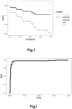
- Fig. 1 It was investigated how the quality parameters change when the threshold changes in the range of values 0.3-0.7 in the function of determining the class label. It is shown in Fig. 1 .
- the classifier can be optimized against either PPV or NPV, and the results reveal the relationships between the different groups of parameters. Optimizing a classifier against PPV increases its specificity, and against NPV increases its sensitivity or accuracy. The highest classification accuracy is obtained when the threshold of the classifier function is as small as possible. In contrast, the highest PPV has a classifier with a high threshold for the classifier model but has lower classification accuracy and sensitivity. The highest NPV has a classifier for the lowest thresholds - classification accuracy is also highest for this setting.
- a high PPV indicates the probability of correct detection of malignant neoplasm.
- a high NPV indicates the probability of correctly detecting a benign lesion (excluding a malignant lesion).
- Fig. 2 shows the classifier's quality characteristics as the ROC curve for the tested samples.
- the ROC curve shows the characteristics of the classifier's sensitivity and specificity.
- the shape of the ROC curve indicates that the classifier correctly labels benign / malignant.
- the first step of the classifier is normalization using the set of reference genes listed in Table 2: EIF3A (EIF3S10), HADHA, UBE2D2, EIF5, PPIE, RPLPO.
- the selected feature list contains 32 genes presented in Table 1, such as AMOT, C3, CAMK2N1, CFI, DCSTAMP, DIO1, DUSP5, EGR1, EMP2, FAM20A, FAXC, FLRT3, HBG1, IGFBP6, IGSF1, ITGA2, KCNQ3, MET, METTL7B, MPZL2, NOD1, OLR1, PDE5A, PSD3, SDC4, SLC26A4, SLC26A7, SLC5A8, SLPI, TM4SF1, TPO, and TUSC3, with normalized gene expressions as real numbers.
- Table 1 such as AMOT, C3, CAMK2N1, CFI, DCSTAMP, DIO1, DUSP5, EGR1, EMP2, FAM20A, FAXC, FLRT3, HBG1, IGFBP6, IGSF1, ITGA2, KCNQ3, MET, METTL7B, MPZL2, NOD1, OLR1, PDE5A, PSD3, SDC4, SLC26A4, SLC26A7, S
- the classifier displays on the computer screen the result of the classification test with information about the prediction of the class, the probability of malignancy, and the numerical values of all the features of the sample on the basis of which the classification took place.
- the classifier displays boxplots or violin plots for the benign and malignant classes of each gene in the training population with the expression value of the test sample plotted.
- RNA extraction is well known in the field and described in standard molecular biology textbooks.
- Gene expression levels can be determined by quantitative RT-PCR (quantitative reverse transcription PCR, RT-qPCR) or other PCR-based methods, as well as microarray-based methods or NGS transcriptome sequencing, or others known in the field or combinations thereof.
- quantitative RT-PCR quantitative reverse transcription PCR, RT-qPCR
- other PCR-based methods as well as microarray-based methods or NGS transcriptome sequencing, or others known in the field or combinations thereof.
- Classification quality parameters for both sets take high values, indicating that the described method can be implemented to the full range of thyroid nodule samples containing RNA at high as well as low concentrations.
- a fine-needle biopsy of the nodule was performed in a patient with a suspected thyroid nodule.
- the material was assigned to category IV, suspicious for a follicular neoplasm, which may warrant surgery.
- RNA isolated from a fine-needle biopsy material was used in this analysis. RNA isolation was performed using Qiagen's miRNeasy micro kit according to the manufacturer's recommendations. RNA concentration was measured with the high-sensitivity Qubit TM RNA HS Assay Kit using a Qubit 2.0 fluorometer. 20 ng of total RNA was used for the reaction.
- a Sensiscript cDNA reagent kit was used, with Sensiscript Reverse Transcriptase (RT-S), RT buffer, dNTPS mixture, and RNase-free water. Random Nanomers, OligodT(23) primers, and RNase inhibitor at 40 units/ ⁇ l were also used.
- the initial RT buffer was diluted to make 1x concentrated buffer, with which the inhibitor was diluted in a 3:1 ratio.
- composition of the cDNA synthesis is summarized in the table below: Reagents Volume (per reaction) Final concentration 10x Buffer RT 1 ⁇ l 1x dNTP (5nM each) 1 ⁇ l 0,5 nM each Random Nanomer Primers (50 ⁇ M) 0,8 ⁇ l 2,5 ⁇ M OligodT(n23) Primer (40 ⁇ M) 0,25 ⁇ l 1 ⁇ M RNAse Inhibitor (10 units/ ⁇ l) 0,5 ⁇ l 1 unit RT-S Polymerase (4 units/ ⁇ l) 0,5 ⁇ l 0,2 unit RNAse free H 2 O 5,95 ⁇ l Final volume per sample 10 ⁇ l
- pre-amplification An additional pre-amplification step (pre-amplification) is used for RNA samples with low RNA concentrations, allowing real-time PCR reaction without introducing amplification bias. All samples from fine-needle biopsies of thyroid nodules were preamplified.
- the pre-amplification step was carried out using TaqMan PreAmp Master Mix reaction mixture and Taqman Preamp Pool gene-specific primer mix.
- the pre-amplification reaction was carried out using a QuantStudio 12k Flex system in 96-well plates under the following conditions:
- composition of the cDNA pre-amplification reaction is summarized in table below: Reagents Volume (per reaction) Final concentration 2x Taqman Preamp Master mix 5 ⁇ l 1x Taqman Preamp Pool 2,5 ⁇ l cDNA template 2,5 ⁇ l Final volume 10 ⁇ l
- the reaction mixture, RNase-free water, and preamplified cDNA were applied to the 384-well plate.
- the plate was centrifuged for 1 min at 2,000 rpm.
- samples were transferred from the 384-well plate to the OpenArray at the dispensing station.
- the OpenArray plate was sealed with foil and placed in the press for 10s. After removal, the plate was filled with oil, and the cap was sealed.
- composition of the cDNA amplification reaction is summarized in the table below: Reagents Volume (per reaction) 2x TaqmanOpenarray Real-Time PCR Mastermix 2,6 ⁇ l RNAse-free water 1,4 ⁇ l cDNA template 1,5 ⁇ l Final volume 5,5 ⁇ l
- the qPCR reaction was performed on a QuantStudio 12K FLEX system with an OpenArray block according to the Openarray Taqman Gene Expression protocol (Thermofisher).
- the qPCR reaction was performed using reference RNAs. Normalization of the expression measured on the qPCR plate was carried out using a calibrator and six reference genes (EIF5, PPIE, UBE2D2, HADHA, EIF3A (EIF3S10), RPLPO). The result was analyzed, including whether all reference genes returned a signal from the quantitative RT-PCR reaction. The condition for correct normalization is that all reference genes return a signal.
- the steps described above refer to the evaluation of gene expression in the analyzed sample.
- the normalized gene expression values are entered into the classifier to obtain information about the nature of the thyroid nodule.
- Fig.3 shows the expression of individual genes. The values for the test sample are marked with a horizontal line against the population used to teach the model.
- the sample was classified as benign, with a probability of a malignant lesion of 0.016 (1.6%). Further observation of the patient confirmed the nature of the examined nodule as benign.
- a fine-needle biopsy of the nodule was performed in a patient with a suspected thyroid nodule.
- the specimen was assigned to category IV (suspicious for a follicular neoplasm), which may warrant surgery.
- the expression of individual signature genes for the test sample is indicated by a horizontal line against the population used to train the model. Taking into account the expression of the entire set of genes, the sample was classified as malignant, with a probability of a malignant lesion of 0. 981 (98.1%). The postoperative evaluation confirmed the nature of the test lesion as malignant.
- Fig. 5 , Fig. 6 , and Fig.7 Further examples of determining the nature of lesions in fine-needle biopsy samples of thyroid nodules using the expression of the selected 32 genes are shown in Fig. 5 , Fig. 6 , and Fig.7 .
- two samples of thyroid nodules ( Fig. 5 and Fig. 6 ) were classified as benign, with a probability of a malignant lesion of 0.251 (25.1%) and 0.069 (about 7%), respectively.
- the third thyroid nodule sample ( Fig. 7 ) was classified as malignant, with a probability of malignancy of 0.69 (69%).
Landscapes
- Chemical & Material Sciences (AREA)
- Life Sciences & Earth Sciences (AREA)
- Health & Medical Sciences (AREA)
- Organic Chemistry (AREA)
- Proteomics, Peptides & Aminoacids (AREA)
- Engineering & Computer Science (AREA)
- Immunology (AREA)
- Pathology (AREA)
- Analytical Chemistry (AREA)
- Zoology (AREA)
- Genetics & Genomics (AREA)
- Wood Science & Technology (AREA)
- Physics & Mathematics (AREA)
- Biotechnology (AREA)
- Microbiology (AREA)
- Molecular Biology (AREA)
- Hospice & Palliative Care (AREA)
- Biophysics (AREA)
- Oncology (AREA)
- Biochemistry (AREA)
- Bioinformatics & Cheminformatics (AREA)
- General Engineering & Computer Science (AREA)
- General Health & Medical Sciences (AREA)
- Measuring Or Testing Involving Enzymes Or Micro-Organisms (AREA)
Claims (5)
- Une méthode de différenciation des nodules thyroïdiens bénins et malins dans les échantillons de biopsie tumorale, comprenant :- étapes d'isolement de l'ARN, et- détermination des niveaux d'expression des gènes et utilisation d'un classificateur- comparison de l'expression génique de l'échantillon de nodules thyroïdiens avec l'expression génique des échantillons de nodules thyroïdiens dans le kit d'entraînement, puis- identification de la nature maligne ou bénigne du nodule et determination de la probabilité de malignité du nodule thyroïdien,avec le classificateur entraîné sur les données d'expression génique corrélées avec la nature du nodule thyroïdien, en particulier les nodules thyroïdiens bénins et les tumeurs malignes, y compris le carcinome thyroïdien papillaire et folliculaire,caractérisé en ce quele niveau d'expression d'un ensemble de 32 gènes est déterminé, qui se compose de AMOT, C3, CAMK2N1, CFI, DCSTAMP, DIO1, DUSP5, EGR1, EMP2, FAM20A, FAXC, FLRT3, HBG1, IGFBP6, IGSF1, ITGA2, KCNQ3, MET, METTL7B, MPZL2, NOD1, OLR1, PDE5A, PSD3, SDC4, SLC26A4, SLC26A7, SLCSA8, SLPI, TM4SF1, TPO et TUSC3,et la normalisation de l'expression de chaque gène de cet ensemble est effectuée sous le contrôle endogène des six gènes de normalisation suivants EIF3A (EIF3S10), HADHA, UBE2D2, EIF5, PPIE et RPLPO,et la nature maligne ou bénigne de l'échantillon de nodule est déterminée avec une spécificité d'au moins 91 % et une sensibilité de classification d'au moins 83 % pour le niveau d'expression normalisé d'un ensemble de 32 gènes,
- Le procédé, selon la revendication 1, caractérisé en ce que la nature du nodule et la probabilité de malignité de l'échantillon testé sont déterminées selon la règle de décision du classificateur, en tenant compte des données de niveau d'expression de l'ensemble de 32 gènes et du niveau d'expression génique de chaque échantillon du nodule dans l'ensemble d'entraînement, déterminé pour chaque échantillon de l'ensemble d'entraînement pour le même ensemble de 32 gènes et dans les mêmes conditions de normalisation.
- Le procédé selon la revendication. 2, caractérisé en ce que la nature et la probabilité de malignité d'un échantillon de nodule sont déterminées selon le critère de probabilité maximale pour un modèle probabiliste des valeurs d'expression normalisées d'un ensemble de 32 gènes de l'échantillon.
- Le procédé selon la revendication. 1, caractérisé en ce que la RT-qPCR, les microréseaux d'oligonucléotides ou le séquençage du transcriptome ARN-seq sont utilisés pour détecter le niveau d'expression d'un ensemble de gènes.
- Utilisation de l'ensemble de sondes d'hybridation Hs00611096_m1, Hs00163811_m1, Hs00218591_m1, Hs00989715_m1, Hs00229255_m1, Hs00174944_m1, Hs00244839_m1, Hs00152928_m1, Hs00171315_m1, Hs01034070_jn1, Hs00383314_m1, Hs01922255_s1, Hs00361131_g1, Hs00181853jn1, Hs00169943_m1, Hs00158127_m1, Hs01120412_m1, Hs01565584__m1, Hs00378551_m1, Hs01083647_m1, Hs01036720_m1, Hs01552593_m1, Hs00153649_m1, Hs00939867_m1, Hs01120908jfn1, Hs01070627_m1, Hs01104163_m1, Hs00377618_m1, Hs00268204_m1, Hs01547334_m1, Hs00892519_m1, Hs00185147_m1 pour détecter dans un échantillon de nodule thyroïdien le niveau d'expression d'un gène correspondant à partir d'un ensemble de 32 gènes AMOT, C3, CAMK2N1, CFI, DCSTAMP, DIO1, DUSP5, EGR1, EMP2, FAM20A, FAXC, FLRT3, HBG1, IGFBP6, IGSF1, ITGA2, KCNQ3, MET, METTL7B, MPZL2, NOD1, OLR1, PDE5A, DSP3, SDC4, SLC26A4, SLC26A7, SLC5A8, SLPI, TM4SF1, TPO, TUSC3 et pour déterminer la nature du nodule par le procédé selon les revendications 1 à 3, en utilisant un produit logiciel informatique ayant un support lisible par ordinateur et des ressources pour mesurer le niveau d'expression normalisé d'un ensemble de gènes, effectuer une classification et déterminer la probabilité de malignité d'un échantillon de nodule.
Priority Applications (1)
| Application Number | Priority Date | Filing Date | Title |
|---|---|---|---|
| EP22460030.4A EP4303323B1 (fr) | 2022-07-05 | 2022-07-05 | Procédé de différenciation de différenciation de nodules thyroïdiens bénins et malins |
Applications Claiming Priority (1)
| Application Number | Priority Date | Filing Date | Title |
|---|---|---|---|
| EP22460030.4A EP4303323B1 (fr) | 2022-07-05 | 2022-07-05 | Procédé de différenciation de différenciation de nodules thyroïdiens bénins et malins |
Publications (3)
| Publication Number | Publication Date |
|---|---|
| EP4303323A1 EP4303323A1 (fr) | 2024-01-10 |
| EP4303323B1 true EP4303323B1 (fr) | 2025-02-19 |
| EP4303323C0 EP4303323C0 (fr) | 2025-02-19 |
Family
ID=83049933
Family Applications (1)
| Application Number | Title | Priority Date | Filing Date |
|---|---|---|---|
| EP22460030.4A Active EP4303323B1 (fr) | 2022-07-05 | 2022-07-05 | Procédé de différenciation de différenciation de nodules thyroïdiens bénins et malins |
Country Status (1)
| Country | Link |
|---|---|
| EP (1) | EP4303323B1 (fr) |
Families Citing this family (1)
| Publication number | Priority date | Publication date | Assignee | Title |
|---|---|---|---|---|
| CN118326047A (zh) * | 2024-06-14 | 2024-07-12 | 润安医学科技(苏州)有限公司 | 一组microRNA标志物在甲状腺结节良恶性鉴别中的应用 |
Family Cites Families (14)
| Publication number | Priority date | Publication date | Assignee | Title |
|---|---|---|---|---|
| ATE491043T1 (de) | 2004-04-09 | 2010-12-15 | Us Gov Health & Human Serv | Diagnosegerät zur diagnose gutartiger gegenüber bösartigen schilddrüsenläsionen |
| US20070037186A1 (en) | 2005-05-20 | 2007-02-15 | Yuqiu Jiang | Thyroid fine needle aspiration molecular assay |
| US7901888B2 (en) | 2007-05-09 | 2011-03-08 | The Regents Of The University Of California | Multigene diagnostic assay for malignant thyroid neoplasm |
| US9234244B2 (en) | 2007-08-27 | 2016-01-12 | The United States Of America, As Represented By The Secretary, Department Of Health And Human Services | Diagnostic tool for diagnosing benign versus malignant thyroid lesions |
| GB2512153B (en) | 2008-11-17 | 2014-11-12 | Veracyte Inc | Methods and compositions of molecular profiling for disease diagnostics |
| US10236078B2 (en) | 2008-11-17 | 2019-03-19 | Veracyte, Inc. | Methods for processing or analyzing a sample of thyroid tissue |
| WO2010124372A1 (fr) | 2009-04-29 | 2010-11-04 | Genomedx Biosciences, Inc. | Systèmes et procédés pour la classification d'un tissu thyroïdien fondée sur l'expression |
| PL390570A1 (pl) | 2010-03-01 | 2011-09-12 | Centrum Onkologii - Instytut Im. M. Skłodowskiej-Curie | Zestaw, sposób i zastosowanie do wykrywania profilu ekspresji genów charakterystycznego dla raka brodawkowatego tarczycy |
| EP2366800A1 (fr) | 2010-03-01 | 2011-09-21 | Centrum Onkologii-Instytut im M. Sklodowskiej-Curie Oddzial w Gliwicach | Kit, méthode et l'utilisation pour le diagnostic de cancer papillaire de la thyroïde en utilisant un profil d'expression génique |
| EP2569626B1 (fr) | 2010-05-11 | 2019-11-27 | Veracyte, Inc. | Méthodes et compositions utilisées dans le cadre du diagnostic de maladies |
| EP2505664B1 (fr) | 2011-03-30 | 2014-12-03 | Universität Leipzig | Procédé et moyens pour distinguer des échantillons de tumeurs malignes des échantillons de tumeurs bénignes, en particulier dans la biopsie par aspiration à l'aiguille séchée à l'air |
| WO2013138726A1 (fr) | 2012-03-15 | 2013-09-19 | Sabiosciences Corp. | Biomarqueur d'un cancer de la thyroïde |
| CN104937111B (zh) | 2012-11-27 | 2018-05-11 | 智利天主教教皇大学 | 用于诊断甲状腺肿瘤的组合物和方法 |
| PL406033A1 (pl) | 2013-11-14 | 2015-05-25 | Warszawski Uniwersytet Medyczny | Sposób diagnozowania raka brodawkowatego tarczycy, zastosowanie markera mikroRNA do diagnozowania nowotworu tarczycy, oceny stopnia zaawansowania choroby oraz oceny podatności pacjenta i/lub choroby na zaproponowane leczenie oraz zawierający takie markery zestaw diagnostyczny |
-
2022
- 2022-07-05 EP EP22460030.4A patent/EP4303323B1/fr active Active
Also Published As
| Publication number | Publication date |
|---|---|
| EP4303323A1 (fr) | 2024-01-10 |
| EP4303323C0 (fr) | 2025-02-19 |
Similar Documents
| Publication | Publication Date | Title |
|---|---|---|
| US12595516B2 (en) | Neuroendocrine tumors | |
| EP2864500B1 (fr) | Malignité moléculaire dans des lésions mélanocytiques | |
| JP5405110B2 (ja) | 原発不明がんの原発巣を同定するための方法および材料 | |
| US10266902B2 (en) | Methods for prognosis prediction for melanoma cancer | |
| KR20170053617A (ko) | 폐암 상태를 평가하는 방법 | |
| EP2121988B1 (fr) | Survie au cancer de la prostate et récurrence de ce dernier | |
| US12297504B2 (en) | Chromosomal assessment to diagnose urogenital malignancy in dogs | |
| KR20140125239A (ko) | 조기 유방암 예후 예측 진단용 유전자 마커 및 이의 용도 | |
| US12252748B2 (en) | Kit, device and method for detecting prostate cancer | |
| WO2020190819A1 (fr) | Méthodes de prédiction du cancer de la prostate et leurs utilisations | |
| EP4303323B1 (fr) | Procédé de différenciation de différenciation de nodules thyroïdiens bénins et malins | |
| US20210079479A1 (en) | Compostions and methods for diagnosing lung cancers using gene expression profiles | |
| CN119998464B (zh) | 用于预测甲状腺癌转移的标志物组合及其应用 | |
| US20180051342A1 (en) | Prostate cancer survival and recurrence | |
| US10501806B2 (en) | Chromosomal assessment to differentiate histiocytic malignancy from lymphoma in dogs | |
| EP4023770A1 (fr) | Procédé d'examen des gènes pour le diagnostic des tumeurs de la thyroïde, ensemble pour le diagnostic des tumeurs de la thyroïde et application | |
| EP4303324A1 (fr) | Procédé de distinction entre des nodules thyroïdiens bénins et malins | |
| EP3936614A1 (fr) | Kit, dispositif et procédé de détection de léiomyosarcome utérin | |
| US20220042108A1 (en) | Systems and methods of assessing breast cancer |
Legal Events
| Date | Code | Title | Description |
|---|---|---|---|
| PUAI | Public reference made under article 153(3) epc to a published international application that has entered the european phase |
Free format text: ORIGINAL CODE: 0009012 |
|
| STAA | Information on the status of an ep patent application or granted ep patent |
Free format text: STATUS: THE APPLICATION HAS BEEN PUBLISHED |
|
| AK | Designated contracting states |
Kind code of ref document: A1 Designated state(s): AL AT BE BG CH CY CZ DE DK EE ES FI FR GB GR HR HU IE IS IT LI LT LU LV MC MK MT NL NO PL PT RO RS SE SI SK SM TR |
|
| STAA | Information on the status of an ep patent application or granted ep patent |
Free format text: STATUS: REQUEST FOR EXAMINATION WAS MADE |
|
| 17P | Request for examination filed |
Effective date: 20231228 |
|
| RBV | Designated contracting states (corrected) |
Designated state(s): AL AT BE BG CH CY CZ DE DK EE ES FI FR GB GR HR HU IE IS IT LI LT LU LV MC MK MT NL NO PL PT RO RS SE SI SK SM TR |
|
| GRAP | Despatch of communication of intention to grant a patent |
Free format text: ORIGINAL CODE: EPIDOSNIGR1 |
|
| STAA | Information on the status of an ep patent application or granted ep patent |
Free format text: STATUS: GRANT OF PATENT IS INTENDED |
|
| INTG | Intention to grant announced |
Effective date: 20240926 |
|
| GRAS | Grant fee paid |
Free format text: ORIGINAL CODE: EPIDOSNIGR3 |
|
| GRAA | (expected) grant |
Free format text: ORIGINAL CODE: 0009210 |
|
| STAA | Information on the status of an ep patent application or granted ep patent |
Free format text: STATUS: THE PATENT HAS BEEN GRANTED |
|
| AK | Designated contracting states |
Kind code of ref document: B1 Designated state(s): AL AT BE BG CH CY CZ DE DK EE ES FI FR GB GR HR HU IE IS IT LI LT LU LV MC MK MT NL NO PL PT RO RS SE SI SK SM TR |
|
| REG | Reference to a national code |
Ref country code: GB Ref legal event code: FG4D |
|
| REG | Reference to a national code |
Ref country code: CH Ref legal event code: EP |
|
| REG | Reference to a national code |
Ref country code: DE Ref legal event code: R096 Ref document number: 602022010814 Country of ref document: DE |
|
| REG | Reference to a national code |
Ref country code: IE Ref legal event code: FG4D |
|
| U01 | Request for unitary effect filed |
Effective date: 20250314 |
|
| REG | Reference to a national code |
Ref country code: NL Ref legal event code: MP Effective date: 20250219 |
|
| PG25 | Lapsed in a contracting state [announced via postgrant information from national office to epo] |
Ref country code: RS Free format text: LAPSE BECAUSE OF FAILURE TO SUBMIT A TRANSLATION OF THE DESCRIPTION OR TO PAY THE FEE WITHIN THE PRESCRIBED TIME-LIMIT Effective date: 20250519 |
|
| PG25 | Lapsed in a contracting state [announced via postgrant information from national office to epo] |
Ref country code: PL Free format text: LAPSE BECAUSE OF FAILURE TO SUBMIT A TRANSLATION OF THE DESCRIPTION OR TO PAY THE FEE WITHIN THE PRESCRIBED TIME-LIMIT Effective date: 20250219 |
|
| PG25 | Lapsed in a contracting state [announced via postgrant information from national office to epo] |
Ref country code: ES Free format text: LAPSE BECAUSE OF FAILURE TO SUBMIT A TRANSLATION OF THE DESCRIPTION OR TO PAY THE FEE WITHIN THE PRESCRIBED TIME-LIMIT Effective date: 20250219 |
|
| PG25 | Lapsed in a contracting state [announced via postgrant information from national office to epo] |
Ref country code: IS Free format text: LAPSE BECAUSE OF FAILURE TO SUBMIT A TRANSLATION OF THE DESCRIPTION OR TO PAY THE FEE WITHIN THE PRESCRIBED TIME-LIMIT Effective date: 20250619 Ref country code: NO Free format text: LAPSE BECAUSE OF FAILURE TO SUBMIT A TRANSLATION OF THE DESCRIPTION OR TO PAY THE FEE WITHIN THE PRESCRIBED TIME-LIMIT Effective date: 20250519 |
|
| PG25 | Lapsed in a contracting state [announced via postgrant information from national office to epo] |
Ref country code: HR Free format text: LAPSE BECAUSE OF FAILURE TO SUBMIT A TRANSLATION OF THE DESCRIPTION OR TO PAY THE FEE WITHIN THE PRESCRIBED TIME-LIMIT Effective date: 20250219 |
|
| PG25 | Lapsed in a contracting state [announced via postgrant information from national office to epo] |
Ref country code: GR Free format text: LAPSE BECAUSE OF FAILURE TO SUBMIT A TRANSLATION OF THE DESCRIPTION OR TO PAY THE FEE WITHIN THE PRESCRIBED TIME-LIMIT Effective date: 20250520 |
|
| U07 | Unitary effect registered |
Designated state(s): AT BE BG DE DK EE FI FR IT LT LU LV MT NL PT RO SE SI Effective date: 20250618 |
|
| U20 | Renewal fee for the european patent with unitary effect paid |
Year of fee payment: 4 Effective date: 20250701 |
|
| PG25 | Lapsed in a contracting state [announced via postgrant information from national office to epo] |
Ref country code: SM Free format text: LAPSE BECAUSE OF FAILURE TO SUBMIT A TRANSLATION OF THE DESCRIPTION OR TO PAY THE FEE WITHIN THE PRESCRIBED TIME-LIMIT Effective date: 20250219 |
|
| PG25 | Lapsed in a contracting state [announced via postgrant information from national office to epo] |
Ref country code: CZ Free format text: LAPSE BECAUSE OF FAILURE TO SUBMIT A TRANSLATION OF THE DESCRIPTION OR TO PAY THE FEE WITHIN THE PRESCRIBED TIME-LIMIT Effective date: 20250219 |
|
| PG25 | Lapsed in a contracting state [announced via postgrant information from national office to epo] |
Ref country code: SK Free format text: LAPSE BECAUSE OF FAILURE TO SUBMIT A TRANSLATION OF THE DESCRIPTION OR TO PAY THE FEE WITHIN THE PRESCRIBED TIME-LIMIT Effective date: 20250219 |
|
| PLBE | No opposition filed within time limit |
Free format text: ORIGINAL CODE: 0009261 |
|
| STAA | Information on the status of an ep patent application or granted ep patent |
Free format text: STATUS: NO OPPOSITION FILED WITHIN TIME LIMIT |
|
| REG | Reference to a national code |
Ref country code: CH Ref legal event code: L10 Free format text: ST27 STATUS EVENT CODE: U-0-0-L10-L00 (AS PROVIDED BY THE NATIONAL OFFICE) Effective date: 20251231 |
|
| 26N | No opposition filed |
Effective date: 20251120 |
|
| REG | Reference to a national code |
Ref country code: CH Ref legal event code: H13 Free format text: ST27 STATUS EVENT CODE: U-0-0-H10-H13 (AS PROVIDED BY THE NATIONAL OFFICE) Effective date: 20260224 |
|
| PG25 | Lapsed in a contracting state [announced via postgrant information from national office to epo] |
Ref country code: CH Free format text: LAPSE BECAUSE OF NON-PAYMENT OF DUE FEES Effective date: 20250731 |