EP4332577A1 - Applications de crmp2 et d'anticorps anti-crmp2 - Google Patents
Applications de crmp2 et d'anticorps anti-crmp2 Download PDFInfo
- Publication number
- EP4332577A1 EP4332577A1 EP21939076.2A EP21939076A EP4332577A1 EP 4332577 A1 EP4332577 A1 EP 4332577A1 EP 21939076 A EP21939076 A EP 21939076A EP 4332577 A1 EP4332577 A1 EP 4332577A1
- Authority
- EP
- European Patent Office
- Prior art keywords
- crmp2
- antibody
- reagent
- nervous system
- autoimmune disease
- Prior art date
- Legal status (The legal status is an assumption and is not a legal conclusion. Google has not performed a legal analysis and makes no representation as to the accuracy of the status listed.)
- Pending
Links
Images


Classifications
-
- G—PHYSICS
- G01—MEASURING; TESTING
- G01N—INVESTIGATING OR ANALYSING MATERIALS BY DETERMINING THEIR CHEMICAL OR PHYSICAL PROPERTIES
- G01N33/00—Investigating or analysing materials by specific methods not covered by groups G01N1/00 - G01N31/00
- G01N33/48—Biological material, e.g. blood, urine; Haemocytometers
- G01N33/50—Chemical analysis of biological material, e.g. blood, urine; Testing involving biospecific ligand binding methods; Immunological testing
- G01N33/53—Immunoassay; Biospecific binding assay; Materials therefor
- G01N33/564—Immunoassay; Biospecific binding assay; Materials therefor for pre-existing immune complex or autoimmune disease, i.e. systemic lupus erythematosus, rheumatoid arthritis, multiple sclerosis, rheumatoid factors or complement components C1-C9
-
- A—HUMAN NECESSITIES
- A61—MEDICAL OR VETERINARY SCIENCE; HYGIENE
- A61K—PREPARATIONS FOR MEDICAL, DENTAL OR TOILETRY PURPOSES
- A61K31/00—Medicinal preparations containing organic active ingredients
- A61K31/70—Carbohydrates; Sugars; Derivatives thereof
- A61K31/7088—Compounds having three or more nucleosides or nucleotides
- A61K31/713—Double-stranded nucleic acids or oligonucleotides
-
- G—PHYSICS
- G01—MEASURING; TESTING
- G01N—INVESTIGATING OR ANALYSING MATERIALS BY DETERMINING THEIR CHEMICAL OR PHYSICAL PROPERTIES
- G01N33/00—Investigating or analysing materials by specific methods not covered by groups G01N1/00 - G01N31/00
- G01N33/48—Biological material, e.g. blood, urine; Haemocytometers
- G01N33/50—Chemical analysis of biological material, e.g. blood, urine; Testing involving biospecific ligand binding methods; Immunological testing
- G01N33/68—Chemical analysis of biological material, e.g. blood, urine; Testing involving biospecific ligand binding methods; Immunological testing involving proteins, peptides or amino acids
- G01N33/6854—Immunoglobulins
-
- A—HUMAN NECESSITIES
- A61—MEDICAL OR VETERINARY SCIENCE; HYGIENE
- A61K—PREPARATIONS FOR MEDICAL, DENTAL OR TOILETRY PURPOSES
- A61K45/00—Medicinal preparations containing active ingredients not provided for in groups A61K31/00 - A61K41/00
-
- A—HUMAN NECESSITIES
- A61—MEDICAL OR VETERINARY SCIENCE; HYGIENE
- A61K—PREPARATIONS FOR MEDICAL, DENTAL OR TOILETRY PURPOSES
- A61K45/00—Medicinal preparations containing active ingredients not provided for in groups A61K31/00 - A61K41/00
- A61K45/06—Mixtures of active ingredients without chemical characterisation, e.g. antiphlogistics and cardiaca
-
- A—HUMAN NECESSITIES
- A61—MEDICAL OR VETERINARY SCIENCE; HYGIENE
- A61P—SPECIFIC THERAPEUTIC ACTIVITY OF CHEMICAL COMPOUNDS OR MEDICINAL PREPARATIONS
- A61P25/00—Drugs for disorders of the nervous system
-
- A—HUMAN NECESSITIES
- A61—MEDICAL OR VETERINARY SCIENCE; HYGIENE
- A61P—SPECIFIC THERAPEUTIC ACTIVITY OF CHEMICAL COMPOUNDS OR MEDICINAL PREPARATIONS
- A61P29/00—Non-central analgesic, antipyretic or antiinflammatory agents, e.g. antirheumatic agents; Non-steroidal antiinflammatory drugs [NSAID]
-
- A—HUMAN NECESSITIES
- A61—MEDICAL OR VETERINARY SCIENCE; HYGIENE
- A61P—SPECIFIC THERAPEUTIC ACTIVITY OF CHEMICAL COMPOUNDS OR MEDICINAL PREPARATIONS
- A61P37/00—Drugs for immunological or allergic disorders
- A61P37/02—Immunomodulators
-
- C—CHEMISTRY; METALLURGY
- C12—BIOCHEMISTRY; BEER; SPIRITS; WINE; VINEGAR; MICROBIOLOGY; ENZYMOLOGY; MUTATION OR GENETIC ENGINEERING
- C12N—MICROORGANISMS OR ENZYMES; COMPOSITIONS THEREOF; PROPAGATING, PRESERVING, OR MAINTAINING MICROORGANISMS; MUTATION OR GENETIC ENGINEERING; CULTURE MEDIA
- C12N15/00—Mutation or genetic engineering; DNA or RNA concerning genetic engineering, vectors, e.g. plasmids, or their isolation, preparation or purification; Use of hosts therefor
- C12N15/09—Recombinant DNA-technology
- C12N15/63—Introduction of foreign genetic material using vectors; Vectors; Use of hosts therefor; Regulation of expression
-
- G—PHYSICS
- G01—MEASURING; TESTING
- G01N—INVESTIGATING OR ANALYSING MATERIALS BY DETERMINING THEIR CHEMICAL OR PHYSICAL PROPERTIES
- G01N33/00—Investigating or analysing materials by specific methods not covered by groups G01N1/00 - G01N31/00
- G01N33/48—Biological material, e.g. blood, urine; Haemocytometers
- G01N33/50—Chemical analysis of biological material, e.g. blood, urine; Testing involving biospecific ligand binding methods; Immunological testing
- G01N33/68—Chemical analysis of biological material, e.g. blood, urine; Testing involving biospecific ligand binding methods; Immunological testing involving proteins, peptides or amino acids
- G01N33/6893—Chemical analysis of biological material, e.g. blood, urine; Testing involving biospecific ligand binding methods; Immunological testing involving proteins, peptides or amino acids related to diseases not provided for elsewhere
-
- C—CHEMISTRY; METALLURGY
- C07—ORGANIC CHEMISTRY
- C07K—PEPTIDES
- C07K16/00—Immunoglobulins [IG], e.g. monoclonal or polyclonal antibodies
- C07K16/18—Immunoglobulins [IG], e.g. monoclonal or polyclonal antibodies against material from animals or humans
-
- G—PHYSICS
- G01—MEASURING; TESTING
- G01N—INVESTIGATING OR ANALYSING MATERIALS BY DETERMINING THEIR CHEMICAL OR PHYSICAL PROPERTIES
- G01N2800/00—Detection or diagnosis of diseases
- G01N2800/24—Immunology or allergic disorders
-
- G—PHYSICS
- G01—MEASURING; TESTING
- G01N—INVESTIGATING OR ANALYSING MATERIALS BY DETERMINING THEIR CHEMICAL OR PHYSICAL PROPERTIES
- G01N2800/00—Detection or diagnosis of diseases
- G01N2800/28—Neurological disorders
-
- G—PHYSICS
- G01—MEASURING; TESTING
- G01N—INVESTIGATING OR ANALYSING MATERIALS BY DETERMINING THEIR CHEMICAL OR PHYSICAL PROPERTIES
- G01N2800/00—Detection or diagnosis of diseases
- G01N2800/52—Predicting or monitoring the response to treatment, e.g. for selection of therapy based on assay results in personalised medicine; Prognosis
Definitions
- the present disclosure relates to the technical field of biomedicine, in particular to the application of CRMP2 and anti-CRMP2 antibodies.
- Autoimmune diseases of nervous system are a type of diseases caused by inflammation and other injuries due to autoimmune molecules and immune cells recognizing the cell components (autoantigens) of tissues of the nervous system, which can occur in central nervous system, peripheral nervous system and neuromuscular junction, and includes autoimmune encephalitis (AE), central demyelinating disease, autoimmune peripheral neuropathy, etc. Of these, AE is the most common.
- AE mainly affects young and middle-aged people, with high mortality and disability rates.
- an autoantibody against neuronal surface receptor AE namely anti-NMDAR (N-methyl-D-aspartate receptor) antibody
- CSF cerebrospinal fluid
- Autoantigens (or proteins) corresponding to such AEs include: (1) neurotransmitter receptor AMPAR (alpha-amino-3-hydroxy-5-methyl-4-isoxazolepropionic acid receptor), mGluR5 (Metabotropic glutamate receptor 5), GABA A R/GABA B R (Gamma-Aminobutyric Acid A/B receptor), GlyR1 (Glycine receptor), D2R (Dopamine-2 receptor); (2) transmembrane protein CASPR2 (Contactin-associated protein-like 2), DNER (Delta/Notch Like EGF Repeat Containing), DDP6/DPPX (Dipetidyl Peptidase Like 6), DCC (Netrin 1 Receptor DCC), IgL
- the diagnosis of AE first requires a comprehensive analysis of the patient's clinical manifestations, an examination of cerebrospinal fluid, neuroimaging, electroencephalogram and other ancillary detection results to determine that the patient is suffering from encephalitis (suspected AE or possible AE), and subsequent a known AE related antibody test is performed to diagnose. If the antibody test is positive and consistent with the clinical manifestation and other possible causes are excluded, AE can be definitively diagnosed as an AE directed against a certain known autoantigen, such as anti-NMDAR encephalitis. Therefore, the positive result for the detection of the corresponding antibody is a necessary condition for the diagnosis of AE but not a sufficient condition. Since the clinical manifestations of autoimmune encephalitis in children are different from those in adults, it is difficult to identify specific autoimmune encephalitis syndrome. Therefore, the detection of autoantibodies is very important for the definite diagnosis of autoimmune encephalitis in children.
- AE-specific antibodies are mainly detected using cell based assay (CBA) based on the expression of specific antigens.
- CBA cell based assay
- the Chinese Academy of Medical Sciences conducted anti NMDAR antibody testing on the cerebrospinal fluid of 284 patients with unexplained encephalitis using a single molecule fluorescence CBA method. Among them, 46 of cases were positive for the antibody, with a prevalence rate of 16.2%. This indicates that anti-NMDAR encephalitis is not rare in China. Therefore, it is necessary to conduct anti-NMDAR antibody testing for cases of encephalitis of unknown etiology.
- the pathogenesis of AE is still in the initial stage of research, and the occurrence and clinical manifestations of the disease triggered by different autoantibodies may vary.
- the anti-NMDAR antibody binds to neuron NMDAR, the number of synapses is greatly reduced; the anti-GABABR antibody inhibits the activity of such receptor, but has no significant effect on the density of synapses; anti-LGI1 and CASPR2 antibodies can interfere with synaptic protein-protein interaction; anti-neurexin-3 ⁇ antibody can alter the formation of synapses; and anti-IgLON5 antibody can lead to the formation of new forms of Tau protein lesions.
- Tumors and viral infections are risk factors for AE.
- Anti-NMDAR encephalitis is often accompanied by teratoma, and anti-GABABR encephalitis is often accompanied by small cell lung cancer.
- Tumor cells may secrete autoantigens and thus activate the immune system, leading to the development of AE.
- neuron-like tissue cells in teratomas secrete NMDAR, and the inflammatory response of tumor tissue in patients with AE is higher than that in those without AE.
- the exception is encephalitis with DNER, mGluR1, and mGluR5 accompanied by Hodgkin lymphoma, where the tumor does not produce any AE-associated antigens.
- the object of the present disclosure is to provide a novel antibody marker and establish a detection system thereof for diagnosis and treatment of an autoimmune disease of nervous system.
- the technical solution adopted by the present disclosure is as follows:
- the present disclosure provides use of CRMP2 as a marker for an autoimmune disease of nervous system.
- the CRMP2 comprises an amino acid sequence as shown in any one of SEQ ID NOs: 1 to 3.
- the CRMP2 comprises a nucleotide sequence as shown in any one of SEQ ID NOs: 4 to 6.
- the present disclosure also provides use of anti-CRMP2 antibody as a marker for an autoimmune disease of nervous system.
- the present disclosure also provides use of a reagent detecting anti-CRMP2 antibody in the preparation of a reagent for detection, diagnosis, treatment or prognosis evaluation of an autoimmune disease of nervous system.
- a method for detecting the anti-CRMP2 antibody is at least one of CBA, immunoblotting, membrane strip method, ELISA, chemiluminescence, radioimmunoassay, liquid chip method, lateral chromatography, electrochemistry, and flow cytometry.
- the present disclosure also provides use of a reagent inhibiting expression or translation of anti-CRMP2 antibody in preparation of a medicine for an autoimmune disease of a nervous system.
- a preparation inhibiting expression or translation of the anti-CRMP2 antibody comprises a gene promoter inhibitor of the anti-CRMP2 antibody, a gene transcription inhibitor of the anti-CRMP2 antibody, a protein synthesis inhibitor of the anti CRMP2 antibody, or a siRNA of the anti CRMP2 antibody gene.
- the present disclosure also provides use of a preparation reducing or inactivating an activity of an anti-CRMP2 antibody in preparation of a medicine for an autoimmune disease of nervous system.
- a reagent reducing or inactivating activity of the anti-CRMP2 antibody comprises a substance for inhibiting the activity of the anti-CRMP2 antibody, a substance for degrading the activity of the anti-CRMP2 antibody, or a gene tool for reducing the protein level of the anti-CRMP2 antibody.
- the present disclosure also provides an auxiliary diagnostic reagent for an autoimmune disease of nervous system, the reagent contains a reagent for detecting the expression amount of an anti-CRMP2 antibody.
- a detected sample is cerebrospinal fluid or blood.
- the autoimmune disease of the nervous system is encephalitis or encephalomyelitis.
- the beneficial effects of the present disclosure are that:
- the present disclosure discovers that the CRMP2 is a novel encephalitis or encephalomyelitis-associated autoantibody-targeted molecular marker through research.
- this novel disease is called anti-CRMP2 encephalitis or encephalomyelitis, and the affected area or symptom location may be brain, cerebellum and/or spinal cord.
- CRMP2 can be used for definitive diagnosis of the disease and provide basis for immunotherapy regimens, and can monitor therapeutic effect and determine prognosis and recurrence by detecting the change in the titer of the autoantibody.
- CRMP2 is known to have 3 protein isoforms, and the sequences are as follows: Isoform 1: length: 677 amino acids; species: human (Homo sapiens), with the amino acid sequence as shown in SEQ ID NO: 1; isoform 2: length: 572 amino acids; species: human (Homo sapiens), with the amino acid sequence as shown in SEQ ID NO: 2; isoform 3: length: 536 amino acids; species: human (Homo sapiens), and with the amino acid sequence as shown in SEQ ID NO: 3.
- CRMP2 is known to have 3 transcripts encoding proteins corresponding to the above 3 protein isoforms, and the sequences are as follows: Transcript 1: length: 2034 bp; species: human (Homo sapiens), with the nucleotide sequence as shown in SEQ ID NO: 4; transcript 2: length: 1719 bp; species: human (Homo sapiens), with the nucleotide sequence as shown in SEQ ID NO: 5; transcript 3: length: 1611bp; species: human (Homo sapiens), with the nucleotide sequence as shown in SEQ ID NO: 6.
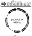
- the CRMP2 sequence used in the pcDNA3.1-CRMP2 plasmid is transcript 1 for expression of CRMP2 protein type 1, and covers all the sequences of 3 transcripts and 3 isoforms in order to detect anti-CRMP2 antibodies without omission. Of these, the map of the pcDNA3.1-CRMP2 vector is shown in FIG. 1 .
- the eukaryotic expression vector used for pcDNA3.1-CRMP2 plasmid is pcDNA3.1+, and the map is as follows: the enzyme cleavage sites used for vector construction are BamH I (5' end) and EcoR I (3' end), with the inserted CRMP2 transcript 1 sequence in the middle. The specific sequence is as shown in SEQ ID NO: 7.
- the three patients in this Example were numbered P1, P2 and P3, respectively.
- P1 was the first case in which CRMP 2 was identified using cerebrospinal fluid and blood samples therefrom, and P2 and P3 were positive cases of anti-CRMP2 antibody detected by retrospective analysis using CBA method.
- P1, P2 and P3 all had symptoms of dizziness or headache, visual impairment and unsteady walking; as well as white matter lesions; with effective immunotherapy, positive TBA detection in mouse brain slices of blood and/or cerebrospinal fluid, positive detection for anti-CRMP2 antibody and negative detection for other AE associated antibodies.
- P1 and P3 had speech barriers and inaccuracy in bilateral finger-nose test, and P2 had chest and abdominal pain, and spinal cord lesions.
- cerebrospinal fluid was tested positive for Mycoplasma pneumoniae, and for P3, cerebrospinal fluid was tested positive for human herpesvirus type 6 and Klebsiella pneumoniae, and the IgM of influenza B virus in blood was positive.
- Table 1 Clinical data of 3 patients with anti-CRMP2 antibody positive encephalitis/encephalomyelitis Patie nt Gender Age (year of age, approximat e age,for protection of privacy) Symptoms Physical examination Cerebrospinal fluid Brain slice P1 Female About 35 Dizziness, rotation of vision, unsteady walking, and fever Poetry-like speech, normal muscle strength of limbs, poor stability and inaccuracy in bilateral finger-nose test, poor coordination movement and slight resistance of neck Leukocyte 38/ ⁇ L, mononuclear cell 90% Brain section and cerebellu m section: cerebrosp inal fluid (+) P2 Female About 40 Headache, nausea, emesis, chest pain on the left side, weakness in the right upper limb, followed by weakness in both lower limbs.
- Vague speech free movement of both eyeballs in all directions, and visible nystagmus; limb muscle strength at grade 4, and poor vibration perception of tuning forks in both ankles, and normal feelings on depth of the whole body; inaccuracy in bilateral finger-nose test and heel-knee-tibi a test.
- MRI visible long-segment lesion of spinal cord, multiple flaky abnormal signals of white matter regions of bilateral cerebral hemispheres and brainstem, and considering demyelinating lesion 1. Acute disseminated encephalomy elitis; 2. tuberculous meningitis?
- thyroid-associated antibodies antibodies of thyrotropin receptor, antibodies of anti-thyroid peroxidase, antibodies of anti-thyroglobulin; 4. tumor antigen: carbohydrate antigen (CA-125), non-small cell lung cancer-associated antigen, squamous cell carcinoma-associated antigen (SCC), serum pancreas and gastrointestinal tumor-associated antigen (CA 242), gastric cancer-associated antigen (CA-724), gastrointestinal cancer-associated antigen (CA-199), carcinoembryonic antigen (CEA); 5.
- tumor antigen carbohydrate antigen (CA-125), non-small cell lung cancer-associated antigen, squamous cell carcinoma-associated antigen (SCC), serum pancreas and gastrointestinal tumor-associated antigen (CA 242), gastric cancer-associated antigen (CA-724), gastrointestinal cancer-associated antigen (CA-199), carcinoembryonic antigen (CEA); 5.
- CA-125 carbohydrate antigen
- SCC squamous cell carcinoma-associated antigen
- serum autoimmune antibodies antibody of antinuclear, antibody of anti-double-stranded DNA, antibody of anti-Jo-1, antibody of anti-ssB, antibody of anti-Sm, antibody of anti-UI-nRNP, antibody of anti-ribosomal P protein, antibody of anti-proliferative nuclear antigen, antibody of anti-mitochondrial, antibody of anti-centromere, antibody of anti-PM-Scl, antibody of anti-SCL-70, antibody of anti-ssA, antibody of anti-nucleosome, antibody of anti-histone, antibody of anti-recombinant Ro52.
- MRI magnetic resonance
- mRS modified Rankin scale
- Immunohistochemical experiment based on rat brain slices is a routine examination of autoimmune encephalitis, including immunoenzyme chemical method and immunofluorescence (also known as TBA, tissue based assay).
- the adult rat was anesthetized and then the heart of adult rat was infused with PBS (phosphate buffer) to remove circulating blood.
- PBS phosphate buffer
- the brain including hippocampus was taken for sagittal frozen sections.
- the cerebellum was taken to be longitudinally sliced, fixed by cold acetone for 10 minutes and dried for later use. Slices were washed with PBST solution (PBS containing 0.05% of Tween-20) for 3 times, 3 minutes each time.
- PBST solution PBS containing 0.05% of Tween-20
- immunoenzyme chemical and immunofluorescence methods were performed for detection respectively: (1) immunoenzyme chemical method was used: the slices were then treated with 3% hydrogen peroxide for 10 minutes, washed with PBST for 3 times prior to the next step of blocking, and blocked with 10% goat serum/PBST solution for 1 hour at room temperature; (2) immunofluorescence method was used: without hydrogen peroxide treatment, the slices were blocked with 10% goat serum/PBST solution for 1 hour at room temperature.
- the blocking buffer was shaken off after the two groups were blocked, and the cerebrospinal fluid from patients P1 was added, and incubated at 37°C for 1 hour. After incubation, the slices were washed with PBST for 3 times, 5 minutes each time.
- Results The detection results are shown in FIG. 3 , in which Images A and C in FIG. 3 are the results of immunoenzyme chemical detection. Wherein the comparison results of cerebrospinal fluid staining of negative non-autoimmune patients are shown in Image A in FIG. 3 , and positive cerebrospinal fluid staining for patient P1 is shown in Image C in FIG. 3 . Images B and D in FIG. 3 show the results of immunofluorescence detection, Image B in FIG. 3 shows the results of detection in hippocampus of brain, and Image D in FIG. 3 shows the results of detection in Purkinje cells of cerebellum, all of which are positive. This indicates that the cerebrospinal fluid of patient P1 contained autoantibodies against cerebral neurons, and patient P1 has a suspected AE.
- Antigens against autoantibodies in cerebrospinal fluid from patient P1 were identified using immunoprecipitation combined with protein mass spectrometry.
- the suspected target Coomassie brilliant blue staining bands (about 70KDa) were selected, cut off gel for peptide fingerprint mass spectrometry protein identification.
- the results are shown in FIG. 4 .
- Image A in FIG. 4 shows the result of immunoprecipitation.
- Image B in FIG. 4 shows the result of Coomassie brilliant blue staining, where the immunoprecipitation and Coomassie brilliant blue staining indicates that a target protein band may be at about 70Kd,.
- Image C in FIG. 4 shows the secondary mass spectrometry of one of the polypeptides
- FIG. 4 shows the distribution of the polypeptide (underlined) identified by protein mass spectrometry on CRMP2 protein, in which the CRMP2 protein coverage of the polypeptide is 40%.
- the polypeptide underlined with a dotted line in image D in FIG. 4 corresponds to the polypeptide in image C in FIG. 4 .
- CRMP2 was located in the cytoplasm, axons and dendrites of neurons.
- Anti-CRMP2 antibodies in cerebrospinal fluid and serum samples from patients with anti-CRMP2 encephalitis/encephalomyelitis should be able to bind to neurons, which is an important mechanism arising AE.
- the serum stained neurons of patient P1 are positive, and the signals are distributed on the cytoplasm, dendrites and axons of neurons, which were consistent with the localization of CRMP2 and consistent with the characteristics of anti-CRMP2 encephalitis/encephalomyelitis. Negative control sera were not stained.
- Detection for cerebrospinal fluid and serum samples of patients by immunofluorescence i.e., the CBA method
- 293T cells overexpressing target antigen is an important way to diagnose autoimmune encephalitis/encephalomyelitis.
- Implementation process a 293T cell slide was prepared, and when the cells grew to 50% - 90% of density, the eukaryotic expression plasmid pcDNA3.1-CRMP2 was transfected into 293T cells to make the 293T cells overexpress the CRMP2 recombinant protein.
- the 293T cells were washed with PBS for 3 times, then fixed by acetone for 5 minutes and dried.
- the slide was washed with PBST solution for 3 times, 3 minutes each time. Blocked with 10% goat serum/PBST solution for 1 hour at room temperature, the sealing solution was abandoned, and serum from patient P1 or P3 (diluted at 1: 20) was added, and incubated at 37 °C for 1 hour.
- an anti-CRMP2 antibody (ab129082, Abeam, a dilution of 1:200) was added as a positive control, at the same time as the secondary antibody, a red fluorescence-labeled goat anti-rabbit IgG secondary antibody (ab 150092, Abeam) was added to for immunofluorescence co-staining.
- This example uses serum from patients with non-autoimmune diseases as a negative control. The results are shown in FIG. 6 .
- this example successfully overexpressed CRMP2 protein in 293T cells, and the serum staining from patients P1 and P3 substantially overlapped with CRMP2.
- the negative control serum were not stained. It indicated that the serum from patients P1 and P3 contained autoimmune antibodies against CRMP2.
- Reduction or elimination of autoimmune antibodies by immunotherapy is a main way to treat autoimmune encephalitis/encephalomyelitis.
- the serum from patient P2 before treatment was positive for CBA staining and co-localized with CRMP2, which indicates that the serum from patient P2 before treatment contained autoimmune antibodies against CRMP2.
- the serum from patient P2 was negative for CBA staining, which indicates that the anti-CRMP2 antibodies in the serum disappeared after treatment of patient P2.
- Immunoblotting is another method for detecting autoimmune antibodies.
- the CRMP2 protein was prepared. 293T cells were cultured in a 10 cm cell culture dish to a density of 80%, pcDNA3.1-CRMP2 eukaryotic expression plasmids were transfected into the 293T cells using EntransterTM-D4000 (4000-2, Engreen), and the 293T cells were continuously cultured for 24 to 48 hours for overexpression of CRMP2 recombinant proteins. Then, the 293T cells were treated with 1 mL of protein lysate agent at 4 °C for 30 min. Centrifugation was performed at 12000 rpm at 4°C for 15 min and the supernatant was collected to obtain CRMP2-containing 293T protein lysate solution.
- the eluent was separated by 10% SDS-PAGE, transferred to a nitrocellulose membrane, blocked with 5% skim milk powder/TBST at room temperature for 1 hour, incubated with 1:20 diluted cerebrospinal fluid from P2 patient at room temperature for 2 hours, washed 3 times for 5 min by TBST, incubated with 1:5000 diluted HRP-labeled goat anti-human IgG at room temperature for 1 hour, washed 3 times for 5 min by TBST, and developed with ECL substrate for detection of the anti-CRMP2 antibodies in the cerebrospinal fluid of the P2 patient. Normal rabbit IgG was used as a negative control for anti-CRMP2 antibody,.
- the pcDNA3.1 empty vector transfected 293T was used as a negative control for the CRMP2 protein.
- the results are shown in FIG. 8 .
- a clear band appeared at the position of the target CRMP2 size, and the corresponding negative control had no band, which indicates that the cerebrospinal fluid of the P2 patient contained anti-CRMP2 antibody.
- immunoblotting membrane strip method
- FIG. 9 The results of sequence alignment of the 3 isoforms of CRMP2 are shown in FIG. 9 , which shows that the amino acid sequences of the 3 isoforms are completely consistent at the positions 142 to 677. Therefore, the CRMP2 sequence in the pcDNA3.1-CRMP2 plasmid used in Examples 5, 6 and 7 is transcript 1 for expression of CRMP2 protein type 1, and covers all the sequences of 3 transcripts and 3 isoforms in order to detect anti-CRMP2 antibodies without omission.
- CRMP2 also known as DPYL2
- results are shown in FIG. 10 . It can be seen from FIG. 10 that CRMP2 proteins of different species are highly homologous, with a homology of 96.853%.
- the CRMP2 proteins from different species can be used to detect anti-CRMP 2 antibodies in human cerebrospinal fluid or serum samples.
Landscapes
- Health & Medical Sciences (AREA)
- Life Sciences & Earth Sciences (AREA)
- Engineering & Computer Science (AREA)
- Chemical & Material Sciences (AREA)
- Immunology (AREA)
- Biomedical Technology (AREA)
- Molecular Biology (AREA)
- Hematology (AREA)
- General Health & Medical Sciences (AREA)
- Urology & Nephrology (AREA)
- Medicinal Chemistry (AREA)
- Biotechnology (AREA)
- Biochemistry (AREA)
- Microbiology (AREA)
- Physics & Mathematics (AREA)
- Pathology (AREA)
- Food Science & Technology (AREA)
- Cell Biology (AREA)
- General Physics & Mathematics (AREA)
- Analytical Chemistry (AREA)
- Veterinary Medicine (AREA)
- Public Health (AREA)
- Animal Behavior & Ethology (AREA)
- Pharmacology & Pharmacy (AREA)
- Organic Chemistry (AREA)
- Genetics & Genomics (AREA)
- Bioinformatics & Cheminformatics (AREA)
- Proteomics, Peptides & Aminoacids (AREA)
- Epidemiology (AREA)
- General Chemical & Material Sciences (AREA)
- Nuclear Medicine, Radiotherapy & Molecular Imaging (AREA)
- Chemical Kinetics & Catalysis (AREA)
- Rheumatology (AREA)
- Wood Science & Technology (AREA)
- Zoology (AREA)
- General Engineering & Computer Science (AREA)
- Rehabilitation Therapy (AREA)
- Plant Pathology (AREA)
- Biophysics (AREA)
- Neurosurgery (AREA)
Applications Claiming Priority (2)
| Application Number | Priority Date | Filing Date | Title |
|---|---|---|---|
| CN202110461611.6A CN113238045B (zh) | 2021-04-27 | 2021-04-27 | Crmp2及抗crmp2抗体的应用 |
| PCT/CN2021/138864 WO2022227617A1 (fr) | 2021-04-27 | 2021-12-16 | Applications de crmp2 et d'anticorps anti-crmp2 |
Publications (2)
| Publication Number | Publication Date |
|---|---|
| EP4332577A1 true EP4332577A1 (fr) | 2024-03-06 |
| EP4332577A4 EP4332577A4 (fr) | 2025-06-04 |
Family
ID=77129604
Family Applications (1)
| Application Number | Title | Priority Date | Filing Date |
|---|---|---|---|
| EP21939076.2A Pending EP4332577A4 (fr) | 2021-04-27 | 2021-12-16 | Applications de crmp2 et d'anticorps anti-crmp2 |
Country Status (4)
| Country | Link |
|---|---|
| US (1) | US20240210416A1 (fr) |
| EP (1) | EP4332577A4 (fr) |
| CN (1) | CN113238045B (fr) |
| WO (1) | WO2022227617A1 (fr) |
Families Citing this family (6)
| Publication number | Priority date | Publication date | Assignee | Title |
|---|---|---|---|---|
| CN113238045B (zh) * | 2021-04-27 | 2021-12-28 | 南方医科大学南方医院 | Crmp2及抗crmp2抗体的应用 |
| CN114019161B (zh) * | 2021-11-08 | 2022-08-09 | 陕西脉元生物科技有限公司 | 抗camk2a自身抗体的试剂在制备诊断神经系统症状相关疾病的试剂盒中的应用 |
| CN114878828B (zh) * | 2021-12-24 | 2023-03-10 | 天津天海新域生物科技有限公司 | 用于检测自身免疫性脑炎的抗体组合物及其应用 |
| CN115466785A (zh) * | 2022-09-29 | 2022-12-13 | 中国医学科学院肿瘤医院深圳医院 | 一种抗nmdar脑炎的生物标志物及其应用 |
| CN116359512A (zh) * | 2023-03-08 | 2023-06-30 | 南方医科大学南方医院 | Crmp3作为癫痫的诊断、治疗、预后分子标志物的应用 |
| CN119563589B (zh) * | 2024-10-23 | 2026-03-13 | 上海交通大学医学院附属瑞金医院 | 抗IgLON5抗体相关脑病主动免疫动物模型构建 |
Family Cites Families (20)
| Publication number | Priority date | Publication date | Assignee | Title |
|---|---|---|---|---|
| CA2461692A1 (fr) * | 2001-09-07 | 2003-03-20 | Institut National De La Sante Et De La Recherche Medicale (Inserm) | Utilisation d'une proteine de la famille des crmps pour le traitement des maladies liees au systeme immunitaire |
| US8163896B1 (en) * | 2002-11-14 | 2012-04-24 | Rosetta Genomics Ltd. | Bioinformatically detectable group of novel regulatory genes and uses thereof |
| WO2004078925A2 (fr) * | 2003-02-28 | 2004-09-16 | Irm Llc | Procedes et compositions pour traiter et prevenir les maladies neurodegeneratives |
| ATE420160T1 (de) * | 2003-06-18 | 2009-01-15 | Genelux Corp | Modifizierte rekombinante vacciniaviren, verwendungen davon |
| US7851468B2 (en) * | 2006-05-15 | 2010-12-14 | Cephalon, Inc. | Substituted pyrazolo[3,4-d]pyrimidines |
| US20170315136A9 (en) * | 2009-06-19 | 2017-11-02 | Banyan Biomarkers, Inc. | Biomarker assay of neurological condition |
| AU2010262952B2 (en) * | 2009-06-19 | 2016-01-07 | Banyan Biomarkers, Inc. | Biomarker assay of neurological condition |
| CA2785431A1 (fr) * | 2009-07-16 | 2011-01-20 | Institut National De La Sante Et De La Recherche Medicale (Inserm) | Cmrp2 clivee et phosphorylee comme marqueur sanguin de maladies inflammatoires du systeme nerveux central |
| CN105911275B (zh) * | 2009-08-14 | 2018-06-29 | 加利福尼亚大学董事会 | 诊断自闭症的试剂盒 |
| AU2010291933B2 (en) * | 2009-09-14 | 2016-09-08 | Banyan Biomarkers, Inc. | Micro-RNA, autoantibody and protein markers for diagnosis of neuronal injury |
| EP2483417B1 (fr) * | 2009-09-29 | 2017-03-29 | The Trustees Of The University Of Pennsylvania | Méthodes de diagnostic et de traitement d'encéphalite ou d'épilepsie |
| EP2521915A4 (fr) * | 2010-01-04 | 2013-05-29 | Mayo Foundation | Autoanticorps spécifiques à la périphérine utilisés en tant que marqueurs de maladies neurologiques et du système endocrinien |
| EP2553466A4 (fr) * | 2010-04-01 | 2013-10-16 | Banyan Biomarkers Inc | Marqueurs et dosages destinés à détecter la neurotoxicité |
| WO2011160096A2 (fr) * | 2010-06-17 | 2011-12-22 | Banyan Biomarkers, Inc. | Protéine acide fibrillaire gliale, auto-antigènes et auto-anticorps contre ceux-ci en tant que biomarqueurs de lésion neurale ou de trouble ou affection neurologique |
| CN104004830B (zh) * | 2013-05-07 | 2016-09-14 | 南方医科大学南方医院 | Kin17基因或蛋白在制备子宫颈癌诊断及治疗药剂中的应用 |
| HK1254818A1 (zh) * | 2015-06-26 | 2019-07-26 | The Regents Of The University Of California | 抗原肽及其用於诊断和治疗自闭症的用途 |
| CN106367438B (zh) * | 2016-08-29 | 2019-10-22 | 南方医科大学南方医院 | 表达神经退行性疾病保护肽的aav9-cip病毒及其制备方法 |
| US11061027B2 (en) * | 2016-09-27 | 2021-07-13 | Mayo Foundation For Medical Education And Research | Materials and methods for evaluating and treating cancer |
| EP3629021A1 (fr) * | 2018-09-26 | 2020-04-01 | Euroimmun Medizinische Labordiagnostika AG | Diagnostic d'une maladie neuro-autoimmune |
| CN113238045B (zh) * | 2021-04-27 | 2021-12-28 | 南方医科大学南方医院 | Crmp2及抗crmp2抗体的应用 |
-
2021
- 2021-04-27 CN CN202110461611.6A patent/CN113238045B/zh active Active
- 2021-12-16 WO PCT/CN2021/138864 patent/WO2022227617A1/fr not_active Ceased
- 2021-12-16 US US18/288,506 patent/US20240210416A1/en active Pending
- 2021-12-16 EP EP21939076.2A patent/EP4332577A4/fr active Pending
Also Published As
| Publication number | Publication date |
|---|---|
| WO2022227617A1 (fr) | 2022-11-03 |
| EP4332577A4 (fr) | 2025-06-04 |
| CN113238045B (zh) | 2021-12-28 |
| US20240210416A1 (en) | 2024-06-27 |
| CN113238045A (zh) | 2021-08-10 |
Similar Documents
| Publication | Publication Date | Title |
|---|---|---|
| EP4332577A1 (fr) | Applications de crmp2 et d'anticorps anti-crmp2 | |
| US11112407B2 (en) | Diagnosis of a neurological disease | |
| CN106243211B (zh) | 神经自身免疫疾病的诊断 | |
| CN104640877B (zh) | 用于检测自身免疫性疾病的诊断方法和相关主题 | |
| CN105579853B (zh) | 用于检测gaba(a)相关性自身免疫疾病的诊断方法和相关主题 | |
| US11644463B2 (en) | Detection of an autoantibody | |
| US20230039907A1 (en) | Assessing and treating autoimmune ataxia | |
| EP3519592B1 (fr) | Matières et méthodes pour l'évaluation du cancer | |
| Xu et al. | Identification of anti-Collapsin response mediator protein 2 antibodies in patients with encephalitis or encephalomyelitis | |
| US10877033B2 (en) | Method of detecting the presence or absence of autoantibodies | |
| EP3938787A1 (fr) | Auto-anticorps utilisés comme biomarqueur de l'encéphalite paranéoplasique associée au cancer testiculaire | |
| CN116559465A (zh) | 抗ncam1自身抗体的试剂在制备检测神经系统症状相关疾病的产品中的应用及试剂盒 | |
| Xu et al. | Characterization of A Novel Autoimmune Encephalitis Associated Antibody against CRMP2 | |
| CN102792163A (zh) | 作为神经系统和内分泌疾病标记的外周蛋白特异性自身抗体 | |
| US20220120744A1 (en) | Assessing and treating germ cell tumors and paraneoplastic autoimmunity | |
| CN116008535A (zh) | 抗sytl2自身抗体的试剂在制备诊断神经系统疾病的试剂盒中的应用 | |
| CN119438599A (zh) | 检测抗snap91自身抗体的试剂在制备检测和/或诊断神经系统自身免疫疾病的产品中的应用 | |
| JP7033840B2 (ja) | 神経自己免疫疾患の診断 | |
| Yamanaka et al. | Case report: Anti-Neural Cell Adhesion Molecule 1 Antibody-positive Encephalitis Presenting with Schizophrenia-like Symptoms and an Ovarian Teratoma | |
| Constantinescu et al. | Antibodies to the GABA (B) receptor in limbic encephalitis with seizures: Case series and characterisation of the... |
Legal Events
| Date | Code | Title | Description |
|---|---|---|---|
| STAA | Information on the status of an ep patent application or granted ep patent |
Free format text: STATUS: THE INTERNATIONAL PUBLICATION HAS BEEN MADE |
|
| PUAI | Public reference made under article 153(3) epc to a published international application that has entered the european phase |
Free format text: ORIGINAL CODE: 0009012 |
|
| STAA | Information on the status of an ep patent application or granted ep patent |
Free format text: STATUS: REQUEST FOR EXAMINATION WAS MADE |
|
| 17P | Request for examination filed |
Effective date: 20231025 |
|
| AK | Designated contracting states |
Kind code of ref document: A1 Designated state(s): AL AT BE BG CH CY CZ DE DK EE ES FI FR GB GR HR HU IE IS IT LI LT LU LV MC MK MT NL NO PL PT RO RS SE SI SK SM TR |
|
| DAV | Request for validation of the european patent (deleted) | ||
| DAX | Request for extension of the european patent (deleted) | ||
| RAP1 | Party data changed (applicant data changed or rights of an application transferred) |
Owner name: GUANGZHOU SOUTHMED KINDSTAR BIOTECHNOLOGY CO., LTD |
|
| A4 | Supplementary search report drawn up and despatched |
Effective date: 20250506 |
|
| RIC1 | Information provided on ipc code assigned before grant |
Ipc: G01N 33/68 20060101AFI20250428BHEP |
|
| STAA | Information on the status of an ep patent application or granted ep patent |
Free format text: STATUS: EXAMINATION IS IN PROGRESS |
|
| 17Q | First examination report despatched |
Effective date: 20251203 |