EP4335360A1 - Nichtinvasive blutdruckmessung - Google Patents
Nichtinvasive blutdruckmessung Download PDFInfo
- Publication number
- EP4335360A1 EP4335360A1 EP22195047.0A EP22195047A EP4335360A1 EP 4335360 A1 EP4335360 A1 EP 4335360A1 EP 22195047 A EP22195047 A EP 22195047A EP 4335360 A1 EP4335360 A1 EP 4335360A1
- Authority
- EP
- European Patent Office
- Prior art keywords
- patient
- risk
- blood pressure
- parameter
- model
- Prior art date
- Legal status (The legal status is an assumption and is not a legal conclusion. Google has not performed a legal analysis and makes no representation as to the accuracy of the status listed.)
- Withdrawn
Links
Images
Classifications
-
- A—HUMAN NECESSITIES
- A61—MEDICAL OR VETERINARY SCIENCE; HYGIENE
- A61B—DIAGNOSIS; SURGERY; IDENTIFICATION
- A61B5/00—Measuring for diagnostic purposes; Identification of persons
- A61B5/02—Detecting, measuring or recording for evaluating the cardiovascular system, e.g. pulse, heart rate, blood pressure or blood flow
- A61B5/021—Measuring pressure in heart or blood vessels
- A61B5/022—Measuring pressure in heart or blood vessels by applying pressure to close blood vessels, e.g. against the skin; Ophthalmodynamometers
- A61B5/02225—Measuring pressure in heart or blood vessels by applying pressure to close blood vessels, e.g. against the skin; Ophthalmodynamometers using the oscillometric method
-
- A—HUMAN NECESSITIES
- A61—MEDICAL OR VETERINARY SCIENCE; HYGIENE
- A61B—DIAGNOSIS; SURGERY; IDENTIFICATION
- A61B5/00—Measuring for diagnostic purposes; Identification of persons
- A61B5/02—Detecting, measuring or recording for evaluating the cardiovascular system, e.g. pulse, heart rate, blood pressure or blood flow
- A61B5/021—Measuring pressure in heart or blood vessels
- A61B5/02108—Measuring pressure in heart or blood vessels from analysis of pulse wave characteristics
- A61B5/02125—Measuring pressure in heart or blood vessels from analysis of pulse wave characteristics of pulse wave propagation time
-
- A—HUMAN NECESSITIES
- A61—MEDICAL OR VETERINARY SCIENCE; HYGIENE
- A61B—DIAGNOSIS; SURGERY; IDENTIFICATION
- A61B5/00—Measuring for diagnostic purposes; Identification of persons
- A61B5/02—Detecting, measuring or recording for evaluating the cardiovascular system, e.g. pulse, heart rate, blood pressure or blood flow
- A61B5/021—Measuring pressure in heart or blood vessels
- A61B5/022—Measuring pressure in heart or blood vessels by applying pressure to close blood vessels, e.g. against the skin; Ophthalmodynamometers
- A61B5/0225—Measuring pressure in heart or blood vessels by applying pressure to close blood vessels, e.g. against the skin; Ophthalmodynamometers the pressure being controlled by electric signals, e.g. derived from Korotkoff sounds
- A61B5/02255—Measuring pressure in heart or blood vessels by applying pressure to close blood vessels, e.g. against the skin; Ophthalmodynamometers the pressure being controlled by electric signals, e.g. derived from Korotkoff sounds the pressure being controlled by plethysmographic signals, e.g. derived from optical sensors
-
- A—HUMAN NECESSITIES
- A61—MEDICAL OR VETERINARY SCIENCE; HYGIENE
- A61B—DIAGNOSIS; SURGERY; IDENTIFICATION
- A61B5/00—Measuring for diagnostic purposes; Identification of persons
- A61B5/02—Detecting, measuring or recording for evaluating the cardiovascular system, e.g. pulse, heart rate, blood pressure or blood flow
- A61B5/024—Measuring pulse rate or heart rate
- A61B5/02438—Measuring pulse rate or heart rate with portable devices, e.g. worn by the patient
-
- A—HUMAN NECESSITIES
- A61—MEDICAL OR VETERINARY SCIENCE; HYGIENE
- A61B—DIAGNOSIS; SURGERY; IDENTIFICATION
- A61B5/00—Measuring for diagnostic purposes; Identification of persons
- A61B5/05—Detecting, measuring or recording for diagnosis by means of electric currents or magnetic fields; Measuring using microwaves or radio waves
- A61B5/053—Measuring electrical impedance or conductance of a portion of the body
-
- A—HUMAN NECESSITIES
- A61—MEDICAL OR VETERINARY SCIENCE; HYGIENE
- A61B—DIAGNOSIS; SURGERY; IDENTIFICATION
- A61B5/00—Measuring for diagnostic purposes; Identification of persons
- A61B5/08—Measuring devices for evaluating the respiratory organs
- A61B5/082—Evaluation by breath analysis, e.g. determination of the chemical composition of exhaled breath
-
- A—HUMAN NECESSITIES
- A61—MEDICAL OR VETERINARY SCIENCE; HYGIENE
- A61B—DIAGNOSIS; SURGERY; IDENTIFICATION
- A61B5/00—Measuring for diagnostic purposes; Identification of persons
- A61B5/24—Detecting, measuring or recording bioelectric or biomagnetic signals of the body or parts thereof
- A61B5/316—Modalities, i.e. specific diagnostic methods
- A61B5/318—Heart-related electrical modalities, e.g. electrocardiography [ECG]
- A61B5/346—Analysis of electrocardiograms
-
- A—HUMAN NECESSITIES
- A61—MEDICAL OR VETERINARY SCIENCE; HYGIENE
- A61B—DIAGNOSIS; SURGERY; IDENTIFICATION
- A61B5/00—Measuring for diagnostic purposes; Identification of persons
- A61B5/72—Signal processing specially adapted for physiological signals or for diagnostic purposes
- A61B5/7235—Details of waveform analysis
-
- A—HUMAN NECESSITIES
- A61—MEDICAL OR VETERINARY SCIENCE; HYGIENE
- A61B—DIAGNOSIS; SURGERY; IDENTIFICATION
- A61B5/00—Measuring for diagnostic purposes; Identification of persons
- A61B5/72—Signal processing specially adapted for physiological signals or for diagnostic purposes
- A61B5/7235—Details of waveform analysis
- A61B5/7264—Classification of physiological signals or data, e.g. using neural networks, statistical classifiers, expert systems or fuzzy systems
-
- A—HUMAN NECESSITIES
- A61—MEDICAL OR VETERINARY SCIENCE; HYGIENE
- A61B—DIAGNOSIS; SURGERY; IDENTIFICATION
- A61B5/00—Measuring for diagnostic purposes; Identification of persons
- A61B5/72—Signal processing specially adapted for physiological signals or for diagnostic purposes
- A61B5/7271—Specific aspects of physiological measurement analysis
- A61B5/7275—Determining trends in physiological measurement data; Predicting development of a medical condition based on physiological measurements, e.g. determining a risk factor
-
- G—PHYSICS
- G16—INFORMATION AND COMMUNICATION TECHNOLOGY [ICT] SPECIALLY ADAPTED FOR SPECIFIC APPLICATION FIELDS
- G16H—HEALTHCARE INFORMATICS, i.e. INFORMATION AND COMMUNICATION TECHNOLOGY [ICT] SPECIALLY ADAPTED FOR THE HANDLING OR PROCESSING OF MEDICAL OR HEALTHCARE DATA
- G16H50/00—ICT specially adapted for medical diagnosis, medical simulation or medical data mining; ICT specially adapted for detecting, monitoring or modelling epidemics or pandemics
- G16H50/30—ICT specially adapted for medical diagnosis, medical simulation or medical data mining; ICT specially adapted for detecting, monitoring or modelling epidemics or pandemics for calculating health indices; for individual health risk assessment
-
- A—HUMAN NECESSITIES
- A61—MEDICAL OR VETERINARY SCIENCE; HYGIENE
- A61B—DIAGNOSIS; SURGERY; IDENTIFICATION
- A61B2505/00—Evaluating, monitoring or diagnosing in the context of a particular type of medical care
- A61B2505/03—Intensive care
-
- A—HUMAN NECESSITIES
- A61—MEDICAL OR VETERINARY SCIENCE; HYGIENE
- A61B—DIAGNOSIS; SURGERY; IDENTIFICATION
- A61B5/00—Measuring for diagnostic purposes; Identification of persons
- A61B5/02—Detecting, measuring or recording for evaluating the cardiovascular system, e.g. pulse, heart rate, blood pressure or blood flow
- A61B5/024—Measuring pulse rate or heart rate
- A61B5/0245—Measuring pulse rate or heart rate by using sensing means generating electric signals, i.e. ECG signals
-
- A—HUMAN NECESSITIES
- A61—MEDICAL OR VETERINARY SCIENCE; HYGIENE
- A61B—DIAGNOSIS; SURGERY; IDENTIFICATION
- A61B5/00—Measuring for diagnostic purposes; Identification of persons
- A61B5/05—Detecting, measuring or recording for diagnosis by means of electric currents or magnetic fields; Measuring using microwaves or radio waves
- A61B5/053—Measuring electrical impedance or conductance of a portion of the body
- A61B5/0535—Impedance plethysmography
-
- G—PHYSICS
- G16—INFORMATION AND COMMUNICATION TECHNOLOGY [ICT] SPECIALLY ADAPTED FOR SPECIFIC APPLICATION FIELDS
- G16H—HEALTHCARE INFORMATICS, i.e. INFORMATION AND COMMUNICATION TECHNOLOGY [ICT] SPECIALLY ADAPTED FOR THE HANDLING OR PROCESSING OF MEDICAL OR HEALTHCARE DATA
- G16H20/00—ICT specially adapted for therapies or health-improving plans, e.g. for handling prescriptions, for steering therapy or for monitoring patient compliance
- G16H20/10—ICT specially adapted for therapies or health-improving plans, e.g. for handling prescriptions, for steering therapy or for monitoring patient compliance relating to drugs or medications, e.g. for ensuring correct administration to patients
-
- G—PHYSICS
- G16—INFORMATION AND COMMUNICATION TECHNOLOGY [ICT] SPECIALLY ADAPTED FOR SPECIFIC APPLICATION FIELDS
- G16H—HEALTHCARE INFORMATICS, i.e. INFORMATION AND COMMUNICATION TECHNOLOGY [ICT] SPECIALLY ADAPTED FOR THE HANDLING OR PROCESSING OF MEDICAL OR HEALTHCARE DATA
- G16H40/00—ICT specially adapted for the management or administration of healthcare resources or facilities; ICT specially adapted for the management or operation of medical equipment or devices
- G16H40/60—ICT specially adapted for the management or administration of healthcare resources or facilities; ICT specially adapted for the management or operation of medical equipment or devices for the operation of medical equipment or devices
- G16H40/63—ICT specially adapted for the management or administration of healthcare resources or facilities; ICT specially adapted for the management or operation of medical equipment or devices for the operation of medical equipment or devices for local operation
-
- G—PHYSICS
- G16—INFORMATION AND COMMUNICATION TECHNOLOGY [ICT] SPECIALLY ADAPTED FOR SPECIFIC APPLICATION FIELDS
- G16H—HEALTHCARE INFORMATICS, i.e. INFORMATION AND COMMUNICATION TECHNOLOGY [ICT] SPECIALLY ADAPTED FOR THE HANDLING OR PROCESSING OF MEDICAL OR HEALTHCARE DATA
- G16H40/00—ICT specially adapted for the management or administration of healthcare resources or facilities; ICT specially adapted for the management or operation of medical equipment or devices
- G16H40/60—ICT specially adapted for the management or administration of healthcare resources or facilities; ICT specially adapted for the management or operation of medical equipment or devices for the operation of medical equipment or devices
- G16H40/67—ICT specially adapted for the management or administration of healthcare resources or facilities; ICT specially adapted for the management or operation of medical equipment or devices for the operation of medical equipment or devices for remote operation
-
- G—PHYSICS
- G16—INFORMATION AND COMMUNICATION TECHNOLOGY [ICT] SPECIALLY ADAPTED FOR SPECIFIC APPLICATION FIELDS
- G16H—HEALTHCARE INFORMATICS, i.e. INFORMATION AND COMMUNICATION TECHNOLOGY [ICT] SPECIALLY ADAPTED FOR THE HANDLING OR PROCESSING OF MEDICAL OR HEALTHCARE DATA
- G16H50/00—ICT specially adapted for medical diagnosis, medical simulation or medical data mining; ICT specially adapted for detecting, monitoring or modelling epidemics or pandemics
- G16H50/20—ICT specially adapted for medical diagnosis, medical simulation or medical data mining; ICT specially adapted for detecting, monitoring or modelling epidemics or pandemics for computer-aided diagnosis, e.g. based on medical expert systems
-
- G—PHYSICS
- G16—INFORMATION AND COMMUNICATION TECHNOLOGY [ICT] SPECIALLY ADAPTED FOR SPECIFIC APPLICATION FIELDS
- G16H—HEALTHCARE INFORMATICS, i.e. INFORMATION AND COMMUNICATION TECHNOLOGY [ICT] SPECIALLY ADAPTED FOR THE HANDLING OR PROCESSING OF MEDICAL OR HEALTHCARE DATA
- G16H50/00—ICT specially adapted for medical diagnosis, medical simulation or medical data mining; ICT specially adapted for detecting, monitoring or modelling epidemics or pandemics
- G16H50/70—ICT specially adapted for medical diagnosis, medical simulation or medical data mining; ICT specially adapted for detecting, monitoring or modelling epidemics or pandemics for mining of medical data, e.g. analysing previous cases of other patients
Definitions
- This invention relates to the field of non-invasive blood pressure monitoring.
- ABSP Arterial Blood Pressure
- the gold standard for blood pressure measurement is the invasive measurement. This uses intra-arterial cannulation. This provides the most accurate measurements, and allows for continuous measurements. However, it necessitates trained medical staff and for this reason is usually applied only in the highest acuity settings, where very close monitoring and real-time alarming is required.
- NIBP non-invasive blood pressure measurement
- standard cuff-based ABP measurement using automated oscillometry This provides intermittent measurements.
- intervals are set at fixed or standardized time intervals in dependence upon patient condition. For example, a typical time interval might be every 15 minutes in an ICU setting. However, this leaves open a significant risk that a change in patient condition could occur suddenly during one of the intervals.
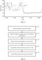
- Fig. 1 illustrates an example of a real-time systolic blood pressure (y-axis) of a patient as a function of time (trace 22), as compared with blood pressure measurements recorded by a measurement device at regular intervals (trace 24).
- the interval time is 15 minutes in this example. It can be seen from this example how, if the blood pressure drops suddenly during an inter-measurement interval, this could be missed for a significant time until the next measurement is taken. This is apparent for example within the interval from around 16:15 to around 16:30, where the patient's real-time blood pressure has dropped significantly, but this occurs within the measurement interval so that approximately 15 minutes pass before this change in condition can be addressed by a clinician.
- a computer-implemented method comprises receiving biological measurement data for a patient.
- the method further comprises applying a patient risk model to compute a risk parameter, p, indicative of a risk level of at least one pre-defined adverse clinical event, wherein at least one input to the model is the biological measurement data.
- the method further comprises obtaining a clinician-defined patient risk parameter, A.
- the method further comprises computing a measurement time interval, T, for successive non-invasive blood pressure measurements of the patient based on A and p and in accordance with the relationship: T ⁇ A p ⁇ where a is pre-defined parameter, preferably having a value greater than zero.
- the method further comprises controlling a non-invasive blood pressure measurement apparatus to acquire blood pressure measurements at a frequency defined by the determined time interval, T.
- embodiments of the invention provide a method of adaptively configuring the timing intervals between automatically triggered non-invasive blood pressure measurements based on a risk model which takes into account a current value of at least one biological parameter of the patient, and which advantageously also takes into account a clinician assessment of risk.
- a risk model which takes into account a current value of at least one biological parameter of the patient, and which advantageously also takes into account a clinician assessment of risk.
- T keeps the product of the model-based probability for a critical event, p, and the blood pressure measurement interval time constant.
- the model may be designed or trained to output a combined risk level of the patient suffering any of a set of different adverse events. Additionally or alternatively, the model may output a risk associated with only one adverse clinical event but wherein the method comprises running multiple versions of the model, each configured for predicting probability of a different type of adverse event, and selecting one the determined probability parameters based on a pre-defined criterion, or based on an input from the clinician.
- the at least one adverse or critical event may include hypotension and/or hypertension of the patient; a shock state of the patient, a cardiac arrest, or any cardiac event.
- the model is such that p is the probability of the occurrence of the event in a pre-defined time span.
- the biological measurement data includes real-time sensor data, or data derived therefrom.
- the biological measurement data includes historical biological measurement data for the patient retrieved from a datastore.
- this might include heart rate or other vital signs, previous blood pressure measurement data, pathology tests or any other biological parameters.
- the risk parameter p is recalculated recurrently over a monitoring session for the patient.
- the biological measurement data is preferably updated for each recalculation.
- the risk model is a Bayesian model.
- the Bayesian model is a personalized risk model for the patient and/or a clinician treating the patient.
- the Bayesian model is pre-configured in accordance with prior information including one or more of: patient medical history; patient condition severity/acuity; a training level of a clinician treating the patient; and a measure of a speed of physician reaction to condition changes.
- the method further comprises recurrently adjusting or updating the risk model based on a patient monitoring database comprising monitoring data for a plurality of patients.
- a self-learning model which can learn over time e.g. via retrospective analysis of monitored patients. This could be based on monitoring data acquired at a same medical institution or department, and/or could be based on monitoring data acquired more widely, for example population-wide across multiple different medical institutions.
- the biological measurement data includes data from one or more of: an Electro-cardiogram (ECG) sensing apparatus; a Photo-plethysmogram (PPG) sensing apparatus; a capnographic measurement apparatus; and a bioimpedance measurement apparatus.
- ECG Electro-cardiogram
- PPG Photo-plethysmogram
- the method can be implemented in software form.
- a processing unit comprising: an input/output; and one or more processors adapted to: receive at the input/output biological measurement data for a patient; apply a patient risk model to compute a risk parameter, p, indicative of a risk level of at least one pre-defined adverse clinical event, wherein at least one input to the model is the biological measurement data; obtain a clinician-defined patient risk parameter, A ; compute a measurement time interval, T, for successive non-invasive blood pressure measurements of the patient based on A and p and in accordance with the relationship: T ⁇ A p ⁇ where a is pre-defined parameter; and control, via generating control signals for output at the input/output, a non-invasive blood pressure measurement apparatus to acquire blood pressure measurements at a frequency defined by the determined time interval, T.
- Another aspect of the invention is a system comprising the processing unit as outlined above or in accordance with any embodiment described in this disclosure; and a non-invasive blood pressure measurement apparatus operatively coupled with the processing unit.
- the system further comprises one or more biological parameter sensing devices, for acquiring biological measurement data, operatively coupled with the processing unit.
- the one or more biological parameter sensing devices include a PPG sensor integrated in a section of a non-invasive blood pressure measurement device.
- the invention provides a method for adaptively scheduling non-invasive blood pressure measurements with time intervals determined based on using a risk model to compute a risk of a patient suffering a pre-defined one or more adverse clinical events, for example within a pre-defined time window, and also based on a clinician risk assessment for a patient.
- Blood pressure is typically measured via intermittent cuff non-invasive blood pressure (NIBP) measurements.
- NIBP non-invasive blood pressure
- medical staff must decide which blood pressure monitoring interval is appropriate based on an assessment of the hemodynamic stability of the patient.
- the NIBP measurement intervals would ideally need continuous adaptation to reflect changing patient risk levels. However, in practical situations, this is not implemented, which can result in a number of possible adverse consequences, including:
- Fig. 1 illustrates that if NIBP monitoring is performed using intermittent measurements at fixed measurement time intervals (line 24), many episodes in which large BP changes (line 22) occur are missed. The selection of the measurement time interval by the clinician was not appropriated for this patient. It is noted that the illustrated graph of Fig. 1 shows the blood pressure trend line according to a zero-order hold representation (e.g. the discrete-time blood pressure signal is represented as a continuous-time signal by holding each sample value for one sample interval).
- a zero-order hold representation e.g. the discrete-time blood pressure signal is represented as a continuous-time signal by holding each sample value for one sample interval.
- Blood pressure can be used as an indicator of a wide variety of medical adverse events in the body of the patient.
- Two adverse events of common concern for clinicians monitoring a patient are hypertension and hypotension phases.
- Hypertensive events are phases of very high blood pressure, typically understood to correspond to systolic blood pressure greater than about 140 mm/Hg.
- Hypotensive events are phases of very low blood pressure, typically understood to correspond to systolic blood pressure of less than about 90 mm/Hg.
- the probability of a patient death or longer-term health issues can increase significantly if time is spent in a hypotensive phase.
- the post-operative phase of care in particular carries a significant risk of myocardial infarction and death even if only short amounts of time are spent in hypotensive phase, due to the resulting low perfusion (ischemia) of the heart and brain.
- McEvoy, M et al Perioperative Quality Initiative consensus statement on postoperative blood pressure, risk and outcomes for elective surgery, British Journal of Anaesthesia, Volume 122, Issue 5, 2019, Pages 575-586 .
- a clinician must act quickly through administration of medication or other interventions to bring the patient back into a normal hemodynamic state.
- a hypotensive event starts at the moment the patient's true blood pressure value falls below a certain pre-defined critical threshold.
- the duration of the hypotensive blood pressure event can be defined as the interval time (minutes) from the moment when the actual blood pressure crosses below the critical blood pressure threshold to the moment it rises back above the pre-defined critical threshold.
- DDD depth-duration dose metric
- hypotension threshold there are different ways in which this can be defined. Regardless of the definition, it has been shown in studies that in different populations (e.g. non-cardiac surgery patients and traumatic brain injury patients) that patient outcome is strongly dependent on the duration and depth (meaning how low the blood pressure drops, e.g. the minimum value) of the hypotensive episode. This is because damage is done cumulatively over the course of a continuous hypotensive episode. For example, by quantifying the hypotension dose (hypotension depth integrated across exposure time) of 7521 traumatic brain injury patients, it was shown in one study ( ibid ) that hypotension was associated with 20% increased mortality.
- hypotension dose hypertension depth integrated across exposure time
- Embodiments of the present invention provide a system and method to estimate and automatically configure optimal/appropriate blood pressure measurement intervals, personalized to a patient risk profile and physician opinion.
- Fig. 1 outlines in block diagram form steps of an example method according to one or more embodiments. The steps will be recited in summary, before being explained further in the form of example embodiments.
- the method comprises receiving 12 biological measurement data for a patient.
- the method further comprises applying 14 a patient risk model to compute a risk parameter, p, indicative of a risk level of at least one pre-defined adverse clinical event, wherein at least one input to the model is the biological measurement data.
- the method further comprises obtaining 16 a clinician-defined patient risk parameter, A.
- the method further comprises computing 18 a measurement time interval, T, for successive non-invasive blood pressure measurements of the patient based on A and p and in accordance with the relationship T ⁇ A p ⁇ where a is pre-defined parameter.
- the method further comprises controlling 20 a non-invasive blood pressure measurement apparatus to acquire blood pressure measurements at a frequency defined by the determined time interval, T.
- the method can also be embodied in hardware form, for example in the form of a processing unit which is configured to carry out a method in accordance with any example or embodiment described in this document, or in accordance with any claim of this application.
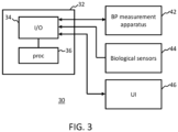
- FIG. 2 presents a schematic representation of an example processing unit 32 configured to execute a method in accordance with one or more embodiments of the invention, and also schematically showing the processing flow in more detail.
- the processing unit 32 comprises an input/output 34 and one or more processors 36.
- the one or more processors are configured to perform the following steps:
- FIG. 2 shows the processing unit 32 operating within the context of such a system.
- the system 30 may further comprise a non-invasive blood pressure measurement apparatus 42 operatively coupled with the processing unit, for example a cuff-based oscillometric blood pressure measurement apparatus.
- the system 30 may further comprise one or more biological parameter sensing devices 44, for acquiring biological measurement data, operatively coupled with the processing unit 32.
- the one or more biological parameter sensing devices include a PPG sensor.
- a PPG sensor can be used to acquire pulse rate or heart rate, as well as blood oxygen concentration. It is also possible to infer a patient respiration rate from a PPG sensor signal.
- the PPG sensor may advantageously be integrated in a section of a non-invasive blood pressure measurement device.
- the system 30 may further comprise a user interface 46, for example for use in receiving a user-input indicative of the clinician-defined patient risk parameter, A, and/or for displaying a representation of the determined blood pressure measurement interval, a current value of the computed risk parameter, one or more biological measurements for the patient, or any combination thereof.
- a user interface 46 for example for use in receiving a user-input indicative of the clinician-defined patient risk parameter, A, and/or for displaying a representation of the determined blood pressure measurement interval, a current value of the computed risk parameter, one or more biological measurements for the patient, or any combination thereof.
- a patient in a medical institution is monitored with a set of one or more measurement devices 42, e.g. sensors, each configured to measure at least one biological parameter, e.g. a physiological parameter.
- Data outputs from the set of one or more measurement devices are received at the processing unit 32.
- This data forms a biological measurement data set for the patient.
- the processing unit could be a processing unit of a patient monitoring system in some examples.
- the biological measurement data set may include data retrieved from a datastore which may include for example historical values for one or more biological parameters, or variables relating to the patient's medical history. It could include parameters which cannot be measured with sensors, such as pathology test results.
- the processing unit 32 is further adapted to obtain an indication of a clinician-defined risk parameter, A.
- this parameter may be obtained based on a user input received from a user interface device 46.
- a clinician who is responsible for monitoring the patient may input the parameter.
- the parameter A may be determined for the patient based on a look-up table.
- a clinician may have determined in advance a set of relationships between different patient status classifications and associated values for the parameter A, and these may be encoded in a stored look-up table.
- the processing unit 32 is further adapted to retrieve from a datastore a patient risk model.
- the patient risk model is adapted to receive input values for one or more variables corresponding to one or more respective biological measurements. These may include some or all of the values forming the received biological measurement dataset.
- the patient risk model is configured to generate as an output a value of a parameter, p , indicative of a risk level of at least one pre-defined adverse clinical event, such as hypotension, hypertension, or a cardiac event for instance.
- the value of p may be a probability value, and may take values ranging from 0 to 1.
- the model may be a generic model for any patient, or may be personalized to the patient, as will be explained in more detail later. The model could additionally or alternatively in some cases be personalized to the clinician monitoring the patient, as will also be explained in more detail later.
- the processing unit 32 having retrieved the model, applies the model by supplying to it as input the relevant biological measurements, to thereby generate an output risk parameter value, p.
- the processing unit 32 then computes a measurement time interval, T, for successive non-invasive blood pressure measurements of the patient based on first computing a value of A p ⁇ , and computing a value of T based on the relationship T ⁇ A p ⁇ , where a is further variable parameter.
- ⁇ may be a function of any or all of A, p , and ⁇ .
- parameter ⁇ is to provide a transfer or conversion of the value of A p ⁇ to a suitable value for T, in the correct units.
- the parameter ⁇ can be pre-defined to provide the particular mapping between T and the ratio A p ⁇ which is desired.
- the parameter a provides a further degree of freedom in the computation, for refining the computation of the time interval.
- the parameter in effect defines a relative weighting to be applied to the computer-determined risk parameter, p , as compared to the clinician-defined risk parameter, A. It will be recognized that as the value of a increases, so the scaling of the value of p ⁇ , as a function of p increases in steepness. This has the effect that a higher value of a means more relative weight being placed on the computationally defined risk parameter p as compared with the clinician defined risk parameter A.
- the parameter a might in some examples be freely determined by a user. In some examples, it might be determined using a pre-defined lookup table or mapping which relates different patient or clinician statuses or classes to different values of a. For example, for a higher acuity patient, it might be preferred to set a lower value for a, since (for a value of p which varies between 0 and 1) this will have the effect of reducing the time intervals between blood pressure measurements if risk parameter p increases during a time period between a clinician updating of their own assessment of risk, A. Therefore a higher value for a in these circumstances weighs toward greater caution.
- the value of a might be set lower compared to the case of a less experienced clinician. This would have the effect (assuming a values of p which varies between 0 and 1) of giving higher relative weighting to the clinician-defined parameter A as compared to a case of a lower value of a.
- the processing unit 32 may store in a memory a record of the newly calculated value of the computed interval time T. This may be updated each time T is re-calculated. A log may be recorded of computed values of T over time. T may be re-calculated recurrently over a monitoring session for the patient. The risk parameter p may be recalculated recurrently over the monitoring session for the patient, with the biological measurement data being updated for each recalculation. Using the newly calculated value for p, the processing unit can recalculate T.
- the processing unit 32 is further configured to control the NIBP measurement apparatus to acquire the BP measurements at a frequency defined by the determined time interval, T.
- the actual control of the NIBP measurement apparatus may be implemented by the processing unit 32 itself, or by a subsystem with which the processing unit is operatively coupled.
- the step of controlling the NIBP measurement apparatus to acquire the BP measurements at a frequency defined by the determined time interval may comprise simply communicating a control command to the NIBP measurement apparatus control subsystem to cause the subsystem to acquire the measurements at the computed interval T.
- the processing unit may simply update a value of the parameter T in a local register, and wherein the processing unit runs a control routine or program for triggering the BP measurements at time intervals determined by the current value of T in the register.
- the value of T be re-calculated recurrently.
- this could be performed at regular fixed time intervals, at intervals set by a clinician, or at intervals which are automatically determined or triggered.
- one approach might be to re-calculate T each time a new BP measurement is triggered.
- the value of T itself is updated at a frequency determined by the current value of T, so that as each measurement interval ends, a new blood pressure measurement is acquired, and at the same time, a new value of the interval time T is calculated.
- new values of each of the one or more biological measurements may be sampled, and these used as inputs for re-running the patient risk model to generate a new value for the parameter p, following which the new value of T is computed using the relationship already outlined above.
- the patient risk model is naturally trained or programmed for modelling the patient risk for a specific adverse clinical event.
- a plurality of these versions of the risk model might be run, to derive a set of initial risk parameters p i , each indicative of the probability of occurrence of a respective adverse clinical event. These initial risk parameters may then be further processed to derive the overall risk parameter p , indicative of risk of one or more adverse clinical events.
- just one of the initial risk parameters, p i might be selected, corresponding to the adverse event which is most likely, or for which consequences for the patient are most severe. The selection may be based on a user input at the user interface. For instance, the user interface displays the set of computed initial risk parameters, along with an indication of the adverse clinical event to which each corresponds, and the user can select one to be used as the overall patient risk parameter.
- some or all parameters of the set of initial risk parameters may be combined to form the overall patient risk parameter, p.
- the probabilities may simply be added, to give a cumulative probability of any of the respective adverse clinical events occurring. Alternatively, an average might be computed.
- the biological measurement data which is obtained as part of the method, it is preferred that this is received continuously, or in real-time, from one or more measurement devices or sensors arranged for monitoring the patient.
- the patient risk model is provided with information which is up-to-date for the patient.
- the alternative approach would be to use archived biological measurement data for the patient.
- the biological measurement data may include one or more of the following:
- the Photo-plethysmogram (PPG) sensor can be provided as an auxiliary sensor, separate to the blood pressure measurement apparatus, e.g. a finger PPG sensor.
- the PPG sensor may be provided integrated in the blood pressure measurement apparatus, for example integrated in the blood pressure measurement cuff, and arranged such that, when the cuff is worn on a body part for measuring blood pressure, the PPG sensor optical area is in optical communication with a surface of the patient's skin.
- biological measurement data can include historical biological measurement data for the patient retrieved from a datastore.
- this preferably comprises a cuff-based measurement device.
- the cuff might in use be applied to the patient's arm or wrist.
- a typical example of such a device operates by the cuff being actively pressurized e.g. with a pump or pressure reservoir, and the cuff pressure measured with a pressure sensor.
- Other ways to operate a cuff-based NIBP measurement device also exist and will be known to the skilled person. Any other type of NIBP measurement device can also be used as an alternative to a cuff-based system.
- NIBP measurement devices are very well understood in the field, and the skilled person will understand how to implement this feature.
- a guiding insight behind the approach proposed by the inventors is the concept that, for a constant value of the clinician-defined parameter, A, a product of the probability, p (potentially adjusted by power laws), for a critical event and the NIBP interval time, T, be maintained constant.
- p potentially adjusted by power laws
- T is the recommended measurement interval time.
- A is a physician-defined parameter, representing the physician's assessment of a risk level for a particular (pre-defined) adverse or critical event (e.g. hypotension, hypertension) of a patient for a coming monitoring period. It expected the physician would choose this in dependence upon their judgment of the situation, using their knowledge and experience to assess the risk level.
- the parameter, p is the probability of the occurrence of the same pre-defined adverse event within a defined time window, as computed based on a patient risk model.
- the index/power parameter a can be adjusted so as to give more weight or less weight to the computer risk parameter, p , as compared to the clinician risk parameter A.
- ⁇ might be a single value constant.
- ⁇ might be a function of any or all of A, p and a, so that it provides a transfer function between the value of A p ⁇ and suitable timing values T for the measurement intervals.
- reference to use of A p ⁇ alone can be understood to optionally include the coefficient or transfer function ⁇ as a further multiplier.
- this calibration might be already incorporated into the allowable value ranges for A, p and a.
- each of these parameters might take values across respective ranges, and/or at respective intervals across that range, which are configured such that evaluation of A p ⁇ automatically results in a value of T with the correct units.
- This equation has the effect that an increased risk level of a patient potentially suffering a critical event results in a decreased measurement interval time.
- the parameter A can be specified by a particular clinician treating a patient, representing the clinician's assessment of the risk level for the patient. Additionally or alternatively, the parameter A could be set based on a standardized guideline in a particular hospital department or based on generic guidelines, e.g. based on a lookup table relating different classifications of patient condition to different values for the parameter A.
- a time-dependent estimation of the probability p of a critical event allows up to real-time adjustments of the interval time T.
- the method may further comprise determining an estimate of an uncertainty of T.
- an uncertainty parameter ⁇ T may be computed.
- a simple means for estimating an uncertainty is to define uncertainty interval values ⁇ A and ⁇ p .
- ⁇ p can be estimated from the Bayesian model.
- the Bayesian model inherently outputs a probability distribution, along with an uncertainty dp, and this uncertainty can be used for ⁇ p.
- the value for ⁇ A can be pre-defined for example using a look-up table, or defined according to a hospital policy in dependence upon patient condition or other factors.
- the computed value of the uncertainty parameter ⁇ T could be presented to the user (clinician) on a display of a user interface, along with the current computed value of T.
- the user could, via the user interface, be provided the option whether to accept T as the current measurement interval or not.
- the value of the uncertainty parameter provides additional information that assists the user in making this decision. If the uncertainty is high, then the user (clinician) might decide to lean toward caution and set the interval at a shorter time than the current value of T.
- the user could be provided a set of three preset options for the measurement interval time: T, T+ ⁇ T, or T- ⁇ T. For example, if the uncertainty is relatively high, then the user might choose T- ⁇ T.
- the value of ⁇ A can be set to zero or estimated from historical data or from guidelines.
- a part of the method is the use of a patient risk model to compute the risk parameter, p, indicative of a risk level of at least one pre-defined adverse clinical event. Details in respect of the patient risk model will now be discussed in further detail.
- the patient risk model is configured such as to receive, in operation, inputs which comprise at least: values for each of one or more biological measurement parameters for the patient (discussed above).
- the patient risk model is configured to generate as output a value, p, indicative of probability of occurrence of a pre-defined adverse clinical event within a certain time interval.
- a particularly well-suited approach to estimating a probability of this kind is to use a Bayesian statistical model.
- a statistical model using Bayesian principles is able to determine a posterior probability distribution for a particular outcome, based on one or a plurality of input variables, wherein the model has been constructed based on one or a plurality of prior probability distributions for one or more events.
- Bayesian principles in this way to derive estimates of risk based on current values of one or more parameters and prior distributions for those parameters will be well known to the skilled person.
- a Bayesian model might be constructed such as to provide a value of p which indicates an estimated risk of blood pressure dropping below value x for y minutes, where x and y are defined values set so as to correspond to a hypotensive event of concern.
- Prior distributions can be determined in advance for this scenario, as a function of variations in the measured biological parameters, d(t).
- d (t) is a vector representing the set of all biological parameters (and/or other parameters) which are to be used as the inputs to the model.
- T A p BP ⁇ x mmHg for y min , K
- the probability p can preferably be estimated recurrently, e.g. continuously or quasi-continuously, from acquired real-time biological measurement data and/or available historical data.
- Each of these variables could be codified according to a respective standardized score metric, so that prior distributions of p can be measured over variable values of each.
- the model can be effectively made to mimic the mental process of a human being taking into account new data.
- the method may further comprise updating the patient risk model based on monitoring data for patients in the medical institution.
- monitoring data could include blood pressure measurement data and biological measurement data so that relationships between these parameters can be used in training the model.
- the model can learn over time via retrospective analysis of monitored patients.
- changes in clinician training level can be monitored and reflected in updates to the prior information used in training the model.
- the method may further include a model update phase, which might be run once or at intervals which might be the same or different to the intervals for any (optional) updating of the parameters p and T.
- the update phase may include execution of an algorithm for obtaining and collating the relevant updated model prior distributions, and updating the model with the updated prior distributions.
- a machine learning model might be used in further examples comprising one or more machine learning algorithms trained to receive as input at least the biological measurement data for the patient and generate as output the risk parameter, p , indicative of a risk level of at least one pre-defined adverse clinical event.
- Suitable machine learning algorithms include, by way of non-limiting example, a multivariate regression model, or an artificial neural network such as a convolutional neural network.
- the invention can be embodied in software form.
- a computer program product comprising code means configured, when run on a processor, to cause the processor to perform a method in accordance with any example or embodiment of the invention described in this document, or in accordance with any claim of this patent application.
- the method can be executed by a processing unit at any of a variety of different locations.
- the method could be executed by a processing unit of a bed-side patient monitoring device, or by a patient monitoring system, or by a separate computing system, or by a cloud-based computing system.
- Embodiments of the invention described above employ a processing unit.
- the processing unit may in general comprise a single processor or a plurality of processors. It may be located in a single containing device, structure or unit, or it may be distributed between a plurality of different devices, structures or units. Reference therefore to the processing unit being adapted or configured to perform a particular step or task may correspond to that step or task being performed by any one or more of a plurality of processing components, either alone or in combination. The skilled person will understand how such a distributed processing arrangement can be implemented.
- the processing unit may include a communication module or input/output for receiving data and outputting data to further components.
- the one or more processors of the processing unit can be implemented in numerous ways, with software and/or hardware, to perform the various functions required.
- a processor typically employs one or more microprocessors that may be programmed using software (e.g., microcode) to perform the required functions.
- the processor may be implemented as a combination of dedicated hardware to perform some functions and one or more programmed microprocessors and associated circuitry to perform other functions.
- circuitry examples include, but are not limited to, conventional microprocessors, application specific integrated circuits (ASICs), and field-programmable gate arrays (FPGAs).
- ASICs application specific integrated circuits
- FPGAs field-programmable gate arrays
- the processor may be associated with one or more storage media such as volatile and non-volatile computer memory such as RAM, PROM, EPROM, and EEPROM.
- the storage media may be encoded with one or more programs that, when executed on one or more processors and/or controllers, perform the required functions.
- Various storage media may be fixed within a processor or controller or may be transportable, such that the one or more programs stored thereon can be loaded into a processor.
- a single processor or other unit may fulfill the functions of several items recited in the claims.
- a computer program may be stored/distributed on a suitable medium, such as an optical storage medium or a solid-state medium supplied together with or as part of other hardware, but may also be distributed in other forms, such as via the Internet or other wired or wireless telecommunication systems.
- a suitable medium such as an optical storage medium or a solid-state medium supplied together with or as part of other hardware, but may also be distributed in other forms, such as via the Internet or other wired or wireless telecommunication systems.
Landscapes
- Health & Medical Sciences (AREA)
- Life Sciences & Earth Sciences (AREA)
- Engineering & Computer Science (AREA)
- Medical Informatics (AREA)
- Public Health (AREA)
- Physics & Mathematics (AREA)
- General Health & Medical Sciences (AREA)
- Pathology (AREA)
- Biomedical Technology (AREA)
- Animal Behavior & Ethology (AREA)
- Veterinary Medicine (AREA)
- Surgery (AREA)
- Molecular Biology (AREA)
- Cardiology (AREA)
- Biophysics (AREA)
- Heart & Thoracic Surgery (AREA)
- Physiology (AREA)
- Vascular Medicine (AREA)
- Artificial Intelligence (AREA)
- Psychiatry (AREA)
- Computer Vision & Pattern Recognition (AREA)
- Signal Processing (AREA)
- Ophthalmology & Optometry (AREA)
- Radiology & Medical Imaging (AREA)
- Nuclear Medicine, Radiotherapy & Molecular Imaging (AREA)
- Pulmonology (AREA)
- Fuzzy Systems (AREA)
- Primary Health Care (AREA)
- Epidemiology (AREA)
- Databases & Information Systems (AREA)
- Data Mining & Analysis (AREA)
- Evolutionary Computation (AREA)
- Mathematical Physics (AREA)
- Measuring Pulse, Heart Rate, Blood Pressure Or Blood Flow (AREA)
- Medical Treatment And Welfare Office Work (AREA)
Priority Applications (6)
| Application Number | Priority Date | Filing Date | Title |
|---|---|---|---|
| EP22195047.0A EP4335360A1 (de) | 2022-09-12 | 2022-09-12 | Nichtinvasive blutdruckmessung |
| EP23762233.7A EP4586899A1 (de) | 2022-09-12 | 2023-08-29 | Nichtinvasive blutdruckmessung |
| PCT/EP2023/073610 WO2024056362A1 (en) | 2022-09-12 | 2023-08-29 | Non-invasive blood pressure measurement |
| CN202380065514.2A CN119923220A (zh) | 2022-09-12 | 2023-08-29 | 无创血压测量 |
| US19/109,183 US20260076581A1 (en) | 2022-09-12 | 2023-08-29 | Non-invasive blood pressure measurement |
| JP2025508448A JP2025530077A (ja) | 2022-09-12 | 2023-08-29 | 非侵襲的血圧測定 |
Applications Claiming Priority (1)
| Application Number | Priority Date | Filing Date | Title |
|---|---|---|---|
| EP22195047.0A EP4335360A1 (de) | 2022-09-12 | 2022-09-12 | Nichtinvasive blutdruckmessung |
Publications (1)
| Publication Number | Publication Date |
|---|---|
| EP4335360A1 true EP4335360A1 (de) | 2024-03-13 |
Family
ID=83283534
Family Applications (2)
| Application Number | Title | Priority Date | Filing Date |
|---|---|---|---|
| EP22195047.0A Withdrawn EP4335360A1 (de) | 2022-09-12 | 2022-09-12 | Nichtinvasive blutdruckmessung |
| EP23762233.7A Pending EP4586899A1 (de) | 2022-09-12 | 2023-08-29 | Nichtinvasive blutdruckmessung |
Family Applications After (1)
| Application Number | Title | Priority Date | Filing Date |
|---|---|---|---|
| EP23762233.7A Pending EP4586899A1 (de) | 2022-09-12 | 2023-08-29 | Nichtinvasive blutdruckmessung |
Country Status (5)
| Country | Link |
|---|---|
| US (1) | US20260076581A1 (de) |
| EP (2) | EP4335360A1 (de) |
| JP (1) | JP2025530077A (de) |
| CN (1) | CN119923220A (de) |
| WO (1) | WO2024056362A1 (de) |
Citations (2)
| Publication number | Priority date | Publication date | Assignee | Title |
|---|---|---|---|---|
| US20180042493A1 (en) * | 2015-02-24 | 2018-02-15 | Koninklijke Philips N.V. | Apparatus and method for providing a control signal for a blood pressure measurement device |
| WO2021062292A1 (en) * | 2019-09-26 | 2021-04-01 | Etiometry Inc. | Systems and methods for transitioning patient care from signal based monitoring to risk-based monitoring |
-
2022
- 2022-09-12 EP EP22195047.0A patent/EP4335360A1/de not_active Withdrawn
-
2023
- 2023-08-29 JP JP2025508448A patent/JP2025530077A/ja active Pending
- 2023-08-29 US US19/109,183 patent/US20260076581A1/en active Pending
- 2023-08-29 WO PCT/EP2023/073610 patent/WO2024056362A1/en not_active Ceased
- 2023-08-29 EP EP23762233.7A patent/EP4586899A1/de active Pending
- 2023-08-29 CN CN202380065514.2A patent/CN119923220A/zh active Pending
Patent Citations (2)
| Publication number | Priority date | Publication date | Assignee | Title |
|---|---|---|---|---|
| US20180042493A1 (en) * | 2015-02-24 | 2018-02-15 | Koninklijke Philips N.V. | Apparatus and method for providing a control signal for a blood pressure measurement device |
| WO2021062292A1 (en) * | 2019-09-26 | 2021-04-01 | Etiometry Inc. | Systems and methods for transitioning patient care from signal based monitoring to risk-based monitoring |
Non-Patent Citations (5)
Also Published As
| Publication number | Publication date |
|---|---|
| EP4586899A1 (de) | 2025-07-23 |
| US20260076581A1 (en) | 2026-03-19 |
| CN119923220A (zh) | 2025-05-02 |
| JP2025530077A (ja) | 2025-09-11 |
| WO2024056362A1 (en) | 2024-03-21 |
Similar Documents
| Publication | Publication Date | Title |
|---|---|---|
| US11844624B2 (en) | System and method for monitoring cerebral activity | |
| JP5608102B2 (ja) | 血行動態(Hemodynamic)モニタ及びアラーム | |
| EP2849639B1 (de) | Messung des herzzeitvolumens | |
| US8668649B2 (en) | System for cardiac status determination | |
| JP6757734B2 (ja) | 血圧測定デバイス用の制御信号を供給するための装置及び方法 | |
| US12243649B2 (en) | Physiological parameter monitoring system | |
| US11166666B2 (en) | Enhanced acute care management combining imaging and physiological monitoring | |
| US10869631B2 (en) | Method and system for assessing fluid responsiveness using multimodal data | |
| US20140046153A1 (en) | Monitoring volaemic condition in a human or animal subject | |
| US20250275726A1 (en) | Method for training a machine learning model, monitoring device, and monitoring method | |
| US20160171170A1 (en) | Measuring respiration or other periodic physiological processes | |
| US20260076581A1 (en) | Non-invasive blood pressure measurement | |
| JP7716679B2 (ja) | 健康管理システム、及び、健康管理方法 | |
| EP4272633B1 (de) | Vorrichtung zur bestimmung von zustandsinformationen | |
| JP7669084B1 (ja) | バイタルデータ評価方法、バイタルデータ評価システムおよびプログラム | |
| US20250114004A1 (en) | Device and method for detecting orthostatic vital signs | |
| EP4094682A1 (de) | Verfahren, vorrichtung und computerprogrammprodukt zur analyse eines pulswellensignals | |
| CN117795614A (zh) | 诊疗辅助系统、诊疗辅助装置以及程序 | |
| CN117795608A (zh) | 诊疗辅助系统、诊疗辅助装置以及程序 | |
| WO2025258403A1 (ja) | バイタルデータ評価方法、バイタルデータ評価装置、バイタルデータ評価システムおよびプログラム | |
| JP2024140945A (ja) | プログラム、情報処理方法および情報処理装置 | |
| CN121311941A (zh) | 预测针对临床对象的参数的值的可靠性 |
Legal Events
| Date | Code | Title | Description |
|---|---|---|---|
| PUAI | Public reference made under article 153(3) epc to a published international application that has entered the european phase |
Free format text: ORIGINAL CODE: 0009012 |
|
| STAA | Information on the status of an ep patent application or granted ep patent |
Free format text: STATUS: THE APPLICATION HAS BEEN PUBLISHED |
|
| AK | Designated contracting states |
Kind code of ref document: A1 Designated state(s): AL AT BE BG CH CY CZ DE DK EE ES FI FR GB GR HR HU IE IS IT LI LT LU LV MC MK MT NL NO PL PT RO RS SE SI SK SM TR |
|
| STAA | Information on the status of an ep patent application or granted ep patent |
Free format text: STATUS: THE APPLICATION IS DEEMED TO BE WITHDRAWN |
|
| 18D | Application deemed to be withdrawn |
Effective date: 20240914 |