EP4336183A2 - Verbesserte verfahren und vorrichtungen zur genauen diagnose von infektionen - Google Patents
Verbesserte verfahren und vorrichtungen zur genauen diagnose von infektionen Download PDFInfo
- Publication number
- EP4336183A2 EP4336183A2 EP23209434.2A EP23209434A EP4336183A2 EP 4336183 A2 EP4336183 A2 EP 4336183A2 EP 23209434 A EP23209434 A EP 23209434A EP 4336183 A2 EP4336183 A2 EP 4336183A2
- Authority
- EP
- European Patent Office
- Prior art keywords
- infection
- bacterial
- sample
- viral
- mxa
- Prior art date
- Legal status (The legal status is an assumption and is not a legal conclusion. Google has not performed a legal analysis and makes no representation as to the accuracy of the status listed.)
- Pending
Links
Images












Classifications
-
- C—CHEMISTRY; METALLURGY
- C12—BIOCHEMISTRY; BEER; SPIRITS; WINE; VINEGAR; MICROBIOLOGY; ENZYMOLOGY; MUTATION OR GENETIC ENGINEERING
- C12Q—MEASURING OR TESTING PROCESSES INVOLVING ENZYMES, NUCLEIC ACIDS OR MICROORGANISMS; COMPOSITIONS OR TEST PAPERS THEREFOR; PROCESSES OF PREPARING SUCH COMPOSITIONS; CONDITION-RESPONSIVE CONTROL IN MICROBIOLOGICAL OR ENZYMOLOGICAL PROCESSES
- C12Q1/00—Measuring or testing processes involving enzymes, nucleic acids or microorganisms; Compositions therefor; Processes of preparing such compositions
- C12Q1/68—Measuring or testing processes involving enzymes, nucleic acids or microorganisms; Compositions therefor; Processes of preparing such compositions involving nucleic acids
- C12Q1/6876—Nucleic acid products used in the analysis of nucleic acids, e.g. primers or probes
- C12Q1/6888—Nucleic acid products used in the analysis of nucleic acids, e.g. primers or probes for detection or identification of organisms
- C12Q1/689—Nucleic acid products used in the analysis of nucleic acids, e.g. primers or probes for detection or identification of organisms for bacteria
-
- C—CHEMISTRY; METALLURGY
- C12—BIOCHEMISTRY; BEER; SPIRITS; WINE; VINEGAR; MICROBIOLOGY; ENZYMOLOGY; MUTATION OR GENETIC ENGINEERING
- C12Q—MEASURING OR TESTING PROCESSES INVOLVING ENZYMES, NUCLEIC ACIDS OR MICROORGANISMS; COMPOSITIONS OR TEST PAPERS THEREFOR; PROCESSES OF PREPARING SUCH COMPOSITIONS; CONDITION-RESPONSIVE CONTROL IN MICROBIOLOGICAL OR ENZYMOLOGICAL PROCESSES
- C12Q1/00—Measuring or testing processes involving enzymes, nucleic acids or microorganisms; Compositions therefor; Processes of preparing such compositions
- C12Q1/70—Measuring or testing processes involving enzymes, nucleic acids or microorganisms; Compositions therefor; Processes of preparing such compositions involving virus or bacteriophage
-
- G—PHYSICS
- G01—MEASURING; TESTING
- G01N—INVESTIGATING OR ANALYSING MATERIALS BY DETERMINING THEIR CHEMICAL OR PHYSICAL PROPERTIES
- G01N33/00—Investigating or analysing materials by specific methods not covered by groups G01N1/00 - G01N31/00
- G01N33/48—Biological material, e.g. blood, urine; Haemocytometers
- G01N33/50—Chemical analysis of biological material, e.g. blood, urine; Testing involving biospecific ligand binding methods; Immunological testing
- G01N33/53—Immunoassay; Biospecific binding assay; Materials therefor
- G01N33/569—Immunoassay; Biospecific binding assay; Materials therefor for microorganisms, e.g. protozoa, bacteria, viruses
- G01N33/56911—Bacteria
-
- G—PHYSICS
- G01—MEASURING; TESTING
- G01N—INVESTIGATING OR ANALYSING MATERIALS BY DETERMINING THEIR CHEMICAL OR PHYSICAL PROPERTIES
- G01N33/00—Investigating or analysing materials by specific methods not covered by groups G01N1/00 - G01N31/00
- G01N33/48—Biological material, e.g. blood, urine; Haemocytometers
- G01N33/50—Chemical analysis of biological material, e.g. blood, urine; Testing involving biospecific ligand binding methods; Immunological testing
- G01N33/53—Immunoassay; Biospecific binding assay; Materials therefor
- G01N33/569—Immunoassay; Biospecific binding assay; Materials therefor for microorganisms, e.g. protozoa, bacteria, viruses
- G01N33/56983—Viruses
-
- G—PHYSICS
- G01—MEASURING; TESTING
- G01N—INVESTIGATING OR ANALYSING MATERIALS BY DETERMINING THEIR CHEMICAL OR PHYSICAL PROPERTIES
- G01N2333/00—Assays involving biological materials from specific organisms or of a specific nature
- G01N2333/435—Assays involving biological materials from specific organisms or of a specific nature from animals; from humans
- G01N2333/46—Assays involving biological materials from specific organisms or of a specific nature from animals; from humans from vertebrates
- G01N2333/47—Assays involving proteins of known structure or function as defined in the subgroups
-
- G—PHYSICS
- G01—MEASURING; TESTING
- G01N—INVESTIGATING OR ANALYSING MATERIALS BY DETERMINING THEIR CHEMICAL OR PHYSICAL PROPERTIES
- G01N2333/00—Assays involving biological materials from specific organisms or of a specific nature
- G01N2333/435—Assays involving biological materials from specific organisms or of a specific nature from animals; from humans
- G01N2333/46—Assays involving biological materials from specific organisms or of a specific nature from animals; from humans from vertebrates
- G01N2333/47—Assays involving proteins of known structure or function as defined in the subgroups
- G01N2333/4701—Details
- G01N2333/4737—C-reactive protein
-
- G—PHYSICS
- G01—MEASURING; TESTING
- G01N—INVESTIGATING OR ANALYSING MATERIALS BY DETERMINING THEIR CHEMICAL OR PHYSICAL PROPERTIES
- G01N2333/00—Assays involving biological materials from specific organisms or of a specific nature
- G01N2333/435—Assays involving biological materials from specific organisms or of a specific nature from animals; from humans
- G01N2333/575—Hormones
- G01N2333/585—Calcitonins
Definitions
- the invention pertains to the field of identifying infections. More particularly, the invention pertains to using the biomarkers MxA, C-reactive protein and/or procalcitonin to accurately screen for and diagnose viral and bacterial infections and to differentiate colonization from active infection.
- Fever is a common cause of childhood visits to urgent care centers for both family practice and pediatric offices. Most commonly, this relates to either a respiratory infection or gastroenteritis. The high incidence of fever in children and the precautious administration of unnecessary antibiotics is reason to develop a more accurate screening test for viral and/or bacterial infection.
- MxA myxovirus resistance protein A
- PCT procalcitonin
- CRP C-Reactive Protein
- Other acute respiratory infections include sinusitis, otitis media, rhinopharyngitis, rhinosinusitis, pharyngotonsillitis, epiglottitis, laryngitis, rhinitis, bronchitis, bronchiolitis and pneumonia.
- Severe community-acquired pneumonia is caused by bacterial infections in around 60% of cases, requiring admission to an intensive care unit (ICU) for about 10% of patients. The remaining 30% are related to respiratory viruses.
- ICU intensive care unit
- Mx proteins are members of the superfamily of high molecular weight GTPases. Accordingly, these GTPases are upregulated by type I alpha/beta or type II interferons (IFN). The Mx GTPases are expressed exclusively in IFN alpha/beta but not IFN gamma treated cells. Type I interferons play important roles in innate immune responses and have immunomodulatory, antiproliferative, and antiviral functions. Human MxA, a 78 kDa protein, accumulates in the cytoplasm of IFN ⁇ / ⁇ treated cells and inhibits the replication of a wide range of viruses.
- IFN type II interferons
- MxA protein may offer certain advantages as a biomarker for viral infection over the other induced proteins such as 2', 5'- oligoadenylate synthetase, because of its lower basal concentration, longer half-life (2.3 days) and fast induction.
- MxA mRNA is detectable in isolated peripheral blood white blood cells stimulated with IFN within 1 to 2 h of IFN induction, and MxA protein begins to accumulate shortly thereafter.
- MxA protein expression in peripheral blood is a sensitive and specific marker for viral infection.
- the higher MxA levels in the viral infection group compared with the bacterial infection group can be explained by the fact that the MxA protein is induced exclusively by type IIFN and not by IFN-gamma, IL-1, TNF-alpha, or any of the other cytokines by bacterial infection. Serum type I IFN levels remain within normal limits, even in patients with severe bacterial infections.
- C-Reactive Protein CRP
- CRP C-Reactive Protein
- C-reactive protein is found to be superior to that of the erythrocyte sedimentation rate (ESR) and superior or equal to that of the white blood cell count (WBC).
- Procalcitonin is another marker of bacterial infection. While procalcitonin has no known hormonal activity, it is a 116 amino acid peptide precursor of the hormone calcitonin which is involved with calcium homeostasis. When a patient is healthy, procalcitonin is only present in the parafollicular cells (C cells) of the thyroid gland and by the neuroendocrine cells of the lung and the intestine. If a bacterial infection is present, however, intact procalcitonin is found in the blood. The level of procalcitonin is related to the proinflammatory stimulus by severity of bacterial infection and sepsis. Procalcitonin levels do not rise significantly with viral or non-infectious inflammations.
- procalcitonin has been used in identifying bacterial infections, however, similar to C-reactive protein, some viral infections such as Influenza A and B and Adenovirus may cause modest elevations in procalcitonin levels.
- Diagnostic and screening devices and methods test for the presence of immune response markers for viral infection and immune response markers for bacterial infection, to effectively assist in the identification of the presence of a clinically significant infection, assist in the differentiation of viral and bacterial infections and to distinguish colonization/carrier state from active infection.
- One method of differentiating between colonization/carrier state and active infection includes the step of performing a test for a presence of bacteria and/or virus in a sample.
- This first test may include, but is not limited to, PCR, viral culture, viral or bacterial IFA, viral antigen testing, bacterial antigen testing, or a bacterial culture. If the sample is positive for a typical pathogen (viral or bacterial), a second test is performed to confirm the existence of a bonafide infection. Bacterial confirmation in the presence of at least approximately 0.10 ng/ml to.015 ng/ml procalcitonin and/or a presence of at least approximately 15 mg/L to 20 mg/L of C-reactive protein represents a true infection and not a carrier state or colonization.
- a third test is performed to detect a presence of the mean plus 2-3.5 times the standard deviation of the normal population baseline values of the viral biomarker, or at least approximately 25 ng/ml to 35 ng/ml of MxA depending on the reference standard.
- a presence of at least approximately 25 ng/ml MxA indicates an active viral infection.
- the absence of at least approximately 25 ng/ml MxA indicates an absence of a bonafide viral infection and represents the carrier state or colonization.
- tests for only bacteria or only viruses are performed.
- the biomarkers may be qualitative and set with thresholds at the cut-off or provide quantitative results or a combination of qualitative or quantitative results.
- a method for differentiating between colonization/carrier state and an active infection includes the step of determining the presence of a viral or bacterial pathogen utilizing antigen testing, molecular testing, and/or cell culture in combination with serological confirmation of a systemic response via an elevation in MxA, CRP, procalcitonin or any other specific bacterial biomarker.
- Other potential biomarkers include but are not limited to serum amyloid A, IL-6 (Interleukin-6), IFIT, or human neutrophile lipocalin (HNL).
- MxA, procalcitonin, and/or C-reactive protein are used to distinguish between bacterial infection, viral infection, and colonization/carrier state.
- these markers of immune response are used to diagnose patients with an illness that was microbiologically unconfirmed with the standard laboratory methods (for example, PCR, culture, and radiography).
- MxA in combination with either C-reactive protein or procalcitonin, or another serologic bacterial marker, helps to confirm the presence of a clinically significant infection from an insignificant infection that does not require immediate treatment.
- a first sample is assayed for the presence of elevated MxA, C-reactive protein and/or procalcitonin. If these assays give a negative result, a second sample is taken from the patient within four to seventy two hours (preferably, within 48 hours) of the initial sample, and tested a second time for the presence of elevated MxA, C-reactive protein and/or procalcitonin.
- the first and second sample are tested for MxA, with the second sample being taken within four to forty-eight hours of the first sample.
- the first sample and the second sample are tested for MxA and either C-reactive protein or procalcitonin.
- HNL human neutrophil lipocalin
- IL-6 or serum amyloid A are assayed instead of C-reactive protein or procalcitonin.
- the second sample in this embodiment is taken within four to forty-eight hours of the first sample.
- additional research and testing is done to try to determine if a patient with a microbiologically unconfirmed diagnosis has an emergent disease or illness.
- a method of increasing the specificity of detection of a bacterial host biomarker without compromising sensitivity includes the step of assaying for at least one viral host biomarker and at least one bacterial host biomarker.
- the bacterial host biomarker is C-reactive protein and the viral host biomarker is MxA.
- the bacterial host biomarker is procalcitonin and the viral host biomarker is MxA.
- the viral host biomarker is interferon or an IFIT (an interferon-induced protein with tetratricopeptide repeats).
- a method for determining whether an infection is bacterial and/or viral includes the step of determining a presence of MxA, C-reactive protein, and procalcitonin in a sample.
- One or more additional levels of CRP at 80 mg/ml - 100 mg/ml or PCT between 0.25ng/ml and 1.0 ng/ml may be added to the assay to determine the intensity or severity of the bacterial infection.
- a kit for diagnosing whether an infection is bacterial and/or viral includes at least one reagent for determining a presence of a first level of C-reactive protein in a sample, at least one reagent determining a presence of a second level of C-reactive protein that is higher than the first level of C-reactive protein in the sample, at least one reagent for determining a presence of MxA in the sample, and at least one reagent for determining a presence of procalcitonin in the sample.
- a single multiparametric device tests for the presence of MxA, a low level of C-reactive protein, a high level of C-reactive protein, and procalcitonin in a sample. In another preferred embodiment, a single multiparametric device tests for the presence of MxA and procalcitonin in a sample.
- One method screens a patient with a respiratory infection for bacterial colonization.
- a first test is performed for a presence of bacteria. If the first test indicates bacteria is present, a second test is performed to quantitatively determine a level of procalcitonin or C-reactive protein in a patient sample.
- a cell culture result of greater than 1 ⁇ 10 6 CFU/ml atypical bacteria in the first test and a level of procalcitonin less than 0.15 ng/ml indicates colonization.
- a cell culture result of greater than 1 ⁇ 10 6 CFU/ml atypical bacteria in the first test, a level of procalcitonin greater than or equal to 0.15 ng/ml and less than 0.25 ng/ml, a white blood cell count less than 12,000, and no bands indicates colonization.
- a cell culture result of greater than 1 ⁇ 10 6 CFU/ml atypical bacteria in the first test and a level of C-reactive protein less than 20 mg/l indicates colonization.
- a cell culture result of greater than 1 ⁇ 10 6 CFU/ml atypical bacteria in the first test, a level of C-reactive protein greater than or equal to 20 mg/l and less than 80 mg/l, a white blood cell count less than 12,000, and no bands also indicates colonization.
- the method tests for Streptolysin O antibody and a white blood cell count.
- a Streptolysin O antibody of less than 80% and a white blood cell count of less than 12,000 indicates colonization.
- Negative paired serology also indicates colonization.
- a method of screening a patient with a respiratory infection for colonization includes the step of performing a first test for a presence of a bacterial or viral infection. If the first test is positive for presence of a virus, a second test is performed to determine a level of MxA in a patient sample. A level of MxA in the patient sample greater than or equal to 25 ng/ml indicates a viral infection and a level of MxA in the patient sample less than 25 ng/ml indicates no systemic host response.
- the method may also include a third test for a presence of bacteria. If the third test indicates bacteria is present, a fourth test is performed to determine a level of procalcitonin or C-reactive protein in a patient sample.
- a patient sample testing positive for atypical bacteria using PCR in the third test and a level of procalcitonin in a patient sample less than 0.1 ng/ml or a level of C-reactive protein in the patient sample is less than 20 mg/l indicates negative for infection.
- a cell culture result of greater than 1 ⁇ 10 6 CFU/ml atypical bacteria in the third test and a level of procalcitonin less than 0.15 ng/ml indicates colonization.
- a cell culture result of greater than 1 ⁇ 10 6 CFU/ml atypical bacteria in the third test, a level of procalcitonin greater than or equal to 0.15 ng/ml and less than 0.25 ng/ml, a white blood cell count less than 12,000, and no bands indicates colonization.
- a cell culture result of greater than 1 ⁇ 10 6 CFU/ml atypical bacteria in the third test and a level of C-reactive protein less than 20 mg/l indicates colonization.
- a cell culture result of greater than 1 ⁇ 10 6 CFU/ml atypical bacteria in the third test, a level of C-reactive protein greater than or equal to 20 mg/l and less than 80 mg/l, a white blood cell count less than 12,000, and no bands also indicates colonization.
- a sample is tested for Streptolysin O antibody and a white blood cell count.
- AStreptolysin O antibody of less than 80% and a white blood cell count of less than 12,000 indicates colonization and negative paired serology also indicates colonization.
- a method of screening a symptomatic patient for active infection includes performing a test to assay for a host viral biomarker selected from the group consisting of MxA and an interferon induced protein with tetratricopeptide repeats and a host bacterial biomarker selected from the group consisting of C-reactive protein, procalcitonin, interleukin-6, serum amyloid A, and human neutrophil lipocalin.
- the first test is performed using a membrane and buffer that directly lyses cells, separates blood into plasmalserum, and filters cellular debris to detect the host viral biomarker and the host viral biomarker without any pre-processing steps.
- a value of the host viral biomarker greater than approximately 2 times the mean in the normal population times the standard deviation indicates a viral infection.
- a value of the host bacterial biomarker greater than approximately 2 times the mean in the normal population times the standard deviation indicates a bacterial infection.
- a value of the host viral biomarker greater than approximately 2 times the mean in the normal population times the standard deviation and a value of the host bacterial biomarker greater than approximately 2 times the mean in the normal population times the standard deviation indicates a viral infection.
- a value of the host viral biomarker less than approximately 2 times the mean in the normal population times the standard deviation and a value of the host bacterial biomarker less than approximately 2 times the mean in the normal population times the standard deviation indicates a microbiologically unconfirmed state.
- a method includes the step of assaying a patient sample for MxA. If the patient sample has an MxA level greater than or equal to 25 ng/ml, the patient is diagnosed with a viral infection, regardless of elevated levels of at least one bacterial host biomarker in the sample.
- a method of differentiating between colonization and active infection includes the step of performing at least one first test for a presence of bacteria or virus in a sample. If the sample is positive for bacteria, a second test is performed for a presence of at least approximately 0.10 ng/ml procalcitonin and/or a presence of at least approximately 20 mg/L of C-reactive protein. A presence of at least approximately 0.10 ng/ml procalcitonin or at least approximately 20 mg/L of C-reactive protein indicates an active bacterial infection. An absence of at least approximately 0.10 ng/ml procalcitonin or at least approximately 20 mg/L C-reactive protein indicates bacterial colonization.
- a third test is performed for a presence of at least approximately 25 ng/ml MxA. Apresence of at least approximately 25 ng/ml MxA indicates an active viral infection. An absence of at least approximately 25 ng/ml MxA indicates viral colonization.
- a method of differentiating between colonization and active infection includes the step of performing at least one first test for a presence of bacteria in a sample. If the sample is positive for bacteria, a second test is performed to assay for a presence of at least approximately 0.10 ng/ml procalcitonin and/or a presence of at least approximately 20 mg/L of C-reactive protein. A presence of at least approximately 0.10 ng/ml procalcitonin or at least approximately 20 mg/L indicates an active bacterial infection. An absence of at least approximately 0.10 ng/ml procalcitonin or at least approximately 20 mg/L C-reactive protein indicates bacterial colonization.
- a method of differentiating between colonization and active infection includes the step of performing at least one first test for a presence of virus in a sample. If the sample is positive for virus, a second test is performed to assay for a presence of at least approximately 25 ng/ml MxA. A presence of at least approximately 25 ng/ml MxA indicates an active viral infection and an absence of at least approximately 25 ng/ml MxA indicates viral colonization.
- a method of screening a patient for a bacterial or viral infection includes the steps of testing for a level of MxA greater than 25 ng/ml in a first sample and testing the first sample for a level of a host bacterial biomarker.
- the host bacterial biomarker is selected from the group consisting of a level of C-reactive protein in the first sample greater than 20 mg/L and a level of procalcitonin in the first sample greater than .10 ng/ml.
- a second sample is taken approximately 4-48 hours after the first sample has been taken and the sample is re-assayed for a presence of MxA and either C-reactive protein or procalcitonin. In some preferred embodiments, the second sample is taken 6-8 hours after the first sample has been taken. In some preferred embodiments, both the first sample are assayed using a quantitative assay or both samples are assayed using a qualitative assay.
- a method determines a presence of MxA and procalcitonin in a sample using a single multiparametric assay device that assays for the presence of both MxA and procalcitonin.
- test platform any test platform.
- Other potential biomarkers include, but are not limited to, serum amylase A, or human neutrophil lipocalin (HNL).
- HNL human neutrophil lipocalin
- Some device examples include, but are not limited to, lateral flow devices, ELISA, fluorescence, or chemiluminescence.
- the results may be qualitative or quantitative or a combination thereof.
- the test may represent a single use disposable format or a portable or desktop analyzer. Other examples are also described herein.
- the present invention provides methods and devices for differentiating between viral and bacterial infections.
- the assays and methods described herein test for diagnostic markers that are specifically produced in a host in response to general, unspecified bacterial infection and general, unspecified viral infection.
- the diagnostic markers are preferably markers of an unspecified and/or unknown illness of bacterial or viral origin.
- the diagnostic markers are specific markers for an immune response to an unspecified and/or unknown bacterial and/or viral infection.
- the methods and devices herein are also able to distinguish between colonization and active infection. It was unexpected that someone can have an active infection and be asymptomatic. Conversely, someone who is symptomatic may not have an active infection.
- colonization or a “carrier state” refer to a clinically insignificant local infection without an associated systemic immune or serological response. These two terms will be used interchangeably herein.
- a multiplexed diagnostic device tests markers for both viral and bacterial infection and can effectively identify clinically significant infections by choosing a threshold significantly above baseline values seen in the normal population and based on the relative values of the biomarkers, and can assist in the rapid differentiation between viral and bacterial infections and/or between active infection and colonization, for example at the outpatient office or during an urgent care visit. This ability can dramatically reduce health care costs by limiting misdiagnosis and the subsequent overuse of antibiotics. Such a practice may limit antibiotic allergies, adverse events, and antibiotic resistance.
- the methods and devices described herein test for the presence of MxA, C-reactive protein (preferably a first level and a second level, where the second level of C-reactive protein is higher than the first level of C-reactive protein but alternatively one level of C-reactive protein may be assayed), and/or procalcitonin, or another bacterial biomarker. Testing for this unique combination of viral (MxA) and bacterial (C-reactive protein and procalcitonin) immune response markers allows for a much more accurate diagnosis of a patient.
- the combination of MxA, interferon, or IFIT in the presence of C-reactive protein, procalcitonin, or another bacterial biomarker shifts the receiver operator curve to allow for higher sensitivity thresholds to be used for bacterial infection confirmation because the specificity of the bacterial biomarker is enhanced by the presence of the viral marker.
- a patient has an elevated viral marker in the presence of elevated C-reactive protein and/or procalcitonin or other bacterial biomarkers, it confirms a viral infection yet an elevation of the bacterial markers independent of the viral markers would confirm a bacterial infection. Without the presence of the viral biomarkers, the cutoff for the bacterial infection determination would need to be set much higher to generate improved specificity at the cost of sensitivity.
- This combination of the biomarkers dramatically improves the bacterial sensitivity by shifting the receiver operator curves in favor of higher sensitivity cutoffs.
- a combined single diagnostic sample analysis device tests for a presence of MxA, a low level of C-reactive protein, a high level of C-reactive protein, and procalcitonin.
- a first combined diagnostic device tests for a presence of two or more of MxA, a low level of C-reactive protein, a high level of C-reactive protein, or procalcitonin and one or more additional diagnostic devices tests for a presence of at least one of MxA, a low level of C-reactive protein, a high level of C-reactive protein, or procalcitonin.
- a first combined diagnostic sample analysis device tests for a presence of MxA, a low level of C-reactive protein, and a high level of C-reactive protein
- a second sample analysis device tests for the presence of procalcitonin.
- different devices test for each of MxA, a low level of C-reactive protein, a high level of C-reactive protein, and procalcitonin.
- obtaining results for two levels of C-reactive protein differentiates between a non-aggressive bacterial infection needing appropriate oral antibiotics (positive result for low level of C-reactive protein only in the range of 20 mg/L) versus an aggressive, severe bacterial infection needing aggressive therapeutic intervention such as intravenous antibiotics or other more drastic interventions (positive result for both low level and high level of C-reactive protein in the range of greater than 80 mg/L).
- the presence of a second higher cutoff line may also assist in identifying patients more likely requiring hospital admission.
- High C-reactive protein levels help determine the aggressiveness or clinical significance of a bacterial infection because of the semiquantitative aspect of the test.
- assay formats for determining the presence of C-reactive protein, MxA and/or procalcitonin include, but are not limited to, immunoassays, immunoblotting methods, agglutination reactions, a complement-fixation reaction, a hemolytic reaction, a precipitation reaction, a gold colloid method, a chromatography method, phosphorescence, radioactivity, colorimetry, gravimetry, X-ray diffraction, X-ray absorption, magnetism, fluorescent resonant emissions, or an immunostaining method.
- immunoassays include, but are not limited to, immunoprecipitation, radioimmunoassays (RIA), enzyme immunoassays (EIA or ELISA), a Vidas ® immunoassay device (Biomerieux, Hazelwood, Missouri), an i-Stat ® portable handheld system (Abbott Laboratories, Abbott Park, Illinois), a Philips Handheld diagnostic system (Philips Handheld Diagnostics, The Netherlands), fluorescent immunoassays (FIA), chemiluminescent immunoassays, physiochemical assays (TIA, LAPIA, or PCIA), lateral flow immunoassays, or flow cytometry.
- RIA radioimmunoassays
- EIA or ELISA enzyme immunoassays
- a Vidas ® immunoassay device Biomerieux, Hazelwood, Missouri
- an i-Stat ® portable handheld system Abbott Laboratories, Abbott Park, Illinois
- Philips Handheld diagnostic system Philips
- MxA monoclonal antibodies have been used in modified flow cytometry ( Itazawa et al., Increased lymphoid MxA expression in acute asthma exacerbation in children., Allergy Sep 2001 56(9): 895-8 ).
- Some preferred immunoassays for these biomarkers include, but are no limited to, ELISAs, fluorescence immunoassays, magnetic assays, paramagnetic assays, and chemiluminiscent assays.
- the mRNA or gene transcripts may be used.
- the assays are automated.
- a device to determine the presence of C-reactive protein, MxA and/or procalcitonin is a multiparametric immunoassay system that is able to detect two or more of these targets in the same device.
- One such device is a Vidas ® immunoassay device (Biomerieux, Hazelwood, Missouri), which could test for the presence of one, two, three, or all four of these targets simultaneously.
- the Vidas ® immunoassay device is an Enzyme Linked Fluorescent assay (ELFA) (also available in a compact version called Mini Vidas ® ) and is widely used in clinical laboratories.
- ELFA Enzyme Linked Fluorescent assay
- Vitek ® immunodiagnostic system Biomerieux, Hazelwood, Missouri
- Luminex ® immunoassay system Luminex Corporation, Austin, Texas
- Another example is a device similar to an i-Stat ® portable handheld system (Abbott Laboratories, Abbott Park, Illinois, see the devices disclosed in US Patent Nos. 5,638,828 , 5,666,967 , 5,653,243 , 5,779,650 , 6,010,463 , 6,845,327 , 6,896,778 , 7,419,821 , and 8,017,382 , all herein incorporated by reference).
- Yet another example is a device that combines magnetic particle separation with chemiluminescent detection, such as the BioFlash multiparametric immunoassay system (Biokit, Barcelona, Spain).
- chemiluminescent detection such as the BioFlash multiparametric immunoassay system (Biokit, Barcelona, Spain).
- a Philips handheld diagnostics device Philips Handheld Diagnostics, The Netherlands.
- Viral and bacterial infections are highly contagious and difficult to clinically differentiate due to a significant overlap in signs and symptoms, which often leads to the over prescription of systemic antibiotics and fosters antibiotic resistance.
- acute respiratory infections are the leading cause of morbidity, accounting for: 20% of medical consultations, 30% of absences from work, and 75% of all antibiotic prescriptions.
- physician office visits annually for acute respiratory infection.
- the ability to detect an immune response to an infection aids in the clinical diagnostic ability to differentiate clinically significant infections and those resulting from a viral and/or bacterial etiology and to differentiate between colonization and active infection.
- the marker for viral infection is MxA and the markers for bacterial infection are procalcitonin (PCT), and two levels of C-reactive protein. High MxA protein levels are strongly correlated with systemic viral infection and increased C-reactive protein and procalcitonin are more associated with bacterial infections.
- the present invention includes infectious screening tests for identifying MxA, C-reactive protein and procalcitonin in samples. MxA is present in leukocytes (white blood cells). Therefore, the sample can be taken anywhere leukocytes are available, for example in a peripheral blood sample, nasopharyngeal aspirates, tears, spinal fluid, and middle ear aspirates. In one preferred embodiment, the sample is taken from whole blood.
- lysing buffer is used to treat the whole blood in a vacuum tube.
- whole blood is preferably lysed before the sample is assayed for the host biomarkers.
- a membrane and buffer are used to directly lyse the whole blood cells, separate blood into plasmalserum, and filter cellular debris, to detect a combination of intracellular and extracellular biomarkers.
- the C 50 concentration for a particular test depends on an individual's visual acuity.
- the C 50 concentration is also known as the cut-off concentration or the threshold concentration above which the test is considered positive. Some times it is also called a Medical Decision Point above which a relevant decision is made by the clinician
- the Applicant has found the C 50 values to be ⁇ 25 ng/ml to 35 ng/ml for MxA, ⁇ 15 mg/L to 20 mg/L for low CRP (serum equivalent) and ⁇ 80 mg/L to 100 mg/L for high CRP (serum equivalent). Below C 50 , for example at C 5 there is a 5% chance the result is scored as positive.
- the C 5 concentrations begin at 10 ng/ml for MxA, at about 10 mg/L for low CRP and about 30 mg/L for high CRP. These are not false positives because there is some analyte present in the sample.
- the threshold concentration of procalcitonin in a sample needed to elicit a positive result is greater than approximately 0.1 ng/ml. In another preferred embodiment, the threshold concentration of procalcitonin in a sample to elicit a positive result is equal to or greater than approximately 0.15 ng/ml. In another preferred embodiment, the threshold concentration of procalcitonin in a sample to elicit a positive result is equal to or greater than approximately 0.25 ng/ml. In one preferred embodiment, the procalcitonin cut off value is defined as the mean in the normal population + 2-3.5 times the standard deviation.
- the threshold concentration of MxA in a sample to elicit a positive result may be as low as approximately 15 ng/ml; however, the threshold concentration may by higher, in a range from approximately 20 ng/ml to approximately 400 ng/ml. In one preferred embodiment, the threshold concentration to obtain a positive result for MxA is equal to or greater than approximately 25 ng/ml. In another preferred embodiment, the threshold concentration to obtain a positive result for MxA is equal to or greater than approximately 30 ng/ml. In other preferred embodiments, a threshold concentration to obtain a positive result for MxA is equal to or greater than approximately 40 ng/ml.
- the cutoff value (threshold concentration) for assaying MxA depends on whether a quantitative or qualitative assay is being performed.
- the cutoff value for assaying MxA in lateral flow assays is preferably 40 ng/ml because it is a qualitative assay.
- a 25 ng/ml cut off value or a 35 ng/ml cut off value is preferable when performing a quantitative assays.
- Any MxA values between approximately 25 ng/ml and 40 ng/ml could preferably used in a quantitative assay.
- the cut off values are preferably technology independent and the standards used may alter the cut off values slightly. The important thing is to determine whether the MxA biomarker is elevated.
- the MxA cut off value is defined as the mean in the normal population + 2-3.5 times the standard deviation.
- a threshold concentration to obtain a positive result for the low level of C-reactive protein is equal to or greater than a serum equivalent of approximately 6-20 mg/L of C-reactive protein. In other preferred embodiments, the threshold concentration to obtain a positive result for the low level of C-reactive protein is equal to or greater than a serum equivalent of approximately 10 mg/L of C-reactive protein.
- the threshold concentration to obtain a positive result for the low level of C-reactive protein is equal to or greater than a serum equivalent of approximately 20 mg/L.
- the C-reactive protein cut off value is defined as the mean in the normal population + 2-3.5 times the standard deviation.
- the threshold concentration to obtain a positive result for the high level of C-reactive protein is equal to or greater than a serum equivalent of approximately 60-100 mg/L. In another preferred embodiment, the threshold concentration to obtain a positive result for the high level of C-reactive protein is equal to or greater than a serum equivalent of approximately 80 mg/L. In other preferred embodiments, the threshold concentration to obtain a positive result for the high level of C-reactive protein is equal to or greater than a serum equivalent of approximately 65 mg/L.
- the threshold concentrations of each of the targets may depend on the size of the sample being applied to the assay device (for example a test strip), as well as its dilution, if applicable.
- the devices and methods described herein allow for the rapid, visual, qualitative in vitro detection of MxA, C-reactive protein and procalcitonin directly from peripheral whole blood.
- the test measures an immune response to a suspected viral and/or bacterial infection in patients older than one year that present within seven days of onset of a fever, with respiratory symptoms consistent with respiratory disease, and with a suspected diagnosis of acute pharyngitis or community acquired pneumonia. Negative results do not necessarily preclude respiratory infection and should not be used as the sole basis for diagnosis, treatment, or other management decisions.
- additional laboratory testing e.g., bacterial and viral culture, immunofluorescence, viral polymerase chain reaction, and radiography
- clinical presentation are preferably additionally used to confirm whether a specific lower respiratory or pharyngeal pathogen exists.
- the devices and methods are intended for professional use in an outpatient office or urgent care clinic and should be used in conjunction with other clinical (laboratory or radiographic) and epidemiological information.
- a method of differentiating between colonization and active infection includes the step of performing a first test for a presence of bacteria or virus in a sample.
- the first test may include, but is not limited to, PCR, a radiological test, IFA, a rapid antigen test, or a bacterial culture. If the sample is positive for bacteria, a second test is performed for a presence of at least approximately 0.10 ng/ml procalcitonin and/or a presence of at least approximately 15-20 mg/L of C-reactive protein. A presence of at least approximately 0.10 ng/ml procalcitonin or at least approximately 15-20 mg/L C-reactive protein indicates an active bacterial infection.
- An absence of at least approximately 0.10 ng/ml procalcitonin or at least approximately 15-20 mg/L C-reactive protein in combination with other factors indicates bacterial colonization. If the sample is positive for virus, a third test is performed for a presence of at least approximately 25 ng/ml MxA. A presence of at least approximately 25 ng/ml MxA indicates an active viral infection. The absence of at least approximately 25 ng/ml MxA indicates negative or a non-systemic host response. In other embodiments, tests for only bacteria or only viruses are performed.
- the infections being distinguished are respiratory infections.
- other types of infections which can be bacterial or viral, are differentiated using the system of the present invention.
- Some examples include, but are not limited to, gastric infections, encephalitis, meningitis, gastroenteritis, febrile respiratory illness (including bronchitis, pharyngitis, pneumonia), cellulitis, sinusitis, otitis media, urinary tract infections, and conjunctivitis.
- Sensitivity is the ability to detect a positive result. For example, a more sensitive test is less likely to miss a positive with a very low concentration. In a qualitative test where the results are scored either as positive or negative, the ability to determine correctly the positive samples which have low concentrations of the analyte by having a lower limit of detection is of paramount importance. This is especially true during the early time course of any infection or disease where the target analyte is generally at low concentrations. The higher the sensitivity, the lower the false negatives in the system.
- Specificity is the ability to identify the specific analyte without interference from other components. Specificity is also the likelihood that a test will be negative when the analyte is absent from the sample. The higher the specificity, the lower the false positives in the system.
- MxA In isolation, neither MxA nor procalcitonin alone is sensitive or specific at identifying both viral and bacterial infection.
- Procalcitonin is specific to identify bacterial infection, but is not sensitive for viral infection.
- MxA is specific to identify viral infection, but it is not sensitive for bacterial infection.
- Using both procalcitonin and MxA together provides a sensitive and specific way to identify an immune response to a viral and/or bacterial infection.
- the fingerstick blood pattern of test results shows a positive result with a serum equivalence to a procalcitonin cut-off between approximately 0.10 ng/ml and 0.15 ng/ml and a MxA cut-off in a range between approximately 25 ng/ml and 40 ng/ml.
- the fingerstick blood pattern of test results shows a positive result with a serum equivalence to a low CRP level cut-off between approximately 10 mg/L and 20 mg/L, a serum equivalence to a high CRP level cut-off between approximately 65 mg/L and 100 mg/L, and a MxA cut-off between approximately 25 ng/ml and 40 ng/ml.
- the blood pattern of test results shows a positive result with a serum equivalence to a low CRP level cut-off between approximately 10 mg/L and 20 mg/L, a serum equivalence to a high CRP level cut-off between approximately 60 mg/L and 100 mg/L, and a MxA cut-off between approximately 15 ng/ml and 25 ng/ml.
- the fingerstick blood pattern of test results shows a positive result with a serum equivalence to a low CRP level cut-off between approximately 10 and 20 mg/L, a serum equivalence to a high CRP level cut-off between approximately 80 mg/L and 100 mg/L, a serum equivalence of procalcitonin between approximately 0.10 ng/ml and 0.15 ng/ml and a MxA cut-off between approximately 25 ng/ml and 40 ng/ml.
- These preferred values are shown in Table 3.
- Elevated C-reactive protein or procalcitonin levels alone are nonspecific indicators. For example, in influenza infection, there is an elevated level of C-reactive protein which may erroneously lead a clinician to prescribe antibiotics. When C-reactive protein or procalcitonin are multiplexed with MxA, the true etiology (viral or non-viral) is identified which can lead to appropriate and timely therapeutic intervention.
- Microbial clinical relevance is based on a host response. Overtreatment of colonizing bacteria and under-treatment of potential significant bacterial infection is thwarted with the methods described herein.
- NICE guideline development group Prescribing of antibiotics for self-limiting respiratory tract infections in adults and children in primary care. 2014, herein incorporated by reference.
- the National Institute for Health and Care Excellence (NICE) currently recommends using a strategy of either no antibiotic prescriptions or a delayed antibiotic prescription for dealing with uncomplicated acute sore throats and other respiratory infections.
- CRP C-reactive protein
- a diagnostic strategy that incorporates sensitive biomarkers (e.g., infection present yes/no) followed by pathogen-specific tests that are linked to a rapid assessment of drug resistance could not only bring antibiotic stewardship to the outpatient setting but also revolutionize sepsis management.
- Clinical studies that evaluated the presence of respiratory viruses in asymptomatic patients indicate that the old doctrine, which considered the presence of any respiratory virus clinically significant, is no longer true.
- Detected nucleic acids may be from nonviable organisms or from commensal (nonpathogenic) or colonizing bacteria or viruses that are noncontributory to the disease.
- Pathogen-based testing also needs to take into account colonization rates in children, especially due to their high pneumococcal colonization rates.
- Procalcitonin, MxA and C-reactive protein are promising biomarkers that can be used in addition to fever, leukocytosis, and clinical syndrome as a predictor of bacterial (PCT and CRP) or viral (MxA) infection.
- colonization is the presence of a bacteria or virus without an associated immune antibody response detectable in the blood.
- the ability to use serology to detect antibody responses requires two patient visits, an initial visit at the start of symptoms and a subsequent visit 2-4 weeks later. Because of the inherent time delay, it is not practical to perform this testing to confirm an active infection.
- most doctors simply rely on antigen testing, culture, or PCR to identify the presence of a bacteria or virus instead of using paired serology, which combines identification with an antibody response. This results in the significant over-estimation of true infection and subsequent over-prescription of unnecessary antibiotics.
- a clinically significant infection is the local microbiological confirmation of a pathogen by cell culture, molecular techniques, and antigen in association with a systemic immune response (C-reactive protein, procalcitonin, MxA, or serological response).
- Patients with acute febrile respiratory symptoms may be first categorized as having a clinically significant immune response or not.
- the definition of a bona fide infection is one where a virus or bacteria is identified via antigen detection, cell culture or PCR and is associated with an immune response. The lack of an associated immune response confirms colonization.
- the patients with a clinically significant immune response they may be categorized as viral or bacterial.
- MxA may be used to differentiate an invasive viral infection from asymptomatic/colonization.
- procalcitonin and/or C-reactive protein may be used to differentiate bacterial colonization from infection.
- Distinguishing asymptomatic infection (carrier state) without a host response from a clinically significant infection is critically important in defining an objective comparative gold standard.
- Routine oropharyngeal bacterial cultures frequently grow bacteria that are not pathogenic or responsible for an active infection. Bacterial growth in the presence of a host immune response is far more indicative of a clinically significant bacterial infection.
- Molecular tests are so sensitive, used independently, that they will often provide clinically misleading information, since molecular tests cannot differentiate an active infection from a carrier state. For some viral pathogens, the combination of a positive molecular test in association with a host response is the differentiating feature of a clinically important infection.
- the methods described herein are able to distinguish between infection/immune response (for example, using MxA, procalcitonin and/or C-reactive protein levels) and colonization (concentration of organisms at a site, although the organism is causing no deleterious signs or symptoms). Colonization can persist for days to years, with resolution influenced by the immune response to the organism, competition at the site from other organisms and, sometimes, use of antimicrobials.
- a carrier is a colonized person that may transmit the organism to other people. Separately, contamination occurs when a microbe is introduced into the specimen from another site.
- PCR assays allow detection of even trace amounts of viral nucleic acids. This may lead to positive results even in the absence of symptoms or without the pathogen being the etiological agent. It is very likely that this is at least partly explained by the extreme sensitivity of PCR which makes it prone to detect subclinical infections, carriage, persistence, and contamination.
- Staphylococcus aureus can be isolated from the oral cavity in 20-40% of healthy people ( Barlow and Chattaway. (1969). Observations on the carriage of Candida albicans in man. British Journal of Dermatology 81, 103-106 ; Wheat et al. (1981). Effect of Rifampin on nasal carriers of coagulase-positive staphylococci.
- Boe et al. reported an isolation rate of 31% in patients admitted to a medical ward ( Boe et al., 1964. Perineal carriers of staphylococci. Br. Med. J. 5404:280-281 , herein incorporated by reference) and Uemura et al. reported an isolation rate of 29% in a group of healthy adult volunteers ( Uemura et al., 2004. Comparative characterization of Staphylococcus aureus isolates from throats and noses of healthy volunteers. Jpn. J. Infect. Dis. 57:21-24 , herein incorporated by reference). Berkovitch et. al.
- Examples of widespread colonization of gram positive bacteria include the fact that up to 25%-40% of patients are colonized with group A streptococcus (GAS) in the oropharynx ( Schwartz RH et al. Penicillin V for group A streptococcal pharyngotonsillitis. A randomized trial of seven vs ten days' therapy. JAMA 1981; 246:1790 ; Strömberg A et al. Scand J Infect Dis. 1988;20(4):411-7 ; Shulman ST et al. Pediatr Infect Dis J. 1994;13:1-7 ; Le TM et al. Arch Otolaryngol Head Neck Surg. 2007;133(10):969-72 ; Del Mar C.
- GAS group A streptococcus
- the oropharyngeal cavity possesses defense mechanisms against colonization with aerobic Enterobacteriaceae, Pseudomonadaceae and Acinetobacter species.
- Several factors contribute to the colonization defense integrity, including the appropriate anatomy and physiology (e.g. pH of saliva), motility, secretory immunoglobulin A, mucosal cell turnover, and indigenous oral flora ( Spijkervet et al., Colonization index of the oral cavity: a novel technique for monitoring colonization defense. Microbial Ecology in Health and Disease; 1989;2:145-151 , incorporated herein by reference). All of these factors interact within the host to contribute to an effective clearance of Enterobacteriaceae, Pseudomonadaceae, Acinetobacter species. If one of the defense factors is altered such as with ageing, underlying disease, medical intervention, or history of antibacterial agents, these gram negative bacteria tend to colonize.
- Chlamydophila carriers who represent 2-5% of the population may be the source of the infection but they do not exhibit the symptoms of an acute infection.
- the infection can take a symptomatic form in immunocompromised conditions. Carrier-state does not require treatment ( Choroszy-Krol et al., Infections caused by Chlamydophila pneumoniae. Adv Clin Exp Med. 2014 Jan-Feb;23(1):123-6 , herein incorporated by reference).
- C. pneumoniae and M. pneumonia detection There is a large spectrum of symptoms of the respiratory tract in case of C. pneumoniae and M. pneumonia detection ranging from asymptomatic infection (or carriage) to severe pneumonia. Both C. pneumoniae and M. pneumoniae can colonize or persist in the respiratory tract for weeks and even months after acute infection and resolution of symptoms associated with the initial infection.
- Clinical data, pharyngeal and nasopharyngeal specimens, and serum samples were collected.
- the primary objective was to differentiate between colonization and symptomatic infection with M. pneumoniae by current diagnostic methods, especially real-time PCR.
- Pertussis can lead to asymptomatic infection.
- immunizations have led to the harboring of bacteria with adults serving as the reservoir.
- four children with pertussis and their 18 family members were subjects of a 1-year study to detect infection and antibody responses to Bordetella pertussis.
- Attack rate for pertussis infection in contacts was 83%.
- Two-thirds of cases in these immunized contacts were subclinical.
- immunity to disease is greater than is protection from infection ( Long et al., Widespread silent transmission of pertussis in families: antibody correlates of infection and symptomatology. J Infect Dis. 1990 Mar;161(3):480-6.1990 , herein incorporated by reference).
- group A streptococcus ( Reed et al., Prevalence of Chlamydia trachomatis and Mycoplasma pneumoniae in children with and without pharyngitis. J Fam Pract. 1988;26(4):387-392 ; Lieu et al., Clinical evaluation of a latex agglutination test for streptococcal pharyngitis: performance and impact on treatment rates. Pediatr Infect Dis J. 1988;7(12):847-854 ; Shulman et al., Streptococcal pharyngitis: The case for penicillin therapy.
- GAS group A streptococcus
- Group A beta-haemolytic Streptococcus is considered to be the predominant bacterial cause (10-26% of all acute tonsillitis cases) of acute tonsillitis and in most countries is the only pathogen for which antibiotic therapy is currently recommended ( Christensen et al., Are procalcitonin or other infection markers useful in the detection of group A streptococcal acute tonsillitis? Scand J Infect Dis. 2014 May;46(5):376-83. 2014 , herein incorporated by reference).
- the clinical specificity is decreased due to the poor capability of the test to differentiate between patients with GAS acute tonsillitis and GAS carriers with a tonsillar infection of other origin.
- Respiratory viruses such as Influenza A/B, Parainfluenza virus 1-4, Metapneumovirus, and Respiratory Syncytial Virus 1-2 are considered true pathogens.
- Herpes Simplex virus, Epstein Barr virus, and Cytomegalovirus can result in asymptomatic shedding in the pharynx and mouth, which is of no clinical significance.
- Rhinovirus and Coronavirus are known to colonize the nasopharynx ( van der Zalm et al., Respiratory pathogens in children with and without respiratory symptoms. J Pediatr. 2009 Mar;154(3):396-400, 400.e1 , herein incorporated by reference).
- Rhinovirus and Coronaviruses are the most frequently identified of the respiratory viruses found in nasopharyngeal testing of both symptomatic cases and asymptomatic cases ( van der Zalm, 2009). Human Rhinoviruses were detected in 20% to 50% of samples and Coronaviruses in 10% of asymptomatic patients ( van Benten I et al. Pediatr Allergy Immunol. 2003;14(5):363-70 ; van der Zalm MM et al. J Pediatr. 2009;154(3):396-400, 400.e1 ; Rhedin S et al. Pediatrics. 2014;133(3):e538-45 ; Nokso-Koivisto J et al.
- coronavirus and rhinovirus do not typically cause fever although they colonize the nasopharynx and oropharynx in up to 10-40% of normal, healthy persons.
- 662 outpatients with acute respiratory illness ( ⁇ 7 days) were tested with a multiplex MRT-PCR (SRT-PCR) to examine the distribution of viruses and characteristics of patients using multinomial logistic regression.
- SRT-PCR multiplex MRT-PCR
- Viral contributing factors for disease equal the proportion of all hospitalized cases related to a specific virus/rate of presence in asymptomatic children ( Singleton et al. J Med Virol 2010;82(7): 1282-90 , herein incorporated by reference).
- Group 1 includes viruses with a significantly greater contribution to respiratory symptoms, including RSV, Metapneumovirus, Parainfluenza viruses, and Influenza viruses.
- Group 2 viruses including human Rhinoviruses, Adenoviruses, and Coronaviruses, are less likely to cause significant active infection.
- Rhinovirus infection remains localized in the upper respiratory tract. This occurs for one very important reason: rhinoviruses are extremely inefficient replicators at temperatures above 33°C. The virus may find its way to the lower portion of the lungs, but temperatures there will be several degrees warmer (approximately 37°C) and will not be conducive to rhinoviral infection. The virus will also be swallowed and end up in the stomach where both increased temperature and decreased pH work to prevent infection. Unlike Poliovirus, the Rhinovirus capsid (protective protein coat) irreversibly disassembles at low pH, effectively inactivating the virus. Rhinovirus mRNA has been detected in children for prolonged periods, even after symptoms have resolved ( Blomqvist et al.
- MxA protein is not induced by human rhinovirus (HRV) infections when PCR was used to detect rhino viruses in nasopharyngeal aspirates. This discrepancy between PCR detection and lack of systemic MxA was thought to be due to lack of a genuine respiratory infection related to long-term carriage of, or a latent infection by, rhinovirus ( Josekelä et al., Eur J Clin Microb Inf Dis., 18: 655-668, 1999 , incorporated herein by reference).
- the presence of the combination of the viral and bacterial biomarkers discussed herein in a patient sample indicate the presence of clinically significant infection.
- a symptomatic patient is negative for the viral or bacterial biomarkers, it is much more likely the patient has an underlying hypersensitivity reaction such as asthma, hayfever, or COPD exacerbation. Viruses and some bacteria may induce this allergic reaction without causing invasive disease. Without the biomarkers, these patients would be diagnosed as having a primary infectious condition that likely would lead to overtreatment.
- Another embodiment of the methods and devices described herein includes the inclusion of allergic biomarkers such as total serum IgE.
- Zambrano et al. Zambrano et al., J Allergy Clin Immunol. 111(5): 1008-1116, May 2003 , herein incorporated by reference
- the subjects with asthma and high levels of total IgE had lower respiratory tract symptoms that were significantly greater than symptoms reported by the subjects with asthma and low IgE levels or by the control subjects without asthma, despite having viral loads that were, if anything, lower throughout the infection in the evaluation.
- the subjects with asthma with high and low IgE levels had respiratory tract symptoms (both upper and lower) that were increased compared with control subjects during the period of symptom resolution, even though the viral loads were not significantly different from the control subjects without asthma.
- the results indicate that viral load is not likely to influence the persistence of symptoms in the subjects with asthma, which supports the hypothesis that the prolongation of symptoms may result from the amplification of allergic inflammation provoked by HRV.
- airway hyper-responsiveness is defined as the increased sensitivity of the small airways to bronchoconstriction in response to inhaled substances, such as histamine or methacholine. It is therefore of great interest that viral respiratory infections can transiently increase airway responsiveness in humans and in animals.
- HRV or influenza viruses The use of an experimentally induced infection of volunteers with HRV or influenza viruses has enabled longitudinal examination of lung physiology before, during, and after infections. Cheung et al. inoculated 14 subjects with mild asthma with either HRV-16 (type 16 rhinovirus) or placebo and found that airway responsiveness transiently increased during the acute infection, and returned to baseline levels by 1 week after the inoculation.
- HRV-16 infection increased the maximal response to inhaled methacholine, and, in contrast to changes in airway responsiveness, the maximal responses remained elevated for up to 15 days after the acute infection.
- viral infections can enhance both the reactivity of the lower airway and the magnitude of bronchoconstriction in response to inhaled contractile substances in asthma, and the latter effect can persist for weeks after the acute infection ( Cheung et al. Rhinovirus inhalation causes long-lasting excessive airway narrowing in response to methacholine in asthmatic subjects in vivo. Am. J. Respir. Crit. Care Med. 1995. 152:1490-1496 , herein incorporated by reference).
- Johnston et al. determined that 80 to 85% of school-aged children with wheezing episodes tested positive for a virus and that the virus most commonly detected was HRV ( Johnston et al., Br. Med. J. 310:1226-1229, 1995 , incorporated herein by reference) followed by coronavirus. Furthermore, about half of the exacerbations in adults with asthma are associated with HRV infection ( Nicholson et al., Br. Med. J. 307: 982-986, 1993 , incorporated herein by reference). Moreover, virus-induced asthma may be severe: seasonal patterns of upper respiratory virus prevalence correlate closely with hospital admissions for asthma, especially in children ( Johnston et al., Am. J. Respir.
- the Applicant ran MxA ELISA (using Biovendor's commercially available CE marked MxA ELISA) tests on samples that had been confirmed to be Rhinovirus positive using the BioFire TM PCR system.
- the quantitative data demonstrates that Rhinovirus was only elevated above 40 ng/mL in 3/51 patients or 5.9% of the subjects, independent of age, that tested Biofire PCR positive, as seen in the Table 4.
- Table 4 shows the age of the patient, that all of the samples were positive in the BioFire TM PCR test, and the MxA ELISA result.
- Anti-streptolysin O (ASO) titer is a blood test to measure antibodies against streptolysin O, a substance produced by group A Streptococcus bacteria. The presence of an immune response to either GAS somatic or extracellular antigens remains the most reliable means for documentation of bona fide infection. Approximately 60% of patients with culture positive group A cultures had an elevation of either anti-streptolysin O (ASO) and anti-DNase B (ADB) confirming an immune response.
- ASO anti-streptolysin O
- ADB anti-DNase B
- the presence of antistreptolysin O antibody is used in association with elevated procalcitonin values to define true infection since the presence of the antistreptolysin O antibody supports the existence of an immune response.
- Antistreptococcal antibody (ASO) titers have no value in the diagnosis of acute GAS pharyngitis, but are useful in prospective epidemiologic studies to differentiate true GAS infections from GAS carriage.
- Demonstration of a significant or four-fold rise in titer on paired serum samples taken at an interval of 7 to 14 days apart will indicate an ongoing or an acute infection ( Johnson et al., Laboratory diagnosis of group A streptococcal infections. World Health Organization, Geneva, 1996 , incorporated herein by reference).
- titer obtained with a single serum sample can be interpreted based on a cut-off value defined as the upper limit of normal (ULN).
- UPN represents the highest level of antibodies that can be observed in 20% of normal individuals who have demonstrable antibodies in them. Any ASO titer above these cut-off values would be suggestive of a GAS infection ( Brahmadathan and Gladstone, Indian J Med Microbiol., 24(2): 92-6, Apr 2006 , incorporated herein by reference).
- the upper limit of normal for streptococcal serology has been defined by separating the upper 20% from the lower 80% of the group distribution in a dichotomous fashion ( Ayoub and Wannamaker, Pediatrics 29: 527-538, 1962 ;; Klein, 1971; Wannamaker 1960, all incorporated herein by reference).
- the choice of the 80th percentile cutoff rather than more traditional upper-limit-of-normal calculations is based upon studies that found that more than 80 to 90% of patients with acute rheumatic fever or post-streptococcal glomerulonephritis have streptococcal titers that are above the 80th percentile for the healthy controls with no clinical evidence of recent streptococcal infection (Ayoub 1962; Wannamaker 1960). Therefore, it is assumed that in any population a proportion of apparently healthy individuals will have had a recent, subclinical GAS infection (Ayoub 1962).
- streptococcal titers in the population primarily reflect the incidence of pharyngeal infection with GAS; therefore, the titers in healthy people are low in early childhood, rise to a peak in children aged 5 to 15 years, decrease in late adolescence and early adulthood, and then flatten off after that.
- the U.S. ULN have been defined.
- Table 5 shows the differences between contamination, colonization, and active infection (adapted from Lorrot M et al. Procalcitonin in pediatric emergencies: comparison with C-reactive protein, interleukin 6 and interferon alpha in the differentiation between bacterial and viral infections. Presse Med 2000;29:128-134 , herein incorporated by reference).
- Normal WBC, IgG, and C-reactive protein values excluded bacterial infections with a predictive value of 100% in children presenting with fever.
- Lorrot compared procalcitonin with C reactive protein, interleukin-6 and interferon-alpha. MxA levels (a way of measuring host immune response) were not measured in the Lorrot study.
- Table 5 Condition DNA Detection Culture Growth Antigen Detection Host Immune Response Contamination No Yes No No Colonization (carrier state) Yes Yes Yes Yes Yes No Active infection Yes Yes Yes Yes Yes Yes Yes Yes Yes Yes Yes Yes Yes Yes Yes Yes
- the primary bacterial pathogens for upper respiratory infections are shown in Table 6 ( Bisno et al. Clin Infect Dis 2002;15;35(2):113-25 ; Wenzel and Fowler. Clinical practice: acute bronchitis. N Engl J Med 2006; 355:2125-30 , both herein incorporated by reference).
- the primary lower respiratory tract infection bacterial pathogens include S.
- sensitivity for the streptococcal Rapid antigen detection test ranges from 70-90 percent and specificity ranges from 90-100 percent in multiple studies ( Del Mar et al. Antibiotics for sore throat. Cochrane Database Syst Rev. 2006 Oct 18;(4):CD000023 ; Gerber MA and Shulman ST. Rapid diagnosis of pharyngitis caused by group A streptococci. Clin Microbiol Rev 2004; 17:571 ; Gieseker et al. Comparison of two rapid Streptococcus pyogenes diagnostic tests with a rigorous culture standard.
- procalcitonin can be found in the serum of a healthy person ( ⁇ 0.12 ng/mL) and the current assays demonstrate an interassay precision of approximately 10% ( Aouifi et al. Usefulness of procalcitonin for diagnosis of infection in cardiac surgical patients. Crit Care Med 2000; 28:3171-6 , herein incorporated by reference), a recommended cutoff for definitive bacterial infection is 0.15 ng/ml.
- procalcitonin between 0.15 and 0.25 ng/ml does not exclude an infection, because localized infections (without systemic signs) may be associated with such low levels.
- WBC level is linked to this elevated procalcitonin for bacterial infection and elevated procalcitonin ⁇ 0.15 ng/ml and ⁇ 0.25 ng/ml in the presence of low WBC level will be deemed viral.
- procalcitonin level below 0.15 ng/ml, the diagnosis of a bacterial respiratory tract infection is considered highly unlikely, and the use of antibiotics is discouraged.
- a procalcitonin level above 0.25 ng/mL a bacterial respiratory tract infection is considered the most likely diagnosis and the use of antibiotics is recommended (Mayo Clinic).
- Table 8 shows how procalcitonin may be used in the embodiments described herein to determine active viral or bacterial infection, or colonization. If there are no viral or bacterial pathogens detected, and the procalcitonin level is less than .15 ng/ml, the diagnosis is no bacterial or viral infection. If only a viral pathogen is detected, and the procalcitonin level is less than .15 ng/ml, the diagnosis is viral infection. In some embodiments, this is preferably further confirmed by testing for a level of ⁇ 25 ng/ml MxA in the sample. In other embodiments, this is preferably further confirmed by testing for a level ranging from 15 ng/ml to 40 ng/ml in the sample.
- the diagnosis is bacterial colonization from non-primary pathogens. If only a bacterial pathogen is detected, and the procalcitonin level is greater than or equal to .10 ng/ml, the diagnosis is bacterial infection from primary pathogens. If only a bacterial pathogen is detected, and the procalcitonin level is greater than or equal to .15 ng/ml, the diagnosis is bacterial infection from non-primary pathogens.
- the diagnosis is viral and bacterial co-infection, with the bacterial infection being from primary pathogens. In some embodiments, this is preferably further confirmed by testing for a level of ⁇ 25ng/ml MxA in the sample. In other embodiments, this is preferably further confirmed by testing for a level ranging from 15 ng/ml to 40 ng/ml in the sample..
- the diagnosis is viral and bacterial co-infection, with the bacterial infection being from non-primary pathogens. In some embodiments, this is preferably further confirmed by testing for a level of ⁇ 25 ng/ml MxA in the sample In other embodiments, this is preferably further confirmed by testing for a level ranging from 15 ng/ml to 40 ng/ml in the sample.
- Van Gageldonk-Lafeber et al observed no association between detected bacterial and viral pathogens and either diagnoses made by general practitioners (GP) or subject's reported symptoms ( van Gageldonk-Lafeber et al., A case-control study of acute respiratory tract infection in general practice patients in the netherlands. Clin Infect Dis. 2005;41(4):490-497 , herein incorporated by reference). Moreover, physical examination alone was shown to have a sensitivity of 50% to 70% and specificity of 60% to 75% ( Lieberman et al., Aetiology of respiratory tract infections: Clinical assessment versus serological tests. Br J Gen Pract.
- C-reactive protein measurement is recommended as the first-line method of screening suspected bacterial inflammation ( Korppi et al., White blood cells, C-reactive protein and erythrocyte sedimentation rate in pneumococcal pneumonia in children. Eur Respir J. 1997;10(5):1125-1129 , herein incorporated by reference).
- C-reactive protein is feasible and accurate at differentiating pneumonia from acute bronchitis ( van der Meer et al., Diagnostic value of C reactive protein in infections of the lower respiratory tract: Systematic review. BMJ. 2005;331(7507):26.
- C-reactive protein and procalcitonin (PCT) concentrations have been used to initiate and monitor antibiotic use for LRTI ( Cals et al., Effect of point of care testing for C reactive protein and training in communication skills on antibiotic use in lower respiratory tract infections: Cluster randomised trial. BMJ. 2009;338:b1374. doi: 10.113 6/bmj. b 1374 ; Schuetz et al., Effect of procalcitonin-based guidelines vs standard guidelines on antibiotic use in lower respiratory tract infections: The ProHOSP randomized controlled trial. JAMA. 2009;302(10):1059-1066. doi: 10.1001/jama.2009.1297 , both herein incorporated by reference).
- Procalcitonin has been suggested ( Briel et al., Procalcitonin-guided antibiotic use vs a standard approach for acute respiratory tract infections in primary care. Arch Intern Med. 2008;168(18):2000-7 ; discussion 2007-8, herein incorporated by reference) for monitoring community-acquired outpatient infections, but since it is not available as a POC test, costs for measuring procalcitonin are relatively higher, making it undesirable for high-incidence infections in family practice ( Cals et al., Procalcitonin-based guidelines and lower respiratory tract infections. JAMA. 2010;303(5):418. doi: 10.1001/jama.2010.52 , herein incorporated by reference).
- C-reactive protein as a single biomarker is a useful and highly specific parameter to suggest the bacterial etiology of an infection at high concentrations, but lower concentrations of C-reactive protein are often observed during both viral and bacterial infections (ten Oever et al., Combination of biomarkers for the discrimination between bacterial and viral lower respiratory tract infections. J Infect Dis. 2012;65(6):490-495 , herein incorporated by reference). Attempts at panel tests, including C-reactive protein combined with IL-18 because of its role in anti-viral immunity, have been unsuccessful at differentiating viral from bacterial infection (ten Oever et al., 2012)
- MxA protein is induced exclusively by type 1 IFN and not by IFN-gamma, IL-1, TNF-alpha, or any of the other cyotokines induced by bacterial infection ( Simon et al., Interferon-regulated mx genes are not responsive to interleukin-1, tumor necrosis factor, and other cytokines. J Virol. 1991;65(2):968-971 , herein incorporated by reference).
- Serum type 1 IFN levels remain within normal limits, even in patients with severe bacterial infections ( Calandra et al., Prognostic values of tumor necrosis factor/cachectin, interleukin-1, interferon-alpha, and interferon-gamma in the serum of patients with septic shock. swiss-dutch J5 immunoglobulin study group. J Infect Dis. 1990;161(5):982-987 ; Girardin E, Grau GE, Dayer JM, Roux-Lombard P, Lambert PH. Tumor necrosis factor and interleukin-1 in the serum of children with severe infectious purpura. N Engl J Med. 1988;319(7):397-400.
- the magnitude of the IFN response varies with the type of inciting virus (Melendi et al., 2007). Moreover, a deficiency in the receptor for IFN is reported to increase the severity of respiratory viral infection ( Lee et al., IFN-gamma production during initial infection determines the outcome of reinfection with respiratory syncytial virus. Am J Resp Crit Care Med. 2008;177(2):208-218 , herein incorporated by reference).
- MxA has been found to be elevated in common respiratory viral infections as well as common viral gastrointestinal infections ( Forster et al., MxA protein in infants and children with respiratory tract infection. Acta Paediatr. 1996;85(2):163-167 ; Halminen et al., Expression of MxA protein in blood lymphocytes discriminates between viral and bacterial infections in febrile children. Pediatr Res. 1997;41(5):647-650 ; Chieux et al., The MxA protein levels in whole blood lysates of patients with various viral infections. J Virol Methods. 1998;70(2):183-191.25-27 ; Chieux et al., MxA protein in capillary blood of children with viral infections. J Med Virol.
- Nokso-Koivisto showed that 81% of the children with virus-positive samples had previous respiratory symptoms or had family members with concurrent respiratory symptoms ( Nokso-Koivisto et al., Human picornavirus and coronavirus RNA in nasopharynx of children without concurrent respiratory symptoms. J Med Virol. 2002;66(3):417-420 , herein incorporated by reference).
- viruses such as influenza, parainfluenza, metapneumovirus, and RSV are rarely detected in asymptomatic subjects and when present, suggest active infection ( Rhedin et al., Clinical utility of PCR for common viruses in acute respiratory illness. Pediatrics.
- Rhinovirus is considered a relatively mild pathogen that can colonize the nasal mucosa without causing symptoms ( van Benten et al., Predominance of rhinovirus in the nose of symptomatic and asymptomatic infants. Pediatr Allergy Immunol. 2003;14(5):363-370 , herein incorporated by reference).
- Viruses such as rhinovirus and coronavirus cause the common cold and do not typically cause an invasive infection, fever in immunocompetent hosts, or stimulate IFN or MxA ( Nokso-Koivisto et al., Human picornavirus and coronavirus RNA in nasopharynx of children without concurrent respiratory symptoms. J Med Virol.
- Molecular testing, antigen testing and cell culture only determine the presence or absence of a pathogen. They do not differentiate a bonafide infection from a carrier state or colonization. The presence of a systemic response is required to confirm a true active infection.
- a novel diagnostic method must be able to differentiate between bacterial cell culture growth, a colonizing bacteria, and a host immune response.
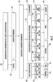
- Figure 1 shows an existing clinical method for diagnoses of upper respiratory infections (100).
- Nasopharyngeal or oropharyngeal swabs may be taken (102), viral transport medium (104) is taken, and may be subject to real time PCR (106) or respiratory panel PCR (107) (for example Biofire ® respiratory panel PCR).
- IgM is positive (108) in the real time PCR for Epstein barr virus
- the diagnosis (110) is a viral infection.
- the respiratory panel PCR is positive for HSV, CMV, Rhinovirus, Coronavirus, Influenza A, Influenza B, Parainfluenza, or RSV (118)
- the diagnosis (110) is also a viral infection.
- the respiratory panel PCR is positive for Bordetella, Chlamydia, or Mycoplasma (116)
- the diagnosis (120) is a bacterial infection.
- An alternative sample that could be collected is a urine sample (112), collected via a urine collection transport system (114). If the urine sample is positive for Pneumococcus or Legionella antigen (122), the diagnosis (120) is a bacterial infection.
- An oropharyngeal sample (124) may alternatively be subject to cell culture, using a culture swab collection transport system (126). Any bacteria with growth > 10 6 or any group A strep growth (128) indicates (120) a bacterial infection.
- the samples are negative for PCR, antigen (130), and cell culture (132), they are considered microbiologically unconfirmed (134) and subject to further testing. If the procalcitonin levels in the sample are less than 1.0 ng/ml and there are any white blood cells (136), the diagnosis (146) is negative. If the procalcitonin levels are between 0.1 ng/ml and 0.25 ng/ml plus any white blood cells (138), the diagnosis (154) is a viral infection. If the procalcitonin levels are between 0.25 ng/ml and 0.5 ng/ml and the white blood cell count is less than 8,000 (140), the diagnosis (154) is also a viral infection.
- the diagnosis (150) is a bacterial infection. If the procalcitonin levels are greater than or equal to 0.5 ng/ml and there are any white blood cells (144), the diagnosis (150) is also a bacterial infection. Patient history, physical exam, white blood cell count and other laboratory tests (152) may be taken to confirm a final clinical diagnosis (156).
- Figure 2 shows an existing clinical method for diagnoses of lower respiratory tract infections.
- the initial testing (160) for diagnosis, including PCR, culture, and antigen detection, is similar to what is described with respect to Figure 1 .
- Positive samples (162) are identified with either a bacterial or viral diagnosis.
- a radiological/laboratory confirmation for example using a chest x-ray.
- the diagnosis (174) is a bacterial infection.
- the procalcitonin levels are less than 0.25 ⁇ g/L and the white blood cell count is less than 15,000 (172), the diagnosis (176) is a viral infection.
- the diagnosis (186) is a bacterial infection. If the procalcitonin levels in these patients are between 0.25 ⁇ g/L and 0.50 ⁇ g/L and the white blood cell count is greater than 12,000 (182), the diagnosis (186) is also a bacterial infection. In these patients, if their procalcitonin levels are between 0.25 ⁇ g/L and 0.50 ⁇ g/L and the white blood cell count is less than or equal to 12,000 (184), the diagnosis (188) is a viral infection. In these patients, if the procalcitonin values are between 0.1 ⁇ g/L and 0.25 ⁇ g/L and there are any white blood cells (190), the diagnosis (188) is also a viral infection.
- Both Figure 1 and Figure 2 primarily test for specific pathogens or a general immune response. These methods do not differentiate between colonization and active infection.
- Identifying an appropriate immune response is best accomplished in two ways. First, bacterial cell culture growth without an associated elevation of procalcitonin or C-reactive protein is unlikely to represent a clinically significant bacterial infection and is more likely to represent colonization. Secondly, PCR studies have repeatedly demonstrated that both Rhinovirus and Coronavirus can persist in the nasopharynx without a host response and are not related to a clinically significant infection. Similarly, patients with a history of HSV and CMV may have periodic DNA shedding which is not associated with active infection. Although molecular testing can be very useful for viral detection, molecular testing alone cannot determine clinical significance. It is common for patients to not have a confirmed microbiological diagnosis. Reliance on the host response is therefore critical for the safe and effective management of these patients.
- the methods described herein use MxA to differentiate between an active viral infection, and a non-systemic host response. Following negative molecular testing for viral pathogens, the methods described herein also use elevated procalcitonin or C-reactive protein as a differentiator of the presence of a probable bacterial infection.
- One method of differentiating between colonization, a non-systemic host response and active infection includes the step of performing a first test for a presence of bacteria or virus in a sample.
- the first test may include, but is not limited to, PCR, a radiological test, viral culture, viral IFA, viral antigen testing, or a bacterial culture. If the sample is positive for virus, a second test is performed for a presence of at least approximately 25 ng/ml MxA in the sample (or is positive for paired serology). In the absence of 25 ng/ml MxA or paired serology, the sample is classified as no systemic host response. If the sample is positive for at least 25 ng/ml MxA, the infection is classified as a viral infection, regardless of whether or not there are additional indications of a bacterial infection.
- a second test is performed to determine a level of procalcitonin or C-reactive protein in a patient sample.
- procalcitonin or C-reactive protein in the absence of at least 0.1 ng/ml procalcitonin or 20 mg/L of C-reactive protein, in combination with other factors, indicates bacterial colonization.
- Other levels of procalcitonin between .10 ng/ml and .25 ng/ml, in combination with other factors, or C-reactive protein between 20 mg/l and 80 mg/l, in combination with other factors, may also indicate colonization.
- tests for only bacteria or only viruses are performed.
- the host biomarkers (such as MxA, C-reactive protein, and/or procalcitonin) levels are determined initially, and if they are negative in a symptomatic patient, it is assumed clinically insignificant and additional testing is not necessary. However, if additional tests are performed and show any growth or detection of pathogen, this would represent colonization.
- Typical/IDSA-listed bacteria include Group A and C beta-hemolytic streptococcous, Neisseria gonorrhea, Arcanobacterium haemolyticum, and Fusobacterium necrophorum.
- Atypical (non-IDSA) bacteria include Bordetella pertussis, Chlamydophila pneumoniae and Mycoplasma pneumoniae.
- Paired serology is a pathogen-specific antibody titer increase by a factor of 4 or more between the acute-phase serum specimen and the convalescent-phase.
- Figure 3 shows an embodiment of a method to diagnose upper respiratory infections and colonization using MxA, procalcitonin and/or CRP and other sample testing.
- Figure 3 shows a variety of different testing that can be performed to screen and diagnose an upper respiratory infection (500), for example in a clinical trial. In clinical settings, however, typically very few, if any, of these tests are initially performed. Instead, a rapid test, if available, is preferably performed to initially screen for pathogens.
- Nasopharyngeal and oropharyngeal samples may be collected for PCR (501) (see top left side of Figure 3 ). If Epstein-Barr virus (EBV) PCR (502) is negative (503) for IgM EBV (serum sample), the result is considered microbiologically unconfirmed (511). If the EBV PCR is positive (504) for IgM EBV (serum sample), there is microbiological confirmation (505) of a viral infection.
- EBV Epstein-Barr virus
- Respiratory panel PCR may also or alternatively be performed.
- One example of the respiratory panel PCR that could be performed is BioFire ® respiratory panel PCR, but alternative respiratory panel PCR systems could be used.
- the sample is positive for any of the atypical bacteria Bordetella pertussis, Chlamydophila pneumoniae or Mycoplasma pneumoniae (512), and either procalcitonin levels are less than 0.1 ng/ml or C-reactive protein levels are less than 20 mg/l (513), the diagnosis (514) is negative. If the sample is positive (512) for any of these atypical bacteria and the procalcitonin level is greater than or equal to 0.1 ng/ml or the C-reactive protein levels are greater than or equal to 20 mg/l or there is paired serology(517), a bacterial infection is microbologically confirmed (518).
- Oropharyngeal samples for cell culture (Cx) or PCR (515) may alternatively or additionally be taken. If the sample is positive for Group A Strep (cell culture), Group C strep (cell culture), Arcanobacterium (cell culture), or Fusobacterium (PCR) (516), which are all typical IDSA-listed bacteria, and procalcitonin levels are greater than or equal to 0.1 ng/ml or C-reactive protein levels are greater than or equal to 20 mg/l or there is positive paired serology (517), there is microbiological confirmation (518) of a bacterial infection.
- Group A Strep cell culture
- Group C strep cell culture
- Arcanobacterium cell culture
- Fusobacterium PCR
- non-IDSA listed bacteria of 10 6 or greater in cell culture 519 (bacterial growth greater than 1 ⁇ 10 6 colony forming units (CFU)/mL)
- the procalcitonin level is greater than or equal to 0.25 ng/ml
- the procalcitonin levels are greater than or equal to 0.15 ng/ml but less than 0.25 ng/ml and either the white blood cell count is greater than 12,000 or bands are present
- the C-reactive protein level is greater than 80 mg/l
- the C-reactive protein is greater than or equal to 20 mg/l but less than 80 mg/l and either the white blood cell count is greater than or equal to 12,000 or there are bands (520)
- microbiological confirmation 518) of bacterial infection.
- PCR is positive for Neisseria (521)
- the infection is confirmed (518) as bacterial.
- cell culture is positive for Group A or Group C strep (522)
- the procalcitonin levels are less than 0.1 ng/ml or the C-reactive protein levels are less than 20 mg/l (523)
- ASO ULN Streptolysin O antibody
- the procalcitonin levels are less than 0.1 ng/ml or the C-reactive protein levels are less than 20 mg/l (523), there is negative paired serology or less than 80% ULN Streptolysin O antibody (ASO) and the white blood cell count is less than 12,000 (529), there is colonization (527) (and the sample is considered microbiologically unconfirmed (511)).
- non-IDSA listed bacteria of 10 6 or greater in cell culture 525 (bacterial growth greater than 1 ⁇ 10 6 colony forming units (CFU)/mL)
- the levels of procalcitonin are less than 0.15 ng/ml
- the procalcitonin levels are greater than or equal to 0.15 ng/ml but less than 0.25 ng/ml and the white blood cell count is less than 12,000 and there are no bands
- the C-reactive protein levels are less than 20 mg/l
- the C-reactive protein levels are greater than or equal to 20 mg/l but less than 80 mg/l and the white blood cell count is less than 12,000 and there are no bands
- there is colonization (527) and the sample is considered microbiologically unconfirmed (511)).
- PCR is highly sensitive, it is unlikely that a viral or atypical bacterial will not be detected. Thus, any elevated procalcitonin greater than or equal to 0.1 ng/ml is more likely bacterial.
- results may then be further analyzed (see bottom portion of Figure 3 ). Further analysis is only performed if there has been no confirmation of bacterial or viral infection. The clinician does not perform further analysis if he has confirmed either bacterial or viral infection.
- procalcitonin levels are less than 0.15 ng/ml or C-reactive protein levels are less than 20 mg/l in these samples (530), the patient is diagnosed (531) as negative. If procalcitonin levels are greater than or equal to 0.15 ng/ml but less than 0.25 ng/ml in these samples, the white blood cell count is less than 15,000 and there are no bands, or the C-reactive protein levels are greater than or equal to 20 mg/l but less than 80 mg/l, the white blood cell count is less than 15,000 and there are no bands (532), the patient is diagnosed (533) with a viral infection.
- procalcitonin levels are greater than or equal to 0.25 ng/ml, procalcitonin levels are greater than or equal to 0.15 ng/ml but less than 0.25 ng/ml and the white blood cell count is greater than or equal to 15,000 or bands are present, the C-reactive protein levels are greater or equal to 80 mg/l, or the C-reactive protein levels are greater than or equal to 20 mg/l but less than 80 mg/l and the white blood cell count is greater than or equal to 15,000 or bands are present (534), the patient is diagnosed (535) with a bacterial infection. The final clinical diagnosis (536) is either negative, viral or bacterial.
- the diagnosis is considered viral regardless of what the C-reactive protein or procalcitonin value is.
- the practitioner should not prescribe antibiotics, and instead take a watchful waiting approach, re-evaluating later or doing reflex testing.
- Figure 4 shows an embodiment of a method to diagnose lower respiratory infections (540) and colonization using MxA, C-reactive protein, procalcitonin and other assays.
- Figure 4 shows a variety of different testing that can be performed to screen and diagnose a lower respiratory tract infection (540), for example in a clinical trial. In clinical settings, however, typically very few, if any, of these tests are initially performed. Instead, a rapid test, if available, is preferably performed to initially screen for pathogens.
- Nasopharyngeal and oropharyngeal samples may be collected for PCR (542) (top left side of Figure 4 ). If Epstein-Barr virus (EBV) PCR (581) is negative (582) for IgM EBV (serum sample), the result is considered microbiologically unconfirmed (565). If the EBV PCR is positive (583) for IgM EBV (serum sample), there is microbiological confirmation (545) of a viral infection.
- EBV Epstein-Barr virus
- Respiratory panel PCR may also or alternatively be performed. Although a Biofire ® respiratory panel PCR is identified in this figure, other respiratory panel PCR systems could alternatively be used.
- the sample is positive for Bordetella pertussis, Chlamydophila pneumoniae or Mycoplasma pneumoniae (547), and procalcitonin levels are less than 0.1 ng/ml or C-reactive protein levels are less than 20 mg/l (548), the diagnosis (549) is negative. If the sample is positive for any of these atypical bacteria (547) and the procalcitonin level is greater than or equal to 0.1 ng/ml or the C-reactive protein levels are greater than or equal to 20 mg/l or there is paired serology (551), a bacterial infection is microbologically confirmed (554).
- Urinary antigen testing or PCR may be used for Pneumococcus and Legionella testing according to use for lower respiratory tract infections.
- Oropharyngeal samples for cell culture (Cx) or PCR (552) may alternatively or additionally be taken. If the sample is positive for Group A Strep (cell culture), Group C strep (cell culture), Arcanobacterium (cell culture), or Fusobacterium (PCR) (553), which are all typical IDSA-listed bacteria, and procalcitonin levels are greater than or equal to 0.1 ng/ml or C-reactive protein levels are greater than or equal to 20 mg/l or there is positive paired serology (551), there is microbiological confirmation (554) of a bacterial infection.
- Group A Strep cell culture
- Group C strep cell culture
- Arcanobacterium cell culture
- Fusobacterium PCR
- non-IDSA listed bacteria of 10 6 or greater in cell culture (555) bacteria growth greater than 1 ⁇ 10 6 colony forming units (CFU)/mL
- the procalcitonin level is greater than or equal to 0.25 ng/ml
- the procalcitonin levels are greater than or equal to 0.15 ng/ml but less than 0.25 ng/ml and either the white blood cell count is greater than 12,000 or bands are present
- the C-reactive protein level is greater than 80 mg/l
- the C-reactive protein is greater than or equal to 20 mg/l but less than 80 mg/l and either the white blood cell count is greater than or equal to 12,000 or there are bands (556)
- microbiological confirmation 554) of bacterial infection.
- PCR is positive for Neisseria (557)
- the infection is confirmed (554) as bacterial.
- cell culture is positive for Group A or Group C strep (558)
- the procalcitonin levels are less than 0.1 ng/ml or the C-reactive protein levels are less than 20 mg/l (560)
- ASO ULN Streptolysin O antibody
- the procalcitonin levels are less than 0.1 ng/ml or the C-reactive protein levels are less than 20 mg/l (560), there is negative paired serology or less than 80% ULN Streptolysin O antibody (ASO) and the white blood cell count is less than 12,000 (561), there is colonization (562) (and the sample is considered microbiologically unconfirmed (565)).
- non-IDSA listed bacteria of 10 6 or greater in cell culture 563 (bacterial growth greater than 1 ⁇ 10 6 colony forming units (CFU)/mL)
- the levels of procalcitonin are less than 0.15 ng/ml
- the procalcitonin levels are greater than or equal to 0.15 ng/ml but less than 0.25 ng/ml and the white blood cell count is less than 12,000 and there are no bands
- the C-reactive protein levels are less than 20 mg/l
- the C-reactive protein levels are greater than or equal to 20 mg/l but less than 80 mg/l and the white blood cell count is less than 12,000 and there are no bands (564)
- there is colonization 562) (and the sample is considered microbiologically unconfirmed (565)).
- PCR is highly sensitive, it is unlikely that a viral or atypical bacterial will not be detected; thus, any elevated procalcitonin greater than or equal to 0.1 ng/ml is more likely bacterial.
- the patients with microbiologically unconfirmed results may be subject to a chest X-ray (571) to determine if infiltrate is identified. Further analysis is only performed if there has been no confirmation of bacterial or viral infection. The clinician does not perform further analysis if he has confirmed either bacterial or viral infection.
- the white blood cell count is less than 15,000 and there are no bands, or the C-reactive protein levels are greater than or equal to 20 mg/l but less than 80 mg/l, the white blood cell count is less than 15,000 and there are no bands (575), the patient is diagnosed (576) with a viral infection.
- procalcitonin levels are greater than or equal to 0.25 ng/ml, procalcitonin levels are greater than or equal to 0.15 ng/ml but less than 0.25 ng/ml and the white blood cell count is greater than or equal to 15,000 or bands are present, the C-reactive protein levels are greater or equal to 80 mg/l, or the C-reactive protein levels are greater than or equal to 20 mg/l but less than 80 mg/l and the white blood cell count is greater than or equal to 15,000 or bands are present (573), the patient is diagnosed (574) with a bacterial infection.
- the white blood cell count is less than 15,000 and there are no bands, or the C-reactive protein levels are greater than or equal to 20 mg/l but less than 80 mg/l, the white blood cell count is less than 15,000 and there are no bands (575), the patient is diagnosed (576) with a viral infection.
- the white blood cell count is less than 15,000 and there are no bands, or the C-reactive protein levels are greater than or equal to 20 mg/l but less than 80 mg/l, the white blood cell count is less than 15,000 and there are no bands (575), the patient is diagnosed (576) with a viral infection. If the procalcitonin level is less than 0.15 ng/ml or the C-reactive protein levels are less than 20 mg/l (578), the illness is classified as negative (579). The final clinical diagnosis (580) is either negative, viral or bacterial.
- the diagnosis is considered viral regardless of what the C-reactive protein or procalcitonin value is.
- the practitioner should not prescribe antibiotics, and instead take a watchful waiting approach, re-evaluating later or doing reflex testing.
- Figures 3 and 4 show embodiments for diagnosing respiratory infections
- the MxA, C-reactive protein and procalcitonin values in the figures could alternatively be used to diagnose other types of bacterial and viral infections (for example gastric infections, meningitis, encephalitis, cellulitis, urinary tract infections, otitis and conjunctivitis), as well as identifying colonization.
- any of the devices shown in Figures 13 , 15 or 17 could be used to differentiate an active infection from colonization of a virus or bacteria.
- a direct Antigen detection test such as Strep test or any PCR cannot differentiate colonization from active infection. Only a test that detects the host's immune response such as biomarkers of the host's origin is able to accomplish such a differentiation.
- Assaying for both MxA and C-reactive protein (or PCT) permits the user to obtain screening data for both bacterial and viral infections, increasing the ability of determining colonization.
- Figures 18 and 19 use C-reactive protein to diagnose microbiologically unconfirmed patients with a respiratory infection.
- Figure 18 shows diagnostic methods for patients with a suspected upper respiratory infection. If microbiological tests, such as PCR, culture, or antigen detection (900), are positive (902) for bacterial or virus, the patient is diagnosed (918), (920) with a bacterial or viral infection. If the microbiologically confirmatory tests (e.g.- PCR, culture, and/or antigen detection) are negative (904), further laboratory confirmation (906) is performed. If the patient has C-reactive protein values of greater than or equal to 60 mg/L and any white blood cell value (908), the patient is diagnosed (918) with a bacterial infection.
- microbiological tests such as PCR, culture, or antigen detection (900)
- the patient is diagnosed (918), (920) with a bacterial or viral infection.
- the microbiologically confirmatory tests e.g.- PCR, culture, and/or antigen detection
- further laboratory confirmation
- the patient has a C-reactive protein value 20 mg/L ⁇ CRP ⁇ 60mg/L and a white blood cell count greater than or equal to 10,000 (910), they are also diagnosed (918) with a bacterial infection. If the patient has a C-reactive protein value 20 mg/L ⁇ CRP ⁇ 60mg/L and a white blood cell count less than 10,000 (912), the patient is diagnosed (920) with a viral infection. If the patient has a C-reactive protein value 10 mg/L ⁇ CRP ⁇ 20mg/L and any white blood cell value (914), the patient is also diagnosed (920) with a viral infection.
- the patient has a C-reactive protein value of less than 10 mg/L and a white blood cell count of less than 10,000 (916), the patient is diagnosed (922) as negative for infection.
- White blood cell counts are less than 50% specific and cannot differentiate a viral or bacterial infection.
- White blood cell count elevation is not diagnostic of a clinically significant infection.
- Figure 19 shows diagnostic methods for patients with a suspected lower respiratory infection. If microbiological tests, such as PCR, culture, or antigen detection (925), are positive (926) for bacteria or virus, the patient is diagnosed with a bacterial or viral infection. In the presence of a systemic immune response, any PCR is considered positive or if the patient has a radiologically confirmed pneumonia. If the microbiologically confirmatory tests (e.g.- PCR, culture, and/or antigen detection) are negative (928), further radiological or laboratory confirmation (930) is performed. The presence of radiologic evidence of diffuse infiltrates by chest X-ray suggests a viral infection while the presence of radiologic evidence of a focal lobar process or infiltrate by chest X-ray suggests a bacterial infection.
- microbiological tests such as PCR, culture, or antigen detection (925)
- 925 PCR, culture, or antigen detection
- any PCR is considered positive or if the patient has a radiologically confirmed pneumonia.
- the microbiologically confirmatory tests
- the patient has a focal/lobar infiltrate (932) and the C-reactive protein levels are greater than or equal to 20 mg/L or the white blood cell count is greater than or equal to 10,000 (934), the patient is diagnosed (938) with a bacterial infection. If the patient has a focal/lobar infiltrate (932) and the C-reactive protein levels are less than 20 mg/L and the white blood cell count is less than 10,000 (936), the patient is diagnosed (940) with a viral infection. If a patient has C-reactive protein levels less than 20 mg/L and a white blood cell count greater than 10,000 (not shown), they may have a noninfectious conditions such as asthma or COPD exacerbation.
- the patient has diffuse/interstitial infiltrate or no infiltrates (948), a C-reactive protein level greater than or equal to 60 mg/L and any white blood cell value (942), the patient is diagnosed (944) with a bacterial infection. If the patient has diffuse/interstitial infiltrate or no infiltrates (948), a C-reactive protein level 20 mg/L ⁇ CRP ⁇ 60mg/L and a white blood cell count of greater than or equal to 10,000 (946), the patient is also diagnosed (944) with a bacterial infection.
- the patient has diffuse/interstitial infiltrate or no infiltrates (948), a C-reactive protein level 20 mg/L ⁇ CRP ⁇ 60mg/L and a white blood cell count of less than 10,000 (950), the patient is diagnosed (954) with a viral infection. If the patient has diffuse/interstitial infiltrate or no infiltrates (948), a C-reactive protein level 10 mg/L ⁇ CRP ⁇ 20mg/L and any white blood cell count (952), the patient is also diagnosed (954) with a viral infection. If the patient has no infiltrate (956), a C-reactive protein value of less than 10 mg/L and a WBC count of less than 10,000 (958), the patient is diagnosed as negative (960) for infection, and the result may indicate colonization.
- the novel method may differentiate colonizing virus from active invasive infection in various different ways.
- the method is used to detect only Influenza A/B, Metapneumovirus, Adenovirus, RSV, Parainfluenza Virus, and Epstein-Barr Virus while excluding Rhinovirus, Coronavirus, HSV, and CMV.
- Herpes Simplex virus, Cytomegalovirus, Rhinovirus, and Coronavirus will be deemed to be true positives if they are PCR positive and associated with a normal procalcitonin level and elevated MxA ⁇ 25 ng/ml.
- Epstein-Barr, HSV, or CMV is positive by PCR
- the presence of a simultaneous positive IgM blood test would confirm true positive.
- the risk is that it takes 5-7 days for an IgM antibody to develop.
- This embodiment is not applicable to Rhinovirus or CMV.
- a positive viral (EBV, HSV, or CMV) PCR and a positive (EBV, HSV, or CMV) VCA (viral capsid antigen) IgG Test indicates new viral infection.
- a positive viral (EBV, HSV, or CMV) PCR and a negative (EBV, HSV, or CMV) VCA IgG Test indicates no acute infection.
- a negative viral (EBV, HSV, or CMV) PCR and a positive (EBV, HSV, or CMV) VCA IgG Test indicates a false positive.
- novel methods described herein use procalcitonin or C-reactive protein to differentiate colonization from true bacterial infection.
- the novel methods also use MxA to differentiate between a viral infection and no systemic host response.
- Testing for C-reactive protein and MxA measures a clinically significant immune response to a suspected invasive viral and/or bacterial infection in patients older than 1 year that present within 3 days of an acute onset fever and within 7 days of new onset respiratory symptoms consistent with a suspected community acquired upper respiratory infection (rhinopharyngitis, tonsillopharyngitis, laryngotracheitis) or lower respiratory tract infection (tracheobronchitis, bronchiolitis, or pneumonia).
- MxA and C-reactive protein can result in a positive viral infection (if the MxA level creates a positive MxA result), a positive bacterial infection (if the C-reactive protein levels create positive C-reactive protein results), or co-infection (if both the MxA and C-reactive protein levels are positive).
- test strips that use MxA and C-reactive protein levels to diagnose infection are described further below.
- Additional testing can confirm that the negative result is truly negative for infection.
- Some examples of ways to confirm that a patient with a respiratory illness is negative for infection, in addition to testing them for MxA, C-reactive protein, and/or procalcitonin include a negative PCR respiratory panel (for example, a BioFire TM respiratory panel, Biofire Diagnostics, Inc., Salt Lake City, Utah), a negative sputum, blood, or throat cultures, negative additional viral PCR testing, negative urine antigen tests or a negative chest X-ray.
- microbiologically unconfirmed patients have one or more of these negative results, they are presumed noninfectious.
- These noninfectious patients may have, for example, an allergy, drug fever, cancer, connective tissue disease, thyroid disease, gout, inflammatory bowel disease, sarcoidosis, vaccination, or blood clots.
- additional testing may be performed to try to microbiologically confirm the etiology of their illness.
- Microbiologically unconfirmed illnesses can be due to a true negative, colonization, or a microbiologically unconfirmed (MU) illness that could be an emergent, previously unidentified illness.
- MU microbiologically unconfirmed
- additional steps are taken to try to determine a diagnosis.
- One additional step is taking a second sample from the patient and retesting for one or more of the same biomarkers tested in the first sample.
- the second sample is preferably taken between four and seventy two hours after the first sample. In some preferred embodiments, the second sample is taken within twenty four hours after the first sample has been taken. In other preferred embodiments, the second sample is taken within four to twelve hours after the first sample has been taken. In other preferred embodiments, the second sample is taken within six to eight hours after the first sample has been taken.
- another sample is taken from the patient within four to seventy two hours after the initial sample was taken to test for MxA, C-reactive protein, and/or procalcitonin, and tested a second time for the presence of elevated MxA, C-reactive protein and/or procalcitonin.
- the second test is preferably a quantitative test, to determine if the levels of one or more of these biomarkers has increased.
- the second sample is tested for MxA within four to eight hours of the initial test.
- additional research and testing is done to try to determine if a patient with a microbiologically unconfirmed diagnosis has an emergent disease or illness.
- the devices in Figures 13 and 15 provide a rapid test that can differentiate an active infection from a respiratory or other type of illness of either viral or bacterial etiology.
- Procalcitonin and C-reactive protein alone cannot. No treatment is needed for microbiologically unconfirmed illnesses.
- additional MxA and C-reactive protein (or procalcitonin) testing done within a shorter time period, such as taking a second sample four to seventy two hourshours after the first sample and assaying it for the presence of these biomarkers, may be prudent to rule out the prodrome effect.
- the second sample to test for MxA, C-reactive protein, and/or procalcitoninis is preferably taken between four and seventy two hours after the first sample. In some preferred embodiments, the second sample is taken within twenty four hours after the first sample has been taken. In other preferred embodiments, the second sample is taken within four to twelve hours after the first sample has been taken. In other preferred embodiments, the second sample is taken within six to eight hours after the first sample has been taken.
- a second sample could be taken from that patient and the second sample could be assayed on the device of Figures 13 , 15 or 17 again for MxA, low CRP and high CRP. If that patient has a viral or bacterial infection, the second sample would indicate a positive result for either MxA or C-reactive protein, respectively.
- a sample tested for C-reactive protein and MxA using the device of Figure 9 or other assay methods known in the art is negative for both C-reactive protein and MxA
- a second sample could be taken from that patient and the second sample could be assayed on the device of Figure 9 again for MxA and C-reactive protein. If that patient has a viral or bacterial infection, the second sample would indicate a positive result for either MxA or C-reactive protein, respectively.
- a sample could be tested for procalcitonin and MxA using devices known in the art. If the sample is initially negative for both MxA and procalcitonin, a second sample could be taken from that patient and the second sample could be assayed again for MxA and procalcitonin. If that patient has a viral or bacterial infection, the second sample would indicate a positive result for either MxA or procalcitonin, respectively.
- results from the second test in any of these examples would guide the practitioner in their decisions whether to prescribe antibiotics. This guidance would be provided much earlier than in the prior art diagnostic system, which relied on a "wait and see” approach, and would wait at least 48 hours to see if the patient worsened before performing any additional testing.
- the Applicant performed prospective, multicenter, blinded clinical trials for identifying an immune response to viral and/or bacterial infection related to an acute community-acquired febrile respiratory infection.
- the study was performed on subjects older than 1 year of age presenting to primary care and urgent care outpatient offices and emergency departments in geographically diverse clinical trial sites across the United States.
- the data substantiates the accuracy of combining these markers either in quantitative or qualitative fashion on any type of device including, but not limited to, lateral flow devices, chemoluminescence, bead, fluorescence ELISA, Automated Immunoassay/immunoanalysis testing systems (for example BioMerieux Vidas ® or mini Vidas ® immunoassay systems).
- Figures 5A-5L show results from 148 patients in the trial. Each row identifies a different patient in the trial. Column one identifies each patient with an arbitrary number. Column two shows the quantitative venous procalcitonin levels (ng/ml) and column three shows the quantitative MxA ELISA levels. Column four lists the organism/infection, if any, that was picked up by a throat culture. Column five lists the clinical diagnosis given to that patient. Column six provides a description and comments regarding how the diagnosis was reached. For the negative diagnoses that do not include MxA values, the MxA assay was not run.
- Bacterial infection was defined and diagnosed in the trial as follows:
- Viral infection was defined and diagnosed in the trial as follows:
- the combined test accurately identified 83 out of 84 microbiologically unconfirmed (MU) respiratory infections, 26 out of 35 viral infections, and 29 out of 29 bacterial infections.
- the combined test reduces the amount of unnecessary antibiotic prescriptions because of its ability to differentiate between viral and bacterial infection.
- Figure 6 shows the bacteria identified in one trial.
- Figure 7 shows the bacteria identified using the novel method described herein. In Figure 6 , all of the bacteria identified in the trial is considered a bacterial infection, and does not consider colonization. In Figure 7 , the patients positive for bacterial infection change because only the true infections are being counted.
- lateral flow devices are used to detect MxA and/or C-reactive protein and other assay devices are used to detect procalcitonin.
- Lateral flow devices are known, and are described in, e.g., U.S. Patent No. 7,723,124 and US Patent Publication No. and 2007/0059682 . The contents of both of these applications are incorporated herein by reference.
- Other lateral flow devices known in the art could alternatively be used with the systems and methods of the present invention.
- U.S. Patent No. 7,723,124 discloses a method for detecting targets, such as pathogens and/or allergy-associated components, in a human body fluid where the body fluid sample is collected by a collection device, such as a swab member.
- the samples are transferred from the swab member to a sample analysis device, on which an analysis of the targets can occur by immunochemical or enzymatic means.
- the test result is capable of being displayed within a very short period of time and can be directly read out by the user. This enables point-of-care testing with results available during a patient visit.
- FIG. 8 One example of a rapid screening test for distinguishing viral and bacterial infection is shown in Figure 8 .
- MxA is a diagnostic marker for viral infection
- C-reactive protein is a diagnostic marker for bacterial infection.
- a blue line (“control line” in A-D of the Figure) represents the control.
- a green line represents a C-reactive protein (CRP) level > 15 mg/L ("CRP test" in A-D of the figure).
- a red line represents an MxA level > 20 ng/ml (“MxA test" in A-D of the figure).
- a positive result for the MxA protein, with a negative result for the CRP protein indicates only a viral infection (Visual Test Result A).
- a positive result for the (CRP) with a negative result for the MxA protein indicates only a bacterial infection (Visual Test Result B).
- a positive result for both MxA and C-reactive protein indicates co-infection (infection with both a bacteria and a virus) (Visual Test Result C).
- No bacterial or viral infection is indicated by a negative result for both MxA and C-reactive protein (Visual Test Result D). While particular color lines are discussed in this example, other colors, or the same colors at different locations on the test strip to indicate viral or bacterial markers, are within the spirit of the present invention.
- the lines may or may not be physically separated by space.
- the labels are chosen such that the color seen when both markers are present is different from the colors seen when the individual markers are present.
- the presence of the viral marker may be indicated by a red line; the presence of the bacterial marker by a blue line; and the presence of both by a purple line (combined red and blue).
- the test strip may also include a control section which indicates the functionality of the test strip.
- Figure 8 shows a control line.
- the control section can be designed to convey a signal to the user that the device has worked.
- the control section may contain a reagent (e.g., an antibody) that will bind to the labeled reagents from the reagent zone.
- rabbit anti-chicken is used as the control line and chicken IgY conjugated to a label, for example blue latex beads, is the control conjugate.
- the control section may contain an anhydrous reagent that, when moistened, produces a color change or color formation, e.g.
- control section could contain immobilized viral and bacterial markers which will react with excess labeled reagent from the reagent zone.
- the control section may be located upstream or downstream from the detection zone. A positive control indicator tells the user that the sample has permeated the required distance through the test device.
- Figures 9A and 9B show a chromatographic test strip (400) with a test line (402) corresponding to the presence of a viral marker such as MxA and a second, separate test line (403) that detects the presence of a bacterial marker such as C-reactive protein or procalcitonin.
- the sample is applied to the application zone (401) of the chromatographic test strip (400).
- the sample passes a reagent zone (460) containing at least one labeled viral binding partner and at least one labeled bacterial binding partner that is eluted by and then able to migrate with a sample transport liquid (e.g. a buffer solution).
- a sample transport liquid e.g. a buffer solution
- the reagent zone (460) is located upstream of the sample application zone (401) such that the labeled binding partners in the reagent zone are eluted by the sample transport liquid and travel to the sample.
- the labeled viral binding partner is capable of specifically binding to a viral marker of interest to form a complex which in turn is capable of specifically binding to another specific reagent or binding partner in the detection zone.
- the labeled bacterial binding partner is capable of specifically binding to a bacterial marker of interest to form a complex which in turn is capable of specifically binding to another specific reagent or binding partner in the detection zone.
- an absorbent pad as well as other known lateral flow immunoassay components including, but not limited to, a waste zone, a carrier backing, a housing, and an opening in the housing for result read out, may optionally also be a component of the test strip (400) in these embodiments.
- the test strip (400) also includes a detection zone (405) containing at least one first section for detection of a viral marker, e.g. a test line (402), including an immobilized specific binding partner, complementary to the viral reagent complex formed by the viral marker and its labeled binding partner.
- a viral marker e.g. a test line (402)
- detection zone binding partners trap the labeled viral binding partners from the reagent zone (460) along with their bound viral markers. This localization of the viral marker with its labeled binding partners gives rise to an indication at the test line (402).
- the presence of the viral marker is determined by qualitative and/or quantitative readout of the test line (402) indication resulting from the accumulation of labeled binding partners.
- the detection zone (405) also includes at least one second section for detection of a bacterial marker, e.g. a test line (403), including an immobilized specific binding partner, complementary to the bacterial reagent complex formed by the bacterial marker and its labeled binding partner.
- a test line (403) including an immobilized specific binding partner, complementary to the bacterial reagent complex formed by the bacterial marker and its labeled binding partner.
- detection zone binding partners trap the labeled bacterial binding partners from the reagent zone (460) along with their bound bacterial markers. This localization of the bacterial marker with its labeled binding partners gives rise to an indication at the test line (403).
- the presence of the bacterial marker is determined by qualitative and/or quantitative readout of the test line (403) indication resulting from the accumulation of labeled binding partners.
- test line (402) is upstream of test line (403) relative to the direction of flow (408) in the figures, in alternative embodiments, test line (403) is upstream of test line (402). In still other embodiments, test lines (402) and (403) are located in the same location on the test strip.
- the detection zone (405) may contain further test lines to detect other viral and/or bacterial markers, as well as a control line (404).
- C-reactive protein and MxA may be detected on the same test strip.
- the control line (404) indicates that the labeled specific binding partner traveled through the length of the assay, even though it may not have bound any viral or bacterial markers, thus confirming proper operation of the assay.
- the control zone (404) is preferably downstream of the test lines (402) and (403). However, in other embodiments, the control zone (404) may be located upstream of either or both of the test lines (402) and (403).
- control line (404) includes an antibody or other recombinant protein which binds to a component of the elution medium or other composition being used in the test.
- control line (404) preferably includes a nucleic acid complementary to the labeled nucleic acid being used as a binding partner for the target nucleic acid.
- test lines for both or either of the viral and bacterial markers
- the presence of each target preferably corresponds to a separate test line (402) or (403).
- both the bacterial marker and the viral marker are detected on a single test line.
- the presence of both a bacterial marker and a viral marker on the same test line has different characteristics than the presence of either a bacterial or viral marker alone. For example, the presence of both a bacterial marker and a viral marker on the same test line may be visually indicated by a different color than the presence of either a bacterial marker or a viral marker alone.
- the devices and methods of the present invention include a lysis zone to help differentiate viral and bacterial infections.
- the sample that has been collected is not lysed prior to collection and transfer to the sample analysis device. This decreases the number of steps needed to collect and prepare the sample for analysis.
- a lysis agent improves assay efficiency is in assaying for the presence of MxA. As discussed herein, the presence of this protein can help to distinguish between bacterial and viral infection in febrile children.
- In situ lysis using a combination of 1% to 6% weight/volume CHAPS and 0.5% to 2% weight/volume NP40 as the lysis agent improves detection of MxA in fresh or frozen whole blood. In other embodiments, in situ lysis uses urea, Tween 80, and/or a combination of these two lysis agents.
- the sample traveling with the transport liquid will encounter the lysis agent.
- the lysis agent will have preferably been pre-loaded onto the test strip and is eluted by the transport liquid.
- the lysis agent has been dried into the test strip.
- the lysis agent may be pre-dried by freeze drying or lyophilizing and then pre-loaded into the test strip.
- the lysis agent may be absorbed, adsorbed, embedded or trapped on the test strip.
- the initially dried lysis agent is preferably localized between the sample application zone and a reagent zone. In embodiments where the reagent zone is upstream of the sample application zone, the lysis zone is downstream of the sample application zone.
- the lysing agent is preferably soluble in the sample transport liquid, and the lysing agent is solubilized and activated upon contact with the sample transport liquid.
- the sample transport liquid then contains both lysing agent in solution or suspension and sample components in suspension. Any lysis-susceptible components in a sample, then being exposed in suspension to the lysing agent, are themselves lysed in situ.
- the running buffer then carries the analyte, including any lysis-freed components, to the detection zone.
- the location where the lysis agent is pre-loaded and dried can be varied as needed.
- the dried, absorbed, adsorbed, embedded, or trapped lysis agent may be located in or just downstream of the sample application zone.
- the dried lysis agent may be located closer to the reagent zone.
- the lysis agent may be included in the running buffer.
- NP-40 and sarkosyl lysis agents are included in a Tris-containing running buffer.
- Tween 80 and urea are used as the lysis agents on a lateral flow chromatography test strip.
- lysis agents on the strip including Tween 80 and urea
- lysis agents in a Tris-containing running buffer including NP-40 and Sarkosyl are used in combination.
- the concentration of lysis agent pre-loaded onto a test strip is preferably between 0.001% and 5% weight/volume.
- the volume to be pre-loaded depends on where the lysis agent is pre-loaded. Appropriate ranges are 1 to 10 microliters when pre-loaded into the sample collector fleece (the sample application zone) or 5 to 50 microliters when pre-loaded into the absorbent pad or into other locations within the test strip. Ideally, the amount pre-loaded should be approximately 3 microliters pre-loaded into the sample collector fleece or approximately 10 microliters pre-loaded into the absorbent pad or into other locations within the test strip.
- a pH below 4.0 tends to precipitate materials, especially proteins.
- Higher pH, above approximately 10.0 tends to lyse materials such as proteins and cells walls. Therefore, a pH of approximately 10.0 or above is preferable for many applications. Alternatively, lower pH may be preferred for nucleic acid targets.
- both the high and low ionic strength may be used to lyse.
- a lower ionic strength (hypotonic) tends to break up erythrocytes.
- water by itself can lyse erythrocytes.
- Higher ionic strength environments may be used to rupture certain cell walls and membranes.
- lysis agents they may be grouped and selected based on their properties: salts, amphoteric and cationic agents, ionic and non-ionic detergents.
- the salt Ammonium Chloride (NH 4 Cl), lyses erythrocytes.
- Other salts including, but not limited to, high concentrations of Sodium Chloride (NaCl) and Potassium Chloride (KCl), may rupture certain cell walls and membranes.
- Other lysis agents are amphoteric agents including, but not limited to, Lyso PC, CHAPS, and Zwittergent.
- cationic agents including, but not limited to, C16 TAB and Benzalkonium Chloride may be used as a lysis agent.
- ionic and non-ionic detergents are often used to break or lyse the cell wall or cell membrane components such as lipoproteins and glycoproteins.
- Common ionic detergents include, but are not limited to, SDS, Cholate, Sodium lauroyl sarcosinate (also known as sarkosyl) and Deoxycholate. Ionic detergents are good solubilizing agents. Antibodies retain their activity in 0.1% SDS or less.
- Common non-ionic detergents include, but are not limited to, Octylglucoside, Digitonin, C12E8, Lubrol, Triton X-100, Noniodet P-40, NP-40 (for example Tergitol ® NP-40), Tween 20, and Tween 80.
- Non-ionic and mild ionic detergents are weaker denaturants and often are used to solubilize membrane proteins such as viral surface proteins.
- Additional lysis agents include, but are not limited to, urea and enzymes. Combinations of different lysis agents may be used to optimize the lysing environment.
- Tween 80 and urea are used as the lysis agents on a lateral flow chromatography test strip.
- lysis agents in a Tris-containing running buffer include NP-40 and Sarkosyl.
- lysis agents on the strip (including Tween 80 and urea) and lysis agents in a Tris-containing running buffer (including NP-40 and Sarkosyl) are used in combination.
- Surfactants are generally wetting agents and lower the surface tension of a liquid. This then allows easier spreading by lowering the interfacial tension between liquids. So, surfactants can interfere with the natural binding of antigen and antibody or ligand and receptors.
- the concentrations are, therefore, experimentally chosen for each class of lysis agent. Once lysis occurs, it is important that the desired binding reactions not be hindered. Generally, 0.001% lysis agent concentration is considered the lower limit, and the upper limit is approximately 1%. There is an additive or synergistic effect when combinations of lysis agents are used. This expands the working range of concentration to run from approximately 0.001% to 1%. Finally, some undesirable non-specific binding may be prevented at a Tween 20 concentration of 5%. In all cases, the total amount of lysis agent pre-loaded onto all locations of an individual test strip must be sufficient to lyse barriers to immunodetection, permitting practical operation of the test strip.
- the lysis agent itself should not interfere with any other assay detector or indicator agents and thus does not interfere with any other assay interactions and reactions to such an extent as to prevent practical operation of the assay.
- a lysis agent should have sufficient shelf life to allow manufacture, distribution and storage before use of a test strip in point-of-care testing.
- in situ lysis using a combination of 1% to 6% weight/volume CHAPS and 0.5% to 2% weight/volume NP40 as the lysis agent is preferably used.
- 2 microliters of 100 mM HEPES buffer (pH 8.0) containing 5% CHAPS and 2% NP-40 with 150 mM Sodium Chloride, 0.1% BSA, and 0.1% Sodium Azide (all percentages weight/volume) are dried onto a lysis zone of a test strip.
- the sample is applied to the application zone (201) on a chromatographic test strip (200).
- the sample passes a lysis zone (250), where a lysis agent will have preferably been pre-loaded onto the test strip and is eluted by the transport liquid.
- the lysis agent lyses any lysis-susceptible components in the sample in situ.
- the chromatographic test strip contains a sample application zone (201), a lysis zone (250) containing a lysis agent, and a reagent zone (260) containing at least one labeled binding partner that binds to a viral marker and at least one labeled binding partner that binds to a bacterial marker that are eluted by and then able to migrate with a sample transport liquid (e.g. a buffer solution).
- a sample transport liquid e.g. a buffer solution.
- the reagent zone (260) is shown downstream of the sample application zone in these figures, in alternative embodiments, the reagent zone (260) could be upstream of the sample application zone (see Figure 10B ), as long as the reagents encounter the sample at some point after the sample reaches the lysis zone and is effectively lysed.
- the labeled binding partners are capable of specifically binding to a viral or bacterial marker of interest to form a complex which in turn is capable of specifically binding to another specific reagent or binding partner in the detection zone.
- an absorbent pad as well as other known lateral flow immunoassay components including, but not limited to, a waste zone, a carrier backing, a housing, and an opening in the housing for result read out, may optionally also be a component of the test strip (200) in these embodiments.
- the lysis agent is localized in the lysis zone (250) between the sample application zone (201) and the reagent zone (260).
- the lysis agent is preferably soluble or miscible in the sample transport liquid, and the lysis agent is solubilized and activated upon contact with the sample transport liquid.
- the sample transport liquid then contains both lysis agent in solution or suspension and sample components in suspension. Any lysis-susceptible components in a sample, then being exposed in suspension to the lysis agent, are themselves lysed in situ.
- the running buffer then carries the sample, including any lysis-freed components, to the detection zone (205).
- the lysis zone (250) is preferably located between the sample application zone (201) and the reagent zone (260), as shown in Figure 10A .
- the lysis zone (250) overlaps the sample application zone (201), the reagent zone (260) or both the sample application zone (201) and the reagent zone (260) as shown in Figures 10B , 10C, and 10D , respectively.
- the figures are schematic, and are not drawn to scale. The amount of overlap between the different zones (as shown in Figures 10B through 10D ) may be highly variable.
- the test strip (200) also includes a detection zone (205) containing a first section for detection of at least one bacterial marker, e.g. a test line (203), including an immobilized specific binding partner, complementary to the bacterial conjugate formed by the bacterial marker and its labeled binding partner.
- a detection zone binding partners trap the bacterial labeled binding partners from the reagent zone (260) along with their bound bacterial markers. This localization of the bacterial markers with their labeled binding partners gives rise to an indication at the test line (203).
- the presence of a bacterial marker is determined by qualitative and/or quantitative readout of the test line (203) indication resulting from the accumulation of labeled binding partners.
- the detection zone (205) also includes a second section for detection of at least one viral marker, e.g. a test line (202), including an immobilized specific binding partner, complementary to the viral conjugate formed by the viral marker and its labeled binding partner.
- a test line (202) including an immobilized specific binding partner, complementary to the viral conjugate formed by the viral marker and its labeled binding partner.
- detection zone binding partners trap the viral labeled binding partners from the reagent zone (260) along with their bound viral markers. This localization of the viral markers with their labeled binding partners gives rise to an indication at the test line (202).
- the presence of a viral marker is determined by qualitative and/or quantitative readout of the test line (202) indication resulting from the accumulation of labeled binding partners.
- test line (203) is upstream of test line (202) relative to the direction of flow (208) in the figures, in alternative embodiments, test line (202) is upstream of test line (203). In still other embodiments, test lines (202) and (203) are located in the same location on the test strip.
- the detection zone (205) may contain further test lines to detect other bacterial and/or viral markers, as well as a control line (204).
- the control line (204) indicates that the labeled specific binding partner traveled through the length of the assay, even though it may not have bound any markers, thus confirming proper operation of the assay.
- the control zone (204) is preferably downstream of the test lines (203) and (202). However, in other embodiments, the control zone (204) may be located upstream of either or both of the test lines (203) and (202).
- control line (204) includes an antibody or other recombinant protein which binds to a component of the elution medium or other composition being used in the test.
- control line (204) preferably includes a nucleic acid complementary to the labeled nucleic acid being used as a binding partner for the target nucleic acid.
- test lines are within the spirit of the invention.
- the presence of each target preferably corresponds to a separate test line (202).
- the presence of multiple targets may be indicated on the same test line such that the presence of more than one target has different characteristics than the presence of a single target. For example, the presence of multiple targets on the same test line may be visually indicated by a different color than the presence of each of the targets alone.
- lysis agents in the running buffer include NP-40 and Sarkosyl.
- one or more lysis agents are dried onto the sample application zone of a lateral flow strip.
- the lysis agent is made of approximately 2 microliters of 100 mM HEPES buffer (pH 8.0) containing 5% CHAPS and 2% NP-40 with 150 mM Sodium Chloride, 0.1% BSA, and 0.1% Sodium Azide (all percentages weight/volume).
- HEPES buffer pH 8.0
- CHAPS 5% CHAPS
- 2% NP-40 150 mM Sodium Chloride
- BSA 0.1% BSA
- 0.1% Sodium Azide all percentages weight/volume
- MxA protein is released from inside white blood cells to react with an MxA monoclonal antibody on a visual tag (colloidal gold or visible latex beads).
- This complex traverses with a running buffer containing Triton X-100 and is captured by MxA monoclonal antibodies immobilized at the test line of the nitrocellulose membrane. This binding at the test line gives rise to a visible indication.
- Tween 80 and urea are used as the lysis agents on a lateral flow chromatography test strip.
- assay formats for determining procalcitonin when MxA and/or C-reactive protein are being determined lateral flow include, but are not limited to, immunoassays, immunoblotting methods, agglutination reactions, a complement-fixation reaction, a hemolytic reaction, a precipitation reaction, a gold colloid method, a chromatography method, phosphorescence, radioactivity, colorimetry, gravimetry, X-ray diffraction, X-ray absorption, magnetism, fluorescent resonant emissions, or an immunostaining method.
- immunoassays include, but are not limited to, immunoprecipitation, radioimmunoassays (RIA), enzyme immunoassays (EIA or ELISA), a Vidas ® immunoassay device (Biomerieux, Hazelwood, Missouri), fluorescent immunoassays (FIA), an i-Stat ® portable handheld system (Abbott Laboratories, Abbott Park, Illinois), a Philips Handheld diagnostic system (Philips Handheld Diagnostics, The Netherlands), chemiluminescent immunoassays, physiochemical assays (TIA, LAPIA, or PCIA), lateral flow immunoassays, or flow cytometry.
- RIA radioimmunoassays
- EIA or ELISA enzyme immunoassays
- FIA fluorescent immunoassays
- i-Stat ® portable handheld system Abbott Laboratories, Abbott Park, Illinois
- Philips Handheld diagnostic system Philips Handheld Diagnostics, The Netherlands
- Some preferred immunoassays for these biomarkers include, but are no limited to, ELISAs, fluorescence immunoassays, magnetic assays, paramagnetic assays, and chemiluminescent assays.
- the mRNA or gene transcripts may be used.
- the assays are automated.
- Assays for MxA and/or C-reactive protein may also alternatively use any of the assay systems and devices above.
- a device to determine the presence of C-reactive protein, MxA and/or procalcitonin is a multiparametric immunoassay system that is able to detect two or more of these targets in the same device.
- the devices are able to detect MxA levels greater than or equal to between 25 ng/ml and 35 ng/ml, low CRP levels greater than or equal to 20 mg/l, high CRP levels greater than or equal to 80 mg/L, and procalcitonin levels of at least 0.1 ng/ml.
- the devices are able to quantitate the levels of these biomarkers.
- One multiparametric immunoassay system that could be used is a Vidas ® immunoassay device (Biomerieux, Hazelwood, Missouri), which could test for the presence of one, two, three, or all four of these targets (MxA, procalcitonin, low-CRP and high-CRP) simultaneously.
- the Vidas ® immunoassay device is an Enzyme Linked Fluorescent assay (ELFA) (also available in a compact version called Mini Vidas ® ) and is widely used in clinical laboratories.
- ELFA Enzyme Linked Fluorescent assay
- Other devices that could be used include a Vitek ® immunodiagnostic system (Biomerieux, Hazelwood, Missouri), or a Luminex ® immunoassay system (Luminex Corporation, Austin, Texas).
- Another example is a device similar to an i-Stat ® portable handheld system (Abbott Laboratories, Abbott Park, Illinois, see the devices disclosed in US Patent Nos. 5,638,828 , 5,666,967 , 5,653,243 , 5,779,650 , 6,010,463 , 6,845,327 , 6,896,778 , 7,419,821 , and 8,017,382 , all herein incorporated by reference).
- Yet another example is a device that combines magnetic particle separation with chemiluminescent detection, such as the BioFlash multiparametric immunoassay system (Biokit, Barcelona, Spain). Any non-subjective read-outs such as machine-read devices could be used to determine the levels of the biomarkers discussed herein.
- Bimodal dual test strips can be used to differentiate bacterial and viral infection in humans, but also may be used in veterinary applications for animals. Since C-reactive protein differs depending upon the species, there are not common antibodies to C-reactive protein between species. Therefore, the veterinary tests need to include C-reactive protein specific to the particular species being tested. MxA is well conserved among species, so it is possible to use human MxA in veterinary tests. However, MxA to a particular species could alternatively be used to try to further increase specificity.
- Veterinary tests using the bimodal dual test strips described herein may be developed for a specific species, including, but not limited to, cats, dogs, rabbits, pigs, sheep, horses, cows, monkeys, chimpanzees, baboons, and orangutans.
- a strip with MxA and low CRP could be made with any configuration, for example the configurations shown in Figures 9A and 9B , or Figures 10A through 10D , where MxA is the viral marker being detected and relatively low levels of C-reactive protein is the bacterial marker being detected.
- the MxA test line and the C-reactive protein test line could overlap, or be in the same location on the test strip.
- the presence of low CRP and MxA on the same test line has different characteristics than the presence of either a bacterial or viral marker alone. For example, the presence of both low CRP and MxA on the same test line may be visually indicated by a different color than the presence of either MxA or low CRP alone.
- a positive result for MxA would give a different color or indication than a positive result for low CRP, so that the person reading the assay could distinguish between a completely negative result, a positive result for MxA, a positive result for low CRP, and a positive result for both MxA and low CRP.
- a positive result for MxA could result in a red test line
- a positive result for low CRP could result in a blue test line. So, when a sample is positive for both MxA and low CRP, the line is visibly purple.
- FIGS 11A-11B and 12A-12D Some embodiments for lateral flow assay devices to detect high levels of CRP are shown in Figures 11A-11B and 12A-12D . These configurations are similar to the configurations shown in Figures 9A-9B and 10A-10D , without a test line for a viral marker, and the same reference numerals are used for the same components of the strip (600), (700).
- FIGs 11A and 11B show a chromatographic test strip (600) with a test line (623) that detects the presence of a bacterial marker, such as high levels of C-reactive protein.
- the sample is applied to the application zone (401) of the chromatographic test strip (600).
- the sample travels along the direction of flow (408).
- the sample then passes a reagent zone (660) containing at least one labeled bacterial binding partner that is eluted by and then able to migrate with a sample transport liquid (e.g. a buffer solution).
- a sample transport liquid e.g. a buffer solution
- the reagent zone (660) is located upstream of the sample application zone (401) such that the labeled binding partners in the reagent zone are eluted by the sample transport liquid and travel to the sample.
- the labeled bacterial binding partner is capable of specifically binding to a bacterial marker of interest, for example high levels of C-reactive protein, to form a complex which in turn is capable of specifically binding to another specific reagent or binding partner in the detection zone.
- an absorbent pad as well as other known lateral flow immunoassay components including, but not limited to, a waste zone, a carrier backing, a housing, and an opening in the housing for result read out, may optionally also be a component of the test strip (600) in these embodiments.
- the test strip (600) also includes a detection zone (605) containing a section for detection of a bacterial marker, e.g. a test line (623), including an immobilized specific binding partner, complementary to the bacterial reagent complex formed by the bacterial marker and its labeled binding partner.
- a detection zone containing a section for detection of a bacterial marker, e.g. a test line (623), including an immobilized specific binding partner, complementary to the bacterial reagent complex formed by the bacterial marker and its labeled binding partner.
- detection zone binding partners trap the labeled bacterial binding partners from the reagent zone (660) along with their bound bacterial markers. This localization of the bacterial marker with its labeled binding partners gives rise to an indication at the test line (623).
- the presence of the bacterial marker is determined by qualitative and/or quantitative readout of the test line (623) indication resulting from the accumulation of labeled binding partners.
- the detection zone (605) may contain further test lines to detect other bacterial and/or viral markers, as well as a control line (404).
- the control line (404) indicates that the labeled specific binding partner traveled through the length of the assay, even though it may not have bound any bacterial markers, thus confirming proper operation of the assay.
- the control zone (404) is preferably downstream of the test line (623). However, in other embodiments, the control zone (404) may be located upstream of the test line (623).
- control line (404) includes an antibody or other recombinant protein which binds to a component of the elution medium or other composition being used in the test.
- control line (404) preferably includes a nucleic acid complementary to the labeled nucleic acid being used as a binding partner for the target nucleic acid.
- the sample passes a lysis zone (250), where a lysis agent will have preferably been pre-loaded onto the test strip and is eluted by the transport liquid.
- the lysis agent lyses any lysis-susceptible components in the sample in situ.
- the chromatographic test strip (700) contains a sample application zone (201), a lysis zone (250) containing a lysis agent, and a reagent zone (760) containing at least one labeled binding partner that binds to a bacterial marker, for example high levels of C-reactive protein, that is eluted by and then able to migrate with a sample transport liquid (e.g. a buffer solution).
- a sample transport liquid e.g. a buffer solution.
- the reagent zone (760) is shown downstream of the sample application zone in these figures, in alternative embodiments, the reagent zone (760) could be upstream of the sample application zone (see Figure 11B ), as long as the reagents encounter the sample at some point after the sample reaches the lysis zone and is effectively lysed.
- the labeled binding partner is capable of specifically binding to a bacterial marker of interest, for example high levels of C-reactive protein, to form a complex which in turn is capable of specifically binding to another specific reagent or binding partner in the detection zone.
- a bacterial marker of interest for example high levels of C-reactive protein
- an absorbent pad as well as other known lateral flow immunoassay components including, but not limited to, a waste zone, a carrier backing, a housing, and an opening in the housing for result read out, may optionally also be a component of the test strip (700) in these embodiments.
- the lysis agent is localized in the lysis zone (250) between the sample application zone (201) and the reagent zone (760).
- the lysis agent is preferably soluble or miscible in the sample transport liquid, and the lysis agent is solubilized and activated upon contact with the sample transport liquid.
- the sample transport liquid then contains both lysis agent in solution or suspension and sample components in suspension. Any lysis-susceptible components in a sample, then being exposed in suspension to the lysis agent, are themselves lysed in situ.
- the running buffer then carries the sample, including any lysis-freed components, to the detection zone (705).
- the lysis zone (250) is preferably located between the sample application zone (201) and the reagent zone (760), as shown in Figure 12A .
- the lysis zone (250) overlaps the sample application zone (201), the reagent zone (760) or both the sample application zone (201) and the reagent zone (260) as shown in Figures 12B , 12C, and 12D , respectively.
- the figures are schematic, and are not drawn to scale. The amount of overlap between the different zones (as shown in Figures 12B through 12D ) may be highly variable.
- the test strip (700) also includes a detection zone (705) containing a section for detection of at least one bacterial marker, e.g. a test line (723), including an immobilized specific binding partner, for example, a specific binding partner for a high level of C-reactive protein, complementary to the bacterial conjugate formed by the bacterial marker and its labeled binding partner.
- a detection zone binding partners trap the bacterial labeled binding partners from the reagent zone (760) along with their bound bacterial markers. This localization of the bacterial markers with their labeled binding partners gives rise to an indication at the test line (723).
- the presence of a bacterial marker is determined by qualitative and/or quantitative readout of the test line (723) indication resulting from the accumulation of labeled binding partners.
- the detection zone (705) may contain further test lines to detect other bacterial and/or viral markers, as well as a control line (204).
- the control line (204) indicates that the labeled specific binding partner traveled through the length of the assay, even though it may not have bound any markers, thus confirming proper operation of the assay.
- the control zone (204) is preferably downstream of the test line (723). However, in other embodiments, the control zone (204) may be located upstream of the test line (723).
- control line (204) includes an antibody or other recombinant protein which binds to a component of the elution medium or other composition being used in the test.
- control line (204) preferably includes a nucleic acid complementary to the labeled nucleic acid being used as a binding partner for the target nucleic acid.
- the sample analysis device or test card (800) includes a closable housing (835) with two sides (836), (837) and a spine or hinged portion (831).
- the test card (800) is approximately 11.5 cm long (L) ⁇ 7 cm wide (W) when the two sides (836), (837) are closed.
- any size test card (800) that accommodates all of the components may be used.
- the first side (836) also includes an absorbent pad (840) and preferably a waste pad (860).
- the first test strip (815) preferably includes a detection zone (805) with an MxA test line (802), a low CRP test line (803) and a control line (804).
- the second test strip (825) preferably includes a detection zone (805) with a high CRP test line (823) and a control line (804). All of the test lines are visible through the windows (865) on the second side (837) of the housing (835) when the housing (835) is closed.
- the absorbent pad (840) is preferably a single pad that the running buffer is added to to start lateral flow.
- the waste pad (860) is preferably a single pad that collects excess running buffer at the end of the test.
- each strip could have a separate absorbent pad (840) and/or waste pad (860).
- the second side (837) of the housing (835) includes three separate sections (838), (839) and (870).
- the sample compressor (870) is any of the sample compressors described in US Patent No. 8,609,433, entitled “Multiplanar Lateral flow Assay with Sample Compressor", issued December 17, 2013 , herein incorporated by reference.
- a window (843) is located in the lower portion (838) of the second side (837) of the housing so that the buffer can be added to the absorbent pad (840) when the housing (835) is closed.
- the viewing windows (865) for the detection zones (805) are on the upper portion (839) of the second side (837) of the housing (835).
- the upper portion (839) and the lower portion (838) of the second side (837) of the housing (835) also preferably each include at least one knob, peg or protrusion (875) that mates with one or more holes (895) so that the upper and lower portions (838), (839) may be easily fastened onto the first side (836) of the housing (835).
- the holes (895) are on the second side (837) of the housing (835) and the pegs (875) are on the first side (836) of the housing (835).
- other reversible fastening mechanisms could be used to secure the upper portion (838) and/or lower portion (839) of the second side (837) of the housing (835) to the first side (836) of the housing (835).
- the upper and lower sections (838), (839) are permanently closed, for example using an adhesive, before use.
- the flap (870), also known as a sample compressor, on the second side (837) of the housing includes two conjugate zones (872), (874) and two sample application zones (873), (876), and can be easily opened and closed.
- the flap (870) also preferably includes at least one knob, peg or protrusion (875) that mates with one or more holes (895) so that the flap (870) is easily correctly closed onto the first side (836) of the housing (835) after sample has been added to the sample application zones (873), (876).
- the holes (895) are on the second side (837) of the housing (835) and the pegs (875) are on the first side (836) of the housing (835).
- other reversible fastening mechanisms could be used to secure the flap (870) to the first side (836) of the housing (835).
- the conjugate zones (872), (874) and the sample application zones (873), (876) preferably overlap.
- the conjugate zones (872), (874) are colored due to the dyes in the sample conjugates and control conjugates, and the sample is placed directly on the colored portion of the flap (870).
- the conjugate zone (872) that is used for the first test strip (815) contains an MxA binding partner that is labeled with a red dye, a low CRP binding partner that is labeled with a black dye, and a control binding partner that is labeled with a blue dye.
- the conjugate zone (872) appears purplish.
- the other conjugate zone (874) contains a high CRP binding partner that is labeled with a black dye and a control binding partner that is labeled with a blue dye. In this embodiment, the conjugate zone (874) appears bluish.
- the diverting zone (850) preferably includes a gap or barrier that interrupts lateral flow, diverting the running buffer up into the flap (870) that includes the conjugate zones (872), (874) and the sample application zones (873), (876).
- the diverting zone is any of the diverting zones described in US Patent No. 8,815,609, entitled “Multiplanar Lateral flow Assay with Zone", issued August 26, 2014 , herein incorporated by reference.
- the upper and lower portions (838), (839) of the second side (837) of the housing (835) are preferably snapped closed before use by securing the pegs (875) to the holes (895).
- the sample analysis device, or test card (800) is preferably placed on a flat surface. If the flap (870) is not already open, the user opens it to access the sample application zones (873), (876).
- a blood sample to be tested is taken from the patient. The sample may be taken by any procedure known in the art.
- a sample of 5 ⁇ l of blood is added to each of the sample application zones (873), (876) and then the flap (870) is closed. Each of the 5 ⁇ l samples is preferably collected independently of each other.
- the blood samples are preferably added directly to the device (800), without any pretreatment.
- Running buffer is added to the absorbent pad (840), which initiates lateral flow (885).
- the running buffer includes one or more lysis agents, for example detergents, to lyse the blood sample and expose the intracellular MxA in the sample.
- the lysis agents NP-40 and sarkosyl are included in a Tris-containing running buffer.
- the running buffer When the running buffer reaches the diverting zone (850), it is diverted up into the flap (870). It travels through the conjugate zones (872), (874), collecting any complexes formed between the MxA binding partner and MxA in the sample, the low CRP binding partner and low levels of C-reactive protein in the sample, the high CRP binding partner and high levels of C-reactive protein in the sample, as well as the control conjugate.
- the running buffer which now contains sample, conjugate, and the complexes described above, then travels into the transfer pad (855), and to the detection zones (805) on each of the test strips (815), (825). If MxA is present in the sample, the MxA test line (802) on the first test strip (815) will be red. If a threshold low level of C-reactive protein is present in the sample, the low CRP test line (803) on the first test strip (815) will be black. If a threshold high level of C-reactive protein is present in the sample, the high CRP test line (823) on the second test strip (825) will be black.
- the control lines (804) on both the first strip (815) and the second test strip (825) will be blue.
- the levels of detection are 40 ng/ml for MxA, 10 mg/L for low CRP on the first test strip (815) and 80 mg/L for high CRP on the second test strip (825). In other preferred embodiments, the levels of detection are 25 ng/ml for MxA, 20 mg/L for low CRP on the first test strip (815) and 80 mg/L for high CRP on the second test strip (825). Any combinations of these different cutoff values could be used.
- the results of the test should be visible after approximately 5-20 minutes, preferably within about 10 minutes.
- control binding partner is on the sample compressor or flap (870) and not on either of the test strips (815), (825), there is a true procedural control to this configuration. If the flap (870) is not closed properly, nothing will show up in the detection zone (805), indicating that the test was run improperly.
- Figures 14A through 14F show test results using the device (800) shown in Figures 13A through 13C , with two test strips (815), (825) side by side, where a first test strip (815) tests for the presence of both MxA and low levels of C-reactive protein and the second test strip (825) tests for high levels of C-reactive protein.
- Figure 14A shows a negative result at the MxA test line (802) and a negative result at the low CRP test line (803) on the first test strip (815), as well as a negative result at the high CRP test line (823) on the second test strip (825). More specifically, the only visible lines in the detection zone (805) of the lateral flow assay (800) are the two blue control lines (804). This result indicates that the sample is negative for both viral and bacterial infection. A patient with this result, absent additional testing, would be considered microbiologically unconfirmed.
- Figures 14B and 14C are positive for viral infection.
- the presence of two blue control lines (804) and a red MxA line (802) indicate a viral infection.
- Figure 14C the presence of two blue control lines (804) and a red MxA line (802) indicate a viral infection. Since there is also a black low CRP line (803) in Figure 14C , there is a possibility of bacterial co-infection, although there is an absence of a high CRP line (823). Any time MxA is positive in this test, it indicates a viral infection.
- Figures 14D and 14E are positive for bacterial infection.
- the presence of two blue control lines (804) and a black low CRP line (803) indicates a bacterial infection.
- Figure 14E the presence of two blue control lines (804), a black low CRP line (803), and a black high CRP line (823) also indicates a bacterial infection.
- the MxA line is absent in both Figures 14D and 14E , indicating an absence of a viral infection.
- Figure 14F indicates viral infection, or co-infection (both bacterial and viral infection).
- the presence of two blue control lines (804), a red MxA line (802), a black low CRP line (803), and a black high CRP line (823) indicates viral infection or a possible co-infection. In most cases, the patient will have a viral infection only. While there is a possibility for co-infection, the Applicants have not observed co-infection in their studies.
- FIG. 15A through 15C Another preferred configuration for a bimodal dual test strip sample analysis device (1000) is shown in Figures 15A through 15C .
- This configuration is similar to the configuration (800) shown in Figures 13A through 13C , but the sample application zones (1073), (1076) are located on each of the test strips (1015), (1025), downstream of the diverting zone (850).
- the sample analysis device or test card (1000) includes a closable housing (835) with two sides (836), (837) and a spine or hinged portion (831).
- the test card (1000) is approximately 11.5 cm long (L) ⁇ 7 cm wide (W) when the two sides (836), (837) are closed.
- any size test card (1000) that accommodates all of the components may be used.
- the first side (836) of the housing (835) there are two test strips (1015), (1025), each including a receiving pad (845), a diverting zone (850), a transfer pad (1055) and a detection zone (805).
- the first side (836) also includes an absorbent pad (840) and preferably a waste pad (860).
- the first test strip (1015) preferably includes a detection zone (805) with an MxA test line (802), a low CRP test line (803) and a control line (804).
- the second test strip (1025) preferably includes a detection zone (805) with a high CRP test line (823) and a control line (804).
- the absorbent pad (840) is preferably a single pad to which the running buffer is added to start lateral flow.
- the waste pad (860) is preferably a single pad that collects excess running buffer at the end of the test.
- each strip could have a separate absorbent pad (840) and/or waste pad (860).
- the second side (837) of the housing (835) includes three separate sections (838), (839) and (1070).
- the middle portion, or flap (1070) also known as a sample compressor, preferably includes two conjugate zones (872), (874), each including a labeled binding partner for at least one analyte, and a labeled control.
- a window (843) is located in the lower portion (838) of the second side (837) of the housing so that the buffer can be added when the housing (835) is closed.
- the viewing windows (865) for the detection zones (805) are on the upper portion (839) of the second side (837) of the housing (835).
- the upper portion (839) and the lower portion (838) of the second side (837) of the housing (835) also preferably each include at least one knob, peg or protrusion (875) that mates with one or more holes (895) so that the upper and lower portions (838), (839) may be easily fastened onto the first side (836) of the housing (835).
- the holes (895) are on the second side (837) of the housing (835) and the pegs (875) are on the first side (836) of the housing (835).
- other reversible fastening mechanisms could be used to secure the upper portion (838) and/or lower portion (839) of the second side (837) of the housing (835) to the first side (836) of the housing (835).
- the upper and lower sections (838), (839) are permanently closed, for example using an adhesive, before use.
- the flap (1070) on the second side (837) of the housing includes two conjugate zones (872), (874) and can be easily opened and closed.
- the flap (1070) also preferably includes at least one knob, peg or protrusion (875) that mates with one or more holes (895) so that the flap (1070) is easily correctly closed onto the first side (836) of the housing (835) after sample has been added to the sample application zones (1073), (1076) on the test strips (1015), (1025).
- the holes (895) are on the second side (837) of the housing (835) and the pegs (875) are on the first side (836) of the housing (835).
- other reversible fastening mechanisms could be used to secure the flap (1070) to the first side (836) of the housing (835).
- the conjugate zones (872), (874) are colored due to the dyes in the sample conjugates and control conjugates.
- the conjugate zone (872) that is used for the first test strip (1015) contains an MxA binding partner that is labeled with a red dye, a low CRP binding partner that is labeled with a black dye, and a control binding partner that is labeled with a blue dye.
- the conjugate zone (872) appears purplish.
- the other conjugate zone (874) contains a high CRP binding partner that is labeled with a black dye and a control binding partner that is labeled with a blue dye. In this embodiment, the conjugate zone (874) appears bluish.
- the diverting zone (850) which preferably includes a gap or barrier, interrupts lateral flow, diverting the running buffer up into the flap (1070) that includes the conjugate zones (872), (874).
- the upper and lower portions (838), (839) of the second side (837) of the housing (835) are preferably snapped closed before use by securing the pegs (875) to the holes (895).
- the sample analysis device, or test card (1000) is preferably placed on a flat surface. If the flap (1070) is not already open, the user opens it to access the sample application zones (1073), (1076).
- the sample application zones (1073), (1076) may be located in any portion of the transfer pad (1055).
- a blood sample to be tested is taken from the patient. The sample may be taken by any procedure known in the art. In a preferred embodiment, a sample of 5 ⁇ l of blood is added to each of the sample application zones (1073), (1076) zones and then the flap (1070) is closed.
- Each of the 5 ⁇ l samples is preferably collected independently of each other.
- the blood is preferably added directly to the device (1000), without any pretreatment.
- an arrow (1002) or other indication shown in Figure 15A ), for example the words “add sample here” shows the user where to place the sample on the test strips (1015), (1025).
- Running buffer is added to the absorbent pad (840), which initiates lateral flow (885).
- the running buffer includes one or more lysis agents, for example detergents, to lyse the blood sample and expose the intracellular MxA in the sample.
- the running buffer reaches the diverting zone (850), it is diverted up into the flap (1070). It travels through the conjugate zones (872), (874), collecting the MxA binding partners, the low CRP binding partners, and the high CRP binding partners, as well as the control conjugate.
- the running buffer which now contains conjugate, then travels into the transfer pad (1055), which includes the sample application zones (1073), (1076), and to the detection zones (805) on each of the test strips (1015), (1025). If MxA is present in the sample, the MxA test line (802) on the first test strip (1015) will be red. If a threshold low level of C-reactive protein is present in the sample, the low CRP test line (803) on the first test strip (1015) will be black.
- the high CRP test line (823) on the second test strip (1025) will be black.
- the levels of detection are 40 ng/ml for MxA, 10 mg/L for low CRP on the first test strip (1015) and 80 mg/L for high CRP on the second test strip (1025). In other preferred embodiments, the levels of detection are 25 ng/ml for MxA, 20 mg/L for low CRP on the first test strip (1015) and 80 mg/L for high CRP on the second test strip (1025). Any combinations of these different cutoff values could be used.
- the results of the test should be visible after approximately 5-20 minutes, preferably within about 10 minutes. If the test was run correctly, the control lines (804) on both the first test strip (1015) and the second test strip (1025) will be blue.
- control binding partner is on the flap (1070) and not on either of the test strips (1015), (1025), there is a true procedural control to this configuration. If the flap (1070) is not closed properly, nothing will show up in the detection zone (805), indicating that the test was run improperly.
- the sample application zones (1073), (1076) are located on the receiving pad (845), before the diverting zone (850).
- the running buffer travels through the sample application zones (1073), (1076), and then is diverted into the flap (1070).
- greater than approximately 1.2 ml of running buffer is placed on the absorbent pad (840). If less than 1.0 ml is added in embodiments where the diverting zone (850) is a gap, the buffer gets stalled at the gap because the gap holds approximately 1.0 ml.
- a kit (1100) includes the sample analysis device (800), (1000), a lancet (1102), one or more pipettes (1101), and a running buffer (1103).
- the lancet (1102) is used to make a skin puncture and one or more pipettes (1101) are used to collect the blood from the puncture site.
- 5 ⁇ l of blood is transferred from a first pipette (1101) to the first conjugate zone (872, see Figures 13B and 15B ) and another 5 ⁇ l of blood is transferred from a second pipette (1101) and added to the second conjugate zone (874, see Figures 13B and 15B ).
- the flap (870) is closed, and the running buffer (1103) is added to the absorbent pad (840), as described in the description of Figs. 13A through 13C and 15A through 15C .
- the diverting zone (850) preferably includes at least one feature that interrupts flow in the plane in which flow is occurring.
- the diverting zone may include a barrier, a gap, a ditch, or any combination of these features.
- the barrier is preferably an impermeable membrane (or substantially impermeable membrane) that may be made of any material that prevents the flow of liquid from continuing to flow in the same plane.
- Some materials for the barrier include, but are not limited to, inert materials, semipermeable materials, plastics, hydrocarbons, metal, hydrophobic materials, Sephadex, Sepharose, cellulose acetate, a hygroscopic material (for example CaCl 2 , CaSO 4 or silica gel), or hydrogels.
- the gap or ditch is any break in the plane of the lateral flow test strip that extends to a depth sufficient to stop flow. In one preferred embodiment, the gap is preferably at least approximately 0.1 mm deep.
- the diverting zone (850) in Figures 13A through 13C and 15A through 15C delays or completely stops flow until the sample compressor/flap (870), (1070) is brought into contact with the rest of the device, and creates a bridge along which the fluid can flow.
- the sample compressor (870), (1070) acts as a bridge and redirects flow into a different plane. Flow is diverted into the sample compressor (870), (1070). This increases collection of the reagents on the sample compressor (870), (1070). For example, in embodiments where the conjugate is on the sample compressor (870), (1070), collection of the conjugate increases in devices with a diverting zone (850).
- the sample and conjugate both encounter the running buffer when it is diverted into the sample compressor (870), (1070), and a 1 ⁇ 2 sandwich or full sandwich (depending upon where the second binding partner for the analyte is located on the sample analysis device) is formed before the running buffer is diverted back to the test strips if the analyte is present in the sample.
- Embodiments with a diverting zone (850) and a sample compressor (870), (1070) increase speed, allow for better interactions between the conjugate and the sample, and allow for more sensitivity because more conjugate is placed into the fluid.
- all of the fluid preferably interacts with the conjugate. This is a significant improvement over compressor embodiments without redirection, where approximately 20-30% of the fluid interacts with the conjugate.
- FIG 17 Another preferred configuration for a bimodal dual test strip sample analysis device (1200) is shown in Figure 17 .
- This configuration is similar to the configurations (800), (1000) shown in Figures 13A through 13C and Figures 15A through 15C , without a second section (837) of the housing (1235) or a diverting zone (850). Instead, all of the components of the test are located in the same plane and flow proceeds laterally from the absorbent pad (840) to the waste pad (860).
- this embodiment could also include a housing with a window to facilitate application of the buffer to the absorbent pad (840), a window located above each sample application zone (1273), (1276) for applying sample to the device (1200), and viewing windows for the detection zone (805).
- the sample analysis device (1200) is approximately 11.5 cm long (L) x 7 cm wide (W).
- any size test card (1200) that accommodates all of the components may be used.
- the detection zone (805) of the first test strip (1215) preferably includes an MxA test line (802), a low CRP test line (803) and a control line (804).
- the detection zone (805) on the second test strip (1225) also preferably includes a high CRP test line (823) and a control line (804).
- the absorbent pad (840) is preferably a single pad that the running buffer is added to to start lateral flow.
- the waste pad (860) is preferably a single pad that collects excess running buffer at the end of the test.
- each strip could have a separate absorbent pad (840) and/or waste pad (860).
- the conjugate zones (1272), (1274) are colored due to the dyes in the sample conjugates and control conjugates.
- the conjugate zone (1272) that is used for the first test strip (1215) contains an MxA binding partner that is labeled with a red dye, a low CRP binding partner that is labeled with a black dye, and a control binding partner that is labeled with a blue dye.
- the conjugate zone (1272) appears purplish.
- the other conjugate zone (1274) contains a high CRP binding partner that is labeled with a black dye and a control binding partner that is labeled with a blue dye. In this embodiment, the conjugate zone (1274) appears bluish.
- a blood sample to be tested is taken from the patient.
- the sample may be taken by any procedure known in the art.
- a sample of 5 ⁇ l of blood is added to each of the sample application zones (1273), (1276).
- Each of the 5 ⁇ l samples is preferably collected independently of each other.
- an arrow (1002) or other indication shown in Figure 15A ), for example the words "add sample here" shows the user where to place the sample on the test strips (1215), (1225).
- Running buffer is added to the absorbent pad (840), which initiates lateral flow (1285).
- the running buffer includes one or more lysis agents, for example detergents, to lyse the blood sample and expose the intracellular MxA in the sample. It travels through the conjugate zones (1272), (1274), collecting the MxA binding partners, the low CRP binding partners, the high CRP binding partners, as well as the control conjugate.
- the lysis agents NP-40 and sarkosyl are included in a Tris-containing running buffer.
- the running buffer which now contains conjugate, then travels into the transfer pad (1255), which includes the sample application zones (1273), (1276), and to the detection zones (805) on each of the test strips (1215), (1225).
- MxA is present in the sample
- the MxA test line (802) on the first test strip (1215) will be red.
- the low CRP test line (803) on the first test strip (1215) will be black.
- the high CRP test line (823) on the second test strip (1225) will be black.
- the levels of detection are 40 ng/ml for MxA, 10 mg/L for low CRP on the first test strip (1215) and 80 mg/L for high CRP on the second test strip (1225).
- the results of the test should be visible after approximately 5-20 minutes, preferably within about 10 minutes. If the test was run correctly, the control lines (804) on both the first strip (1215) and the second test strip (1225) will be blue.
- the sample application zones (1273), (1276) are located upstream of the conjugate zones (1272), (1274).
- the running buffer travels through the sample application zones (1273), (1276), and then to the conjugate zones (1272), (1274).
- the conjugate zones (1272), (1274) overlap the sample application zones (1273), (1276).
- the conjugate zones (1272), (1274), and/or the sample application zones (1273), (1276) may be located in the receiving pad (845).
- the control is rabbit anti-chicken and the control conjugate is blue latex beads coupled to chicken IgY.
- the lysis agents NP-40 and sarkosyl are included in a Tris-containing running buffer.
- the bimodal test strips with reagents to detect MxA and C-reactive protein were designed to detect a host immune response (only), not specific viral or bacterial antigens, nucleic acids (including viral DNA shedding) or bacterial cell culture growth. If these test strips are negative, they result in a microbiologically unconfirmed respiratory illness.
- the bimodal dual strip test allows for the rapid, visual, qualitative in vitro detection of both MxA and C-reactive protein directly from peripheral whole blood.
- the test measures a clinically significant immune response to a suspected invasive viral and/or bacterial infection in patients older than 1 year that present within 3 days of an acute onset fever and within 7 days of new onset respiratory symptoms consistent with a suspected community acquired upper respiratory infection (rhinopharyngitis, tonsillopharyngitis, laryngotracheitis) or lower respiratory tract infection (tracheobronchitis, bronchiolitis, or pneumonia).
- the dual strip bimodal test helps to identify 1) patients with an underlying invasive viral infection from either Influenza A/B, Adenovirus, Respiratory Syncytial Virus, Metapneumovirus, Parainfluenza Virus, or Epstein-Barr Virus; 2) patients with a clinically significant elevated host response consistent with an underlying bacterial infection.
- test is intended for professional use in an outpatient setting and should be used in conjunction with other clinical evidence including laboratory, radiographic, and epidemiological information.
- Negative results do not preclude respiratory infection and should not be used as the sole basis for diagnosis, treatment, or other clinical and patient management decisions.
- additional laboratory testing e.g., bacterial and viral culture, immunofluorescence, and viral polymerase chain reaction
- additional laboratory testing e.g., bacterial and viral culture, immunofluorescence, and viral polymerase chain reaction
- the bimodal test strips described in Figures 13-17 are used in combination with a separate sample analysis device that tests for the presence of procalcitonin.
- a level of procalcitonin can be determined in the patients with the results shown in Figure 14A , where the MxA and C-reactive protein results are all negative and the patient's diagnosis is microbiologically unconfirmed.
- procalcitonin values are taken instead of C-reactive protein values.
- a procalcitonin level equal to or greater than approximately 0.15 ng/ml creates a presumption that the patient has a bacterial infection.
- a procalcitonin level less than 0.15 ng/ml creates a presumption that the patient is noninfectious. The combination of results creates a more accurate diagnosis for the patients.
- assay formats for determining the presence of procalcitonin include, but are not limited to, immunoassays, immunoblotting methods, agglutination reactions, a complement-fixation reaction, a hemolytic reaction, a precipitation reaction, a gold colloid method, a chromatography method, phosphorescence, radioactivity, colorimetry, gravimetry, X-ray diffraction, X-ray absorption, magnetism, fluorescent resonant emissions, or an immunostaining method.
- immunoassays include, but are not limited to, immunoprecipitation, radioimmunoassays (RIA), enzyme immunoassays (EIA or ELISA), a Vidas ® immunoassay device (Biomerieux, Hazelwood, Missouri), fluorescent immunoassays (FIA), chemiluminescent immunoassays, physiochemical assays (TIA, LAPIA, or PCIA), lateral flow immunoassays, or flow cytometry.
- FIA fluorescent immunoassays
- chemiluminescent immunoassays chemiluminescent immunoassays
- TIA physiochemical assays
- LAPIA LAPIA
- PCIA lateral flow immunoassays
- flow cytometry Some preferred immunoassays for these biomarkers include, but are no limited to, ELISAs, fluorescence immunoassays and chemiluminiscent assays. In some preferred embodiments, the assays are automated.
- C-Reactive Protein C-Reactive Protein
- PCT procalcitonin
- C-reactive protein testing is part of the routine evaluation of patients with respiratory infections in general practice, and its use has proved cost-effective, despite the significant overlap in viral and bacterial signs and symptoms at moderate C-reactive protein levels.
- C-reactive protein is found to be valuable aid in reducing unnecessary antibiotics even if it is of modest value at differentiating viral from bacterial disease independently.
- procalcitonin has high sensitivity and low specificity at differentiating a viral from bacterial infection yet at high concentrations, the reverse is true, and sensitivity falls and specificity is increased.
- MxA is a biomarker that is specific for viral infection.
- the Applicant has found that testing for the presence of MxA, a host biomarker of viral infection, combined together with either C-reactive protein or procalcitonin creates an unexpected synergy, greater than an additive phenomenon, and increases both the specificity and sensitivity of both of the bacterial markers.
- a bacterial biomarker has an ability to detect bacterial infection with an optimized point that maximizes sensitivity and specificity to identify a bacterial infection.
- MxA allows the curve to shift to maximal sensitivity (a lower C-reactive protein or procalcitonin cutoff concentration) without sacrificing the specificity because the MxA identifies the viral patients that have elevated C-reactive protein or procalcitonin (leading to reduced specificity), and correctly recategorizes these patients as viral.
- any patient with an elevated C-reactive protein or procalcitonin in the presence of MxA has a viral disease and any elevation of C-reactive protein or procalcitonin (now at a much lower cutoff concentration) in the absence of MxA is bacterial.
- the lack of elevation of C-reactive protein, procalcitonin, or MxA has an extremely high negative predictive value for the presence of a clinically significant infection.
- Other host biomarkers of viral infection for example IFITs (interferon-induced proteins with tetratricopeptide repeats) may alternatively be used to increase sensitivity and specificity of any of the bacterial biomarkers tested alone.
- the optimized ROC value of C-reactive protein alone is 40 mg/L (74% sensitivity and 73% specificity). Testing for a combination of 20 mg/L C-reactive protein and 40 ng/ml of MxA increases sensitivity and specificity to 95% and 90%, respectively. Testing for a combination of 40 mg/L C-reactive protein and 40 ng/ml of MxA (not shown in the figures) increases sensitivity and specificity to 100% and 90%, respectively.
- MxA in combination with C-reactive protein permits a more accurate interpretation with detection of lower levels of C-reactive protein and relying on MxA to provide the specificity.
- the optimized ROC of procalcitonin alone is 0.4 ng/ml (95% sensitivity and 57% specificity).
- Testing for a combination of 0.4 ng/ml procalcitonin and 40 ng/ml of MxA increases sensitivity and specificity to 100% and 90%, respectively.
- a combination of MxA and procalcitonin allows not only higher sensitivity but also a dramatic increase in specificity.
- C-reactive protein or procalcitonin or other bacterial host biomarkers obtained by any means
- MxA levels are significantly improved in the context of MxA levels.
- a clinically significant infection is the local microbiological confirmation of a pathogen by cell culture, molecular techniques, and antigen in association with a systemic immune response (C-reactive protein, procalcitonin, MxA, or serological response).
- a positive low C-reactive protein result plus a positive MxA result does not indicate viral-bacterial co-infection. Instead, a patient with that result has a viral infection only. In fact, the Applicant believes that viral-bacterial co-infection only infrequently exists.
- True infections are either solely bacterial or solely viral.
- a diagnosis of "co-infection" is the product of the erroneous definitions of infection. The presence of a true infection (versus colonization of a virus or bacteria) requires both the presence of a pathogen and a host response (systemic response) to that infection. In prior art methods, technicians and doctors would culture a sample and ignore whether or not there was a simultaneous presence of a host response.
- a rapid lateral flow test aids the primary and urgent care physicians in the outpatient setting to make a rapid assessment of the clinical significance of an acute respiratory infection. Further, the test helps to differentiate infections with a systemic host response from local infections or colonization as well as identify patients as having a viral or bacterial infection versus those with a microbiologically unconfirmed (MU) respiratory illness.
- the test uses a combination of two biomarkers, including myxovirus resistance protein A (MxA), a novel viral biomarker, and C-reactive protein (CRP).
- MxA myxovirus resistance protein A
- CRP C-reactive protein
- MxA is an intracellular blood protein that is induced by type 1 interferon and is therefore specific for true viral infections (as opposed to viral carriage).
- the biomarker is normally very low in the blood but has fast induction in case of a viral infection, a long half-life, and stays elevated in the presence of elevated interferon.
- the test is preferably a single use disposable test that uses a fingerstick blood (5 ⁇ l) sample near the bedside. The time to result is approximately 15 minutes and no additional sample processing is required.
- the readout of the test is interpreted either as a viral infection when MxA is elevated (MxA positive, C-reactive protein positive or negative) or as a bacterial infection whenever C-reactive protein is elevated in the presence of normal MxA (MxA negative, low or high C-reactive protein positive).
- Dual strip formats for a lateral flow test that detects the presence of MxA and C-reactive protein are shown in Figs. 13A-13C and 15A-15C , and described in US Patent No. 8,962,260, entitled “Method and Device for Combined Detection of Viral and Bacterial Infections", issued February 24, 2015 , and US Patent Publication No. 2013/0196310, entitled “Method and Device for Combined Detection of Viral and Bacterial Infections", published August 1, 2013 , both incorporated herein by reference.
- the lateral flow test demonstrated a sensitivity and specificity of 80% and 96%, respectively, to identify a bacterial infection, and a sensitivity and specificity of 86% and 94%, respectively for detecting a viral infection.
- the patients in this study with positive C-reactive protein (low and/or high CRP) and positive MxA were identified as having a viral infection.
- the test lines for low CRP, high CRP and MxA were all positive, and these patients were all identified as having a viral infection. "Unconfirmed" results are preferably interpreted as "negative”.
- the test may alternatively be in an even simpler format with only one strip that includes MxA and C-reactive protein (preferably low CRP).
- MxA is the viral host biomarker and the bacterial host biomarker is procalcitonin.
- IFITs as viral host biomarkers
- IFITs Interferon Induced Proteins with tetratricopeptide repeats
- Qualifying patients with a clinical diagnosis of pharyngitis or LRTI had the following samples collected: a fingerstick blood sample was applied to a rapid, point-of-care immunoassay (testing MxA and C-reactive protein, see assays above described in Figures 13-17 ), followed by the collection of four oropharyngeal samples, one venous blood sample and a urine sample. Two oropharyngeal samples were sent for viral PCR testing and two oropharyngeal samples were sent for routine bacterial cell culture. A venous blood sample measured C-reactive protein and MxA levels with an ELISA and atypical bacteria confirmed with paired serological testing.
- a viral infection was confirmed if oropharyngeal PCR testing was positive for viral pathogens.
- a bacterial infection was confirmed when throat cultures identified Group A beta hemolytic strep growth or other bacterial growth greater than 1 ⁇ 10 6 colony forming units (CFU)/mL. If the Streptococcus pneumoniae or Legionella urine antigen assay was positive, it confirmed a bacterial infection.
- Bacterial infection was confirmed in positive throat or sputum cultures. Elevated IgM antibodies or two-fold increase in IgG antibodies between acute and convalescent phase indicated atypical bacteria. Positive Streptococcus or Legionella urine antigen assays also confirmed bacterial infection.
- the immunoassay was interpreted by identifying the presence of the control lines or result lines according to Table 10 and Figs. 14A through Fig. 14F .
- Table 10 Cont MxA CRP CRP Test Outcome + + Viral Infection + + + Viral Infection* + + + + + Bacterial /Co-Infection + + B acterial/Co-Infection + + + Bacterial/Co-Infection + Negative Invalid *cannot preclude co-infection
- the presence of two control lines (blue) and an MxA line (red) indicates viral infection.
- the presence of two control lines, an MxA line and a low CRP line (black) indicates a viral infection, but does not preclude co-infection.
- the presence of only control lines indicates a negative result.
- the presence of two control lines and a low CRP line indicates a bacterial infection.
- the presence of two control lines, a low CRP line, and a high CRP line (black) indicates a bacterial infection.
- the presence of two control lines, an MxA line, a low CRP line and a high CRP line indicates a bacterial or co-infection. No control lines indicate an invalid test.
- Diagnosis of Chlamydia pneumoniae and Mycoplasma pneumoniae was determined by PCR and performed by means of paired serology at the time of enrollment and at 4-6 weeks thereafter.
- Commercially available ELISA tests (Ani Labsystems Ltd. Oy.; Vantaa, Finland) were used according to the manufacturer's instructions for the detection of immunoglobulin M (IgM) and IgG antibodies to M. pneumoniae and C. pneumoniae.
- IgM immunoglobulin M
- IgG immunoglobulin M
- Atypical bacterial infection was confirmed if there was identification of Mycoplasma pneumoniae and Chlamydia pneumoniae by PCR, the presence of Mycoplasma pneumoniae and Chlamydia pneumoniae IgM antibodies, or a two-fold increase in IgG antibodies between acute and convalescent phase samples.
- a definitive typical bacterial infection was considered when a bacterium was cultured from blood, sputum, or if the urine antigen assay for Legionella or Streptococcus was found to be positive. All subjects suspected of a LRTI had peripheral venous blood collected and sent for plating on routine bacterial blood cultures. Upon reaching the clinical laboratory, the specimens were divided into samples for plating on blood and chocolate agar. All specimens were processed within 24 hours of collection and a single colony-forming unit (CFU)/mL of a single bacterial species indicated an infection and not colonization.
- CFU colony-forming unit
- Expectorated sputum was collected from subjects with a productive cough and a presumptive LRTI. Each sputum sample was assessed according to the classification scheme of Miller ( Miller, A study of techniques for the examination of sputum in a field survey of chronic bronchitis. Am Rev Respir Dis. 1963;88:473-483 , herein incorporated by reference). In accordance with the criteria of Murray & Washington ( Murray et al., Microscopic and bacteriologic analysis of expectorated sputum. Mayo Clin Proc.
- Urine samples were collected and assayed for Streptococcal pneumoniae and Legionella pneumophila antigen.
- Immunochromatographic membrane tests (Alere BinaxNOW S. pneumoniae and BinaxNOW Legionella; Waltham, MA) were performed on urine samples for detection of Streptococcus pneumoniae and Legionella pneumophila antigens. Identification of Legionella pneumophila by PCR also confirmed the diagnosis of Legionella.
- a definitive viral infection was confirmed if the oropharyngeal PCR respiratory panel (Luminex xTAG; Austin, TX) or other viral PCR was positive for viral nucleic acid.
- Viral pathogens detected by PCR included Influenza A, Influenza B, Parainfluenza 2, Parainfluenza 3, and HSV-1. Three asymptomatic controls had rhinovirus detected but this was deemed likely colonization and was excluded from the microbiological confirmation.
- URI such as pharyngitis
- LRTI community acquired pneumonia
- microbiological testing such as PCR and/or bacterial culture in combination with an accompanying immune response with elevated MxA is suggestive of a true viral infection while C-reactive protein elevates in the presence of bacterial infection.
- the combined semi-quantitative C-reactive protein and MxA ten-minute fingerstick immunoassay demonstrated encouraging sensitivity and specificity at identifying clinically significant infections and helped differentiate viral and/or bacterial acute febrile infections.
- the test did not differentiate bacterial infections from bacterial/viral co-infections. Since the presence of a bacterial infection drives antibiotic therapy in cases of co-infection, this was not considered a significant limitation.
- the immunoassay's interplay between an MxA value and a semi-quantitative C-reactive protein value can aid in the differentiation of infectious etiology.
- MxA nor C-reactive protein alone is sensitive or specific at identifying both viral and/or bacterial infection.
- the pattern of results in a 10-minute, point-of-care test provides a sensitive and specific method for differentiating acute febrile respiratory infections. Global use of this type of rapid test may reduce antibiotic overuse, reduce antibiotic resistance, and lower healthcare costs.
- the draft NICE clinical guidelines recommend using a point-of-care C-reactive protein (CRP) test with 20 mg/L as a cut-off for identifying clinically significant lower respiratory tract infections requiring antibiotic therapy.
- CRP C-reactive protein
- the dual strip immunoassay test (testing Myxovirus resistance protein A [MxA] as well as semi-quantitative C-reactive protein, see Figures 8-17 and description above) was compared against using C-reactive protein alone to determine potential antibiotic prescription outcomes.
- URI febrile upper respiratory infection
- One oropharyngeal and the nasopharyngeal sample were combined and sent for testing with the BioFire PCR respiratory panel and additional viral PCR testing for Herpes Simplex Virus, Cytomegalovirus, Epstein-Barr Virus (EBV).
- the other oropharyngeal sample was sent for routine bacterial cell culture.
- a venous blood sample measured Procalcitonin (PCT), C-reactive protein, MxA, white blood cell count, and EBV IgM/IgG levels. Personnel performing the immunoassay were blinded to confirmatory test results.
- the threshold levels for measuring C-reactive protein and MxA in this study were 20 mg/L for low CRP levels, 65 mg/L for high CRP levels, and 25 ng/ml for MxA.
- a viral infection was confirmed if oropharyngeal PCR testing was positive for viral pathogens.
- a bacterial infection was confirmed when throat cultures identified Group A beta hemolytic strep growth or other bacterial growth in association with PCT ⁇ 0.1 ng/ml.
- Subjects who did not have definitive microbiological confirmation of disease were characterized according to the methods shown in Figure 20 .
- Subjects that were negative for MxA levels greater than 25 ng/ml were negative for viral infection.
- microbiological tests such as PCR, culture, PCT ⁇ 0.1 ng/ml or antigen detection (970)
- the patient is diagnosed with a bacterial or viral infection. If the microbiologically confirmatory tests are negative (974), further laboratory confirmation (976) using PCT levels is performed. If the PCT levels in the sample are greater than or equal to 0.15 ng/ml (978), the patient is diagnosed (980) with a bacterial infection. If the PCT levels are less than 0.15 ng/ml (982), the patient is diagnosed (984) as negative for infection.
- the dual strip immunoassay test combines an MxA value with a semi-quantitative C-reactive protein value to help identify clinically significant immune responses and can aid in the differentiation of infectious etiology.
- Use of the dual strip immunoassay test would reduce the over prescription of antibiotics in 26% (9/34) of confirmed cases of viral infection compared to using C-reactive protein alone.
- the dual strip immunoassay test can support antibiotic stewardship in the outpatient setting and limit antibiotic resistance, adverse events, and healthcare costs.
- C-reactive protein can help to identify patients with a clinically significant underlying immune response consistent with a suspected respiratory infection from those patients representing a microbiologically unconfirmed (MU) illness.
- MU microbiologically unconfirmed
- These markers will also simultaneously aid in the differentiation of viral and bacterial acute febrile respiratory infections. Examined together in a 10-minute point-of-care (POC) test, these markers provide a sensitive and specific means to assess clinical significance and differentiate acute febrile respiratory infections.
- procalcitonin levels for microbiologically unconfirmed patients adds a valuable diagnostic indicator to the diagnostic testing.
Landscapes
- Life Sciences & Earth Sciences (AREA)
- Health & Medical Sciences (AREA)
- Chemical & Material Sciences (AREA)
- Engineering & Computer Science (AREA)
- Immunology (AREA)
- Molecular Biology (AREA)
- Organic Chemistry (AREA)
- Analytical Chemistry (AREA)
- Proteomics, Peptides & Aminoacids (AREA)
- Physics & Mathematics (AREA)
- Virology (AREA)
- Hematology (AREA)
- Urology & Nephrology (AREA)
- Zoology (AREA)
- Wood Science & Technology (AREA)
- Microbiology (AREA)
- Biotechnology (AREA)
- Biomedical Technology (AREA)
- Biochemistry (AREA)
- General Health & Medical Sciences (AREA)
- Medicinal Chemistry (AREA)
- Pathology (AREA)
- Biophysics (AREA)
- Tropical Medicine & Parasitology (AREA)
- General Physics & Mathematics (AREA)
- Food Science & Technology (AREA)
- Cell Biology (AREA)
- Bioinformatics & Cheminformatics (AREA)
- General Engineering & Computer Science (AREA)
- Genetics & Genomics (AREA)
- Investigating Or Analysing Biological Materials (AREA)
- Measuring Or Testing Involving Enzymes Or Micro-Organisms (AREA)
Applications Claiming Priority (4)
| Application Number | Priority Date | Filing Date | Title |
|---|---|---|---|
| US201562245431P | 2015-10-23 | 2015-10-23 | |
| US15/012,897 US10808287B2 (en) | 2015-10-23 | 2016-02-02 | Methods and devices for accurate diagnosis of infections |
| EP16858264.1A EP3365459B1 (de) | 2015-10-23 | 2016-10-21 | Verbesserte verfahren und vorrichtungen zur genauen diagnose von infektionen |
| PCT/US2016/058031 WO2017070422A1 (en) | 2015-10-23 | 2016-10-21 | Improved methods and devices for accurate diagnosis of infections |
Related Parent Applications (1)
| Application Number | Title | Priority Date | Filing Date |
|---|---|---|---|
| EP16858264.1A Division EP3365459B1 (de) | 2015-10-23 | 2016-10-21 | Verbesserte verfahren und vorrichtungen zur genauen diagnose von infektionen |
Publications (2)
| Publication Number | Publication Date |
|---|---|
| EP4336183A2 true EP4336183A2 (de) | 2024-03-13 |
| EP4336183A3 EP4336183A3 (de) | 2024-07-10 |
Family
ID=58557772
Family Applications (2)
| Application Number | Title | Priority Date | Filing Date |
|---|---|---|---|
| EP16858264.1A Active EP3365459B1 (de) | 2015-10-23 | 2016-10-21 | Verbesserte verfahren und vorrichtungen zur genauen diagnose von infektionen |
| EP23209434.2A Pending EP4336183A3 (de) | 2015-10-23 | 2016-10-21 | Verbesserte verfahren und vorrichtungen zur genauen diagnose von infektionen |
Family Applications Before (1)
| Application Number | Title | Priority Date | Filing Date |
|---|---|---|---|
| EP16858264.1A Active EP3365459B1 (de) | 2015-10-23 | 2016-10-21 | Verbesserte verfahren und vorrichtungen zur genauen diagnose von infektionen |
Country Status (6)
| Country | Link |
|---|---|
| US (3) | US10808287B2 (de) |
| EP (2) | EP3365459B1 (de) |
| AU (2) | AU2016342268B2 (de) |
| CA (1) | CA3041458C (de) |
| ES (1) | ES2976232T3 (de) |
| WO (2) | WO2017070419A1 (de) |
Families Citing this family (11)
| Publication number | Priority date | Publication date | Assignee | Title |
|---|---|---|---|---|
| EP3489686B1 (de) | 2017-11-22 | 2020-12-30 | Dewact Labs GmbH | Verfahren und vorrichtung zur unterscheidung zwischen virus- und bakterieninfektionen |
| US20230005575A1 (en) * | 2019-11-26 | 2023-01-05 | Koninklijke Philips N.V. | Systems and methods for recommending medical tests |
| EP4070104A2 (de) * | 2019-12-04 | 2022-10-12 | ProciseDx Inc. | Differentieller nachweis von viralen und bakteriellen infektionen |
| EP4717174A3 (de) * | 2019-12-11 | 2026-06-17 | Magnolia Medical Technologies, Inc. | Flüssigkeitstransfervorrichtungen mit integriertem flussbasiertem test |
| WO2021222610A2 (en) * | 2020-04-29 | 2021-11-04 | Rapid Pathogen Screening, Inc. | Method and device for detection of severe acute respiratory syndrome coronavirus 2 using mxa proteins |
| CN112763728B (zh) * | 2021-04-09 | 2023-10-13 | 北京华益精点生物技术有限公司 | 用于检测液态样品的浓度的检测方法和测试条 |
| CN114093524A (zh) * | 2021-11-02 | 2022-02-25 | 深圳市儿童医院 | 一种儿童抗菌药物使用评估系统、计算机可读存储介质及终端 |
| CN114942335B (zh) * | 2022-03-31 | 2025-01-24 | 南京柯瑞生物医疗科技有限公司 | 一种以MxA作为标志物进行重症病毒性肺炎相关疾病的诊断的用途 |
| CN116298243B (zh) * | 2023-02-01 | 2023-09-08 | 河北艾驰生物科技有限公司 | 一种抗链球菌脱氧核糖核酸酶b测定试剂盒及其制备方法 |
| CN121068930A (zh) * | 2025-06-18 | 2025-12-05 | 北京水木翼锋诊断技术有限公司 | Mxa在制备甄别病毒感染的检测试剂中的用途 |
| CN120526862B (zh) * | 2025-07-24 | 2026-05-08 | 中国疾病预防控制中心传染病预防控制所 | 一种基于5种标记物的感染病原体类型检测系统 |
Citations (11)
| Publication number | Priority date | Publication date | Assignee | Title |
|---|---|---|---|---|
| US5638828A (en) | 1993-10-28 | 1997-06-17 | I-Stat Corporation | Fluid sample collection and introduction device and method |
| US6845327B2 (en) | 2001-06-08 | 2005-01-18 | Epocal Inc. | Point-of-care in-vitro blood analysis system |
| US6896778B2 (en) | 2001-06-04 | 2005-05-24 | Epocal Inc. | Electrode module |
| US20070059682A1 (en) | 2005-09-13 | 2007-03-15 | Rapid Pathogen Screening Inc. | Method to increase specificity and/or accuracy of lateral flow immunoassays |
| US7419821B2 (en) | 2002-03-05 | 2008-09-02 | I-Stat Corporation | Apparatus and methods for analyte measurement and immunoassay |
| US7723124B2 (en) | 2004-02-09 | 2010-05-25 | Rapid Pathogen Screening, Inc. | Method for the rapid diagnosis of targets in human body fluids |
| US20100297611A1 (en) | 2008-05-20 | 2010-11-25 | Rapid Pathogen Screening, Inc. | Method and Device For Combined Detection Of Viral And Bacterial Infections |
| US20130196310A1 (en) | 2008-05-20 | 2013-08-01 | Rapid Pathogen Screening, Inc. | Method and Device for Combined Detection of Viral and Bacterial Infections |
| US8609433B2 (en) | 2009-12-04 | 2013-12-17 | Rapid Pathogen Screening, Inc. | Multiplanar lateral flow assay with sample compressor |
| US8815609B2 (en) | 2008-05-20 | 2014-08-26 | Rapid Pathogen Screening, Inc. | Multiplanar lateral flow assay with diverting zone |
| US8962260B2 (en) | 2008-05-20 | 2015-02-24 | Rapid Pathogen Screening, Inc. | Method and device for combined detection of viral and bacterial infections |
Family Cites Families (191)
| Publication number | Priority date | Publication date | Assignee | Title |
|---|---|---|---|---|
| US4299916A (en) | 1979-12-26 | 1981-11-10 | Syva Company | Preferential signal production on a surface in immunoassays |
| JPS5745460A (en) | 1980-09-02 | 1982-03-15 | Fuji Photo Film Co Ltd | Inspection sheet for measuring trace component and inspecting method using said sheet |
| JPS5745454A (en) | 1980-09-02 | 1982-03-15 | Fuji Photo Film Co Ltd | Immunochemical measuring method for various minor components |
| DE3170134D1 (en) | 1980-09-02 | 1985-05-30 | Fuji Photo Film Co Ltd | Method for immunochemical measurement |
| US4508820A (en) | 1982-08-13 | 1985-04-02 | The United States Of America As Represented By The Department Of Health And Human Services | Method for simultaneously monitoring turnover rate in multiple proteins |
| CA1225317A (en) | 1984-12-04 | 1987-08-11 | Gerald Krystal | Protein assay by silver binding |
| DE3445816C1 (de) | 1984-12-15 | 1986-06-12 | Behringwerke Ag, 3550 Marburg | Flaechenfoermiges diagnostisches Mittel |
| US6060237A (en) | 1985-02-26 | 2000-05-09 | Biostar, Inc. | Devices and methods for optical detection of nucleic acid hybridization |
| US5026653A (en) | 1985-04-02 | 1991-06-25 | Leeco Diagnostic, Inc. | Scavenger antibody mixture and its use for immunometric assay |
| US4960692A (en) | 1986-03-18 | 1990-10-02 | Fisher Scientific Company | Assay employing binding pair members on particles and on a filter or membrane |
| US4703016A (en) | 1986-05-05 | 1987-10-27 | The United States Of America As Represented By The Department Of Health And Human Services | Silver stain for rapid, quantitative detection of polypeptides and nucleic acids |
| US4960691A (en) | 1986-09-29 | 1990-10-02 | Abbott Laboratories | Chromatographic test strip for determining ligands or receptors |
| US4920046A (en) | 1987-02-20 | 1990-04-24 | Becton, Dickinson And Company | Process, test device, and test kit for a rapid assay having a visible readout |
| US5120643A (en) | 1987-07-13 | 1992-06-09 | Abbott Laboratories | Process for immunochromatography with colloidal particles |
| US4981786A (en) | 1987-09-04 | 1991-01-01 | Syntex (U.S.A.) Inc. | Multiple port assay device |
| ATE86746T1 (de) | 1987-09-11 | 1993-03-15 | Abbott Lab | Geraet fuer die laterale immunochromatographische bestimmung. |
| US4956302A (en) | 1987-09-11 | 1990-09-11 | Abbott Laboratories | Lateral flow chromatographic binding assay device |
| US4963325A (en) | 1988-05-06 | 1990-10-16 | Hygeia Sciences, Inc. | Swab expressor immunoassay device |
| US5985327A (en) | 1988-10-05 | 1999-11-16 | Flinders Technologies Pty. Ltd. | Solid medium and method for DNA storage |
| US5807527A (en) | 1991-05-29 | 1998-09-15 | Flinders Technologies Pty. Ltd. | Solid medium and method for DNA storage |
| US5972386A (en) | 1995-12-19 | 1999-10-26 | Flinders Technologies Pty, Ltd. | Dry solid medium for storage and analysis of genetic material |
| US5496562A (en) | 1988-10-05 | 1996-03-05 | Flinders Technologies Pty Ltd | Solid medium and method for DNA storage |
| US5756126A (en) | 1991-05-29 | 1998-05-26 | Flinders Technologies Pty. Ltd. | Dry solid medium for storage and analysis of genetic material |
| US5348891A (en) | 1988-11-15 | 1994-09-20 | H.B.T. Holland Biotechnology | Method for an increased visualization of the reaction product of a specifically binding substance and a corresponding bindable substance and test kit therefor |
| US5252496A (en) | 1989-12-18 | 1993-10-12 | Princeton Biomeditech Corporation | Carbon black immunochemical label |
| US5783687A (en) | 1990-03-14 | 1998-07-21 | The Regents Of The University Of California | Dyes designed for high sensitivity detection of double-stranded DNA |
| US5312921A (en) | 1990-03-14 | 1994-05-17 | Regents Of The University Of California | Dyes designed for high sensitivity detection of double-stranded DNA |
| US5763162A (en) | 1990-03-14 | 1998-06-09 | The Regents Of University Of California | Multichromophore fluorescent DNA intercalation complexes |
| US5221678A (en) | 1990-07-26 | 1993-06-22 | Merck Frosst Canada, Inc. | (quinolin-2-ylmethoxy)tetrahydrocarbazoles as inhibitors of the biosynthesis of leukotrienes |
| US5648274A (en) | 1991-05-29 | 1997-07-15 | Smithkline Diagnostics, Inc. | Competitive immunoassay device |
| US5998220A (en) | 1991-05-29 | 1999-12-07 | Beckman Coulter, Inc. | Opposable-element assay devices, kits, and methods employing them |
| US5607863A (en) | 1991-05-29 | 1997-03-04 | Smithkline Diagnostics, Inc. | Barrier-controlled assay device |
| US5877028A (en) | 1991-05-29 | 1999-03-02 | Smithkline Diagnostics, Inc. | Immunochromatographic assay device |
| US5869345A (en) | 1991-05-29 | 1999-02-09 | Smithkline Diagnostics, Inc. | Opposable-element assay device employing conductive barrier |
| US5637469A (en) | 1992-05-01 | 1997-06-10 | Trustees Of The University Of Pennsylvania | Methods and apparatus for the detection of an analyte utilizing mesoscale flow systems |
| DK88293A (da) | 1992-08-03 | 1994-02-04 | Becton Dickinson Co | Fastfaseanalyse |
| US5658751A (en) | 1993-04-13 | 1997-08-19 | Molecular Probes, Inc. | Substituted unsymmetrical cyanine dyes with selected permeability |
| US5436134A (en) | 1993-04-13 | 1995-07-25 | Molecular Probes, Inc. | Cyclic-substituted unsymmetrical cyanine dyes |
| US5415994A (en) | 1993-08-02 | 1995-05-16 | Quidel Corporation | Lateral flow medical diagnostic assay device with sample extraction means |
| CA2184386C (en) | 1994-03-05 | 2005-10-18 | Rosemarie Kientsch-Engel | Interference eliminating agent for application in immunoassays |
| US5714341A (en) | 1994-03-30 | 1998-02-03 | Epitope, Inc. | Saliva assay method and device |
| US6037127A (en) | 1994-03-31 | 2000-03-14 | E. I. Du Pont De Nemours And Company | Method for detection of non-denatured nucleic acid fragments |
| US5405430A (en) | 1994-04-12 | 1995-04-11 | Groves; William D. | Recovery of precious metals from evaporite sediments |
| DE4439429C2 (de) | 1994-07-25 | 1997-11-20 | Boehringer Mannheim Gmbh | Verfahren zum Nachweis der Kontamination einer Oberfläche mit einem Analyten |
| EP0699906B1 (de) | 1994-07-25 | 2002-04-24 | Roche Diagnostics GmbH | Verfahren zum Nachweis der Kontamination einer Oberfläche mit einem Analyten |
| US5569608A (en) | 1995-01-30 | 1996-10-29 | Bayer Corporation | Quantitative detection of analytes on immunochromatographic strips |
| GB9502112D0 (en) | 1995-02-03 | 1995-03-22 | British Biocell Int | Assay device and method |
| WO1996028715A1 (en) | 1995-03-14 | 1996-09-19 | Howard Milne Chandler | Sample collection device |
| AUPN214095A0 (en) | 1995-04-03 | 1995-04-27 | Australian Water Technologies Pty Ltd | Method for detecting microorganisms using flow cytometry |
| US5695949A (en) | 1995-04-07 | 1997-12-09 | Lxn Corp. | Combined assay for current glucose level and intermediate or long-term glycemic control |
| WO1996036878A1 (en) | 1995-05-19 | 1996-11-21 | Universal Healthwatch, Inc. | Rapid self-contained assay format |
| US5705353A (en) | 1995-06-07 | 1998-01-06 | Beckman Instruments, Inc. | Method of reducing interferences in assays |
| US5989813A (en) | 1995-07-13 | 1999-11-23 | Molecular Innovations, Inc. | Detection of amplified nucleic acid sequences using bifunctional haptenization and dyed microparticles |
| US20040110167A1 (en) | 1995-07-13 | 2004-06-10 | Gerdes John C. | Lateral flow system for nucleic acid detection |
| DE19609838A1 (de) | 1996-03-13 | 1997-09-18 | Boehringer Mannheim Gmbh | Verfahren und Teststreifen zur Bestimmung eines Analyten |
| US6077665A (en) | 1996-05-07 | 2000-06-20 | The Board Of Trustees Of The Leland Stanford Junior University | Rapid assay for infection in neonates |
| DE69739357D1 (de) | 1996-06-04 | 2009-05-28 | Univ Utah Res Found | Überwachung der hybridisierung während pcr |
| DE19622503C2 (de) | 1996-06-05 | 1998-07-09 | Securetec Sicherheitstechnolog | Verfahren zum Nachweis von Analyten auf einer Oberfläche |
| AUPO071396A0 (en) | 1996-06-28 | 1996-07-25 | Chandler, Howard Milne | Chromatographic assay or test device |
| US5945345A (en) | 1996-08-27 | 1999-08-31 | Metrika, Inc. | Device for preventing assay interference using silver or lead to remove the interferant |
| US6358752B1 (en) | 1996-09-27 | 2002-03-19 | Cornell Research Foundation, Inc. | Liposome-enhanced test device and method |
| ATE286252T1 (de) | 1996-10-25 | 2005-01-15 | Idexx Lab Inc | Immunoassay-vorrichtung mit teilbarem gehäuse |
| DE19748287A1 (de) | 1996-10-31 | 1999-01-07 | Peter Von Dr Med Wussow | Verfahren zur indirekten Bestimmung der Typ I Interferon-Produktion in vivo mittels Antikörper gegen humane Mx-Proteine sowie Antikörper hierzu |
| US5939252A (en) | 1997-05-09 | 1999-08-17 | Lennon; Donald J. | Detachable-element assay device |
| US6040195A (en) | 1997-06-10 | 2000-03-21 | Home Diagnostics, Inc. | Diagnostic sanitary test strip |
| US6566101B1 (en) | 1997-06-16 | 2003-05-20 | Anthony P. Shuber | Primer extension methods for detecting nucleic acids |
| US5888778A (en) | 1997-06-16 | 1999-03-30 | Exact Laboratories, Inc. | High-throughput screening method for identification of genetic mutations or disease-causing microorganisms using segmented primers |
| US5985675A (en) | 1997-12-31 | 1999-11-16 | Charm Sciences, Inc. | Test device for detection of an analyte |
| US6002734A (en) | 1997-07-22 | 1999-12-14 | Steinman; Don K. | Method and systems for gold assay in large ore samples |
| US6106779A (en) | 1997-10-02 | 2000-08-22 | Biosite Diagnostics, Inc. | Lysis chamber for use in an assay device |
| ATE395597T1 (de) | 1997-10-06 | 2008-05-15 | Enterix Inc | Vorrichtung und verfahren zum nachweis von analyten |
| US7626017B2 (en) | 1997-10-31 | 2009-12-01 | Pressure Biosciences, Inc. | Pressure-enhanced extraction and purification |
| US6087184A (en) | 1997-11-10 | 2000-07-11 | Beckman Coulter, Inc. | Opposable-element chromatographic assay device for detection of analytes |
| DE19816550A1 (de) | 1997-12-24 | 1999-06-24 | Roche Diagnostics Gmbh | Universell verwendbarer Aufbau eines Analysenelements und dessen Einsatz zur Analytbestimmung |
| US6548309B1 (en) | 1998-03-19 | 2003-04-15 | Binax, Inc. | Procedure for assay of liquids containing undissolved solids, semisolids or colloids |
| NZ506887A (en) | 1998-03-30 | 2003-12-19 | Bioshaf Ltd | Flow cytometer analysis of cells and body fluids for fertility testing |
| SE9801563D0 (sv) | 1998-04-30 | 1998-04-30 | Pharmacia & Upjohn Diag Ab | Förfarande med separation och kit att användas vid förfarandet |
| US6046058A (en) | 1998-11-20 | 2000-04-04 | Sun; Ming | Color-coded test strip |
| US6136610A (en) | 1998-11-23 | 2000-10-24 | Praxsys Biosystems, Inc. | Method and apparatus for performing a lateral flow assay |
| AUPP915799A0 (en) | 1999-03-11 | 1999-04-15 | Enterix Inc. | Sample collection and testing system |
| DE19927783A1 (de) | 1999-06-18 | 2000-12-21 | Roche Diagnostics Gmbh | Element zur Bestimmung eines Analyts in einer Flüssigkeit, entsprechendes Bestimmungsverfahren unter Verwendung des Elementes sowie Kit zur Bestimmugn eines Analyts |
| ATE273516T1 (de) | 1999-06-23 | 2004-08-15 | Cornell Res Foundation Inc | Entwässerung/rehydratisierung von markierten liposomen auf einer prüfeinrichtung |
| US6350578B1 (en) | 1999-06-25 | 2002-02-26 | The Regents Of The University Of California | Method of quantitating dsDNA |
| US6498192B1 (en) | 1999-08-30 | 2002-12-24 | The University Of Tennessee Research Corporation | Prevention of apoptosis in non-cancerous cells |
| US6875619B2 (en) | 1999-11-12 | 2005-04-05 | Motorola, Inc. | Microfluidic devices comprising biochannels |
| US6727073B1 (en) | 1999-11-19 | 2004-04-27 | Binax, Inc. | Method for detecting enteric disease |
| GB9929272D0 (en) | 1999-12-10 | 2000-02-02 | Diagnology Limited | Assay |
| DE10009503A1 (de) | 2000-02-29 | 2001-08-30 | Roche Diagnostics Gmbh | Verfahren zur Immobilisierung von Konjugaten in diagnostischen Tests |
| AU2001276870B2 (en) | 2000-07-11 | 2006-07-20 | Northwestern University | Method of detection by enhancement of silver staining |
| DE10054093B4 (de) | 2000-11-01 | 2006-02-09 | peS Gesellschaft für medizinische Diagnose-Systeme mbH | Verfahren zur Bestimmung der Antigenkonzentration in einer Probe mittels Fluoreszenzimmuntests |
| US20030108940A1 (en) | 2000-11-15 | 2003-06-12 | Hidetoshi Inoko | Novel polymorphic microsatellite markers in the human MHC class II region |
| US6767710B2 (en) | 2001-03-30 | 2004-07-27 | Praxsys Biosystems, Llc | Prewetting stop flow test strip |
| US6818452B2 (en) | 2001-04-23 | 2004-11-16 | Branan Medical Corp. | Lateral flow contact test apparatus |
| US6565808B2 (en) | 2001-05-18 | 2003-05-20 | Acon Laboratories | Line test device and methods of use |
| JP4216184B2 (ja) | 2001-06-19 | 2009-01-28 | アイダホ リサーチ ファウンデーション | 妊娠状態の決定方法 |
| ATE437216T1 (de) | 2001-09-06 | 2009-08-15 | Genomic Profiling Systems Inc | Schnellnachweis replizierender zellen |
| SE0103072D0 (sv) | 2001-09-17 | 2001-09-17 | Pharmacia Diagnostics Ab | Multi-analyte assay device with multi-spot detection zone |
| WO2003043402A2 (en) | 2001-10-19 | 2003-05-30 | Proligo Llc | Nucleic acid probes and methods to detect and/or quantify nucleic acid analytes |
| US20030119209A1 (en) | 2001-12-21 | 2003-06-26 | Kaylor Rosann Marie | Diagnostic methods and devices |
| US7384598B2 (en) | 2001-12-21 | 2008-06-10 | Kimberly-Clark Worldwide, Inc. | Diagnostic device |
| WO2003062824A1 (en) | 2002-01-23 | 2003-07-31 | Boditech Inc. | Lateral flow quantitative assay method and strip and laser-induced fluoerescence detection device therefor |
| US20030186463A1 (en) | 2002-03-18 | 2003-10-02 | Robert Hudak | Method for clearing color and debris from or adding adjuvants or reactants to a selected portion of a chromatographic strip alone or in combination with a cell lysing step |
| CN1656378A (zh) | 2002-03-22 | 2005-08-17 | 协和美帝克斯股份有限公司 | 诊断病毒感染的方法 |
| JP2005536187A (ja) | 2002-04-03 | 2005-12-02 | バイエル・ヘルスケア・アクチェンゲゼルシャフト | N−ホルミルペプチド受容体様1(fprl1)に関連する疾患の診断および治療 |
| JP2005529599A (ja) | 2002-06-11 | 2005-10-06 | アイダホ リサーチ ファウンデーション | ウィルス感染を検出するためのタイプiインターフェロン誘導タンパク質 |
| EP1525055A1 (de) | 2002-07-12 | 2005-04-27 | British Biocell International Limited | Vorrichtung zur Nukleinsäuresequenzanalyse mittels Querflussaufbau und Verfahrenzur Verwendung desselben |
| US7314763B2 (en) | 2002-08-27 | 2008-01-01 | Kimberly-Clark Worldwide, Inc. | Fluidics-based assay devices |
| US7135329B2 (en) | 2002-11-18 | 2006-11-14 | Princeton Biomeditech Corporation | Immunoassay device for diagnosing congestive heart failure and predicting mortality in congestive heart failure patients |
| EP2248912B8 (de) | 2002-11-26 | 2013-04-17 | University of Maryland, Baltimore County | Hochempfindliche Assays für die Pathogendetektion unter Verwendung der metallverstärkten Fluoreszenz |
| GB0301225D0 (en) | 2003-01-20 | 2003-02-19 | Univ Sunderland | Surface layer immuno-chromatography |
| US7379167B2 (en) | 2003-02-11 | 2008-05-27 | International Technidyne Corporation | Hemoglobin test strip and analysis system |
| US7459314B2 (en) | 2003-02-13 | 2008-12-02 | Inverness Medical Switzerland Gmbh | Lateral flow immunoassay controls |
| US7425302B2 (en) | 2003-02-24 | 2008-09-16 | Binax, Inc. | Dry chemistry, lateral flow-reconstituted chromatographic enzyme-driven assays |
| US20040241779A1 (en) | 2003-02-24 | 2004-12-02 | Piasio Roger N. | Dry chemistry, lateral flow-reconstituted chromatographic enzyme-driven assays |
| US20070003992A1 (en) | 2003-04-09 | 2007-01-04 | Pentyala Srinivas N | Methods and kits for detecting cerebrospinal fluid in a sample |
| JP4659734B2 (ja) | 2003-05-07 | 2011-03-30 | コリス バイオコンセプト エスピーアールエル | 一ステップオリゴクロマトグラフィー装置及びその使用方法 |
| US7393697B2 (en) | 2003-06-06 | 2008-07-01 | Advantage Diagnostics Corporation | Diagnostic test for analytes in a sample |
| CA2534402C (en) | 2003-08-04 | 2013-06-25 | Emory University | Porous materials embedded with nanospecies, methods of fabrication thereof, and methods of use thereof |
| US7341837B2 (en) | 2003-09-02 | 2008-03-11 | Lawton Robert L | Soluble analyte detection and amplification |
| EP1664781B1 (de) | 2003-09-22 | 2013-05-08 | Quidel Corporation | Vorrichtungen zum nachweis mehrerer analyte in einer probe |
| ATE509275T1 (de) | 2003-09-23 | 2011-05-15 | Epinex Diagnostic Inc | Schnelltest für glykiertes albumin |
| AU2003295951A1 (en) | 2003-11-26 | 2005-07-21 | Binax, Inc. | Methods and kits for predicting an infectious disease state |
| DE602004024236D1 (en) | 2003-12-12 | 2009-12-31 | Inverness Medical Switzerland | Assay |
| CA2834041C (en) | 2003-12-31 | 2017-05-16 | President And Fellows Of Harvard College | Assay device and method |
| JP2007523348A (ja) | 2004-02-17 | 2007-08-16 | バイナックス インコーポレイティッド | クロマトグラフ排除凝集アッセイ法およびそれらの使用法 |
| WO2006033662A2 (en) | 2004-02-17 | 2006-03-30 | Binax, Inc. | Methods and kits for detection of multiple pathogens |
| CN105087777A (zh) | 2004-04-07 | 2015-11-25 | 安克塞斯生物公司 | 核酸检测系统 |
| US20080003141A1 (en) | 2004-04-30 | 2008-01-03 | Kazuya Iketani | Sample Analyzing Tool |
| WO2005118877A2 (en) | 2004-06-02 | 2005-12-15 | Vicus Bioscience, Llc | Producing, cataloging and classifying sequence tags |
| US20060019406A1 (en) | 2004-07-23 | 2006-01-26 | Ning Wei | Lateral flow device for the detection of large pathogens |
| IL169884A (en) | 2004-07-29 | 2010-11-30 | Savyon Diagnostics Ltd | Assay device |
| EP1807212A4 (de) | 2004-10-06 | 2009-06-03 | Binax Inc | Probenaufnahmevorrichtung und verwendungsverfahren dafür |
| US7583379B2 (en) | 2005-07-28 | 2009-09-01 | University Of Georgia Research Foundation | Surface enhanced raman spectroscopy (SERS) systems and methods of use thereof |
| EP1657550A1 (de) | 2004-11-10 | 2006-05-17 | Coris Bioconcept SPRL | Doppelseitige chromatographische Vorrichtung und Verfahren zu ihrer Verwendung |
| US7465587B2 (en) | 2004-12-03 | 2008-12-16 | Genzyme Corporation | Diagnostic assay device |
| NL1027737C2 (nl) | 2004-12-14 | 2006-06-16 | Teststrip B V | Chromatografische analyse-inrichting en test kit. |
| US7297502B2 (en) | 2004-12-17 | 2007-11-20 | Oakville Hong Kong Company Limited | Devices and methods for analyte assays with built-in result reporting using recognizable symbols |
| JP4628110B2 (ja) | 2005-01-06 | 2011-02-09 | シスメックス株式会社 | イムノクロマトグラフ法用試験具 |
| CA2595676A1 (en) | 2005-01-25 | 2006-08-03 | Apollo Life Sciences Limited | Molecules and chimeric molecules thereof |
| EP1686378B1 (de) | 2005-01-28 | 2010-11-17 | DNT Scientific Research, LLC | Durchflussdiffusor für immunologische Testvorrichtungen und Verfahren |
| US8445293B2 (en) | 2005-02-09 | 2013-05-21 | Rapid Pathogen Screening, Inc. | Method to increase specificity and/or accuracy of lateral flow immunoassays |
| US8669052B2 (en) | 2008-06-10 | 2014-03-11 | Rapid Pathogen Screening, Inc. | Lateral flow nucleic acid detector |
| US20090291508A1 (en) | 2008-05-20 | 2009-11-26 | Rapid Pathogen Screening Inc. | Nanoparticles in diagnostic tests |
| US8470608B2 (en) | 2008-05-20 | 2013-06-25 | Rapid Pathogen Screening, Inc | Combined visual/fluorescence analyte detection test |
| US8614101B2 (en) | 2008-05-20 | 2013-12-24 | Rapid Pathogen Screening, Inc. | In situ lysis of cells in lateral flow immunoassays |
| US7189522B2 (en) | 2005-03-11 | 2007-03-13 | Chembio Diagnostic Systems, Inc. | Dual path immunoassay device |
| WO2006098804A2 (en) | 2005-03-11 | 2006-09-21 | Chembio Diagnostic Systems, Inc. | Dual path immunoassay device |
| US20060205090A1 (en) | 2005-03-14 | 2006-09-14 | Newton Michael W | Water-soluble conjugates for electrochemical detection |
| US20060240569A1 (en) | 2005-04-20 | 2006-10-26 | Becton, Dickinson And Company | Semi-quantitative immunochromatographic device |
| EP1880218A2 (de) | 2005-04-29 | 2008-01-23 | Beckman Coulter | Querfluss-fluoreszenzimmuntest |
| GB0508998D0 (en) | 2005-05-04 | 2005-06-08 | Lateral Lab Ltd | Liquid flow assays utilising a combined detection and control zone |
| GB2427271A (en) | 2005-06-16 | 2006-12-20 | Porvair Filtration Group Ltd | Diagnostic device |
| US7910381B2 (en) | 2005-10-13 | 2011-03-22 | BioAssay Works | Immuno gold lateral flow assay |
| US7687062B2 (en) | 2005-11-29 | 2010-03-30 | Rnl Bio Co. Ltd. | Method for diagnosing colon cancer |
| GB0524746D0 (en) | 2005-12-03 | 2006-01-11 | Inverness Medical Switzerland | Assay device and method |
| US7618810B2 (en) | 2005-12-14 | 2009-11-17 | Kimberly-Clark Worldwide, Inc. | Metering strip and method for lateral flow assay devices |
| CN101341407A (zh) | 2006-01-10 | 2009-01-07 | 因韦尔尼斯医药瑞士股份有限公司 | 免疫检测的装置和方法 |
| CN101000343B (zh) | 2006-01-14 | 2012-11-14 | 霍夫曼-拉罗奇有限公司 | 具有改进的对照区的免疫检测件 |
| JP2009524834A (ja) | 2006-01-27 | 2009-07-02 | オクソニカ・インコーポレーテッド | 被包検出様式を用いた側方流動イムノアッセイ |
| US20070224701A1 (en) | 2006-02-16 | 2007-09-27 | Becton, Dickinson And Company | Combination vertical and lateral flow immunoassay device |
| AU2007217765A1 (en) | 2006-02-21 | 2007-08-30 | Nanogen, Inc. | Methods and compositions for analyte detection |
| CN101395473B (zh) | 2006-03-20 | 2012-09-19 | 阿莱瑞士股份有限公司 | 利用水溶性轭合物的电化学检测方法 |
| GB2436616A (en) | 2006-03-29 | 2007-10-03 | Inverness Medical Switzerland | Assay device and method |
| US7879623B2 (en) | 2006-03-31 | 2011-02-01 | Guirguis Raouf A | Integrated device for analyte, testing, confirmation, and donor identity verification |
| US7741103B2 (en) | 2006-03-31 | 2010-06-22 | Guirguis Raouf A | Integrated screening and confirmation device |
| GB2437311A (en) | 2006-04-07 | 2007-10-24 | Mologic Ltd | A protease detection product |
| US8007999B2 (en) | 2006-05-10 | 2011-08-30 | Theranos, Inc. | Real-time detection of influenza virus |
| JP2007322310A (ja) | 2006-06-02 | 2007-12-13 | Nippon Kayaku Co Ltd | 検体中の分析対象物質の検出あるいは測定方法 |
| AU2007280929B2 (en) | 2006-07-26 | 2012-03-22 | Abbott Rapid Diagnostics International Unlimited Company | Analysis device for biological sample |
| US20090047673A1 (en) | 2006-08-22 | 2009-02-19 | Cary Robert B | Miniaturized lateral flow device for rapid and sensitive detection of proteins or nucleic acids |
| US20080102473A1 (en) | 2006-10-31 | 2008-05-01 | Fouquet Julie E | Method and system to improve contrast in lateral flow assay |
| US7897360B2 (en) | 2006-12-15 | 2011-03-01 | Kimberly-Clark Worldwide, Inc. | Enzyme detection techniques |
| CN101021526B (zh) | 2007-03-16 | 2012-01-25 | 艾博生物医药(杭州)有限公司 | 一种直观显示检测结果的装置和方法 |
| GB0717043D0 (en) | 2007-04-10 | 2007-10-10 | Inverness Medical Switzerland | Assay device |
| US7732132B2 (en) | 2007-04-24 | 2010-06-08 | National Chung Hsing University | Monoclonal antibody to the common epitope of NSs protein of watermelon silver mottle virus and assay for tospovirus |
| GB2453356A (en) | 2007-10-03 | 2009-04-08 | James Homrig | Assay device comprising control analytes to confirm assay completion |
| WO2009108224A1 (en) | 2007-11-16 | 2009-09-03 | Eugene Tu | Viral detection apparatus and method |
| CA2721064C (en) | 2008-04-09 | 2016-11-01 | Kristin Weidemaier | Sensitive immunoassays using coated nanoparticles |
| JP5117928B2 (ja) | 2008-05-27 | 2013-01-16 | 富士フイルム株式会社 | イムノクロマトグラフデバイス |
| WO2009152209A2 (en) | 2008-06-10 | 2009-12-17 | Rapid Pathogen Screening, Inc. | Combined visual/fluorescence analyte detection test |
| EP2313527A4 (de) | 2008-07-15 | 2012-11-21 | Rapid Pathogen Screening Inc | Lateralfluss-nukleinsäuredetektor |
| US20100173423A1 (en) | 2009-01-06 | 2010-07-08 | Inverness Medical Switzerland Gmbh | Multiple testing apparatus and method |
| WO2010132756A2 (en) | 2009-05-14 | 2010-11-18 | Streck, Inc. | Sample processing cassette, system, and method |
| WO2011008349A2 (en) | 2009-05-26 | 2011-01-20 | Duke University | Methods of identifying infectious disease and assays for identifying infectious disease |
| US8993318B2 (en) | 2009-12-11 | 2015-03-31 | The Brigham And Women's Hospital, Inc. | Pathogen restriction factors |
| WO2011132086A2 (en) | 2010-04-21 | 2011-10-27 | MeMed Diagnostics, Ltd. | Signatures and determinants for distinguishing between a bacterial and viral infection and methods of use thereof |
| RU2444017C1 (ru) * | 2010-12-03 | 2012-02-27 | Учреждение Российской академии медицинских наук Дальневосточный научный центр физиологии и патологии дыхания Сибирского отделения РАМН | Способ прогнозирования развития осложнений у больных внебольничной пневмонией |
| US8603835B2 (en) | 2011-02-10 | 2013-12-10 | Chembio Diagnostic Systems, Inc. | Reduced step dual path immunoassay device and method |
| US9677071B2 (en) | 2011-04-20 | 2017-06-13 | King Faisal Specialist Hospital And Research Centre | Multiple interferon and virus response element cell-based fluorescence system |
| US9421242B2 (en) | 2013-01-18 | 2016-08-23 | Mayo Foundation For Medical Education And Research | Methods and materials for reducing the severity of viral infections |
| KR102489679B1 (ko) | 2013-03-07 | 2023-01-18 | 레피드 페써겐 스크리닝, 아이엔씨. | 바이러스 및 박테리아 감염의 복합 검출을 위한 방법 및 장치 |
-
2016
- 2016-02-02 US US15/012,897 patent/US10808287B2/en active Active
- 2016-10-21 EP EP16858264.1A patent/EP3365459B1/de active Active
- 2016-10-21 AU AU2016342268A patent/AU2016342268B2/en active Active
- 2016-10-21 WO PCT/US2016/058025 patent/WO2017070419A1/en not_active Ceased
- 2016-10-21 CA CA3041458A patent/CA3041458C/en active Active
- 2016-10-21 WO PCT/US2016/058031 patent/WO2017070422A1/en not_active Ceased
- 2016-10-21 ES ES16858264T patent/ES2976232T3/es active Active
- 2016-10-21 EP EP23209434.2A patent/EP4336183A3/de active Pending
-
2020
- 2020-09-14 US US17/020,628 patent/US20210108250A1/en not_active Abandoned
-
2022
- 2022-09-09 AU AU2022228181A patent/AU2022228181B2/en active Active
-
2025
- 2025-01-16 US US19/024,596 patent/US20250154609A1/en active Pending
Patent Citations (17)
| Publication number | Priority date | Publication date | Assignee | Title |
|---|---|---|---|---|
| US5638828A (en) | 1993-10-28 | 1997-06-17 | I-Stat Corporation | Fluid sample collection and introduction device and method |
| US5653243A (en) | 1993-10-28 | 1997-08-05 | I-Stat Corporation | Fluid sample collection and introduction device and method |
| US5666967A (en) | 1993-10-28 | 1997-09-16 | I-Stat Corporation | Fluid sample collection and introduction device |
| US5779650A (en) | 1993-10-28 | 1998-07-14 | I-Stat Corporation | Fluid sample collection and introduction device and method |
| US6010463A (en) | 1993-10-28 | 2000-01-04 | I-Stat | Fluid sample collection and introduction device and method |
| US6896778B2 (en) | 2001-06-04 | 2005-05-24 | Epocal Inc. | Electrode module |
| US6845327B2 (en) | 2001-06-08 | 2005-01-18 | Epocal Inc. | Point-of-care in-vitro blood analysis system |
| US7419821B2 (en) | 2002-03-05 | 2008-09-02 | I-Stat Corporation | Apparatus and methods for analyte measurement and immunoassay |
| US8017382B2 (en) | 2002-03-05 | 2011-09-13 | Abbott Point Of Care Inc. | Apparatus and methods for analyte measurement and immunoassay |
| US7723124B2 (en) | 2004-02-09 | 2010-05-25 | Rapid Pathogen Screening, Inc. | Method for the rapid diagnosis of targets in human body fluids |
| US20070059682A1 (en) | 2005-09-13 | 2007-03-15 | Rapid Pathogen Screening Inc. | Method to increase specificity and/or accuracy of lateral flow immunoassays |
| US20100297611A1 (en) | 2008-05-20 | 2010-11-25 | Rapid Pathogen Screening, Inc. | Method and Device For Combined Detection Of Viral And Bacterial Infections |
| US20130130367A1 (en) | 2008-05-20 | 2013-05-23 | Rapid Pathogen Screening, Inc. | Method And Device For Combined Detection Of Viral And Bacterial Infections |
| US20130196310A1 (en) | 2008-05-20 | 2013-08-01 | Rapid Pathogen Screening, Inc. | Method and Device for Combined Detection of Viral and Bacterial Infections |
| US8815609B2 (en) | 2008-05-20 | 2014-08-26 | Rapid Pathogen Screening, Inc. | Multiplanar lateral flow assay with diverting zone |
| US8962260B2 (en) | 2008-05-20 | 2015-02-24 | Rapid Pathogen Screening, Inc. | Method and device for combined detection of viral and bacterial infections |
| US8609433B2 (en) | 2009-12-04 | 2013-12-17 | Rapid Pathogen Screening, Inc. | Multiplanar lateral flow assay with sample compressor |
Non-Patent Citations (188)
| Title |
|---|
| ADVANI ET AL.: "Detecting respiratory viruses in asymptomatic children", PEDIATR INFECT DIS J., vol. 31, no. 12, December 2012 (2012-12-01), pages 1221 - 6 |
| AOUIFI ET AL., CRIT CARE MED., vol. 28, 2000, pages 3171 - 6 |
| AOUIFI ET AL.: "Usefulness of procalcitonin for diagnosis of infection in cardiac surgical patients", CRIT CARE MED, vol. 28, 2000, pages 3171 - 6, XP009097023, DOI: 10.1097/00003246-200009000-00008 |
| AYOUBWANNAMAKER, PEDIATRICS, vol. 29, 1962, pages 527 - 538 |
| BARDIN ET AL., EUR. RESPIR. J., vol. 9, 1996, pages 2250 - 2255 |
| BARLOWCHATTAWAY: "Observations on the carriage of Candida albicans in man", BRITISH JOURNAL OF DERMATOLOGY, vol. 81, 1969, pages 103 - 106 |
| BELL SMSMITH DD, LANCET, vol. 2, no. 7976, 1976, pages 62 - 3 |
| BELL: "Quantitative throat-swab culture in the diagnosis of streptococcal pharyngitis in children", LANCET, vol. 2, no. 7976, 10 July 1976 (1976-07-10), pages 62 - 3 |
| BERKLEY ET AL.: "Viral etiology of severe pneumonia among Kenyan infants and children", JAMA, vol. 303, no. 20, 2010, pages 2051 - 2057 |
| BERKOVITCH ET AL.: "Colonization rate of bacteria in the throat of healthy infants", INT. J. PEDIATR. OTORHINOLARYNGOL, vol. 63, 2002, pages 19 - 24 |
| BIERBAUM ET AL.: "Performance of a novel microarray multiplex PCR for the detection of 23 respiratory pathogens (SYMP-ARI study", EUR J CLIN MICROBIOL INFECT DIS., vol. 31, 2012, pages 2851 - 61, XP037067201, DOI: 10.1007/s10096-012-1639-1 |
| BISNO ET AL., CLIN INFECT DIS, vol. 15, no. 2, 2002, pages 113 - 25 |
| BISNO ET AL.: "Practice Guidelines for the Diagnosis and Management of Group A Streptococcal Pharyngitis", CLINICAL INFECTIOUS DISEASES, vol. 35, 2002, pages 113 - 25 |
| BLASCHKE: "Interpreting assays for the detection of Streptococcus pneumoniae", CLIN INFECT DIS., vol. 52, no. 4, May 2011 (2011-05-01), pages 5331 - 7 |
| BLOMQVIST ET AL.: "Virological and serological analysis of rhinovirus infections during the first two years of life in a cohort of children", J MED VIROL, vol. 66, 2002, pages 263 - 8 |
| BOE ET AL.: "Perineal carriers of staphylococci", BR. MED. J., vol. 5404, 1964, pages 280 - 281 |
| BOUADMA, L. ET AL.: "M, PRORATA trial group: Use of procalcitonin to reduce patients' exposure to antibiotics in intensive care units (PRORATA trial): a multicentre randomised controlled trial", LANCET, vol. 375, 2010, pages 463 - 474 |
| BRAHMADATHANGLADSTONE, INDIAN J MED MICROBIOL., vol. 24, no. 2, April 2006 (2006-04-01), pages 92 - 6 |
| BRIEL ET AL.: "Procalcitonin-guided antibiotic use vs a standard approach for acute respiratory tract infections in primary care", ARCH INTERN MED., vol. 168, no. 18, 2008, pages 2000 - 7 |
| BRIEL, M. ET AL.: "Procalcitonin-guided antibiotic use vs a standard approach for acute respiratory tract infections in primary care", ARCHIVES INTERN MED., vol. 168, 2008, pages 2000 - 7 |
| BURKHARDT, O. ET AL.: "Procalcitonin guidance and reduction of antibiotic use in acute respiratory tract infection", EUR RESPIR J, vol. 36, 2010, pages 601 - 607 |
| CALANDRA ET AL.: "Prognostic values of tumor necrosis factor/cachectin, interleukin-1, interferon-alpha, and interferon-gamma in the serum of patients with septic shock. swiss-dutch J5 immunoglobulin study group", J INFECT DIS., vol. 161, no. 5, 1990, pages 982 - 987 |
| CALIENDO AM ET AL.: "Better tests, better care: improved diagnostics for infectious diseases", CLIN INFECT DIS., vol. 57, no. 3, December 2013 (2013-12-01), pages 139 - 70 |
| CALS ET AL.: "Effect of point of care testing for C reactive protein and training in communication skills on antibiotic use in lower respiratory tract infections: Cluster randomised trial", BMJ, vol. 338, 2009, pages bl374 |
| CALS ET AL.: "Procalcitonin-based guidelines and lower respiratory tract infections", JAMA, vol. 303, no. 5, 2010, pages 418 |
| CAPELASTEGUI ET AL.: "Etiology of community- acquired pneumonia in a population-based study: link between etiology and patients characteristics, process-of-care, clinical evolution and outcomes", BMC INFECT DIS., vol. 12, 2012, pages 134, XP021106907, DOI: 10.1186/1471-2334-12-134 |
| CHALMERS ET AL.: "C-reactive protein is an independent predictor of severity in community-acquired pneumonia", AM J MED., vol. 121, no. 3, 2008, pages 219 - 225, XP022510916, DOI: 10.1016/j.amjmed.2007.10.033 |
| CHARTRAND ET AL.: "Accuracy of rapid influenza diagnostic tests: a meta-analysis", ANN INTERN MED, vol. 156, 2012, pages 500 |
| CHEUNG ET AL.: "Rhinovirus inhalation causes long-lasting excessive airway narrowing in response to methacholine in asthmatic subjects in vivo", AM. J. RESPIR. CRIT. CARE MED., vol. 152, 1995, pages 1490 - 1496 |
| CHI ET AL.: "Etiology of acute pharyngitis in children: is antibiotic therapy needed?", J MICROBIOL IMMUNOL INFECT, vol. 36, no. 1, March 2003 (2003-03-01), pages 26 - 30 |
| CHIEUX ET AL.: "MxA protein in capillary blood of children with viral infections", J MED VIROL, vol. 59, no. 4, 1999, pages 547 - 551, XP008018243, DOI: 10.1002/(SICI)1096-9071(199912)59:4<547::AID-JMV20>3.0.CO;2-B |
| CHIEUX ET AL.: "The MxA protein levels in whole blood lysates of patients with various viral infections", J VIROL METHODS, vol. 70, no. 2, 1998, pages 183 - 191, XP000974950, DOI: 10.1016/S0166-0934(97)00177-8 |
| CHOROSZY-KROL ET AL.: "Infections caused by Chlamydophila pneumoniae", ADV CLIN EXP MED., vol. 23, no. 1, January 2014 (2014-01-01), pages 123 - 6 |
| CHRIST-CRAIN M.: "Procalcitonin guidance of antibiotic therapy in community-acquired pneumonia: a randomized trial", AM J RESPIR CRIT CARE MED, vol. 174, 2006, pages 84 - 93, XP055021728, DOI: 10.1164/rccm.200512-1922OC |
| CHRIST-CRAIN, M. ET AL.: "Effect of procalcitonin-guided treatment on antibiotic use and outcome in lower respiratory tract infections: cluster- randomised, single-blinded intervention trial", LANCET, vol. 363, 2004, pages 600 - 607, XP004747009, DOI: 10.1016/S0140-6736(04)15591-8 |
| CHRISTENSEN ET AL.: "Are procalcitonin or other infection markers useful in the detection of group A streptococcal acute tonsillitis?", SCAND J INFECT DIS., vol. 46, no. 5, May 2014 (2014-05-01), pages 376 - 83 |
| CHUNGBARNES, THORAX, vol. 55, 1999, pages 825 - 857 |
| DAHLER-ERIKSEN ET AL.: "Near-patient test for C-reactive protein in general practice: Assessment of clinical, organizational, and economic outcomes", CLIN CHEM., vol. 45, no. 4, 1999, pages 478 - 485 |
| DEL MAR C.: "Managing sore throat: a literature review", I. MAKING THE DIAGNOSIS. MED J AUST, vol. 156, 1992, pages 572 - 575 |
| DEL MAR ET AL.: "Antibiotics for sore throat", COCHRANE DATABASE SYST REV., vol. 18, no. 4, October 2006 (2006-10-01), pages CD000023 |
| DEL MAR: "Managing sore throat: a literature review. I. Making the diagnosis", MED J AUST, vol. 156, 1992, pages 572 - 575 |
| DENLINGER ET AL., AM J RESPIR CRIT CARE MED., vol. 184, no. 9, 2011, pages 1007 - 14 |
| DIEDERICHSEN ET AL.: "Randomised controlled trial of C-reactive protein rapid test as a guide to treatment of respiratory infections in general practice", SCAND J PRIM HEALTH CARE, vol. 18, no. 1, 2000, pages 39 - 43 |
| DORIGO-ZETSMA ET AL.: "Results of molecular detection of Mycoplasma pneumoniae among patients with acute respiratory infection and in their household contacts reveals children as human reservoirs", J INFECT DIS., vol. 183, no. 4, 15 February 2001 (2001-02-15), pages 675 - 8 |
| DRUMMOND ET AL.: "Community acquired pneumonia--a prospective UK study", ARCH DIS CHILD., vol. 83, no. 5, November 2000 (2000-11-01), pages 408 - 12 |
| DUFF ET AL., PEDIATRICS, vol. 92, 1993, pages 535 - 540 |
| DURRANI ET AL., J. ALLERGY CLIN IMMUNOL, vol. 130, 2012, pages 489 - 495 |
| ELSAMMAK ET AL.: "Diagnostic value of serum procalcitonin and C-reactive protein in Egyptian children with streptococcal tonsillopharyngitis", PEDIATR INFECT DIS J, vol. 25, 2006, pages 174 - 6 |
| ENDEMAN ET AL.: "Clinical features predicting failure of pathogen identification in patients with community acquired pneumonia", SCAND J INFECT DIS., 2008, pages 1 - 6 |
| ESPOSITO ET AL.: "Aetiology of acute pharyngitis: the role of atypical bacteria", J MED MICROBIOL, vol. 53, 2004, pages 645 - 51 |
| EWIG ET AL.: "Factors associated with unknown aetiology in patients with community-acquired pneumonia", EUR RESPIR J., vol. 20, 2002, pages 1254 - 62 |
| FALSEY AR ET AL.: "Bacterial complications of respiratory tract viral illness: a comprehensive evaluation", J INFECT DIS., vol. 208, no. 3, 1 August 2013 (2013-08-01), pages 432 - 41 |
| FILE TM: "Community Acquired Pneumonia", LANCET, vol. 362, 2003, pages 1991 - 2001 |
| FLANDERS ET AL.: "Performance of a bedside c-reactive protein test in the diagnosis of community-acquired pneumonia in adults with acute cough", AM J MED., vol. 116, no. 8, 2004, pages 529 - 535 |
| FORSTER ET AL.: "MxA protein in infants and children with respiratory tract infection", ACTA PAEDIATR, vol. 85, no. 2, 1996, pages 163 - 167, XP008018220 |
| FRAENKEL ET AL., AM J RESPIR. CRIT. CARE MED., vol. 151, 1997, pages 879 - 886 |
| GERBER ET AL.: "Antigen detection test for streptococcal pharyngitis: evaluation of sensitivity with respect to true infection", J.PEDIATR, vol. 108, 1986, pages 654 - 658 |
| GERBER ET AL.: "The group A streptococcal carrier state. A reexamination", AM. J. DIS. CHILD., vol. 142, 1988, pages 562 - 565 |
| GERBER MASHULMAN ST: "Rapid diagnosis of pharyngitis caused by group A streptococci", CLIN MICROBIOL REV, vol. 17, 2004, pages 571 |
| GERN ET AL., AM. J. RESPIR. CRIT. CARE MED., vol. 155, 1997, pages 1872 - 1876 |
| GIESEKER ET AL.: "Comparison of two rapid Streptococcus pyogenes diagnostic tests with a rigorous culture standard", PEDIATR INFECT DIS J, vol. 21, 2002, pages 922 |
| GILBERT: "Use of plasma procalcitonin levels as an adjunct to clinical microbiology", J CLIN MICROBIOL, vol. 48, no. 7, July 2010 (2010-07-01), pages 2325 - 9 |
| GILL ET AL., J IMMUNOL., vol. 184, 2010, pages 5999 - 60006 |
| GIRARDIN EGRAU GEDAYER JMROUX-LOMBARD PLAMBERT PH: "Tumor necrosis factor and interleukin-1 in the serum of children with severe infectious purpura", N ENGL J MED., vol. 319, no. 7, 1988, pages 397 - 400 |
| GREEN ET AL., BMJ, vol. 324, 2002, pages 763 |
| H. INFLUENZAEM. CATARRHALIS: "whilst the patient with cystic fibrosis or bronchiectasis is commonly colonized by P. aeruginosa (Niederman, Gram-negative colonization of the respiratory tract: pathogenesis and clinical consequences", SEMIN RESPIR INFECT, vol. 5, 1990, pages 173 - 184 |
| HALMINEN ET AL.: "Expression of MxA protein in blood lymphocytes discriminates between viral and bacterial infections in febrile children", PEDIATR RES., vol. 41, no. 5, 1997, pages 647 - 650, XP008018242 |
| HOPSTAKEN ET AL.: "Contributions of symptoms, signs, erythrocyte sedimentation rate, and C-reactive protein to a diagnosis of pneumonia in acute lower respiratory tract infection", BR J GEN PRACT., vol. 53, no. 490, 2003, pages 358 - 364 |
| HUIJSKENS ET AL.: "The value of signs and symptoms in differentiating between bacterial, viral and mixed aetiology in patients with community-acquired pneumonia", J MED MICROBIOL, vol. 63, 2014, pages 441 - 52 |
| HUIJSKENS ET AL.: "Viral and bacterial aetiology of community-acquired pneumonia in adults", INFLUENZA OTHER RESPI VIRUSES, vol. 7, 2013, pages 567 - 73 |
| HUOVINEN ET AL., PHARYNGITIS IN ADULTS: THE PRESENCE AND COEXISTENCE OF VIRUSES AND BACTERIAL ORGANISMS ANN INTERN MED., vol. 110, no. 8, 1989, pages 612 - 616 |
| INFLUENZA OTHER RESPIR VIRUSES, vol. 8, no. 4, July 2014 (2014-07-01), pages 397 - 405 |
| ITAZAWA ET AL.: "Increased lymphoid MxA expression in acute asthma exacerbation in children.", ALLERGY, vol. 56, no. 9, September 2001 (2001-09-01), pages 895 - 8, XP071458003, DOI: 10.1034/j.1398-9995.2001.00312.x |
| JANSEN ET AL.: "Frequent detection of respiratory viruses without symptoms: toward defining clinically relevant cutoff values", J CLIN MICROBIOL, vol. 49, 2011, pages 2631 - 2636 |
| JANSEN ET AL.: "Frequent detection of respiratory viruses without symptoms: Toward defining clinically relevant cutoff values", J CLIN MICROBIOL, vol. 49, no. 7, 2011, pages 2631 - 2636 |
| JANSEN ET AL.: "Frequent detection of respiratory viruses without symptoms: Toward defining clinically relevant cutoff values", J CLIN MICROBIOL., vol. 49, no. 7, 2011, pages 2631 - 2636 |
| JANSEN RRWIERINGA JKOEKKOEK SM ET AL.: "Frequent detection of respiratory viruses without symptoms: toward defining clinically relevant cutoff values", J CLIN MICROBIOL, vol. 49, no. 7, July 2011 (2011-07-01), pages 2631 - 6 |
| JARTTI ET AL.: "New molecular virus detection methods and their clinical value in lower respiratory tract infections in children", PAEDIATR RESPIR REV., vol. 14, no. 1, 2013, pages 38 - 45 |
| JARTTI ET AL.: "Persistence of rhinovirus and enterovirus RNA after acute respiratory illness in children", J MED VIROL, vol. 72, 2004, pages 695 - 9 |
| JARTTI ET AL.: "Serial viral infections in infants with recurrent respiratory illnesses", EUR RESPIR J, vol. 32, 2008, pages 314 - 20 |
| JARTTI T ET AL., PEDIATR INFECT DIS J, vol. 27, no. 12, 2008, pages 1103 - 1107 |
| JENNINGS ET AL.: "Incidence and characteristics of viral community-acquired pneumonia in adults", THORAX, vol. 63, no. 1, January 2008 (2008-01-01), pages 42 - 8 |
| JOHANSSON ET AL.: "Etiology of community-acquired pneumonia: increased microbiological yield with new diagnostic methods", CLIN INFECT DIS., vol. 50, 2010, pages 202 - 9 |
| JOHNSON DR ET AL.: "The human immune response to streptococcal extracellular antigens: clinical, diagnostic, and potential pathogenetic implications", CLIN INFECT DIS., vol. 50, no. 4, 15 February 2010 (2010-02-15), pages 481 - 90 |
| JOHNSTON ET AL., AM. J. RESPIR. CRIT. CARE MED., vol. 154, 1996, pages 654 - 660 |
| JOHNSTON ET AL., BR. MED. J., vol. 310, 1995, pages 1226 - 1229 |
| JOHNSTON ET AL.: "Use of polymerase chain reaction for diagnosis of picornavirus infection in subjects with and without respiratory symptoms", J CLIN MICROBIOL, vol. 31, no. 1, 1993, pages 111 - 117 |
| KAPLAN ET AL., J. INFECT. DIS., vol. 123, 1971, pages 490 - 501 |
| KAPLAN ET AL., PEDIATRICS, vol. 101, 1998, pages 86 - 88 |
| KAPLAN ET AL.: "Diagnosis of streptococcal pharyngitis: differentiation of active infection from the carrier state in the symptomatic child", THE JOURNAL OF INFECTIOUS DISEASES, vol. 123, no. 5, May 1971 (1971-05-01), pages 490 - 501 |
| KAWAMURA ET AL.: "New sandwich-type enzyme-linked immunosorbent assay for human MxA protein in a whole blood using monoclonal antibodies against GTP-binding domain for recognition of viral infection", J CLIN LAB ANAL, vol. 26, no. 3, 2012, pages 174 - 183, XP055844447, DOI: 10.1002/jcla.21507 |
| KELLOGG ET AL.: "Detection of group A streptococci in the laboratory of physician's office. Culture vs. antibody methods", J. AM. MED. ASSOC., vol. 255, 1986, pages 2638 - 2642 |
| KENNEDY ET AL., AM J RESPIR CRIT CARE MED., vol. 189, no. 5, 2014, pages 532 - 9 |
| KLEIN ET AL., APPL. MICROBIOL, vol. 21, 1971, pages 999 - 1001 |
| KLING ET AL.: "Persistence of rhinovirus RNA after asthma exacerbation in children", CLIN EXP ALLERGY, vol. 35, 2005, pages 672 - 8, XP071885597, DOI: 10.1111/j.1365-2222.2005.02244.x |
| KORPPI ET AL.: "White blood cells, C-reactive protein and erythrocyte sedimentation rate in pneumococcal pneumonia in children", EUR RESPIR J., vol. 10, no. 5, 1997, pages 1125 - 1129 |
| KOSKENVUO ET AL., PEDIATR HEMATOL ONCOL., vol. 23, no. 8, December 2006 (2006-12-01), pages 649 - 660 |
| KRISTOFFERSEN, KB ET AL.: "Antibiotic treatment interruption of suspected lower respiratory tract infections based on a single procalcitonin measurement at hospital admission-a randomized trial", CLIN MICROBIOL INFECT, vol. 15, 2009, pages 481 - 487 |
| LAHDE ET AL.: "HRCT findings in the lungs of primary care patients with lower respiratory tract infection", ACTA RADIOL., vol. 43, no. 2, 2002, pages 159 - 163 |
| LAING ET AL.: "Community-acquired pneumonia in Christchurch and Waikato 1999-2000: microbiology and epidemiology", N Z MED J., vol. 114, no. 1143, 9 November 2001 (2001-11-09), pages 488 - 92 |
| LE ET AL., ARCH OTOLARYNGOL HEAD NECK SURG., vol. 133, no. 10, 2007, pages 969 - 72 |
| LE TM ET AL., ARCH OTOLARYNGOL HEAD NECK SURG, vol. 133, no. 10, 2007, pages 969 - 72 |
| LEE ET AL.: "IFN-gamma production during initial infection determines the outcome of reinfection with respiratory syncytial virus", AM J RESP CRIT CARE MED., vol. 177, no. 2, 2008, pages 208 - 218 |
| LEEKHA S ET AL.: "Viral detection using a multiplex polymerase chain reaction-based assay in outpatients with upper respiratory infection", DIAGN MICROBIOL INFECT DIS., vol. 75, 2013, pages 169 - 73 |
| LIEBERMAN ET AL.: "Aetiology of respiratory tract infections: Clinical assessment versus serological tests", BR J GEN PRACT., vol. 51, no. 473, 2001, pages 998 - 1000 |
| LIEU ET AL.: "Clinical evaluation of a latex agglutination test for streptococcal pharyngitis: performance and impact on treatment rates", PEDIATR INFECT DIS J., vol. 7, no. 12, 1988, pages 847 - 854 |
| LINDE A.: "The importance of specific virus diagnosis and monitoring for antiviral treatment", ANTIVIRAL RES., vol. 51, August 2001 (2001-08-01), pages 81 - 94 |
| LONG ET AL.: "Widespread silent transmission of pertussis in families: antibody correlates of infection and symptomatology", J INFECT DIS., vol. 161, no. 3, March 1990 (1990-03-01), pages 480 - 6 |
| LONG, W. ET AL.: "Procalcitonin-guidance for reduction of antibiotic use in low-risk outpatients with community acquired pneumonia", RESPIROLOGY, vol. 16, 2011, pages 819 - 824 |
| LONG, W. ET AL.: "The value of serum procalcitonin in treatment of community acquired pneumonia in outpatient", ZHONGHUA NEI KE ZA ZHI, vol. 48, 2009, pages 216 - 219 |
| LORROT M ET AL.: "Procalcitonin in pediatric emergencies: comparison with C-reactive protein, interleukin 6 and interferon alpha in the differentiation between bacterial and viral infections", PRESSE MED, vol. 29, 2000, pages 128 - 134 |
| MAKELA ET AL., EUR J CLIN MICROB INF DIS., vol. 18, 1999, pages 655 - 668 |
| MARTIN ET AL.: "The epidemiology of sepsis in the united states from 1979 through 2000", N ENGL J MED., vol. 348, 2003, pages 1546 - 1554 |
| MATHISEN ET AL.: "Respiratory viruses in Nepalese children with and without pneumonia: A case-control study", PEDIATR INFECT DIS J., vol. 29, no. 8, 2010, pages 731 - 735 |
| MELBYE ET AL.: "Diagnosis of pneumonia in adults in general practice, relative importance of typical symptoms and abnormal chest signs evaluated against a radiographic reference standard", SCAND J PRIM HEALTH CARE, vol. 10, no. 3, 1992, pages 226 - 233 |
| MELBYE ET AL.: "The course of C-reactive protein response in untreated upper respiratory tract infection", BR J GEN PRACT., vol. 54, no. 506, September 2004 (2004-09-01), pages 653 - 8 |
| MELENDI ET AL., MOREOVER, A DEFICIENCY IN THE RECEPTOR FOR IFN IS REPORTED TO INCREASE THE SEVERITY OF RESPIRATORY VIRAL INFECTION, 2007 |
| MELENDI ET AL.: "Cytokine profiles in the respiratory tract during primary infection with human metapneumovirus, respiratory syncytial virus or influenza virus in infants", PEDIATRICS, vol. 120, no. 2, 2007, pages e410 - e415 |
| METLAYFINE: "Testing strategies in the initial management of patients with community-acquired pneumonia", ANN INTERN MED., vol. 138, no. 2, 2003, pages 109 - 118 |
| MILLER: "A study of techniques for the examination of sputum in a field survey of chronic bronchitis", AM REV RESPIR DIS., vol. 88, 1963, pages 473 - 483 |
| MULLER, F ET AL.: "Procalcitonin levels predict bacteremia in patients with community-acquired pneumonia: a prospective cohort trial", CHEST, vol. 138, 2010, pages 121 - 129 |
| MURRAY ET AL.: "Microscopic and bacteriologic analysis of expectorated sputum", MAYO CLIN PROC, vol. 50, no. 6, 1975, pages 339 - 344 |
| MUSHER DM ET AL.: "Can an etiologic agent be identified in adults who are hospitalized for community-acquired pneumonia: results of a one-year study", J INFECT, vol. 67, no. 1, July 2013 (2013-07-01), pages 11 - 8, XP028556169, DOI: 10.1016/j.jinf.2013.03.003 |
| N ENGL J MED, vol. 355, 2006, pages 2125 - 30 |
| NAKABAYASHI ET AL.: "MxA-based recognition of viral illness in febrile children by a whole blood assay", PEDIATR RES., vol. 60, 2006, pages 770 - 774, XP055193733, DOI: 10.1203/01.pdr.0000246098.65888.5b |
| NAKHOULHICKNER: "Management of adults with acute streptococcal pharyngitis: minimal value for backup strep testing and overuse of antibiotics", J GEN INTERN MED, vol. 28, 2013, pages 830 |
| NICHOLSON ET AL., BR. MED. J., vol. 307, 1993, pages 982 - 986 |
| NICHOLSON ET AL.: "Acute viral infections of upper respiratory tract in elderly people living in the community: comparative, prospective, population based study of disease burden", BMJ, vol. 315, 1997, pages 1060 - 4 |
| NILSSONRIPA: "Staphylococcus aureus throat colonization is more frequent than colonization in the anterior nares", J CLIN MICROBIOL, vol. 44, 2006, pages 3334 - 9 |
| NOKSO-KOIVISTO ET AL.: "Human picornavirus and coronavirus RNA in nasopharynx of children without concurrent respiratory symptoms", J MED VIROL, vol. 66, no. 3, March 2002 (2002-03-01), pages 417 - 20 |
| NOKSO-KOIVISTO ET AL.: "Human picornavirus and coronavirus RNA in nasopharynx of children without concurrent respiratory symptoms", J MED VIROL., vol. 66, no. 3, 2002, pages 417 - 420 |
| NOKSO-KOIVISTO J ET AL.: "Human picornavirus and coronavirus RNA in nasopharynx of children without concurrent respiratory symptoms", J MED VIROL, vol. 66, no. 3, 2002, pages 417 - 20 |
| O'BRIEN KL ET AL., PEDIATR INFECT DIS J., vol. 22, 2003, pages 133 - 40 |
| OOSTERHEERT ET AL.: "Impact of rapid detection of viral and atypical bacterial pathogens by real-time polymerase chain reaction for patients with lower respiratory tract infection", CLIN INFECT DIS., vol. 41, no. 10, 15 November 2005 (2005-11-15), pages 1438 - 44 |
| OSHITA: "Semi-quantitative procalcitonin test for the diagnosis of bacterial infection Clinical use and experience in japan", J MICROBIOL IMMUNOL INFECT, vol. 43, no. 3, 2010, pages 222 - 227, XP027245934 |
| PELTOLA ET AL.: "Rhinovirus transmission within families with children: incidence of symptomatic and asymptomatic infections", J INFECT DIS, vol. 197, 2008, pages 382 - 9 |
| REED ET AL.: "Prevalence of Chlamydia trachomatis and Mycoplasma pneumoniae in children with and without pharyngitis", J FAM PRACT, vol. 26, no. 4, 1988, pages 387 - 392 |
| RHEDIN S ET AL.: "Clinical utility of PCR for common viruses in acute respiratory illness", PEDIATRICS, vol. 133, no. 3, 2014, pages e538 - e545 |
| RHEDIN SLINDSTRAND AROTZEN-OSTLUND M ET AL.: "Clinical utility of PCR for common viruses in acute respiratory illness", PEDIATRICS, vol. 133, no. 3, March 2014 (2014-03-01), pages e538 - 45 |
| RIEDEL, S ET AL.: "Procalcitonin as a marker for the detection of bacteremia and sepsis in the emergency department", AM J CLIN PATHOL, vol. 135, 2011, pages 182 - 189, XP055296898, DOI: 10.1309/AJCP1MFYINQLECV2 |
| ROBERTS ET AL.: "Detection of group A Streptococcus in tonsils from pediatric patients reveals high rate of asymptomatic streptococcal carriage", BMC PEDIATR, vol. 12, 2012, pages 3, XP021163597, DOI: 10.1186/1471-2431-12-3 |
| ROSENTHALTAGER: "Prevalence of Gram- negative rods in the normal pharyngeal flora", ANNALS OF INTERNAL MEDICINE, vol. 83, 1975, pages 355 - 357 |
| SATO ET AL.: "Differences in serum cytokine levels between influenza virus A and B infections in children", CYTOKINE, vol. 47, no. 1, 2009, pages 65 - 68, XP026211846, DOI: 10.1016/j.cyto.2009.05.003 |
| SCHMIDT ET AL.: "Chlamydia pneumoniae carriage and infection in hospitalized children with respiratory tract diseases", INFECTION, vol. 31, no. 6, December 2003 (2003-12-01), pages 410 - 6 |
| SCHUETZ ET AL.: "Effect of procalcitonin-based guidelines vs standard guidelines on antibiotic use in lower respiratory tract infections: The ProHOSP randomized controlled trial", JAMA, vol. 302, no. 10, 2009, pages 1059 - 1066 |
| SCHUETZ ET AL.: "Role of procalcitonin in managing adult patients with respiratory tract infections", CHEST, vol. 141, no. 4, April 2012 (2012-04-01), pages 1063 - 73 |
| SCHUETZ, P. ET AL.: "ProHOSP Study Group: Effect of procalcitonin-based guidelines vs standard guidelines on antibiotic use in lower respiratory tract infections: the ProHOSP randomized controlled trial", JAMA, vol. 302, 2009, pages 1059 - 1066 |
| SCHUETZ, P. ET AL.: "Serum procalcitonin for discrimination of blood contamination from bloodstream infection due to coagulase- negative staphylococci", INFECTION, vol. 35, 2007, pages 352 - 355, XP019536526, DOI: 10.1007/s15010-007-7065-0 |
| SCHWARTZ RH ET AL.: "Penicillin V for group A streptococcal pharyngotonsillitis", A RANDOMIZED TRIAL OF SEVEN VS TEN DAYS' THERAPY. JAMA, vol. 246, 1981, pages 1790 |
| SHULMAN ET AL.: "Streptococcal pharyngitis: The case for penicillin therapy", PEDIATR INFECT DIS J, vol. 13, 1994, pages 1 - 7 |
| SHULMAN ST ET AL., PEDIATR INFECT DIS J., vol. 13, 1994, pages 1 - 7 |
| SIMON ET AL.: "Interferon-regulated mx genes are not responsive to interleukin-1, tumor necrosis factor, and other cytokines", J VIROL., vol. 65, no. 2, 1991, pages 968 - 971 |
| SIMON ET AL.: "Serum procalcitonin and C-reactive protein levels as markers of bacterial infection: A systematic review and meta-analysis", CLIN INFECT DIS., vol. 39, no. 2, 2004, pages 206 - 217 |
| SINGLETON ET AL., J MED VIROL, vol. 82, no. 7, 2010, pages 1282 - 90 |
| SMITHLIPWORTH: "C-reactive protein in simple community-acquired pneumonia", CHEST, vol. 107, no. 4, 1995, pages 1028 - 1031 |
| SOTO-QUIROS ET AL., J ALLERGY CLIN IMMUNOL, vol. 129, 2012, pages 1499 - 1505 |
| SPIJKERVET ET AL.: "Colonization index of the oral cavity: a novel technique for monitoring colonization defense", MICROBIAL ECOLOGY IN HEALTH AND DISEASE, vol. 2, 1989, pages 145 - 151 |
| SPUESENS ET AL.: "Carriage of Mycoplasma pneumoniae in the upper respiratory tract of symptomatic and asymptomatic children: an observational study", PLOS MED, vol. 10, 2013, pages e1001444 |
| STEER ET AL., CLIN VACCIN IMMUNOL, vol. 16, no. 2, February 2009 (2009-02-01), pages 172 - 5 |
| STOLZ, D. ET AL.: "Antibiotic treatment of exacerbations of COPD: a randomized, controlled trial comparing procalcitonin-guidance with standard therapy", CHEST, vol. 131, 2007, pages 9 - 19 |
| STOLZ, D. ET AL.: "Procalcitonin for reduced antibiotic exposure in ventilator-associated pneumonia: a randomised study", EUR RESPIR J, vol. 34, 2009, pages 1364 - 1375 |
| STOVERLITWIN: "The Epidemiology of Upper Respiratory Infections at a Tertiary Care Center: Prevalence, Seasonality, and Clinical Symptoms", JOURNAL OF RESPIRATORY MEDICINE, vol. 2014, no. 469393, 2014, pages 8 |
| STROMBERG A ET AL., SCAND J INFECT DIS., vol. 20, no. 4, 1988, pages 411 - 7 |
| TANZ ET AL.: "Performance of a rapid antigen-detection test and throat culture in community pediatric offices: implications for management of pharyngitis", PEDIATRICS, vol. 123, 2009, pages 437 |
| TEMPLETON ET AL.: "Improved diagnosis of the etiology of community- acquired pneumonia with real-time polymerase chain reaction", CLIN INFECT DIS., vol. 41, 2005, pages 345 - 51 |
| TEN OEVER ET AL.: "Combination of biomarkers for the discrimination between bacterial and viral lower respiratory tract infections", J INFECT DIS., vol. 65, no. 6, 2012, pages 490 - 495 |
| TRIGG CJ ET AL., RESPIR MED., vol. 85, 1991, pages 301 - 8 |
| TURNER ET AL., J. INFECT. DIS., vol. 145, 1982, pages 849 - 853 |
| UEMURA ET AL.: "Comparative characterization of Staphylococcus aureus isolates from throats and noses of healthy volunteers", JPN. J. INFECT. DIS., vol. 57, 2004, pages 21 - 24 |
| VAN BENTEN ET AL.: "Predominance of rhinovirus in the nose of symptomatic and asymptomatic infants", PEDIATR ALLERGY IMMUNOL, vol. 14, no. 5, October 2003 (2003-10-01), pages 363 - 70 |
| VAN BENTEN I ET AL.: "Predominance of rhinovirus in the nose of symptomatic and asymptomatic infants", PEDIATR ALLERGY IMMUNOL, vol. 14, no. 5, 2003, pages 363 - 370 |
| VAN DER MEER ET AL.: "Diagnostic value of C reactive protein in infections of the lower respiratory tract: Systematic review", BMJ, vol. 331, no. 7507, 2005, pages 26 |
| VAN DER ZALM ET AL.: "Respiratory pathogens in children with and without respiratory symptoms", J PEDIATR., vol. 154, no. 3, 2009, pages 396 - 400, XP025954855, DOI: 10.1016/j.jpeds.2008.08.036 |
| VAN DER ZALM MM ET AL., J PEDIATR, vol. 154, no. 3, 2009, pages 396 - 400 |
| VAN DER ZALM MMVAN EWIJK BEWILBRINK B ET AL.: "Respiratory pathogens in children with and without respiratory symptoms", J PEDIATR, vol. 154, no. 3, March 2009 (2009-03-01), pages 396 - 400, XP025954855, DOI: 10.1016/j.jpeds.2008.08.036 |
| VAN GAGELDONK-LAFEBER ABHEIJNEN MLBARTELDS AI ET AL.: "A case-control study of acute respiratory tract infection in general practice patients in The Netherlands", CLIN INFECT DIS., vol. 41, no. 4, 15 August 2005 (2005-08-15), pages 490 - 7 |
| VAN GAGELDONK-LAFEBER ET AL.: "A case-control study of acute respiratory tract infection in general practice patients in the netherlands", CLIN INFECT DIS., vol. 41, no. 4, 2005, pages 490 - 497 |
| VAN GAGELDONK-LAFEBER ET AL.: "The aetiology of community-acquired pneumonia and implications for patient management", NETH J MED., vol. 71, 2013, pages 418 - 25 |
| VAN NIEUWKOOP, C. ET AL.: "Procalcitonin reflects bacteremia and bacterial load in urosepsis syndrome: a prospective observational study", CRIT CARE, vol. 14, 2010, pages R206, XP021091682, DOI: 10.1186/cc9328 |
| VERBAKEL ET AL.: "Analytical accuracy and user-friendliness of the afinion point-of-care C-reactive protein test", J CLIN PATHOL, vol. 67, no. 1, 2014, pages 83 - 86 |
| WANNAMAKERAYOUB, CIRCULATION, vol. 21, 1960, pages 598 - 614 |
| WARK ET AL., J EXP MED., vol. 201, 2005, pages 937 - 947 |
| WHEAT ET AL.: "Effect of Rifampin on nasal carriers of coagulase-positive staphylococci", JOURNAL OF INFECTIOUS DISEASES, vol. 144, 1981, pages 177 |
| WINTHER ET AL., ACTA OTOLARYNGOL, vol. 97, 1984, pages 309 - 318 |
| WINTHER ET AL., ACTA OTOLARYNOGOL.(STOCKH, vol. 413, 1984, pages 19 - 24 |
| WISELKA ET AL., THORAX, vol. 46, 1991, pages 706 - 11 |
| ZAMBRANO ET AL., J ALLERGY CLIN IMMUNOL, vol. 111, no. 5, May 2003 (2003-05-01), pages 1008 - 1116 |
| ZIMMERMAN ET AL., INFLUENZA OTHER RESPIR VIRUSES, vol. 8, no. 4, 2014, pages 397 - 405 |
Also Published As
| Publication number | Publication date |
|---|---|
| ES2976232T3 (es) | 2024-07-26 |
| CA3041458C (en) | 2022-10-25 |
| US20170114392A1 (en) | 2017-04-27 |
| US10808287B2 (en) | 2020-10-20 |
| CA3041458A1 (en) | 2017-04-27 |
| EP4336183A3 (de) | 2024-07-10 |
| EP3365459B1 (de) | 2023-12-06 |
| WO2017070422A1 (en) | 2017-04-27 |
| AU2016342268B2 (en) | 2022-06-09 |
| US20250154609A1 (en) | 2025-05-15 |
| AU2016342268A1 (en) | 2018-06-14 |
| EP3365459A4 (de) | 2019-03-27 |
| AU2022228181B2 (en) | 2025-10-30 |
| EP3365459A1 (de) | 2018-08-29 |
| US20210108250A1 (en) | 2021-04-15 |
| WO2017070419A1 (en) | 2017-04-27 |
| AU2022228181A1 (en) | 2022-09-29 |
Similar Documents
| Publication | Publication Date | Title |
|---|---|---|
| US20250154609A1 (en) | Improved methods and devices for accurate diagnosis of infections | |
| Sheu et al. | Serum and urine levels of interleukin-6 and interleukin-8 in children with acute pyelonephritis | |
| Abdel-Razik et al. | Ascitic fluid calprotectin and serum procalcitonin as accurate diagnostic markers for spontaneous bacterial peritonitis | |
| Linder et al. | Heparin-binding protein: a diagnostic marker of acute bacterial meningitis | |
| Alcoba et al. | A three-step diagnosis of pediatric pneumonia at the emergency department using clinical predictors, C-reactive protein, and pneumococcal PCR | |
| Bello et al. | Inflammatory response in mixed viral-bacterial community-acquired pneumonia | |
| US20190242895A1 (en) | Early diagnosis of infections | |
| Miyashita et al. | Diagnostic value of symptoms and laboratory data for pertussis in adolescent and adult patients | |
| CN103975241B (zh) | 用于提高a型链球菌免疫测定法的特异性的n-乙酰基-d-葡糖胺 | |
| Ivaska et al. | Aetiology of febrile pharyngitis in children: potential of myxovirus resistance protein A (MxA) as a biomarker of viral infection | |
| US20140120174A1 (en) | Methods of prognosis and diagnosis of sepsis | |
| US8541180B1 (en) | Compositions and methods for assessing gastrointestinal health | |
| Bamba et al. | Prospective surveillance for atypical pathogens in children with community-acquired pneumonia in Japan | |
| WO2021222610A2 (en) | Method and device for detection of severe acute respiratory syndrome coronavirus 2 using mxa proteins | |
| CN1942577A (zh) | 预测传染病状态的方法和试剂盒 | |
| HK40107625A (en) | Improved methods and devices for accurate diagnosis of infections | |
| Khalifa et al. | Ascitic fluid lactoferrin as a diagnostic marker for spontaneous bacterial peritonitis | |
| Adamczyk et al. | Diagnostics of atypical pulmonary infections | |
| Watanabe et al. | Clinical factors associated with negative urinary antigen tests implemented for the diagnosis of community-acquired pneumococcal pneumonia in adult patients | |
| Romero-Gómez et al. | Rapid antigen test for adenovirus in children: Age and onset of symptoms are important | |
| HK1260059B (en) | Improved methods and devices for accurate diagnosis of infections | |
| HK1260059A1 (en) | Improved methods and devices for accurate diagnosis of infections | |
| Lind et al. | Epidemiology of meningococcal disease in Denmark 1974–1999: contribution of the laboratory surveillance system | |
| Ogawa et al. | Severe Bacterial Pneumonia Due to Legionella longbeachae Transmitted from Potting Soil in Japan | |
| Plummer et al. | Combining antigen detection and serology for the diagnosis of selected infectious diseases |
Legal Events
| Date | Code | Title | Description |
|---|---|---|---|
| PUAI | Public reference made under article 153(3) epc to a published international application that has entered the european phase |
Free format text: ORIGINAL CODE: 0009012 |
|
| STAA | Information on the status of an ep patent application or granted ep patent |
Free format text: STATUS: THE APPLICATION HAS BEEN PUBLISHED |
|
| AC | Divisional application: reference to earlier application |
Ref document number: 3365459 Country of ref document: EP Kind code of ref document: P |
|
| AK | Designated contracting states |
Kind code of ref document: A2 Designated state(s): AL AT BE BG CH CY CZ DE DK EE ES FI FR GB GR HR HU IE IS IT LI LT LU LV MC MK MT NL NO PL PT RO RS SE SI SK SM TR |
|
| REG | Reference to a national code |
Ref country code: DE Ref legal event code: R079 Free format text: PREVIOUS MAIN CLASS: G01N0033569000 Ipc: C12Q0001040000 |
|
| PUAL | Search report despatched |
Free format text: ORIGINAL CODE: 0009013 |
|
| AK | Designated contracting states |
Kind code of ref document: A3 Designated state(s): AL AT BE BG CH CY CZ DE DK EE ES FI FR GB GR HR HU IE IS IT LI LT LU LV MC MK MT NL NO PL PT RO RS SE SI SK SM TR |
|
| RIC1 | Information provided on ipc code assigned before grant |
Ipc: G01N 30/90 20060101ALI20240531BHEP Ipc: G01N 33/68 20060101ALI20240531BHEP Ipc: G01N 33/569 20060101ALI20240531BHEP Ipc: C12Q 1/04 20060101AFI20240531BHEP |
|
| REG | Reference to a national code |
Ref country code: HK Ref legal event code: DE Ref document number: 40107625 Country of ref document: HK |
|
| STAA | Information on the status of an ep patent application or granted ep patent |
Free format text: STATUS: REQUEST FOR EXAMINATION WAS MADE |
|
| 17P | Request for examination filed |
Effective date: 20250109 |
|
| STAA | Information on the status of an ep patent application or granted ep patent |
Free format text: STATUS: EXAMINATION IS IN PROGRESS |
|
| 17Q | First examination report despatched |
Effective date: 20260701 |