EP4343694B1 - Procédé et système basés sur l'intelligence artificielle (ia) pour analyser une plaie - Google Patents
Procédé et système basés sur l'intelligence artificielle (ia) pour analyser une plaie Download PDFInfo
- Publication number
- EP4343694B1 EP4343694B1 EP23192424.2A EP23192424A EP4343694B1 EP 4343694 B1 EP4343694 B1 EP 4343694B1 EP 23192424 A EP23192424 A EP 23192424A EP 4343694 B1 EP4343694 B1 EP 4343694B1
- Authority
- EP
- European Patent Office
- Prior art keywords
- wound
- image
- images
- output image
- hardware processors
- Prior art date
- Legal status (The legal status is an assumption and is not a legal conclusion. Google has not performed a legal analysis and makes no representation as to the accuracy of the status listed.)
- Active
Links
Images



Classifications
-
- G—PHYSICS
- G06—COMPUTING OR CALCULATING; COUNTING
- G06T—IMAGE DATA PROCESSING OR GENERATION, IN GENERAL
- G06T7/00—Image analysis
- G06T7/0002—Inspection of images, e.g. flaw detection
- G06T7/0012—Biomedical image inspection
-
- B—PERFORMING OPERATIONS; TRANSPORTING
- B29—WORKING OF PLASTICS; WORKING OF SUBSTANCES IN A PLASTIC STATE IN GENERAL
- B29C—SHAPING OR JOINING OF PLASTICS; SHAPING OF MATERIAL IN A PLASTIC STATE, NOT OTHERWISE PROVIDED FOR; AFTER-TREATMENT OF THE SHAPED PRODUCTS, e.g. REPAIRING
- B29C64/00—Additive manufacturing, i.e. manufacturing of three-dimensional [3D] objects by additive deposition, additive agglomeration or additive layering, e.g. by 3D printing, stereolithography or selective laser sintering
- B29C64/30—Auxiliary operations or equipment
- B29C64/386—Data acquisition or data processing for additive manufacturing
- B29C64/393—Data acquisition or data processing for additive manufacturing for controlling or regulating additive manufacturing processes
-
- G—PHYSICS
- G06—COMPUTING OR CALCULATING; COUNTING
- G06N—COMPUTING ARRANGEMENTS BASED ON SPECIFIC COMPUTATIONAL MODELS
- G06N3/00—Computing arrangements based on biological models
- G06N3/02—Neural networks
- G06N3/04—Architecture, e.g. interconnection topology
- G06N3/045—Combinations of networks
- G06N3/0455—Auto-encoder networks; Encoder-decoder networks
-
- G—PHYSICS
- G06—COMPUTING OR CALCULATING; COUNTING
- G06N—COMPUTING ARRANGEMENTS BASED ON SPECIFIC COMPUTATIONAL MODELS
- G06N3/00—Computing arrangements based on biological models
- G06N3/02—Neural networks
- G06N3/04—Architecture, e.g. interconnection topology
- G06N3/0464—Convolutional networks [CNN, ConvNet]
-
- G—PHYSICS
- G06—COMPUTING OR CALCULATING; COUNTING
- G06N—COMPUTING ARRANGEMENTS BASED ON SPECIFIC COMPUTATIONAL MODELS
- G06N3/00—Computing arrangements based on biological models
- G06N3/02—Neural networks
- G06N3/08—Learning methods
- G06N3/096—Transfer learning
-
- G—PHYSICS
- G06—COMPUTING OR CALCULATING; COUNTING
- G06T—IMAGE DATA PROCESSING OR GENERATION, IN GENERAL
- G06T7/00—Image analysis
- G06T7/10—Segmentation; Edge detection
- G06T7/11—Region-based segmentation
-
- G—PHYSICS
- G06—COMPUTING OR CALCULATING; COUNTING
- G06T—IMAGE DATA PROCESSING OR GENERATION, IN GENERAL
- G06T7/00—Image analysis
- G06T7/60—Analysis of geometric attributes
- G06T7/62—Analysis of geometric attributes of area, perimeter, diameter or volume
-
- G—PHYSICS
- G16—INFORMATION AND COMMUNICATION TECHNOLOGY [ICT] SPECIALLY ADAPTED FOR SPECIFIC APPLICATION FIELDS
- G16H—HEALTHCARE INFORMATICS, i.e. INFORMATION AND COMMUNICATION TECHNOLOGY [ICT] SPECIALLY ADAPTED FOR THE HANDLING OR PROCESSING OF MEDICAL OR HEALTHCARE DATA
- G16H30/00—ICT specially adapted for the handling or processing of medical images
- G16H30/40—ICT specially adapted for the handling or processing of medical images for processing medical images, e.g. editing
-
- G—PHYSICS
- G16—INFORMATION AND COMMUNICATION TECHNOLOGY [ICT] SPECIALLY ADAPTED FOR SPECIFIC APPLICATION FIELDS
- G16H—HEALTHCARE INFORMATICS, i.e. INFORMATION AND COMMUNICATION TECHNOLOGY [ICT] SPECIALLY ADAPTED FOR THE HANDLING OR PROCESSING OF MEDICAL OR HEALTHCARE DATA
- G16H50/00—ICT specially adapted for medical diagnosis, medical simulation or medical data mining; ICT specially adapted for detecting, monitoring or modelling epidemics or pandemics
- G16H50/20—ICT specially adapted for medical diagnosis, medical simulation or medical data mining; ICT specially adapted for detecting, monitoring or modelling epidemics or pandemics for computer-aided diagnosis, e.g. based on medical expert systems
-
- A—HUMAN NECESSITIES
- A61—MEDICAL OR VETERINARY SCIENCE; HYGIENE
- A61B—DIAGNOSIS; SURGERY; IDENTIFICATION
- A61B2576/00—Medical imaging apparatus involving image processing or analysis
- A61B2576/02—Medical imaging apparatus involving image processing or analysis specially adapted for a particular organ or body part
-
- A—HUMAN NECESSITIES
- A61—MEDICAL OR VETERINARY SCIENCE; HYGIENE
- A61B—DIAGNOSIS; SURGERY; IDENTIFICATION
- A61B5/00—Measuring for diagnostic purposes; Identification of persons
- A61B5/44—Detecting, measuring or recording for evaluating the integumentary system, e.g. skin, hair or nails
- A61B5/441—Skin evaluation, e.g. for skin disorder diagnosis
- A61B5/445—Evaluating skin irritation or skin trauma, e.g. rash, eczema, wound, bed sore
-
- G—PHYSICS
- G06—COMPUTING OR CALCULATING; COUNTING
- G06N—COMPUTING ARRANGEMENTS BASED ON SPECIFIC COMPUTATIONAL MODELS
- G06N3/00—Computing arrangements based on biological models
- G06N3/02—Neural networks
- G06N3/04—Architecture, e.g. interconnection topology
- G06N3/048—Activation functions
-
- G—PHYSICS
- G06—COMPUTING OR CALCULATING; COUNTING
- G06N—COMPUTING ARRANGEMENTS BASED ON SPECIFIC COMPUTATIONAL MODELS
- G06N3/00—Computing arrangements based on biological models
- G06N3/02—Neural networks
- G06N3/08—Learning methods
- G06N3/09—Supervised learning
-
- G—PHYSICS
- G06—COMPUTING OR CALCULATING; COUNTING
- G06T—IMAGE DATA PROCESSING OR GENERATION, IN GENERAL
- G06T2200/00—Indexing scheme for image data processing or generation, in general
- G06T2200/24—Indexing scheme for image data processing or generation, in general involving graphical user interfaces [GUIs]
-
- G—PHYSICS
- G06—COMPUTING OR CALCULATING; COUNTING
- G06T—IMAGE DATA PROCESSING OR GENERATION, IN GENERAL
- G06T2207/00—Indexing scheme for image analysis or image enhancement
- G06T2207/20—Special algorithmic details
- G06T2207/20036—Morphological image processing
-
- G—PHYSICS
- G06—COMPUTING OR CALCULATING; COUNTING
- G06T—IMAGE DATA PROCESSING OR GENERATION, IN GENERAL
- G06T2207/00—Indexing scheme for image analysis or image enhancement
- G06T2207/20—Special algorithmic details
- G06T2207/20081—Training; Learning
-
- G—PHYSICS
- G06—COMPUTING OR CALCULATING; COUNTING
- G06T—IMAGE DATA PROCESSING OR GENERATION, IN GENERAL
- G06T2207/00—Indexing scheme for image analysis or image enhancement
- G06T2207/20—Special algorithmic details
- G06T2207/20084—Artificial neural networks [ANN]
-
- G—PHYSICS
- G06—COMPUTING OR CALCULATING; COUNTING
- G06T—IMAGE DATA PROCESSING OR GENERATION, IN GENERAL
- G06T2207/00—Indexing scheme for image analysis or image enhancement
- G06T2207/30—Subject of image; Context of image processing
- G06T2207/30004—Biomedical image processing
- G06T2207/30088—Skin; Dermal
-
- G—PHYSICS
- G06—COMPUTING OR CALCULATING; COUNTING
- G06T—IMAGE DATA PROCESSING OR GENERATION, IN GENERAL
- G06T2207/00—Indexing scheme for image analysis or image enhancement
- G06T2207/30—Subject of image; Context of image processing
- G06T2207/30004—Biomedical image processing
- G06T2207/30096—Tumor; Lesion
Definitions
- the disclosure herein generally relates to the field of wound management, and, more particularly, to an artificial intelligence (AI) method and system for analyzing a wound of a person.
- AI artificial intelligence
- Wound measurement is one of the important components in the wound management system, the accuracy of which influences the diagnosis and treatment by the healthcare professionals. Also, it is critical to the evaluation of wound healing trajectory and to determine the future treatment for the patients by the doctors. In addition, the wound area gives an effective and reliable index of later complete wound healing. The most healthcare professionals depend only on imprecise manual measurement and optical assessment of wounds, which is time-consuming and often inaccurate causing negative impact on patients such as infection risks, inaccurate measurements, and discomfort to patients. These problems can be solved by exploiting image processing and computer vision techniques paired with artificial intelligence (AI). Evaluation of wound images using image processing and AI is a challenging task due to the complexities involved in the wound capturing process such as variable lighting condition, time constraints in clinical laboratories.
- AI artificial intelligence
- Treuillet et al. "Three-Dimensional Assessment of Skin Wounds Using a Standard Digital Camera", IEEE TRANSACTIONS ON MEDICAL IMAGING, IEEE, USA, vol. 28, no. 5, May 2009, pages 752-762 builds 3D models of skin wounds from color images.
- the method can deal with uncalibrated images acquired with a handheld digital camera with free zooming.
- An original iterative matching scheme is used to generate a dense estimation of the surface geometry from 2 widely separated views. From the therefrom reconstructed 3D model of the skin wound, accurate volumetric measurements are available.
- WO 2016/069463 A2 Tissue Analystics Inc. describes collecting an image of a region of skin condition to obtain visual recording representing the skin condition and a reference model. Visual recording representing wound and the reference model is normalized such that an image of the reference model in the visual recording conforms to objective visual characteristics of the reference model for normalizing an image of the wound in the visual recording. Respective parameter values are compared at successive times using the image of the wound in the visual recording to be normalized.
- a system for analyzing a wound includes a user interface, one or more hardware processors and a memory.
- the user interface receives a plurality of images of a plurality of wounds as an input.
- the memory is in communication with the one or more hardware processors, wherein the one or more first hardware processors are configured to execute programmed instructions stored the memory, the memory further comprises: a training module configured to train a deep learning model using the plurality of preprocessed images, wherein the deep learning model having an encoder decoder architecture, the encoder decoder architecture comprises: a pre-trained Resnet50 as the encoder, an Atrous spatial pyramid pooling (ASPP) comprising of dilated convolution, wherein the ASPP is connected to the encoder, a bilinear up sampler configured to up sample the plurality of images by a factor of four, and a channel wise attention mechanism using squeeze and excitation (SE) network, designed to improve representational power of the network by enabling to perform dynamic channel-wise feature recalibration; an image segmentation module configured to: preprocess an input image of the wound to be analyzed; generate an output image from preprocessed image using the trained deep learning model; and post-process the output image using a binary segmentation mask with a
- a method for analyzing a wound includes a plurality of images of a plurality of wounds is received as an input. The plurality of images then preprocessed. In the next step, a deep learning model is trained using the plurality of preprocessed images, wherein the deep learning model having an encoder decoder architecture, the encoder decoder architecture comprises: a pre-trained Resnet50 as the encoder, an Atrous spatial pyramid pooling (ASPP) comprising of dilated convolution, wherein the ASPP is connected to the encoder, a bilinear up sampler configured to up sample the plurality of images by a factor of four, and a channel wise attention mechanism using a squeeze and excitation (SE) network, designed to improve representational power of the SE network by enabling to perform dynamic channel-wise feature recalibration.
- the deep learning model having an encoder decoder architecture, the encoder decoder architecture comprises: a pre-trained Resnet50 as the encoder, an Atrous spatial pyramid pooling (ASPP) comprising
- an image of the wound to be analyzed is provided.
- the image is then preprocessed.
- an output image is generated from preprocessed image using the trained deep learning model. Further, the output image is post-processed using a binary segmentation mask with a fixed threshold of a predefined number to get a segmented output image.
- morphology operations are performed on the segmented output image to remove small regions and spurious noises to fill small holes within the wound to improve a true positive rate.
- connected component analysis is performed on the morphology operated image to label connected region.
- a plurality of wound parameters of the wound is calculated by measuring the labelled connected regions. And finally, the calculated plurality of wound parameters and the wound segmentation boundary or region is overlaid on the image of the wound for user/physician's diagnosis.
- one or more non-transitory machine-readable information storage mediums comprising one or more instructions which when executed by one or more hardware processors cause analyzing a wound. Initially, a plurality of images of a plurality of wounds is received as an input. The plurality of images then preprocessed.
- a deep learning model is trained using the plurality of preprocessed images, wherein the deep learning model having an encoder decoder architecture, the encoder decoder architecture comprises: a pre-trained Resnet50 as the encoder, an Atrous spatial pyramid pooling (ASPP) comprising of dilated convolution, wherein the ASPP is connected to the encoder, a bilinear up sampler configured to up sample the plurality of images by a factor of four, and a channel wise attention mechanism using a squeeze and excitation (SE) network, designed to improve representational power of the SE network by enabling to perform dynamic channel-wise feature recalibration.
- an image of the wound to be analyzed is provided. The image is then preprocessed.
- an output image is generated from preprocessed image using the trained deep learning model. Further, the output image is post-processed using a binary segmentation mask with a fixed threshold of a predefined number to get a segmented output image.
- morphology operations are performed on the segmented output image to remove small regions and spurious noises to fill small holes within the wound to improve a true positive rate.
- connected component analysis is performed on the morphology operated image to label connected region.
- a plurality of wound parameters of the wound is calculated by measuring the labelled connected regions. And finally, the calculated plurality of wound parameters and wound segmented boundary is overlaid on the image of the wound.
- Wound measurement is one of the important components in the wound management system, the accuracy of which influences the diagnosis and treatment by the healthcare professionals. Also, it is critical to the evaluation of wound healing trajectory and to determine the future treatment for the patients by the doctors.
- the wound management system comprises wound segmentation.
- the wound segmentation has been roughly categorized into traditional image processing and deep learning-based methods.
- the former category focuses on combining image processing techniques with machine learning approaches by using hand-crafted features for processing wound images. Most of these methods suffer from one or more of the following limitations: (a) no guarantee of optimal result as they depend on manually curated parameters and empirically handcrafted features, (b) the handcrafted features are affected by illumination, image resolution and skin pigmentation, (c) they are not immune to severe pathologies and rare cases that are very impractical from a clinical perspective.
- the latter category focuses on the exploitation of deep learning (DL) networks on wound segmentation.
- DL based-methods combine both feature extraction and decision making.
- the present disclosure provides an artificial intelligence (AI) based system and method for analyzing wounds on a person.
- the system is configured to take an image of the wound taken from a camera of a person.
- the AI module estimate the healing index based on the wound parameter for example using the area of wound history. This image is then provided to the physician after the analysis and physician is able to provide feedback to the person in terms of a healing index.
- the system provides a fully automatized wound segmentation and quantify the parameters that assist wound care professionals.
- An AI based estimation module is provided, implemented with morphological operations, connected component analysis, and shape analysis, improving accuracy and providing the wound parameter and metrics such as area, perimeter, circle diameter, major and minor axis length of an ellipse.
- FIG. 1 through FIG. 6B where similar reference characters denote corresponding features consistently throughout the figures, there are shown preferred embodiments and these embodiments are described in the context of the following exemplary system and/or method.
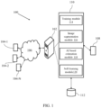
- FIG. 1 illustrates a block diagram a system 100 for analyzing a wound in the person or a patient.
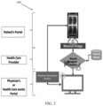
- FIG. 2 shows a schematic architecture of the system 100.
- the system 100 may also be referred as a wound management system 100.
- the network 106 may be a wireless or a wired network, or a combination thereof.
- the network 106 can be implemented as a computer network, as one of the different types of networks, such as virtual private network (VPN), intranet, local area network (LAN), wide area network (WAN), the internet, and such.
- VPN virtual private network
- LAN local area network
- WAN wide area network
- the internet and such.
- the network 106 may either be a dedicated network or a shared network, which represents an association of the different types of networks that use a variety of protocols, for example, Hypertext Transfer Protocol (HTTP), Transmission Control Protocol/Internet Protocol (TCP/IP), and Wireless Application Protocol (WAP), to communicate with each other.
- HTTP Hypertext Transfer Protocol
- TCP/IP Transmission Control Protocol/Internet Protocol
- WAP Wireless Application Protocol
- the network 106 may include a variety of network devices, including routers, bridges, servers, computing devices, storage devices. The network devices within the network 106 may interact with the system 100 through communication links.
- the system 100 may be implemented in a workstation, a mainframe computer, a server, and a network server.
- the computing device 102 further comprises one or more hardware processors 108, one or more memory 110, hereinafter referred as a memory 110 and a data repository 112, for example, a repository 112.
- the memory 110 is in communication with the one or more hardware processors 108, wherein the one or more hardware processors 108 are configured to execute programmed instructions stored in the memory 110, to perform various functions as explained in the later part of the disclosure.
- the repository 112 may store data processed, received, and generated by the system 100.
- the memory 110 further comprises a plurality of modules for performing various functions.
- the plurality of modules comprises a training module 114, an image segmentation module 116, an AI based estimation module 118, and a self-learning module 120 as shown in the block diagram of FIG. 1 .
- the system 100 supports various connectivity options such as BLUETOOTH ® , USB, ZigBee and other cellular services.
- the network environment enables connection of various components of the system 100 using any communication link including Internet, WAN, MAN, and so on.
- the system 100 is implemented to operate as a stand-alone device.
- the system 100 may be implemented to work as a loosely coupled device to a smart computing environment. The components and functionalities of the system 100 are described further in detail.
- the system 100 comprises a patient portal 302, a physician portal 304 and a server 306 or any other place where wound analysis happens.
- the patient portal 302 is an application present in the mobile phone of the patient, where the patient can click an image of a wound to be analyzed and share it to the server for wound analysis.
- the patient portal 302 comprises following features: a camera for capturing the image to be analyzed, an interface through which patient ID, date and time stamp can be entered, converting image to byte string array, ability to upload json to server, camera calibration and a display screen displaying a healing index as feedback from the analysis.
- the date and time stamp can be taken automatically from the processor.
- the system 100 is also configured to a plurality of patient parameters as input to the physician analyzing the image via the patient portal 302, wherein the plurality of patient parameters comprises patient name, ID, patient medical history, and patient physical statistics.
- FIG. 3 also shows the server such as Amazon web service (AWS) cloud gateway, the server 306 comprises authentication, database and feature for appending json to database.
- the physician portal 304 comprises a GUI showing patient details, input image and the analyzed image.
- the physician portal is in communication with an AWS-Python API Server.
- the user interface 104 is configured to receive a plurality of images.
- the plurality of images is further used for training a deep learning mode using the training module 114.
- the system 100 comprises the training module 114.
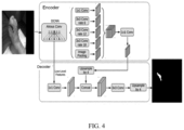
- the training module 114 is configured to train a deep learning (DL) model using the plurality of preprocessed images, wherein the deep learning model having an encoder decoder architecture as shown in FIG. 4 .
- the encoder decoder architecture comprises: a pre-trained Resnet50 as the encoder, an Atrous spatial pyramid pooling (ASPP) comprising of dilated convolution, wherein the ASPP is connected to the encoder, a bilinear up sampler configured to up sample the plurality of images by a factor of four, and a channel wise attention mechanism using squeeze and excitation (SE) network, designed to improve representational power of the network by enabling to perform dynamic channel-wise feature recalibration.
- DL deep learning
- SE squeeze and excitation
- transfer learning was used for the deep learning model. Instead of randomly initializing the weights, the Resnet50 model, pre-trained on the Image Net dataset is loaded before the model is trained on the plurality of data. Transfer learning with the pre-trained model is beneficial to the training process in the sense that the weights converge faster and better.
- DeepLabV3+ architecture was used. This is an encoder decoder architecture with Atrous Spatial Pyramid Pooling (ASPP) and bilinear up sampling.
- the network begins with a pre-trained Resnet50 as encoder, which is followed by ASPP.
- the ASPP consists of dilated convolution which helps to encode multi-scale contextual information. It is followed by a bilinear up sampling by a factor of 4 to get the output mask.
- SE - Squeeze and Excitation
- Atrous Spatial Pyramid Pooling is a semantic segmentation module for resampling a given feature layer at multiple rates prior to convolution. This amounts to probing the original image with multiple filters that have complementary effective fields of view, thus capturing objects as well as useful image context at multiple scales. Rather than resampling features, the mapping is implemented using multiple parallel Atrous convolutional layers with different sampling rates.
- Squeeze and Excitation Networks Architecture consists of three operations: Squeeze, Excitation, and Scaling.
- the squeeze operation is mainly used to extract the global information from each channel of the feature map.
- the feature map is basically the output of the convolution layer, which is a 4D tensor of size B ⁇ H ⁇ W ⁇ C.
- GAP Global Average Pooling
- GAP Global Average Pooling
- GAP Global Average Pooling
- GAP Global Max Pooling
- the feature map is now reduced to a smaller dimension (B x 1 x 1 x C)
- B x 1 x 1 x C basically for each channel of size H x W is reduced to a singular vector.
- MLP multiLayer perceptron
- the MLP is used to generate the weights to scale each channel of the feature map adaptively.
- the excitation operation passes the "excited" tensor of shape B x 1 x 1 x C. This tensor is then passed through a sigmoid activation function.
- the sigmoid activation function converts the tensor values in the range of 0 and 1. Then an element-wise multiplication is performed between the output of the sigmoid activation function and the input feature map. If the value is close to 0 means the channel is less important so, the values of the feature channel would be reduced, and if the value is close to 1, this means that is channel is important.
- the Squeeze and Excitation Network basically scale each channel information. It reduces the non-relevant channel information and the relevant channel are not much affected. So, after the whole operation, the feature map only contains the relevant information, which increases the representational power of the entire network.
- the system 100 comprises the image segmentation module 116.
- the image segmentation module 116 configured to preprocess an input image of the wound to be analyzed; generate an output image from the preprocessed image using the trained deep learning model; and post-process the output image using a binary segmentation mask with a fixed threshold of a predefined number.
- the binary segmentation masks predicted by the trained DL model are grayscale images with pixel intensities that range from 0 to 255.
- binary segmentation masks are first generated from thresholding with a fixed threshold of 127, which is half the max intensity.
- the system 100 comprises the AI based estimation module 118.
- the AI based estimation module 118 configured to perform morphology operations on the segmented image to remove the small regions and spurious noises to fill the small holes within the wound to improve a true positive rate; perform connected component analysis on the binary image to label connected regions; calculate a plurality of wound parameters of the wound by measuring the labelled connected regions; and overlay the calculated plurality of parameters and wound segmented boundary on the image of the wound.
- the block diagram of the AI based estimation module 118 is shown in Fig. 5 , which includes morphological operations, connected component analysis, and shape analysis followed by wound measurements.
- Input to the post processing stage is the binary segmentation mask that is generated from the deep learning output by thresholding. Morphology operations are performed on the binary mask to remove the small regions/spurious noises and to fill the small holes within the wound to improve the true positive rate.
- the blood stain could be identified as wound by the deep learning network causing small false-positive region/noise in the segmented mask. This small false-positive region is detected and removed by finding small, connected components in the segmented mask.
- the abnormal tissue like fibrinous tissue inside the wound could be treated as non-wound by the network representing it as small holes inside the segmented mask. These holes are detected and filled by finding the small, connected components.
- Connected component analysis is used to label the connected regions followed by measurement of those labeled connected regions.
- the measurements such as, area, perimeter, circle diameter, major and minor axis length of ellipse etc. are used in conjunction with shape analysis to find the approximate shape of the wound and its measurements. Finally, the measured wound parameters and wound segmented boundary are overlaid on the wound image.
- the system 100 also comprises the self-learning module.
- the self-learning module configured to receive annotating on the output image of the wound provided by a user, if the segmented output image is not matching with the input image based on an evaluation done by the user; and utilizing the annotation provided by the user to self-learn the training module to improve the accuracy of the system.
- the user can provide an annotation tool that has been developed based on graph-cut method with minimum touch points of two or more that distinguish the foreground and background of the image.
- the user can provide the annotation tool using a manual re-draw approach.
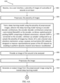
- FIG. 6A-6B illustrates a flow chart of an AI based method 600 for analyzing the wound in the person, in accordance with an example embodiment of the present disclosure.
- the method 600 depicted in the flow chart may be executed by a system, for example, the system 100 of FIG. 1 .
- the system 100 may be embodied in the computing device.
- Operations of the flowchart, and combinations of operations in the flowchart may be implemented by various means, such as hardware, firmware, processor, circuitry and/or other device associated with execution of software including one or more computer program instructions.
- one or more of the procedures described in various embodiments may be embodied by computer program instructions.
- the computer program instructions, which embody the procedures, described in various embodiments may be stored by at least one memory device of a system and executed by at least one processor in the system. Any such computer program instructions may be loaded onto a computer or other programmable system (for example, hardware) to produce a machine, such that the resulting computer or other programmable system embody means for implementing the operations specified in the flowchart.
- the operations of the method 600 are described with help of system 100. However, the operations of the method 600 can be described and/or practiced by using any other system.
- the plurality of images of a plurality of wounds is received as the input via the user interface 104.
- the plurality of images is then preprocessed.
- the deep learning model is trained using the plurality of preprocessed images, wherein the deep learning model having an encoder decoder architecture, the encoder decoder architecture comprises: a pre-trained Resnet50 as the encoder, an Atrous spatial pyramid pooling (ASPP) comprising of dilated convolution, wherein the ASPP is connected to the encoder, a bilinear up sampler configured to up sample the plurality of images by a factor of four, and a channel wise attention mechanism using a squeeze and excitation (SE) network, designed to improve representational power of the SE network by enabling to perform dynamic channel-wise feature recalibration.
- ABP Atrous spatial pyramid pooling
- SE squeeze and excitation
- the image of the wound to be analyzed is provided.
- the image to be analyzed is preprocessed.
- the output image is generated from the preprocessed image using the trained deep learning model.
- the output image is preprocessed using the binary segmentation mask with the fixed threshold of the predefined number to get the segmented output image.
- next step 616 of the method 600 morphology operations are performed on the segmented output image to remove the small regions and spurious noises to fill the small holes within the wound to improve a true positive rate.
- next step 618 connected component analysis is performed on the morphology operated image to label connected regions.
- the plurality of wound parameters of the wound is calculated by measuring the labelled connected regions.
- step 622 the calculated plurality of wound parameters and wound segmented boundary is overlaid on the image of the wound. This overlaid image can be displayed on the display screen which can further be analyzed by the physician.
- the method 100 further comprises receiving annotation on the segmented output image of the wound provided by a user, if the segmented output image is not matching with the input image based on an evaluation done by the user; and utilizing the annotation provided by the user to self-learn the training module 114 to improve the accuracy of the system 100.
- the system 100 also comprises a 3D printer.
- the 3D printer is configured to provide a wound dress patch estimated using the plurality of wound parameter.
- the system 100 can also be explained with the help of experimental results.
- preexisting wound care data have been merged segmentation dataset, which leads to 2119 images.
- images were resized and augmented as a process homogeneousness, extending the dataset as 10937 images.
- the DeepLab V3+, and Resnet-50 encoder was adapted with pre-trained weights from image net dataset, resulting in a dice score of 0.9206 and IOU -0.85.
- the performance was improved to a dice score of 0.926 and IOU-0.86 for DeepLab-V3+ with squeeze & Excite model.
- the AI module is implemented with morphological operations, connected component analysis, and shape analysis, improving accuracy and providing the wound parameter and metrics such as area, perimeter, circle diameter, major and minor axis length of an ellipse.
- a lightweight approach was attempted using the U-Net model with Mobile Net Encoder, which yields a dice score of 0.91 and IOU of 0.83.
- the images were resized to 256*256 and the Image Augmentation techniques which includes, Gamma, Saturation, Hue, Horizontal and Vertical Flips etc. were adapted, which made the training data set as 10746 wound images.
- the augmentations were not applied to the testing and validation sets and contained 191 images in each set.
- the deep learning model in the presented work was implemented in python with Keras and TensorFlow backend.
- the model was trained on Tesla P100 GPU with 16 GB memory.
- the Adam optimization algorithm was employed for updating the parameters in the network.
- Dice loss was used as the loss function and also Precision, Recall and the Dice score were monitored as the evaluation matrices.
- the initial learning rate was set to 0.0001 and each minibatch contained 16 images for balancing the training accuracy and efficiency. A drop out of 0.1 was specified in order to prevent any overfitting.
- Early stopping was used to terminate the training so that the best result was saved when there was no improvement for more than 10 epochs in terms of validation loss.
- the deep learning model was trained for around 30 epochs before overfitting illustrates the training and validation losses.
- the embodiments of present disclosure herein addresses unresolved problem of accurate, timely and cost effective analysis of the wounds of the person.
- the embodiment thus providesan AI based method and a system for analyzing the wounds.
- Such computer-readable storage means contain program-code means for implementation of one or more steps of the method, when the program runs on a server or mobile device or any suitable programmable device.
- the hardware device can be any kind of device which can be programmed including e.g., any kind of computer like a server or a personal computer, or the like, or any combination thereof.
- the device may also include means which could be e.g., hardware means like e.g., an application-specific integrated circuit (ASIC), a field-programmable gate array (FPGA), or a combination of hardware and software means, e.g., an ASIC and an FPGA, or at least one microprocessor and at least one memory with software processing components located therein.
- the means can include both hardware means and software means.
- the method embodiments described herein could be implemented in hardware and software.
- the device may also include software means. Alternatively, the embodiments may be implemented on different hardware devices, e.g., using a plurality of CPUs.
- the embodiments herein can comprise hardware and software elements.
- the embodiments that are implemented in software include but are not limited to, firmware, resident software, microcode, etc.
- the functions performed by various components described herein may be implemented in other components or combinations of other components.
- a computer-usable or computer readable medium can be any apparatus that can comprise, store, communicate, propagate, or transport the program for use by or in connection with the instruction execution system, apparatus, or device.
- a computer-readable storage medium refers to any type of physical memory on which information or data readable by a processor may be stored.
- a computer-readable storage medium may store instructions for execution by one or more processors, including instructions for causing the processor(s) to perform steps or stages consistent with the embodiments described herein.
- the term "computer-readable medium” should be understood to include tangible items and exclude carrier waves and transient signals, i.e., be non-transitory. Examples include random access memory (RAM), read-only memory (ROM), volatile memory, nonvolatile memory, hard drives, CD ROMs, DVDs, flash drives, disks, and any other known physical storage media.
Landscapes
- Engineering & Computer Science (AREA)
- Physics & Mathematics (AREA)
- Health & Medical Sciences (AREA)
- Theoretical Computer Science (AREA)
- General Health & Medical Sciences (AREA)
- General Physics & Mathematics (AREA)
- Medical Informatics (AREA)
- Biomedical Technology (AREA)
- Computer Vision & Pattern Recognition (AREA)
- Data Mining & Analysis (AREA)
- Chemical & Material Sciences (AREA)
- Public Health (AREA)
- Materials Engineering (AREA)
- Radiology & Medical Imaging (AREA)
- Nuclear Medicine, Radiotherapy & Molecular Imaging (AREA)
- Software Systems (AREA)
- Life Sciences & Earth Sciences (AREA)
- Biophysics (AREA)
- Evolutionary Computation (AREA)
- Molecular Biology (AREA)
- Computing Systems (AREA)
- General Engineering & Computer Science (AREA)
- Mathematical Physics (AREA)
- Computational Linguistics (AREA)
- Artificial Intelligence (AREA)
- Primary Health Care (AREA)
- Epidemiology (AREA)
- Quality & Reliability (AREA)
- Optics & Photonics (AREA)
- Databases & Information Systems (AREA)
- Pathology (AREA)
- Mechanical Engineering (AREA)
- Manufacturing & Machinery (AREA)
- Geometry (AREA)
- Image Analysis (AREA)
Claims (15)
- Système (100) pour analyser une plaie, le système comprenant :une interface utilisateur (104) pour recevoir une pluralité d'images d'une pluralité de plaies en entrée,un ou plusieurs processeurs matériels (108) ; etune mémoire (110) en communication avec les un ou plusieurs processeurs matériels, les un ou plusieurs premiers processeurs matériels étant configurés pour exécuter des instructions programmées stockées dans la mémoire, la mémoire comprenant en outre :un module d'entraînement configuré pour entraîner un modèle d'apprentissage profond à l'aide de la pluralité d'images prétraitées, le modèle d'apprentissage profond ayant une architecture codeur-décodeur, l'architecture codeur-décodeur comprenant :un Resnet50 préentraîné comme encodeur,une agrégation par pyramide spatiale Atrous (ASPP) comprenant une convolution dilatée, l'ASPP étant reliée à l'encodeur,un suréchantillonneur bilinéaire configuré pour suréchantillonner la pluralité d'images d'un facteur quatre, etun mécanisme d'attention canal par canal utilisant un réseau de compression et d'excitation (SE), conçu pour améliorer la puissance de représentation du réseau en permettant la réalisation d'un réétalonnage dynamique des caractéristiques canal par canal ;un module de segmentation d'image configuré pour :prétraiter une image d'entrée de la plaie à analyser ;générer une image de sortie à partir d'une image prétraitée à l'aide du modèle d'apprentissage profond entraîné ; etpost-traiter l'image de sortie à l'aide d'un masque de segmentation binaire avec un seuil fixe d'un nombre prédéfini ;un module d'estimation à base d'IA configuré pour :effectuer des opérations de morphologie sur l'image segmentée pour supprimer les petites régions et les bruits parasites afin de combler les petits trous dans la plaie pour améliorer le taux de vrais positifs ;effectuer une analyse en composantes connexes sur l'image binaire pour étiqueter les régions connexes ;calculer une pluralité de paramètres de la plaie en mesurant les régions connexes étiquetées ; etsuperposer la pluralité de paramètres de plaie calculés sur l'image de la plaie.
- Système selon la revendication 1 comprenant en outre un module d'autoapprentissage, le module d'autoapprentissage étant configuré pour :recevoir une annotation sur l'image de sortie de la plaie fournie par un utilisateur, si l'image de sortie segmentée ne correspond pas à l'image d'entrée sur la base d'une évaluation effectuée par l'utilisateur ; etutiliser l'annotation fournie par l'utilisateur pour l'autoapprentissage du module de formation afin d'améliorer la précision du système.
- Système selon la revendication 1 comprenant en outre une imprimante 3D configurée pour fournir une pièce de pansement estimée à l'aide de la pluralité de paramètres de plaie.
- Procédé mis en oeuvre par processeur (600) pour analyser une plaie, le procédé comprenant :la réception, via une interface utilisateur, d'une pluralité d'images d'une pluralité de plaies en entrée (602) ;le prétraitement, via un ou plusieurs processeurs matériels, de la pluralité d'images (604) ;l'entraînement, via les un ou plusieurs processeurs matériels, d'un modèle d'apprentissage profond à l'aide de la pluralité d'images prétraitées, le modèle d'apprentissage profond ayant une architecture codeur-décodeur, l'architecture codeur-décodeur comprenant :un Resnet50 préentraîné comme encodeur,une agrégation par pyramide spatiale Atrous (ASPP) comprenant une convolution dilatée, l'ASPP étant reliée à l'encodeur,un suréchantillonneur bilinéaire configuré pour suréchantillonner la pluralité d'images d'un facteur quatre, etun mécanisme d'attention canal par canal utilisant un réseau de compression et d'excitation (SE), conçu pour améliorer la puissance de représentation du réseau SE en permettant la réalisation d'un réétalonnage dynamique des caractéristiques canal par canal (606) ;la fourniture, via les un ou plusieurs processeurs matériels, d'une image de la plaie à analyser (608) ;le prétraitement, via un ou plusieurs processeurs matériels, de l'image (610) ;la génération, via un ou plusieurs processeurs matériels, d'une image de sortie à partir d'une image prétraitée à l'aide du modèle d'apprentissage profond entraîné (612) ;le post-traitement, via un ou plusieurs processeurs matériels, de l'image de sortie à l'aide d'un masque de segmentation binaire avec un seuil fixe d'un nombre prédéfini pour obtenir une image de sortie segmentée (614) ;la réalisation, via les un ou plusieurs processeurs matériels, d'opérations de morphologie sur l'image de sortie segmentée pour supprimer les petites régions et les bruits parasites afin de combler les petits trous dans la plaie pour améliorer le taux de vrais positifs (616) ; la réalisation, via les un ou plusieurs processeurs matériels, d'une analyse en composantes connexes sur l'image traitée par les opérations de morphologie pour étiqueter les régions connexes (618) ;le calcul, via les un ou plusieurs processeurs matériels, d'une pluralité de paramètres de la plaie en mesurant les régions connexes étiquetées (620) ; etla superposition, via les un ou plusieurs processeurs matériels, de la pluralité calculée de paramètres de plaie sur l'image de la plaie (622).
- Procédé mis en oeuvre par processeur selon la revendication 4, comprenant en outre :la réception d'une annotation sur l'image de sortie segmentée de la plaie fournie par un utilisateur, si l'image de sortie segmentée ne correspond pas à l'image d'entrée sur la base d'une évaluation effectuée par l'utilisateur ; etl'utilisation de l'annotation fournie par l'utilisateur pour l'autoapprentissage du module de formation afin d'améliorer la précision du système.
- Procédé mis en oeuvre par le processeur selon la revendication 4 comprenant en outre la présentation d'un indice de guérison de la plaie analysée à la personne.
- Procédé mis en oeuvre par processeur selon la revendication 4, dans lequel la pluralité de paramètres de plaie comprend une surface, un périmètre, un diamètre de cercle, une longueur de grand axe et de petit axe d'une ellipse.
- Procédé mis en oeuvre par processeur selon la revendication 4, dans lequel le prétraitement comprend le redimensionnement de l'image et l'augmentation de l'image.
- Procédé mis en oeuvre par processeur selon la revendication 4, dans lequel le masque de segmentation binaire est une image en niveaux de gris avec des intensités de pixels allant de 0 à 255.
- Procédé mis en oeuvre par processeur selon la revendication 4, dans lequel l'ASPP est configuré pour coder des informations contextuelles à échelles multiples à partir de la pluralité d'images.
- Procédé mis en oeuvre par processeur selon la revendication 4 comprenant en outre une pluralité de paramètres de patient en entrée pour un médecin analysant l'image, la pluralité de paramètres de patient comprenant le nom du patient, l'identifiant, les antécédents médicaux du patient et les statistiques physiques du patient.
- Procédé mis en oeuvre par processeur selon la revendication 4 comprenant l'intégration avec une imprimante 3D pour fournir une pièce de pansement estimée à l'aide de la pluralité de paramètres de plaie.
- Un ou plusieurs supports de stockage d'informations non transitoires lisibles par machine comprenant une ou plusieurs instructions qui, lorsqu'elles sont exécutées par un ou plusieurs processeurs matériels, provoquent :la réception d'une pluralité d'images d'une pluralité de plaies en entrée ;le prétraitement de la pluralité d'images ;l'entraînement d'un modèle d'apprentissage profond à l'aide de la pluralité d'images prétraitées, le modèle d'apprentissage profond ayant une architecture codeur-décodeur, l'architecture codeur-décodeur comprenant :un Resnet50 préentraîné comme encodeur,une agrégation par pyramide spatiale Atrous (ASPP) comprenant une convolution dilatée, l'ASPP étant reliée à l'encodeur,un suréchantillonneur bilinéaire configuré pour suréchantillonner la pluralité d'images d'un facteur quatre, etun mécanisme d'attention canal par canal utilisant un réseau de compression et d'excitation (SE), conçu pour améliorer la puissance de représentation du réseau SE en permettant la réalisation d'un réétalonnage dynamique des caractéristiquescanal par canal ;la fourniture d'une image de la plaie à analyser ;le prétraitement de l'image ;la génération d'une image de sortie à partir d'une image prétraitée à l'aide du modèle d'apprentissage profond entraîné ;le post-traitement de l'image de sortie à l'aide d'un masque de segmentation binaire avec un seuil fixe d'un nombre prédéfini pour obtenir une image de sortie segmentée ;la réalisation d'opérations de morphologie sur l'image de sortie segmentée pour supprimer les petites régions et les bruits parasites afin de combler les petits trous dans la plaie pour améliorer le taux de vrais positifs ;la réalisation d'une analyse en composantes connexes sur l'image traitée par les opérations de morphologie pour étiqueter les régions connexes ;le calcul d'une pluralité de paramètres de plaie en mesurant les régions connexes étiquetées ; etla superposition de la pluralité de paramètres de plaie calculés sur l'image de la plaie.
- Un ou plusieurs supports de stockage d'informations lisibles par machine non transitoires selon la revendication 13, comprenant en outre :la réception d'une annotation sur l'image de sortie segmentée de la plaie fournie par un utilisateur, si l'image de sortie segmentée ne correspond pas à l'image d'entrée sur la base d'une évaluation effectuée par l'utilisateur ; etl'utilisation de l'annotation fournie par l'utilisateur pour l'autoapprentissage du module de formation afin d'améliorer la précision du système.
- Un ou plusieurs supports de stockage d'informations lisibles par machine non transitoires selon la revendication 13, comprenant en outre la présentation d'un indice de guérison de la plaie analysée à la personne.
Applications Claiming Priority (1)
| Application Number | Priority Date | Filing Date | Title |
|---|---|---|---|
| IN202221052530 | 2022-09-14 |
Publications (3)
| Publication Number | Publication Date |
|---|---|
| EP4343694A1 EP4343694A1 (fr) | 2024-03-27 |
| EP4343694B1 true EP4343694B1 (fr) | 2024-10-09 |
| EP4343694C0 EP4343694C0 (fr) | 2024-10-09 |
Family
ID=87760437
Family Applications (1)
| Application Number | Title | Priority Date | Filing Date |
|---|---|---|---|
| EP23192424.2A Active EP4343694B1 (fr) | 2022-09-14 | 2023-08-21 | Procédé et système basés sur l'intelligence artificielle (ia) pour analyser une plaie |
Country Status (2)
| Country | Link |
|---|---|
| US (1) | US20240087116A1 (fr) |
| EP (1) | EP4343694B1 (fr) |
Families Citing this family (1)
| Publication number | Priority date | Publication date | Assignee | Title |
|---|---|---|---|---|
| CN120299220B (zh) * | 2025-06-11 | 2025-08-19 | 四川省国土空间生态修复与地质灾害防治研究院 | 基于深度学习的滑坡灾害预警方法、设备、介质及产品 |
Family Cites Families (7)
| Publication number | Priority date | Publication date | Assignee | Title |
|---|---|---|---|---|
| US20150140058A1 (en) * | 2013-11-20 | 2015-05-21 | Dermagenesis Llc | Skin printing and auto-grafting |
| CN107106020A (zh) * | 2014-10-29 | 2017-08-29 | 组织分析股份有限公司 | 用于分析并且传输与哺乳动物皮损病有关的数据、图像和视频的系统与方法 |
| US10037603B2 (en) * | 2015-05-04 | 2018-07-31 | Siemens Healthcare Gmbh | Method and system for whole body bone removal and vascular visualization in medical image data |
| US10223788B2 (en) * | 2016-08-31 | 2019-03-05 | International Business Machines Corporation | Skin lesion segmentation using deep convolution networks guided by local unsupervised learning |
| US10783632B2 (en) * | 2018-12-14 | 2020-09-22 | Spectral Md, Inc. | Machine learning systems and method for assessment, healing prediction, and treatment of wounds |
| US11176677B2 (en) * | 2020-03-16 | 2021-11-16 | Memorial Sloan Kettering Cancer Center | Deep interactive learning for image segmentation models |
| US12236592B2 (en) * | 2021-09-14 | 2025-02-25 | Arizona Board Of Regents On Behalf Of Arizona State University | Systems, methods, and apparatuses for systematically determining an optimal approach for the computer-aided diagnosis of a pulmonary embolism |
-
2023
- 2023-08-21 EP EP23192424.2A patent/EP4343694B1/fr active Active
- 2023-08-24 US US18/455,429 patent/US20240087116A1/en active Pending
Also Published As
| Publication number | Publication date |
|---|---|
| EP4343694A1 (fr) | 2024-03-27 |
| EP4343694C0 (fr) | 2024-10-09 |
| US20240087116A1 (en) | 2024-03-14 |
Similar Documents
| Publication | Publication Date | Title |
|---|---|---|
| CN110909780B (zh) | 一种图像识别模型训练和图像识别方法、装置及系统 | |
| CN110120047B (zh) | 图像分割模型训练方法、图像分割方法、装置、设备及介质 | |
| CN112990232B (zh) | 面向多种高空作业施工现场的安全带佩戴识别与检测方法 | |
| CN110288597B (zh) | 基于注意力机制的无线胶囊内窥镜视频显著性检测方法 | |
| CN105117611B (zh) | 基于卷积神经元网络的中医舌诊模型的确定方法及系统 | |
| Liu et al. | A framework of wound segmentation based on deep convolutional networks | |
| CN111325739A (zh) | 肺部病灶检测的方法及装置,和图像检测模型的训练方法 | |
| CN112862746B (zh) | 基于人工神经网络的组织病变识别的识别方法及识别系统 | |
| CN113160261A (zh) | 一种用于oct图像角膜层分割的边界增强卷积神经网络 | |
| EP4343694B1 (fr) | Procédé et système basés sur l'intelligence artificielle (ia) pour analyser une plaie | |
| CN110349166A (zh) | 一种针对于视网膜图像的血管分割方法、装置及设备 | |
| JP2025541186A (ja) | 診断及び処置のための光干渉断層血管撮影スキャンの機械学習による分析 | |
| Prasanth et al. | Automated Detection of Oropharyngeal Carcinoma Using Super-Resolution Generative Adversarial Network and Convolutional Neural Network | |
| US20240212154A1 (en) | Image segmentation for size estimation and machine learning-based modeling for predicting rates of size changes over time | |
| Micheal et al. | Dual-model approach for diabetic retinopathy and macular edema detection | |
| Chelliah et al. | Prognosticating Pneumonia: A Comparative Analysis of Prediction Models Enhanced by and Devoid of Attention Mechanism | |
| CN111950637B (zh) | 紫质检测方法、装置、皮肤检测仪和可读存储介质 | |
| Kaki et al. | Semantic segmentation of dental caries using improved deeplab v3network | |
| Sabri et al. | Diagnosing Gingiva Disease Using Artificial Intelligence Techniques | |
| CN112598603A (zh) | 一种基于卷积神经网络的口腔龋病图像智能识别方法 | |
| Saky et al. | Enhanced segnet with integrated grad-cam for interpretable retinal layer segmentation in oct images | |
| Bellary et al. | The MRI knee pain classification using CNN algorithm and segmentation using clustering algorithm | |
| CN116664592A (zh) | 基于图像的动静脉血管分离方法、装置、电子设备及介质 | |
| Pyar | Transfer Learning-Based Segmentation of Pneumonia from Chest X-Rays Images | |
| CN117523189A (zh) | 脉络膜血管分割方法、装置、终端设备以及存储介质 |
Legal Events
| Date | Code | Title | Description |
|---|---|---|---|
| PUAI | Public reference made under article 153(3) epc to a published international application that has entered the european phase |
Free format text: ORIGINAL CODE: 0009012 |
|
| STAA | Information on the status of an ep patent application or granted ep patent |
Free format text: STATUS: THE APPLICATION HAS BEEN PUBLISHED |
|
| STAA | Information on the status of an ep patent application or granted ep patent |
Free format text: STATUS: REQUEST FOR EXAMINATION WAS MADE |
|
| AK | Designated contracting states |
Kind code of ref document: A1 Designated state(s): AL AT BE BG CH CY CZ DE DK EE ES FI FR GB GR HR HU IE IS IT LI LT LU LV MC ME MK MT NL NO PL PT RO RS SE SI SK SM TR |
|
| 17P | Request for examination filed |
Effective date: 20240308 |
|
| RBV | Designated contracting states (corrected) |
Designated state(s): AL AT BE BG CH CY CZ DE DK EE ES FI FR GB GR HR HU IE IS IT LI LT LU LV MC ME MK MT NL NO PL PT RO RS SE SI SK SM TR |
|
| GRAP | Despatch of communication of intention to grant a patent |
Free format text: ORIGINAL CODE: EPIDOSNIGR1 |
|
| STAA | Information on the status of an ep patent application or granted ep patent |
Free format text: STATUS: GRANT OF PATENT IS INTENDED |
|
| RIC1 | Information provided on ipc code assigned before grant |
Ipc: G06N 3/09 20230101ALN20240515BHEP Ipc: G06N 3/048 20230101ALN20240515BHEP Ipc: A61B 5/00 20060101ALN20240515BHEP Ipc: G06N 3/096 20230101ALI20240515BHEP Ipc: G06N 3/0464 20230101ALI20240515BHEP Ipc: G06N 3/0455 20230101ALI20240515BHEP Ipc: G06T 7/62 20170101ALI20240515BHEP Ipc: G06T 7/00 20170101AFI20240515BHEP |
|
| INTG | Intention to grant announced |
Effective date: 20240606 |
|
| GRAS | Grant fee paid |
Free format text: ORIGINAL CODE: EPIDOSNIGR3 |
|
| GRAA | (expected) grant |
Free format text: ORIGINAL CODE: 0009210 |
|
| STAA | Information on the status of an ep patent application or granted ep patent |
Free format text: STATUS: THE PATENT HAS BEEN GRANTED |
|
| AK | Designated contracting states |
Kind code of ref document: B1 Designated state(s): AL AT BE BG CH CY CZ DE DK EE ES FI FR GB GR HR HU IE IS IT LI LT LU LV MC ME MK MT NL NO PL PT RO RS SE SI SK SM TR |
|
| REG | Reference to a national code |
Ref country code: CH Ref legal event code: EP |
|
| REG | Reference to a national code |
Ref country code: DE Ref legal event code: R096 Ref document number: 602023000701 Country of ref document: DE |
|
| REG | Reference to a national code |
Ref country code: IE Ref legal event code: FG4D |
|
| U01 | Request for unitary effect filed |
Effective date: 20241016 |
|
| U07 | Unitary effect registered |
Designated state(s): AT BE BG DE DK EE FI FR IT LT LU LV MT NL PT RO SE SI Effective date: 20241104 |
|
| PG25 | Lapsed in a contracting state [announced via postgrant information from national office to epo] |
Ref country code: IS Free format text: LAPSE BECAUSE OF FAILURE TO SUBMIT A TRANSLATION OF THE DESCRIPTION OR TO PAY THE FEE WITHIN THE PRESCRIBED TIME-LIMIT Effective date: 20250209 Ref country code: HR Free format text: LAPSE BECAUSE OF FAILURE TO SUBMIT A TRANSLATION OF THE DESCRIPTION OR TO PAY THE FEE WITHIN THE PRESCRIBED TIME-LIMIT Effective date: 20241009 |
|
| PG25 | Lapsed in a contracting state [announced via postgrant information from national office to epo] |
Ref country code: ES Free format text: LAPSE BECAUSE OF FAILURE TO SUBMIT A TRANSLATION OF THE DESCRIPTION OR TO PAY THE FEE WITHIN THE PRESCRIBED TIME-LIMIT Effective date: 20241009 |
|
| PG25 | Lapsed in a contracting state [announced via postgrant information from national office to epo] |
Ref country code: NO Free format text: LAPSE BECAUSE OF FAILURE TO SUBMIT A TRANSLATION OF THE DESCRIPTION OR TO PAY THE FEE WITHIN THE PRESCRIBED TIME-LIMIT Effective date: 20250109 |
|
| PG25 | Lapsed in a contracting state [announced via postgrant information from national office to epo] |
Ref country code: GR Free format text: LAPSE BECAUSE OF FAILURE TO SUBMIT A TRANSLATION OF THE DESCRIPTION OR TO PAY THE FEE WITHIN THE PRESCRIBED TIME-LIMIT Effective date: 20250110 |
|
| PG25 | Lapsed in a contracting state [announced via postgrant information from national office to epo] |
Ref country code: PL Free format text: LAPSE BECAUSE OF FAILURE TO SUBMIT A TRANSLATION OF THE DESCRIPTION OR TO PAY THE FEE WITHIN THE PRESCRIBED TIME-LIMIT Effective date: 20241009 |
|
| PG25 | Lapsed in a contracting state [announced via postgrant information from national office to epo] |
Ref country code: RS Free format text: LAPSE BECAUSE OF FAILURE TO SUBMIT A TRANSLATION OF THE DESCRIPTION OR TO PAY THE FEE WITHIN THE PRESCRIBED TIME-LIMIT Effective date: 20250109 |
|
| PG25 | Lapsed in a contracting state [announced via postgrant information from national office to epo] |
Ref country code: SM Free format text: LAPSE BECAUSE OF FAILURE TO SUBMIT A TRANSLATION OF THE DESCRIPTION OR TO PAY THE FEE WITHIN THE PRESCRIBED TIME-LIMIT Effective date: 20241009 |
|
| PG25 | Lapsed in a contracting state [announced via postgrant information from national office to epo] |
Ref country code: SK Free format text: LAPSE BECAUSE OF FAILURE TO SUBMIT A TRANSLATION OF THE DESCRIPTION OR TO PAY THE FEE WITHIN THE PRESCRIBED TIME-LIMIT Effective date: 20241009 |
|
| PG25 | Lapsed in a contracting state [announced via postgrant information from national office to epo] |
Ref country code: CZ Free format text: LAPSE BECAUSE OF FAILURE TO SUBMIT A TRANSLATION OF THE DESCRIPTION OR TO PAY THE FEE WITHIN THE PRESCRIBED TIME-LIMIT Effective date: 20241009 |
|
| PLBE | No opposition filed within time limit |
Free format text: ORIGINAL CODE: 0009261 |
|
| STAA | Information on the status of an ep patent application or granted ep patent |
Free format text: STATUS: NO OPPOSITION FILED WITHIN TIME LIMIT |
|
| 26N | No opposition filed |
Effective date: 20250710 |
|
| U20 | Renewal fee for the european patent with unitary effect paid |
Year of fee payment: 3 Effective date: 20250825 |
|
| PG25 | Lapsed in a contracting state [announced via postgrant information from national office to epo] |
Ref country code: MC Free format text: LAPSE BECAUSE OF FAILURE TO SUBMIT A TRANSLATION OF THE DESCRIPTION OR TO PAY THE FEE WITHIN THE PRESCRIBED TIME-LIMIT Effective date: 20241009 |