EP4344646A1 - Procédé et système de réglage d'un paramètre d'imagerie médicale d'un milieu - Google Patents
Procédé et système de réglage d'un paramètre d'imagerie médicale d'un milieu Download PDFInfo
- Publication number
- EP4344646A1 EP4344646A1 EP22315223.2A EP22315223A EP4344646A1 EP 4344646 A1 EP4344646 A1 EP 4344646A1 EP 22315223 A EP22315223 A EP 22315223A EP 4344646 A1 EP4344646 A1 EP 4344646A1
- Authority
- EP
- European Patent Office
- Prior art keywords
- medium
- medical imaging
- medical
- parameter
- imaging parameter
- Prior art date
- Legal status (The legal status is an assumption and is not a legal conclusion. Google has not performed a legal analysis and makes no representation as to the accuracy of the status listed.)
- Pending
Links
Images

Classifications
-
- G—PHYSICS
- G16—INFORMATION AND COMMUNICATION TECHNOLOGY [ICT] SPECIALLY ADAPTED FOR SPECIFIC APPLICATION FIELDS
- G16H—HEALTHCARE INFORMATICS, i.e. INFORMATION AND COMMUNICATION TECHNOLOGY [ICT] SPECIALLY ADAPTED FOR THE HANDLING OR PROCESSING OF MEDICAL OR HEALTHCARE DATA
- G16H30/00—ICT specially adapted for the handling or processing of medical images
- G16H30/40—ICT specially adapted for the handling or processing of medical images for processing medical images, e.g. editing
-
- A—HUMAN NECESSITIES
- A61—MEDICAL OR VETERINARY SCIENCE; HYGIENE
- A61B—DIAGNOSIS; SURGERY; IDENTIFICATION
- A61B8/00—Diagnosis using ultrasonic, sonic or infrasonic waves
- A61B8/54—Control of the diagnostic device
-
- A—HUMAN NECESSITIES
- A61—MEDICAL OR VETERINARY SCIENCE; HYGIENE
- A61B—DIAGNOSIS; SURGERY; IDENTIFICATION
- A61B8/00—Diagnosis using ultrasonic, sonic or infrasonic waves
- A61B8/44—Constructional features of the ultrasonic, sonic or infrasonic diagnostic device
- A61B8/4416—Constructional features of the ultrasonic, sonic or infrasonic diagnostic device related to combined acquisition of different diagnostic modalities, e.g. combination of ultrasound and X-ray acquisitions
-
- A—HUMAN NECESSITIES
- A61—MEDICAL OR VETERINARY SCIENCE; HYGIENE
- A61B—DIAGNOSIS; SURGERY; IDENTIFICATION
- A61B5/00—Measuring for diagnostic purposes; Identification of persons
- A61B5/0033—Features or image-related aspects of imaging apparatus, e.g. for MRI, optical tomography or impedance tomography apparatus; Arrangements of imaging apparatus in a room
- A61B5/0035—Features or image-related aspects of imaging apparatus, e.g. for MRI, optical tomography or impedance tomography apparatus; Arrangements of imaging apparatus in a room adapted for acquisition of images from more than one imaging mode, e.g. combining MRI and optical tomography
-
- A—HUMAN NECESSITIES
- A61—MEDICAL OR VETERINARY SCIENCE; HYGIENE
- A61B—DIAGNOSIS; SURGERY; IDENTIFICATION
- A61B5/00—Measuring for diagnostic purposes; Identification of persons
- A61B5/0033—Features or image-related aspects of imaging apparatus, e.g. for MRI, optical tomography or impedance tomography apparatus; Arrangements of imaging apparatus in a room
- A61B5/0037—Performing a preliminary scan, e.g. a prescan for identifying a region of interest
-
- A—HUMAN NECESSITIES
- A61—MEDICAL OR VETERINARY SCIENCE; HYGIENE
- A61B—DIAGNOSIS; SURGERY; IDENTIFICATION
- A61B5/00—Measuring for diagnostic purposes; Identification of persons
- A61B5/0059—Measuring for diagnostic purposes; Identification of persons using light, e.g. diagnosis by transillumination, diascopy, fluorescence
-
- A—HUMAN NECESSITIES
- A61—MEDICAL OR VETERINARY SCIENCE; HYGIENE
- A61B—DIAGNOSIS; SURGERY; IDENTIFICATION
- A61B5/00—Measuring for diagnostic purposes; Identification of persons
- A61B5/0059—Measuring for diagnostic purposes; Identification of persons using light, e.g. diagnosis by transillumination, diascopy, fluorescence
- A61B5/0073—Measuring for diagnostic purposes; Identification of persons using light, e.g. diagnosis by transillumination, diascopy, fluorescence by tomography, i.e. reconstruction of 3D images from 2D projections
-
- A—HUMAN NECESSITIES
- A61—MEDICAL OR VETERINARY SCIENCE; HYGIENE
- A61B—DIAGNOSIS; SURGERY; IDENTIFICATION
- A61B5/00—Measuring for diagnostic purposes; Identification of persons
- A61B5/05—Detecting, measuring or recording for diagnosis by means of electric currents or magnetic fields; Measuring using microwaves or radio waves
- A61B5/055—Detecting, measuring or recording for diagnosis by means of electric currents or magnetic fields; Measuring using microwaves or radio waves involving electronic [EMR] or nuclear [NMR] magnetic resonance, e.g. magnetic resonance imaging
-
- A—HUMAN NECESSITIES
- A61—MEDICAL OR VETERINARY SCIENCE; HYGIENE
- A61B—DIAGNOSIS; SURGERY; IDENTIFICATION
- A61B6/00—Apparatus or devices for radiation diagnosis; Apparatus or devices for radiation diagnosis combined with radiation therapy equipment
- A61B6/02—Arrangements for diagnosis sequentially in different planes; Stereoscopic radiation diagnosis
- A61B6/03—Computed tomography [CT]
- A61B6/032—Transmission computed tomography [CT]
-
- A—HUMAN NECESSITIES
- A61—MEDICAL OR VETERINARY SCIENCE; HYGIENE
- A61B—DIAGNOSIS; SURGERY; IDENTIFICATION
- A61B6/00—Apparatus or devices for radiation diagnosis; Apparatus or devices for radiation diagnosis combined with radiation therapy equipment
- A61B6/50—Apparatus or devices for radiation diagnosis; Apparatus or devices for radiation diagnosis combined with radiation therapy equipment specially adapted for specific body parts; specially adapted for specific clinical applications
- A61B6/502—Apparatus or devices for radiation diagnosis; Apparatus or devices for radiation diagnosis combined with radiation therapy equipment specially adapted for specific body parts; specially adapted for specific clinical applications for diagnosis of breast, i.e. mammography
-
- A—HUMAN NECESSITIES
- A61—MEDICAL OR VETERINARY SCIENCE; HYGIENE
- A61B—DIAGNOSIS; SURGERY; IDENTIFICATION
- A61B6/00—Apparatus or devices for radiation diagnosis; Apparatus or devices for radiation diagnosis combined with radiation therapy equipment
- A61B6/54—Control of apparatus or devices for radiation diagnosis
- A61B6/545—Control of apparatus or devices for radiation diagnosis involving automatic set-up of acquisition parameters
-
- A—HUMAN NECESSITIES
- A61—MEDICAL OR VETERINARY SCIENCE; HYGIENE
- A61B—DIAGNOSIS; SURGERY; IDENTIFICATION
- A61B8/00—Diagnosis using ultrasonic, sonic or infrasonic waves
- A61B8/44—Constructional features of the ultrasonic, sonic or infrasonic diagnostic device
- A61B8/4444—Constructional features of the ultrasonic, sonic or infrasonic diagnostic device related to the probe
-
- A—HUMAN NECESSITIES
- A61—MEDICAL OR VETERINARY SCIENCE; HYGIENE
- A61B—DIAGNOSIS; SURGERY; IDENTIFICATION
- A61B8/00—Diagnosis using ultrasonic, sonic or infrasonic waves
- A61B8/48—Diagnostic techniques
- A61B8/485—Diagnostic techniques involving measuring strain or elastic properties
-
- A—HUMAN NECESSITIES
- A61—MEDICAL OR VETERINARY SCIENCE; HYGIENE
- A61B—DIAGNOSIS; SURGERY; IDENTIFICATION
- A61B8/00—Diagnosis using ultrasonic, sonic or infrasonic waves
- A61B8/52—Devices using data or image processing specially adapted for diagnosis using ultrasonic, sonic or infrasonic waves
- A61B8/5207—Devices using data or image processing specially adapted for diagnosis using ultrasonic, sonic or infrasonic waves involving processing of raw data to produce diagnostic data, e.g. for generating an image
-
- A—HUMAN NECESSITIES
- A61—MEDICAL OR VETERINARY SCIENCE; HYGIENE
- A61B—DIAGNOSIS; SURGERY; IDENTIFICATION
- A61B8/00—Diagnosis using ultrasonic, sonic or infrasonic waves
- A61B8/58—Testing, adjusting or calibrating the diagnostic device
-
- A—HUMAN NECESSITIES
- A61—MEDICAL OR VETERINARY SCIENCE; HYGIENE
- A61B—DIAGNOSIS; SURGERY; IDENTIFICATION
- A61B8/00—Diagnosis using ultrasonic, sonic or infrasonic waves
- A61B8/58—Testing, adjusting or calibrating the diagnostic device
- A61B8/585—Automatic set-up of the device
-
- G—PHYSICS
- G16—INFORMATION AND COMMUNICATION TECHNOLOGY [ICT] SPECIALLY ADAPTED FOR SPECIFIC APPLICATION FIELDS
- G16H—HEALTHCARE INFORMATICS, i.e. INFORMATION AND COMMUNICATION TECHNOLOGY [ICT] SPECIALLY ADAPTED FOR THE HANDLING OR PROCESSING OF MEDICAL OR HEALTHCARE DATA
- G16H30/00—ICT specially adapted for the handling or processing of medical images
- G16H30/20—ICT specially adapted for the handling or processing of medical images for handling medical images, e.g. DICOM, HL7 or PACS
-
- G—PHYSICS
- G16—INFORMATION AND COMMUNICATION TECHNOLOGY [ICT] SPECIALLY ADAPTED FOR SPECIFIC APPLICATION FIELDS
- G16H—HEALTHCARE INFORMATICS, i.e. INFORMATION AND COMMUNICATION TECHNOLOGY [ICT] SPECIALLY ADAPTED FOR THE HANDLING OR PROCESSING OF MEDICAL OR HEALTHCARE DATA
- G16H40/00—ICT specially adapted for the management or administration of healthcare resources or facilities; ICT specially adapted for the management or operation of medical equipment or devices
- G16H40/40—ICT specially adapted for the management or administration of healthcare resources or facilities; ICT specially adapted for the management or operation of medical equipment or devices for the management of medical equipment or devices, e.g. scheduling maintenance or upgrades
-
- G—PHYSICS
- G16—INFORMATION AND COMMUNICATION TECHNOLOGY [ICT] SPECIALLY ADAPTED FOR SPECIFIC APPLICATION FIELDS
- G16H—HEALTHCARE INFORMATICS, i.e. INFORMATION AND COMMUNICATION TECHNOLOGY [ICT] SPECIALLY ADAPTED FOR THE HANDLING OR PROCESSING OF MEDICAL OR HEALTHCARE DATA
- G16H40/00—ICT specially adapted for the management or administration of healthcare resources or facilities; ICT specially adapted for the management or operation of medical equipment or devices
- G16H40/60—ICT specially adapted for the management or administration of healthcare resources or facilities; ICT specially adapted for the management or operation of medical equipment or devices for the operation of medical equipment or devices
-
- A—HUMAN NECESSITIES
- A61—MEDICAL OR VETERINARY SCIENCE; HYGIENE
- A61B—DIAGNOSIS; SURGERY; IDENTIFICATION
- A61B5/00—Measuring for diagnostic purposes; Identification of persons
- A61B5/0059—Measuring for diagnostic purposes; Identification of persons using light, e.g. diagnosis by transillumination, diascopy, fluorescence
- A61B5/0075—Measuring for diagnostic purposes; Identification of persons using light, e.g. diagnosis by transillumination, diascopy, fluorescence by spectroscopy, i.e. measuring spectra, e.g. Raman spectroscopy, infrared absorption spectroscopy
-
- A—HUMAN NECESSITIES
- A61—MEDICAL OR VETERINARY SCIENCE; HYGIENE
- A61B—DIAGNOSIS; SURGERY; IDENTIFICATION
- A61B5/00—Measuring for diagnostic purposes; Identification of persons
- A61B5/0059—Measuring for diagnostic purposes; Identification of persons using light, e.g. diagnosis by transillumination, diascopy, fluorescence
- A61B5/0082—Measuring for diagnostic purposes; Identification of persons using light, e.g. diagnosis by transillumination, diascopy, fluorescence adapted for particular medical purposes
- A61B5/0091—Measuring for diagnostic purposes; Identification of persons using light, e.g. diagnosis by transillumination, diascopy, fluorescence adapted for particular medical purposes for mammography
-
- A—HUMAN NECESSITIES
- A61—MEDICAL OR VETERINARY SCIENCE; HYGIENE
- A61B—DIAGNOSIS; SURGERY; IDENTIFICATION
- A61B8/00—Diagnosis using ultrasonic, sonic or infrasonic waves
- A61B8/44—Constructional features of the ultrasonic, sonic or infrasonic diagnostic device
- A61B8/4405—Device being mounted on a trolley
Definitions
- transducer for communication, imaging or scanning purposes, for example in the field of medical imaging, radar, sonar, seismology, wireless communications, radio astronomy, acoustics and biomedicine.
- One example comprises ultrasound imaging.
- the aim of ultrasound imaging is to estimate the medium reflectivity.
- an ultrasound transducer device also referred to as an ultrasound probe
- a set of ultrasound transducer elements may be used.
- one or multiple transducers are used to transmit one or successively several ultrasound beam into a medium, corresponding to a transmission operation.
- a reception operation a set of backscattered echo signals are received from the medium by the same set or another set of transducer elements.
- each of the transducer elements converts a received echo signal into for example an electrical signal.
- the signal may further be processed by the ultrasound system or by any associated (optionally dedicated) system. For example, they may be amplified, filtered digitalized and/or a signal conditioning operation may be carried out.
- the transducer elements may be arranged as a transducer line or as a transducer array or any other configuration.
- the received signals are then transmitted to an image processing system.
- the received signals may be processed to generate image data of the scanned medium, for example using a beamforming method, more in particular a delay and sum (DAS) beamforming method.
- DAS delay and sum
- beamforming may be understood as a signal processing technique conventionally used in sensor arrays for directional signal transmission and/or reception. This process is used to generate beamformed data.
- beamforming may be understood as a signal processing technique that is achieved by combining elements in an antenna array (for example an ultrasound transducer) in such a way that the combined signals form constructive interferences.
- the system and method described herein are related to technologies for adjusting a medical imaging parameter of a medium.
- a method of adjusting a medical imaging parameter of a medium comprises obtaining an optical characteristic of the medium.
- the method also comprises adjusting the medical imaging parameter as a function of the optical characteristic.
- the optical characteristic may be obtained, for example, in an optical acquisition method.
- the optical acquisition may be done by an optical spectrometer or optical element(s), for example an optical sensor.
- An optical element may comprise borescopes, fiberscopes, fiber optic faceplates, lenses, prisms, mirrors, light-emitting diodes, photodetectors, optical fibers, fiber optic cables, and/or fiber optic bundles.
- An optical characteristic may be any type of characteristic that may be received by optical acquisition.
- the optical characteristic of the medium is obtained by optical acquisition using light of one wavelength. Also different wavelengths may be used.
- any type of change, determination and/or calculation may be meant. Any type of mathematical operation may be used for this purpose, in particular also iterative methods. Reading from tables may also be used to adjust the medical imaging parameter. Adjusting the medical imaging parameter may also be carried out using a mathematical model.
- a mathematical model may also comprise a machine learning model. In other words, the adjustment may be implemented or may use an artificial-intelligence-based (AI-based) algorithm and/or (pre-trained) machine learning algorithm.
- the medical imaging parameter and the optical characteristic may be associated both with the same spatial region of the medium.
- a spatial region of the medium may also be defined as region of interest (ROI).
- ROI region of interest
- the medical imaging parameter and the optical characteristic may be both determined for the same ROI of a medium. It is also possible that the medical imaging parameter may be determined for a ROI of the medium and the optical characteristic may be determined as an overall characteristic of the medium.
- adjusting the medical imaging parameter with information about the optical characteristic of the medium may increase quality of the medical image data.
- the optical characteristic may comprise a property of the medium.
- the medical imaging parameter may be adjusted to improve the quality of the medical imaging data.
- the obtained optical parameter allows a medical imaging method to become adaptive to particular medium characteristics.
- a real-time adjustment or quasi-real-time adjustment i.e. where a user does not perceive and delay due to the adjustment
- the optical characteristic may be obtained in advance or simultaneously with an acquisition of the medical data (for example ultrasound data).
- an additional marker may be determined for medical imaging when taking into account the optical characteristic.
- An additional marker may help for a more reliable detection of tumours or the like. Additional markers may further help for all kinds of medical imaging methods, for example ultrasound imaging.
- ShearWave TM Elastography (SWE) for determining elasticity may be improved by using the system or method according to any examples of the present disclosure, as described in some examples below.
- the method may be used for the examination of any kind of any tissue or organ.
- the method may be used for breast or liver.
- the method may be used to examine musculoskeletal regions of the bodies.
- the method may be used to examine liquid present in certain regions of a body like the knees.
- the optical characteristic may comprise at least one of a light absorption characteristic, a light scattering characteristic, and a light diffusion characteristic of the medium.
- the optical characteristic may optionally comprise a light absorption characteristic, light scattering characteristic, and/or light diffusion characteristic of the medium as a function of a light wavelength.
- the light wavelength may be for example in a spectral range of 600 to 1500 nm. In this spectral range typical characteristics of different endogenous contents of a human or animal tissue can be detected. Accordingly, it becomes possible to detect a particular endogenous content in the medium based on its spectral characteristics.
- the light diffusion characteristic of the medium may be based on an attenuation of photons passing the medium.
- One advantage of a light absorption characteristic and/or light scattering characteristic is that it may be obtained fast and without impact such as harm to the medium.
- the method may further comprise an operation of determining a biological characteristic of the medium as a function of the optical characteristic, for example as a function of spectral characteristics of the medium, as explained above.
- the medical imaging parameter may be adjusted as a function of the biological characteristic.
- the biological characteristic may comprise a density of the medium.
- a fatty medium i.e. comprising an increased amount of lipid in relation to its liquid content, in particular water
- a less dense medium may be characterized by comprising a relatively high proportion of lipids and/or a relatively low proportion of liquids.
- a liquid-rich (i.e. water-rich) medium i.e. comprising a relatively low amount of lipid in relation to its water content
- a dense medium may be characterized by comprising a relatively low proportion of lipids and/or a relatively high proportion of liquids. Accordingly, by determining the amount of lipid in the medium (based on the optical characteristic), the density of the medium may be determined. The density may also depend on other factors such as the amount of bones, liquid such as blood in vessels, collagen and/or hemoglobin in the medium.
- One advantage of determining a biological characteristic of the medium is that the quality of the medical imaging data may be improved. For example, an ultrasound imaging parameter may be adjusted as a function of the density of the medium. Furthermore, the biological characteristic may be used as an additional marker for examining the medium. For example, the biological characteristic may determine whether a tissue is healthy or sick.
- the biological characteristic may comprise a characteristic of an endogenous content of the medium.
- the endogenous content may comprise information about endogenous substances in the medium.
- the endogenous content may comprise information about a biological tissue.
- the endogenous content may for example comprise a lipids characteristic.
- a lipid characteristic may comprise information about a degree and/or volume and/or amount and/or rate of lipids in the medium.
- a lipid characteristic may also comprise a qualitative determination, for example, a qualitative determination whether a medium is lipids or not.
- a qualitative determination may also determine whether a medium contain an unusually high amount of lipids.
- the endogenous content may comprise information about oxigenated and/or non-oxigenated hemoglobin.
- the endogenous content may also comprise information about other blood related parameters, such as the relative amount of blood in the medium and/or of an oxygen saturation rate or level of the medium (for instance defined by the relation between oxigenated and non-oxigenated hemoglobin).
- the endogenous content may comprise information about collagen characteristics.
- Collagen characteristics may be used as a marker for the elasticity. Therefore, collagen characteristics may be used for example for optimizing a ShearWave TM Elastography (SWE) method.
- SWE ShearWave TM Elastography
- One advantage of a characteristic comprising an endogenous content of the medium is that it may improve the image quality of medical imaging methods. For example, medical imaging parameters of Pulsed-Wave-Doppler may be better adjusted to a particular medium.
- the medical imaging parameter may comprise at least one of: an ultrasound imaging parameter, a tomography imaging parameter, a three-dimensional (3D) imaging parameter, a slicing coefficient in a 3D imaging method, an X-ray imaging parameter, a mammography imaging parameter, and/or magnetic resonance imaging (MRI).
- an ultrasound imaging parameter a tomography imaging parameter
- a three-dimensional (3D) imaging parameter a slicing coefficient in a 3D imaging method
- an X-ray imaging parameter a mammography imaging parameter
- MRI magnetic resonance imaging
- the 3D image may be compiled based on a plurality of 2D images. More in particular, for example, a 3D-ROI may be determined. Thus, it may be useful to adapt a slicing strategy (using the slicing coefficient) based on the optical characteristic to obtain a suitable 3D image of the ROI.
- the X-ray imaging parameter may comprise information about for example an X-ray doses and/or X-ray degree.
- the optical acquisition may be conducted with an ultrasound imaging system.
- the ultrasound imaging system may comprise an optical element (for example an optical sensor) to obtain the optical characteristic.
- the optical element may be included in a probe of the ultrasound imaging system, in particular the probe which also contains ultrasound transducer element(s). In this case, it is advantageously fast and easy for a user to reliably acquire the optical characteristic and the medical data of the same area of a medium.
- the medical imaging parameter may comprise a data acquisition parameter defining a medical data acquisition method.
- the method may also comprise a data processing parameter defining a medical data processing method.
- the data acquisition parameter may define a medical data acquisition method.
- a medical data acquisition method may be, for example, an ultrasound insonification/acquisition method.
- the medical data acquisition method may precede a medical data processing method.
- the medical data acquisition method may, for example, consist of: acquire raw data of the medium, which may be processed by the medical data processing method to construct medical image data.
- the medical data processing method may process the obtained or acquired medical image data.
- the medical data processing method may be a beamforming method.
- the optical acquisition and medical data acquisitions may be simultaneous or at different time. It is also possible that the optical acquisition is before the medical data acquisition, to allow the use of the optical characteristic to configure the medical data acquisition.
- the medical data acquisition may be done before, as the optical characteristic may only be used to (post-) process the imaging data.
- the optical characteristic may be used to adjust the speed of sound in a processing method such as beamforming.
- the data acquisition parameter may comprise at least one of: an ultrasound wave coefficient characterizing an ultrasound wave transmitted into the medium, and/or a radiation doses of X-ray waves transmitted into the medium.
- the data processing parameter may comprise a speed of sound of the medium, an ultrasound attenuation coefficient of the medium, and/or an ultrasound scattering and/or absorption coefficient.
- the medical acquisition method is an ultrasound insonification/acquisition method.
- the ultrasound wave coefficient may be or may comprise, for example, a frequency of transmitted ultrasound waves.
- a reduced frequency of transmitted ultrasound waves may be chosen because ultrasound waves with reduced frequency are less attenuated.
- the ultrasound wave coefficient may comprise an ultrasound stimulation wave coefficient of stimulation waves transmitted into the medium.
- the ultrasound wave coefficient may be SWE stimulation pulses which cause a shear wave in the medium, i.e. so-called "push pulses", for example as described in EP 1546757 B1 of the same applicant.
- push pulses which cause a shear wave in the medium
- a shorter push pulse may be used.
- a longer push may be used.
- a fatty medium with a high degree of lipids may lead to a high viscosity and thus to a low elasticity. Therefore, it may be advantageous to adapt SWE parameters.
- a rheological model for example Voigt's model, may be used to adapt the SWE processing or elasticity estimation of the fat characteristic, as also described in for example: S. Catheline et.al. (2005). Measurement of viscoelastic properties of homogeneous soft solid using transient elastography: An inverse problem approach. The Journal of the Acoustical Society of America. 116. 3734-41. 10.1121/1.1815075 .
- the medical imaging parameter may be a data processing parameter, which is adjusted to match more precisely a real characteristic of the medium.
- the medical imaging parameter may be a data processing parameter, which is adjusted, such that a model used in the medical data processing method matches more precisely a real characteristic of the medium.
- a data processing parameter may be the speed of sound.
- the speed of sound may be adjusted to the real speed of sound, as determined based on the biological and physiological characteristics of the medium.
- the adjustment of a processing parameter may, for example, depend on the degree of fat (i.e. lipids concentration) in the body. It is possible that a mathematical model may be used which takes into account the viscosity and not only elasticity. A mere elasticity model would not precisely reflect the viscous characteristic of the fatty medium. An elasticity model only takes into account the stiffness but not the viscosity. In contrast, a viscosity model may take into account viscosity and elasticity. Therefore, an adaptation of the used model may be made as a function of the biological characteristic of the medium (i.e. in the mentioned example the degree of fat). The adjustment of a processing parameter may only influence the medical data processing method but not the medical data acquisition method.
- a mathematical model may be used which takes into account the viscosity and not only elasticity. A mere elasticity model would not precisely reflect the viscous characteristic of the fatty medium. An elasticity model only takes into account the stiffness but not the viscosity. In contrast, a viscosity model may take into
- the medical imaging parameter may be obtained in a medical imaging method.
- the method may further comprise combining at least two medical imaging methods using the optical characteristic to adjust at least one medical imaging parameter of at least a first one of the two medical imaging methods.
- the medical imaging parameter may be used to configure a medical imaging method.
- Examples of medical imaging methods may be ultrasound imaging, tomography and/or three-dimensional (3D) imaging, X-ray imaging, mammography imaging, and magnetic resonance imaging (MRI).
- 3D three-dimensional
- X-ray imaging X-ray imaging
- mammography imaging X-ray imaging
- MRI magnetic resonance imaging
- the X-ray doses or X-ray degree may be configured based on the optical characteristic, i.e. adapted to the specific medium.
- the length of a push pulse in SWE may be configured based on the optical characteristic.
- a combination of two medical imaging methods may be, for example, the combination of ultrasound imaging and mammography.
- optics might serve as a bridging modality between ultrasound imaging and mammography.
- One advantage of this aspect is that the advantages of at least two medical imaging methods may be combined. It may even be that there is a synergic effect by combining at least two medical methods, as explained in the following exemplary aspect.
- the medical imaging parameter may be a data processing parameter, which is adjusted such that the image data of the first medical imaging method more precisely match with anatomic and/or geometric characteristics of the medium, or other characteristics of the medium, for example the speed of sound in the medium.
- a characteristic defined by the medium for example the speed of sound, may be an average characteristic for the complete medium or a region of interest in the medium. For instance, an average speed of sound across a region of interest in the medium may be determined, or several values for different regions of interest or for different parts of a region of interest.
- the medical imaging parameter may be used to configure a medical data processing method, such as for example a beamforming method.
- a data processing parameter such as the speed of sound
- a data processing parameter such as the speed of sound
- the speed of sound may be adjusted such that the anatomic reconstruction is more precise.
- a more reliable volumetric or geometric matching between images for example of a ROI in a medium, of different medical imaging methods, for example ultrasound imaging and mammography, may be achieved.
- the method may comprise selecting and/or confirming a medical imaging method as a function of, the optical characteristic.
- the medical imaging parameter of the selected and/or confirmed medical imaging method may be adjusted as a function of the optical characteristic.
- the optical characteristic may be used to determine the density of the medium.
- a breast considered to have a 'high density' may require ultrasound imaging after or in place of a mammography for confirmation of the diagnostic. Therefore, in case of a fatty (i.e. less dense) breast, mammography may be used and the X-ray doses may be adapted. Furthermore, in case of a not fatty or lean (i.e. dense) medium, ultrasound imaging may be used and the stimulation waves of SWE may be adapted (mammography may optionally be omitted in this case).
- the present disclosure may also refer to a medical data acquisition method.
- the medical data acquisition method may comprise an adjusting a medical imaging parameter according to any examples of the present disclosure and obtaining medical data of the medium as a function of the adjusted medical imaging parameter.
- the obtained medical data may also be referred to as raw data.
- One advantage of the disclosed medical data acquisition method is an easier image quality, due to the adjusted medical imaging parameter.
- the present disclosure may also refer to a method of processing medical image data.
- the method of processing medical image data may comprise adjusting a medical imaging parameter according to any examples of the present disclosure, and processing medical image, data as a function of the adjusted medical imaging parameter.
- One advantage of this disclosed method is that the method of processing medical image data may be conducted independently from other methods.
- the method of processing medical image data may be conducted independently from the medical data acquisition.
- the present disclosure may also refer to a medical imaging method.
- the medical imaging method may comprise a medical data acquisition method and a method of processing medical image data according to any examples of the present disclosure.
- the medical imaging method may hence comprise the medical data acquisition method to obtain raw data, and the medical data processing method to process the raw data, in order to obtain image data. Those image data may then be displayed on dedicated screens or any other display device having the required specifications. At least one of the two sub-methods may use an adjusted parameter, as defined according to any examples of the present disclosure.
- the medical imaging data may, for example, comprise the medical data acquisition method, and the medical data processing method, as described according to any examples of the present disclosure.
- adjusting the medical imaging parameter and/or configuring at least one of the medical imaging methods and the medical data processing method may be performed in real-time.
- the method may improve the examination speed and the image quality.
- a real-time performance of adjusting the medical imaging parameter may comprise an iterative process to improve the medical imaging parameter and thus obtain a better image quality.
- the present disclosure may also refer to a computer program comprising computer-readable instructions which when executed by a data processing system cause the data processing system to carry out a method according to any examples of the present disclosure.
- the method operations may comprise any (physical) aspects which go beyond a mere data processing (for example an ultrasound wave emission)
- the computer program may further comprise computer-readable instructions which when executed by a data processing system cause any external elements of a system (for example an ultrasound transducer device) to carry out these operations.
- the disclosure further refers to a system for adjusting a medical imaging parameter of a medium.
- the system comprises a processing unit, which is configured to obtain an optical characteristic of the medium and to adjust the medical imaging parameter as a function of the optical characteristic.
- a system for adjusting a medical imaging parameter of a medium may be a compact design.
- elements of the system configured to obtain an optical characteristic of the medium and to adjust the medical imaging parameter as a function of the optical characteristic may be arranged in one compact design, such as a housing or a probe.
- the elements may be arranged to scan the same region in a medium, such that the optical characteristic may be assigned to the acquired medical data. It is however also possible that at least one of the two elements is not physically part of the system, optionally based in another place (i.e. external to the system) and only provides the respective data (i.e. medical data or the optical characteristic) to the system.
- a bimodal system may be configured to acquire medical data and optical characteristic of a medium.
- a multi-modal system may be configured to acquire at least two different types of medical data (for example ultrasound data and mammography data) and optionally also an optical characteristic.
- the optical characteristic may however also be obtained by another system or device.
- Adjusting may also mean selecting the best mode in a multi-modal system.
- the system may automatically select or propose to the user the estimated best medical imaging mode.
- the examiner may use a multi-modal system comprising ultrasound imaging and mammography for examination and the system automatically may select ultrasound as the best mode for the specific examination conditions.
- the system may furthermore be configured to perform any of the method features or operations described above.
- WO 2020178522 A2 describes a bimodal ultrasonic probe comprising an optical device for diagnosis.
- the portable bimodal probe intended to be applied to a biological tissue to be examined.
- the probe comprises an ultrasonic transducer configured to transmit ultrasonic waves into the tissue and to receive ultrasonic waves reflected by the tissue, the transducer extending along a transverse axis.
- the probe comprises at least two optodes arranged on either side of the transverse axis, such that the transducer extends between the two optodes.
- Each optode comprises a light transmitter configured to transmit a light wave to the tissue and an optical detector configured to detect a light wave propagated by the tissue.
- the optodes are designed such that at least one light transmitter and at least one optical detector are arranged on either side of the transducer.
- At least one optical detector has a detection surface formed of a semiconductor material and connected to an electronic card.
- the system and method described herein are related to technologies for optimizing a process for constructing image data of a medium, in particular in the context of medical imaging.
- the method is in particular suitable for processing signal data of a medium scanned by a bimodal device.
- the method may be used in a device such as for instance an ultrasound system.
- Fig. 1 shows a flowchart of a method 100 of adjusting a medical imaging parameter of a medium according to the present disclosure.
- the method comprises an operation (a) of obtaining an optical characteristic of the medium.
- the optical characteristic may comprise a light absorption and the medium may be a human or animal tissue or any material to be examined.
- the optical characteristic may be acquired using for example an optical element as described below. However, the optical characteristic may also be obtained from a data storage or a data interface, i.e. may be acquired by any kind of optical element beforehand and/or remotely.
- the method comprises an operation (b) of adjusting the medical imaging parameter as a function of the optical characteristic.
- the medical imaging parameter may comprise a tomography and/or three-dimensional (3D) imaging parameter.
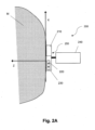
- Fig. 2A shows a schematic drawing of a medical imaging system 200 according to examples of the present disclosure.
- the system 200 shown on Fig. 2A is adapted for adjusting a medical imaging parameter of a medium M.
- the medium M may be for instance living tissues and/or in particular human tissues of a person.
- the system 200 may be any medical imaging or sensor device.
- the system 200 may be for instance a device for tomography, X-ray, mammography or magnetic resonance imaging (MRI).
- the medical imaging parameter may comprise an imaging parameter related to the system 200.
- the system 200 may for example be or comprise a device for ultrasound imaging.
- the system 200 may include for instance:
- the processing unit 240 may send data to an external device, such as for example, a server, a computer on which an artificial intelligence (AI) algorithm may be running, a dedicated workstation, presenting data, a device for displaying images obtained from the processing unit or any of the other external devices.
- the processing unit 240 may be configured to obtain an optical characteristic of the medium and adjust the medical imaging parameter as a function of the optical characteristic. Accordingly, the method according to the present disclosure, in particular a method of adjusting a medical imaging parameter of a medium, may be carried out by the processing unit 240 or any external device.
- the process for constructing the image data may be carried out by the same processing unit 240 as that one for optimizing the optimizing a process, or (at least in part) by another one or several others.
- the system 200 may include at least one processor and memory.
- the processor and memory unit may be incorporated into the system 200 or may be a computer or computer communicatively linked thereto.
- memory storing, instructions to evaluate imaging data or otherwise perform the methods described herein
- RAM volatile
- non-volatile such as RAM, flash memory, etc.
- the system 200 may also include storage devices (removable and/or non-removable) including, but not limited to, magnetic or optical disks or tape.
- the system 200 may also have input device(s) such as keyboard, mouse, pen, voice input, etc.
- connections such as LAN, WAN, point to point, etc.
- the connections may be operable to facility point-to-point communications, connection-oriented communications, connectionless communications, etc.
- the system 200 may typically include at least some form of computer readable media.
- Computer readable media may be any available media that may be accessed by processing unit (or processor) or other devices comprising the operating environment.
- Computer readable media may comprise computer storage media and communication media.
- Computer storage media includes volatile and nonvolatile, removable and non-removable media implemented in any method or technology for storage of information such as computer readable instructions, data structures, program modules or other data.
- Computer storage media does not include communication media.
- Communication media embodies computer readable instructions, data structures, program modules, or other data in a modulated data signal such as a carrier wave or other transport mechanism and includes any information delivery media.
- modulated data signal means a signal that has one or more of its characteristics set or changed in such a manner as to encode information in the signal.
- communication media includes wired media such as a wired network or direct-wired connection, and wireless media such as acoustic, RF, infrared, microwave, and other wireless media. Combinations of the any of the above should also be included within the scope of computer readable media.
- the processing unit 240 may be a single computer operating in a networked environment using logical connections to one or more remote computers.
- the remote computer may be a personal computer, a server, a router, a network PC, a peer device or other common network node, and typically includes many or all of the elements described above as well as others not so mentioned.
- the logical connections may include any method supported by available communications media. Such networking environments are commonplace in offices, enterprise-wide computer networks, intranets and the Internet.
- the optical element 210 may comprise borescopes, fiberscopes, fiber optic faceplates, lenses, prisms, mirrors, light-emitting diodes, photodetectors, optical fibers, fiber optic cables, and/or fiber optic bundles.
- the optical element 210 may be configured to generate and/or record and/or receive signals.
- the optical element 210 and/or the processing unit 240 may be configured to obtain an optical characteristic of the medium M.
- the optical element 210 may for example comprise a laser printed circuit board (PCB) and edge emitting diodes (not shown in fig. 2A ).
- the edge emitting diodes may emit a laser beam.
- the optical element may comprise an optical window, a prism, a detector PCB, and a detector (not shown in fig. 2A ).
- the detector may detect any light, in particular reflections of the laser beam, which was emitted by the edge emitting diodes.
- the transducer element 220 may comprise piezo-crystals and/or other components that may be configured to generate and/or record and/or receive signals.
- the terms transducer and transducer elements may be used synonymously throughout this disclosure unless denoted differently.
- the system 200 may use ultrasound waves, the transducer elements being then adapted to ultrasound waves.
- the method optionally may produce ultrasound images of the medium M and/or may send data to a dedicated server or working station.
- the system 200 may be configured to acquire more than one type of medical data, for example not only ultrasound data, but also mammography data (not shown in fig. 2a ). In other words, the system may also be a multimodal system.
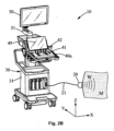
- Fig. 2B shows a schematic drawing of an ultrasound imaging system 10 according to examples of the present disclosure.
- the ultrasound imaging system 10 may be an example of a medical imaging system 200, as described in context of. fig. 1A.
- the ultrasound imaging system 10 may comprise:
- the probe 20 may be connected to the processing unit 30 via a cable 21 or via a wireless connection, and it is able to emit ultrasound waves W into a medium M and to receive ultrasound waves W from the medium M, said received ultrasound waves being consequent or resulting from reflections of said emitted ultrasound waves on diffusing particles inside said medium.
- the probe 20 may be a transducer array comprising a plurality of transducers, each one converting an electric signal into a vibration and reciprocally.
- a transducer is for example a piezoelectric element.
- the transducer array may comprise hundred transducers or more.
- the transducer array is a linear or curved and is disposed on an outer surface of the medium M so as to be coupled to the medium and to vibrate and to emit or receive ultrasound waves W.
- the probe 20 may further comprise an optical element configured to obtain an optical characteristic of the medium.
- the optical characteristic is obtained by another device comprised by, connectable to, or associated with the system 10.
- the system may comprise or be connectable to a distinctive optical sensor, which may for example be attached to the probe.
- the optical sensor may be connected to the system via a cable or via a wireless connection.
- the optical characteristic may also be obtained by any device which is external and/or remote to the system 10, as also described in context of fig. 2A .
- the processing unit 30 may comprise receiving devices for amplifying and/or filtering signals received from the probe 20, and converters (analog to digital converters and digital to analog converters) for transforming the signals into data representing the signal.
- the data may be stored into a memory in the processing unit or directly processed for calculating intermediate processed data (beamformed data).
- the processing unit 30 may use any known processing method to process the image on the bases of the signals received from the probe, such as beamforming.
- the processed ultrasound image data may be:
- the display screen 50 may be a screen for visualizing the image processed by the processing unit 30.
- the display 50 may also visualize other information such as scales used in the image, or configuration information for the processing or any information such as help information or contextual gesture help for the touch pad 42.
- the display screen may by articulated on a support arm 51 for better positioning for the user.
- the display screen is usually a screen of a great size (at least 20 inches) for better image visualization to the user.
- the control panel 40a is for example a portion of the system casing 31, said portion comprising a panel casing having a substantially flat surface inclined towards the user for manipulation by one hand of said user.
- the control panel 40a may be moved by a hanger upwards and downward for being adapted to the user size, and may be optionally moved frontward and rearward for being adapted to the user position.
- the control panel 40a may include a control panel display screen 49 for visualizing several configuration information. This control panel display screen 49 can also be hinged to the control panel casing 48 for being inclined into a different inclination than the control panel surface.
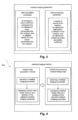
- Fig. 3 gives an overview of the structure of the medical imaging parameter according to examples of the present disclosure.
- the medical imaging parameter comprises a data acquisition parameter and a data processing parameter.
- the data acquisition parameter may define a medical data acquisition method.
- the data acquisition parameter may comprise for example an ultrasound wave coefficient characterizing an ultrasound wave transmitted into the medium, and/or a radiation doses of X-ray waves transmitted into the medium. Accordingly, the data acquisition parameter may influence the way how medical data are obtained from the medium, for example, how the medium is insonified by imaging waves.
- the data processing parameter may define a medical data processing method.
- the data processing parameter may comprise for example at least one of: a speed of sound of the medium, an ultrasound attenuation coefficient of the medium, and an ultrasound scattering and/or absorption coefficient.
- the medical imaging parameter may comprise at least one of: an ultrasound imaging parameter, a tomography imaging parameter, a three-dimensional (3D) imaging parameter, a slicing coefficient in a 3D imaging method, an X-ray imaging parameter, a mammography imaging parameter, and magnetic resonance imaging (MRI).
- an ultrasound imaging parameter a tomography imaging parameter
- a three-dimensional (3D) imaging parameter a slicing coefficient in a 3D imaging method
- an X-ray imaging parameter a mammography imaging parameter
- MRI magnetic resonance imaging
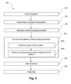
- Fig. 4 shows a flowchart of a medical imaging method 400 according to examples of the present disclosure.
- the medical imaging method may comprise a medical data acquisition method and a method of processing medical image data.
- the medical data acquisition method may comprise adjusting a medical imaging parameter and obtaining medical data of the medium as a function of the adjusted medical imaging parameter.
- the method of processing medical image data may comprise adjusting a medical imaging parameter and processing medical image data as a function of the adjusted medical imaging parameter.
- the medical data acquisition method may precede the method of processing medical image data.
- the acquired medical data i.e. the output of the medical data acquisition method
- Examples of medical imaging methods are shown in more detail in figures 5 and 6 .
- Fig. 5 shows a flowchart of a medical data acquisition method 500 according to examples of the present disclosure.
- an optical acquisition may be conducted, for example using an optical element as described above.
- the optical characteristic may however also be obtained from a data storage or a data interface, i.e. may be acquired by any kind of optical element beforehand and/or remotely.
- a second operation 502 may comprise determining a biological characteristic. In other words, based on the optical characteristic, a biological characteristic, for example the endogenous content of the medium may be determined and thus for example an ultrasound wave absorption characteristic of the medium.
- a third operation 503 comprises the adjusting of a medical imaging parameter, for example using a processing unit.
- an ultrasound scan with adjusted medical parameter is conducted.
- the ultrasound scan may comprise transmitting a pulse 504a into the medium.
- the ultrasound scan may comprise receiving a plurality of signals 504b from the medium.
- the ultrasound scan may also be in the form of any specific ultrasound imaging mode, like for example a B-mode (brightness mode), a doppler mode, or ShearWave TM Elastography (SWE) Smode (for example as described in EP 1546757 B1 of the same applicant).
- the ultrasound scan may also be carried out by any external device, for example remotely to the processing unit.
- the medical imaging parameter may be an ultrasound stimulation wave coefficient of so-called "push pulses" in a SWE mode, as described above.
- a shorter push pulse may be emitted.
- a longer push may be used.
- the acquired ultrasound data may be processed, for example in order to construct image data.
- image data is obtained, i.e. as an output of the operation 605.
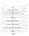
- Fig. 6 shows a flowchart of a method of processing medical image data 600 according to examples of the present disclosure.
- the method of fig. 6 may principally correspond to the method of fig. 5 .
- the data processing operation may be adapted based on the optical characteristic (i.e. in addition or as an alternative to an adaption of the ultrasound scan operation).
- the order of the operations may differ from that one in the method of fig. 5 (as shown in fig. 6 ) or may be similar to the order of the operations of method of fig. 5
- a ultrasound scan may be conducted.
- the ultrasound scan may comprise transmitting a pulse into the medium 601a and receiving in response the plurality of signals from the medium 601b.
- operation 601 is merely optional, as the data may also be obtained from any external device, for example from a data storage device.
- a second operation 602 may comprise obtaining an optical characteristic.
- operation 602 is merely optional, as the optical characteristic may also be obtained from any external device, for example from a data storage device.
- determining of a biological characteristic may be conducted.
- a fourth operation 604 may comprise adjusting a data processing parameter.
- a fifth operation 605 may comprise data processing with an adjusted data processing parameter.
- image data may be obtained, i.e. as an output of the operation 605.
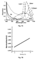
- Fig. 7A shows the absorption spectra of different biological materials.
- Biological material like lipid typically has a maximal absorption at a wavelength of about 920 nm.
- Water at a temperature of 37 °C (usual temperature of an alive human body) has typically a maximal absorption at a wavelength of about 980nm.
- the biological characteristics, i.e. the endogenous content of the medium may thus be derived from a measured spectrum of the medium.
- a high absorption of light at a wavelength of 920 nm may indicate a high content of lipids in the considered medium.
- This approach is especially effective in the field of breast examinations.
- a less dense breast usually comprises relatively high amount of lipid in relation to water, while a dense breast comprises a relatively water-rich tissue in relation to lipid what can precisely be determined based on the spectrum.
- the density of a medium may be determined based on optical characteristics, i.e. absorption spectrum at one or several wavelengths (cf. operations 602 and 603 in fig. 6 ).
- Fig. 7B shows the speed of sound for different densities.
- a correlation between speed of sound and the density of a medium may be detected. For example, it may be that a high speed of sound indicates a high density of the medium.
- the speed of sound may be determined based on the density (cf. operation 604 in fig. 6 ). For example, if the density of medium was determined based on the absorption spectrum, the speed of sound in the medium may be determined using Fig. 7B .
- the adjusted speed of sound may then be used in a medical data processing method, i.e. in a respective beamforming method to construct more precise ultrasound image data (cf. operation 605 in fig. 6 ).
Landscapes
- Health & Medical Sciences (AREA)
- Life Sciences & Earth Sciences (AREA)
- Engineering & Computer Science (AREA)
- Medical Informatics (AREA)
- Nuclear Medicine, Radiotherapy & Molecular Imaging (AREA)
- General Health & Medical Sciences (AREA)
- Biomedical Technology (AREA)
- Public Health (AREA)
- Physics & Mathematics (AREA)
- Radiology & Medical Imaging (AREA)
- Veterinary Medicine (AREA)
- Surgery (AREA)
- Molecular Biology (AREA)
- Heart & Thoracic Surgery (AREA)
- Pathology (AREA)
- Biophysics (AREA)
- Animal Behavior & Ethology (AREA)
- High Energy & Nuclear Physics (AREA)
- Epidemiology (AREA)
- Primary Health Care (AREA)
- Optics & Photonics (AREA)
- Business, Economics & Management (AREA)
- General Business, Economics & Management (AREA)
- Dentistry (AREA)
- Oral & Maxillofacial Surgery (AREA)
- Computer Vision & Pattern Recognition (AREA)
- Pulmonology (AREA)
- Theoretical Computer Science (AREA)
- Ultra Sonic Daignosis Equipment (AREA)
- Measuring And Recording Apparatus For Diagnosis (AREA)
Priority Applications (4)
| Application Number | Priority Date | Filing Date | Title |
|---|---|---|---|
| EP22315223.2A EP4344646A1 (fr) | 2022-09-30 | 2022-09-30 | Procédé et système de réglage d'un paramètre d'imagerie médicale d'un milieu |
| CN202311228735.5A CN117796841A (zh) | 2022-09-30 | 2023-09-22 | 调节介质的医学成像参数的方法和系统 |
| KR1020230127826A KR20240046041A (ko) | 2022-09-30 | 2023-09-25 | 매질의 의료 이미징 파라미터를 조정하는 방법 및 시스템 |
| US18/475,834 US20240108318A1 (en) | 2022-09-30 | 2023-09-27 | Method and system for adjusting a medical imaging parameter of a medium |
Applications Claiming Priority (1)
| Application Number | Priority Date | Filing Date | Title |
|---|---|---|---|
| EP22315223.2A EP4344646A1 (fr) | 2022-09-30 | 2022-09-30 | Procédé et système de réglage d'un paramètre d'imagerie médicale d'un milieu |
Publications (1)
| Publication Number | Publication Date |
|---|---|
| EP4344646A1 true EP4344646A1 (fr) | 2024-04-03 |
Family
ID=83899827
Family Applications (1)
| Application Number | Title | Priority Date | Filing Date |
|---|---|---|---|
| EP22315223.2A Pending EP4344646A1 (fr) | 2022-09-30 | 2022-09-30 | Procédé et système de réglage d'un paramètre d'imagerie médicale d'un milieu |
Country Status (4)
| Country | Link |
|---|---|
| US (1) | US20240108318A1 (fr) |
| EP (1) | EP4344646A1 (fr) |
| KR (1) | KR20240046041A (fr) |
| CN (1) | CN117796841A (fr) |
Citations (5)
| Publication number | Priority date | Publication date | Assignee | Title |
|---|---|---|---|---|
| EP1546757B1 (fr) | 2002-09-02 | 2008-11-26 | Centre National De La Recherche Scientifique (Cnrs) | Procede et dispositif d'imagerie utilisant des ondes de cisaillement |
| US20100087733A1 (en) * | 2008-10-07 | 2010-04-08 | Canon Kabushiki Kaisha | Biological information processing apparatus and biological information processing method |
| US20130204134A1 (en) * | 2010-10-14 | 2013-08-08 | Koninklijke Philips Electronics N.V. | Property determination apparatus for determining a property of an object |
| US20160192840A1 (en) * | 2013-08-01 | 2016-07-07 | Sogang University Research Foundation | Device and method for acquiring fusion image |
| WO2020178522A2 (fr) | 2019-03-05 | 2020-09-10 | Micro Photon Devices Srl | Sonde bimodale à ultrasons comportant un dispositif optique pour un diagnostic |
Family Cites Families (2)
| Publication number | Priority date | Publication date | Assignee | Title |
|---|---|---|---|---|
| US20140005544A1 (en) * | 2011-11-02 | 2014-01-02 | Seno Medical Instruments, Inc. | System and method for providing selective channel sensitivity in an optoacoustic imaging system |
| US10309936B2 (en) * | 2013-10-11 | 2019-06-04 | Seno Medical Instruments, Inc. | Systems and methods for component separation in medical imaging |
-
2022
- 2022-09-30 EP EP22315223.2A patent/EP4344646A1/fr active Pending
-
2023
- 2023-09-22 CN CN202311228735.5A patent/CN117796841A/zh active Pending
- 2023-09-25 KR KR1020230127826A patent/KR20240046041A/ko active Pending
- 2023-09-27 US US18/475,834 patent/US20240108318A1/en active Pending
Patent Citations (5)
| Publication number | Priority date | Publication date | Assignee | Title |
|---|---|---|---|---|
| EP1546757B1 (fr) | 2002-09-02 | 2008-11-26 | Centre National De La Recherche Scientifique (Cnrs) | Procede et dispositif d'imagerie utilisant des ondes de cisaillement |
| US20100087733A1 (en) * | 2008-10-07 | 2010-04-08 | Canon Kabushiki Kaisha | Biological information processing apparatus and biological information processing method |
| US20130204134A1 (en) * | 2010-10-14 | 2013-08-08 | Koninklijke Philips Electronics N.V. | Property determination apparatus for determining a property of an object |
| US20160192840A1 (en) * | 2013-08-01 | 2016-07-07 | Sogang University Research Foundation | Device and method for acquiring fusion image |
| WO2020178522A2 (fr) | 2019-03-05 | 2020-09-10 | Micro Photon Devices Srl | Sonde bimodale à ultrasons comportant un dispositif optique pour un diagnostic |
Non-Patent Citations (1)
| Title |
|---|
| S. CATHELINE: "Measurement of viscoelastic properties of homogeneous soft solid using transient elastography: An inverse problem approach", THE JOURNAL OF THE ACOUSTICAL SOCIETY OF AMERICA, vol. 116, 2005, pages 3734 - 41, XP012072696, DOI: 10.1121/1.1815075 |
Also Published As
| Publication number | Publication date |
|---|---|
| US20240108318A1 (en) | 2024-04-04 |
| CN117796841A (zh) | 2024-04-02 |
| KR20240046041A (ko) | 2024-04-08 |
Similar Documents
| Publication | Publication Date | Title |
|---|---|---|
| US10709419B2 (en) | Dual modality imaging system for coregistered functional and anatomical mapping | |
| US9757092B2 (en) | Method for dual modality optoacoustic imaging | |
| US10433732B2 (en) | Optoacoustic imaging system having handheld probe utilizing optically reflective material | |
| AU2012332233B2 (en) | Dual modality imaging system for coregistered functional and anatomical mapping | |
| CN100353910C (zh) | 非探入式受检体信息成象装置 | |
| US9360551B2 (en) | Object information acquiring apparatus and control method thereof | |
| US20160058426A1 (en) | Methods and systems for automatic control of subjective image quality in imaging of objects | |
| CN112469338B (zh) | 一种基于超声检测肝的装置及超声设备及超声成像方法 | |
| JP2005253827A (ja) | 超音波撮像方法及び装置 | |
| CN106137125A (zh) | 被检体信息获取设备 | |
| US5546945A (en) | Method and apparatus for displaying acoustic signal transit times | |
| US20210022711A1 (en) | Ultrasound elastography method and system | |
| US8282551B2 (en) | Ultrasonic diagnostic apparatus, data analysis method, and data analysis program | |
| JP7812140B2 (ja) | 全波形反転を使用した反射超音波断層撮影画像化 | |
| KR20140137037A (ko) | 초음파 영상 처리 장치 및 방법 | |
| EP3381373B1 (fr) | Appareil de diagnostic ultrasonique et son procédé de commande | |
| JP2017140092A (ja) | 被検体情報取得装置 | |
| EP4344646A1 (fr) | Procédé et système de réglage d'un paramètre d'imagerie médicale d'un milieu | |
| CN117517467A (zh) | 用于估计介质中超声衰减目的的超声数据评估方法和系统 | |
| JP3522488B2 (ja) | 超音波診断装置 | |
| CN113491535B (zh) | 超声波诊断装置、信号处理装置以及记录介质 | |
| CN118119341A (zh) | 用于前列腺成像的光声探针 | |
| JP2017164222A (ja) | 処理装置および処理方法 | |
| US20240219543A1 (en) | Beamforming method and system | |
| EP2773267B1 (fr) | Système d'imagerie bimodal pour cartographie fonctionnelle et cartographie anatomique co-enregistrées |
Legal Events
| Date | Code | Title | Description |
|---|---|---|---|
| PUAI | Public reference made under article 153(3) epc to a published international application that has entered the european phase |
Free format text: ORIGINAL CODE: 0009012 |
|
| STAA | Information on the status of an ep patent application or granted ep patent |
Free format text: STATUS: THE APPLICATION HAS BEEN PUBLISHED |
|
| AK | Designated contracting states |
Kind code of ref document: A1 Designated state(s): AL AT BE BG CH CY CZ DE DK EE ES FI FR GB GR HR HU IE IS IT LI LT LU LV MC MK MT NL NO PL PT RO RS SE SI SK SM TR |
|
| STAA | Information on the status of an ep patent application or granted ep patent |
Free format text: STATUS: REQUEST FOR EXAMINATION WAS MADE |
|
| 17P | Request for examination filed |
Effective date: 20241002 |
|
| RBV | Designated contracting states (corrected) |
Designated state(s): AL AT BE BG CH CY CZ DE DK EE ES FI FR GB GR HR HU IE IS IT LI LT LU LV MC MK MT NL NO PL PT RO RS SE SI SK SM TR |
|
| GRAJ | Information related to disapproval of communication of intention to grant by the applicant or resumption of examination proceedings by the epo deleted |
Free format text: ORIGINAL CODE: EPIDOSDIGR1 |
|
| GRAP | Despatch of communication of intention to grant a patent |
Free format text: ORIGINAL CODE: EPIDOSNIGR1 |
|
| GRAP | Despatch of communication of intention to grant a patent |
Free format text: ORIGINAL CODE: EPIDOSNIGR1 |
|
| STAA | Information on the status of an ep patent application or granted ep patent |
Free format text: STATUS: GRANT OF PATENT IS INTENDED |
|
| RAP3 | Party data changed (applicant data changed or rights of an application transferred) |
Owner name: SUPERSONIC IMAGINE |
|
| INTG | Intention to grant announced |
Effective date: 20251125 |
|
| GRAC | Information related to communication of intention to grant a patent modified |
Free format text: ORIGINAL CODE: EPIDOSCIGR1 |
|
| PUAJ | Public notification under rule 129 epc |
Free format text: ORIGINAL CODE: 0009425 |
|
| INTG | Intention to grant announced |
Effective date: 20260127 |
|
| GRAC | Information related to communication of intention to grant a patent modified |
Free format text: ORIGINAL CODE: EPIDOSCIGR1 |
|
| 32PN | Public notification |
Free format text: COMMUNICATION PURSUANT TO RULE 71(3) EPC (EPO FORM 2004 DATED 27/01/2026) |
|
| INTG | Intention to grant announced |
Effective date: 20260318 |