EP4349976A1 - Verfahren zur bewertung einer antikörperabhängigen verstärkungsreaktion mit einem pseudovirus - Google Patents
Verfahren zur bewertung einer antikörperabhängigen verstärkungsreaktion mit einem pseudovirus Download PDFInfo
- Publication number
- EP4349976A1 EP4349976A1 EP22811031.8A EP22811031A EP4349976A1 EP 4349976 A1 EP4349976 A1 EP 4349976A1 EP 22811031 A EP22811031 A EP 22811031A EP 4349976 A1 EP4349976 A1 EP 4349976A1
- Authority
- EP
- European Patent Office
- Prior art keywords
- antibody
- cells
- virus
- srips
- protein
- Prior art date
- Legal status (The legal status is an assumption and is not a legal conclusion. Google has not performed a legal analysis and makes no representation as to the accuracy of the status listed.)
- Pending
Links
Images


Classifications
-
- G—PHYSICS
- G01—MEASURING; TESTING
- G01N—INVESTIGATING OR ANALYSING MATERIALS BY DETERMINING THEIR CHEMICAL OR PHYSICAL PROPERTIES
- G01N33/00—Investigating or analysing materials by specific methods not covered by groups G01N1/00 - G01N31/00
- G01N33/48—Biological material, e.g. blood, urine; Haemocytometers
- G01N33/50—Chemical analysis of biological material, e.g. blood, urine; Testing involving biospecific ligand binding methods; Immunological testing
- G01N33/53—Immunoassay; Biospecific binding assay; Materials therefor
- G01N33/569—Immunoassay; Biospecific binding assay; Materials therefor for microorganisms, e.g. protozoa, bacteria, viruses
- G01N33/56983—Viruses
-
- C—CHEMISTRY; METALLURGY
- C07—ORGANIC CHEMISTRY
- C07K—PEPTIDES
- C07K16/00—Immunoglobulins [IG], e.g. monoclonal or polyclonal antibodies
- C07K16/08—Immunoglobulins [IG], e.g. monoclonal or polyclonal antibodies against material from viruses
- C07K16/10—RNA viruses
- C07K16/116—Togaviridae (F); Matonaviridae (F); Flaviviridae (F)
-
- C—CHEMISTRY; METALLURGY
- C12—BIOCHEMISTRY; BEER; SPIRITS; WINE; VINEGAR; MICROBIOLOGY; ENZYMOLOGY; MUTATION OR GENETIC ENGINEERING
- C12N—MICROORGANISMS OR ENZYMES; COMPOSITIONS THEREOF; PROPAGATING, PRESERVING, OR MAINTAINING MICROORGANISMS; MUTATION OR GENETIC ENGINEERING; CULTURE MEDIA
- C12N7/00—Viruses; Bacteriophages; Compositions thereof; Preparation or purification thereof
-
- G—PHYSICS
- G01—MEASURING; TESTING
- G01N—INVESTIGATING OR ANALYSING MATERIALS BY DETERMINING THEIR CHEMICAL OR PHYSICAL PROPERTIES
- G01N33/00—Investigating or analysing materials by specific methods not covered by groups G01N1/00 - G01N31/00
- G01N33/48—Biological material, e.g. blood, urine; Haemocytometers
- G01N33/50—Chemical analysis of biological material, e.g. blood, urine; Testing involving biospecific ligand binding methods; Immunological testing
- G01N33/68—Chemical analysis of biological material, e.g. blood, urine; Testing involving biospecific ligand binding methods; Immunological testing involving proteins, peptides or amino acids
- G01N33/6854—Immunoglobulins
-
- C—CHEMISTRY; METALLURGY
- C07—ORGANIC CHEMISTRY
- C07K—PEPTIDES
- C07K14/00—Peptides having more than 20 amino acids; Gastrins; Somatostatins; Melanotropins; Derivatives thereof
- C07K14/005—Peptides having more than 20 amino acids; Gastrins; Somatostatins; Melanotropins; Derivatives thereof from viruses
-
- C—CHEMISTRY; METALLURGY
- C12—BIOCHEMISTRY; BEER; SPIRITS; WINE; VINEGAR; MICROBIOLOGY; ENZYMOLOGY; MUTATION OR GENETIC ENGINEERING
- C12N—MICROORGANISMS OR ENZYMES; COMPOSITIONS THEREOF; PROPAGATING, PRESERVING, OR MAINTAINING MICROORGANISMS; MUTATION OR GENETIC ENGINEERING; CULTURE MEDIA
- C12N2770/00—MICROORGANISMS OR ENZYMES; COMPOSITIONS THEREOF; PROPAGATING, PRESERVING, OR MAINTAINING MICROORGANISMS; MUTATION OR GENETIC ENGINEERING; CULTURE MEDIA ssRNA viruses positive-sense
- C12N2770/00011—Details
- C12N2770/24011—Flaviviridae
- C12N2770/24111—Flavivirus, e.g. yellow fever virus, dengue, JEV
- C12N2770/24122—New viral proteins or individual genes, new structural or functional aspects of known viral proteins or genes
-
- C—CHEMISTRY; METALLURGY
- C12—BIOCHEMISTRY; BEER; SPIRITS; WINE; VINEGAR; MICROBIOLOGY; ENZYMOLOGY; MUTATION OR GENETIC ENGINEERING
- C12N—MICROORGANISMS OR ENZYMES; COMPOSITIONS THEREOF; PROPAGATING, PRESERVING, OR MAINTAINING MICROORGANISMS; MUTATION OR GENETIC ENGINEERING; CULTURE MEDIA
- C12N2770/00—MICROORGANISMS OR ENZYMES; COMPOSITIONS THEREOF; PROPAGATING, PRESERVING, OR MAINTAINING MICROORGANISMS; MUTATION OR GENETIC ENGINEERING; CULTURE MEDIA ssRNA viruses positive-sense
- C12N2770/00011—Details
- C12N2770/24011—Flaviviridae
- C12N2770/24111—Flavivirus, e.g. yellow fever virus, dengue, JEV
- C12N2770/24123—Virus like particles [VLP]
-
- C—CHEMISTRY; METALLURGY
- C12—BIOCHEMISTRY; BEER; SPIRITS; WINE; VINEGAR; MICROBIOLOGY; ENZYMOLOGY; MUTATION OR GENETIC ENGINEERING
- C12N—MICROORGANISMS OR ENZYMES; COMPOSITIONS THEREOF; PROPAGATING, PRESERVING, OR MAINTAINING MICROORGANISMS; MUTATION OR GENETIC ENGINEERING; CULTURE MEDIA
- C12N2770/00—MICROORGANISMS OR ENZYMES; COMPOSITIONS THEREOF; PROPAGATING, PRESERVING, OR MAINTAINING MICROORGANISMS; MUTATION OR GENETIC ENGINEERING; CULTURE MEDIA ssRNA viruses positive-sense
- C12N2770/00011—Details
- C12N2770/24011—Flaviviridae
- C12N2770/24111—Flavivirus, e.g. yellow fever virus, dengue, JEV
- C12N2770/24151—Methods of production or purification of viral material
- C12N2770/24152—Methods of production or purification of viral material relating to complementing cells and packaging systems for producing virus or viral particles
-
- C—CHEMISTRY; METALLURGY
- C12—BIOCHEMISTRY; BEER; SPIRITS; WINE; VINEGAR; MICROBIOLOGY; ENZYMOLOGY; MUTATION OR GENETIC ENGINEERING
- C12Q—MEASURING OR TESTING PROCESSES INVOLVING ENZYMES, NUCLEIC ACIDS OR MICROORGANISMS; COMPOSITIONS OR TEST PAPERS THEREFOR; PROCESSES OF PREPARING SUCH COMPOSITIONS; CONDITION-RESPONSIVE CONTROL IN MICROBIOLOGICAL OR ENZYMOLOGICAL PROCESSES
- C12Q1/00—Measuring or testing processes involving enzymes, nucleic acids or microorganisms; Compositions therefor; Processes of preparing such compositions
- C12Q1/66—Measuring or testing processes involving enzymes, nucleic acids or microorganisms; Compositions therefor; Processes of preparing such compositions involving luciferase
-
- G—PHYSICS
- G01—MEASURING; TESTING
- G01N—INVESTIGATING OR ANALYSING MATERIALS BY DETERMINING THEIR CHEMICAL OR PHYSICAL PROPERTIES
- G01N2333/00—Assays involving biological materials from specific organisms or of a specific nature
- G01N2333/005—Assays involving biological materials from specific organisms or of a specific nature from viruses
- G01N2333/08—RNA viruses
- G01N2333/18—Togaviridae; Flaviviridae
- G01N2333/183—Flaviviridae, e.g. pestivirus, mucosal disease virus, bovine viral diarrhoea virus, classical swine fever virus (hog cholera virus) or border disease virus
- G01N2333/185—Flaviviruses or Group B arboviruses, e.g. yellow fever virus, japanese encephalitis, tick-borne encephalitis, dengue
-
- Y—GENERAL TAGGING OF NEW TECHNOLOGICAL DEVELOPMENTS; GENERAL TAGGING OF CROSS-SECTIONAL TECHNOLOGIES SPANNING OVER SEVERAL SECTIONS OF THE IPC; TECHNICAL SUBJECTS COVERED BY FORMER USPC CROSS-REFERENCE ART COLLECTIONS [XRACs] AND DIGESTS
- Y02—TECHNOLOGIES OR APPLICATIONS FOR MITIGATION OR ADAPTATION AGAINST CLIMATE CHANGE
- Y02A—TECHNOLOGIES FOR ADAPTATION TO CLIMATE CHANGE
- Y02A50/00—TECHNOLOGIES FOR ADAPTATION TO CLIMATE CHANGE in human health protection, e.g. against extreme weather
- Y02A50/30—Against vector-borne diseases, e.g. mosquito-borne, fly-borne, tick-borne or waterborne diseases whose impact is exacerbated by climate change
Definitions
- the present invention relates to a method for functional evaluation of antibody, which uses pseudovirus.
- Dengue virus which belongs to the genus Flavivirus of the Flaviviridae family, is a +strand RNA virus, and its genome consists of three structural protein regions (C, prM, E) and seven non-structural protein regions (NS1, NS2a, NS2b, NS3, NS4a, NS4b, NS5).
- Dengue virus includes four types of viruses (DENV-1 to DENV-4) and they are biologically classified as serotypes.
- Dengue virus is transmitted by mosquitoes. When a dengue virus-carrying mosquito bites a human, the injected virus first infects dendritic cells, monocytes, and lymphocytes in the skin and undergoes primary proliferation.
- the virus then travels through the bloodstream to various organs in the body and undergoes secondary proliferation.
- the condition in which the proliferated virus is present in the blood is called viremia, and when a mosquito sucks such blood, it becomes a new virus-carrying mosquito and can transmit the virus to other humans.
- Aedes albopictus which lives in temperate regions including Japan, also transmits dengue virus.
- the Aedes albopictus was the vector animal.
- the population at risk of being infected with dengue virus is estimated to be 3.9 billion people, and 390 million people, which is one-tenth the number, are infected with some kind of dengue virus every year. Even when humans are infected with the dengue virus, many of the infections are inapparent infections, and only about 100 million of them actually develop symptoms.
- dengue fever a transient febrile disease.
- the main symptoms of dengue fever are relatively mild, including fever, headache, muscular pain, and joint pain. Most cases of dengue fever are mild, but a part of patients with dengue fever develop a severe form, dengue hemorrhagic fever.
- the symptoms of dengue hemorrhagic fever are severe and can lead to death if proper treatment is not provided. It has been epidemiologically proven that first infection often causes dengue fever and the second infection with a dengue virus of a serotype different from the first infection increases the risk of developing dengue hemorrhagic fever. It is said that a third or subsequent infection does not occur.
- ADE Antibody-dependent enhancement
- ADE test Simple rapid kits (immunochromatography, ELISA method) are commercially available for antibody test in infectious diseases. They are all qualitative and quantitative chemical test methods and do not reveal biological functional activities of antibody.
- the outline of the ADE test is to examine how much infection of cells can be increased by adding antibodies in the presence of the minimum necessary amount of virus.
- a method for evaluating ADE using fixed cells has been reported (Non Patent Literature 1).
- Existing ADE tests can examine biological functions of antibodies, but require live dengue virus as the assay antigen. Dengue virus is classified as a Category IV infectious disease under the Infectious Disease Control Act. Therefore, biosafety level 2 (BSL2) equipment is required when handling Dengue virus.
- BSL2 biosafety level 2
- SRIPs Single Round Infectious Particles
- SRIPs that retain the outer shell of a specific virus can be produced by using the outer shell gene of the virus when producing SRIPs (Non Patent Literatures 1 to 4).
- SRIP contains in its inside a self-replicating gene called replicon. Although replicon genes are replicated within SRIPs-infected cells, they cannot be amplified as particles (viruses) and can be safely handled because they do not cause cell destruction or reinfection. By encoding nanoluciferase as a reporter gene, infected cells can be measured based on luciferase activity.
- SRIPs bind to antibodies as pseudoviruses, they can be used as antigens for tests of antibody functions such as antibody neutralization activity and ADE activity.
- ADE activity measurement using SRIPs and the luciferase activity produced thereby has also been reported (Non Patent Literature 4), but this method also required three days for measurement.
- Non Patent Literatures 5 to 6 A neutralization test using SRIPs as substitute antigens and detecting the luciferase activity produced by SRIPs that infected cells has been reported (Non Patent Literatures 5 to 6). However, it requires 3 days of culture since it takes time for the replicon to increase within the cells after the reaction with the neutralizing antibody.
- the present inventors have conducted intensive studies of Single Round Infectious Particles (SRIPs) suitable as assay antigens for neutralization tests and ADE tests. As a result, they have found that the use of yellow fever virus (YFV) gene as the backbone of the replicon plasmid results in rapid uptake of SRIPs into cells and accelerated replication thereof, thereby shortening the time up to the detection, and successfully developed a rapid diagnostic method for antibody function.
- SRIPs Single Round Infectious Particles
- the present invention relates to the following.
- antibody function can be determined in a shorter period of time (1 to 2 days) than conventional (3 days), whereby rapid antibody tests can be performed. This allows for rapid determination of the possibility that test subjects develop ADE due to reinfection, as well as its use for property evaluation of antibodies (neutralization activity evaluation) in the development of pharmaceutical products and evaluation of induction potency of ADE active antibodies of vaccine.
- the present invention relates to single round infectious virus particles (SRIPs) containing a gene having a region encoding a labeled protein and a region encoding non-structural (NS) proteins 1 to 5 of the yellow fever virus genome, a capsid protein of a virus, and an outer shell protein (Envelope) of an evaluation target virus.
- SRIPs single round infectious virus particles
- SRIPs in the present specification have a region encoding a labeled protein and a region encoding non-structural (NS) proteins 1 to 5 of the yellow fever virus genome in the gene thereof.
- the "labeled protein” is not particularly limited as long as it is a protein capable of labeling cells infected with the aforementioned SRIPs. Preferably, it can be expressed only in cells infected with the aforementioned SRIPs.
- the labeled protein is preferably a fluorescent protein or a luminescent protein.
- green fluorescent protein GFP
- red fluorescent protein RFP
- blue fluorescent protein BFP
- cyan fluorescent protein CFP
- yellow fluorescent protein YFP
- fluorescent proteins improved from these fluorescent proteins (see https://www.fpbase.org/), luciferase, and nanoluciferase can be mentioned.
- Yellow fever virus is a virus that belongs to the genus Flavivirus of the Flaviviridae family, and its genome is a single-stranded plus RNA.
- the "gene” may be DNA or RNA.
- a nucleocapsid in which a capsid protein (also called core protein) is bound to a single-stranded RNA is surrounded by membrane protein and envelope proteins.
- One reading frame is present on single-stranded RNA, and three types of structural proteins (C, prM, E) and seven types of non-structural proteins (NS1, NS2A, NS2B, NS3, NS4A, NS4B, NS5) are encoded.
- non-structural (NS) proteins 1 to 5 of the yellow fever virus genome means these seven types of non-structural proteins. Proteins with mutations in a part (e.g., 1 to 5 amino acids) of these proteins are also included in the non-structural (NS) proteins 1 to 5 of the yellow fever virus genome as long as they are considered to be substantially the same as these proteins and the effects of the present invention are not affected.
- the gene of SRIPs in the present specification optionally has, where necessary, a region other than a region encoding the aforementioned labeled protein and a region encoding non-structural (NS) proteins 1 to 5 of the yellow fever virus genome.
- NS non-structural
- it does not contain gene regions encoding at least (i) a capsid protein, and (ii) one or both of a membrane protein and an envelope protein. That is, since the virus particles in the present specification have the Envelope of the evaluation target virus, they can infect cells that are infected by the evaluation target virus, whereas they cannot proliferate within the infected cells because the gene thereof does not encode capsid protein, and/or Envelope and membrane protein precursor.
- a region encoding a labeled protein and a region encoding non-structural (NS) proteins 1 to 5 of the yellow fever virus genome may be linked to transcriptional regulatory regions that can be expressed in cells used in infection evaluation tests.
- Such transcription regulatory region optionally contains a promoter.
- the promoter is not particularly limited as long as it has activity in the aforementioned cells.
- CMV cytomegalovirus
- RSV Rous sarcoma virus
- CAG promoter promoter having cytomegalovirus enhancer, chicken ⁇ -actin promoter, and rabbit ⁇ -globin gene splicing acceptor
- ubiquitin promoter ubiquitin promoter
- SV40 Simian virus 40
- EF-1 ⁇ elongation factor 1 ⁇
- the "capsid protein of a virus” that the SRIPs in the present specification has is a protein that binds to the aforementioned RNA gene to form a nucleocapsid.
- the virus from which the capsid protein is derived is not particularly limited as long as it belongs to the Flaviviridae family. Examples thereof include yellow fever virus (YFV), dengue virus (DENV), Japanese encephalitis virus (JEV), West Nile virus (WNV), and tick-borne encephalitis virus (TBEV), and YFV and DENV are preferred.
- the outer shell protein is a protein that constitutes the outer shell of virus, and has the functions of binding to receptors and membrane fusion.
- the evaluation target virus from which the Envelope is derived is not particularly limited as long as it belongs to the aforementioned Flavivirus genus, and may be, for example, DENV.
- DENV includes four kinds of type 1 to type 4, which are called DENV-1, DENV-2, DENV-3, and DENV-4. Throughout the present specification, DENV may be any one of DENV-1 to DENV-4, a plurality of them, or all of them.
- SRIPs in the present specification may further contain a membrane protein in addition to those mentioned above.
- SRIPs in the present specification can be obtained by introducing a replicon vector containing a gene having a region encoding a labeled protein and a region encoding non-structural (NS) proteins 1 to 5 of the yellow fever virus genome, a vector containing a gene encoding a capsid protein, and a vector containing a gene encoding Envelope and a gene encoding a membrane protein precursor (prM) into the same cell ( Fig. 1 ).
- NS non-structural
- the present invention relates to a method for producing SRIPs, including introducing a vector containing a gene having a region encoding non-structural (NS) proteins 1 to 5 of the yellow fever virus genome and a region encoding a labeled protein, a vector containing a gene encoding a capsid protein of a virus, and a vector containing a gene encoding an outer shell protein of an evaluation target virus into animal cells, and collecting SRIPs produced in the introduced cells.
- the vector may be preferably a plasmid vector.
- the animal cells may be human cells or monkey cells and, for example, HEK293T cells can be used. Each viral gene is known in the technical field and can be obtained from various databases.
- the neutralization activity of antibody can be measured in a short time by infecting Fc ⁇ receptor-non-expressing cells with the aforementioned SRIPs in the presence of a test antibody. Therefore, the present invention provides a method for determining neutralization activity of an antibody, including contacting Fc ⁇ receptor-non-expressing cells with the aforementioned SRIPs in the presence of a test antibody, culturing the aforementioned cells for 24 to 72 hr, and measuring a label in a culture medium resulting from the aforementioned culture, wherein when the measured label is less than the label measured in negative control cells similarly contacted with SRIPs in the absence of the antibody or in the presence of a negative control antibody, the test antibody is determined to have neutralization activity on the aforementioned evaluation target virus.
- the Fc ⁇ receptor-non-expressing cell may be any cell that does not express Fc ⁇ receptors, and is preferably an epithelial cell, such as human or primate (monkey, chimpanzee, etc.) cell.
- an epithelial cell such as human or primate (monkey, chimpanzee, etc.) cell.
- a specific example of the Fc ⁇ receptor-non-expressing cell is Vero cell.
- the aforementioned SRIPs may be brought into contact with Fc ⁇ receptor-non-expressing cells in the presence of a test antibody by adding the aforementioned SRIPs to the culture medium of the Fc ⁇ receptor-non-expressing cells after or simultaneously with the addition of the test antibody to the culture medium, or by adding the test antibody and the aforementioned SRIPs mixed in advance to the culture medium of the Fc ⁇ receptor-non-expressing cells.
- a test antibody and SRIPs are mixed in advance, and the mixture is allowed to stand at about 37°C for 1 to 6 hr (preferably, 2 hr) and then contacted with Fc ⁇ receptor-non-expressing cells.
- an antibody composition as well as a purified antibody can be used.
- serum derived from a test subject can be used as is or used after dilution.
- dilution it can be diluted 5120-fold or less, 640-fold or less, 320-fold or less, 160-fold or less, 80-fold or less, 40-fold or less, or 10-fold or less.
- the time of contact of SRIPs in the presence of a test antibody is not less than 24 hr and less than 72 hr, preferably 24 to 48 hr.
- the label can be measured as appropriate according to the kind of the selected label.
- luminescence can be measured using a luminometer.
- Neutralization activity is determined by comparing the measured level of label with the level of label in the negative control.
- a negative control is an Fc ⁇ receptor-non-expressing cell infected with SRIPs under the same conditions as the test for the test antibody except that neutralization activity is not present.
- the negative control may be an Fc ⁇ receptor-non-expressing cell infected with SRIPs in the absence of a test antibody, or an Fc ⁇ receptor-non-expressing cell infected with SRIPs in the presence of an antibody that evidently does not have neutralization activity (e.g., antibodies that do not bind to the evaluation target virus).
- the level of the label of the negative control can also be obtained by performing the above-mentioned neutralization activity evaluation method simultaneously with the test antibody, or may be obtained by performing the above-mentioned neutralization activity evaluation method separately from the test antibody.
- the test antibody can be determined to have the neutralization activity.
- the patient serum is used as the test antibody and the level of the label in the presence of the serum is lower than that of a negative control, the patient can be determined to have an antibody with neutralization activity.
- the "level” means an indicator of quantified abundance and includes, for example, concentration, amount, or an indicator that can be used in place of these. Therefore, the level may be a measured value itself such as fluorescence intensity, or may be a value converted to concentration. In addition, the level may be an absolute numerical value (abundance, abundance per unit area, etc.), or may be a relative numerical value compared to a comparison control set as necessary.
- the antibody-dependent enhancement ability (ADE) of antibody can be measured in a short time by infecting Fc ⁇ receptor-expressing cells with the aforementioned SRIPs in the presence of a test antibody. Therefore, the present invention provides a method for determining antibody-dependent enhancement ability of an antibody, including contacting Fc ⁇ receptor-expressing cells with the aforementioned SRIPs in the presence of a test antibody, culturing the aforementioned cells for 30 min to 24 hr, and measuring a label in the aforementioned culture medium, wherein when the level of the aforementioned measured label is higher than that measured in a negative control cell, the test antibody is determined to have antibody-dependent enhancement ability for the aforementioned evaluation target virus.
- the Fc ⁇ receptor-expressing cell may be any cell that expresses Fc ⁇ receptors, and is preferably a cell of the immune system, such as human or primate (monkey, chimpanzee, etc.) cell.
- a specific example of the Fc ⁇ receptor-expressing cell includes Mylc cell (MiCAN Technologies, Inc., Japan) and K562 cell.
- the aforementioned SRIPs may be brought into contact with Fc ⁇ receptor-expressing cells in the presence of a test antibody by adding the aforementioned SRIPs to the culture medium of the Fc ⁇ receptor-expressing cells after or simultaneously with the addition of the test antibody to the culture medium, or by adding the test antibody and the aforementioned SRIPs mixed in advance to the culture medium of the Fc ⁇ receptor-expressing cells.
- a test antibody an antibody composition as well as a purified antibody can be used.
- the antibody composition for example, serum derived from a test subject can be used as is or used after dilution.
- dilution it can be diluted 10-fold or more, 40-fold or more, 160-fold or more and/or 10240-fold or less, 2560-fold or less, 640-fold or less, or may be any combination of these upper limit values and lower limit values, for example, 10-fold or more and 10240-fold or less, or 40-fold or more and 2560-fold or less.
- SRIPs obtained by the above-mentioned method can be diluted as appropriate and used in this method.
- the dilution rate can be 2-fold or more, and may be, for example, 10-fold or more, 100-fold or more, or 1000-fold or more.
- Cell culture can be performed, for example, on a plate capable of cultivating immune cells, such as a 96-well plate.
- the number of cells to be cultured when using a 96-well plate can be set to not less than 6.9 ⁇ 10 cells/well, not less than 2.1 ⁇ 10 2 cells/well, not less than 6.2 ⁇ 10 2 cells/well, not less than 1.9 ⁇ 10 3 cells/well, not less than 5.6 ⁇ 10 3 cells/well, or not less than 1.7 ⁇ 10 4 cells/well.
- the number of cells to be cultured can be appropriately determined according to the dilution rate of SRIPs and the cells to be used. For example, when Mylc cells are used, the number of cells on a 96-well plate is preferably set to not less than 1.9 ⁇ 10 3 cells/well.
- the time for contacting SRIPs with cells in the presence of a test antibody is 30 min to 24 hr, or may also be, for example, 2 to 24 hr, 5 to 24 hr, 30 min to 16 hr, 2 to 16 hr, or 5 to 16 hr.
- Label can be measured as appropriate according to the kind of the label selected. For example, luminescence can be measured using a luminometer.
- ADE ability is determined by comparing the measured level of label with the level of label in the negative control.
- a negative control is an Fc ⁇ receptor-expressing cell infected with SRIPs under the same conditions as the test for the test antibody except that neutralization activity is not present.
- the negative control may be an Fc ⁇ receptor-expressing cell infected with SRIPs in the absence of a test antibody, or an Fc ⁇ receptor-expressing cell infected with SRIPs in the presence of an antibody that evidently does not have ADE ability (e.g., antibodies that do not bind to the evaluation target virus).
- the level of the label of the negative control can also be obtained by performing the above-mentioned ADE ability evaluation method simultaneously with the test antibody, or may be obtained by performing the above-mentioned ADE ability evaluation method separately from the test antibody.
- the test antibody can be determined to have the ADE ability.
- the patient serum is used as the test antibody and the level of the label in the presence of the serum is higher than that of a negative control, the patient can be determined to have an antibody with ADE ability.
- the SRIPs of the present invention can be used for the measurement of the functions (neutralization activity, ADE ability) of an antibody. Therefore, the present invention relates to a composition for determining antibody function containing the aforementioned SRIPs as an active ingredient.
- the antibody function here is a function related to viral infection and is typically neutralization activity or antibody-dependent enhancement ability.
- the composition of the present invention can be a composition for use in the aforementioned neutralization activity evaluation method or ADE ability evaluation method.
- the composition can contain components such as stabilizer, buffer, and the like in addition to the aforementioned SRIPs.
- the present invention relates to a kit for determining antibody function, containing the aforementioned SRIPs.
- the antibody function here is a function related to viral infection and is typically antibody-dependent enhancement ability or neutralization activity.
- the kit may further contain a negative control antibody or a standard antibody, and/or an Fc ⁇ receptor-non-expressing cell (when the antibody function is neutralization activity) or an Fc ⁇ receptor-expressing cell (when the antibody function is ADE).
- the kit of the present invention may further contain an outer box, container, diluent, culture medium, and/or instructions.
- different components may be packaged in separate containers and included in one kit, or only some components (including at least the aforementioned SRIPs) may be included in the kit, and other components may be provided separately from the kit.
- luciferase-expressing SRIPs were produced based on the prM/E gene information of dengue type 1 virus, dengue type 2 virus (JX042506), dengue type 3 virus (EU081222), and dengue type 4 virus (AY618988).
- Schematic diagram of SRIPs production process is shown in Fig. 1 .
- a YFV subgenomic replicon plasmid was constructed using the 17D-204 strain of yellow fever virus (YFV) ( Fig. 1 , circle 1).
- Virus RNA was extracted from YFV-infected Vero cells, reverse transcribed into cDNA, and amplified as a separate dsDNA fragment containing cytomegalovirus (CMV) promoter, nanoluciferase (NanoLuc) gene before foot-and-mouth disease virus (FMDV) 2A gene, and hepatitis delta virus ribozyme (HDV-RZ).
- CMV cytomegalovirus
- NanoLuc nanoluciferase
- FMDV foot-and-mouth disease virus
- HDV-RZ hepatitis delta virus ribozyme
- the NanoLuc gene was introduced at a position 21aa after the C-coding region, followed by the RMDV-2A gene and at a position 24aa before the C-terminal transmembrane domain of the E protein-coding region.
- Some individual fragments required for the production of replicon-length cDNA were inserted into the low copy number plasmid pACYC177 and named pCMV-YF-nluc-rep.
- RNA was extracted from Vero cells infected with YFV and reverse transcribed into cDNA.
- a cDNA encoding 101aa mature capsid (C) was amplified from the obtained cDNA.
- the obtained fragment was cloned into pCAGGS to construct a plasmid pCAG-YF-C expressing the YFV capsid ( Fig. 1 , circle 2).
- Plasmid pcD1ME YG1 encoding pre-membrane/membrane (prM/M) of dengue type 1 virus (DENV-1) (D1/Hu/Saitama/NIID100/2014) and envelope (E) protein was prepared by a previously reported method ( Konishi et al., Vaccine; 24(12):pp.2200-2207 (2006 )).
- 293T cells were proliferated in a 10 cm dish and cotransfected with three kinds of plasmids using polyethyleneamine ( FIG. 1 ): 2.5 ⁇ g replicon plasmid, 1.25 ⁇ g YFV capsid-expression plasmid, and 1.25 ⁇ g each DENV prME-expression plasmid.
- Fig. 1 polyethyleneamine
- the medium was removed and replaced with a new medium containing 10 mM HEPES buffer.
- the infection titer of purified SRIPs was estimated by luciferase assay of infected Vero cells ( Fig. 2 ).
- SRIPs constituted of YFV capsid (C) and replicon plasmid and DENV-1 pre-membrane/membrane (prM/M), and envelope (E) was named D1-SRIPs.
- SRIPs constituted of YFV capsid (C) and replicon plasmid and DENV-2, DENV-3 and DENV-4 pre-membrane/membrane (prM/M), and envelope (E) were respectively named D2-SRIPs, D3-SRIPs, and D4-SRIPs.
- K562 cells which are human leukemia cells
- Mylc cells which are immortalized myeloid cells (MiCAN Technologies, Inc.) were used as Fc ⁇ receptor-expressing cells.
- Vero cells Fc ⁇ receptor-non-expressing
- RPMI-1640 was used for K562 cells
- ⁇ MEM ⁇ -modified Eagle Minimum Essential Medium
- DMEM Dulbecco's modified Eagle medium
- FBS fetal bovine serum
- the cell lysate (10 ⁇ L) and Promega KK's luciferase substrate, Nano-Glo (registered trademark) Luciferase Assay System (N1110) (10 ⁇ L) were mixed in a white microplate, and luciferase activity was measured with a 96-well plate compatible luminometer GloMax (registered trademark) Navigator System of Promega K.K.
- K562 cells and Mylc cells were infected with D2-SRIPs, D3-SRIPs, and D4-SRIPs and the luciferase activity thereof was measured.
- Fig. 2A The results of D1-SRIPs are shown in Fig. 2A .
- Antigen dilution rate-dependent luciferase activity was detected in all cells. This suggests that SRIPs can infect K562, Mylc, and Vero cells and replicate intracellularly.
- the luciferase activity of K562 cells was compared between 24 hr culture and 48 hr culture. As a result, luciferase activity was sufficiently detected even after 24 hr of culture, but shorter culture time clearly reduced luciferase activity.
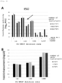
- Vero cells were passaged in a 96-well microplate (0.25 ⁇ 10 5 cells per well). The next day, 75 ⁇ L of 10 to 10240-fold step-diluted dengue antibody-positive serum (2 samples: #1 and #2) was mixed with 75 ⁇ L of an equal volume of 100-fold diluted D1-SRIPs, reacted at 37°C for 2 hr, and 40 ⁇ L of the serum-SRIP mixture was inoculated into Vero cells (Fc ⁇ receptor-non-expressing cells) that were passaged the previous day and allowed to adsorb for additional 2 hr at 37°C. Thereafter, 100 ⁇ L of warmed 10% FBS-added DMEM was dispensed into each well, and intracellular luciferase activity was measured after 48 hr of culture at 37°C.
- Fig. 4A The results are shown in Fig. 4A .
- increased luciferase activity was observed as an ADE action in the group to which diluted serum was added. Luciferase activity increased in proportion to the number of cells. SRIP antigen concentration was also positively correlated with luciferase activity.
- Fold enhancement was determined ( Fig. 4B ). Fold enhancement was determined by subtracting the luciferase activity of the serum-free Control from the luciferase activity (index) of the serum-added sample based on the data in Fig. 4A . Fold enhancement suggested an inversely proportional tendency with SRIP antigen concentration.
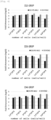
- Fig. 5A The results are shown in Fig. 5A .
- luciferase activity increased in the group to which diluted serum was added as compared with the value of serum-free Control.
- Mylc cells luciferase activity decreased inversely proportional to the number of cells.
- SRIP antigen concentration was positively correlated with luciferase activity.
- Fold enhancement was determined and an inversely proportional tendency with SRIP antigen concentration was suggested ( Fig. 5B ).
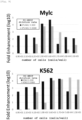
- ADE activity of dengue antibody-positive serum (#1) against SRIPs of each serotype was shown by Fold enhancement ( Fig. 6 ). Similar to the results of ADE activity using D1-SRIPs, both K562 cells and Mylc cells stably showed high Fold enhancement (1000- to 100000-fold). The number of cells did not have a remarkable effect on Fold enhancement under the conditions of this assay.
- Dengue antibody-positive serum (#1) was fixed to a 1:500 antibody dilution rate, D1-SRIPs antigen dilution rate was set to 1:100 or 1:1000, and ADE test using K562 or Mylc cells was performed with shorter incubation times (30 min to 16 h).
- Fig. 7A shows the luciferase activity
- Fig. 7B shows the Fold enhancement calculated therefrom.
- the luciferase activity of Control was already less than Logic 3 at 16 hr.
- lowering the antigen concentration had a large effect on luciferase activity, but the effect on Fold enhancement was relatively small. This suggests that Fold enhancement can be used as an evaluation index without being greatly influenced by antigen concentration.
- Dengue antibody-positive serum (#1) was fixed to a 1:500 antibody dilution rate, D1-SRIPs antigen dilution rate was set to 1:2 and 1:10, and ADE test was performed using Mylc and K562 cells by 5 hr culture. Specifically, 36 ⁇ L of diluted (1:500) dengue antibody-positive serum (#1) and 36 ⁇ L of equal volume of D1-SRIPs (1:2 or 1:10 dilution) were mixed in a 96-well poly-L lysine-coated microplate and reacted for 2 hr at 37°C, and then mixed with 6.9 ⁇ 10 1 to 1.5 ⁇ 10 5 Mylc cells or K562 (Fc ⁇ receptor-expressing cells) (50 ⁇ L). After 5 hr of culture at 37°C, intracellular luciferase activity was measured.
- Fig. 8 shows Fold enhancement obtained from luciferase activity in each cell after culture for 5 hr. SRIP antigen concentration and Fold enhancement were inversely related. Furthermore, it was suggested that ADE activity could be measured in a wider range of the number of cells in Mylc cells than in K562 cells (peak at 5.6 ⁇ 10 3 cells). On the other hand, Fold enhancement tended to increase in proportion to the number of cells in K562 cells. These results suggest that Mylc cells can be assayed under lower cell number conditions than K562 cells.
- Dengue antibody-positive serum (#1) was fixed to a 1:500 antibody dilution rate, D1-SRIPs antigen dilution rate was set to 1:100 and 1:1000, and ADE test was performed using Mylc and K562 cells by 48 hr culture. Specifically, 36 ⁇ L of diluted (1:500) dengue antibody-positive serum (#1) and 36 ⁇ L of equal volume of D1-SRIPs (1:10 or 1:1000 dilution) were mixed in a 96-well poly-L lysine-coated microplate and reacted for 2 hr at 37°C, and then mixed with 6.9 ⁇ 10 1 to 1.5 ⁇ 10 5 Mylc cells or K562 (Fc ⁇ receptor-expressing cells) (50 ⁇ L). After 48 hr of culture at 37°C, intracellular luciferase activity was measured.
- Fig. 9 shows Fold enhancement obtained from luciferase activity in each cell after culture for 48 hr. It was suggested that too many cells at the time of infection result in lower fold enhancement in Mylc cells (peak at 5.6 ⁇ 10 3 cells). On the other hand, Fold enhancement tended to increase in proportion to the number of cells in K562 cells (when D1-SRIPs were used at 1:1000 dilution). From the above, in order to obtain high Fold enhancement, the optimal conditions for the enhancement test are suggested to be that cells are prepared to be "5 ⁇ 10 3 cells/well" when Mylc cells are used and "1 ⁇ 10 5 cells/well" when K562 cells are used.
- Dengue antibody positive serum was step-diluted, reacted with each of D1-SRIPs to D4-SRIPs antigens, infected with Mylc and K562 cells, and ADE test was performed.
- the assay was performed under two different conditions below.
- Fig. 10 shows Fold enhancement obtained from luciferase activity in each cell after culture for (i) 5 hr or (ii) 24 hr.
- Fig. 10A shows the concentration-dependent enhancement activity of dengue antibody-positive serum (#1) against SRIPs of each serotype. There was no significant difference in waveform between Mylc cells and K562 cells in the concentration dependence curve after 5 hours of culture. Mylc cells after 24 hr showed lower enhancement activity against D3-SRIPs than the waveform of K562 cells (serum dilution rate 1:10 to 1:160).
- Fig. 10B shows concentration dependent enhancement activity of dengue antibody positive serum (#2) against SRIPs of each serotype.
Landscapes
- Health & Medical Sciences (AREA)
- Life Sciences & Earth Sciences (AREA)
- Chemical & Material Sciences (AREA)
- Engineering & Computer Science (AREA)
- Immunology (AREA)
- Virology (AREA)
- Biomedical Technology (AREA)
- Molecular Biology (AREA)
- Urology & Nephrology (AREA)
- Hematology (AREA)
- Medicinal Chemistry (AREA)
- General Health & Medical Sciences (AREA)
- Biochemistry (AREA)
- Biotechnology (AREA)
- Microbiology (AREA)
- Organic Chemistry (AREA)
- Genetics & Genomics (AREA)
- Physics & Mathematics (AREA)
- Pathology (AREA)
- Cell Biology (AREA)
- General Physics & Mathematics (AREA)
- Analytical Chemistry (AREA)
- Food Science & Technology (AREA)
- Bioinformatics & Cheminformatics (AREA)
- Zoology (AREA)
- Wood Science & Technology (AREA)
- Tropical Medicine & Parasitology (AREA)
- Proteomics, Peptides & Aminoacids (AREA)
- General Engineering & Computer Science (AREA)
- Biophysics (AREA)
- Measuring Or Testing Involving Enzymes Or Micro-Organisms (AREA)
- Micro-Organisms Or Cultivation Processes Thereof (AREA)
Applications Claiming Priority (2)
| Application Number | Priority Date | Filing Date | Title |
|---|---|---|---|
| JP2021089309 | 2021-05-27 | ||
| PCT/JP2022/016731 WO2022249757A1 (ja) | 2021-05-27 | 2022-03-31 | 疑似ウイルスを使用した抗体依存性感染増強反応の評価法 |
Publications (2)
| Publication Number | Publication Date |
|---|---|
| EP4349976A1 true EP4349976A1 (de) | 2024-04-10 |
| EP4349976A4 EP4349976A4 (de) | 2025-07-30 |
Family
ID=84228706
Family Applications (1)
| Application Number | Title | Priority Date | Filing Date |
|---|---|---|---|
| EP22811031.8A Pending EP4349976A4 (de) | 2021-05-27 | 2022-03-31 | Verfahren zur bewertung einer antikörperabhängigen verstärkungsreaktion mit einem pseudovirus |
Country Status (5)
| Country | Link |
|---|---|
| US (1) | US20250076299A1 (de) |
| EP (1) | EP4349976A4 (de) |
| JP (1) | JPWO2022249757A1 (de) |
| CN (1) | CN117751183A (de) |
| WO (1) | WO2022249757A1 (de) |
Family Cites Families (4)
| Publication number | Priority date | Publication date | Assignee | Title |
|---|---|---|---|---|
| EP0977587B1 (de) * | 1997-02-28 | 2005-06-15 | Acambis Inc. | Chimäre impfstoffe gegen flaviviren |
| KR101499750B1 (ko) * | 2006-02-27 | 2015-03-06 | 더 보드 오브 리전츠 오브 더 유니버시티 오브 텍사스 시스템 | 가감염성 플라비바이러스 및 이들의 용도 |
| WO2019036617A1 (en) * | 2017-08-18 | 2019-02-21 | The United States Of America, As Represented By The Secretary, Department Of Health And Human Services | DENGUE / WESTERN NILE VIRUSES OF GENETIC RAPPORTEUR GENES, AND USE THEREOF |
| JP6681636B1 (ja) | 2019-12-02 | 2020-04-15 | 株式会社Techno−idea | 識別体、物品管理システム、及び暗証番号管理システム |
-
2022
- 2022-03-31 EP EP22811031.8A patent/EP4349976A4/de active Pending
- 2022-03-31 US US18/520,251 patent/US20250076299A1/en active Pending
- 2022-03-31 WO PCT/JP2022/016731 patent/WO2022249757A1/ja not_active Ceased
- 2022-03-31 JP JP2023524069A patent/JPWO2022249757A1/ja active Pending
- 2022-03-31 CN CN202280038045.0A patent/CN117751183A/zh active Pending
Also Published As
| Publication number | Publication date |
|---|---|
| US20250076299A1 (en) | 2025-03-06 |
| CN117751183A (zh) | 2024-03-22 |
| EP4349976A4 (de) | 2025-07-30 |
| JPWO2022249757A1 (de) | 2022-12-01 |
| WO2022249757A1 (ja) | 2022-12-01 |
Similar Documents
| Publication | Publication Date | Title |
|---|---|---|
| Mori et al. | Nuclear localization of Japanese encephalitis virus core protein enhances viral replication | |
| Scholle et al. | trans-Packaged West Nile virus-like particles: infectious properties in vitro and in infected mosquito vectors | |
| Matsuda et al. | High-throughput neutralization assay for multiple flaviviruses based on single-round infectious particles using dengue virus type 1 reporter replicon | |
| Shan et al. | An infectious cDNA clone of Zika virus to study viral virulence, mosquito transmission, and antiviral inhibitors | |
| US10632185B2 (en) | Chimeric west nile/zika viruses and methods of use | |
| Yamanaka et al. | Evaluation of single-round infectious, chimeric dengue type 1 virus as an antigen for dengue functional antibody assays | |
| Weber et al. | A neutralization assay for chikungunya virus infections in a multiplex format | |
| Yamanaka et al. | Utility of Japanese encephalitis virus subgenomic replicon-based single-round infectious particles as antigens in neutralization tests for Zika virus and three other flaviviruses | |
| Yamanaka et al. | Seroprevalence of flavivirus neutralizing antibodies in Thailand by high-throughput neutralization assay: Endemic circulation of zika virus before 2012 | |
| Tran et al. | Roles of ESCRT proteins ALIX and CHMP4A and their interplay with interferon-stimulated gene 15 during tick-borne flavivirus infection | |
| Araf et al. | Dengue outbreak is a global recurrent crisis: Review of the literature | |
| US10533997B2 (en) | Reverse genetics system of Zika virus | |
| US11634459B2 (en) | Chimeric reporter West Nile/dengue viruses and their use | |
| Van Den Hurk et al. | Vector competence of Australian mosquitoes for yellow fever virus | |
| EP4349976A1 (de) | Verfahren zur bewertung einer antikörperabhängigen verstärkungsreaktion mit einem pseudovirus | |
| Kato et al. | Evaluation of Macaca radiata as a non-human primate model of Dengue virus infection | |
| Yoshii et al. | Establishment of a neutralization test involving reporter gene-expressing virus-like particles of tick-borne encephalitis virus | |
| Ackermann‐Gäumann et al. | A reporter virus particle seroneutralization assay for tick‐borne encephalitis virus overcomes ELISA limitations | |
| Lin et al. | Fast preparation of a long chimeric armored RNA as controls for external quality assessment for molecular detection of Zika virus | |
| Liang et al. | Development of a SARS‐CoV‐2 neutralization assay based on a pseudotyped virus using a HIV system | |
| Yang et al. | Analytical methods for detection of Zika virus | |
| Kolokoltsov et al. | Pseudotyped viruses permit rapid detection of neutralizing antibodies in human and equine serum against Venezuelan equine encephalitis virus | |
| CN105602991A (zh) | 一种乙脑假病毒及其制备方法 | |
| Meyrath et al. | Nanoluciferase-based cell fusion assay for rapid and high-throughput assessment of SARS-CoV-2-neutralizing antibodies in patient samples | |
| Lodmell et al. | Viral RNA in the bloodstream suggests viremia occurs in clinically ill rabies-infected mice |
Legal Events
| Date | Code | Title | Description |
|---|---|---|---|
| STAA | Information on the status of an ep patent application or granted ep patent |
Free format text: STATUS: THE INTERNATIONAL PUBLICATION HAS BEEN MADE |
|
| PUAI | Public reference made under article 153(3) epc to a published international application that has entered the european phase |
Free format text: ORIGINAL CODE: 0009012 |
|
| STAA | Information on the status of an ep patent application or granted ep patent |
Free format text: STATUS: REQUEST FOR EXAMINATION WAS MADE |
|
| 17P | Request for examination filed |
Effective date: 20231222 |
|
| AK | Designated contracting states |
Kind code of ref document: A1 Designated state(s): AL AT BE BG CH CY CZ DE DK EE ES FI FR GB GR HR HU IE IS IT LI LT LU LV MC MK MT NL NO PL PT RO RS SE SI SK SM TR |
|
| DAV | Request for validation of the european patent (deleted) | ||
| DAX | Request for extension of the european patent (deleted) | ||
| A4 | Supplementary search report drawn up and despatched |
Effective date: 20250630 |
|
| RIC1 | Information provided on ipc code assigned before grant |
Ipc: C12N 7/01 20060101AFI20250624BHEP Ipc: C12N 5/10 20060101ALI20250624BHEP Ipc: C12N 15/33 20060101ALI20250624BHEP Ipc: C12Q 1/02 20060101ALI20250624BHEP Ipc: G01N 33/53 20060101ALI20250624BHEP |
|
| RIC1 | Information provided on ipc code assigned before grant |
Ipc: C12N 7/01 20060101AFI20250715BHEP Ipc: C12N 5/10 20060101ALI20250715BHEP Ipc: C12N 15/33 20060101ALI20250715BHEP Ipc: C12Q 1/02 20060101ALI20250715BHEP Ipc: G01N 33/53 20060101ALI20250715BHEP |
|
| RAP1 | Party data changed (applicant data changed or rights of an application transferred) |
Owner name: MICAN TECHNOLOGIES INC. Owner name: JAPAN INSTITUTE FOR HEALTH SECURITY Owner name: OSAKA UNIVERSITY |