EP4349999A1 - Automatisiertes system und verfahren zur mikromassstab-überwachung von in einer petrischale enthaltenen c.-elementen mit transparentem medium, erhaltene überwachungsaufzeichnung und verwendung davon - Google Patents
Automatisiertes system und verfahren zur mikromassstab-überwachung von in einer petrischale enthaltenen c.-elementen mit transparentem medium, erhaltene überwachungsaufzeichnung und verwendung davon Download PDFInfo
- Publication number
- EP4349999A1 EP4349999A1 EP22815409.2A EP22815409A EP4349999A1 EP 4349999 A1 EP4349999 A1 EP 4349999A1 EP 22815409 A EP22815409 A EP 22815409A EP 4349999 A1 EP4349999 A1 EP 4349999A1
- Authority
- EP
- European Patent Office
- Prior art keywords
- elegans
- movable
- petri dish
- camera
- monitoring
- Prior art date
- Legal status (The legal status is an assumption and is not a legal conclusion. Google has not performed a legal analysis and makes no representation as to the accuracy of the status listed.)
- Withdrawn
Links
Images





Classifications
-
- G—PHYSICS
- G01—MEASURING; TESTING
- G01N—INVESTIGATING OR ANALYSING MATERIALS BY DETERMINING THEIR CHEMICAL OR PHYSICAL PROPERTIES
- G01N35/00—Automatic analysis not limited to methods or materials provided for in any single one of groups G01N1/00 - G01N33/00; Handling materials therefor
- G01N35/00584—Control arrangements for automatic analysers
-
- G—PHYSICS
- G01—MEASURING; TESTING
- G01N—INVESTIGATING OR ANALYSING MATERIALS BY DETERMINING THEIR CHEMICAL OR PHYSICAL PROPERTIES
- G01N33/00—Investigating or analysing materials by specific methods not covered by groups G01N1/00 - G01N31/00
- G01N33/48—Biological material, e.g. blood, urine; Haemocytometers
- G01N33/50—Chemical analysis of biological material, e.g. blood, urine; Testing involving biospecific ligand binding methods; Immunological testing
- G01N33/5005—Chemical analysis of biological material, e.g. blood, urine; Testing involving biospecific ligand binding methods; Immunological testing involving human or animal cells
- G01N33/5008—Chemical analysis of biological material, e.g. blood, urine; Testing involving biospecific ligand binding methods; Immunological testing involving human or animal cells for testing or evaluating the effect of chemical or biological compounds, e.g. drugs, cosmetics
- G01N33/5082—Supracellular entities, e.g. tissue, organisms
- G01N33/5085—Supracellular entities, e.g. tissue, organisms of invertebrates
-
- C—CHEMISTRY; METALLURGY
- C12—BIOCHEMISTRY; BEER; SPIRITS; WINE; VINEGAR; MICROBIOLOGY; ENZYMOLOGY; MUTATION OR GENETIC ENGINEERING
- C12Q—MEASURING OR TESTING PROCESSES INVOLVING ENZYMES, NUCLEIC ACIDS OR MICROORGANISMS; COMPOSITIONS OR TEST PAPERS THEREFOR; PROCESSES OF PREPARING SUCH COMPOSITIONS; CONDITION-RESPONSIVE CONTROL IN MICROBIOLOGICAL OR ENZYMOLOGICAL PROCESSES
- C12Q1/00—Measuring or testing processes involving enzymes, nucleic acids or microorganisms; Compositions therefor; Processes of preparing such compositions
- C12Q1/02—Measuring or testing processes involving enzymes, nucleic acids or microorganisms; Compositions therefor; Processes of preparing such compositions involving viable microorganisms
- C12Q1/025—Measuring or testing processes involving enzymes, nucleic acids or microorganisms; Compositions therefor; Processes of preparing such compositions involving viable microorganisms for testing or evaluating the effect of chemical or biological compounds, e.g. drugs, cosmetics
-
- C—CHEMISTRY; METALLURGY
- C12—BIOCHEMISTRY; BEER; SPIRITS; WINE; VINEGAR; MICROBIOLOGY; ENZYMOLOGY; MUTATION OR GENETIC ENGINEERING
- C12M—APPARATUS FOR ENZYMOLOGY OR MICROBIOLOGY; APPARATUS FOR CULTURING MICROORGANISMS FOR PRODUCING BIOMASS, FOR GROWING CELLS OR FOR OBTAINING FERMENTATION OR METABOLIC PRODUCTS, i.e. BIOREACTORS OR FERMENTERS
- C12M23/00—Constructional details, e.g. recesses, hinges
- C12M23/02—Form or structure of the vessel
- C12M23/10—Petri dish
-
- C—CHEMISTRY; METALLURGY
- C12—BIOCHEMISTRY; BEER; SPIRITS; WINE; VINEGAR; MICROBIOLOGY; ENZYMOLOGY; MUTATION OR GENETIC ENGINEERING
- C12M—APPARATUS FOR ENZYMOLOGY OR MICROBIOLOGY; APPARATUS FOR CULTURING MICROORGANISMS FOR PRODUCING BIOMASS, FOR GROWING CELLS OR FOR OBTAINING FERMENTATION OR METABOLIC PRODUCTS, i.e. BIOREACTORS OR FERMENTERS
- C12M23/00—Constructional details, e.g. recesses, hinges
- C12M23/48—Holding appliances; Racks; Supports
-
- C—CHEMISTRY; METALLURGY
- C12—BIOCHEMISTRY; BEER; SPIRITS; WINE; VINEGAR; MICROBIOLOGY; ENZYMOLOGY; MUTATION OR GENETIC ENGINEERING
- C12M—APPARATUS FOR ENZYMOLOGY OR MICROBIOLOGY; APPARATUS FOR CULTURING MICROORGANISMS FOR PRODUCING BIOMASS, FOR GROWING CELLS OR FOR OBTAINING FERMENTATION OR METABOLIC PRODUCTS, i.e. BIOREACTORS OR FERMENTERS
- C12M41/00—Means for regulation, monitoring, measurement or control, e.g. flow regulation
- C12M41/46—Means for regulation, monitoring, measurement or control, e.g. flow regulation of cellular or enzymatic activity or functionality, e.g. cell viability
-
- C—CHEMISTRY; METALLURGY
- C12—BIOCHEMISTRY; BEER; SPIRITS; WINE; VINEGAR; MICROBIOLOGY; ENZYMOLOGY; MUTATION OR GENETIC ENGINEERING
- C12M—APPARATUS FOR ENZYMOLOGY OR MICROBIOLOGY; APPARATUS FOR CULTURING MICROORGANISMS FOR PRODUCING BIOMASS, FOR GROWING CELLS OR FOR OBTAINING FERMENTATION OR METABOLIC PRODUCTS, i.e. BIOREACTORS OR FERMENTERS
- C12M41/00—Means for regulation, monitoring, measurement or control, e.g. flow regulation
- C12M41/48—Automatic or computerized control
-
- G—PHYSICS
- G01—MEASURING; TESTING
- G01N—INVESTIGATING OR ANALYSING MATERIALS BY DETERMINING THEIR CHEMICAL OR PHYSICAL PROPERTIES
- G01N15/00—Investigating characteristics of particles; Investigating permeability, pore-volume or surface-area of porous materials
- G01N15/10—Investigating individual particles
- G01N15/14—Optical investigation techniques, e.g. flow cytometry
- G01N15/1429—Signal processing
- G01N15/1433—Signal processing using image recognition
-
- G—PHYSICS
- G01—MEASURING; TESTING
- G01N—INVESTIGATING OR ANALYSING MATERIALS BY DETERMINING THEIR CHEMICAL OR PHYSICAL PROPERTIES
- G01N15/00—Investigating characteristics of particles; Investigating permeability, pore-volume or surface-area of porous materials
- G01N15/10—Investigating individual particles
- G01N15/14—Optical investigation techniques, e.g. flow cytometry
- G01N15/1434—Optical arrangements
-
- G—PHYSICS
- G01—MEASURING; TESTING
- G01N—INVESTIGATING OR ANALYSING MATERIALS BY DETERMINING THEIR CHEMICAL OR PHYSICAL PROPERTIES
- G01N15/00—Investigating characteristics of particles; Investigating permeability, pore-volume or surface-area of porous materials
- G01N15/10—Investigating individual particles
- G01N15/14—Optical investigation techniques, e.g. flow cytometry
- G01N15/1468—Optical investigation techniques, e.g. flow cytometry with spatial resolution of the texture or inner structure of the particle
-
- G—PHYSICS
- G01—MEASURING; TESTING
- G01N—INVESTIGATING OR ANALYSING MATERIALS BY DETERMINING THEIR CHEMICAL OR PHYSICAL PROPERTIES
- G01N21/00—Investigating or analysing materials by the use of optical means, i.e. using sub-millimetre waves, infrared, visible or ultraviolet light
- G01N21/84—Systems specially adapted for particular applications
-
- G—PHYSICS
- G01—MEASURING; TESTING
- G01N—INVESTIGATING OR ANALYSING MATERIALS BY DETERMINING THEIR CHEMICAL OR PHYSICAL PROPERTIES
- G01N33/00—Investigating or analysing materials by specific methods not covered by groups G01N1/00 - G01N31/00
- G01N33/48—Biological material, e.g. blood, urine; Haemocytometers
- G01N33/483—Physical analysis of biological material
-
- G—PHYSICS
- G01—MEASURING; TESTING
- G01N—INVESTIGATING OR ANALYSING MATERIALS BY DETERMINING THEIR CHEMICAL OR PHYSICAL PROPERTIES
- G01N35/00—Automatic analysis not limited to methods or materials provided for in any single one of groups G01N1/00 - G01N33/00; Handling materials therefor
- G01N35/0099—Automatic analysis not limited to methods or materials provided for in any single one of groups G01N1/00 - G01N33/00; Handling materials therefor comprising robots or similar manipulators
-
- G—PHYSICS
- G16—INFORMATION AND COMMUNICATION TECHNOLOGY [ICT] SPECIALLY ADAPTED FOR SPECIFIC APPLICATION FIELDS
- G16H—HEALTHCARE INFORMATICS, i.e. INFORMATION AND COMMUNICATION TECHNOLOGY [ICT] SPECIALLY ADAPTED FOR THE HANDLING OR PROCESSING OF MEDICAL OR HEALTHCARE DATA
- G16H10/00—ICT specially adapted for the handling or processing of patient-related medical or healthcare data
- G16H10/40—ICT specially adapted for the handling or processing of patient-related medical or healthcare data for data related to laboratory analysis, e.g. patient specimen analysis
-
- H—ELECTRICITY
- H04—ELECTRIC COMMUNICATION TECHNIQUE
- H04N—PICTORIAL COMMUNICATION, e.g. TELEVISION
- H04N23/00—Cameras or camera modules comprising electronic image sensors; Control thereof
- H04N23/45—Cameras or camera modules comprising electronic image sensors; Control thereof for generating image signals from two or more image sensors being of different type or operating in different modes, e.g. with a CMOS sensor for moving images in combination with a charge-coupled device [CCD] for still images
-
- H—ELECTRICITY
- H04—ELECTRIC COMMUNICATION TECHNIQUE
- H04N—PICTORIAL COMMUNICATION, e.g. TELEVISION
- H04N23/00—Cameras or camera modules comprising electronic image sensors; Control thereof
- H04N23/90—Arrangement of cameras or camera modules, e.g. multiple cameras in TV studios or sports stadiums
-
- A—HUMAN NECESSITIES
- A61—MEDICAL OR VETERINARY SCIENCE; HYGIENE
- A61B—DIAGNOSIS; SURGERY; IDENTIFICATION
- A61B5/00—Measuring for diagnostic purposes; Identification of persons
- A61B5/103—Measuring devices for testing the shape, pattern, colour, size or movement of the body or parts thereof, for diagnostic purposes
-
- G—PHYSICS
- G01—MEASURING; TESTING
- G01N—INVESTIGATING OR ANALYSING MATERIALS BY DETERMINING THEIR CHEMICAL OR PHYSICAL PROPERTIES
- G01N15/00—Investigating characteristics of particles; Investigating permeability, pore-volume or surface-area of porous materials
- G01N15/10—Investigating individual particles
- G01N2015/1006—Investigating individual particles for cytology
-
- G—PHYSICS
- G01—MEASURING; TESTING
- G01N—INVESTIGATING OR ANALYSING MATERIALS BY DETERMINING THEIR CHEMICAL OR PHYSICAL PROPERTIES
- G01N15/00—Investigating characteristics of particles; Investigating permeability, pore-volume or surface-area of porous materials
- G01N15/10—Investigating individual particles
- G01N15/14—Optical investigation techniques, e.g. flow cytometry
- G01N15/1434—Optical arrangements
- G01N2015/144—Imaging characterised by its optical setup
-
- G—PHYSICS
- G01—MEASURING; TESTING
- G01N—INVESTIGATING OR ANALYSING MATERIALS BY DETERMINING THEIR CHEMICAL OR PHYSICAL PROPERTIES
- G01N2333/00—Assays involving biological materials from specific organisms or of a specific nature
- G01N2333/435—Assays involving biological materials from specific organisms or of a specific nature from animals; from humans
- G01N2333/43504—Assays involving biological materials from specific organisms or of a specific nature from animals; from humans from invertebrates
- G01N2333/43526—Assays involving biological materials from specific organisms or of a specific nature from animals; from humans from invertebrates from worms
- G01N2333/4353—Assays involving biological materials from specific organisms or of a specific nature from animals; from humans from invertebrates from worms from nematodes
- G01N2333/43534—Assays involving biological materials from specific organisms or of a specific nature from animals; from humans from invertebrates from worms from nematodes from Caenorhabditis
-
- G—PHYSICS
- G16—INFORMATION AND COMMUNICATION TECHNOLOGY [ICT] SPECIALLY ADAPTED FOR SPECIFIC APPLICATION FIELDS
- G16H—HEALTHCARE INFORMATICS, i.e. INFORMATION AND COMMUNICATION TECHNOLOGY [ICT] SPECIALLY ADAPTED FOR THE HANDLING OR PROCESSING OF MEDICAL OR HEALTHCARE DATA
- G16H40/00—ICT specially adapted for the management or administration of healthcare resources or facilities; ICT specially adapted for the management or operation of medical equipment or devices
-
- G—PHYSICS
- G16—INFORMATION AND COMMUNICATION TECHNOLOGY [ICT] SPECIALLY ADAPTED FOR SPECIFIC APPLICATION FIELDS
- G16H—HEALTHCARE INFORMATICS, i.e. INFORMATION AND COMMUNICATION TECHNOLOGY [ICT] SPECIALLY ADAPTED FOR THE HANDLING OR PROCESSING OF MEDICAL OR HEALTHCARE DATA
- G16H50/00—ICT specially adapted for medical diagnosis, medical simulation or medical data mining; ICT specially adapted for detecting, monitoring or modelling epidemics or pandemics
- G16H50/30—ICT specially adapted for medical diagnosis, medical simulation or medical data mining; ICT specially adapted for detecting, monitoring or modelling epidemics or pandemics for calculating health indices; for individual health risk assessment
Definitions
- the present invention relates to an automated system for monitoring Caenorhabditis elegans ( C. elegans ) contained in a Petri dish with transparent medium, the system being provided with at least one movable camera and at least one fixed camera with respect to the Petri dish for monitoring of C. elegans contained in said Petri dish, which cameras are positioned along the X, Y, and Z axes of a robot.
- C. elegans Caenorhabditis elegans contained in a Petri dish with transparent medium
- the system being provided with at least one movable camera and at least one fixed camera with respect to the Petri dish for monitoring of C. elegans contained in said Petri dish, which cameras are positioned along the X, Y, and Z axes of a robot.
- the movable camera thereby captures framed high-resolution images for each C. elegans nematode, and the fixed camera captures low-resolution global images of the entire Petri dish.
- the presence of the movable camera and the fixed camera together with a laser pointer, lighting means and a robot controlling the position of the movable camera with respect to the dish allows each C. elegans nematode to be monitored and precise movement characteristics thereof over a given time period to be extracted.
- the object of the invention is to provide a system that automatically establishes a multiscale tracking of multiple C. elegans nematodes coexisting in a Petri dish with great freedom of movement.
- C. elegans is a nematode about 1 mm long, which is inexpensive to maintain and can be cultured in large numbers in a laboratory.
- C. elegans has many tissues and organs that are present in higher organisms, such as the nervous system, the digestive tract, the motor (muscle) system and the reproductive system, which can be easily monitored in vivo, using optical methods, because they are transparent throughout their entire life cycle (less than three weeks) for the wild type. Due to these characteristics, C. elegans has become one of the most important models in many fields of research, including aging and neurobiology.
- Healthspan assays with C. elegans for neurodegenerative diseases are based on the search for new potential therapeutic components with which nematodes with a certain pathology are fed to monitor the evolution of their mobility patterns. At present, generally these assays are performed manually by skilled operators and are quite laborious.
- the technologies being developed to detect the movement of C. elegans automatically are based on optical techniques.
- patent number WO2008062347 discloses a small organism locomotor recording procedure and device. The disclosed procedure detects interruptions of infrared microbeams when C. elegans nematodes move through the light beam. C. elegans nematode movements generate a transient fluctuation in the signal received by the phototransistor, and movement is detected by means of digital analysis of the phototransistor output.
- the article " The Caenorhabditis elegans lifespan machine” (Stroustrup, N. et al., Nature Methods, 10(7), 665-670 (2013 )) discloses a machine that scans standard dishes twice per hour and can distinguish dead C. elegans nematodes for lifespan assays. However, due to a low capture rate, C. elegans nematode movement cannot be tracked.
- non-patent literature reference " Improving lifespan automation for Caenorhabditis elegans by using image processing and a post-processing adaptive data filter" (Puchalt, Joan Caries et al., Scientific Reports 10, 8729 (2020 )) discloses a backlight control strategy which allows image segmentation automation during monitoring of an entire Petri dish with a fixed camera.
- the mentioned reference discloses the use of different types of image processing to automate lifespan assays.
- the imaging processes applied depend on whether the C. elegans nematodes are located in the center of the dish or in an area very close to the wall of the Petri dish, where the image capture conditions are disturbed by lighting refractions.
- the mentioned reference also proposes a correction filter adaptive to lifespan curves.
- the monitoring of an entire Petri dish with a fixed camera is also disclosed in this case. That is, the mentioned reference does not include the use of a movable camera which can offer, in combination with the images captured by the fixed camera, a microscale monitoring of each nematode either.
- non-patent literature reference “Active backlight for automating visual monitoring: An analysis of a lighting control technique for Caenorhabditis elegans cultured on standard Petri plates” (Puchalt, Joan Carles. et al., PLoS One APR 16 (2019 )) describes a backlight control strategy which allows image segmentation automation during the monitoring of an entire Petri dish with a fixed camera.
- the main drawback of the mentioned systems resides in the fact that they work with low-resolution images, or with a limited field of view of a few millimeters. That is, the known configurations do not allow control of the process of capturing high-resolution images with complete information about the dish captured at a lower resolution at the same time (to perform global tracking).
- the applicant of the present patent detects the need to propose a system which allows automation of the microscale monitoring of each nematode, and for this purpose includes in said configuration the combination of a fixed camera and a movable camera of different resolutions and the possibility that the Petri dish is movable along the X, Y, and Z axes with respect to the movable camera(s).
- the inspection system proposed below overcomes the drawbacks set forth above by proposing a system and method capable of automating the microscale extraction of characteristic of C. elegans.
- the mentioned automation allows saving in the time to be invested in the assay and provides greater objectivity, with constant monitoring of C. elegans nematodes and more precise measurements.
- a notable advantage of the invention is the monitoring of assays with multiple C. elegans nematodes where each Petri dish contains multiple C. elegans nematodes and the microscale extraction of mobility characteristics of each of the C. elegans nematodes contained in a Petri dish is possible.
- the present invention is capable of extracting precise mobility characteristics of multiple C. elegans nematodes coexisting in a Petri dish, preferably having dimensions between 150 and 35 mm in diameter. Monitoring is feasible due to the transparency of both the Petri dish and the medium. It should be noted that the main advantage of using Petri dishes having these dimensions is that they offer greater freedom of movement to C. elegans nematodes and furthermore allow for an increase in the number of C. elegans nematodes per Petri dish, thereby reducing the number of dishes needed and therefore reducing the cost of preparing the assay.
- the present invention enables monitoring of multiple C. elegans nematodes contained in standard Petri dishes having large dimensions (for example, 55 mm in diameter) in comparison with the size of a C. elegans nematode (about 1 mm in length).
- the automated system of the invention requires the Petri dish to be monitored to be moved in a controlled manner along the X, Y, and Z axes with respect to at least one movable camera.
- an automated and robotic system is proposed, where the Petri dish and the fixed camera are mounted on a movable trolley in order to move along the X and Y axes, and the movable cameras are independently mounted on a movable head that moves along the Z axis to enable focusing.
- the automated system of the invention includes at least one fixed camera with respect to the Petri dish which monitors the entire dish with minimal precision (projection of the width of C. elegans in 3 pixels).
- the fixed camera is provided with an optic that captures a low-resolution sequence of global images and allows the entire Petri dish to be framed.
- the purpose of the fixed camera is to provide global information about the location of all the C. elegans nematodes in the Petri dish, allowing control of their identities.
- the system of the present invention is provided with at least one movable camera with respect to the Petri dish which monitors a small frame around a single individual C. elegans nematode with maximum (microscale) precision.
- the movable camera is provided with an optic that captures a high-resolution sequence of images for each C. elegans nematode of the Petri dish to be monitored and allows a single individual C. elegans nematode to be tightly framed.
- the optic of the movable camera is an optic having a focal length of between 80 mm and 135 mm.
- the automated system of the invention also includes a robot which enables the movable camera to track the movement of a C. elegans nematode in the Petri dish while extracting the precise characteristics of its movement.
- the fixed camera, lighting means, and a support where the Petri dish is placed are fixed in a movable trolley that moves along the X and Y axes by means of the robot.
- the movable camera and a laser pointer are mounted on a movable head that moves along the Z axis by means of the robot. That is, the robot operates the movable trolley along the X and Y axes and the movable head along the Z axis.
- the fixed camera, the lighting means, and the support for the Petri dish are mounted together on the movable trolley that moves along the X and Y axes, with the automated system allowing two different configurations based on the type of lighting to be implemented.
- the arrangement of the lighting means is fundamental to obtaining well contrasted images, so there are defined two different lighting strategies defined by the two preferred embodiments of the system of the present invention, which will be described in detail in the following section.
- the two preferred embodiments of the invention have the same configuration, differing only in the configuration of the movable trolley.
- a fixed low-resolution camera is positioned at the base of the support, specifically below the Petri dish, which is transparent.
- at least one movable camera is mounted on the movable head to move it along the Z axis. That is, the movable camera captures images above the Petri dish, providing a microscale function; in this way, the robot, upon operating the movable head, allows focusing, and upon operating the movable trolley, allows varying the microscale field of view.
- the lighting means are arranged at the base of the movable trolley on which the Petri dish rests, emitting a backlighting light beam with respect to the Petri dish.
- This second embodiment proposes using a light splitter to enable the possibility of using backlighting.
- the movable camera mounted in the movable head works on a microscale level, moving along the Z axis, and the fixed camera works on a global level.
- the lighting means are coupled to the movable trolley together with the support for the Petri dish and the fixed camera.
- the rest of the automated system for both embodiments of the invention is the same, with active vision techniques which allow the individual microscale tracking and focusing on each C. elegans nematode being applied.
- the system offers a global view that provides global mapping information where all C. elegans nematodes will be located and identified continuously. This mapping allows guiding the microscale image acquisition process, providing precise descriptors to solve posture tracking algorithms, event detection and movement pattern recognition.
- the movable camera captures a high-resolution sequence of images for each C. elegans nematode, which include the light emitted by the laser pointer in a position centered in each image, the high-resolution images being focused due to the position along the Z axis of the movable head with respect to the Petri dish and the high-resolution images being framed for each C. elegans nematode to be monitored due to the position along the X and Y axes of the movable trolley on which the support for the Petri dish is mounted .
- the laser pointer mounted on the movable head that moves along the Z axis is used to start the multiscale tracking of each C. elegans nematode.
- the laser pointer points to the field of view of the movable camera.
- the light emitted by the laser pointer is detected at the same time by the fixed camera.
- the presence of the light emitted by the laser pointer in the image captured by the fixed camera allows control over which C. elegans nematode is framed with the movable camera, controlling the movement executed by the robot of the Petri dish along the X and Y axes, such that with the visual feedback from the fixed camera, the laser pointer is successfully positioned above the chosen C. elegans nematode for microscale monitoring.
- the X, Y, and Z axes are controlled with the visual feedback from the movable camera, with the C. elegans nematode being centered and focused in the image as described in detail in the method used based on the system of the present invention.
- the method of the invention includes the following steps, which will be described in detail in the following section:
- a first individual C. elegans nematode is chosen using the images captured by the fixed cameras of the automated system of the invention, and a framing operation is performed with the help of the laser pointer and the tracking of the individual nematode is performed with the movable cameras.
- the framing operation is completed when the light emitted by the laser pointer is centered on the C. elegans nematode chosen to be monitored.
- the next individual C. elegans nematode to be monitored is chosen, and so on until all the nematodes are monitored, such that a monitoring record for each C. elegans nematode is obtained.
- the method described in detail above allows the monitoring of C. elegans based on multiple images, provided by at least one fixed camera which allows the control of the sequencing of the monitoring of all the C. elegans nematodes in a standard Petri dish that is several centimeters in diameter.
- the individual microscale images are provided by at least one movable camera which allows the precise characteristics of movement during a given period of time (for example, 30 seconds) to be extracted.
- the automated system of the invention and its automated method of monitoring allows a monitoring record to be obtained. It should be noted that, advantageously, the record obtained allows a sequence of microscale images to be provided that enables precise posture patterns of C. elegans nematodes to be extracted so as to determine new mobility characteristics.
- the automated system and the automated method developed are used in various applications such as: compound irritability, compound toxicity, and pharmacological tests.
- the present invention enables the automation of healthspan assays for C. elegans, which are used in sectors such as the pharmaceutical sector, the food sector, and the cosmetic sector.
- the pharmaceutical sector uses these assays to search for new drugs that relieve the effects of neurodegenerative diseases such as Alzheimer's, Parkinsons or Huntington's.
- the food sector uses these assays to design new, healthier probiotic foods, adding value to foodstuffs and/or analyzing their toxicity.
- the cosmetics sector uses these assays to analyze the irritability or toxicity of new products.
- Figures 1 , 2 , and 3 set out in the present specification allow a depiction of some elements that are part of the automated system for microscale monitoring of C. elegans contained in a Petri dish with transparent medium to be observed.
- the two movable cameras (2) and the laser pointer (5) are mounted on the movable head (7) such that the two movable cameras (2) are positioned with respect to the laser pointer (5) in such a way that all the images taken by the movable cameras (2) contain the light emitted by the laser pointer (5) in a centered position.
- the automated system of the invention is provided with a robot that operates the movable trolley (6) along the X and Y axes (9) and (9') and the movable head (7) along the Z axis (11), enabling the movement of the movable cameras (2) along the X, Y, and Z axes with respect to the Petri dish.
- the automated system of the present invention includes a fixed camera (1) and lighting means which are not depicted in Figures 1 , 2 , and 3 , since the two preferred embodiments of the invention described in detail below materialize depending on their location.
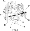
- Figures 4 and 5 specifically depict the automated system according to the first preferred embodiment of the invention.
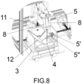
- Figures 6 , 7 , and 8 correspond to depictions of the automated system of the present invention according to the second preferred embodiment.
- the fixed camera (1) is provided with an optic that captures a low-resolution sequence of global images and allows the entire Petri dish (4) to be framed, with the Petri dish (4) being lit by the lighting means.
- the lighting means are cold and homogeneous and made up of LEDs to project homogeneous lighting onto the Petri dish (4) to be monitored.
- the laser pointer (5) emits a light which allows the framing of the movable camera (2) to be located with respect to the image captured by the fixed camera (1).
- the movable camera (2) thereby captures a high-resolution sequence of images for each C. elegans nematode, which include the light emitted by the laser pointer (5) in a position centered in each image, the high-resolution images being focused due to the position along the Z axis of the movable head (7) with respect to the Petri dish (4), and the high-resolution images being framed for each C. elegans nematode to be monitored due to the position along the X and Y axes (9) and (9') of the movable trolley (6) on which the support (3) for the Petri dish (4) is mounted .
- the fixed camera (1) is mounted on the movable trolley (6) which is provided with a support (3).
- the fixed camera (1) thereby captures images from an angle opposite the movable camera (2), with the lighting means (10) being transverse with respect to the Petri dish (4), being arranged in such a way that they emit a transverse ring-shaped light beam in the support (3).
- the lighting means (10) in the first preferred embodiment of the invention are centered at the height of the surface of the transparent medium of the Petri dish (4) to offer more homogeneous lighting of the Petri dish (4).
- the fixed camera (1') is arranged in such a way that it captures images from one side of the support (3) and the lighting means (10') are arranged at the base of the movable trolley (6). In that sense, the lighting means (10') emit a backlighting light beam with respect to the Petri dish (4).
- the automated system is provided with a beam splitter (12).
- the second embodiment of the invention has a configuration in which the inclusion of two fixed cameras (1') arranged in alignment along the X axis (9) and facing one another is foreseen, as observed in Figure 7 .
- the support (3) is provided with two housings arranged along the X axis (9) for the placement of two Petri dishes (4).
- the monitoring of two Petri dishes (4) is thereby enabled, since movement along the X and Y axes (9) and (9') of the movable trolley (6) which contains the support (3) with two Petri dishes (4) allows the mobility of all the C. elegans nematodes contained in both Petri dishes (4) to be monitored.
- Figure 7 includes an example of images captured by the fixed camera (1'), image A, and the movable camera (2), image B.
- image A captures a global view of the Petri dish (4) while image B shows the individual C. elegans nematode selected for monitoring.
- the backlighting light beam orthogonally goes through the Petri dish (4) such that a part of the light goes through the light splitter towards the movable camera (2) (located at the top in the movable head (7)) and another part of the light beam is reflected and strikes the fixed camera (1').
- the beam splitter (12) is arranged at 45o, as depicted in Figure 7 , such that the part of the light beam that is reflected and strikes the fixed camera (1') does so by being reflected at 45°.
- the automated method used in any of the preferred embodiments of the system of the invention follows steps that allow precise mobility information to be extracted from all the C. elegans nematodes contained in a Petri dish.
- This step comprises the following sub-steps:
- the global tracking step is maintained in parallel to the step of choosing the C. elegans nematode to be monitored and framing same and the step of individual tracking.
- the step of individual tracking begins and is also executed in parallel with the global tracking step.
Landscapes
- Health & Medical Sciences (AREA)
- Chemical & Material Sciences (AREA)
- Life Sciences & Earth Sciences (AREA)
- Engineering & Computer Science (AREA)
- General Health & Medical Sciences (AREA)
- Biochemistry (AREA)
- Analytical Chemistry (AREA)
- Organic Chemistry (AREA)
- Bioinformatics & Cheminformatics (AREA)
- Wood Science & Technology (AREA)
- Zoology (AREA)
- Immunology (AREA)
- Biomedical Technology (AREA)
- Physics & Mathematics (AREA)
- General Physics & Mathematics (AREA)
- Pathology (AREA)
- Biotechnology (AREA)
- Microbiology (AREA)
- General Engineering & Computer Science (AREA)
- Genetics & Genomics (AREA)
- Sustainable Development (AREA)
- Molecular Biology (AREA)
- Signal Processing (AREA)
- Cell Biology (AREA)
- Dispersion Chemistry (AREA)
- Medicinal Chemistry (AREA)
- Urology & Nephrology (AREA)
- Hematology (AREA)
- Multimedia (AREA)
- Clinical Laboratory Science (AREA)
- Toxicology (AREA)
- Proteomics, Peptides & Aminoacids (AREA)
- Biophysics (AREA)
- Food Science & Technology (AREA)
- Robotics (AREA)
- Computer Hardware Design (AREA)
- Human Computer Interaction (AREA)
- Medical Informatics (AREA)
- Epidemiology (AREA)
- Public Health (AREA)
Applications Claiming Priority (2)
| Application Number | Priority Date | Filing Date | Title |
|---|---|---|---|
| ES202130492A ES2883331B2 (es) | 2021-05-31 | 2021-05-31 | Sistema y procedimiento automatizado para la monitorizacion a nivel microescala de los c. elegans contenidos en una placa petri con medio transparente, registro de monitorizado obtenido y uso del mismo |
| PCT/ES2022/070254 WO2022254058A1 (es) | 2021-05-31 | 2022-04-27 | Sistema y procedimiento automatizado para la monitorización a nivel microescala de los c. elegans contenidos en una placa petri con medio transparente, registro de monitorizado obtenido y uso del mismo |
Publications (1)
| Publication Number | Publication Date |
|---|---|
| EP4349999A1 true EP4349999A1 (de) | 2024-04-10 |
Family
ID=78819719
Family Applications (1)
| Application Number | Title | Priority Date | Filing Date |
|---|---|---|---|
| EP22815409.2A Withdrawn EP4349999A1 (de) | 2021-05-31 | 2022-04-27 | Automatisiertes system und verfahren zur mikromassstab-überwachung von in einer petrischale enthaltenen c.-elementen mit transparentem medium, erhaltene überwachungsaufzeichnung und verwendung davon |
Country Status (4)
| Country | Link |
|---|---|
| US (1) | US20240210427A1 (de) |
| EP (1) | EP4349999A1 (de) |
| ES (1) | ES2883331B2 (de) |
| WO (1) | WO2022254058A1 (de) |
Family Cites Families (17)
| Publication number | Priority date | Publication date | Assignee | Title |
|---|---|---|---|---|
| DE10112798A1 (de) * | 2001-03-16 | 2002-10-02 | Elegene Gmbh | Vorrichtung und Verfahren zur automatischen Erzeugung einer Wärmevermeidungsreaktion von Nematoden oder ähnlichen Lebewesen |
| AR058206A1 (es) | 2006-11-21 | 2008-01-23 | Consejo Nac Invest Cient Tec | Procedimiento y dispositivo de registro locomotor de organismos pequenos, registro comportamental obtenido y aplicacion del mismo |
| US8961877B2 (en) * | 2007-08-09 | 2015-02-24 | Massachusetts Institute Of Technology | High-throughput, whole-animal screening system |
| US9723817B2 (en) * | 2011-11-10 | 2017-08-08 | State Of Oregon Acting By And Through The State Board Of Higher Education On Behalf Of The University Of Oregon | Electropharyngeogram arrays and methods of use |
| US9305205B2 (en) * | 2013-02-14 | 2016-04-05 | Microbrightfield, Inc. | Methods and systems for tracking movement of microscopic worms and worm-like organisms, and software therefor |
| US10061111B2 (en) * | 2014-01-17 | 2018-08-28 | The Trustees Of Columbia University In The City Of New York | Systems and methods for three dimensional imaging |
| US11795423B2 (en) | 2014-06-26 | 2023-10-24 | The Trustees Of The University Of Pennsylvania | Micro-fabricated multi-well chips for long term automated imaging of C. elegans growth and behavior |
| CN104542495A (zh) * | 2014-07-01 | 2015-04-29 | 上海交通大学医学院附属新华医院 | 一种新型的秀丽线虫培养及实时动态观测装置 |
| KR101755232B1 (ko) * | 2014-10-24 | 2017-07-07 | 포항공과대학교 산학협력단 | 선충의 3차원 운동 분석시스템 및 방법 |
| US20190225923A1 (en) * | 2016-09-30 | 2019-07-25 | Nanyang Technological University | Apparatus for embryo biopsy |
| GB201803724D0 (en) * | 2018-03-08 | 2018-04-25 | Cambridge Entpr Ltd | Methods |
| CN110502037A (zh) * | 2018-05-16 | 2019-11-26 | 上海医药工业研究院 | 线虫追踪成像分析装置 |
| US11762184B2 (en) * | 2018-07-09 | 2023-09-19 | The Regents Of The University Of Michigan | Microscope surveillance system |
| GB2578144A (en) * | 2018-10-18 | 2020-04-22 | The Univ Of Durham | Method and apparatus for tracking nematode worms |
| EP3931545A4 (de) * | 2019-02-27 | 2023-01-04 | Synthego Corporation | Laser-photoablation von zellkulturen |
| US20200408691A1 (en) * | 2019-06-27 | 2020-12-31 | Nikita Vladimirov | Multi-view light-sheet microscope with an optical arm combiner |
| MX2022016575A (es) * | 2020-11-06 | 2023-04-11 | Illumina Inc | Aparato y metodo para obtener una imagen de una muestra en movimiento. |
-
2021
- 2021-05-31 ES ES202130492A patent/ES2883331B2/es active Active
-
2022
- 2022-04-27 WO PCT/ES2022/070254 patent/WO2022254058A1/es not_active Ceased
- 2022-04-27 EP EP22815409.2A patent/EP4349999A1/de not_active Withdrawn
- 2022-04-27 US US18/287,478 patent/US20240210427A1/en active Pending
Also Published As
| Publication number | Publication date |
|---|---|
| ES2883331B2 (es) | 2022-10-03 |
| ES2883331A1 (es) | 2021-12-07 |
| WO2022254058A1 (es) | 2022-12-08 |
| US20240210427A1 (en) | 2024-06-27 |
Similar Documents
| Publication | Publication Date | Title |
|---|---|---|
| KR101495011B1 (ko) | 카메라 시스템 좌표계의 측정 및 변환 | |
| US11754392B2 (en) | Distance determination of a sample plane in a microscope system | |
| JP6975474B2 (ja) | 空気試料の自動分析を実行するためのシステム及び方法 | |
| US20200041776A1 (en) | Sample observation device and sample observation method | |
| US20250391023A1 (en) | Plaque detection method for imaging of cells | |
| JP2017223951A (ja) | 微細対象物観察装置 | |
| CN111239044A (zh) | 细胞检测方法、装置与系统 | |
| CN114441260B (zh) | 载片架确定系统 | |
| EP4349999A1 (de) | Automatisiertes system und verfahren zur mikromassstab-überwachung von in einer petrischale enthaltenen c.-elementen mit transparentem medium, erhaltene überwachungsaufzeichnung und verwendung davon | |
| JP4779149B2 (ja) | レーザ走査型顕微鏡の操作方法 | |
| US10690902B2 (en) | Image processing device and microscope system | |
| JP7296878B2 (ja) | 細胞観察システム | |
| WO2022225890A1 (en) | Plaque detection method and apparatus for imaging of cells | |
| JP2011017620A (ja) | 形状測定方法、画像処理プログラム及び観察装置 | |
| JP7288141B2 (ja) | 顕微鏡画像撮像方法および顕微鏡画像撮像装置 | |
| WO2022099185A1 (en) | Plaque counting assay method | |
| CN113906482A (zh) | 用于在具有腔体的测试板中测定活性物质对螨类、昆虫和其他生物的作用的系统 | |
| US9036017B2 (en) | Biological observation apparatus and biological observation method | |
| WO2023141189A2 (en) | Method and apparatus for imaging of cells for counting cells, confluence measurement and plaque detection | |
| JP7403834B2 (ja) | 撮像装置と撮像方法 | |
| JP2024506290A (ja) | マシンビジョンシステムにおける欠陥を検出する装置および方法 | |
| CN107003237A (zh) | 用于检测接种的方法和装置及设有这种检测装置的自动接种设备 | |
| EP1967841B1 (de) | Fluoreszenzbeobachtungsvorrichtung | |
| JP6980898B2 (ja) | 細胞画像処理装置 | |
| CN208187359U (zh) | 一种高速高通量生物样本形态检测系统 |
Legal Events
| Date | Code | Title | Description |
|---|---|---|---|
| STAA | Information on the status of an ep patent application or granted ep patent |
Free format text: STATUS: THE INTERNATIONAL PUBLICATION HAS BEEN MADE |
|
| PUAI | Public reference made under article 153(3) epc to a published international application that has entered the european phase |
Free format text: ORIGINAL CODE: 0009012 |
|
| STAA | Information on the status of an ep patent application or granted ep patent |
Free format text: STATUS: REQUEST FOR EXAMINATION WAS MADE |
|
| 17P | Request for examination filed |
Effective date: 20231025 |
|
| AK | Designated contracting states |
Kind code of ref document: A1 Designated state(s): AL AT BE BG CH CY CZ DE DK EE ES FI FR GB GR HR HU IE IS IT LI LT LU LV MC MK MT NL NO PL PT RO RS SE SI SK SM TR |
|
| DAX | Request for extension of the european patent (deleted) | ||
| RAV | Requested validation state of the european patent: fee paid |
Extension state: MA Effective date: 20231025 |
|
| STAA | Information on the status of an ep patent application or granted ep patent |
Free format text: STATUS: THE APPLICATION IS DEEMED TO BE WITHDRAWN |
|
| 18D | Application deemed to be withdrawn |
Effective date: 20251101 |