EP4360657A1 - Mir-29 pour le traitement du cancer - Google Patents
Mir-29 pour le traitement du cancer Download PDFInfo
- Publication number
- EP4360657A1 EP4360657A1 EP22383047.2A EP22383047A EP4360657A1 EP 4360657 A1 EP4360657 A1 EP 4360657A1 EP 22383047 A EP22383047 A EP 22383047A EP 4360657 A1 EP4360657 A1 EP 4360657A1
- Authority
- EP
- European Patent Office
- Prior art keywords
- mir
- cells
- expression
- composition
- precursor
- Prior art date
- Legal status (The legal status is an assumption and is not a legal conclusion. Google has not performed a legal analysis and makes no representation as to the accuracy of the status listed.)
- Withdrawn
Links
Images





Classifications
-
- C—CHEMISTRY; METALLURGY
- C12—BIOCHEMISTRY; BEER; SPIRITS; WINE; VINEGAR; MICROBIOLOGY; ENZYMOLOGY; MUTATION OR GENETIC ENGINEERING
- C12N—MICROORGANISMS OR ENZYMES; COMPOSITIONS THEREOF; PROPAGATING, PRESERVING, OR MAINTAINING MICROORGANISMS; MUTATION OR GENETIC ENGINEERING; CULTURE MEDIA
- C12N15/00—Mutation or genetic engineering; DNA or RNA concerning genetic engineering, vectors, e.g. plasmids, or their isolation, preparation or purification; Use of hosts therefor
- C12N15/09—Recombinant DNA-technology
- C12N15/63—Introduction of foreign genetic material using vectors; Vectors; Use of hosts therefor; Regulation of expression
- C12N15/79—Vectors or expression systems specially adapted for eukaryotic hosts
- C12N15/85—Vectors or expression systems specially adapted for eukaryotic hosts for animal cells
- C12N15/86—Viral vectors
-
- A—HUMAN NECESSITIES
- A61—MEDICAL OR VETERINARY SCIENCE; HYGIENE
- A61P—SPECIFIC THERAPEUTIC ACTIVITY OF CHEMICAL COMPOUNDS OR MEDICINAL PREPARATIONS
- A61P35/00—Antineoplastic agents
-
- C—CHEMISTRY; METALLURGY
- C12—BIOCHEMISTRY; BEER; SPIRITS; WINE; VINEGAR; MICROBIOLOGY; ENZYMOLOGY; MUTATION OR GENETIC ENGINEERING
- C12N—MICROORGANISMS OR ENZYMES; COMPOSITIONS THEREOF; PROPAGATING, PRESERVING, OR MAINTAINING MICROORGANISMS; MUTATION OR GENETIC ENGINEERING; CULTURE MEDIA
- C12N15/00—Mutation or genetic engineering; DNA or RNA concerning genetic engineering, vectors, e.g. plasmids, or their isolation, preparation or purification; Use of hosts therefor
- C12N15/09—Recombinant DNA-technology
- C12N15/11—DNA or RNA fragments; Modified forms thereof; Non-coding nucleic acids having a biological activity
- C12N15/113—Non-coding nucleic acids modulating the expression of genes, e.g. antisense oligonucleotides; Antisense DNA or RNA; Triplex- forming oligonucleotides; Catalytic nucleic acids, e.g. ribozymes; Nucleic acids used in co-suppression or gene silencing
-
- C—CHEMISTRY; METALLURGY
- C12—BIOCHEMISTRY; BEER; SPIRITS; WINE; VINEGAR; MICROBIOLOGY; ENZYMOLOGY; MUTATION OR GENETIC ENGINEERING
- C12N—MICROORGANISMS OR ENZYMES; COMPOSITIONS THEREOF; PROPAGATING, PRESERVING, OR MAINTAINING MICROORGANISMS; MUTATION OR GENETIC ENGINEERING; CULTURE MEDIA
- C12N15/00—Mutation or genetic engineering; DNA or RNA concerning genetic engineering, vectors, e.g. plasmids, or their isolation, preparation or purification; Use of hosts therefor
- C12N15/09—Recombinant DNA-technology
- C12N15/63—Introduction of foreign genetic material using vectors; Vectors; Use of hosts therefor; Regulation of expression
- C12N15/79—Vectors or expression systems specially adapted for eukaryotic hosts
- C12N15/85—Vectors or expression systems specially adapted for eukaryotic hosts for animal cells
-
- C—CHEMISTRY; METALLURGY
- C12—BIOCHEMISTRY; BEER; SPIRITS; WINE; VINEGAR; MICROBIOLOGY; ENZYMOLOGY; MUTATION OR GENETIC ENGINEERING
- C12N—MICROORGANISMS OR ENZYMES; COMPOSITIONS THEREOF; PROPAGATING, PRESERVING, OR MAINTAINING MICROORGANISMS; MUTATION OR GENETIC ENGINEERING; CULTURE MEDIA
- C12N5/00—Undifferentiated human, animal or plant cells, e.g. cell lines; Tissues; Cultivation or maintenance thereof; Culture media therefor
- C12N5/06—Animal cells or tissues; Human cells or tissues
- C12N5/0602—Vertebrate cells
- C12N5/0634—Cells from the blood or the immune system
- C12N5/0645—Macrophages, e.g. Kuepfer cells in the liver; Monocytes
-
- C—CHEMISTRY; METALLURGY
- C12—BIOCHEMISTRY; BEER; SPIRITS; WINE; VINEGAR; MICROBIOLOGY; ENZYMOLOGY; MUTATION OR GENETIC ENGINEERING
- C12N—MICROORGANISMS OR ENZYMES; COMPOSITIONS THEREOF; PROPAGATING, PRESERVING, OR MAINTAINING MICROORGANISMS; MUTATION OR GENETIC ENGINEERING; CULTURE MEDIA
- C12N5/00—Undifferentiated human, animal or plant cells, e.g. cell lines; Tissues; Cultivation or maintenance thereof; Culture media therefor
- C12N5/06—Animal cells or tissues; Human cells or tissues
- C12N5/0602—Vertebrate cells
- C12N5/0679—Cells of the gastro-intestinal tract
-
- C—CHEMISTRY; METALLURGY
- C12—BIOCHEMISTRY; BEER; SPIRITS; WINE; VINEGAR; MICROBIOLOGY; ENZYMOLOGY; MUTATION OR GENETIC ENGINEERING
- C12N—MICROORGANISMS OR ENZYMES; COMPOSITIONS THEREOF; PROPAGATING, PRESERVING, OR MAINTAINING MICROORGANISMS; MUTATION OR GENETIC ENGINEERING; CULTURE MEDIA
- C12N5/00—Undifferentiated human, animal or plant cells, e.g. cell lines; Tissues; Cultivation or maintenance thereof; Culture media therefor
- C12N5/06—Animal cells or tissues; Human cells or tissues
- C12N5/0602—Vertebrate cells
- C12N5/0693—Tumour cells; Cancer cells
-
- A—HUMAN NECESSITIES
- A61—MEDICAL OR VETERINARY SCIENCE; HYGIENE
- A61K—PREPARATIONS FOR MEDICAL, DENTAL OR TOILETRY PURPOSES
- A61K31/00—Medicinal preparations containing organic active ingredients
- A61K31/70—Carbohydrates; Sugars; Derivatives thereof
- A61K31/7088—Compounds having three or more nucleosides or nucleotides
- A61K31/7105—Natural ribonucleic acids, i.e. containing only riboses attached to adenine, guanine, cytosine or uracil and having 3'-5' phosphodiester links
-
- A—HUMAN NECESSITIES
- A61—MEDICAL OR VETERINARY SCIENCE; HYGIENE
- A61K—PREPARATIONS FOR MEDICAL, DENTAL OR TOILETRY PURPOSES
- A61K35/00—Medicinal preparations containing materials or reaction products thereof with undetermined constitution
- A61K35/12—Materials from mammals; Compositions comprising non-specified tissues or cells; Compositions comprising non-embryonic stem cells; Genetically modified cells
- A61K35/14—Blood; Artificial blood
- A61K35/15—Cells of the myeloid line, e.g. granulocytes, basophils, eosinophils, neutrophils, leucocytes, monocytes, macrophages or mast cells; Myeloid precursor cells; Antigen-presenting cells, e.g. dendritic cells
-
- A—HUMAN NECESSITIES
- A61—MEDICAL OR VETERINARY SCIENCE; HYGIENE
- A61K—PREPARATIONS FOR MEDICAL, DENTAL OR TOILETRY PURPOSES
- A61K35/00—Medicinal preparations containing materials or reaction products thereof with undetermined constitution
- A61K35/12—Materials from mammals; Compositions comprising non-specified tissues or cells; Compositions comprising non-embryonic stem cells; Genetically modified cells
- A61K35/14—Blood; Artificial blood
- A61K35/17—Lymphocytes; B-cells; T-cells; Natural killer cells; Interferon-activated or cytokine-activated lymphocytes
-
- C—CHEMISTRY; METALLURGY
- C12—BIOCHEMISTRY; BEER; SPIRITS; WINE; VINEGAR; MICROBIOLOGY; ENZYMOLOGY; MUTATION OR GENETIC ENGINEERING
- C12N—MICROORGANISMS OR ENZYMES; COMPOSITIONS THEREOF; PROPAGATING, PRESERVING, OR MAINTAINING MICROORGANISMS; MUTATION OR GENETIC ENGINEERING; CULTURE MEDIA
- C12N2310/00—Structure or type of the nucleic acid
- C12N2310/10—Type of nucleic acid
- C12N2310/14—Type of nucleic acid interfering nucleic acids [NA]
- C12N2310/141—MicroRNAs, miRNAs
-
- C—CHEMISTRY; METALLURGY
- C12—BIOCHEMISTRY; BEER; SPIRITS; WINE; VINEGAR; MICROBIOLOGY; ENZYMOLOGY; MUTATION OR GENETIC ENGINEERING
- C12N—MICROORGANISMS OR ENZYMES; COMPOSITIONS THEREOF; PROPAGATING, PRESERVING, OR MAINTAINING MICROORGANISMS; MUTATION OR GENETIC ENGINEERING; CULTURE MEDIA
- C12N2510/00—Genetically modified cells
-
- C—CHEMISTRY; METALLURGY
- C12—BIOCHEMISTRY; BEER; SPIRITS; WINE; VINEGAR; MICROBIOLOGY; ENZYMOLOGY; MUTATION OR GENETIC ENGINEERING
- C12N—MICROORGANISMS OR ENZYMES; COMPOSITIONS THEREOF; PROPAGATING, PRESERVING, OR MAINTAINING MICROORGANISMS; MUTATION OR GENETIC ENGINEERING; CULTURE MEDIA
- C12N2740/00—Reverse transcribing RNA viruses
- C12N2740/00011—Details
- C12N2740/10011—Retroviridae
- C12N2740/16011—Human Immunodeficiency Virus, HIV
- C12N2740/16041—Use of virus, viral particle or viral elements as a vector
- C12N2740/16043—Use of virus, viral particle or viral elements as a vector viral genome or elements thereof as genetic vector
-
- C—CHEMISTRY; METALLURGY
- C12—BIOCHEMISTRY; BEER; SPIRITS; WINE; VINEGAR; MICROBIOLOGY; ENZYMOLOGY; MUTATION OR GENETIC ENGINEERING
- C12N—MICROORGANISMS OR ENZYMES; COMPOSITIONS THEREOF; PROPAGATING, PRESERVING, OR MAINTAINING MICROORGANISMS; MUTATION OR GENETIC ENGINEERING; CULTURE MEDIA
- C12N2840/00—Vectors comprising a special translation-regulating system
- C12N2840/20—Vectors comprising a special translation-regulating system translation of more than one cistron
- C12N2840/203—Vectors comprising a special translation-regulating system translation of more than one cistron having an IRES
Definitions
- the present invention discloses a method of therapeutically treating cancer, in particular CRC (colorectal cancer), or of reducing the risk or the progression of cancer, in particular CRC disease progression, in a patient in need thereof, the method comprising expressing miR-326 in the epithelial tumor cells and/or immune cells of said patient, preferably in NK (Natural Killer cells) and macrophages, and suitable means for said methods.
- CRC colonal cancer
- NK Natural Killer cells
- microRNAs can be found in different extracellular fluids including blood, serum, urine and saliva, among others, which allows an easy detection (Kim 2015).
- miRNAs are highly stable molecules that avoid degradation by resisting room temperatures for several days and extreme conditions such as boiling, multiple freeze-thaw cycles, and high or low pHs (Huang 2017). These properties make their detection an easy and reproducible procedure worldwide.
- MicroRNA expression profile can be detected and monitored by qRT-PCR, a simple, cheap, reliable and well-established method to detect specific genes (Khoury et al. 2020).
- microRNAs are part of the pathophysiological mechanisms involved in the development and progression of the disease, which allow them to debut as early biomarkers that help to anticipate the diagnosis and the treatment of the tumour.
- most of the microRNAs are tissue-specific which lets ensuring a certain miRNA profile in plenty of patients suffering from a specific disease.
- miRNAs Due to all of this, miRNAs have been unleashed as novel targets for developing therapies against diseases (Huang 2017). To date, there are not microRNAs used in therapy to treat colorectal cancer, but there are some miRNAs modulators commercialised in other pathologies such as MiRavirsen, an intramuscular inhibitor of miR-122 for the treatment of Hepatitis C (Gebert et al. 2014), or ABX464, a molecule that triggers the increase of miR-124 to reduce the symptoms of inflammatory bowel disease for ulcerative colitis and Crohn's disease patients (Vermeire et al. 2021).
- MiRavirsen an intramuscular inhibitor of miR-122 for the treatment of Hepatitis C
- ABX464 ABX464
- miR-326 participates in essential processes of many types of tumours such as cellular apoptosis, tumour growth, cell invasion, embryonic development, immunomodulation, chemotherapy resistance, and oncogenesis (Y. Pan et al. 2019). Moreover, has been generally reported that this microRNA is downregulated in most cancers. In non-small lung cancer, miR-326 inhibits tumour proliferation by targeting CCND1 and high expression of the miRNA decreases tumour metastasis by targeting Adam17, NSBP1 and Phox2a (Cai et al., 2015; Sun et al., 2016; Wang et al., 2015).
- miR-326 In gastric cancer, low expression of miR-326 is a prognostic factor for poor patient outcomes and downregulation of the miRNA promotes metastasis by targeting FSCN1 (Li et al. 2015). In breast cancer, miR-326 has been reported to repress cell metastasis by targeting B7-H3, a protein which correlates with adverse prognosis (Sun et al. 2014). miR-326 controls cancer invasiveness regulating VEGF-C and accelerates cervical cancer metastasis (Cheng et al. 2018; Zhang et al. 2017). The miRNA controls PKM2 which is overexpressed in thyroid cancer and plays an oncogenic role in metastasis (Yu et al., 2018).
- miR-326 targets the Bcl-2 axis inhibiting proliferation and promoting apoptosis in endometrial cancer (Cai et al. 2021). miR-326 expression level is restrained by abnormal PI3K signalling, resulting in a downregulation in glioblastoma (Nawaz et al. 2016).
- miR-326 is acting in many types of cancer as a tumour suppressor miRNA, but little is known about the function of miR-326 in CRC.
- miR-326 modulation could be a potential therapeutic intervention to reduce or block tumour progression and could also be used as a prognostic biomarker in CRC.
- miR-326 as a key mediator involved in the development and progression of colorectal cancer unveiling this molecule as a biomarker and therapeutic target in this tumoral context.
- hsa-miR-326 as a potential biomarker of CRC due to its expression associated with different histopathological features like tumour appearance, tumour location, stroma percentage, immune infiltrate, tumour staging and invasion in a cohort of 192 CRC patients. Moreover, we demonstrated that high levels of miR-326 are associated with a better patient's overall survival. Furthermore, we have determined that miR-326 is mostly located in tumour epithelial cells, macrophages, and natural killer cells in colorectal cancer patients' biopsies. Moreover, we have induced and inhibited miR-326 expression in Caco-2, an epithelial colorectal cancer cell line, and in M ⁇ , M1 and M2 murine macrophages.
- miR-326 expression levels are found in M ⁇ , M1 and M2 murine macrophages polarized upon different stimuli. Modulation of miR-326 by transient transfection does not regulate macrophage polarization but is involved in the process that leads macrophages to a M1 phenotype. Additionally, we suggest ITGA5 as a potential target of miR-326 in M ⁇ and M1 macrophages. Finally, we have demonstrated that overexpression of miR-326 reduces PD-L1 levels in Caco-2 cells, M ⁇ , M1 and M2 murine macrophages and inhibition of the miRNA leads to the opposite effect, PD-L1 levels increased.
- the present invention is the first one that analyses miR-326 expression in a large cohort of CRC patients with 192 biopsies from primary tumours and relates the microRNA with tumour-infiltrating immune cells and overall survival.
- miR-326 expression is an early diagnostic biomarker of colorectal cancer.
- miR-326 was upregulated in left-side tumours compared to right-side tumours.
- the distinction between right- and left-sided colon is based on the embryological origin and both sides exhibit different histology. Since LCRC is clearer to identify in the early stages of carcinogenesis because presents polypoid morphology, RCRC exhibits flat morphology that is difficult to detect, presenting a more aggressive phenotype (Baran et al. 2018; Mukund et al. 2020). This sidespecificity exhibits therapy responses completely different, LCRC patients benefit more from chemotherapies treatments than RCRC which have demonstrated promising results with immunotherapy. Moreover, the side-distinction presents post-transcriptional regulation miRNAs differences. All of this points out that miR-326 associates with the less aggressive phenotype and suggest that miR-326 could be used as a predictive biomarker with reference to the different treatments employed for CRC patients.
- miR-326 expression was associated with tumour stroma percentage.
- Our results show that high levels of miR-326 were linked to reduced stroma patients and correlate with longer overall survival.
- the stroma is a critical component of the tumour microenvironment and possesses certain tumour-suppressing abilities. Despite this, tumour malignancy changes the stroma profile and tumour-associated stromal cells contribute to local and distant progression of the malignant tumour (Bremnes et al., 2011; Valkenburg et al., 2018). Tumours with a high stroma percentage exhibited poor patient prognosis in CRC patients (Park et al. 2014). All this suggests that this miRNA could be able to predict the prognosis of the patients with the differential expression of miR-326.
- miR-326 presented a strong correlation with immune infiltrate profile in our patient's cohort showing that high levels of miR-326 correlated with high tumour infiltrating immune cells. This suggests that miR-326 could be established as a good prognostic biomarker in terms of immune infiltrate profile of CRC patients.
- miR-326 was associated with tumour stage and invasion in our patients' cohort. Increased levels of miR-326 were detected in earlier stages of the disease (TNM I-II) compared to late stages (TNM III-IV) of CRC patients. Consistently to the tumour stage, miR-326 presented a correlation with tumour invasiveness grade (T1-T4). miR-326 was upregulated in our patients' cohort when the tumour had not been extremely invasive (Tis-T1-T2) compared to late stages of invasiveness (T3-T4). Moreover, miR-326 was also related to the lymph nodes affectation. In our cohort, miR-326 levels were upregulated when lymph nodes had not been affected compared to lymph-node's affectation.
- miR-326 expression levels were associated with patient's overall survival. High levels of the microRNA were associated with better patients' overall survival. Therefore, again, our results demonstrated for the first time that miR-326 acted as a potential prognostic biomarker in CRC patients and could be considered a potential therapeutic target for colorectal cancer.
- the malignant phenotypes of the different types of cancers are defined not only by the intrinsic activities of tumour cells but also by the immune cells recruited in the tumour-related microenvironment (Ben-Baruch 2002), and the interaction between tumour and immune cells has significant prognostic relevance in tumour progression (Bremnes et al., 2011). Therefore, in recent years, researchers have focused their investigation on the role of miRNAs in inflammatory and anti-inflammatory processes. Many studies have confirmed that miRNAs dysregulation in cells and targeted tissues is correlated with immune disequilibrium and autoimmune diseases (Jadideslam et al. 2018). Recent research has described miR-326 is critical in regulating functions of inflammatory cells mostly in the pathogenesis of neurodegenerative and autoimmune disorders.
- miR-326 coordinates TH-17 differentiation and correlates with leukocytes in multiple sclerosis (Du et al. 2009; Honardoost et al. 2014), plays a role in autoimmune thyroiditis regulating Th17/Treg balance (Zhao et al. 2021) or contributes to B cell hyperactivity in systemic lupus erythematosus (Xia et al. 2018).
- CD68 the classical M1 macrophage marker
- CD56 the classical marker of natural killer cells.
- miR-326 In vitro cell proliferation and suppresses in vivo tumour growth in different kinds of tumours such as melanoma (Kang et al. 2017), endometrial cancer (Liu et al., 2017), cervical cancer (Zhang et al. 2017), breast cancer (Ghaemi et al.
- miR-326 has been already reported to reduce cell proliferation in SW480 colorectal cancer cells by inhibiting E2F1, a member of the E2F gene family involved in cell cycle regulation (Gao et al., 2021). However, the decrease of proliferation process overexpressing miR-326 had not been described to date in Caco-2 cells.
- p27 is a cyclin-dependent kinase inhibitor that blocks the cell cycle and accumulates the cells in the G0/G1 phase until mitogenic signals lead the cell to the S-phase (Chu et al. 2008). High p27 levels arrest the cell cycle in G1 -phase (Kullmann et al. 2013).
- miR-326 is suppressing the inhibitor of G1-S transition and allows the cells to reach the DNA synthesis phase (S-phase) without regulation.
- miR-326 overexpression increases the sub-G1 cell proportion and decreases the G0/G1, S, and G2/M cell proportions by reducing Cyclin D1 and Cyclin D2 levels in breast cancer (Ghaemi et al. 2019).
- overexpressed miR-326 blocks the G2/M phase due to the effect of the miRNA on the cell cycle regulator NOB1 (Ji et al. 2017).
- miR-326 overexpression arrests the cell cycle in HCT116 and SW620 cells, increasing the G1-number of cells targeting NOB1 (Wu et al. 2015).
- miR-326 overexpression leads to a decrease in the inhibition of the G0/G1 phase and increases the proportion of cells reaching the S-phase by targeting the kinase-inhibitor p-27 in Caco-2 cells.
- Epithelial-mesenchymal transition is another highly repetitive process that carry out cancer cells and is characterised by the loss of cell adhesion ability and the acquisition of motility and invasive capacity.
- Well-established transcription factors such as ZEB1/2, SNAI1/2 and TWIST1/2 mediate the EMT process in response to signalling factors and these inducers can downregulate E-cadherin and upregulate N-cadherin and Vimentin, between others (Shibue et al. 2017).
- miR-326 overexpression reduces TWIST1, TWIST2 and SNAI1 genes, and inhibition of the miRNA promotes the opposite effect increasing the three transcription factors. At the same time, overexpression of the miRNA increased E-cadherin levels.
- miR-326 is regulating colorectal cancer progression process repressing some mesenchymal genes such as TWIST1, TWIST2 and SNAI1 and increasing epithelial genes like E-cadherin in Caco-2 cells.
- some publications have described that miR-326 regulates EMT genes in lung carcinoma by targeting ADAM17 and ZEB1 genes (Cai et al. 2015; Liu et al. 2021).
- miR-326 regulates EMT in endometrial cancer by controlling TWIST1 (Liu et al., 2017).
- TWIST1 Liu et al., 2017
- no existing publication has characterized the role of miR-326 modulating EMT transcription factors in colorectal cancer cells.
- ITGA5 as a direct target of miR-326 in Caco-2 cells.
- ITGA5 exhibited an inverse correlation with miR-326 expression in our colorectal cancer patients.
- High levels of the miRNA correlated with low levels of ITGA5.
- This data suggest ITGA5 behaves as a direct target of miR-326 in tumour biopsies.
- miR-326 expression decreases along the CRC disease and high levels of the miRNA are found in the first stages and low levels of miR-326 correlate with the late stages of the disease.
- Fibronectin is one of the proteins constituting the extracellular matrix that participates in the earliest steps in primary tumour formation and the promotion of invasive tumour behaviour. It is widely accepted that FN levels are increased in several tumour types and this upregulation relates to the poor prognosis of the disease (Efthymiou et al. 2020; Wang et al. 2017). To that, cell adhesion to fibronectin assays were carried out in Caco-2 cells, considering fibronectin as a malignant tumoral matrix linked to tumour progression (Spada 2021).
- miR-326 regulates SNAIL expression in Caco-2 cells. Overexpression of the miRNA decreases SNAIL and inhibition of miR-326 levels increases SNAIL expression. SNAIL is a biomarker of poor prognosis in metastatic cancers and regulates integrin expression. High levels of SNAIL correlates with high levels of ITGA5 expression in prostate cancer cells (Neal et al. 2011). Moreover, SNAIL upregulates fibronectin, and this association is related to increased migration, invasion and metastasis (Kaufhold et al. 2014). We have previously described that miR-326 regulates SNAI1 expression gene and our results confirm that this miRNA regulates SNAIL protein levels too. Overall, we can confirm miR-326 is regulating cell adhesion to malignant fibronectin through the axis ITGA5/SNAIL, downregulating the expression levels of these proteins to avoid a progression of colorectal cancer in Caco-2 cells.
- CXCR4 is another direct target of miR-326 in Caco-2 cells.
- the microRNA modulates the CXCR4 expression and miR-326 directly binds to CXCR4 3'UTR.
- CXCR4 expression shows an inverse correlation with miR-326 levels in our colorectal cancer patients.
- CXCR4 expression is a prognostic factor for several gastrointestinal tumors and CXCR4 inhibitors are being studied in several clinal trials to treat colorectal cancer patients nowadays (Khare et al. 2021).
- miR-326 and its target CXCR4 can be regulating the tumour colorectal cancer cells and the miR-326/CXCR4 axis could be pointed as a good therapeutic target for CRC patients' treatment.
- miR-326 localizes in inflammatory cellularity, more specifically in NK cells and M1 macrophages.
- M1 and M2 macrophages which behave differently in inflammation-related diseases.
- M1 macrophages are characterized by the production of pro-inflammatory cytokines, antimicrobial and tumoricidal activity.
- M2 macrophages are part of immunosuppression, tumorigenesis, wound repair and elimination of parasites (Essandoh et al. 2016).
- Our in vitro experiments have demonstrated, in murine macrophages stimulated toward these two different phenotypes, that miR-326 is differently expressed in the cells depending on the stimulus received.
- M1 macrophages presented a low miR-326 expression and high expression of the miRNA was found in M2 macrophages.
- miR-326 induces an increase of the M1 proinflammatory macrophage marker (NOS2).
- NOS2 M1 proinflammatory macrophage marker
- miR-326 modulation did not affect NOS2 expression in M ⁇ and M1 contexts.
- the inhibition of the miRNA in an M2 context decreases the M2 macrophage marker (YM1 marker).
- miR-326 modulation did not affect YM1 expression in M ⁇ and M1 contexts.
- the discrepancy in the results suggest that miR-326 is not regulating macrophages polarization because the overexpression and the inhibition of the miRNA leads to the same effect, both pre-miR-326 and anti-miR-326 are trying to shift an M2 to an M1 phenotype in an M2 environment.
- a first aspect of the invention refers to the use of miR-326 expression in tumour biopsies of CRC as a prognostic biomarker of CRC, preferably of the histopathological features of CRC tumors such as tumour location, stromal component, and presence of inflammatory infiltrate, all related differently to disease progression and prognosis.
- the use, preferably by qRT-PCR, of miR-326 expression in primary tumour biopsies is associated with overall survival (OS) as shown in the examples.
- OS overall survival
- expression levels of miR-326 were associated with the patient's overall survival. High levels of miR-326 were significantly associated with better patients' OS.
- a second aspect of the invention refers to the use of miR-326 expression in tumour biopsies of CRC for differentiating between tumoral and non-tumoral tissue, indicating that CRC associates with a decrease in miR-326 expression.
- a third aspect of the invention refers to a composition
- a composition comprising, in a preferably therapeutically effective amount, macrophages and/or natural killer (NK) cells, wherein these cells have been generated by transforming, transducing or transfecting these cells with miR-326, the seed sequence of miR-326 or a precursor of miR-326, or with a vector, linear piece of DNA or plasmid comprising a sequence coding for miR-326 or a precursor of miR-326, wherein the transformation, transduction or transfection is capable of significantly increasing the intracellular content or expression of miR-326 or the precursor of miR-326, transiently or permanently, in comparison to an untreated control macrophage or NK, and optionally simultaneously or subsequently to the transformation, transduction or transfection culturing these cells in the presence of a pro-inflammatory medium; for use in the treatment of cancer in a subject in need thereof.
- NK natural killer
- said transduction, transformation or transfection in NK cells provides for an increased cytotoxicity characterized by a significant increased expression of cytotoxicity activation marker NKG2C and preferably, a significantly decreased expression of the cytotoxicity inhibition markers CD158b and CD158e with respect to an untreated control NK which does not express miR-326, the seed sequence of miR-326 or a precursor of miR-326 or that only expresses basal levels of these sequences.
- a preferred embodiment refers to a composition
- a composition comprising, in a preferably therapeutically effective amount, natural killer (NK) cells, wherein these cells have been generated by transforming, transducing or transfecting these cells with miR-326, the seed sequence of miR-326 or a precursor of miR-326, or with a vector comprising a sequence coding for miR-326, the seed sequence of miR-326 or a precursor of miR-326, wherein the transformation, transduction or transfection is capable of significantly increasing the intracellular content or expression of miR-326, the seed sequence of miR-326 or a precursor of miR-326, transiently or permanently, in comparison to an untreated control NK, wherein said transduction, transformation or transfection in NK cells provides for an increased cytotoxicity characterized by a significant increased expression of cytotoxicity activation marker NKG2C and preferably a significantly decreased expression of the cytotoxicity inhibition markers CD158b and CD158e with respect to an untreated control NK which does not express miR-3
- said transduction, transformation or transfection in macrophages provides for an increased predisposition for these cells to shift from an M2 phenotype to a M1 phenotype when exposed or culture in a pro-inflammatory context or medium.
- inflammatory context is understood as a tumour microenvironment (stromal cells that are comprised of fibroblasts and vascular cells and inflammatory immune cells), where cytokines can modulate an antitumoral response, or can also induce cell transformation and malignancy, conditional on the balance of a pro-tumoral response.
- the most common cytokines involved in an inflammatory context are TNF-a, IL-6, TGF- ⁇ and IL-10.
- culturing in the presence of an inflammatory context is understood as culturing the cells in a suitable culture medium that mimics the inflammatory context, wherein said culture medium comprises cytokines involved in an inflammatory context selected from the list consisting of TNF-a, IL-6, TGF- ⁇ and/or IL-10, or any combination thereof.
- suitable culture medium would be an RPMI medium containing or comprising 10% of FBS, 100 U/ml Penicillin-Streptomycin, 2,5% of HEPES Solution Buffer pH 7.5, and 10 ng/ml of Recombinant Mouse M-CSF.
- RPMI media for macrophage Polarization from M2 to M1 would be completed RPMI media in which polarization factors were added.
- Cells can be for example stimulated with LPS (10 ng/ml) and IFN- ⁇ (10 ng/ml) to promote M1 phenotype and IL-4 (10 ng/ml) to promote M2 phenotype.
- the vector, linear piece of DNA or plasmid comprising a sequence coding for miR-326, the seed sequence of miR-326 or a precursor of miR-326 and capable of significantly increasing the intracellular expression of miR-326, the seed sequence of miR-326 or a precursor of miR-326, comprises an expression construct to be carried into the cell by said vector, linear piece of DNA or a plasmid.
- the construct coding for miR-326, the seed sequence of miR-326 or a precursor of miR-326 is characterized by comprising at least one or more transcription promoter sequences, one or more ORFs encoding the miR-326, the seed sequence of miR-326 or a precursor of miR-326, and one or more transcription termination sequences.
- the promoter sequence might be any known sequence in the state of the art able to promote transcription in an epithelial cell and/or immune cell such as a macrophage or a NK cell.
- the expression construct might comprise several ORFs in tandem, each one flanked by a promoter and a termination, transcriptional termination, sequence upstream and downstream of the ORF, respectively, therefore built for allowing independent expression of each ORF, controlled by a specific promoter.
- the expression construct might have one or several ORFs within an operonlike structure with polycistronic expression controlled by a common promoter.
- the miR-326 is hsa-miR-326 MIMAT0000756 of SEQ ID NO 1: CCUCUGGGCCCUUCCUCCAG, or of any nucleotide sequence that has at least 80%, 85%, 90%, 91%, 92%, 93%, 94%, 95%, 96%, 97%, 98%, or 99% identity with the full/length sequence of SEQ ID No. 1.
- the miR-326 is at least 10, 11, 12, 13, 14, 15, 16, 17, 18, 19, 20 nucleotides in length.
- the miR-326 comprises, consists, or consists essentially of a fragment composed of a succession of at least 7, 8, 9, 10, 11, 12, 13, 14, 15, or 16 consecutive nitrogen bases of nucleotide or nucleotide analogue units that are identical in at least 60%, 70%, 80%, 85%, 90%, 91%, 92%, 93%, 94%, 95%, 96%, 97%, 98%, 99% or 99.5%, or 100% to SEQ ID NO 1.
- sequence of the nitrogen bases of the nucleotide or nucleotide analogue units comprised in the miR-326 is identical in at least 60%, 70%, 80%, 85%, 90%, 91%, 92%, 93%, 94%, 95%, 96%, 97%, 98%, 99% or 99.5%, or 100% to SEQ ID NO 1.
- the miR-326 is the seed of hsa-miR-326 MIMAT0000756 of SEQ ID NO 3: CUCUGGG, or of any nucleotide sequence that has at least 70%, 80%, 85%, 90%, 91%, 92%, 93%, 94%, 95%, 96%, 97%, 98%, or 99% identity with the full/length sequence of SEQ ID No. 3.
- the precursor of miR-326 is hsa-miR-326 MI0000808 of SEQ ID NO 2: or of any sequence that has at least 80%, 85%, 90%, 91%, 92%, 93%, 94%, 95%, 96%, 97%, 98%, or 99% identity with the full/length sequence of SEQ ID No. 2.
- the precursor of miR-326 is at least 10, 11, 12, 13, 14, 15, 16, 17, 18, 19, 20, 25, 30, 35, 40, 45, 50, 55, or 60 nucleotides in length.
- the precursor miR-326 comprises, consists, or consists essentially of a fragment composed of a succession of at least 7, 8, 9, 10, 11, 12, 13, 14, 15, 20, 25, 30, 35, 40, 45, 50, 55, or 60 consecutive nitrogen bases of nucleotide or nucleotide analogue units that are identical in at least 60%, 70%, 80%, 85%, 90%, 91%, 92%, 93%, 94%, 95%, 96%, 97%, 98%, 99% or 99.5%, or 100% to SEQ ID NO 2.
- sequence of the nitrogen bases of the nucleotide or nucleotide analogue units comprised in the precursor of miR-326 is identical in at least 60%, 70%, 80%, 85%, 90%, 91%, 92%, 93%, 94%, 95%, 96%, 97%, 98%, 99% or 99.5%, or 100% to SEQ ID NO 2.
- sequence identity refers to two or more sequences or subsequences that are the same (“identical”) or have a specified percentage of nucleotides that are identical (“percent identity”) when compared and aligned for maximum correspondence with a second molecule, as measured using a sequence comparison algorithm (e.g., by a BLAST alignment, or any other algorithm known to persons of skill), or alternatively, by visual inspection.
- sequence comparison algorithm e.g., by a BLAST alignment, or any other algorithm known to persons of skill
- M1 phenotype is also called classically activated macrophages or pro-inflammatory macrophages. They are differentiated from monocytes mainly by granulocytemacrophage colony-stimulating factor (GMCSF) and activated by interferon-y (IFNy) mostly released by lymphocytes, lipopolysaccharide (LPS) through toll-like receptor-4 (TLR4), Th1 cytokines, such as tumour necrosis factor alpha (TNF- ⁇ ), and colony stimulating factor 2 (CSF2).
- GMCSF granulocytemacrophage colony-stimulating factor
- IFNy interferon-y
- LPS lipopolysaccharide
- TLR4 toll-like receptor-4
- Th1 cytokines such as tumour necrosis factor alpha (TNF- ⁇ )
- TNF- ⁇ tumour necrosis factor alpha
- CSF2 colony stimulating factor 2
- M1 macrophages are characterized by their high expression of proinflammatory molecules includingTNF ⁇ , HLA-DR, CD11c, CD86, pSTAT1, IL-1 ⁇ , IL-6, IL-12, IL-13, and ROS (nitric oxygen, NO, and inducible nitric oxide synthase, NOS2).
- proinflammatory molecules includingTNF ⁇ , HLA-DR, CD11c, CD86, pSTAT1, IL-1 ⁇ , IL-6, IL-12, IL-13, and ROS (nitric oxygen, NO, and inducible nitric oxide synthase, NOS2).
- M2 phenotype is also known as alternatively activated macrophages or anti-inflammatory macrophages. They are differentiated from M ⁇ or M1 macrophages byTh2-derived cytokines such as IL4, IL10, IL13, transforming growth factor beta (TGF ⁇ ) or prostaglandin E2 (PGE2).
- Th2-derived cytokines such as IL4, IL10, IL13, transforming growth factor beta (TGF ⁇ ) or prostaglandin E2 (PGE2).
- M2 phenotype have signatures including high IL-10 and low IL-12 cytokine expression levels, suppressor of cytokine signaling-3 (SOC3), macrophage colony-stimulating factor (M-CSF), CD206, found in inflammatory zone-1 (Fizz1 or RENTLA), chitinase-3-like protein 3 (Ym1 or CD163) and arginase-1 (Arg1), CD204, VEGF and cMAF.
- SOC3 suppressor of cytokine signaling-3
- M-CSF macrophage colony-stimulating factor
- CD206 found in inflammatory zone-1 (Fizz1 or RENTLA), chitinase-3-like protein 3 (Ym1 or CD163) and arginase-1 (Arg1), CD204, VEGF and cMAF.
- NK or natural killer NK cells are innate lymphocytes that belong to the rapidly expanding family of innate lymphoid cells (ILC) and represent 5-20% of all circulating lymphocytes in humans.
- NK cell is a specialized immune effector cell type that plays a critical role in immunity against viruses and in the immune surveillance of tumors.
- NK cells can be identified by the presence of CD56 and the absence of CD3 (CD56+, CD3-).
- NK cells exert their cytotoxic activity through the secretion of granzyme and perforin. Another mechanism of regulation of this cytotoxic activity is through activation receptors such as NKG2D and NKG2C or inhibitory receptors such as NKG2A, CD158a, CD158b and CD158e.
- CD16 (FcylllA) plays a role in antibodydependent cell-mediated cytotoxicity.
- Macrophages are a subtype of leukocytes and constitute crucial cells of innate immunity implicated in homeostasis maintenance and inflammation response. They are recruited to the inflammation site, as blood peripheral monocytes that scape from the blood flow and migrate into tissues, where they differentiate into macrophages responding to specific signalling.
- the most relevant characteristic of macrophages is their diversification capacity and functional plasticity in response to specific stimuli from the microenvironment, which has been described as macrophage polarization in different phenotypes.
- the major phenotypes reported are naive macrophages (M ⁇ ), M1 pro-inflammatory macrophages, and M2 anti-inflammatory macrophages.
- miR-326 It is a non-coding RNA involved in post-transcriptional regulation of gene expression in multicellular organisms by affecting both the stability and translation of mRNAs.
- miR-326 is transcribed by RNA polymerase II as part of capped and polyadenylated primary transcript (pri-miR-326) that is cleaved by the Drosha ribonuclease III enzyme to produce an approximately 70-nt stemloop precursor miRNA (pre-miRNA), which is further cleaved by the cytoplasmic Dicer ribonuclease to generate the mature miRNA and antisense miRNA product.
- RNA polymerase II as part of capped and polyadenylated primary transcript
- pre-miRNA Drosha ribonuclease III enzyme
- miR-326 is incorporated into an RNA-induced silencing complex (RISC), which recognizes target mRNAs through imperfect base pairing with the miRNA and most commonly results in translational inhibition or destabilization of the target mRNA.
- RISC RNA-induced silencing complex
- the precursor of miR-326 is the resulting RNA derived from the primary transcript termed pri-miR-326.
- the pre-miR-326 is delivered to the cytoplasm via a process that involves Exportin-5. In cytoplasm, the pre-miR-326 is further cleaved by the RNase III enzyme Dicer to generate the double-stranded RNA miR-326.
- the precursor of miR-326 is defined by nucleotide sequence as indicated above.
- the seed sequence of miR-326 or a precursor of miR-326 in comparison to an untreated control macrophage or NK is understood as the relative expression or content of miR-326, the seed sequence of miR-326 or a precursor of miR-326 between a control of non-expression such as pre-miR-Scrambled and the expression or content of miR-326, the seed sequence of miR-326 or a precursor of miR-326 in a transformed, transfected or transduced NK or macrophage, where the p-value is preferably lower than 0.05, preferably lower than 0.01, much preferably lower than 0.001.
- the increase of this expression or content may also be understood as of more than 2, 3, 4, 5, 6, 7, 8, 9 or preferably 10-fold times the expression or content of miR-326, the seed sequence of miR-326 or a precursor of miR-326 relative to the expression or content of this marker in a non-expressing cell such as an untreated control macrophage or NK which do not express miR-326, the seed sequence of miR-326 or a precursor of miR-326 or express basal levels of these sequences.
- Expression can be for example measured by qPCR.
- cytotoxicity activation marker NKG2C is understood as the relative expression of NKG2C between an NK cell having a significant expression or content of miR-326, the seed sequence of miR-326 or a precursor of miR-326, in comparison to an untreated NK cell not expressing miR-326, the seed sequence of miR-326 or a precursor of miR-326 or expressing basal values of these sequences, wherein preferably the p-value is lower than 0.05, preferably lower than 0.01, much preferably lower than 0.001.
- the increase of this expression may also be understood as of more than 2, 3, 4, 5, 6, 7, 8, 9 or preferably 10-fold times the expression of NKG2C relative to the expression of this marker in a non-expressing cell such as an untreated control NK which does not express miR-326, the seed sequence of miR-326 or a precursorof miR-326 or basal levels of these sequences.
- the cells are transformed, for example as indicated in the examples of the present specification, with miR-326, the seed sequence of miR-326 or a precursor of miR-326 as defined above.
- the transformation is preferably carried out by transfection by lipofectamine.
- the transfection is preferably performed when cells reach 70-80% confluence.
- lipofectamine is incubated with Opti-MEM for 10 minutes.
- pre-miRs are diluted in Opti-MEM at appropriated concentration.
- the lipofectamine is added to miRNAs and incubated to allow the formation of transfection complexes.
- Cell cultures are washed twice with 1X PBS, and one last time with Opti-MEM. After 20 minutes of incubation, the mixes of each condition were added to the cell culture. 5 hours later, DMEM was added and incubated overnight. After 18h, the culture medium was removed from the plates, and complete DMEM medium was added. Cells were kept in culture for up to 24-, 48- or 72-hours post-transfection depending on the experiment.
- the vector use to transduced or transfect the cells is a viral vector such as lentivirus, adenoviruses or adenoassociated viruses, polymer based carriers, such as polyethyleneimine (PEI), lipid nanoparticles and liposomes, nanoliposomes, ceramide-containing nanoliposomes, proteoliposomes, both natural and synthetically-derived exosomes, natural, synthetic and semi-synthetic lamellar bodies, nanoparticulates, calcium phosphor-silicate nanoparticulates, calcium phosphate nanoparticulates, silicon dioxide nanoparticulates, nanocrystalline particulates, semiconductor nanoparticulates, poly(D-arginine), sol-gels, nanodendrimers, starch-based delivery systems, micelles, emulsions, niosomes, multi-domain-block polymers (vinyl polymers, polypropyl acrylic acid polymers, dynamic polyconjugates), dry powder formulations, plasmids, viruses, calcium phosphor-
- the composition comprising macrophages is simultaneously or subsequently to the transformation, transduction or transfection of the cells, culture in the presence of a proinflammatory medium to increase the proportion of the M1 phenotype in the macrophages present in the composition, said proportion is increased in at least 1%, 5%, 10% or at least 20%, that is the number of M1 macrophages is increased in at least 1%, 5%, 10% or at least 20%, in comparison to the number of M1 macrophages present prior to the culturing step in the presence of a proinflammatory medium.
- said macrophages or natural killer (NK) cells are human.
- said macrophages or natural killer (NK) cells are autologous or allogeneic to a human subject having cancer and treated with the composition of the present invention.
- the disease or disorder is colorectal cancer.
- a fourth aspect of the invention refers to a composition comprising the therapeutically effective amount of macrophages and/or natural killer (NK) cells as defined in the third aspect of the invention.
- the composition is a pharmaceutical composition optionally further comprising a carrier and/or adjuvant.
- the present invention thus provides, among other things, methods and compositions for the effective therapeutical treatment of cancer, preferably of CRC.
- a fifth aspect of the invention refers to the composition of the fourth aspect for use in therapy.
- a sixth aspect of the invention refers to a therapeutically effective amount of a composition comprising a miR-326, the seed sequence of miR-326 or a precursor of miR-326, or a delivery vector of miR-326, the seed sequence of miR-326 or a precursor of miR-326 such as a plasmid or vector comprising a sequence coding for miR-326, the seed sequence of miR-326 or a precursor of miR-326, wherein said composition is capable of significantly increasing the intracellular concentration of miR-326, the seed sequence of miR-326 or a precursor of miR-326 in the macrophages or natural killer (NK) cells of a subject in need thereof in comparison to an untreated control macrophage or NK; for use in the treatment of cancer.
- NK natural killer
- miR-326 the seed sequence of miR-326 or a precursor of miR-326 are as defined in the third aspect of the invention.
- said composition comprises a delivery vehicle of miR-326, the seed sequence of miR-326 or a precursor of miR-326.
- the delivery vehicle is selected from the list consisting of viral vectors such as lentivirus, adenoviruses or adenoassociated viruses, polymer based carriers, such as polyethyleneimine (PEI), lipid nanoparticles and liposomes, nanoliposomes, ceramide-containing nanoliposomes, proteoliposomes, both natural and synthetically-derived exosomes, natural, synthetic and semi-synthetic lamellar bodies, nanoparticulates, calcium phosphor-silicate nanoparticulates, calcium phosphate nanoparticulates, silicon dioxide nanoparticulates, nanocrystalline particulates, semiconductor nanoparticulates, poly(D-arginine), sol-gels, nanodendrimers, starch-based delivery systems, micelles, emulsions, ni
- viral vectors such as lentivirus, adenoviruses or a
- said composition is administered by a route selected from the list consisting of oral, rectal, vaginal, transmucosal, or intestinal administration, parenteral delivery, including intradermal, transdermal (topical), intramuscular, subcutaneous, intramedullary injections, as well as intrathecal, direct intraventricular, intravenous, intraperitoneal, or intranasal.
- the disease or disorder is colorectal cancer and the treatment at the effective therapeutic dose is carried out at an administration interval such that at least one symptom or feature of colorectal cancer is reduced in intensity, severity, or frequency or has delayed in onset.
- the invention further refers to a method of production of a composition comprising macrophages and/or natural killer (NK) cells, wherein the method comprises the steps of transforming, transducing or transfecting these cells in vitro with miR-326, the seed sequence of miR-326 or a precursor of miR-326, or with a vector comprising a sequence coding for miR-326, the seed sequence of miR-326 or a precursor of miR-326, wherein the transformation, transduction or transfection is capable of significantly increasing the intracellular expression of miR-326, the seed sequence of miR-326 or a precursor of miR-326, transiently or permanently, in comparison to an untreated control macrophage or NK, and optionally, with the proviso that if the composition comprises macrophages, simultaneously or subsequently to the transformation, transduction or transfection culturing these cells in the presence of a pro-inflammatory medium to increased the proportion of M1 macrophages according to the invention.
- NK natural killer
- the composition comprises natural killer (NK) cells
- the method comprises the steps of transforming, transducing or transfecting these cells with miR-326, the seed sequence of miR-326 or a precursor of miR-326, or with a vector comprising a sequence coding for miR-326, the seed sequence of miR-326 or a precursor of miR-326, wherein the transformation, transduction or transfection is capable of significantly increasing the intracellular content or expression of miR-326, the seed sequence of miR-326 or a precursor of miR-326, transiently or permanently, in comparison to an untreated control NK, wherein said transduction, transformation or transfection in NK cells provides for an increased cytotoxicity characterized by a significant increased expression of cytotoxicity activation marker NKG2C with respect to an untreated control NK which does not express miR-326, the seed sequence of miR-326 or a precursor of miR-326 or that expresses basal levels of these sequences.
- NK natural killer
- the vector is a viral vector such as lentivirus, adenoviruses or adenoassociated viruses, polymer based carriers, such as polyethyleneimine (PEI), lipid nanoparticles and liposomes, nanoliposomes, ceramide-containing nanoliposomes, proteoliposomes, both natural and synthetically-derived exosomes, natural, synthetic and semi-synthetic lamellar bodies, nanoparticulates, calcium phosphor-silicate nanoparticulates, calcium phosphate nanoparticulates, silicon dioxide nanoparticulates, nanocrystalline particulates, semiconductor nanoparticulates, poly(D-arginine), sol-gels, nanodendrimers, starch-based delivery systems, micelles, emulsions, niosomes, multi-domain-block polymers (vinyl polymers, polypropyl acrylic acid polymers, dynamic polyconjugates), dry powder formulations, plasmids, viruses, calcium phosphate
- polymer based carriers such as polyethylene
- said macrophages or natural killer (NK) cells are of human origin. More preferably, said macrophages or natural killer (NK) cells are autologous or allogeneic to a human subject which is going to be subjected to a cancer treatment.
- any of the delivery vehicles above mentioned can be formulated in combination with one or more additional nucleic acids, carriers, targeting ligands or stabilizing reagents, or in pharmacological compositions where it is mixed with suitable excipients.
- additional nucleic acids, carriers, targeting ligands or stabilizing reagents or in pharmacological compositions where it is mixed with suitable excipients.
- Provided liposomally-encapsulated or associated miR-326 agents and compositions containing the same may be administered and dosed in accordance with current medical practice, taking into account the clinical condition of the subject, the site and method of administration, the scheduling of administration, the subject's age, sex, body weight and other factors relevant to clinicians of ordinary skill in the art.
- the "effective amount" for the purposes herein may be determined by such relevant considerations as are known to those of ordinary skill in experimental clinical research, pharmacological, clinical and medical arts.
- the amount administered is effective to achieve at least some stabilization, improvement or elimination of symptoms and other indicators as are selected as appropriate measures of disease progress, regression or improvement by those of skill in the art.
- a therapeutically effective amount is largely determined based on the total amount of the therapeutic agent contained in the pharmaceutical compositions of the present invention. Generally, a therapeutically effective amount is sufficient to achieve a meaningful benefit to the subject (e.g., treating, modulating, curing, preventing and/or ameliorating cancer). For example, a therapeutically effective amount may be an amount sufficient to achieve a desired therapeutic and/or prophylactic effect.
- the amount of the miR-326 agents, such as miR-326 or precursors of miR-326, administered to a subject in need thereof will depend upon the characteristics of the subject. Such characteristics include the condition, disease severity, general health, age, sex and body weight of the subject. One of ordinary skill in the art will be readily able to determine appropriate dosages depending on these and other related factors. In addition, both objective and subjective assays may optionally be employed to identify optimal dosage ranges.
- a therapeutically effective amount is commonly administered in a dosing regimen that may comprise multiple unit doses.
- a therapeutically effective amount (and/or an appropriate unit dose within an effective dosing regimen) may vary, for example, depending on route of administration, on combination with other pharmaceutical agents.
- the specific therapeutically effective amount (and/or unit dose) for any particular patient may depend upon a variety of factors including the disorder being treated and the severity of the disorder; the activity of the specific pharmaceutical agent employed; the specific composition employed; the age, body weight, general health, sex and diet of the patient; the time of administration, route of administration, and/or rate of excretion or metabolism of the specific protein employed; the duration of the treatment; and like factors as is well known in the medical arts.
- modulate refers to the ability of a compound to increase the activity and/or expression of miR-326, where such function may include transcription regulatory activity and/or nucleic acid-binding.
- the "subject” is defined herein to include animals such as mammals, including, but not limited to, primates (e.g., humans), cows, sheep, goats, horses, dogs, cats, rabbits, rats, mice and the like. In some prefer embodiments, the subject is a human.
- treat include partially or completely delaying, alleviating, mitigating or reducing the intensity of one or more attendant symptoms of a disorder or condition and/or alleviating, mitigating or impeding one or more causes of a disorder or condition.
- Treatments according to the invention are applied preventively, prophylactically, palliatively or remedially.
- administering refers to oral administration, administration as a suppository, topical contact, intravenous, intraperitoneal, intramuscular, intralesional, intranasal or subcutaneous administration, or the implantation of a slow-release device e.g., a mini-osmotic pump, to a subject.
- Administration is by any route, including parenteral and transmucosal (e.g., buccal, sublingual, palatal, gingival, nasal, vaginal, rectal, or transdermal).
- Parenteral administration includes, e.g., intravenous, intramuscular, intra-arteriole, intradermal, subcutaneous, intraperitoneal, intraventricular, and intracranial.
- Other modes of delivery include, but are not limited to, the use of liposomal formulations, intravenous infusion, transdermal patches, etc.
- miR-326, a pre- miR-326 or a miR-326 modulator as described herein may be delivered to the tumour of a subject in need thereof as naked RNA (unpackaged), in the case of miR-326 or pre-miR-326, or via delivery vehicles.
- delivery vehicle the terms “delivery vehicle,” “transfer vehicle,” “Nanoparticle” or grammatical equivalent, are used interchangeably.
- miR-326 agents we refer to the combination of terms miR-326, the seed sequence of miR-326 or a precursor of miR-326 as "miR-326 agents”.
- miR-326 agents according to the present invention may be synthesized according to any of a variety of known methods.
- miRNAs according to the present invention may be synthesized via in vitro transcription (IVT). Briefly, IVT is typically performed with a linear or circular DNA template containing a promoter, a pool of ribonucleotide triphosphates, a buffer system that may include DTT and magnesium ions, and an appropriate RNA polymerase (e.g., T3, T7 or SP6 RNA polymerase), DNAse I, pyrophosphatase, and/or RNAse inhibitor. The exact conditions will vary according to the specific application.
- RNA polymerase e.g., T3, T7 or SP6 RNA polymerase
- a DNA template is transcribed in vitro.
- a suitable DNA template typically has a promoter, for example a T3, T7 or SP6 promoter, for transcription, followed by desired nucleotide sequence for desired mRNA and a termination signal.
- miR-326 agents according to the present invention may be synthesized as unmodified or modified miRNA.
- miRNAs are modified to enhance stability.
- Modifications of miR-326 can include, for example, modifications of the nucleotides of the RNA.
- a modified miR-326 according to the invention can thus include, for example, backbone modifications, sugar modifications or base modifications.
- miR-326 or a pre- miR-326 may be synthesized from naturally occurring nucleotides and/or nucleotide analogues (modified nucleotides) including, but not limited to, purines (adenine (A), guanine (G)) or pyrimidines (thymine (T), cytosine (C), uracil (U)), and as modified nucleotides analogues or derivatives of purines and pyrimidines, such as e.g.
- miR-326 or a pre- miR-326 may contain RNA backbone modifications.
- a backbone modification is a modification in which the phosphates of the backbone of the nucleotides contained in the RNA are modified chemically.
- Exemplary backbone modifications typically include, but are not limited to, modifications from the group consisting of methylphosphonates, methylphosphoramidates, phosphoramidates, phosphorothioates (e.g. cytidine 5'-O-(1-thiophosphate)), boranophosphates, positively charged guanidinium groups etc., which means by replacing the phosphodiester linkage by other anionic, cationic or neutral groups.
- miR-326 or a pre- miR-326 may contain sugar modifications.
- a typical sugar modification is a chemical modification of the sugar of the nucleotides it contains including, but not limited to, sugar modifications chosen from the group consisting of 2'-deoxy-2'-fluorooligoribonucleotide (2'-fluoro-2'-deoxycytidine 5'-triphosphate, 2'-fluoro-2'-deoxyuridine 5'-triphosphate), 2'-deoxy-2'-deamine-oligoribonucleotide (2'-amino-2'-deoxycytidine 5'-triphosphate, 2'-amino-2'-deoxyuridine 5'-triphosphate), 2'-O-alkyloligoribonucleotide, 2'-deoxy-2'-C-alkyloligoribonucleotide(2'-O-methylcytidine 5'-triphosphate, 2'-methyluridine 5'-triphosphate), 2'-C
- miR-326 or a pre- miR-326 may contain modifications of the bases of the nucleotides (base modifications).
- a modified nucleotide which contains a base modification is also called a base-modified nucleotide.
- base-modified nucleotides include, but are not limited to, 2-amino-6-chloropurine riboside 5'-triphosphate, 2-aminoadenosine 5'-triphosphate, 2-thiocytidine 5'-triphosphate, 2-thiouridine 5'-triphosphate, 4-thiouridine 5'-triphosphate, 5-aminoallylcytidine 5'-triphosphate, 5-aminoallyluridine 5'-triphosphate, 5-bromocytidine 5'-triphosphate, 5-bromouridine 5'-triphosphate, 5-iodocytidine 5'-triphosphate, 5-iodouridine 5'-triphosphate, 5-methylcytidine 5'-triphosphate
- Caco-2, HT-29, SW620, RKO and CCD841 from ATCC were cultured in DMEM medium (#31885049, Life Technologies), supplemented with 10% FBS (#10099-141, Life Technologies) and 1% penicillin-streptomycin-glutamine (#10378016, Life Technologies).
- MC-38, from Kerafast were cultured in DMEM medium supplemented with 10% FBS, 1% penicillin-streptomycin-glutamine, 0.1 mM nonessential amino acids (#11140-035, Life Technologies), 10 mM Hepes (#15630080, Life Technologies). Cultures were maintained in a regular incubator in a humidified atmosphere with 5% CO 2 at 37°C. Cells lines are adherent, and 0.25% Trypsin-1X EDTA (#25200072, Life Technologies) was used to detach adherent cells for further uses.
- monocytes were extracted from the bone marrow of the mouse femur and tibiae. For that, an incision in the midline of the abdomen was carried out in C57BL/6J males between 2-3 weeks of age. Skin and muscles were dissected from abdomen and hind legs. The 2 femurs and 2 tibiae were removed from the hind legs by cutting through the pelvis close to the hip joint with the aid of a scalpel and dissecting scissors.
- the end of the bones was cut, and bones were collected in a 200 uL empty Eppendorf which was located in a 1,5 mL Eppendorf with 200 uL of completed RPMI medium. Tubes were centrifugated 30 seconds at room temperature at 7500 rpm in a microcentrifuge to pellet cells. Once the bone marrow was expelled the bone become transparent. After, cells were resuspended in fresh media to reach macrophages differentiation.
- Macrophage differentiation media is RPMI (#10379144, Life Technologies) containing 10% of FBS (#DE14-801F, batch 9SB024, Lonza), 100 U/ml Penicillin-Streptomycin (#15070-063, Invitrogen), 2,5% of HEPES Solution Buffer pH 7.5 (#17-737E, Biowhittaker), and 10 ng/ml of Recombinant Mouse M-CSF (#315-02-B, PeproTech). After 5 days, the monocytes differentiate to macrophages. Erythrocytes and other unattached cells were removed by washing with PBS, and then media was replaced with fresh macrophage differentiation media.
- this miRNA was modulated in Caco-2 cells by transient transfection with Pre-miR-326 (hsa-miR-326, miRVana TM , #4464066, microRNA mimic, Ambion) and Pre-miR-Scrambled (miRVana TM , microRNA mimic Negative Control, #4464088, Ambion) at a final concentration of 0.25 nM.
- Pre-miR-326 hsa-miR-326, miRVana TM , #4464066, microRNA mimic, Ambion
- Pre-miR-Scrambled miRVana TM , microRNA mimic Negative Control, #4464088, Ambion
- Anti-miR-326 (hsa-miR-326, miRVana TM , microRNA Inhibitor, #4464084 Ambion) and Anti-miR-Scrambled (miRVana TM , microRNA Inhibitor Negative Control #4464077, Ambion) were used at a final concentration of 50 nM.
- the transfection reagent and culture medium were Lipofectamine 2000 (#11668027, Invitrogen), and Opti-MEM (#31985054, Life Technologies), respectively.
- the transfection was performed in p60/p6-well plates when cells reach 70-80% confluence.
- lipofectamine was incubated with Opti-MEM for 10 minutes following the instructions of the transfection agent manufacturer.
- pre-miRs and anti-miRs were diluted in Opti-MEM at the concentrations listed above. After 10 minutes at room temperature, the transfection reagent was added to miRNAs and incubated for 20 minutes to allow the formation of transfection complexes.
- the final transfection volume was adjusted to the size of the plate, p60-well transfection was performed in 2 ml and p6-well in 1 ml final volume.
- Cell cultures were washed twice with 1X PBS (#10010056, Life Technologies), and one last time with Opti-MEM. After 20 minutes of incubation, the mixes of each condition were added to the cell culture. 5 hours later, 2 ml or 1 ml (depending on well size) of full DMEM was added and incubated overnight. After 18h, the culture medium was removed from the plates to avoid excessive toxicity associated to lipofectamine, and complete DMEM medium was added.
- miR-326 expression was modulated in cultured murine macrophages by transient transfection with Pre-miR-326 to overexpress miRNA at a final concentration of 10 nM, and Anti-miR-326 to inhibit the miRNA expression at a final concentration of 50 nM.
- Pre-miR-Scrambled and Anti-miR-Scrambled were used as negative control tools of transfection. It is important to mention that in this work, in vitro modulation of miR-326 expression was performed after murine macrophages were stimulated towards different phenotypes in p12-well plates.
- the transfection reagent and medium used were 2,5 ⁇ L/well of Lipofectamine 2000 and 500 ⁇ L/well of standard RPMI, respectively. Transfection procedure in macrophages followed the same protocol detailed above for epithelial cancer cells and miR-326 expression was determined by qRT-PCR in order to confirm miR-326 modulation.
- this miRNA was modulated in MC-38 cells by transient transfection with Pre-miR-326 and Pre-miR-Scrambled at a final concentration of 0.25 nM.
- Anti-miR-326 and Anti-miR-Scrambled were used at a final concentration of 50 nM, following the same protocol described in the previous section carried out in Caco-2 cells.
- Lentiviral vector (LV)-based technologies allow the long-term overexpression of genes/microRNAs.
- miR-326-PA-1 Mouse pre-microRNA Expression Construct miR-326
- MMIR-000-PA-1 Mouse precursor Scrambled negative control construct
- Figure 1 A second tittering was made in the laboratory to ensure the number of infectious virus particles of each condition.
- MMIR-326 was tittered as 9 ⁇ 10 7 IFU/ml and MMIR-000 as 8,43 ⁇ 10 7 compared to 2,09 ⁇ 10 9 and 7,76 ⁇ 10 9 from Biosciences' titters.
- MOI multiplicity of infection
- MC-38 MOl 2 ⁇ 10 5 cells were seeded in p-6 well plates with complete DMEM.
- the lentivirus stocks (MMIR-326-PA-1 and MMIR-000-PA-1) were diluted in serial dilutions from 1, 5, 10, 20, 50 and 100x into a final volume of 2 ml of complete DMEM containing 8 ⁇ g/ml of polybrene (#H9268, Sigma-Aldrich) to determine the MC-38 MOI.
- a non-infected well is used to set up the background negative control.
- 48-hours after infection lentivirus expression was read by a Gallios Flow Cytometer (Beckman Coulter) and Kalluza Software.
- copGFP marker expression was measured by qRT-PCR as positive control for lentiviral expression and miR-326 levels were measured by qRT-PCR to ensure the miRNA modulation by LVs.
- This assay was carried out in Caco-2 and MC-38 cells at 48h and 72h post miR-326 modulation, respectively, as specified above. The following day, cells were harvested and 1,5 ⁇ 10 4 Caco-2 and 1x10 4 MC-38 cells were seeded on p96-well by triplicate. Furthermore, a triplicate of non-transfected cells was used as background control. Blank determination was performed by adding DMEM without cells. 24 hours later, 10 ⁇ L of 1/100 BrdU Labeling Reagent diluted with DMEM was added and incubated for 24h at 37°C. Background wells had not BrdU Labeling Reagent.
- BrdU processing was carried out following the manufacturer's instructions (#11647229001, Roche): 200 ⁇ L of FixDenant for 30 minutes at room temperature; after removing this, 100 ⁇ L of 1/100 Anti-BrdU-POD Working Solution was diluted with Antibody dilution Solution in darkness during 90 minutes at room temperature. Then, 3 washes of 250 ⁇ L 1/10 Washing Buffer in distilled water were made. Finally, 100 ⁇ L of Substrate Solution was added to each sample for quantification in an absorbance spectrophotometer at 370 nm (Multiskan GO). The optimal time for the substrate reaction was 10 and 5 minutes in Caco-2 and MC-38 respectively. Blank provides information about the unspecific binding of BrdU and anti-BrdU-POD conjugate to the plate. Background control provides information about the unspecific binding of the anti-BrdU-POD conjugate to the cells in the absence of BrdU.
- miR-326 expression was modulated in Caco-2 cells with Pre-miRs and Anti-miRs. After 72 hours, cells were harvested, washed with 1X PBS, and fixed in cold 70% ethanol at -20°C in FACS tubes for 48h. Following, cells were washed, resuspended in 500 ⁇ L of 1X PBS and treated with 0.5 ⁇ g of RNase A (#12091021, Thermo Fisher), this ensures only DNA, not RNA, is stained.
- CXCR4 overexpression has been identified as a negative prognostic factor and distant dissemination in CRC (Ottaiano et al. 2021a). Based on this evidence, we hypothesized that these genes could be regulated by miR-326 in our in vitro system.
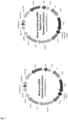
- Integrin alpha 5 (ITGA5) (NM_002205; #SC212067, Origene) and CXCR4 (NM_001008540; #SC207204) 3' UTR sequence were cloned separately, into downstream of the Firefly luciferase gene, in the Multiple Cloning Site pMiRTarget vector ( Figure 2 ).
- the empty pMiRTarget vector was used as the control of the assay.
- DH5 ⁇ Competent Cells (#18265017, Thermo Fisher) were used to amplify these three vectors according to the manufacturer's instructions.
- DNA Plasmids were obtained after purification using EndoFree Mini-Prep and Maxi-prep Plasmid Kits (#12123 #12162, Qiagen).
- pRL-SV40 Vector Promoter-Driven Control Renilla Luciferase Vector, #E6911, Promega was used for normalization in luciferase assays.
- luciferase assays Caco-2 cells were cultured in p24-well plates and transfection was performed at 70-80% of confluence. 500 ng of pMiRTarget Vector (containing a firefly luciferase gene, ITGA5 3'UTR or CXCR4 3'UTR), 0.5 ng of pRL-SV40 Vector (containing a Renilla luciferase gene) and Pre-miR-326 or Pre-miR-Scrambled with a final concentration of 0.25 nM, were simultaneously transfected per well using 2.5 ⁇ L of lipofectamine. Each transfection condition was carried out in quadrupled.
- RNA from cell cultures was extracted with guanidine thiocyanate-phenol-chloroform method using TriPure Isolation Reagent solution (#11667165001, Roche), following the manufacturer's instructions: mechanical lysis, phenol/chloroform extraction, and subsequent alcohols precipitation.
- glycogen #10814010, Invitrogen
- Tripure Isolation Reagent 20 ⁇ l/ml
- linear acrylamide #AM9520, Invitrogen
- RNA enriched in miRNAs was extracted with miRNeasy FFPE kit (# 217504, Qiagen), following the manufacturer's indications using enrichment columns purification and samples were stored at -80°C before qRT-PCRs.
- RNA integrity was checked by electrophoresis in agarose gels loading a solution mix composed of 1 ⁇ l of total RNA, 1 ⁇ l of DNA-Dye NonTox (A9555, PanReac AppliChem) and 4 ⁇ l of nuclease-free water. After electrophoresis, RNA bands were visualized under a UV-transilluminator, ChemiDoc MP Imaging System (BioRad). Finally, RNA was stored at -80°C until its use.
- RT Reverse transcription
- RNA from each sample were used for the reaction.
- RT finished 1 ⁇ l of the obtained cDNA was used as a template for the qRT-PCR, together with SYBR Green I Master Mix (#04887352001, Roche).
- Amplification was carried out in a LightCycler 480 thermal cycler (Roche) with the following temperature protocol: 40 cycles at 95°C for 10s, 60°C for 20s, and 72°C for 20s.
- Primers were synthetized by TIB Molbiol in a 100 ⁇ M stock concentration and diluted in a use concentration of 1:10 in nuclease-free water. Table 2. Oligonucleotides used for messenger expression by qRT-PCR. Product specifications and Forward and Reverse sequences are detailed.
- cDNA for miRNAs expression study was generated using the miRCURY LNA Universal RT microRNA, Polyadenylation, and cDNA Synthesis Kit II (#339340, EXIQON). Drown from the manufacturer's recommendations, 100-200 ng of isolated RNA from cells or paraffin-embedded tissue were used. As a control of cDNA synthesis efficiency, an external RNA (cel-miR-39) was added and further amplified.
- ⁇ CT miRNA CT - housekeeping CT, using the 2 - ⁇ CT formula, or folds, using the 2 - ⁇ CT formula.
- Normfinder and Bestkeeper software were used to determine the most stable housekeeping miRNAs.
- miR-191-5p and miR-103a-3p were finally selected as housekeeping for data normalization in case of colorectal cancer cells and murine macrophages.
- the mean of 5S rRNA Cq and RNU6B Cq as housekeeping was used for normalization in tissue samples from patients' biopsies. All LNA miRNAs probes used in this work were obtained from Qiagen and used at a final concentration of 10 ⁇ M (Error! Reference source not found.).
- In situ hybridization uses a complementary RNA probe to localize a specific RNA sequence, in this case, a miRNA in the tissue.
- a complementary RNA probe to localize a specific RNA sequence, in this case, a miRNA in the tissue.
- deparaffinized and rehydrated colorectal sections were treated with Proteinase K (#158918, Qiagen) (3 ⁇ L/ml in PBS 1X diluted in DEPC water) for 15 minutes at 37°C to digest the tissue and allow the probe to penetrate in the cells. Then, they were washed with diethylpyrocarbonate-treated (DEPC-treated) water for 5 min at room temperature to remove the exceeded product.
- DEPC-treated diethylpyrocarbonate-treated
- Protein extraction was performed by scraping the p60/p6 culture plates with 600/300 ⁇ L RIPA Buffer (#R0278, Sigma-Aldrich), supplemented with protease inhibitors: 1mM DTT (#43815, Sigma), 10 ⁇ g/ml Leupeptin (#L9783, Sigma), 10 ⁇ g/ml Aprotinin (#A1153, Sigma), 1mM Pefabloc (#76307, Fluka) and a cocktail of phosphatase inhibitors (#524625, Millipore). The cell lysate was collected and kept in constant agitation on a shaker rotatory mixer for 20 minutes at 4°C. Finally, it was centrifuged for 10 minutes at 15000 rpm at 4°C to precipitate the insoluble residues. Supernatants were collected and stored at - 80°C for experimental approaches.
- Total protein concentration in cell lysate was estimated by Bradford colorimetric technique. A standard curve was made with serial dilutions of BSA (#10711454001, Sigma-Aldrich), and 5 ⁇ L of protein lysate was used for quantification at 595 O.D. in an absorbance spectrophotometer (Multiskan GO, Thermo Fisher). 50 ⁇ g of total proteins were electrophoretically separated into SDS-polyacrylamide gels (Biorad) under denaturing conditions and transferred to nitrocellulose membranes (#iB23001, Invitrogen), in an iBlot TM 2 Gel Transfer Device (Invitrogen).
- Nitrocellulose membranes were incubated in a 1X TBS blocking solution + 5% BSA + 0.1% Tween (#142312, Panreac Qu ⁇ mica S.A.), for 1h at room temperature. After this, the membranes were incubated overnight at 4°C with the corresponding dilutions of primary antibodies in 1X TBS + 5% BSA + 0.1% Tween.
- FFPE paraffin-embedded
- Immunohistochemistry was performed to visualize protein expression content and localization in tissue samples through the detection of specific epitope-antibody interactions, also labelled. 3 ⁇ m sections of FFPE tumour biopsies were rehydrated and heat-treated using a pressure cooker to unmask the antigenic epitopes. Antigen retrieval solution and boiling time suitable for each antibody are specified in Error! Reference source not found. Then, slides were tempered for 20 min immersed in the preferred retrieval solution and washed for 5 min in 1X PBS.
- Immunostaining of CXCR4, ITGA5, and immune infiltrate molecules were evaluated.
- the evaluation of the immunostainings was carried out in two compartments (intratumoral and peritumoral area).
- the intratumoral evaluation was made in the central area of the tumour, excluding as far as possible the superficial area (1/3).
- the peritumoral evaluation was made on the deep border of infiltration.
- Six high magnification fields (40x objective) were chosen randomly in each compartment and positive cells were counted. The result is a SCORE calculated by the average of the six 40x fields.
- CXCR4 was evaluated in the intratumoral and peritumoral areas as was mentioned before. CXCR4 staining was analysed by combining two variables: extension and intensity of the stain and a SCORE was acquired from these two variables.
- - ITGA5 was evaluated in the intratumoral and peritumoral areas of the CRC tissues.
- - CD56 was evaluated in the membrane of neuroectoderm-derived cells and NK cell-derived lymphomas in intratumoral and peritumoral areas of the CRC tissue.
- - CD68 was evaluated in the cytoplasm and the membrane of monocytes and M1 macrophages in intratumoral and peritumoral areas of the CRC tissue.
- - CD8 was evaluated in the membrane of cytotoxic T-cell lymphocytes in intratumoral and peritumoral areas of the CRC tissue.
- - CD25 was evaluated in the cytoplasm and the membrane of T cells and mastocytes in intratumoral and peritumoral areas of the CRC tissue.
- - CD163 was evaluated in the cytoplasm and the membrane of activated or anti-inflammatory M2 macrophages in intratumoral and peritumoral areas of the CRC tissue.
- - CD11c was evaluated in the cytoplasm of dendritic cells in intratumoral and peritumoral areas of the CRC tissue.
- - Myeloperoxidase was evaluated in the cytoplasm of granulocytes and monocytes in intratumoral and peritumoral areas of the CRC tissue.
- Granzyme B was evaluated in the cytoplasm of cytotoxic lymphocytes (CTL or NK cells) in intratumoral and peritumoral areas of the CRC tissue.
- - Perforin was evaluated in the perinuclear cytoplasm of cytotoxic lymphocytes (CD4+, CD8+ or NK cells) in intratumoral and peritumoral areas of the CRC tissue.
- the median value of miR-326 expression in all samples was chosen as the cut-off point for separating low- and high-level expression.
- the correlation between miR-326 and the inflammatory infiltrate, ITGA5 and CXCR4 is carried out through linear regression with 95% of confidence of the number of positive cells and miR-326 expression is expressed as 2 - ⁇ CT .
- SPSS Social Sciences
- miR-326 levels were determined by qRT-PCR in tumour biopsies.
- Statistical analysis demonstrates that miR-326 is upregulated in left-side patients' colorectal cancers (LCRC), compared to right-side colorectal tumours (RCRC), as can be observed in Figure 4A .
- Figure 48 shows that higher expression levels of miR-326 were found in CRC biopsies with reduced stroma compared to the patients with pronounced stroma, which have lower miR-326 expression.
- high levels of miR-326 are associated with an increased inflammatory infiltrate compared to those biopsies exhibiting low infiltrate ( Figure 4C ) .
- miR-326 is upregulated in less invasive tumour grades (Tis (tumour in situ)-T1-T2) compared to late stages of invasiveness (T3-T4). Moreover, Figure 5C shows that miR-326 expression is also related to the lymph node's affectation. miR-326 levels are upregulated in tumors with lymph nodes non-affected.
- CD8 recognise cytotoxic T-cell lymphocytes
- CD25 binds to T cells and mastocytes
- CD68 recognise monocytes and M1 macrophages
- CD56 identify NK cells
- CD163 binds to M2 macrophages
- CD11c recognise dendritic cells
- Perforin and Granzyme B identify CTL and NK cells
- Myeloperoxidase identify granulocytes and monocytes.
- a specialized pathologist examined and quantified the immunostainings. Representative images of these markers are shown ( Figure 8 ) .
- miR-326 expression levels were determined by qRT-PCR in each sample. A correlation analysis between miR-326 levels and staining quantification of each biopsy was performed. There were no statistical differences between miR-326 expression and CD8, CD25, CD163, CD11c, Perforin, Granzyme B and Myeloperoxidase immunostaining. However, we found a positive correlation between the number of CD68, and CD56 positive cells with miR-326 expression levels in CRC biopsies ( Figure 9 ). As we previously mentioned, CD68 is highly expressed in M1 macrophages and CD56 is the archetypal phenotypic marker of NK cells.
- miR-326 basal levels were determined using qRT-PCR in 3 different epithelial cell lines coming from colon adenocarcinoma (Caco-2, HT-29 and RKO), in a cell line derived from lymph nodes of CRC (SW620), and an epithelial non-tumoral colon cell line (CCD841).
- Caco-2 presented the highest expression levels of miR-326. Significant differences are observed between Caco-2 cells and the rest of the cell lines. miR-326 expression in HT-29 cells was 3 times lower than in Caco-2 cells. SW620 and RKO cell lines had lower expression of miR-326 than CCD841.
- FIG 11A an efficient overexpression of miR-326 was observed in the pre-miR-326 condition compared to pre-miR-Scrambled.
- miR-326 is 30-folds increased comparing pre-miR-326 with pre-miR-Scrambled at 24 hours and 20-folds increased at 48 hours post-transfection.
- Figure 17B shows the reduction of miR-326 expression in cells transfected with Anti-miR-326 compared with Anti-miR-Scrambled. The reduction of the miRNA is 10 folds-decreased comparing Anti-miR-326 and Anti-miR-Scrambled at both times points post-transfection.
- miR-326 is involved in the cell proliferation of Caco-2 cells and overexpression of this miRNA decreases cell proliferation. For this reason, we studied the involvement of miR-326 in the cell cycle distribution of Caco-2 cells. These studies were carried out in miR-326 modulated Caco-2 cells by staining with propidium iodide and following analysis by flow cytometry.
- miR-326 was involved in relevant processes for tumor progression such as proliferation, cell-cycle regulation, and EMT in Caco-2 cells.
- a bioinformatic target prediction study using different databases available online, was performed to determine validated and predicted miR-326 targets involved in the development and progression of CRC.
- the integrin ⁇ family more specifically, the gene encoding the ⁇ -5 beta-1 integrin (ITGA5) was found as a miR-326 predicted and non-validated target gene.
- ITGA5 was correlated with proliferation, migration and poor overall survival in CRC (Lu et al. 2019). Based on this evidence, we hypothesized that this gene could be regulated by miR-326 in our in vitro system.
- miRNAs negatively regulate gene expression at mRNA and protein levels by degrading their mRNA targets and/or by silencing translation.
- miR-326 regulates the expression of ITGA5
- miR-326 was modulated in Caco-2 cells and ITGA5 protein expression was determined by immunoblot.
- overexpression of miR-326 in Caco-2 cells reduces ITGA5 protein levels compared with its pre-miR-Scrambled, at 24 and 48 hours post-transfection.
- inhibition of the microRNA increases ITGA5 protein expression mostly at 48 hours post-transfection.
- ITGA5 3'-UTR was cloned into pMiRTarget 3'-UTR Vectors with firefly luciferase as a reporter and luciferase activity assays were carried out. Luciferase gene expression was driven by SV-40 promoter and ITGA5 3'-UTR was cloned after the luciferase gene.
- pMiRTarget Empty Vector was used as control.
- pMiRTarget 3'-UTR ITGA5 and pMiRTarget Empty Vector were co-transfected with both Pre-miR-326 and Pre-miR-Scrambled in Caco-2 cells.
- Renilla luciferase activity was used for normalization control. Data are shown as the percentage of luciferase activity reduction by Pre-miR-326 in comparison to Pre-miR-Scrambled control. This experiment demonstrated that miR-326 overexpression significantly reduced luciferase activity compared with the Scrambled condition. Moreover, miR-326 overexpression did not reduce luciferase activity when was transfected with pMiRTarget Empty Vector. Statistical significance was observed between 3'-UTR ITGA5 and 3'-UTR Empty Vector transfected with miR-326. All these data demonstrate, for the first time, that miR-326 directly regulates ITGA5 expression through recognition and binding to its 3'-UTR in Caco-2 cells ( Figure 17 ).
- ITGA5 is the main receptor for soluble fibronectin. Fibronectin plays an important role in cell adhesion, spreading, migration, differentiation and high levels of fibronectin are related to cancer progression.
- miR-326 was involved in cell adhesion to fibronectin, as a tumoral matrix, through ITGA5 having relevance in tumor development and progression.
- fibronectin matrix adhesion assays were carried out in Caco-2 cells previously modulated with miR-326 and the number of cells attached to a fibronectin were quantified. As can be observed in Figure 18 , a significant decrease in cell adhesion was observed when miR-326 was overexpressed compared to Pre-miR-Scrambled condition. A tendency of increased adhesion when miR-326 is inhibited can be observed but no statistical significance was found.
- SNAI1 gene expression is modulated by miR-326.
- Protein expression of SNAI1, SNAIL protein enhances cell detachment and leads to cell migration by changing the expression of integrins (Haraguchi et al. 2008).
- ITGA5 ITGA5 as a direct target of miR-326
- miR-326 was modulated in Caco-2 cells, proteins were isolated and SNAIL expression was determined by immunoblot.
- overexpression of miR-326 reduces SNAIL protein levels with pre-miR-Scrambled at 24 hours post-transfection and inhibition of the miRNA increases the SNAIL expression at 48 hours post-transfection.
- the relative quantification of SNAIL levels is shown in Figure 19B .
- CXCR4 IS A MIR-326 TARGET IN CACO-2 CELLS
- CXC chemokines family
- miR-326 overexpression did not reduce luciferase activity compared with the Scrambled when transfected with pMiRTarget Empty Vector.
- Statistical significance was observed between 3'-UTR CXCR4 and 3'-UTR Empty Vector transfected with miR-326. Data are shown as the percentage of luciferase activity reduction by Pre-miR-326 in comparison to Scrambled control. All these data demonstrate for the first time that CXCR4 expression is directly regulated by miR-326 through recognition and binding to its 3'UTR in Caco-2 cells.
- Figure 22B shows a representative image of CXCR4 and the pertinent correlation study between miR-326 and CXCR4.
- ITGA5 correlation an inverse correlation between miR-326 and CXCR4 expression is observed.
- our results have demonstrated both molecules as miR-326 targets and confirm the relation between miR-326 and ITGA5 and CXCR4 also in patients' biopsies.
- the M2[IL-4] phenotype as an anti-inflammatory macrophage, expresses higher amounts of YM1 compared to naive macrophages (M ⁇ [Nonstimulated]) and the pro-inflammatory phenotype (M1[LPS + IFNy]).
- miR-326 is highly reduced in M1[LPS + IFNy] and increased in M2[IL4] phenotype compared to M ⁇ [Non-stimulated].
- the next step was to modulate the miRNA expression in the different polarized groups (M ⁇ [Non-stimulated], M1[LPS + IFNy], and M2[IL-4]).
- This modulation was performed by transient transfection to overexpress and inhibit the miRNA with Pre-miR-326 and Anti-miR-326 technology, respectively, and using Pre-miR-Scrambled and Anti-miR-Scrambled sequences as control groups.
- FIG 25 shows the transfection efficiency in each stimulation condition.
- miR-326 was significantly overexpressed in the cells treated with Pre-miR-326 compared to the control group (Pre-miR-Scrambled).
- miR-326 expression was significantly inhibited in Anti-miR-326 treated group compared to the control (Anti-miR-Scrambled).
- This efficient miR-326 modulation was obtained in the three different macrophage stimulations M ⁇ [Non-stimulated], M1[LPS + IFNy] and M2[IL4].
- YM1 expression (M2 macrophage marker) was not affected by miR-326 in M ⁇ [Non-stimulated] and in M1[LPS + IFN ⁇ ] macrophages.
- YM1 expression levels in an M2[IL4] context decreases when miR-326 is inhibited.
- CXCR4 and ITGA5 are two miR-326 direct targets in Caco-2 cells and present an inverse correlation in our tumour biopsies. Moreover, we wanted to determine if these two miR-326 targets in epithelial cells could be also modulated by miR-326 in naive macrophages (M ⁇ [Non-stimulated] and in induced phenotypes; M1[LPS + IFNy], and M2[IL-4]). For this purpose, CXCR4 and ITGA5 expression was studied by qRT-PCR in the different macrophages experimental groups, in which miR-326 was subsequently modulated.
- ITGA5 could be also a direct target of miR-326 in macrophages but further analysis by means of immunoblot and luciferase assays should be done to confirm it.
- MC-38 immunoresponsive murine model which has a favourable response profile to immune activation.
- This in vivo model is based on subcutaneous injection of MC-38, murine colorectal cancer cells, to develop colon adenocarcinoma.
- miR-326 was overexpressed in MC-38 cells by lentiviral vectors before being injected into the mice. Because miRNAs activity is specie-specific, first, we wanted to corroborate that miR-326 was involved in biological processes related to tumour progression in MC-38 murine cells.
- miR-326 was modulated in MC-38 cells following the same transfection protocol carried on with Lipofectamine in human Caco-2 cells.
- miR-326 expression was analyzed by qRT-PCR. Error! Reference source not found.
- 1A shows a clear overexpression of miR-326 in the pre-miR-326 condition compared to Pre-miR-Scrambled at 24-h and 48 hours.
- Figure 31 B shows the reduction of miR-326 expression in cells transfected with Anti-miR-326 compared with Anti-miR-Scrambled at 24-h and 48 hours, as we expected.
- mice pre-microRNA (MMIR-326) and Scrambled negative control (MMIR-000) constructs were generated, amplificated and tittered at ⁇ 9 ⁇ 10 7 IFU/ml.
- MMIR-326 and MMIR-000 Scrambled negative control constructs were generated, amplificated and tittered at ⁇ 9 ⁇ 10 7 IFU/ml.
- MC-38 cells were infected with MMIR-326 and MMIR-000 separately to calculate the multiplicity of infection of these murine CRC cells.
Landscapes
- Health & Medical Sciences (AREA)
- Engineering & Computer Science (AREA)
- Life Sciences & Earth Sciences (AREA)
- Genetics & Genomics (AREA)
- Biomedical Technology (AREA)
- Organic Chemistry (AREA)
- Chemical & Material Sciences (AREA)
- Biotechnology (AREA)
- Wood Science & Technology (AREA)
- Zoology (AREA)
- Bioinformatics & Cheminformatics (AREA)
- General Engineering & Computer Science (AREA)
- General Health & Medical Sciences (AREA)
- Biochemistry (AREA)
- Microbiology (AREA)
- Molecular Biology (AREA)
- Plant Pathology (AREA)
- Physics & Mathematics (AREA)
- Biophysics (AREA)
- Cell Biology (AREA)
- Gastroenterology & Hepatology (AREA)
- Immunology (AREA)
- Oncology (AREA)
- Virology (AREA)
- Hematology (AREA)
- Chemical Kinetics & Catalysis (AREA)
- General Chemical & Material Sciences (AREA)
- Medicinal Chemistry (AREA)
- Nuclear Medicine, Radiotherapy & Molecular Imaging (AREA)
- Pharmacology & Pharmacy (AREA)
- Animal Behavior & Ethology (AREA)
- Public Health (AREA)
- Veterinary Medicine (AREA)
- Measuring Or Testing Involving Enzymes Or Micro-Organisms (AREA)
- Medicines That Contain Protein Lipid Enzymes And Other Medicines (AREA)
Priority Applications (3)
| Application Number | Priority Date | Filing Date | Title |
|---|---|---|---|
| EP22383047.2A EP4360657A1 (fr) | 2022-10-28 | 2022-10-28 | Mir-29 pour le traitement du cancer |
| PCT/EP2023/080239 WO2024089293A1 (fr) | 2022-10-28 | 2023-10-30 | Mir-326 pour le traitement du cancer |
| EP23798943.9A EP4608415A1 (fr) | 2022-10-28 | 2023-10-30 | Mir-326 pour le traitement du cancer |
Applications Claiming Priority (1)
| Application Number | Priority Date | Filing Date | Title |
|---|---|---|---|
| EP22383047.2A EP4360657A1 (fr) | 2022-10-28 | 2022-10-28 | Mir-29 pour le traitement du cancer |
Publications (1)
| Publication Number | Publication Date |
|---|---|
| EP4360657A1 true EP4360657A1 (fr) | 2024-05-01 |
Family
ID=84332137
Family Applications (2)
| Application Number | Title | Priority Date | Filing Date |
|---|---|---|---|
| EP22383047.2A Withdrawn EP4360657A1 (fr) | 2022-10-28 | 2022-10-28 | Mir-29 pour le traitement du cancer |
| EP23798943.9A Pending EP4608415A1 (fr) | 2022-10-28 | 2023-10-30 | Mir-326 pour le traitement du cancer |
Family Applications After (1)
| Application Number | Title | Priority Date | Filing Date |
|---|---|---|---|
| EP23798943.9A Pending EP4608415A1 (fr) | 2022-10-28 | 2023-10-30 | Mir-326 pour le traitement du cancer |
Country Status (2)
| Country | Link |
|---|---|
| EP (2) | EP4360657A1 (fr) |
| WO (1) | WO2024089293A1 (fr) |
Citations (12)
| Publication number | Priority date | Publication date | Assignee | Title |
|---|---|---|---|---|
| US4373071A (en) | 1981-04-30 | 1983-02-08 | City Of Hope Research Institute | Solid-phase synthesis of polynucleotides |
| US4401796A (en) | 1981-04-30 | 1983-08-30 | City Of Hope Research Institute | Solid-phase synthesis of polynucleotides |
| US4415732A (en) | 1981-03-27 | 1983-11-15 | University Patents, Inc. | Phosphoramidite compounds and processes |
| US4458066A (en) | 1980-02-29 | 1984-07-03 | University Patents, Inc. | Process for preparing polynucleotides |
| US4500707A (en) | 1980-02-29 | 1985-02-19 | University Patents, Inc. | Nucleosides useful in the preparation of polynucleotides |
| US4668777A (en) | 1981-03-27 | 1987-05-26 | University Patents, Inc. | Phosphoramidite nucleoside compounds |
| US4973679A (en) | 1981-03-27 | 1990-11-27 | University Patents, Inc. | Process for oligonucleo tide synthesis using phosphormidite intermediates |
| US5047524A (en) | 1988-12-21 | 1991-09-10 | Applied Biosystems, Inc. | Automated system for polynucleotide synthesis and purification |
| US5132418A (en) | 1980-02-29 | 1992-07-21 | University Patents, Inc. | Process for preparing polynucleotides |
| US5153319A (en) | 1986-03-31 | 1992-10-06 | University Patents, Inc. | Process for preparing polynucleotides |
| US5262530A (en) | 1988-12-21 | 1993-11-16 | Applied Biosystems, Inc. | Automated system for polynucleotide synthesis and purification |
| US5700642A (en) | 1995-05-22 | 1997-12-23 | Sri International | Oligonucleotide sizing using immobilized cleavable primers |
-
2022
- 2022-10-28 EP EP22383047.2A patent/EP4360657A1/fr not_active Withdrawn
-
2023
- 2023-10-30 EP EP23798943.9A patent/EP4608415A1/fr active Pending
- 2023-10-30 WO PCT/EP2023/080239 patent/WO2024089293A1/fr not_active Ceased
Patent Citations (12)
| Publication number | Priority date | Publication date | Assignee | Title |
|---|---|---|---|---|
| US4458066A (en) | 1980-02-29 | 1984-07-03 | University Patents, Inc. | Process for preparing polynucleotides |
| US4500707A (en) | 1980-02-29 | 1985-02-19 | University Patents, Inc. | Nucleosides useful in the preparation of polynucleotides |
| US5132418A (en) | 1980-02-29 | 1992-07-21 | University Patents, Inc. | Process for preparing polynucleotides |
| US4415732A (en) | 1981-03-27 | 1983-11-15 | University Patents, Inc. | Phosphoramidite compounds and processes |
| US4668777A (en) | 1981-03-27 | 1987-05-26 | University Patents, Inc. | Phosphoramidite nucleoside compounds |
| US4973679A (en) | 1981-03-27 | 1990-11-27 | University Patents, Inc. | Process for oligonucleo tide synthesis using phosphormidite intermediates |
| US4373071A (en) | 1981-04-30 | 1983-02-08 | City Of Hope Research Institute | Solid-phase synthesis of polynucleotides |
| US4401796A (en) | 1981-04-30 | 1983-08-30 | City Of Hope Research Institute | Solid-phase synthesis of polynucleotides |
| US5153319A (en) | 1986-03-31 | 1992-10-06 | University Patents, Inc. | Process for preparing polynucleotides |
| US5047524A (en) | 1988-12-21 | 1991-09-10 | Applied Biosystems, Inc. | Automated system for polynucleotide synthesis and purification |
| US5262530A (en) | 1988-12-21 | 1993-11-16 | Applied Biosystems, Inc. | Automated system for polynucleotide synthesis and purification |
| US5700642A (en) | 1995-05-22 | 1997-12-23 | Sri International | Oligonucleotide sizing using immobilized cleavable primers |
Non-Patent Citations (4)
| Title |
|---|
| "Remington's Pharmaceutical Sciences", MACK PUBLISHING CO. |
| A. KOZOMARA ET AL: "miRBase: integrating microRNA annotation and deep-sequencing data", NUCLEIC ACIDS RESEARCH, vol. 39, no. Database, 30 October 2010 (2010-10-30), GB, pages D152 - D157, XP055747242, ISSN: 0305-1048, DOI: 10.1093/nar/gkq1027 * |
| CHEN YUN ET AL: "Knockdown of lncRNA PCAT6 suppresses the growth of non-small cell lung cancer cells by inhibiting macrophages M2 polarization via miR-326/KLF1 axis", BIOENGINEERED, vol. 13, no. 5, 2 May 2022 (2022-05-02), US, pages 12834 - 12846, XP093065128, ISSN: 2165-5979, DOI: 10.1080/21655979.2022.2076388 * |
| SHAO LIJUAN ET AL: "MicroRNA-326 attenuates immune escape and prevents metastasis in lung adenocarcinoma by targeting PD-L1 and B7-H3", CELL DEATH DISCOVERY, vol. 7, no. 1, 15 June 2021 (2021-06-15), XP093065195, Retrieved from the Internet <URL:https://www.nature.com/articles/s41420-021-00527-8> DOI: 10.1038/s41420-021-00527-8 * |
Also Published As
| Publication number | Publication date |
|---|---|
| WO2024089293A1 (fr) | 2024-05-02 |
| EP4608415A1 (fr) | 2025-09-03 |
Similar Documents
| Publication | Publication Date | Title |
|---|---|---|
| CN109312341B (zh) | 微小rna及其使用方法 | |
| CA2789404C (fr) | Procedes et compositions destines a la detection et au traitement du cancer impliquant des miarn et des inhibiteurs des miarn ainsi que des cibles | |
| JP6153932B2 (ja) | 骨肉腫のためのマイクロrnaに基づく方法およびアッセイ | |
| CN102725420A (zh) | 用于染色体重排相关肿瘤的诊断、预后和治疗的基于微小rna的方法和组合物 | |
| WO2012006181A2 (fr) | Compositions et procédés pour l'inhibition de micro-arn oncogènes et le traitement du cancer | |
| US10053696B2 (en) | MiRNA-489 in treatment of breast cancer | |
| Tuo et al. | miR-532–3p suppresses proliferation and invasion of ovarian cancer cells via GPNMB/HIF-1α/HK2 axis | |
| WO2012108843A1 (fr) | Traitement du cancer par inhibition de l'expression d'olfm4, de sp5, de tobi, d'arjdia, de fbni ou d'hat1 | |
| US20140045924A1 (en) | Melanoma treatments | |
| CN111440874A (zh) | 口腔鳞癌诊断和治疗用生物标志物 | |
| Hu et al. | MicroRNA-199a-3p suppresses the invasion and metastasis of nasopharyngeal carcinoma through SCD1/PTEN/AKT signaling pathway | |
| CN116024211B (zh) | tRNA衍生物tRF-His-008在诊断、治疗肾癌中的应用 | |
| Wang et al. | MicroRNA-23b regulates nasopharyngeal carcinoma cell proliferation and metastasis by targeting E-cadherin | |
| CN111455059A (zh) | 检测及靶向生物标志物的试剂在口腔鳞癌中的应用 | |
| CN111455060A (zh) | 诊断和治疗口腔鳞癌的相关生物标志物及应用 | |
| CN109321655B (zh) | Nkiras2基因调控区序列、调控序列及其在鼻咽癌中的应用 | |
| CN111172290A (zh) | 肝细胞癌诊断和治疗的miRNA | |
| CN111455061A (zh) | lncRNA生物标志物在口腔鳞癌诊疗中的应用 | |
| WO2024089293A1 (fr) | Mir-326 pour le traitement du cancer | |
| WO2014066498A1 (fr) | Traitement du cancer du sein métastatique | |
| CN107354159A (zh) | 长链非编码RNA SMAD5‑AS1的siRNA在卵巢癌治疗中的应用 | |
| WO2011044504A1 (fr) | Modulation de niveaux de protéines de liaison à l'arn pour le traitement du cancer du sein | |
| CN111440875A (zh) | 基于生物标志物的诊断和治疗癌症的用途 | |
| CN110863055A (zh) | 长链非编码rna在乳腺癌诊治中的应用 | |
| CN110951886A (zh) | Linc01978在诊治乳腺癌中的用途 |
Legal Events
| Date | Code | Title | Description |
|---|---|---|---|
| PUAI | Public reference made under article 153(3) epc to a published international application that has entered the european phase |
Free format text: ORIGINAL CODE: 0009012 |
|
| STAA | Information on the status of an ep patent application or granted ep patent |
Free format text: STATUS: THE APPLICATION HAS BEEN PUBLISHED |
|
| AK | Designated contracting states |
Kind code of ref document: A1 Designated state(s): AL AT BE BG CH CY CZ DE DK EE ES FI FR GB GR HR HU IE IS IT LI LT LU LV MC ME MK MT NL NO PL PT RO RS SE SI SK SM TR |
|
| STAA | Information on the status of an ep patent application or granted ep patent |
Free format text: STATUS: THE APPLICATION IS DEEMED TO BE WITHDRAWN |
|
| 18D | Application deemed to be withdrawn |
Effective date: 20241105 |