EP4386086A1 - Composition pour le traitement d'une maladie liée au cartilage et procédé de préparation associé - Google Patents
Composition pour le traitement d'une maladie liée au cartilage et procédé de préparation associé Download PDFInfo
- Publication number
- EP4386086A1 EP4386086A1 EP22856239.3A EP22856239A EP4386086A1 EP 4386086 A1 EP4386086 A1 EP 4386086A1 EP 22856239 A EP22856239 A EP 22856239A EP 4386086 A1 EP4386086 A1 EP 4386086A1
- Authority
- EP
- European Patent Office
- Prior art keywords
- cells
- cartilage
- pharmaceutical composition
- chondroprogenitor
- present disclosure
- Prior art date
- Legal status (The legal status is an assumption and is not a legal conclusion. Google has not performed a legal analysis and makes no representation as to the accuracy of the status listed.)
- Pending
Links
Images












Classifications
-
- C—CHEMISTRY; METALLURGY
- C12—BIOCHEMISTRY; BEER; SPIRITS; WINE; VINEGAR; MICROBIOLOGY; ENZYMOLOGY; MUTATION OR GENETIC ENGINEERING
- C12N—MICROORGANISMS OR ENZYMES; COMPOSITIONS THEREOF; PROPAGATING, PRESERVING, OR MAINTAINING MICROORGANISMS; MUTATION OR GENETIC ENGINEERING; CULTURE MEDIA
- C12N5/00—Undifferentiated human, animal or plant cells, e.g. cell lines; Tissues; Cultivation or maintenance thereof; Culture media therefor
- C12N5/06—Animal cells or tissues; Human cells or tissues
-
- A—HUMAN NECESSITIES
- A61—MEDICAL OR VETERINARY SCIENCE; HYGIENE
- A61K—PREPARATIONS FOR MEDICAL, DENTAL OR TOILETRY PURPOSES
- A61K35/00—Medicinal preparations containing materials or reaction products thereof with undetermined constitution
- A61K35/12—Materials from mammals; Compositions comprising non-specified tissues or cells; Compositions comprising non-embryonic stem cells; Genetically modified cells
- A61K35/32—Bones; Osteocytes; Osteoblasts; Tendons; Tenocytes; Teeth; Odontoblasts; Cartilage; Chondrocytes; Synovial membrane
-
- A—HUMAN NECESSITIES
- A61—MEDICAL OR VETERINARY SCIENCE; HYGIENE
- A61P—SPECIFIC THERAPEUTIC ACTIVITY OF CHEMICAL COMPOUNDS OR MEDICINAL PREPARATIONS
- A61P19/00—Drugs for skeletal disorders
- A61P19/02—Drugs for skeletal disorders for joint disorders, e.g. arthritis, arthrosis
-
- A—HUMAN NECESSITIES
- A61—MEDICAL OR VETERINARY SCIENCE; HYGIENE
- A61P—SPECIFIC THERAPEUTIC ACTIVITY OF CHEMICAL COMPOUNDS OR MEDICINAL PREPARATIONS
- A61P19/00—Drugs for skeletal disorders
- A61P19/04—Drugs for skeletal disorders for non-specific disorders of the connective tissue
-
- C—CHEMISTRY; METALLURGY
- C07—ORGANIC CHEMISTRY
- C07K—PEPTIDES
- C07K14/00—Peptides having more than 20 amino acids; Gastrins; Somatostatins; Melanotropins; Derivatives thereof
- C07K14/435—Peptides having more than 20 amino acids; Gastrins; Somatostatins; Melanotropins; Derivatives thereof from animals; from humans
- C07K14/46—Peptides having more than 20 amino acids; Gastrins; Somatostatins; Melanotropins; Derivatives thereof from animals; from humans from vertebrates
- C07K14/47—Peptides having more than 20 amino acids; Gastrins; Somatostatins; Melanotropins; Derivatives thereof from animals; from humans from vertebrates from mammals
-
- C—CHEMISTRY; METALLURGY
- C07—ORGANIC CHEMISTRY
- C07K—PEPTIDES
- C07K14/00—Peptides having more than 20 amino acids; Gastrins; Somatostatins; Melanotropins; Derivatives thereof
- C07K14/435—Peptides having more than 20 amino acids; Gastrins; Somatostatins; Melanotropins; Derivatives thereof from animals; from humans
- C07K14/705—Receptors; Cell surface antigens; Cell surface determinants
-
- C—CHEMISTRY; METALLURGY
- C07—ORGANIC CHEMISTRY
- C07K—PEPTIDES
- C07K14/00—Peptides having more than 20 amino acids; Gastrins; Somatostatins; Melanotropins; Derivatives thereof
- C07K14/435—Peptides having more than 20 amino acids; Gastrins; Somatostatins; Melanotropins; Derivatives thereof from animals; from humans
- C07K14/78—Connective tissue peptides, e.g. collagen, elastin, laminin, fibronectin, vitronectin or cold insoluble globulin [CIG]
-
- C—CHEMISTRY; METALLURGY
- C12—BIOCHEMISTRY; BEER; SPIRITS; WINE; VINEGAR; MICROBIOLOGY; ENZYMOLOGY; MUTATION OR GENETIC ENGINEERING
- C12N—MICROORGANISMS OR ENZYMES; COMPOSITIONS THEREOF; PROPAGATING, PRESERVING, OR MAINTAINING MICROORGANISMS; MUTATION OR GENETIC ENGINEERING; CULTURE MEDIA
- C12N13/00—Treatment of microorganisms or enzymes with electrical or wave energy, e.g. magnetism, sonic waves
-
- C—CHEMISTRY; METALLURGY
- C12—BIOCHEMISTRY; BEER; SPIRITS; WINE; VINEGAR; MICROBIOLOGY; ENZYMOLOGY; MUTATION OR GENETIC ENGINEERING
- C12N—MICROORGANISMS OR ENZYMES; COMPOSITIONS THEREOF; PROPAGATING, PRESERVING, OR MAINTAINING MICROORGANISMS; MUTATION OR GENETIC ENGINEERING; CULTURE MEDIA
- C12N5/00—Undifferentiated human, animal or plant cells, e.g. cell lines; Tissues; Cultivation or maintenance thereof; Culture media therefor
- C12N5/06—Animal cells or tissues; Human cells or tissues
- C12N5/0602—Vertebrate cells
- C12N5/0652—Cells of skeletal and connective tissues; Mesenchyme
- C12N5/0655—Chondrocytes; Cartilage
-
- C—CHEMISTRY; METALLURGY
- C12—BIOCHEMISTRY; BEER; SPIRITS; WINE; VINEGAR; MICROBIOLOGY; ENZYMOLOGY; MUTATION OR GENETIC ENGINEERING
- C12N—MICROORGANISMS OR ENZYMES; COMPOSITIONS THEREOF; PROPAGATING, PRESERVING, OR MAINTAINING MICROORGANISMS; MUTATION OR GENETIC ENGINEERING; CULTURE MEDIA
- C12N2506/00—Differentiation of animal cells from one lineage to another; Differentiation of pluripotent cells
- C12N2506/13—Differentiation of animal cells from one lineage to another; Differentiation of pluripotent cells from connective tissue cells, from mesenchymal cells
- C12N2506/1346—Differentiation of animal cells from one lineage to another; Differentiation of pluripotent cells from connective tissue cells, from mesenchymal cells from mesenchymal stem cells
-
- C—CHEMISTRY; METALLURGY
- C12—BIOCHEMISTRY; BEER; SPIRITS; WINE; VINEGAR; MICROBIOLOGY; ENZYMOLOGY; MUTATION OR GENETIC ENGINEERING
- C12N—MICROORGANISMS OR ENZYMES; COMPOSITIONS THEREOF; PROPAGATING, PRESERVING, OR MAINTAINING MICROORGANISMS; MUTATION OR GENETIC ENGINEERING; CULTURE MEDIA
- C12N2513/00—3D culture
Definitions
- the present disclosure relates to a pharmaceutical composition for treatment of a cartilage-related disease prepared by applying an electric stimulus and a method for preparing the same.
- Degenerative arthritis which has been regarded as a geriatric disease, is now a factor that causes arthritis in young people because of incorrect lifestyle habits, increased traumatic arthritis due to excessive exercise, etc.
- stem cells can be differentiated into chondroprogenitor cells that have the characteristics of chondrocytes without expressing the mature chondrocyte marker COL2 by applying an electric stimulus and have completed the present disclosure.
- the present disclosure is directed to providing a pharmaceutical composition for treatment or prevention of a cartilage-related disease, which contains chondroprogenitor cells having the following characteristics or an aggregate thereof as an active ingredient: (a) not expressing COL2; and (b) the chondroprogenitor cells being substrate-stained by one or more selected from a group consisting of alcian blue, safranin O and toluidine blue.
- the present disclosure is also directed to providing a method for treatment or prevention of a cartilage-related disease, which includes a step of administering chondroprogenitor cells having the following characteristics or an aggregate thereof to a subject in need thereof: (a) not expressing COL2; and (b) the chondroprogenitor cells being substrate-stained by one or more selected from a group consisting of alcian blue, safranin O and toluidine blue.
- the present disclosure is also directed to providing a use of chondroprogenitor cells having the following characteristics or an aggregate thereof for preparing a medicine for treatment or prevention of a cartilage-related disease: (a) not expressing COL2; and (b) the chondroprogenitor cells being substrate-stained by one or more selected from a group consisting of alcian blue, safranin O and toluidine blue.
- the present disclosure provides a pharmaceutical composition for treatment or prevention of a cartilage-related disease, which contains chondroprogenitor cells having the following characteristics or an aggregate thereof as an active ingredient:
- the cartilage-related disease may be selected from a group consisting of osteoarthritis, arthritis, meniscus derangement, rheumatoid arthritis, tear of meniscus, triangular fibrocartilage complex tear, traumatic cartilage damage and degenerative arthritis.
- the chondroprogenitor cells may be induced to differentiate from stem cells.
- the stem cells may be mesenchymal stem cells.
- the induction of differentiation may be achieved by an electric stimulus.
- the electric stimulus may have a frequency of 0 to 20 Hz; an amplitude of -20 to 20 V; and a duty ratio of 0 to 80%.
- the chondroprogenitor cells may have a decreased expression level of one or more gene selected from a group consisting of COL1 and COL5 or a protein encoded by the gene as compared to mesenchymal stem cells; and an increased expression level of the COL6 gene or a protein encoded by the gene as compared to mesenchymal stem cells.
- the chondroprogenitor cells may have an increased expression level of one or more gene selected from a group consisting of GJB2, GJC1, PECAM1, CLDN2, CLDN7, CLDN10 and CLDN19 or a protein encoded by the gene as compared to mesenchymal stem cells.
- the pharmaceutical composition may be in a dosage form that is easy to be inserted directly into the cartilage.
- the aggregate of the chondroprogenitor cells may be in a spheroid form.
- the spheroid may have a diameter of 0.5-1.5 mm.
- the present disclosure also provides a method for preparing a pharmaceutical composition for treatment or prevention of a cartilage-related disease, which includes a step of preparing chondroprogenitor cells or an aggregate thereof by applying an electric stimulus to stem cells, wherein the chondroprogenitor cells have the following characteristics: (a) not expressing COL2; and (b) the chondroprogenitor cells being substrate-stained by one or more selected from a group consisting of alcian blue, safranin O and toluidine blue.
- the present disclosure also provides a method for treatment or prevention of a cartilage-related disease, which includes a step of administering chondroprogenitor cells having the following characteristics or an aggregate thereof to a subject in need thereof: (a) not expressing COL2; and (b) the chondroprogenitor cells being substrate-stained by one or more selected from a group consisting of alcian blue, safranin O and toluidine blue.
- the present disclosure also provides a use of chondroprogenitor cells having the following characteristics or an aggregate thereof for preparing a medicine for treatment or prevention of a cartilage-related disease: (a) not expressing COL2; and (b) the chondroprogenitor cells being substrate-stained by one or more selected from a group consisting of alcian blue, safranin O and toluidine blue.
- composition for treatment of a cartilage-related disease according to the present disclosure can be prepared by applying only an electric stimulus to a stem cell aggregate without introducing externally derived growth factors, etc. and it can be prepared without using expensive growth factors, it has the advantage of remarkably reducing medical expenses.
- the composition for treatment of a cartilage-related disease according to the present disclosure does not express COL2, which is a mature chondrocyte marker, as compared to the conventional cells prepared for treating a cartilage-related disease and, thus, has the advantage of less immune-related side effects than mature cells.
- stem cells can be differentiated into chondroprogenitor cells that have the characteristics of chondrocytes without expressing the mature chondrocyte marker COL2 by applying an electric stimulus and have completed the present disclosure.
- the present disclosure provides a pharmaceutical composition for treatment or prevention of a cartilage-related disease, which contains chondroprogenitor cells having the following characteristics or an aggregate thereof as an active ingredient: (a) not expressing COL2; and (b) the chondroprogenitor cells being substrate-stained by one or more selected from a group consisting of alcian blue, safranin O and toluidine blue.
- COL2 (collagen 2) forms the basis of articular cartilage and hyaline cartilage and accounts for 50% of total proteins in the cartilage and 85-90% of collagen in the articular cartilage. It is used as a typical marker of mature chondrocytes for this reason. Since the chondroprogenitor cells of the present disclosure do not express COL2 at all, it is confirmed that they are not mature chondrocytes.
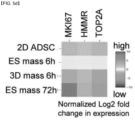
- the chondroprogenitor cells are prepared by applying an electric stimulus to stem cells, specifically mesenchymal stem cells, and are different from the mesenchymal stem cells themselves in that they do not express proliferation markers related with the sternness of the mesenchymal stem cells, such as MKI67, TOP2A and HMMR.
- the chondroprogenitor cells show characteristics similar to those of chondrocytes in that they can be substrate-stained with alcian blue, safranin O, etc. However, they are undifferentiated cells prior to differentiation into mature chondrocytes since they do not express COL2, which is a typical marker of mature chondrocytes, at all. They can be utilized as a formulation that can be easily implanted for treatment, etc. since they spontaneously aggregate into spheroids without external factors such as the addition of growth factors, centrifugation, etc.
- the cartilage-related disease may be selected from a group consisting of osteoarthritis, arthritis, meniscus derangement, rheumatoid arthritis, tear of meniscus, triangular fibrocartilage complex tear, traumatic cartilage damage and degenerative arthritis, although not being limited thereto.
- prevention refers to any action of suppressing symptoms or delaying the onset of a cartilage-related disease by administering the pharmaceutical composition according to the present disclosure.
- treatment refers to any action of improving or favorably changing the symptoms of a cartilage-related disease by administering the pharmaceutical composition according to the present disclosure.
- the active ingredient of the pharmaceutical composition may contain 90% or more, specifically 91%, 92%, 93%, 94%, 95%, 96%, 97%, 98%, 99% or more, of cells homogeneous with the chondroprogenitor cells.
- the cells may be those differentiated from stem cells.
- the stem cells may be mesenchymal stem cells.
- the mesenchymal stem cells (MSCs) of the present disclosure are cells that can differentiate into chondrocyte, osteocytes, adipocytes, myocytes, etc. and can be induced to differentiate into cartilage, bone, muscle, ligament and adipose tissues under specific culture conditions in vitro. Since the mesenchymal stem cells can be extracted easily from the bone marrow, many researches are being conducted on their potential for use as cell therapeutic agents for various intractable diseases. Since the cartilage lacks regenerative capacity, it is very difficult to be repaired once it is damaged. Since degenerative arthritis is caused by degenerative changes in the joint, it cannot be stopped completely. Currently, it is managed by drug therapy, physical therapy, etc.
- cartilage regeneration using the cell therapeutic agent can be applied not only to musculoskeletal diseases but also to diseases of the digestive and urinary systems. That is to say, it can be used for treatment of diseases such as reflux esophagitis, urethral reflux, etc. since it locally regenerates the cartilage tissue.
- the induction of differentiation in the present disclosure may be achieved by an electric stimulus.
- The, electric stimulus may have a frequency of 0 to 20 Hz, specifically 3 to 15 Hz or 5 to 12 Hz, although not being limited thereto.
- the electric stimulus may be a voltage with an amplitude of -20 V to 20 V, specifically -15 V to 15 V, more specifically -10 V to 10 V.
- the chondroprogenitor cells of the present disclosure may have, a decreased expression level of one or more gene selected from a group consisting of COL1 and COL5 or a protein encoded by the gene as compared to mesenchymal stem cells; and an increased expression level of the COL6 gene or a protein encoded by the gene as compared to mesenchymal stem cells; and an increased expression level of one or more gene selected from a group consisting of GJB2, GJC1, PECAM1, CLDN2, CLDN7, CLDN10 and CLDN19 or a protein encoded by the gene as compared to mesenchymal stem cells, although not being limited thereto.
- cells to which the electric stimulus of the present disclosure has been applied can differentiate into chondroprogenitor cells and they can be used to treat a cartilage-related disease.
- the inventors of the present disclosure have confirmed that cell aggregation occurs when an electric stimulus is applied to canine mesenchymal stem cells under a specific condition. It was confirmed that the cell aggregate differentiates into chondroprogenitor cells without addition of exogenous factors and they have the characteristics of chondrocytes since they can be substrate-stained by alcian blue and safranin O (see Example 2-1). It was confirmed that the cells prepared in this way show increased calcium oscillation during the differentiation as compared to the cells to which the electric stimulus was not applied. It was confirmed that cell aggregation occurs due to the electric stimulus and, through this, the mesenchymal stem cells differentiate into chondroprogenitor cells (see Example 2-2).
- the mesenchymal stem cells to which an electric stimulus of a specific condition was applied show no significant difference in viability despite the application of the electric stimulus (see Example 3-2) and the cells to which the electric stimulus was applied are different from mesenchymal stem cells since they do not express stem ness-related factors such as MKI67, TOP2A and HMMR (see Example 3-3) and are different from mature chondrocytes since they do not express COL2, which is a typical marker of mature chondrocytes, at all (see Example 3-4).
- stem ness-related factors such as MKI67, TOP2A and HMMR
- mesenchymal stem cells can be induced to differentiate into chondroprogenitor cells, which are different from mesenchymal stem cells or chondrocytes, by applying an electric stimulus of a specific condition even without addition of expensive growth factors. Since the chondroprogenitor cells of the present disclosure do not express COL2, which is a mature chondrocyte marker, at all, it is expected that they will be free from immune-related problems, which are main problems that occur during transplantation of mature cells, as immature cells.
- the "pharmaceutical composition” refers to a composition prepared for the purpose of preventing or treating a disease and it can be formulated into various forms according to conventional methods. For example, it can be formulated into oral formulations such as a powder, a granule, a tablet, a capsule, a suspension, an emulsion, a syrup, etc. or into a formulation for external application, a suppository, a sterile injectable solution, etc.
- the pharmaceutical composition according to the present disclosure may further contain a pharmaceutically acceptable carrier.
- the pharmaceutically acceptable carrier may be one commonly used in preparation and includes saline, sterile water, Ringer's solution, buffered saline, cyclodextrin, dextrose solution, maltodextrin solution, glycerol, ethanol, liposome, etc., although not being limited thereto.
- the pharmaceutical composition may further contain other common additives such as an antioxidant, a buffer, etc.
- an injectable formulation such as an aqueous solution, a suspension, an emulsion, etc., a pill, a capsule, a granule or a tablet by additionally adding a diluent, a dispersant, a surfactant, a binder, a lubricant, etc.
- Suitable pharmaceutically acceptable carriers and formulations are disclosed in Remington's Pharmaceutical Sciences (19th edition, 1995).
- the pharmaceutical composition of the present disclosure is not particularly limited in dosage form but may be formulated into an injection, an inhalant, a formulation for external application, an oral formulation, etc. It may be in a form that can be implanted easily, specifically a spheroid form.
- the spheroid may have a diameter of 0.5-1.5 mm, specifically 0.8-1.2 mm, more specifically 1.0 mm, although not being limited thereto.
- the pharmaceutical composition of the present disclosure can be administered orally or parenterally (e.g., intravenously, subcutaneously, intraperitoneally or topically) depending on the intended method.
- the administration dosage may be determined adequately by those skilled in the art although it varies depending on the patient's condition and body weight, the severity of a disease, drug type, administration route and administration time.
- the pharmaceutical composition of the present disclosure is administered in a pharmaceutically effective amount.
- the "pharmaceutically effective amount” refers to an amount sufficient to treat a disease with a reasonable benefit/risk ratio applicable to medical treatment.
- the effective dosage level may be determined in consideration of the type and severity of a disease, the activity of a drug, the sensitivity to the drug, administration time, administration route, excretion rate, treatment period, drugs used concurrently and other factors well known in the medical field.
- the pharmaceutical composition according to the present disclosure may be administered as an individual therapeutic agent or together with another therapeutic agent. It may be administered sequentially or simultaneously with a conventional therapeutic agent. It is important to administer a minimum amount that can achieve maximum effect without side effects in consideration of all the factors described above, and it can be easily determined by those skilled in the art.
- the present disclosure also provides a method for prevention or treatment of a cartilage-related disease, which includes a step of administering the pharmaceutical composition to a subject.
- administration refers to provision of the composition of the present disclosure to a subject by any suitable method.
- subject used in the present disclosure refers to a subject in need of treatment of a disease. More specifically, it refers to a mammal such as human, a non-human primate, mouse, dog, cat, horse, cow, etc.
- the present disclosure also provides a use of the pharmaceutical composition for prevention or treatment of a cartilage-related disease.
- the present disclosure also provides a method for preparing a pharmaceutical composition for treatment or prevention of a cartilage-related disease, which includes a step of preparing chondroprogenitor cells or an aggregate thereof by applying an electric stimulus to stem cells, wherein the chondroprogenitor cells have the following characteristics: (a) not expressing COL2; and (b) the chondroprogenitor cells being substrate-stained by one or more selected from a group consisting of alcian blue, safranin O and toluidine blue.
- ADSCs canine adipose-derived stem cells
- the canine ADSCs at passages 3-5 After detaching the canine ADSCs at passages 3-5, they were suspended at a high density (2.5 ⁇ 10 7 cells/mL) in serum-free advanced DMEM/F12 medium (GIBCO) containing 1x antibiotic-antimycotic and 1x GlutaMax (GIBCO). To generate a micromass, 10 ⁇ L of the cell suspension was placed on a 35-mm 2 dish (Corning) and allowed to attach in an incubator (N-Biotek, Korea) under the condition of 37 oC and 5% CO 2 . After 1 hour of incubation, serum-free advanced DMEM/F12 medium (GIBCO) was added. The cells were placed in a multi-channel stimulator that can apply chronic stimulation to cells.
- GBCO serum-free advanced DMEM/F12 medium
- the micromass of the canine primary ADSCs was cultured in the presence or absence of electric stimulation (hereinafter, referred to as ES) with 10 V/cm for 10 ms at a frequency of 2.0 Hz. After applying the electric stimulus for 3 days, the condensed cell mass was observed using a phase-contrast microscope (Eclipse Ti2, Nikon, Japan).
- RNA sequencing Single cell preparation was performed at room temperature using a MACS tissue dissociation kit (Miltenyi Biotech)) and a gentleMACS dissociator (Miltenyi Biotech). The cell suspension was transferred to a cell strainer and washed with a complete medium. Live/Dead viability assay (Molecular Probe) was performed to determine cell viability (live staining: > 90% cell viability as cut off). The RNA-seq analysis service (Macrogen, Korea) was used for RNA sequencing.
- RNA read For preparation of a single-cell library, the cells were applied to the 10x Genomics platform using Next GEM Single Cell 3' Library & Single Cell3' V3.1 gel beads following the manufacturer's instructions that allow to capture approximately 500 cells.
- a uniquely barcoded cDNA library was obtained from RNA, which was reverse-transcribed within single-cell droplets, purified and enriched through PCR.
- the library was sequenced using HiSeqX (Illumina) with a read length of 28 bp for read 1 (cell barcode and unique molecular identifier [UMI]), 8 bp for index read (sample barcode) and 91 bp for read 2 (RNA read).
- Cell Ranger v3.1.0 (10x Genomics) was used to generate FASTQ files. For this, the data were aligned with the canine reference genome (CanFam3.1 release 100) and the expression of genes was measured with UMI and cell barcode. Differential gene expression analysis was performed after the cell cluster was determined. The final aggregation dataset was imported to Seurat 3.1.3 for normalization with multiple datasets. Cells with mitochondria percentage of > 0.2 were filtered to exclude the cells with a low UMI content. UMAP (uniform manifold approximation and projection) analysis was performed on the basis of the statistically significant principal components. The significant markers for every cluster compared with all remaining cells were determined using the minimum fraction of minimum percentage of cells and a Wilcox rank-sum test was performed. Only the significant results were reported.
- CanFam3.1 release 100 canine reference genome
- Differential gene expression analysis was performed after the cell cluster was determined. The final aggregation dataset was imported to Seurat 3.1.3 for normalization with multiple datasets. Cells with mitochondria percentage of >
- transcriptome changes were analyzed using NCBI GEO (Gene Expression Omnibus). For this, GSE32398 (top 250 genes modified in human articular cartilage compared with the growth plate cartilage), GSE51812 (top 239 genes modified in human articular chondrocytes at week 17 of development compared with prechondrocytes at week 6 of embryogenesis) and GSE19664 (top 128 genes modified in healthy human BMSCs (bone marrow-derived stem cells) over time during chondrogenesis) transcriptome datasets were used. GO (Gene Ontology) annotation and PANTHER classification system were used to analyze the significant probe lists.
- Canine primary ADSCs were suspended at a density of 2-2.5 ⁇ 10 7 cells/mL and a 10 ⁇ L drop of the cell suspension was placed on a CellBIND surface 35-mm 2 dish (Corning).
- the micromass of the canine ADSCs was cultured in the presence or absence of electric stimulation (10 V/cm for 10 ms at a frequency of 2.0 Hz).
- a Fluo-4 reagent (Molecular Probe) without probenecid was loaded in the medium at 1:1 ratio for 30 minutes according to the manufacturer's protocol.
- Calcium oscillation was recorded at 1 fps after excitation at 488 nm with 1 second exposure time during 10 minutes without incubation. Time-lapse changes in fluorescence intensity were analyzed using the NIS-Elements Advanced Research Imaging software (Eclipse Ti2, Nikon, Japan).
- GAG glycosaminoglycan
- the single cells prepared in Example 1-3 were stained by treating with Alexa488 anti-dog CD44 (MCA1041A488, Bio-Rad), PE anti-dog CD90 (12-5900-42, BD), PerCP-Cy5.5 anti-dog CD29 (303024, BioLegend), Alexa488 anti-dog CD45 (MCA1042F, Bio-Rad), Alexa647 anti-dog CD73 (Bs-4834R-A647, Bioss), FITC anti-dog CD54 (GTX76274, GeneTex), APC anti-dog CD49d (304308, BioLegend), PE anti-dog CD34 (559369, BD), PE anti-hu/dog HLA-DR (361606, BioLegend) or APC anti-dog CD80 (104714, BD) at 4 oC for 20 minutes. Flow cytometric measurements were conducted using the BD LSRII analysis service at Yonsei University Clinical and Academic Research Institute.
- the expression level of COL1A1 was normalized to the expression level of GAPDH and the relative gene expression level was calculated using the 2- ⁇ CT method.
- Primer sequences were self-determined using Primer-BLAST and were as follows: canine GAPDH forward primer 5'-GGTGATGCTGGTGCTGAGTA, reverse primer 5'-GGCATTGCTGACAATTCTGA; canine COL1A1 forward primer 5'-CCGCTTCACCTACAGTGTCA, reverse primer 5'-CAGACAGGGCCAATATCCAT(Bioneer, Korea).
- the cells were washed 3 times with ice-cold PBS and harvested in a RIPA cell lysis buffer (GenDEPOT) containing phosphatase inhibitor cocktails (GenDEPOT).
- the protein concentration was determined using a BCA protein assay kit (Pierce). Protein samples were separated on SDS-PAGE gel and electrotransferred to a PVDF membrane (ATTO) using standard procedures.
- the membrane was blocked with 5% nonfat dry milk dissolved in 0.05% TBST and then incubated with primary antibodies at oC for 12 hours on a rocking platform.
- the membrane was then washed 3 times with a TBST buffer for 15 minutes and incubated with 1% skim milk in TBST containing HRP-conjugated secondary antibodies (GeneTex) for 1 hour.
- the hybridized membrane was washed with a TBST buffer and visualized using an enhanced chemiluminescence detection kit (Merck) and a LAS500 imaging system (GE Healthcare.
- the cellular condensations induced by ES were disrupted into a uniform single-cell suspension using a nylon mesh with a pore size of 70 ⁇ m.
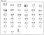
- the cell suspension was collected in a 15-mL tube and then centrifuged at 450 ⁇ g for 5 minutes. After aspiration of the medium, the pellet was suspended in a fresh medium and karyotyping was analyzed using G-banding staining and a chromosome imaging analyzer system at the GenDix Karyotyping Service.
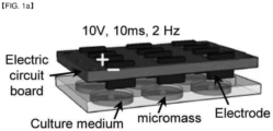
- FIG. 1a After applying ES (10 V/cm, 10 ms, 2 Hz frequency) to canine ADSCs (MSCs), aggregate formation was assessed.
- the micromass of the canine ADSCs was performed without addition of serum and exogenous factors ( FIG. 1a ).
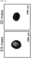
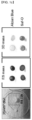
- FIG. 1b After 3 days of ES application, the result of immunohistochemical staining of cartilaginous matrixassociated molecules of the aggregate was investigated by phase-contrast microscopy. As shown in FIG. 1b , the formation of sheet-like aggregates and larger condensation were observed in the micromass subjected to ES compared with those without ES. Furthermore, more intense alcian blue and safranin O staining was observed in the cells subjected to ES, suggesting the deposition of proteoglycan ( FIG. 1c ).
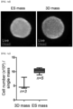
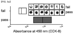
- cell viability was assessed using a Live/Dead reagent. As shown in FIG. 1d , the application of ES did not result in significant difference in cell viability. As shown in FIG. 1e , the aggregate contained approximately 10 5 cells. In order to investigate the change in cell membrane proteins caused by the ES application, flow cytometry analysis was performed using antibodies against surface molecules. The cell masses showed a similar expression pattern of CD44, CD90, CD29, CD73, CD54, CD34, CD49d, CD45, HLA-DR and CD80, compared with ADSCs growing in monolayer ( FIG. 1f ).
- ES Single cells were isolated from the electrically stimulated cell mass.
- the single-cell RNA-seq library was prepared on the 10x Genomics Chromium platform and data were imported into standard Seurat, a toolkit for analysis.
- the overall experiment for confirmation of the cellular transcription profile is shown in FIG. 3a .
- highly variable genes were identified by analyzing the normalized dataset using Loupe Cell Browser and 12 clusters were subdivided into four datasets of ( FIG. 3b ).
- FIG. 3c significant changes in the transcriptome of the cell micromass subjected to ES from the 2D ADSCs in terms of phenotypes and functions.
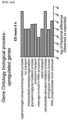
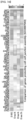
- the gene expression patterns were compared with publicly available datasets of gene expression to understand the role in cartilage development. As shown in FIGS. 4d-f , it was confirmed that the application of ES to the cell micromass for 72 hours induced prechondrogenic changes in considerable number of genes that were upregulated during the articular chondrocyte differentiation from the embryonic chondrogenic condensation. In a research conducted to identify the key molecules involved in the differentiation of osteochondral progenitor cells, it was confirmed that the balance between RUNX2 and SOX9 in the osteochondral progenitor cells plays an essential role in the differentiation of chondrocytes and osteoblasts. The present data for the micromass subjected to ES show that the levels of RUNX2 and its repressor, NKX3.2, were specifically altered to amounts typical for prechondrogenic cells ( FIG. 4g ).
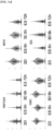
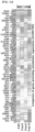
- chondrocytes The main function of chondrocytes is to synthesize extracellular matrix components such as collagen types 2, 4, 6, 10, 11, 12 and 14. Therefore, the level of collagen production was investigated in the cells subjected to ES. As shown in, FIG. 6a , the expression of COL3A1, COL4A1, COL4A2, COL4A5, COL5A3, COL6A1, COL6A3, COL6A5, COL8A1, COL13A1, COL14A1, COL15A1, COL16A1, COL21A1, COL23A1, COL24A1, COL24A1 and COL27A1 was increased in the micromass of canine ADSCs regardless of the ES application.
- COL3A1, COL5A2 and COL6A1/3 increased strongly and uniformly in the micromass of ADSCs.
- the expression of COL6A3 and COL16A1 was suppressed in the micromass as compared to the cells not subjected to ES.
- COL2 which is expressed during the chondrogenesis of MSCs and known as a marker of mature chondrocytes, was not expressed in the presence or absence of ES ( FIG. 6b ). It was confirmed in the comparative examples of a previously filed patent ( KR 10-2015-0047361 ), which discloses a method of differentiating stem cells into chondrocytes by applying an electric stimulus to mesenchymal stem cells, that the expression of COL2, which is used as a mature chondrocyte marker, was increased significantly both in mesenchymal stem cells to which growth factors were added and in mesenchymal stem cells to which the electric stimulus was applied. However, in the present disclosure, it was confirmed that the cell aggregate to which the electric stimulus was applied do not express COL2 at all.
- RT-qPCR real-time quantitative PCR
- the mechanism of the change of the expression level of type 1 collagen in response to electric stimulation was analyzed.
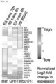
- the open-source database provided by GeneHancer, which integrates enhancers from various sources, was utilized. It was confirmed that the levels of the transcription factors NR4A1, RBFOX2, NFIC, ID3, HDGF, BMI1, YBX1, SMARCE1, HLTF and SMC3, which are known to be associated with the expression of COL1A1 in the cells to which ES was applied, decreased significantly over time ( FIG. 7a ).
- the transcription factors such as TCF12, POLR2A, ATF4, ARID4B, SMARCA5, GTF2F1, LARP7, HDAC2 and YY1 increased in the cell micromass for 72 hours after the application of ES ( FIG. 7b ).
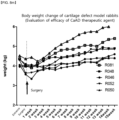
- the implanted electrically stimulated aggregates were derived from adipose-derived mesenchymal stem cells, cyclosporine, an immunosuppressant, was administered intravenously once a day (10 mg/kg) in consideration of xenograft transplantation. The experiment was stopped immediately when normal health condition was not shown within 2 weeks.
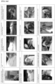
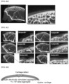
- the femoral cartilage was extracted by performing an autopsy and the defect and regeneration of the femoral cartilage were evaluated with images obtained by imaging with micro-CT having a micrometer-scale resolution. As shown in FIG. 8b and FIG. 8c , it was confirmed that the defect area where the electrically stimulated aggregates were implanted was recovered 4 months later to a level evaluated as the cartilage layer.
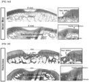
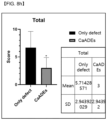
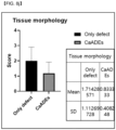
- Cartilage matrix staining was performed to confirm the above result. As shown in FIGS. 8c-8g , differentiation into chondrocytes and recovery of the cartilage matrix as in normal cartilage tissue were confirmed through histological evaluation of the implanted electrically stimulated aggregates through cartilage matrix staining with alcian blue, trichrome and safranin O as compared to the defect. In addition, long-term engraftment in vivo, connection with surrounding normal cartilage and smooth tissue adhesion with the underlying bone tissue were confirmed. These results are consistent with the histopathological staining result confirmed above.
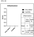
- FIGS. 8h-8m Significant regeneration and recovery were confirmed for the five rabbits.
Landscapes
- Health & Medical Sciences (AREA)
- Life Sciences & Earth Sciences (AREA)
- Chemical & Material Sciences (AREA)
- Engineering & Computer Science (AREA)
- Organic Chemistry (AREA)
- Zoology (AREA)
- Biomedical Technology (AREA)
- Bioinformatics & Cheminformatics (AREA)
- Genetics & Genomics (AREA)
- General Health & Medical Sciences (AREA)
- Wood Science & Technology (AREA)
- Biotechnology (AREA)
- Rheumatology (AREA)
- Biochemistry (AREA)
- Medicinal Chemistry (AREA)
- Cell Biology (AREA)
- Veterinary Medicine (AREA)
- Animal Behavior & Ethology (AREA)
- Public Health (AREA)
- Microbiology (AREA)
- Immunology (AREA)
- General Engineering & Computer Science (AREA)
- Pharmacology & Pharmacy (AREA)
- Orthopedic Medicine & Surgery (AREA)
- Nuclear Medicine, Radiotherapy & Molecular Imaging (AREA)
- Physical Education & Sports Medicine (AREA)
- General Chemical & Material Sciences (AREA)
- Chemical Kinetics & Catalysis (AREA)
- Toxicology (AREA)
- Gastroenterology & Hepatology (AREA)
- Biophysics (AREA)
- Molecular Biology (AREA)
- Proteomics, Peptides & Aminoacids (AREA)
- Virology (AREA)
- Epidemiology (AREA)
- Developmental Biology & Embryology (AREA)
- Micro-Organisms Or Cultivation Processes Thereof (AREA)
- Medicines Containing Material From Animals Or Micro-Organisms (AREA)
Applications Claiming Priority (3)
| Application Number | Priority Date | Filing Date | Title |
|---|---|---|---|
| KR20210106922 | 2021-08-12 | ||
| KR1020210130412A KR102857135B1 (ko) | 2021-08-12 | 2021-09-30 | 연골 관련 질환 치료용 조성물 및 이의 제조방법 |
| PCT/KR2022/011985 WO2023018244A1 (fr) | 2021-08-12 | 2022-08-11 | Composition pour le traitement d'une maladie liée au cartilage et procédé de préparation associé |
Publications (2)
| Publication Number | Publication Date |
|---|---|
| EP4386086A1 true EP4386086A1 (fr) | 2024-06-19 |
| EP4386086A4 EP4386086A4 (fr) | 2025-06-11 |
Family
ID=85200912
Family Applications (1)
| Application Number | Title | Priority Date | Filing Date |
|---|---|---|---|
| EP22856239.3A Pending EP4386086A4 (fr) | 2021-08-12 | 2022-08-11 | Composition pour le traitement d'une maladie liée au cartilage et procédé de préparation associé |
Country Status (4)
| Country | Link |
|---|---|
| US (1) | US20250134933A1 (fr) |
| EP (1) | EP4386086A4 (fr) |
| JP (2) | JP2024529683A (fr) |
| WO (1) | WO2023018244A1 (fr) |
Family Cites Families (9)
| Publication number | Priority date | Publication date | Assignee | Title |
|---|---|---|---|---|
| CN1599793A (zh) * | 2001-12-07 | 2005-03-23 | 杰龙公司 | 人胚胎干细胞衍生的软骨细胞前体 |
| BRPI0413644A (pt) | 2003-08-21 | 2006-10-17 | Novo Nordisk As | métodos para separar as duas formas de um polipeptìdeo tendo uma única racemização de aminoácido, e para separar dois polipeptìdeos, produto de polipeptìdeo, e, composição farmacêutica |
| KR20050044849A (ko) * | 2003-11-07 | 2005-05-13 | 학교법인 인하학원 | 중간엽 줄기세포로부터 연골세포를 분화시키는 방법 |
| KR20070025607A (ko) * | 2005-09-02 | 2007-03-08 | 보령제약 주식회사 | 골막으로부터 연골전구세포를 분리하는 방법 |
| KR101676520B1 (ko) | 2013-10-24 | 2016-11-15 | 제일모직주식회사 | 유기발광소자 봉지용 조성물 및 이를 사용하여 제조된 유기발광소자 표시장치 |
| KR101603475B1 (ko) * | 2015-04-03 | 2016-03-15 | 을지대학교 산학협력단 | 전기자극을 이용하여 중간엽 줄기세포를 연골세포로 분화시키는 방법 |
| KR101992538B1 (ko) * | 2017-10-18 | 2019-06-24 | 서울대학교산학협력단 | 닭 골수 유래 골·연골전구세포 배양액을 유효성분으로 포함하는 골 생성 촉진용 조성물 |
| US11466254B2 (en) * | 2017-10-18 | 2022-10-11 | Seoul National University R & Db Foundation | Composition containing, as active ingredient, culture of chicken bone marrow-derived osteochondral progenitor cells for promoting osteogenesis or inducing chondrogenic differentiation |
| WO2021054449A1 (fr) * | 2019-09-18 | 2021-03-25 | 国立大学法人 岡山大学 | Production de lbm, cpc, opc, et procédés de contrôle de qualité correspondants, kit, matériau de greffe et modèle de maladie |
-
2022
- 2022-08-11 EP EP22856239.3A patent/EP4386086A4/fr active Pending
- 2022-08-11 WO PCT/KR2022/011985 patent/WO2023018244A1/fr not_active Ceased
- 2022-08-11 JP JP2024508478A patent/JP2024529683A/ja active Pending
- 2022-08-11 US US18/681,598 patent/US20250134933A1/en active Pending
-
2026
- 2026-01-05 JP JP2026000530A patent/JP2026053714A/ja active Pending
Also Published As
| Publication number | Publication date |
|---|---|
| JP2026053714A (ja) | 2026-03-25 |
| US20250134933A1 (en) | 2025-05-01 |
| EP4386086A4 (fr) | 2025-06-11 |
| JP2024529683A (ja) | 2024-08-08 |
| WO2023018244A1 (fr) | 2023-02-16 |
Similar Documents
| Publication | Publication Date | Title |
|---|---|---|
| Jiang et al. | Human cartilage-derived progenitor cells from committed chondrocytes for efficient cartilage repair and regeneration | |
| Muruganandan et al. | A FoxA2+ long-term stem cell population is necessary for growth plate cartilage regeneration after injury | |
| JP6696023B2 (ja) | 腎細胞集団及びその使用 | |
| US10047344B2 (en) | Biophysically sorted osteoprogenitors from culture expanded bone marrow derived mesenchymal stromal cells (MSCs) | |
| AU2018290250B2 (en) | Immunoprivileged bioactive renal cells for the treatment of kidney disease | |
| Zhang et al. | RETRACTED ARTICLE: Therapeutic potential of non-adherent BM-derived mesenchymal stem cells in tissue regeneration | |
| Ouyang et al. | Effects of cell type and configuration on anabolic and catabolic activity in 3D co‐culture of mesenchymal stem cells and nucleus pulposus cells | |
| JP2021535135A (ja) | 細胞由来の小胞を含む組成物及びその使用 | |
| Yu et al. | Hyaline cartilage differentiation of fibroblasts in regeneration and regenerative medicine | |
| KR20130063483A (ko) | 개과동물 양막-유래 다분화능 줄기세포 | |
| US20240299459A1 (en) | Therapeutic agent for arthrosis and production method for therapeutic agent for arthrosis | |
| KR102857135B1 (ko) | 연골 관련 질환 치료용 조성물 및 이의 제조방법 | |
| EP3630129A1 (fr) | Cellules satellites, compositions et leurs méthodes de production | |
| Chang et al. | Novel method to repair articular cartilage by direct reprograming of prechondrogenic mesenchymal stem cells | |
| EP4386086A1 (fr) | Composition pour le traitement d'une maladie liée au cartilage et procédé de préparation associé | |
| EP4366745A1 (fr) | Cellules souches mésenchymateuses destinées à être utilisées dans le traitement d'une maladie rénale chronique | |
| WO2023280834A1 (fr) | Cellules souches mésenchymateuses destinées à être utilisées dans le traitement de la dermatite atopique | |
| US20250137072A1 (en) | Markers for skeletal stem cell and uses thereof | |
| WO2023280832A1 (fr) | Cellules souches mésenchymateuses destinées à être utilisées dans le traitement de l'arthrose chez des animaux | |
| CN117881781A (zh) | 用于治疗软骨相关疾病的组合物及其制备方法 | |
| HK40111971A (en) | Therapeutic agent for arthropathy, and method for producing therapeutic agent for arthropathy | |
| HK40106709A (zh) | 肾细胞群及其用途 | |
| RU2805243C2 (ru) | Популяции клеток почек и их применение | |
| Ouyang | Effects of Design Factors and Microenvironment on Mesenchymal Stem Cells and Nucleus Pulposus Cells for Intervertebral Disc Tissue Engineering |
Legal Events
| Date | Code | Title | Description |
|---|---|---|---|
| STAA | Information on the status of an ep patent application or granted ep patent |
Free format text: STATUS: THE INTERNATIONAL PUBLICATION HAS BEEN MADE |
|
| PUAI | Public reference made under article 153(3) epc to a published international application that has entered the european phase |
Free format text: ORIGINAL CODE: 0009012 |
|
| STAA | Information on the status of an ep patent application or granted ep patent |
Free format text: STATUS: REQUEST FOR EXAMINATION WAS MADE |
|
| 17P | Request for examination filed |
Effective date: 20240227 |
|
| AK | Designated contracting states |
Kind code of ref document: A1 Designated state(s): AL AT BE BG CH CY CZ DE DK EE ES FI FR GB GR HR HU IE IS IT LI LT LU LV MC MK MT NL NO PL PT RO RS SE SI SK SM TR |
|
| DAV | Request for validation of the european patent (deleted) | ||
| DAX | Request for extension of the european patent (deleted) | ||
| REG | Reference to a national code |
Ref country code: DE Ref legal event code: R079 Free format text: PREVIOUS MAIN CLASS: C12N0013000000 Ipc: A61K0035320000 |
|
| A4 | Supplementary search report drawn up and despatched |
Effective date: 20250512 |
|
| RIC1 | Information provided on ipc code assigned before grant |
Ipc: C12N 5/07 20100101ALI20250506BHEP Ipc: C07K 14/78 20060101ALI20250506BHEP Ipc: C07K 14/705 20060101ALI20250506BHEP Ipc: C07K 14/47 20060101ALI20250506BHEP Ipc: A61P 19/04 20060101ALI20250506BHEP Ipc: A61P 19/02 20060101ALI20250506BHEP Ipc: C12N 5/077 20100101ALI20250506BHEP Ipc: C12N 13/00 20060101ALI20250506BHEP Ipc: A61K 35/32 20150101AFI20250506BHEP |
|
| STAA | Information on the status of an ep patent application or granted ep patent |
Free format text: STATUS: EXAMINATION IS IN PROGRESS |
|
| 17Q | First examination report despatched |
Effective date: 20260121 |