EP4393386A2 - Procédé et système d'analyse de l'état de flux sanguin - Google Patents
Procédé et système d'analyse de l'état de flux sanguin Download PDFInfo
- Publication number
- EP4393386A2 EP4393386A2 EP24176082.6A EP24176082A EP4393386A2 EP 4393386 A2 EP4393386 A2 EP 4393386A2 EP 24176082 A EP24176082 A EP 24176082A EP 4393386 A2 EP4393386 A2 EP 4393386A2
- Authority
- EP
- European Patent Office
- Prior art keywords
- vascular
- vascular model
- model
- region
- time phase
- Prior art date
- Legal status (The legal status is an assumption and is not a legal conclusion. Google has not performed a legal analysis and makes no representation as to the accuracy of the status listed.)
- Granted
Links
Images








Classifications
-
- G—PHYSICS
- G06—COMPUTING OR CALCULATING; COUNTING
- G06T—IMAGE DATA PROCESSING OR GENERATION, IN GENERAL
- G06T7/00—Image analysis
- G06T7/0002—Inspection of images, e.g. flaw detection
- G06T7/0012—Biomedical image inspection
- G06T7/0014—Biomedical image inspection using an image reference approach
- G06T7/0016—Biomedical image inspection using an image reference approach involving temporal comparison
-
- G—PHYSICS
- G06—COMPUTING OR CALCULATING; COUNTING
- G06T—IMAGE DATA PROCESSING OR GENERATION, IN GENERAL
- G06T17/00—Three-dimensional [3D] modelling for computer graphics
-
- G—PHYSICS
- G06—COMPUTING OR CALCULATING; COUNTING
- G06T—IMAGE DATA PROCESSING OR GENERATION, IN GENERAL
- G06T7/00—Image analysis
- G06T7/0002—Inspection of images, e.g. flaw detection
- G06T7/0012—Biomedical image inspection
-
- A—HUMAN NECESSITIES
- A61—MEDICAL OR VETERINARY SCIENCE; HYGIENE
- A61B—DIAGNOSIS; SURGERY; IDENTIFICATION
- A61B5/00—Measuring for diagnostic purposes; Identification of persons
- A61B5/02—Detecting, measuring or recording for evaluating the cardiovascular system, e.g. pulse, heart rate, blood pressure or blood flow
- A61B5/02007—Evaluating blood vessel condition, e.g. elasticity, compliance
-
- A—HUMAN NECESSITIES
- A61—MEDICAL OR VETERINARY SCIENCE; HYGIENE
- A61B—DIAGNOSIS; SURGERY; IDENTIFICATION
- A61B5/00—Measuring for diagnostic purposes; Identification of persons
- A61B5/02—Detecting, measuring or recording for evaluating the cardiovascular system, e.g. pulse, heart rate, blood pressure or blood flow
- A61B5/021—Measuring pressure in heart or blood vessels
-
- A—HUMAN NECESSITIES
- A61—MEDICAL OR VETERINARY SCIENCE; HYGIENE
- A61B—DIAGNOSIS; SURGERY; IDENTIFICATION
- A61B5/00—Measuring for diagnostic purposes; Identification of persons
- A61B5/02—Detecting, measuring or recording for evaluating the cardiovascular system, e.g. pulse, heart rate, blood pressure or blood flow
- A61B5/026—Measuring blood flow
-
- A—HUMAN NECESSITIES
- A61—MEDICAL OR VETERINARY SCIENCE; HYGIENE
- A61B—DIAGNOSIS; SURGERY; IDENTIFICATION
- A61B5/00—Measuring for diagnostic purposes; Identification of persons
- A61B5/02—Detecting, measuring or recording for evaluating the cardiovascular system, e.g. pulse, heart rate, blood pressure or blood flow
- A61B5/026—Measuring blood flow
- A61B5/0285—Measuring or recording phase velocity of blood waves
-
- A—HUMAN NECESSITIES
- A61—MEDICAL OR VETERINARY SCIENCE; HYGIENE
- A61B—DIAGNOSIS; SURGERY; IDENTIFICATION
- A61B5/00—Measuring for diagnostic purposes; Identification of persons
- A61B5/72—Signal processing specially adapted for physiological signals or for diagnostic purposes
-
- G—PHYSICS
- G06—COMPUTING OR CALCULATING; COUNTING
- G06F—ELECTRIC DIGITAL DATA PROCESSING
- G06F17/00—Digital computing or data processing equipment or methods, specially adapted for specific functions
- G06F17/10—Complex mathematical operations
- G06F17/11—Complex mathematical operations for solving equations, e.g. nonlinear equations, general mathematical optimization problems
-
- G—PHYSICS
- G06—COMPUTING OR CALCULATING; COUNTING
- G06F—ELECTRIC DIGITAL DATA PROCESSING
- G06F30/00—Computer-aided design [CAD]
- G06F30/20—Design optimisation, verification or simulation
-
- G—PHYSICS
- G06—COMPUTING OR CALCULATING; COUNTING
- G06T—IMAGE DATA PROCESSING OR GENERATION, IN GENERAL
- G06T7/00—Image analysis
- G06T7/10—Segmentation; Edge detection
-
- G—PHYSICS
- G06—COMPUTING OR CALCULATING; COUNTING
- G06T—IMAGE DATA PROCESSING OR GENERATION, IN GENERAL
- G06T7/00—Image analysis
- G06T7/10—Segmentation; Edge detection
- G06T7/11—Region-based segmentation
-
- G—PHYSICS
- G06—COMPUTING OR CALCULATING; COUNTING
- G06T—IMAGE DATA PROCESSING OR GENERATION, IN GENERAL
- G06T7/00—Image analysis
- G06T7/30—Determination of transform parameters for the alignment of images, i.e. image registration
- G06T7/32—Determination of transform parameters for the alignment of images, i.e. image registration using correlation-based methods
-
- G—PHYSICS
- G16—INFORMATION AND COMMUNICATION TECHNOLOGY [ICT] SPECIALLY ADAPTED FOR SPECIFIC APPLICATION FIELDS
- G16H—HEALTHCARE INFORMATICS, i.e. INFORMATION AND COMMUNICATION TECHNOLOGY [ICT] SPECIALLY ADAPTED FOR THE HANDLING OR PROCESSING OF MEDICAL OR HEALTHCARE DATA
- G16H10/00—ICT specially adapted for the handling or processing of patient-related medical or healthcare data
-
- G—PHYSICS
- G16—INFORMATION AND COMMUNICATION TECHNOLOGY [ICT] SPECIALLY ADAPTED FOR SPECIFIC APPLICATION FIELDS
- G16H—HEALTHCARE INFORMATICS, i.e. INFORMATION AND COMMUNICATION TECHNOLOGY [ICT] SPECIALLY ADAPTED FOR THE HANDLING OR PROCESSING OF MEDICAL OR HEALTHCARE DATA
- G16H30/00—ICT specially adapted for the handling or processing of medical images
- G16H30/40—ICT specially adapted for the handling or processing of medical images for processing medical images, e.g. editing
-
- G—PHYSICS
- G16—INFORMATION AND COMMUNICATION TECHNOLOGY [ICT] SPECIALLY ADAPTED FOR SPECIFIC APPLICATION FIELDS
- G16H—HEALTHCARE INFORMATICS, i.e. INFORMATION AND COMMUNICATION TECHNOLOGY [ICT] SPECIALLY ADAPTED FOR THE HANDLING OR PROCESSING OF MEDICAL OR HEALTHCARE DATA
- G16H50/00—ICT specially adapted for medical diagnosis, medical simulation or medical data mining; ICT specially adapted for detecting, monitoring or modelling epidemics or pandemics
- G16H50/20—ICT specially adapted for medical diagnosis, medical simulation or medical data mining; ICT specially adapted for detecting, monitoring or modelling epidemics or pandemics for computer-aided diagnosis, e.g. based on medical expert systems
-
- G—PHYSICS
- G16—INFORMATION AND COMMUNICATION TECHNOLOGY [ICT] SPECIALLY ADAPTED FOR SPECIFIC APPLICATION FIELDS
- G16H—HEALTHCARE INFORMATICS, i.e. INFORMATION AND COMMUNICATION TECHNOLOGY [ICT] SPECIALLY ADAPTED FOR THE HANDLING OR PROCESSING OF MEDICAL OR HEALTHCARE DATA
- G16H50/00—ICT specially adapted for medical diagnosis, medical simulation or medical data mining; ICT specially adapted for detecting, monitoring or modelling epidemics or pandemics
- G16H50/50—ICT specially adapted for medical diagnosis, medical simulation or medical data mining; ICT specially adapted for detecting, monitoring or modelling epidemics or pandemics for simulation or modelling of medical disorders
-
- A—HUMAN NECESSITIES
- A61—MEDICAL OR VETERINARY SCIENCE; HYGIENE
- A61B—DIAGNOSIS; SURGERY; IDENTIFICATION
- A61B2576/00—Medical imaging apparatus involving image processing or analysis
- A61B2576/02—Medical imaging apparatus involving image processing or analysis specially adapted for a particular organ or body part
-
- A—HUMAN NECESSITIES
- A61—MEDICAL OR VETERINARY SCIENCE; HYGIENE
- A61B—DIAGNOSIS; SURGERY; IDENTIFICATION
- A61B5/00—Measuring for diagnostic purposes; Identification of persons
- A61B5/72—Signal processing specially adapted for physiological signals or for diagnostic purposes
- A61B5/7235—Details of waveform analysis
- A61B5/7264—Classification of physiological signals or data, e.g. using neural networks, statistical classifiers, expert systems or fuzzy systems
-
- G—PHYSICS
- G06—COMPUTING OR CALCULATING; COUNTING
- G06F—ELECTRIC DIGITAL DATA PROCESSING
- G06F2111/00—Details relating to CAD techniques
- G06F2111/04—Constraint-based CAD
-
- G—PHYSICS
- G06—COMPUTING OR CALCULATING; COUNTING
- G06F—ELECTRIC DIGITAL DATA PROCESSING
- G06F2119/00—Details relating to the type or aim of the analysis or the optimisation
- G06F2119/06—Power analysis or power optimisation
-
- G—PHYSICS
- G06—COMPUTING OR CALCULATING; COUNTING
- G06T—IMAGE DATA PROCESSING OR GENERATION, IN GENERAL
- G06T2207/00—Indexing scheme for image analysis or image enhancement
- G06T2207/30—Subject of image; Context of image processing
- G06T2207/30004—Biomedical image processing
- G06T2207/30101—Blood vessel; Artery; Vein; Vascular
-
- G—PHYSICS
- G06—COMPUTING OR CALCULATING; COUNTING
- G06T—IMAGE DATA PROCESSING OR GENERATION, IN GENERAL
- G06T2207/00—Indexing scheme for image analysis or image enhancement
- G06T2207/30—Subject of image; Context of image processing
- G06T2207/30004—Biomedical image processing
- G06T2207/30101—Blood vessel; Artery; Vein; Vascular
- G06T2207/30104—Vascular flow; Blood flow; Perfusion
Definitions
- CTA and MRA imaging technologies have been widely used in the diagnoses of peripheral vascular diseases, and more particularly, in the diagnoses of the vascular diseases such as vascular stenosis (vertebral artery stenosis), aneurysm, dissecting aneurysm, tumors, tumor-feeding artery, etc.
- the vascular analysis application provides a tool for vascular analysis in precise extraction of fine four grade blood vessels, complete bonelessness, fast automatic measurement, etc.
- medical image analysis systems generally employ an image segmentation technology and an image display technology for a 3-d simulation reconstruction of the blood vessels of an object.
- Doctors may analyze and process the lesions based on vascular morphological index (e.g., a vascular stenosis degree, a hemangioma expansion degree, etc.).
- vascular morphological index e.g., a vascular stenosis degree, a hemangioma expansion degree, etc.
- the morphological index that may be used are not sufficient.
- CFD Computational Fluid Dynamics
- the vascular model or blood flow model is an emerging application that employs computational fluid dynamics. Analyzing a single data using computational fluid mechanics cannot comprehensively reflect the actual condition and the changing rule of the analysis region. Also, selecting the time phase inaccurately may lead to a result of deviation.
- the correlation of the models corresponding to different time phases may include registering the characteristic regions.
- the first vascular region and the second vascular region may include a coronary artery, an abdominal artery, a cerebral artery, or a lower extremity artery.
- the method may further include generating a relationship between a condition of the blood vessel and a time phase, based on the condition of the blood vessel of the first vascular model at the first time phase and the condition of the blood vessel of the second vascular model at the second time phase.
- the determining the condition of the blood vessel of the first vascular model at the first time phase, or the determining a condition of the blood vessel of the second vascular model at the second time phase comprises employing a method of computational fluid dynamics (CFD).
- CFD computational fluid dynamics
- the grids of the first region or the grids of the second region are generated based on Delaunay triangulation (DT).
- DT Delaunay triangulation
- the method may further include: obtaining a 3-dimensional image, wherein the 3-dimensional image may include the one or more regions of interest; and generating 3-dimensional grids corresponding to the 3-dimensional image based on the grids of the first region and the grids of the second region.
- a system including at least one processor and a storage device.
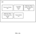
- the system may include a receiving module.
- the receiving module may be configured to: obtain a 2-dimensional image, wherein the 2-dimensional image may include one or more regions of interest.
- the system may further include a multi-time phase feature generation module.
- vascular image corresponding to a “blood vessel” may refer to that the vascular image including an image of the blood vessel.
- a blood vessel may include an aortic or a part thereof, a coronary or a part thereof, etc.
- the third vascular condition may be an average fractional flow reserve (FFR).
- FFR fractional flow reserve
- the method may further include matching the grids of the multiple vascular model.
- the one or more parameters of the first vascular model may include a cross-sectional area of the blood vessel.
- the boundary condition of the second vascular model may include blood pressure, blood velocity, blood viscosity, pressure, or wall stress, of an entrance, an exit, or a side wall of the blood vessel of the second vascular model.
- the parameter of the second vascular model may include flow resistance, blood velocity, blood pressure, wall stress of the blood vessel, wall shear stress (WSS) of the blood vessel, or fractional flow reserve (FFR).
- WSS wall shear stress
- FFR fractional flow reserve
- the determining a parameter of the second vascular model may include performing a computational fluid dynamics (CFD) analysis.
- CFD computational fluid dynamics
- the multi-time phase feature generation module may be configured to: determine, according to the one or more parameters of the first vascular model, a position of the first region of the first vascular model; generate a second vascular model by modifying the first region of the first vascular model; obtain a boundary condition of the second vascular model; determine, according to the boundary conditions of the second vascular model, a parameter of the second vascular model; determine, according to the parameter of the second vascular model, a boundary condition of the first vascular model; and obtain, according to the boundary condition of the first vascular model, a blood flow condition of the first vascular model.
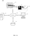
- the storage device 130 may store data or information.
- the data or information may include data obtained by the data collection device 110, processing results or control instructions generated by the processing device 120, user input received by the communication device 140, etc.
- the storage device 130 may be one or more storage mediums with read/write functions.
- the storage device 130 may include but not limited to a static random access memory (SRAM, a random-access memory (RAM), a read-only memory (ROM), a hard disk, a flash memory, etc.
- the storage device 130 may be a remote storage device, such as a cloud disk, etc.
- the grid generation unit 530 may perform a corresponding grid processing to a multi-time phase model for generating a multi-time phase grid processed model.
- the parameter setting unit 520 may set a corresponding parameter or boundary condition of the multi-time phase model or data.
- the automatic segmentation may be performed.
- the selected region is a blood vessel or other section that is difficult to be accurately segmented by machines
- the semi-automatic segmentation may be performed with the user correcting in the segmentation process.
- the images at different time phases may be segmented in sequence or simultaneously.
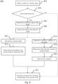
- 616 it may be determined whether all of time phases have been computed. In response to the determination that all of time phases have been computed, 618 may be implemented. In response to the determination that not all of time phases have been traversed, 620 may be implemented.
- FIG. 8 is a flow chart illustrating a process for setting a boundary condition according to some embodiments of the present disclosure.
- 610 may correspond to the process 800.
- process 800 may be implemented by the parameter setting unit 520.
- model 910 may be a coronary artery model with two narrow regions 930 and 940.
- the model 920 may be a normal coronary artery model, and regions (e.g., 935, 945) corresponding to 930, 940 are not narrow.
- 804 may include extracting a centerline of the vascular region model.
- the vascular centerline may refer to an imaginary line located in the blood vessel along the trend of the blood vessel.
- a normal model may be reconstructed based on the abnormal model.
- the methods of reconstruction include lofting or stretching the blood vessel based on diameters or centerlines to generate a normal region.
- the reconstruction technique may include dilating or narrowing the abnormal region. The dilated vascular cross-sectional area may not be larger than vascular cross-sectional areas of adjacent regions. The narrowed vascular cross-sectional area may not be smaller than a vascular cross-sectional areas of adjacent regions.
- the adjacent regions of the reconstructed abnormal region may be smoothed to avoid significant mutations.
- the reconstructed model may be sent to the user.
- the user may modify a part or all of the reconstructed normal model. For example, the user may locally dilate, narrow, smooth the reconstructed normal model.
- the flow volume may be allocated in branches of blood vessel in accordance with the positive relationship from the entrance, until the flow volume of the entrance is allocated to all the branches.
- a computational fluid dynamics (CFD) simulation may be computed to obtain a flow resistance (i.e., a ratio of the entrance pressure to the entrance flow volume) at each entrance of the normal model.
- a total flow resistance of the model may be obtained.
- P inlet and Q may be obtained in 808, and is not repeated here.
- each proximal branch vessel at each level may be determined by analyzing the vascular centerline, the vascular cross-sectional area, and the vascular abnormal region.
- the flow resistance may be allocated based on the size of the diameter of the normal model.
- a boundary resistance corresponding to an actual vascular model may be generated.
- the boundary resistance corresponding to an actual vascular model may be obtained based on a boundary resistance corresponding to the normal model.
- the boundary resistance corresponding to an actual vascular model may be the same as the boundary resistance corresponding to the normal model.
- the boundary resistance of the vascular model may be allocated based on a method described in 820.
- FIG. 10 is a flow chart illustrating a process for a grid division according to some embodiments of the present disclosure.
- process 1000 may correspond to 608.
- the process 1000 may be implemented by the grid generation unit 540.
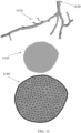
- a boundary region of the model may be obtained. If the model corresponds to a blood vessel or a blood flow related to the blood vessel, the boundary region may be an exit, an entrance, a vascular wall, etc. As shown in FIG. 11 , an entrance 1120 of the model 1110 may be determined to be a boundary region of the model 1110 in 1004.
- volume grids of the model may be generated (also referred to as a 3-dimensional grid division or 3-D grid process) based on the results of the surface grid divisions of the boundary region and the side wall.
- the volume grid division may include dividing the model into 3-dimensional grids.
- the 3-dimensional grids may include a tetrahedral grid, a hexahedral mesh, a prismatic grid (i.e., a boundary layer grid), a mixture grid of tetrahedron and hexahedron, a Cartesian grid, a ball filling grid, etc.
- 1004 through1008 may be omitted, e.g., the model may be directly divided by volume grids.
- FIG. 12 illustrates a flow chart of a process for a grid division according to some embodiments of the present disclosure.
- the process 1200 may be implemented by the multi-time phase feature generation module 430.
- 608 in the FIG. 6 , 1006 in FIG. 10 , etc. may be implemented based on the process 1200.
- a 2-dimensional image may be obtained.
- the 2-dimendional image may be obtained by the data acquisition unit 510.
- the 2-dimendional image may be a 2-dimensional medical image, or an interesting region of the user (e.g., a coronary vascular region, a brain region, etc.).
- the 2-dimensinal image may be a CT image, an MRI image, a PET image, or the like.
- the 2-dimensional image may be presented in grayscale or color.
- the 2-dimensional image may be a 2-dimentional presentation of a model at a time phase.
- the 2-dimensional image may be an image related to a boundary region of model in 1006.
- the 2-dimensional image may display an entrance/exit region of a vascular model (for example, as shown in FIG. 9 ).
- the 2-dimensional image may be an image reconstructed by an image processing device (e.g., the processing device 120).
- the 2-dimensional image may be an image obtained from a local storage device or an external storage device (e.g., the storage device 130).
- the grid generation unit 540 may extract boundary points of a region of interest of a 2-dimensional image.
- extracting boundary points from the region of interest of the 2-dimensional image may include segmenting the region of interest; and extracting the boundary points of the segmented region of interest.
- a technique of segmenting the region of interest may be found elsewhere in the present disclosure.
- the boundary points of the region of interest may include one or more pixels in the boundary of the region of interest (also referred to as "boundary pixels").
- the boundary points at the cross-section of a coronary artery may include one or more boundary pixels located in the wall of coronary artery.
- the boundary pixels of the region of interest may be continuous, partially continuous, or discontinuous.
- the term "continuous" may refer to that a boundary pixel is adjacent to at least one or more other boundary pixels.
- the extracted boundary points may be stored in one or more storage devices (e.g., the storage device 130, the storage module 260, etc.).
- the boundary points may be used by the grid generation unit 540 or other unit/module with a data analysis function in subsequent processes.
- the exemplary i boundary point may include the position of the boundary point, the number of the boundary point, or the like, or any combination thereof.
- one or more regions may be determined based on the boundary points.
- the determination of the one or more regions may be implemented by the grid generation unit 540.
- the one or more regions may be formed by sequentially connecting the boundary points of the region of interest.
- the determination of one or more regions may include determining an initial boundary pixel of the region of interest (for example, a point with the smallest x/y coordinates in the contour pixel may be selected as the initial boundary pixel).
- the boundary pixels of the region of interest may be sorted in a clockwise or counterclockwise direction. Starting from the initial boundary pixel, a previous boundary pixel may be connected to a subsequent boundary pixel by a line to form a short edge.
- a closed boundary curve may be formed.
- the region of interest may be located in a closed boundary curve.
- the region of interest of the model entrance 1140 in FIG. 11 i.e., the region of grid division
- the region of interest may be a region located between two closed boundary curves.
- the region of interest may be a 2-dimensional ring structure, or a structure equivalent to the 2-dimensional ring topology.
- the information of one or more regions may be stored in one or more storage devices (e.g., the storage device 130, the storage module 260, etc.).
- the information of one or more regions may be used by the grid generation unit 540 or other unit/module with a data analysis function in subsequent processes.
- a region needs a grid division.
- the determination may be implemented by the determination unit 580.
- the process 1200 may proceed to 1210.
- the process 1200 may proceed to 1212.
- a condition determined by the determination unit 580 may include whether the region is a region of interest.
- the grid division may be determined to be needed.
- the region of interest may include a region that needs a blood state analysis, e.g., a region where blood flows in a specific blood vessel.
- the region that needs or does not need to be divided may be marked.
- the marking of the region may be implemented by the grid generation unit 540.
- the marking may be performed in a form of a computer readable code or an executable instruction.
- the marked region may be stored in one or more storage devices (e.g., the storage device 130, the storage module 260, etc.).
- the marked region may be read by the grid generation unit 540 or other unit/module with a data analysis function in subsequent processes. For example, the marked region may be removed if a grid division is performed.
- the region may be divided into grids.
- the grid division may be implemented by the grid generation unit 540.
- grid division may be performed based on the boundary points of the region.
- Algorithms used in the grid division may include a triangular grid division, a quadrilateral grid division, a hexagonal grid division, or the like, or a combination thereof.
- Exemplary grid division algorithm S may include a Loop algorithm, a butterfly subdivision algorithm, a Catmull-Clark algorithm, a Doo-Sabin algorithm, a Delaunay triangular division algorithm, etc.
- the grid generation unit 540 may classify the boundary points of the region into different subsets, and sequentially grid-divide the boundary points of each subsets. The grid generation unit 540 may then combine the grid division of the subsets to form a grid division of the region. Particularly, all of the boundary points of the region may be ordered according to the x/y coordinates (for example, the boundary points may be firstly arranged in a non-descending order with respect to the x-coordinates, and then be arranged in a non-descending order with respect to the y-coordinates for the points of the same x-coordinates). The ordered boundary points may be divided into a subset A and a subset B based on their quantity.
- a Delaunay triangular division of the two subsets may be completed respectively. Then the Delaunay triangular division of the subset A and the subset B may be combined to generate a Delaunay triangular division of all of the boundary points.
- the grid division may also include superimposing the boundary curve of the region on the divided grids. In this case, the boundary curve of the region may be maintained in the divided grids (e.g., one or more short edges formed by the boundary pixels as described in 1206).
- the grid division of a region may employ a grid generation technique based on parallel operations. For example, a region division or similar algorithm may be employed to divide the region into multiple sub-regions. Each of the sub-regions may be independently divided into grids. Then, the boundary grids of adjacent sub-regions may be modified to obtain complete grids of the region.
- a grid division control condition may be set for the region.
- the setting of the grid division control condition may be implemented by the grid generation unit 540.
- the grid division control condition may control grid count, size, distribution, shape, or the like, or one or more combinations thereof.
- the grid generation unit 540 may set an area constraint condition for a grid to limit the area of any grid in the region. For example, the grid generation unit 540 may set an area constraint value such that the area of any grid is not larger than the area constraint value.
- the grid generation unit 540 may set an interior angle constraint condition for a grid such that the interior angle of any grid satisfies the interior angle constraint condition.
- the grid generation unit 540 may set an interior angle constraint value for a triangular grid such that the minimum internal angle of any triangular grid is not less than the inner angle constraint value.
- the grid division control condition may be obtained by a user via, for example, the communication device 140.
- the grid division control condition may also be obtained by the grid generation unit 540 or other unit/module with a data analysis function according to analysis of specific conditions.
- the specific conditions may include the time needed to generate the grids, the number of the generated grids, the time of model computation based on the generated grids, an accuracy degree of the obtained result based on the generated grids, etc.
- whether the divided grid satisfies the control condition may be determined.
- the determination of the grid division may be implemented by the grid generation unit 540.
- the process 1200 may proceed to 1218.
- one or more auxiliary points may be inserted in the triangular grid.
- the auxiliary points may be inserted randomly, or be inserted according to the position of the feature points of the original triangular grid.
- the grid generation unit 540 may generate a new grid based on the auxiliary points. For example, an auxiliary point may be inserted inside the triangular grid, e.g., t at the center of gravity of the triangular grid. Connecting the auxiliary point and vertices of the original triangular grid may generate three new triangular grids.
- the flip algorithm may include selecting a quadrilateral containing two adjacent triangular grids (i.e., a diagonal line of the quadrilateral is the adjacent edge of the two triangular grids); selecting another diagonal line as the adjacent edge of two new triangular grids; and obtaining two new triangular grids.
- the inner angle constraint condition may include that the minimum interior angle of the triangular grid unit is not less than an inner angle constraint value.
- the inner angle constraint value may be 5°, 10°, 15°, 20°, 25°, etc.
- the processed grid may return to 1216.
- the grid generation unit 540 may determine whether the processed grid satisfies a control condition. Until the grid satisfies the control condition, process 1200 may proceed to 1220.
- the grid generation unit 540 may generate nodes of grids in a 3-dimensional geometric region by filling based on the sphere packing technique.
- the nodes may be generated with appropriate density according to the geometric features and spatial relations of the geometric model. Then a 3-dimensional grid may be generated by employing the fast Delaunay insertion technique.
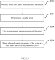
- the multi-time phase feature processing module 440 may obtain extra hemodynamic parameters of the point.
- the extra hemodynamic parameters may be obtained by an interpolation method, or by a user via the communication device 140.
- aspects of the present disclosure may be illustrated and described herein in any of a number of patentable classes or context including any new and useful process, machine, manufacture, or composition of matter, or any new and useful improvement thereof. Accordingly, aspects of the present disclosure may be implemented entirely hardware, entirely software (including firmware, resident software, micro-code, etc.) or combining software and hardware implementation that may all generally be referred to herein as a "block,” “module,” “engine,” “unit,” “component,” or “system.” Furthermore, aspects of the present disclosure may take the form of a computer program product embodied in one or more computer readable media having computer readable program code embodied thereon.
- a computer readable signal medium may include a propagated data signal with computer readable program code embodied therein, for example, in baseband or as part of a carrier wave. Such a propagated signal may take any of a variety of forms, including electro-magnetic, optical, or the like, or any suitable combination thereof.
- a computer readable signal medium may be any computer readable medium that is not a computer readable storage medium and that may communicate, propagate, or transport a program for use by or in connection with an instruction execution system, apparatus, or device.
- Program code embodied on a computer readable signal medium may be transmitted using any appropriate medium, including wireless, wireline, optical fiber cable, RF, or the like, or any suitable combination of the foregoing.
- Computer program code for carrying out operations for aspects of the present disclosure may be written in any combination of one or more programming languages, including an object oriented programming language such as Java, Scala, Smalltalk, Eiffel, JADE, Emerald, C++, C#, VB. NET, Python or the like, conventional procedural programming languages, such as the "C" programming language, Visual Basic, Fortran 2003, Perl, COBOL 2002, PHP, ABAP, dynamic programming languages such as Python, Ruby and Groovy, or other programming languages.
- the program code may execute entirely on the user's computer, partly on the user's computer, as a stand-alone software package, partly on the user's computer and partly on a remote computer or entirely on the remote computer or server.
Landscapes
- Health & Medical Sciences (AREA)
- Engineering & Computer Science (AREA)
- Life Sciences & Earth Sciences (AREA)
- Physics & Mathematics (AREA)
- Medical Informatics (AREA)
- Public Health (AREA)
- General Health & Medical Sciences (AREA)
- Biomedical Technology (AREA)
- Pathology (AREA)
- Cardiology (AREA)
- Theoretical Computer Science (AREA)
- General Physics & Mathematics (AREA)
- Molecular Biology (AREA)
- Surgery (AREA)
- Animal Behavior & Ethology (AREA)
- Heart & Thoracic Surgery (AREA)
- Veterinary Medicine (AREA)
- Biophysics (AREA)
- Physiology (AREA)
- Computer Vision & Pattern Recognition (AREA)
- Epidemiology (AREA)
- Primary Health Care (AREA)
- Hematology (AREA)
- Data Mining & Analysis (AREA)
- Vascular Medicine (AREA)
- Nuclear Medicine, Radiotherapy & Molecular Imaging (AREA)
- Radiology & Medical Imaging (AREA)
- Databases & Information Systems (AREA)
- Mathematical Physics (AREA)
- Quality & Reliability (AREA)
- Software Systems (AREA)
- General Engineering & Computer Science (AREA)
- Pure & Applied Mathematics (AREA)
- Geometry (AREA)
- Mathematical Optimization (AREA)
- Mathematical Analysis (AREA)
- Computational Mathematics (AREA)
- Signal Processing (AREA)
- Computer Graphics (AREA)
- Computer Hardware Design (AREA)
Priority Applications (1)
| Application Number | Priority Date | Filing Date | Title |
|---|---|---|---|
| EP24176082.6A EP4393386B1 (fr) | 2017-01-23 | 2017-01-23 | Analyse de l'état de flux sanguin |
Applications Claiming Priority (3)
| Application Number | Priority Date | Filing Date | Title |
|---|---|---|---|
| PCT/CN2017/072256 WO2018133118A1 (fr) | 2017-01-23 | 2017-01-23 | Système et procédé d'analyse de l'état de la circulation sanguine |
| EP17731461.4A EP3375364B1 (fr) | 2017-01-23 | 2017-01-23 | Système et procédé d'analyse de l'état de la circulation sanguine |
| EP24176082.6A EP4393386B1 (fr) | 2017-01-23 | 2017-01-23 | Analyse de l'état de flux sanguin |
Related Parent Applications (2)
| Application Number | Title | Priority Date | Filing Date |
|---|---|---|---|
| EP17731461.4A Division EP3375364B1 (fr) | 2017-01-23 | 2017-01-23 | Système et procédé d'analyse de l'état de la circulation sanguine |
| EP17731461.4A Division-Into EP3375364B1 (fr) | 2017-01-23 | 2017-01-23 | Système et procédé d'analyse de l'état de la circulation sanguine |
Publications (3)
| Publication Number | Publication Date |
|---|---|
| EP4393386A2 true EP4393386A2 (fr) | 2024-07-03 |
| EP4393386A3 EP4393386A3 (fr) | 2024-09-11 |
| EP4393386B1 EP4393386B1 (fr) | 2026-04-08 |
Family
ID=59724862
Family Applications (2)
| Application Number | Title | Priority Date | Filing Date |
|---|---|---|---|
| EP17731461.4A Active EP3375364B1 (fr) | 2017-01-23 | 2017-01-23 | Système et procédé d'analyse de l'état de la circulation sanguine |
| EP24176082.6A Active EP4393386B1 (fr) | 2017-01-23 | 2017-01-23 | Analyse de l'état de flux sanguin |
Family Applications Before (1)
| Application Number | Title | Priority Date | Filing Date |
|---|---|---|---|
| EP17731461.4A Active EP3375364B1 (fr) | 2017-01-23 | 2017-01-23 | Système et procédé d'analyse de l'état de la circulation sanguine |
Country Status (4)
| Country | Link |
|---|---|
| US (4) | US10360682B2 (fr) |
| EP (2) | EP3375364B1 (fr) |
| CN (3) | CN107123159A (fr) |
| WO (1) | WO2018133118A1 (fr) |
Families Citing this family (60)
| Publication number | Priority date | Publication date | Assignee | Title |
|---|---|---|---|---|
| US10210956B2 (en) * | 2012-10-24 | 2019-02-19 | Cathworks Ltd. | Diagnostically useful results in real time |
| US12026868B2 (en) | 2015-08-14 | 2024-07-02 | Elucid Bioimaging Inc. | Quantitative imaging for detecting histopathologically defined plaque erosion non-invasively |
| US11071501B2 (en) | 2015-08-14 | 2021-07-27 | Elucid Bioiwaging Inc. | Quantitative imaging for determining time to adverse event (TTE) |
| US11113812B2 (en) | 2015-08-14 | 2021-09-07 | Elucid Bioimaging Inc. | Quantitative imaging for detecting vulnerable plaque |
| US12008751B2 (en) | 2015-08-14 | 2024-06-11 | Elucid Bioimaging Inc. | Quantitative imaging for detecting histopathologically defined plaque fissure non-invasively |
| US11087459B2 (en) | 2015-08-14 | 2021-08-10 | Elucid Bioimaging Inc. | Quantitative imaging for fractional flow reserve (FFR) |
| US11676359B2 (en) | 2015-08-14 | 2023-06-13 | Elucid Bioimaging Inc. | Non-invasive quantitative imaging biomarkers of atherosclerotic plaque biology |
| US10176408B2 (en) | 2015-08-14 | 2019-01-08 | Elucid Bioimaging Inc. | Systems and methods for analyzing pathologies utilizing quantitative imaging |
| US11094058B2 (en) | 2015-08-14 | 2021-08-17 | Elucid Bioimaging Inc. | Systems and method for computer-aided phenotyping (CAP) using radiologic images |
| EP4300419A3 (fr) | 2016-05-16 | 2024-04-03 | Cathworks Ltd. | Système d'évaluation vasculaire |
| IL263066B2 (en) | 2016-05-16 | 2023-09-01 | Cathworks Ltd | Selecting blood vessels from images |
| JP6653673B2 (ja) * | 2017-02-28 | 2020-02-26 | 富士フイルム株式会社 | 血流解析装置および方法並びにプログラム |
| CN107610773A (zh) * | 2017-09-12 | 2018-01-19 | 北京即刻叁维数据科技股份有限公司 | 一种基于主动脉医学影像的血管夹层辅助诊断方法 |
| CN108186038B (zh) * | 2018-02-11 | 2020-11-17 | 杭州脉流科技有限公司 | 基于动脉造影影像计算冠脉血流储备分数的系统 |
| CN108257214A (zh) * | 2018-02-12 | 2018-07-06 | 成都信息工程大学 | 心脏左心室流场域数值模拟方法、计算机、计算机程序 |
| CN109065170B (zh) * | 2018-06-20 | 2021-11-19 | 博动医学影像科技(上海)有限公司 | 获取血管压力差的方法及装置 |
| CN109102893A (zh) * | 2018-07-04 | 2018-12-28 | 中山大学 | 一种基于Cross模型的多粒子混合修正的血栓模拟方法 |
| CN110599444B (zh) * | 2018-08-23 | 2022-04-19 | 深圳科亚医疗科技有限公司 | 预测血管树的血流储备分数的设备、系统以及非暂时性可读存储介质 |
| JP6988001B2 (ja) * | 2018-08-30 | 2022-01-05 | オリンパス株式会社 | 記録装置、画像観察装置、観察システム、観察システムの制御方法、及び観察システムの作動プログラム |
| CN110428420B (zh) * | 2018-09-05 | 2022-05-17 | 深圳科亚医疗科技有限公司 | 基于患者的冠状动脉ct血管造影图像来确定冠状动脉的流动信息的方法、装置和介质 |
| CN110367965B (zh) * | 2018-09-19 | 2022-03-08 | 苏州润迈德医疗科技有限公司 | 便捷测量冠状动脉血管评定参数的方法、装置及系统 |
| WO2020083390A1 (fr) * | 2018-10-25 | 2020-04-30 | 苏州润心医疗器械有限公司 | Procédé, dispositif et système d'acquisition de débit sanguin de grande artère sur la surface cardiaque et support de stockage |
| CN109616200A (zh) * | 2018-11-06 | 2019-04-12 | 北京三普威盛科技有限公司 | 用于冠脉狭窄评估的方法,装置,存储介质及电子设备 |
| US11819279B2 (en) * | 2018-11-30 | 2023-11-21 | Koninklijke Philips N.V. | Patient lumen system monitoring |
| CN109620199B (zh) * | 2018-11-30 | 2021-03-16 | 博动医学影像科技(上海)有限公司 | 建立血管截面函数、血管压力差和血管应力的方法及装置 |
| CN109567776B (zh) * | 2018-12-31 | 2021-08-10 | 深圳北芯生命科技股份有限公司 | 用于测试ffr主机系统的导管模拟器 |
| WO2020187383A1 (fr) * | 2019-03-20 | 2020-09-24 | Amid S.R.L. | Procédé et système de calcul de la force échangée entre un fluide et un récipient environnant, en particulier en imagerie cardiovasculaire |
| CN109948622B (zh) * | 2019-03-26 | 2020-12-11 | 数坤(北京)网络科技有限公司 | 一种头颈体动脉瘤检测方法、装置及计算机可读存储介质 |
| IL286814B2 (en) | 2019-04-01 | 2026-03-01 | Cathworks Ltd | Methods and apparatus for selecting angiographic images |
| US10861157B2 (en) | 2019-04-04 | 2020-12-08 | Medtronic Vascular, Inc. | System and methods for determining modified fractional flow reserve values |
| CN110400638B (zh) * | 2019-05-30 | 2022-09-30 | 合肥工业大学 | 用于研究颅内动脉瘤生长过程以及探究生长机制的方法 |
| KR102904994B1 (ko) | 2019-08-05 | 2025-12-30 | 엘루시드 바이오이미징 아이엔씨. | 형태학적 및 혈관주위 질환 표지자의 조합 평가 |
| EP4555935A3 (fr) | 2019-09-23 | 2025-07-30 | Cathworks Ltd. | Procédés, appareil et système de synchronisation entre un modèle vasculaire tridimensionnel et un dispositif d'imagerie |
| CN110910398B (zh) * | 2019-10-28 | 2021-07-20 | 衢州学院 | 一种基于决策层融合的视频复杂场景区域分割方法和装置 |
| US11200669B2 (en) * | 2019-11-19 | 2021-12-14 | Uih America, Inc. | Systems and methods for determining plasma input function used in positron emission tomography imaging |
| CN111161342B (zh) * | 2019-12-09 | 2023-08-29 | 杭州脉流科技有限公司 | 基于冠脉造影图像获取血流储备分数的方法、装置、设备、系统及可读存储介质 |
| CN113487588B (zh) * | 2020-03-02 | 2024-03-22 | 上海博动医疗科技股份有限公司 | 冠状动脉的血流速度的计算方法、装置及电子设备 |
| CN111317455B (zh) * | 2020-03-03 | 2022-02-22 | 上海联影医疗科技股份有限公司 | 血流动力学参数确定方法、装置、设备及存储介质 |
| WO2022001026A1 (fr) * | 2020-06-30 | 2022-01-06 | Shanghai United Imaging Healthcare Co., Ltd. | Systèmes et procédés permettant de déterminer des paramètres de vaisseau sanguin |
| CN112001893B (zh) * | 2020-07-31 | 2024-02-20 | 上海联影医疗科技股份有限公司 | 一种血管参数的计算方法、装置、设备及存储介质 |
| CN111815598B (zh) * | 2020-06-30 | 2024-04-26 | 上海联影医疗科技股份有限公司 | 一种血管参数的计算方法、装置、设备及存储介质 |
| US11439308B2 (en) * | 2020-07-13 | 2022-09-13 | GE Precision Healthcare LLC | Methods and systems for thermal monitoring of tissue with an ultrasound imaging system |
| US12131479B2 (en) * | 2020-08-07 | 2024-10-29 | Canon Medical Systems Corporation | Medical image processing apparatus, system, and method |
| CN112561871B (zh) * | 2020-12-08 | 2021-09-03 | 中国医学科学院北京协和医院 | 一种基于平扫ct图像的主动脉夹层分割方法和装置 |
| CN112434676B (zh) * | 2021-01-26 | 2021-04-20 | 北京圣点云信息技术有限公司 | 一种基于汇流累计量信息的体内静脉识别方法及装置 |
| EP4402703A1 (fr) * | 2021-09-14 | 2024-07-24 | United Therapeutics Corporation | Systèmes et méthodes d'évaluation de tissu |
| US12315076B1 (en) | 2021-09-22 | 2025-05-27 | Cathworks Ltd. | Four-dimensional motion analysis of a patient's coronary arteries and myocardial wall |
| CN113963001B (zh) * | 2021-10-25 | 2023-05-23 | 中国人民解放军总医院第一医学中心 | 一种基于多时相cta的侧支循环血管定量方法 |
| CN113706539A (zh) * | 2021-10-29 | 2021-11-26 | 南京裕隆生物医学发展有限公司 | 一种用于识别肿瘤的人工智能辅助系统 |
| CN114881974A (zh) * | 2022-05-11 | 2022-08-09 | 上海联影医疗科技股份有限公司 | 一种医学图像分析方法和系统 |
| US12482051B2 (en) * | 2021-12-31 | 2025-11-25 | Shanghai United Imaging Healthcare Co., Ltd. | Methods and systems for vascular image processing |
| EP4645221A3 (fr) | 2022-02-10 | 2025-12-31 | Cathworks Ltd. | Système et procédé d'analyse de capteur basée sur l'apprentissage automatique et de segmentation d'arbre vasculaire |
| CN114881975A (zh) * | 2022-05-10 | 2022-08-09 | 王硕 | 预测动脉瘤破裂潜能的系统、方法、电子设备及介质 |
| US20230386647A1 (en) * | 2022-05-31 | 2023-11-30 | Canon Medical Systems Corporation | Medical image processing apparatus, method, and medical system |
| CN115018810B (zh) * | 2022-06-29 | 2025-10-28 | 上海联影医疗科技股份有限公司 | 血流数据的显示方法、装置和存储介质 |
| EP4311496A1 (fr) * | 2022-07-26 | 2024-01-31 | Koninklijke Philips N.V. | Paramètres de débit sanguin |
| CN116636826B (zh) * | 2023-07-27 | 2023-09-22 | 深圳市爱保护科技有限公司 | 血压估算模型的训练方法、装置、电子设备及存储介质 |
| KR20260052078A (ko) | 2023-08-09 | 2026-04-17 | 캐스웍스 엘티디. | 혈관 지수 측정을 위한 향상된 사용자 인터페이스 및 크로스토크 분석 |
| KR20260048313A (ko) | 2023-08-09 | 2026-04-09 | 캐스웍스 엘티디. | Pci 후 관상동맥 분석 |
| US12567489B2 (en) | 2024-06-12 | 2026-03-03 | Cathworks Ltd. | Systems and methods for displaying distal fractional flow reserve values in vascular analysis |
Family Cites Families (46)
| Publication number | Priority date | Publication date | Assignee | Title |
|---|---|---|---|---|
| JP4960162B2 (ja) | 2007-07-17 | 2012-06-27 | 株式会社東芝 | 超音波診断装置 |
| CN101527047B (zh) | 2008-03-05 | 2013-02-13 | 深圳迈瑞生物医疗电子股份有限公司 | 使用超声图像检测组织边界的方法与装置 |
| CN102573638A (zh) * | 2009-10-13 | 2012-07-11 | 新加坡科技研究局 | 一种用于分割图像中的肝脏对象的方法和系统 |
| JP5632203B2 (ja) | 2010-06-08 | 2014-11-26 | 株式会社東芝 | 超音波診断装置、超音波画像処理装置及び超音波画像処理プログラム |
| US8682626B2 (en) | 2010-07-21 | 2014-03-25 | Siemens Aktiengesellschaft | Method and system for comprehensive patient-specific modeling of the heart |
| US8315812B2 (en) | 2010-08-12 | 2012-11-20 | Heartflow, Inc. | Method and system for patient-specific modeling of blood flow |
| US9119540B2 (en) | 2010-09-16 | 2015-09-01 | Siemens Aktiengesellschaft | Method and system for non-invasive assessment of coronary artery disease |
| US9141763B2 (en) | 2011-02-07 | 2015-09-22 | Siemens Aktiengesellschaft | Method and system for patient-specific computational modeling and simulation for coupled hemodynamic analysis of cerebral vessels |
| US9814531B2 (en) | 2011-08-26 | 2017-11-14 | EBM Corporation | System for diagnosing bloodflow characteristics, method thereof, and computer software program |
| US10162932B2 (en) | 2011-11-10 | 2018-12-25 | Siemens Healthcare Gmbh | Method and system for multi-scale anatomical and functional modeling of coronary circulation |
| US10373700B2 (en) | 2012-03-13 | 2019-08-06 | Siemens Healthcare Gmbh | Non-invasive functional assessment of coronary artery stenosis including simulation of hyperemia by changing resting microvascular resistance |
| US9135699B2 (en) * | 2012-03-15 | 2015-09-15 | Siemens Aktiengesellschaft | Method and system for hemodynamic assessment of aortic coarctation from medical image data |
| JP2014028124A (ja) | 2012-06-27 | 2014-02-13 | Toshiba Corp | 医用画像処理装置及びx線診断装置 |
| US10398386B2 (en) | 2012-09-12 | 2019-09-03 | Heartflow, Inc. | Systems and methods for estimating blood flow characteristics from vessel geometry and physiology |
| US9858387B2 (en) | 2013-01-15 | 2018-01-02 | CathWorks, LTD. | Vascular flow assessment |
| EP2916735B1 (fr) | 2012-11-06 | 2019-09-25 | Koninklijke Philips N.V. | Indice de réserve de débit fractionnaire (ffr) |
| JP2014100249A (ja) | 2012-11-19 | 2014-06-05 | Toshiba Corp | 血管解析装置、医用画像診断装置、血管解析方法、及び血管解析プログラム |
| WO2014084382A1 (fr) | 2012-11-30 | 2014-06-05 | 株式会社 東芝 | Dispositif de traitement d'images médicales |
| JP6091870B2 (ja) | 2012-12-07 | 2017-03-08 | 東芝メディカルシステムズ株式会社 | 血管解析装置、医用画像診断装置、血管解析方法、及び血管解析プログラム |
| US9867584B2 (en) * | 2012-12-11 | 2018-01-16 | Koninklijke Philips N.V. | Method of determining the blood flow through coronary arteries |
| CN113729671A (zh) | 2012-12-18 | 2021-12-03 | 皇家飞利浦有限公司 | 用于在从估计的心脏射血输出导出的患者特异性边界条件下模拟血流量的方法和装置 |
| US9042613B2 (en) | 2013-03-01 | 2015-05-26 | Heartflow, Inc. | Method and system for determining treatments by modifying patient-specific geometrical models |
| CA2919714C (fr) | 2013-07-30 | 2018-04-10 | Heartflow, Inc. | Procede et systeme de modelisation d'ecoulement de sang avec conditions aux limites pour performances de diagnostic optimisees |
| US9700219B2 (en) * | 2013-10-17 | 2017-07-11 | Siemens Healthcare Gmbh | Method and system for machine learning based assessment of fractional flow reserve |
| US10595806B2 (en) | 2013-10-22 | 2020-03-24 | Koninklijke Philips N.V. | Fractional flow reserve (FFR) index with adaptive boundary condition parameters |
| JP6362853B2 (ja) * | 2013-11-20 | 2018-07-25 | キヤノンメディカルシステムズ株式会社 | 血管解析装置、および血管解析装置の作動方法 |
| CN104720851B (zh) * | 2013-12-23 | 2019-07-16 | Ge医疗系统环球技术有限公司 | 计算机断层扫描成像的方法和装置 |
| US9390232B2 (en) * | 2014-03-24 | 2016-07-12 | Heartflow, Inc. | Systems and methods for modeling changes in patient-specific blood vessel geometry and boundary conditions |
| CN105096388B (zh) * | 2014-04-23 | 2019-02-05 | 北京冠生云医疗技术有限公司 | 基于计算流体力学的冠状动脉血流仿真系统和方法 |
| EP3140757B1 (fr) * | 2014-05-05 | 2020-06-24 | Siemens Healthcare GmbH | Procédé et système d'évaluation fonctionnelle non invasive de la sténose coronarienne à l'aide de calculs d'écoulement dans des modèles basés sur des patients malades et des modèles anatomiques hypothétiques normaux |
| US10206587B2 (en) * | 2014-05-16 | 2019-02-19 | Toshiba Medical Systems Corporation | Image processing apparatus, image processing method, and storage medium |
| US9747525B2 (en) * | 2014-06-16 | 2017-08-29 | Siemens Healthcare Gmbh | Method and system for improved hemodynamic computation in coronary arteries |
| JP6553099B2 (ja) | 2014-06-30 | 2019-07-31 | コーニンクレッカ フィリップス エヌ ヴェKoninklijke Philips N.V. | 血流予備量比値を算出するための機器 |
| CN106572824B (zh) | 2014-07-18 | 2021-09-07 | 皇家飞利浦有限公司 | 狭窄评估 |
| EP3188059B1 (fr) | 2014-08-29 | 2020-05-13 | KNU-Industry Cooperation Foundation | Méthode de détermination d'informations de vaisseau sanguin spécifiques à un patient |
| CN105513036B (zh) * | 2014-09-26 | 2019-05-31 | 上海联影医疗科技有限公司 | 三维ct图像的分割方法及装置 |
| JP6505444B2 (ja) | 2015-01-16 | 2019-04-24 | キヤノンメディカルシステムズ株式会社 | 観察装置 |
| CN104720894A (zh) * | 2015-02-11 | 2015-06-24 | 中山大学附属第一医院 | 一种血管手术方式的合理性分析方法 |
| CN106033603B (zh) * | 2015-03-10 | 2019-06-11 | 东芝医疗系统株式会社 | 医用图像处理装置和医用图像处理方法 |
| US9928609B2 (en) | 2015-03-10 | 2018-03-27 | Toshiba Medical Systems Corporation | Medical image processing apparatus and medical image processing method |
| US10716513B2 (en) | 2015-04-17 | 2020-07-21 | Heartflow, Inc. | Systems and methods for cardiovascular blood flow and musculoskeletal modeling for predicting device failure or clinical events |
| DE102015207596A1 (de) * | 2015-04-24 | 2016-10-27 | Siemens Healthcare Gmbh | Verfahren sowie Rechen- und Druckeinheit zum Erstellen einer Gefäßstütze |
| CN105654497B (zh) * | 2016-01-20 | 2019-03-01 | 华北电力大学(保定) | 一种血管内光声图像的时间反演重建方法 |
| CN106127819B (zh) | 2016-06-30 | 2019-10-08 | 上海联影医疗科技有限公司 | 医学图像中提取血管中心线的方法及其装置 |
| CN106327487B (zh) * | 2016-08-18 | 2018-01-02 | 苏州润迈德医疗科技有限公司 | 基于x射线冠脉造影图像的冠状动脉血流储备分数计算方法 |
| CN106539622B (zh) * | 2017-01-28 | 2019-04-05 | 北京欣方悦医疗科技有限公司 | 基于血流动力学分析的冠脉虚拟支架植入系统 |
-
2017
- 2017-01-23 EP EP17731461.4A patent/EP3375364B1/fr active Active
- 2017-01-23 WO PCT/CN2017/072256 patent/WO2018133118A1/fr not_active Ceased
- 2017-01-23 EP EP24176082.6A patent/EP4393386B1/fr active Active
- 2017-04-19 CN CN201710258086.1A patent/CN107123159A/zh active Pending
- 2017-04-19 CN CN201710257262.XA patent/CN107115111B/zh active Active
- 2017-04-19 CN CN201710257724.8A patent/CN107123112B/zh active Active
- 2017-04-26 US US15/498,428 patent/US10360682B2/en active Active
- 2017-08-31 US US15/693,375 patent/US10325369B2/en active Active
- 2017-11-10 US US15/809,975 patent/US9984465B1/en active Active
-
2019
- 2019-07-19 US US16/517,514 patent/US10776922B2/en active Active
Also Published As
| Publication number | Publication date |
|---|---|
| US20180211388A1 (en) | 2018-07-26 |
| CN107115111A (zh) | 2017-09-01 |
| US10776922B2 (en) | 2020-09-15 |
| CN107123112A (zh) | 2017-09-01 |
| EP3375364C0 (fr) | 2024-08-28 |
| CN107123159A (zh) | 2017-09-01 |
| WO2018133118A1 (fr) | 2018-07-26 |
| EP4393386A3 (fr) | 2024-09-11 |
| US20190340764A1 (en) | 2019-11-07 |
| EP3375364A4 (fr) | 2019-01-23 |
| EP3375364A1 (fr) | 2018-09-19 |
| CN107123112B (zh) | 2020-08-04 |
| CN107115111B (zh) | 2020-11-27 |
| EP4393386B1 (fr) | 2026-04-08 |
| US20180211386A1 (en) | 2018-07-26 |
| EP3375364B1 (fr) | 2024-08-28 |
| US10360682B2 (en) | 2019-07-23 |
| US10325369B2 (en) | 2019-06-18 |
| US9984465B1 (en) | 2018-05-29 |
Similar Documents
| Publication | Publication Date | Title |
|---|---|---|
| US10776922B2 (en) | Systems and methods for analysis of blood flow state | |
| CN106887000B (zh) | 医学图像的网格化处理方法及其系统 | |
| US12295700B2 (en) | Systems and methods for estimation of blood flow characteristics using reduced order model and/or machine learning | |
| US12059288B2 (en) | Systems and methods for assessing the severity of plaque and/or stenotic lesions using contrast distribution predictions and measurements | |
| US11468570B2 (en) | Method and system for acquiring status of strain and stress of a vessel wall | |
| EP3971906A2 (fr) | Procédé et système permettant de déterminer des traitements par modification des modèles géométriques spécifiques à un patient | |
| WO2021174992A1 (fr) | Systèmes et procédés de détermination de paramètres hémodynamiques | |
| Glaßer et al. | Combined visualization of wall thickness and wall shear stress for the evaluation of aneurysms | |
| CN107077731A (zh) | 成像不确定性的可视化 | |
| Keijzer et al. | Semi-automated 18F-FDG PET segmentation methods for tumor volume determination in Non-Hodgkin lymphoma patients: a literature review, implementation and multi-threshold evaluation | |
| Wu et al. | Automatic segmentation of ultrasound tomography image | |
| CN109087701B (zh) | 提供次要参数的方法、决策支持系统、介质、计算机程序 | |
| US10706548B2 (en) | Automated segmentation of organs, such as kidneys, from magnetic resonance images | |
| EP3839964A1 (fr) | Fabrication de mesures dans des images | |
| CN119993513B (zh) | 脑部血管血流参数的确定方法、装置和计算机设备 | |
| Maher | Automated Segmentation and Uncertainty Quantification for Image-Based Cardiovascular Modeling with Convolutional Neural Networks |
Legal Events
| Date | Code | Title | Description |
|---|---|---|---|
| PUAI | Public reference made under article 153(3) epc to a published international application that has entered the european phase |
Free format text: ORIGINAL CODE: 0009012 |
|
| STAA | Information on the status of an ep patent application or granted ep patent |
Free format text: STATUS: REQUEST FOR EXAMINATION WAS MADE |
|
| 17P | Request for examination filed |
Effective date: 20240515 |
|
| AC | Divisional application: reference to earlier application |
Ref document number: 3375364 Country of ref document: EP Kind code of ref document: P |
|
| AK | Designated contracting states |
Kind code of ref document: A2 Designated state(s): AL AT BE BG CH CY CZ DE DK EE ES FI FR GB GR HR HU IE IS IT LI LT LU LV MC MK MT NL NO PL PT RO RS SE SI SK SM TR |
|
| PUAL | Search report despatched |
Free format text: ORIGINAL CODE: 0009013 |
|
| REG | Reference to a national code |
Ref legal event code: R079 Ipc: G16H0050500000 Ref country code: DE Ref legal event code: R079 Ref document number: 602017094781 Country of ref document: DE Free format text: PREVIOUS MAIN CLASS: A61B0005026000 Ipc: G16H0050500000 |
|
| AK | Designated contracting states |
Kind code of ref document: A3 Designated state(s): AL AT BE BG CH CY CZ DE DK EE ES FI FR GB GR HR HU IE IS IT LI LT LU LV MC MK MT NL NO PL PT RO RS SE SI SK SM TR |
|
| RIC1 | Information provided on ipc code assigned before grant |
Ipc: A61B 5/00 20060101ALN20240808BHEP Ipc: A61B 5/02 20060101ALI20240808BHEP Ipc: A61B 5/021 20060101ALI20240808BHEP Ipc: G16H 30/40 20180101ALI20240808BHEP Ipc: G16H 50/20 20180101ALI20240808BHEP Ipc: G06T 7/00 20170101ALI20240808BHEP Ipc: A61B 5/026 20060101ALI20240808BHEP Ipc: G16H 50/50 20180101AFI20240808BHEP |
|
| STAA | Information on the status of an ep patent application or granted ep patent |
Free format text: STATUS: EXAMINATION IS IN PROGRESS |
|
| 17Q | First examination report despatched |
Effective date: 20250602 |
|
| GRAP | Despatch of communication of intention to grant a patent |
Free format text: ORIGINAL CODE: EPIDOSNIGR1 |
|
| STAA | Information on the status of an ep patent application or granted ep patent |
Free format text: STATUS: GRANT OF PATENT IS INTENDED |
|
| RIC1 | Information provided on ipc code assigned before grant |
Ipc: G16H 50/50 20180101AFI20251027BHEP Ipc: A61B 5/026 20060101ALI20251027BHEP Ipc: G06T 7/00 20170101ALI20251027BHEP Ipc: G16H 50/20 20180101ALI20251027BHEP Ipc: G16H 30/40 20180101ALI20251027BHEP Ipc: A61B 5/021 20060101ALI20251027BHEP Ipc: A61B 5/02 20060101ALI20251027BHEP Ipc: A61B 5/00 20060101ALN20251027BHEP |
|
| INTG | Intention to grant announced |
Effective date: 20251105 |
|
| GRAS | Grant fee paid |
Free format text: ORIGINAL CODE: EPIDOSNIGR3 |
|
| GRAA | (expected) grant |
Free format text: ORIGINAL CODE: 0009210 |
|
| STAA | Information on the status of an ep patent application or granted ep patent |
Free format text: STATUS: THE PATENT HAS BEEN GRANTED |
|
| AC | Divisional application: reference to earlier application |
Ref document number: 3375364 Country of ref document: EP Kind code of ref document: P |
|
| AK | Designated contracting states |
Kind code of ref document: B1 Designated state(s): AL AT BE BG CH CY CZ DE DK EE ES FI FR GB GR HR HU IE IS IT LI LT LU LV MC MK MT NL NO PL PT RO RS SE SI SK SM TR |
|
| REG | Reference to a national code |
Ref country code: CH Ref legal event code: F10 Free format text: ST27 STATUS EVENT CODE: U-0-0-F10-F00 (AS PROVIDED BY THE NATIONAL OFFICE) Effective date: 20260408 Ref country code: GB Ref legal event code: FG4D |
|
| REG | Reference to a national code |
Ref country code: DE Ref legal event code: R096 Ref document number: 602017094781 Country of ref document: DE |