EP4393544A2 - Verfahren zur handhabung von strahlung und strahlungssystem - Google Patents
Verfahren zur handhabung von strahlung und strahlungssystem Download PDFInfo
- Publication number
- EP4393544A2 EP4393544A2 EP23220414.9A EP23220414A EP4393544A2 EP 4393544 A2 EP4393544 A2 EP 4393544A2 EP 23220414 A EP23220414 A EP 23220414A EP 4393544 A2 EP4393544 A2 EP 4393544A2
- Authority
- EP
- European Patent Office
- Prior art keywords
- primary
- radiation
- during
- detector
- inactive
- Prior art date
- Legal status (The legal status is an assumption and is not a legal conclusion. Google has not performed a legal analysis and makes no representation as to the accuracy of the status listed.)
- Pending
Links
Images


Classifications
-
- A—HUMAN NECESSITIES
- A61—MEDICAL OR VETERINARY SCIENCE; HYGIENE
- A61N—ELECTROTHERAPY; MAGNETOTHERAPY; RADIATION THERAPY; ULTRASOUND THERAPY
- A61N5/00—Radiation therapy
- A61N5/10—X-ray therapy; Gamma-ray therapy; Particle-irradiation therapy
- A61N5/1048—Monitoring, verifying, controlling systems and methods
-
- A—HUMAN NECESSITIES
- A61—MEDICAL OR VETERINARY SCIENCE; HYGIENE
- A61B—DIAGNOSIS; SURGERY; IDENTIFICATION
- A61B6/00—Apparatus or devices for radiation diagnosis; Apparatus or devices for radiation diagnosis combined with radiation therapy equipment
- A61B6/42—Arrangements for detecting radiation specially adapted for radiation diagnosis
- A61B6/4208—Arrangements for detecting radiation specially adapted for radiation diagnosis characterised by using a particular type of detector
- A61B6/4241—Arrangements for detecting radiation specially adapted for radiation diagnosis characterised by using a particular type of detector using energy resolving detectors, e.g. photon counting
-
- A—HUMAN NECESSITIES
- A61—MEDICAL OR VETERINARY SCIENCE; HYGIENE
- A61B—DIAGNOSIS; SURGERY; IDENTIFICATION
- A61B6/00—Apparatus or devices for radiation diagnosis; Apparatus or devices for radiation diagnosis combined with radiation therapy equipment
- A61B6/02—Arrangements for diagnosis sequentially in different planes; Stereoscopic radiation diagnosis
- A61B6/03—Computed tomography [CT]
- A61B6/032—Transmission computed tomography [CT]
-
- A—HUMAN NECESSITIES
- A61—MEDICAL OR VETERINARY SCIENCE; HYGIENE
- A61B—DIAGNOSIS; SURGERY; IDENTIFICATION
- A61B6/00—Apparatus or devices for radiation diagnosis; Apparatus or devices for radiation diagnosis combined with radiation therapy equipment
- A61B6/40—Arrangements for generating radiation specially adapted for radiation diagnosis
- A61B6/4007—Arrangements for generating radiation specially adapted for radiation diagnosis characterised by using a plurality of source units
- A61B6/4014—Arrangements for generating radiation specially adapted for radiation diagnosis characterised by using a plurality of source units arranged in multiple source-detector units
-
- A—HUMAN NECESSITIES
- A61—MEDICAL OR VETERINARY SCIENCE; HYGIENE
- A61B—DIAGNOSIS; SURGERY; IDENTIFICATION
- A61B6/00—Apparatus or devices for radiation diagnosis; Apparatus or devices for radiation diagnosis combined with radiation therapy equipment
- A61B6/42—Arrangements for detecting radiation specially adapted for radiation diagnosis
- A61B6/4208—Arrangements for detecting radiation specially adapted for radiation diagnosis characterised by using a particular type of detector
- A61B6/4233—Arrangements for detecting radiation specially adapted for radiation diagnosis characterised by using a particular type of detector using matrix detectors
-
- A—HUMAN NECESSITIES
- A61—MEDICAL OR VETERINARY SCIENCE; HYGIENE
- A61B—DIAGNOSIS; SURGERY; IDENTIFICATION
- A61B6/00—Apparatus or devices for radiation diagnosis; Apparatus or devices for radiation diagnosis combined with radiation therapy equipment
- A61B6/44—Constructional features of apparatus for radiation diagnosis
- A61B6/4429—Constructional features of apparatus for radiation diagnosis related to the mounting of source units and detector units
- A61B6/4435—Constructional features of apparatus for radiation diagnosis related to the mounting of source units and detector units the source unit and the detector unit being coupled by a rigid structure
- A61B6/4441—Constructional features of apparatus for radiation diagnosis related to the mounting of source units and detector units the source unit and the detector unit being coupled by a rigid structure the rigid structure being a C-arm or U-arm
-
- A—HUMAN NECESSITIES
- A61—MEDICAL OR VETERINARY SCIENCE; HYGIENE
- A61B—DIAGNOSIS; SURGERY; IDENTIFICATION
- A61B6/00—Apparatus or devices for radiation diagnosis; Apparatus or devices for radiation diagnosis combined with radiation therapy equipment
- A61B6/48—Diagnostic techniques
- A61B6/483—Diagnostic techniques involving scattered radiation
-
- A—HUMAN NECESSITIES
- A61—MEDICAL OR VETERINARY SCIENCE; HYGIENE
- A61B—DIAGNOSIS; SURGERY; IDENTIFICATION
- A61B6/00—Apparatus or devices for radiation diagnosis; Apparatus or devices for radiation diagnosis combined with radiation therapy equipment
- A61B6/48—Diagnostic techniques
- A61B6/486—Diagnostic techniques involving generating temporal series of image data
-
- A—HUMAN NECESSITIES
- A61—MEDICAL OR VETERINARY SCIENCE; HYGIENE
- A61N—ELECTROTHERAPY; MAGNETOTHERAPY; RADIATION THERAPY; ULTRASOUND THERAPY
- A61N5/00—Radiation therapy
- A61N5/10—X-ray therapy; Gamma-ray therapy; Particle-irradiation therapy
-
- A—HUMAN NECESSITIES
- A61—MEDICAL OR VETERINARY SCIENCE; HYGIENE
- A61N—ELECTROTHERAPY; MAGNETOTHERAPY; RADIATION THERAPY; ULTRASOUND THERAPY
- A61N5/00—Radiation therapy
- A61N5/10—X-ray therapy; Gamma-ray therapy; Particle-irradiation therapy
- A61N5/1048—Monitoring, verifying, controlling systems and methods
- A61N5/1049—Monitoring, verifying, controlling systems and methods for verifying the position of the patient with respect to the radiation beam
-
- G—PHYSICS
- G05—CONTROLLING; REGULATING
- G05B—CONTROL OR REGULATING SYSTEMS IN GENERAL; FUNCTIONAL ELEMENTS OF SUCH SYSTEMS; MONITORING OR TESTING ARRANGEMENTS FOR SUCH SYSTEMS OR ELEMENTS
- G05B15/00—Systems controlled by a computer
- G05B15/02—Systems controlled by a computer electric
-
- H—ELECTRICITY
- H05—ELECTRIC TECHNIQUES NOT OTHERWISE PROVIDED FOR
- H05H—PLASMA TECHNIQUE; PRODUCTION OF ACCELERATED ELECTRICALLY-CHARGED PARTICLES OR OF NEUTRONS; PRODUCTION OR ACCELERATION OF NEUTRAL MOLECULAR OR ATOMIC BEAMS
- H05H9/00—Linear accelerators
-
- A—HUMAN NECESSITIES
- A61—MEDICAL OR VETERINARY SCIENCE; HYGIENE
- A61N—ELECTROTHERAPY; MAGNETOTHERAPY; RADIATION THERAPY; ULTRASOUND THERAPY
- A61N5/00—Radiation therapy
- A61N5/10—X-ray therapy; Gamma-ray therapy; Particle-irradiation therapy
- A61N5/1048—Monitoring, verifying, controlling systems and methods
- A61N5/1049—Monitoring, verifying, controlling systems and methods for verifying the position of the patient with respect to the radiation beam
- A61N2005/1054—Monitoring, verifying, controlling systems and methods for verifying the position of the patient with respect to the radiation beam using a portal imaging system
-
- A—HUMAN NECESSITIES
- A61—MEDICAL OR VETERINARY SCIENCE; HYGIENE
- A61N—ELECTROTHERAPY; MAGNETOTHERAPY; RADIATION THERAPY; ULTRASOUND THERAPY
- A61N5/00—Radiation therapy
- A61N5/10—X-ray therapy; Gamma-ray therapy; Particle-irradiation therapy
- A61N5/1048—Monitoring, verifying, controlling systems and methods
- A61N5/1049—Monitoring, verifying, controlling systems and methods for verifying the position of the patient with respect to the radiation beam
- A61N2005/1061—Monitoring, verifying, controlling systems and methods for verifying the position of the patient with respect to the radiation beam using an x-ray imaging system having a separate imaging source
Definitions
- a radiation source may transmit radiation through an object, such as a patient, and a detector may measure the attenuated radiation.
- the radiation can be converted to electrical signals, a control system can process these signals and the desired images can be provided.
- a primary radiation source and a secondary radiation source are used on the same object.
- Primary radiation from the primary radiation source may then be detected by a primary detector.
- Secondary radiation from the secondary radiation source may or may not be detected by a secondary detector.
- One example of such radiation system uses the primary radiation for imaging a patient and the secondary radiation for treating the patient, such as in radiation therapy or radiotherapy.
- a radiation system comprising a primary radiation source emitting primary radiation onto an object, a secondary radiation source emitting pulsed secondary radiation onto the object, and a primary detector for detecting the primary radiation having interacted with the object
- a primary detector for detecting the primary radiation having interacted with the object
- This risk may be reduced by providing an anti-scatter grid on the primary detector.
- Anti-scatter grids may however provide unsatisfactory performance in that only some of the scatter is blocked, in particular when the secondary radiation has high energies.
- anti-scatter grids add costs and may also deteriorate detection of the primary radiation.
- Primary radiation can be emitted onto an object and detected by a primary detector each time during a plurality of primary active periods.
- Secondary radiation can be emitted onto the object in pulses during a plurality of primary inactive periods but not during the primary active periods.
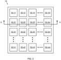
- the primary radiation source 12 and the secondary radiation source 14 may be arranged to rotate in common around the patient 22.
- the primary radiation source 12 and the secondary radiation source 14 may thus be fixed with respect to each other.
- the primary radiation source 12 and the secondary radiation source 14 may for example be spaced with an angular spacing of 90 degrees with respect to a rotation axis centered in the patient 22.
- the control system 18 is operatively connected to the primary radiation source 12, the secondary radiation source 14 and the primary detector 16. In FIG. 1 , the control system 18 controls the primary radiation source 12 to continuously emit the primary radiation 20. Moreover, the control system 18 controls the secondary radiation source 14 to emit the secondary radiation 24 in pulses, as indicated by the dashed lines of the secondary radiation 24.
- the control system 18 is configured to receive imaging data from the primary detector 16, for example in the form of serial data from each readout circuit associated with a respective pixel of the primary detector 16.
- the control system 18 may be configured to generate two-dimensional (2D) projection images based on the imaging data.
- the 2D images may be used by the control system 18 to reconstruct, for example three-dimensional (3D) images, of the patient 22 using inter alia known principles of computed tomography.
- each pixel 32 has a square shape.
- Examples of alternative shapes of the pixels 32 comprise non-square rectangular shapes and hexagonal shapes.
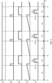
- FIG. 5 shows a plurality of primary active periods 76 and a plurality of primary inactive periods 78.
- the primary active periods 76 and the primary inactive periods 78 are here alternatingly arranged.
- Each primary inactive period 78 is interleaved with a primary active period 76.
- each primary inactive period 78 is constituted by a time period from an end of a previous primary active period 76 to a start of next primary active period 76
- each primary active period 76 is constituted by a time period from an end of a previous primary inactive period 78 to a start of a next primary inactive period 78.
- Each secondary pulse duration 82 may be a few ⁇ s (microseconds), such as at least 1 ⁇ s and/or less than 50 ⁇ s.
- a primary inactive period duration of each primary inactive period 78 may be larger than the associated secondary pulse duration 82 and for example less than 100 ⁇ s.
- the secondary pulses 80 may be repeated every few ms (milliseconds). For example, a time between each two subsequent secondary pulses 80 may be 100 ms, such as at least 1 ms and/or less than 500 ms.
- a primary active period duration of each primary active period 76 may be at least 1 ms. In view of this, it is apparent that the timing diagram in FIG. 5 may not be to scale.
- the reset signal 64 is issued at the end of each primary inactive period 78, detection data 58 from incoming radiation during the primary inactive periods 78 is not acquired, here not counted by the counter 60. Moreover, by sending the collection signal 68 and emitting a secondary pulse 80 during each primary inactive period 78, images that are closed in time to the respective secondary pulses 80 can be provided.
- the readout signal 70 may be sent to the register 66 at any time to cause the register 66 to read out respective counted detection data 62.
- the readout signal 70 may be sent during each primary inactive period 78.
- the readout signal 70 may be sent to the register 66 during only some of the primary inactive periods 78, such as every n'th primary inactive period 78, where n is a positive integer.
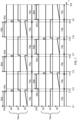
- the secondary radiation source 14b is of the same type as the primary radiation source 12a, e.g., identical thereto.
- the secondary radiation source 14b may be of the same type as the secondary radiation source 14, e.g., identical thereto.
- the secondary radiation source 14b is an MeV radiation source, like the secondary radiation source 14, the secondary detector 16b may be an MeV detector. In this case, the secondary detector 16b may be a direct conversion detector or an indirect conversion detector.
- each secondary active period 76b the secondary detector 16b is in an active state in relation to capturing incoming radiation during this secondary active period 76b, and in each secondary inactive period 78b, the secondary detector 16b is in an inactive state in relation to capturing incoming radiation during this secondary inactive period 78b.
- the method of this example further comprises collecting S22, by the primary detector 16, 16a, detection data 58 indicative of the primary radiation 20, 20a detected during the primary active periods 76, 76a.
- the method of this example further comprises collecting S24, by each register 66 and during each primary inactive period 78, 78a, the counted detection data 62 from the associated counter 60.
- the method of this example further comprises resetting S26 each counter 60 during each primary inactive period 78, 78a.
- the method of this example further comprises reading out S28 the counted detection data 62 from each register 66 during at least some of the primary inactive periods 78, 78a.
- the secondary pulses 80, 80b are synchronized with the primary inactive periods 78, 78a and are emitted during the primary inactive periods 78, 78a.
- one secondary pulse 80, 80b may be emitted during each primary inactive periods 78, 78a.
- the primary radiation 20, 20a may or may not be emitted in primary pulses 80a. In case the primary radiation 20, 20a is emitted in primary pulses 80a, the primary pulses 80a may occur primarily during the primary active periods 76, 76a.
- the secondary radiation source 14, 14b may be separated from the primary radiation source 12, 12a.
- the secondary radiation source 14, 14b may be angled 5 degrees to 170 degrees, such as 90 degrees, relative to the primary radiation source 12, 12a and with respect to a rotation axis centered in the object 22.
- the primary detector 16, 16a does not detect S18 incoming radiation during the primary inactive periods 78, 78a and/or does not collect S20 detection data 58 indicative of incoming radiation during the primary inactive periods 78, 78a. In these manners, the primary detector 16, 16a is inactive in relation to capturing incoming radiation. When the primary detector 16, 16a does not detect incoming radiation or does not collect detection data 58 indicative of incoming radiation, the incoming radiation is not captured by the primary detector 16, 16a.
- the method further comprises collecting S22, by the primary detector 16, 16a, detection data 58 indicative of the primary radiation 20, 20a detected during the primary active periods 76, 76a.
- the collection by the primary detector 16, 16a of the detection data 58 indicative of the primary radiation 20, 20a detected during a primary active period 76, 76a may take place during this primary active period 76, 76a or during a subsequent primary inactive period 78, 78a, e.g., before a secondary pulse 80, 80b is emitted in that primary inactive period 78, 78a.
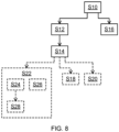
- the primary detector 16, 16a comprises a plurality of pixels 32, one or more counters 60 associated with each pixel 32, each configured to count the detection data 58 indicative of the primary radiation 20, 20a detected during the primary active periods 76, 76a to provide counted detection data 62, and a register 66 associated with each counter 60, each register 66 being configured to collect the counted detection data 62 from the associated counter 60.
- the method further comprises collecting S24, by each register 66 and during each primary inactive period 78, 78a, the counted detection data 62 from the associated counter 60.
- the method further comprises reading out S28 the counted detection data 62 from each register 66 during at least some of the primary inactive periods 78, 78a.
- the method comprises reading out the counted detection data 62 from each register 66 during each primary inactive period 78, 78a.
- a primary active period duration of each primary active period 76, 76a is at least 1 ms.
- the secondary radiation 24, 24b has energies of at least 0.5 MeV.
- emitting the primary radiation 20, 20a onto the object 22 occurs in primary pulses 80a by the primary radiation source 12, 12a.
- the primary pulses 80a may be emitted primarily during the plurality of primary active periods 76, 76a.
- a primary pulse duration 82a of each primary pulse 80a may be less than 100 ⁇ s.
- the primary radiation 20, 20a is detected by the primary detector 16, 16a by direct conversion each time during the plurality of primary active periods 76, 76a.
- the primary detector 16, 16a may be a direct conversion detector.
- the primary radiation 20, 20a may be detected by the primary detector 16, 16a by indirect conversion each time during the plurality of primary active periods 76, 76a.
- the primary detector 16, 16a may be an indirect conversion detector.
- the method further comprises detecting, by a secondary detector 16b, the secondary radiation 24b each time during a plurality of secondary active periods 76b, the secondary radiation 24b having interacted with the object 22.
- the secondary detector 16b may be a direct conversion detector or an indirect conversion detector. In the former case, the detection by the secondary detector 16b may take place during a plurality of secondary active periods 76b.
- the secondary pulses 80b may be emitted during the secondary active periods 76b.
- the secondary detector 16b may be inactive in relation to capturing incoming radiation during this secondary inactive period 78b.
- Each secondary inactive period 78b may follow a unique secondary active period 76b.
- the primary inactive periods 78a may include the secondary active periods 76b, and the secondary active periods 76b may include the secondary pulses 80b.
- the secondary inactive periods 78b may include the primary active periods 76a, and the primary active periods 76a may include the primary pulses 80a.
- Some embodiments include a radiation system 10a, 10b for handling radiation in relation to an object 22, the radiation system 10a, 10b comprising a primary radiation source 12, 12a configured to emit primary radiation 20, 20a; a secondary radiation source 14, 14b configured to emit secondary radiation 24, 24b; a primary detector 16, 16a configured to detect the primary radiation 20, 20a each time during a plurality of primary active periods 76, 76a, the primary radiation 20, 20a having interacted with the object 22; a control system 18 comprising at least one data processing device 26 and at least one memory 28 having at least one computer program stored thereon, the at least one computer program comprising program code which, when executed by the at least one data processing device 26, causes the at least one data processing device 26 to control the primary radiation source 12, 12a to emit the primary radiation 20, 20a onto the object 22; control the secondary radiation source 14, 14b to emit the secondary radiation 24, 24b onto the object 22 in secondary pulses 80, 80b emitted during a plurality of primary inactive periods 78, 78a, interleaving each primary inactive
- the at least one computer program comprises program code which, when executed by the at least one data processing device 26, causes the at least one data processing device 26 to control the primary detector 16, 16a to not detect incoming radiation during the primary inactive periods 78, 78a; and/or control the primary detector 16, 16a to not collect detection data 58 indicative of incoming radiation during the primary inactive periods 78, 78a.
- the at least one computer program comprises program code which, when executed by the at least one data processing device 26, causes the at least one data processing device 26 to control the primary detector 16, 16a to collect detection data 58 indicative of the primary radiation 20, 20a detected during the primary active periods 76, 76a.
- the primary detector 16, 16a comprises a plurality of pixels 32, one or more counters 60 associated with each pixel 32, each configured to count the detection data 58 indicative of the primary radiation 20, 20a detected during the primary active periods 76, 76a to provide counted detection data 62, and a register 66 associated with each counter 60, each register 66 being configured to collect the counted detection data 62 from the associated counter 60.
- each register 66 is configured to collect the counted detection data 62 from the associated counter 60 in response to a collection signal 68.
- the at least one computer program may comprise program code which, when executed by the at least one data processing device 26, causes the at least one data processing device 26 to command sending of the collection signal 68 to each register 66 during each primary inactive period 78, 78a.
- each counter 60 is configured to be reset in response to a reset signal 64.
- the at least one computer program may comprise program code which, when executed by the at least one data processing device 26, causes the at least one data processing device 26 to command sending of the reset signal 64 to each counter 60 during each primary inactive period 78, 78a.
- each register 66 is configured to read out the counted detection data 62 in response to a readout signal 70.
- the at least one computer program may comprise program code which, when executed by the at least one data processing device 26, causes the at least one data processing device 26 to command sending of the readout signal 70 to each register 66 during at least some of the primary inactive periods 78, 78a.
- the secondary radiation source 14, 14b is a linear accelerator.
- a secondary pulse duration 82, 82b of each secondary pulse 80, 80b is less than 100 ⁇ s.
- a primary active period duration of each primary active period 76, 76a is at least 1 ms.
- the secondary radiation 24, 24b has energies of at least 0.5 MeV.
- the at least one computer program comprises program code which, when executed by the at least one data processing device 26, causes the at least one data processing device 26 to control the primary radiation source 12, 12a to emit the primary radiation 20, 20a onto the object 22 in primary pulses 80a.
- the primary pulses 80a may be emitted primarily during the plurality of primary active periods 76, 76a.
- the primary detector is a direct conversion detector.
Landscapes
- Health & Medical Sciences (AREA)
- Life Sciences & Earth Sciences (AREA)
- Engineering & Computer Science (AREA)
- Medical Informatics (AREA)
- Biomedical Technology (AREA)
- Veterinary Medicine (AREA)
- Public Health (AREA)
- Physics & Mathematics (AREA)
- General Health & Medical Sciences (AREA)
- Pathology (AREA)
- Nuclear Medicine, Radiotherapy & Molecular Imaging (AREA)
- Radiology & Medical Imaging (AREA)
- Animal Behavior & Ethology (AREA)
- Heart & Thoracic Surgery (AREA)
- Molecular Biology (AREA)
- Optics & Photonics (AREA)
- Surgery (AREA)
- Biophysics (AREA)
- High Energy & Nuclear Physics (AREA)
- Spectroscopy & Molecular Physics (AREA)
- Theoretical Computer Science (AREA)
- Automation & Control Theory (AREA)
- General Engineering & Computer Science (AREA)
- General Physics & Mathematics (AREA)
- Toxicology (AREA)
- Pulmonology (AREA)
- Plasma & Fusion (AREA)
- Mathematical Physics (AREA)
- Apparatus For Radiation Diagnosis (AREA)
- Measurement Of Radiation (AREA)
- Radiation-Therapy Devices (AREA)
- Analysing Materials By The Use Of Radiation (AREA)
- Geophysics And Detection Of Objects (AREA)
Applications Claiming Priority (1)
| Application Number | Priority Date | Filing Date | Title |
|---|---|---|---|
| SE2251588A SE2251588A1 (en) | 2022-12-29 | 2022-12-29 | Method of handling radiation, and radiation system |
Publications (2)
| Publication Number | Publication Date |
|---|---|
| EP4393544A2 true EP4393544A2 (de) | 2024-07-03 |
| EP4393544A3 EP4393544A3 (de) | 2024-10-09 |
Family
ID=91185342
Family Applications (1)
| Application Number | Title | Priority Date | Filing Date |
|---|---|---|---|
| EP23220414.9A Pending EP4393544A3 (de) | 2022-12-29 | 2023-12-27 | Verfahren zur handhabung von strahlung und strahlungssystem |
Country Status (5)
| Country | Link |
|---|---|
| EP (1) | EP4393544A3 (de) |
| JP (1) | JP2025542124A (de) |
| CN (1) | CN120529867A (de) |
| SE (1) | SE2251588A1 (de) |
| WO (1) | WO2024142026A2 (de) |
Family Cites Families (7)
| Publication number | Priority date | Publication date | Assignee | Title |
|---|---|---|---|---|
| US7634061B1 (en) * | 2004-03-26 | 2009-12-15 | Nova R & D, Inc. | High resolution imaging system |
| US20130229495A1 (en) * | 2012-03-01 | 2013-09-05 | Ali-Reza Bani-Hashemi | Method for calibrating an imaging system |
| EP3368918B1 (de) * | 2015-10-28 | 2019-09-18 | Koninklijke Philips N.V. | Ct-system und ct-verfahren |
| WO2018183748A1 (en) * | 2017-03-30 | 2018-10-04 | Reflexion Medical, Inc. | Radiation therapy systems and methods with tumor tracking |
| WO2019136660A1 (en) * | 2018-01-11 | 2019-07-18 | Shenzhen United Imaging Healthcare Co., Ltd. | Systems and methods for intrafractional ct imaging in image-guided radiotherapy |
| US10960232B2 (en) * | 2018-07-28 | 2021-03-30 | Varian Medical Systems, Inc. | Single-pass imaging and radiation treatment delivery via an extended rotation gantry |
| JP7534038B2 (ja) * | 2018-11-30 | 2024-08-14 | アキュレイ インコーポレイテッド | コーンビームコンピュータ断層撮影における最適なパネル読み出しのための非対称散乱フィッティング |
-
2022
- 2022-12-29 SE SE2251588A patent/SE2251588A1/en unknown
-
2023
- 2023-12-27 EP EP23220414.9A patent/EP4393544A3/de active Pending
-
2024
- 2024-02-29 CN CN202480006196.7A patent/CN120529867A/zh active Pending
- 2024-02-29 WO PCT/IB2024/000095 patent/WO2024142026A2/en not_active Ceased
- 2024-02-29 JP JP2025532493A patent/JP2025542124A/ja active Pending
Also Published As
| Publication number | Publication date |
|---|---|
| WO2024142026A2 (en) | 2024-07-04 |
| WO2024142026A3 (en) | 2024-08-22 |
| SE2251588A1 (en) | 2024-06-30 |
| EP4393544A3 (de) | 2024-10-09 |
| JP2025542124A (ja) | 2025-12-25 |
| CN120529867A (zh) | 2025-08-22 |
Similar Documents
| Publication | Publication Date | Title |
|---|---|---|
| US9417339B2 (en) | Counting digital X-ray detector and method for taking a series of X-ray images | |
| JP4080758B2 (ja) | 放射線検出回路および核医学診断装置 | |
| WO1992014169A1 (en) | Digital gamma ray imaging device | |
| CN102438525B (zh) | 放射线诊断装置及控制方法 | |
| US20180341029A1 (en) | Radiation imaging system, signal processing apparatus, and, radiographic image signal processing method | |
| US10324202B1 (en) | Systems and methods for collecting radiation detection | |
| US10359520B2 (en) | Radiation imaging system, signal processing apparatus, and signal processing method for radiographic image | |
| CN108968992B (zh) | 放射线摄像装置、放射线摄像方法及计算机可读存储介质 | |
| EP4393544A2 (de) | Verfahren zur handhabung von strahlung und strahlungssystem | |
| EP3756033B1 (de) | Verfahren zum auslesen von daten in einem strahlungsdetektor, strahlungsdetektor und bildgebungsvorrichtung | |
| US12436301B2 (en) | Readout circuit, radiation detector, imaging apparatus and method of handling incident radiation | |
| US20070029494A1 (en) | Burst-mode readout for solid state radiation detectors using partitioned pipeline architecture | |
| EP3603746A1 (de) | Bildgeber in strahlentherapieumgebung | |
| JP2017086901A (ja) | データ収集装置、x線ct装置及び核医学診断装置 | |
| JP7652792B2 (ja) | 同期読取り/集積放射線撮像のための多段画素アーキテクチャ、ならびに関連システム、デバイス、および方法 | |
| JPH11304926A (ja) | 核医学診断装置 | |
| WO2004095069A1 (en) | Detector element for combined detection of x-radiation and gamma radiation | |
| US7589325B2 (en) | Method for recording a digital x-ray image, counting x-ray detector and x-ray system | |
| CN113841069A (zh) | 使用单个辐射探测器的x射线和伽马成像 | |
| JPH0448546Y2 (de) | ||
| US20250003895A1 (en) | Image processing apparatus, radiation imaging system, image processing method, and storage medium | |
| JPH10268053A (ja) | 核医学診断装置 | |
| JP4142767B2 (ja) | 核医学診断装置 | |
| JP2007267980A (ja) | 回転機構のない連続処理型x線ct装置 | |
| US20240272094A1 (en) | Two-stage pixel device with adaptive frame grabbing for x-ray imaging with or without automatic exposure control, and related systems, methods and devices |
Legal Events
| Date | Code | Title | Description |
|---|---|---|---|
| PUAI | Public reference made under article 153(3) epc to a published international application that has entered the european phase |
Free format text: ORIGINAL CODE: 0009012 |
|
| STAA | Information on the status of an ep patent application or granted ep patent |
Free format text: STATUS: THE APPLICATION HAS BEEN PUBLISHED |
|
| AK | Designated contracting states |
Kind code of ref document: A2 Designated state(s): AL AT BE BG CH CY CZ DE DK EE ES FI FR GB GR HR HU IE IS IT LI LT LU LV MC ME MK MT NL NO PL PT RO RS SE SI SK SM TR |
|
| REG | Reference to a national code |
Ref country code: DE Ref legal event code: R079 Free format text: PREVIOUS MAIN CLASS: A61N0005100000 Ipc: A61B0006420000 |
|
| PUAL | Search report despatched |
Free format text: ORIGINAL CODE: 0009013 |
|
| AK | Designated contracting states |
Kind code of ref document: A3 Designated state(s): AL AT BE BG CH CY CZ DE DK EE ES FI FR GB GR HR HU IE IS IT LI LT LU LV MC ME MK MT NL NO PL PT RO RS SE SI SK SM TR |
|
| RIC1 | Information provided on ipc code assigned before grant |
Ipc: A61N 5/10 20060101ALI20240903BHEP Ipc: A61B 6/03 20060101ALI20240903BHEP Ipc: A61B 6/00 20240101ALI20240903BHEP Ipc: A61B 6/42 20240101AFI20240903BHEP |
|
| STAA | Information on the status of an ep patent application or granted ep patent |
Free format text: STATUS: REQUEST FOR EXAMINATION WAS MADE |
|
| 17P | Request for examination filed |
Effective date: 20250328 |