EP4394032A2 - Verfahren zur herstellung autologer t-zellen zur behandlung von krebs und zusammensetzungen davon - Google Patents
Verfahren zur herstellung autologer t-zellen zur behandlung von krebs und zusammensetzungen davon Download PDFInfo
- Publication number
- EP4394032A2 EP4394032A2 EP24162638.1A EP24162638A EP4394032A2 EP 4394032 A2 EP4394032 A2 EP 4394032A2 EP 24162638 A EP24162638 A EP 24162638A EP 4394032 A2 EP4394032 A2 EP 4394032A2
- Authority
- EP
- European Patent Office
- Prior art keywords
- cells
- cell
- tumor
- population
- reactive
- Prior art date
- Legal status (The legal status is an assumption and is not a legal conclusion. Google has not performed a legal analysis and makes no representation as to the accuracy of the status listed.)
- Pending
Links
Images


Classifications
-
- C—CHEMISTRY; METALLURGY
- C12—BIOCHEMISTRY; BEER; SPIRITS; WINE; VINEGAR; MICROBIOLOGY; ENZYMOLOGY; MUTATION OR GENETIC ENGINEERING
- C12N—MICROORGANISMS OR ENZYMES; COMPOSITIONS THEREOF; PROPAGATING, PRESERVING, OR MAINTAINING MICROORGANISMS; MUTATION OR GENETIC ENGINEERING; CULTURE MEDIA
- C12N5/00—Undifferentiated human, animal or plant cells, e.g. cell lines; Tissues; Cultivation or maintenance thereof; Culture media therefor
- C12N5/06—Animal cells or tissues; Human cells or tissues
- C12N5/0602—Vertebrate cells
- C12N5/0634—Cells from the blood or the immune system
- C12N5/0636—T lymphocytes
- C12N5/0638—Cytotoxic T lymphocytes [CTL] or lymphokine activated killer cells [LAK]
-
- C—CHEMISTRY; METALLURGY
- C12—BIOCHEMISTRY; BEER; SPIRITS; WINE; VINEGAR; MICROBIOLOGY; ENZYMOLOGY; MUTATION OR GENETIC ENGINEERING
- C12N—MICROORGANISMS OR ENZYMES; COMPOSITIONS THEREOF; PROPAGATING, PRESERVING, OR MAINTAINING MICROORGANISMS; MUTATION OR GENETIC ENGINEERING; CULTURE MEDIA
- C12N5/00—Undifferentiated human, animal or plant cells, e.g. cell lines; Tissues; Cultivation or maintenance thereof; Culture media therefor
- C12N5/06—Animal cells or tissues; Human cells or tissues
- C12N5/0602—Vertebrate cells
- C12N5/0634—Cells from the blood or the immune system
- C12N5/0636—T lymphocytes
-
- A—HUMAN NECESSITIES
- A61—MEDICAL OR VETERINARY SCIENCE; HYGIENE
- A61K—PREPARATIONS FOR MEDICAL, DENTAL OR TOILETRY PURPOSES
- A61K40/00—Cellular immunotherapy
- A61K40/10—Cellular immunotherapy characterised by the cell type used
- A61K40/11—T-cells, e.g. tumour infiltrating lymphocytes [TIL] or regulatory T [Treg] cells; Lymphokine-activated killer [LAK] cells
-
- A—HUMAN NECESSITIES
- A61—MEDICAL OR VETERINARY SCIENCE; HYGIENE
- A61K—PREPARATIONS FOR MEDICAL, DENTAL OR TOILETRY PURPOSES
- A61K40/00—Cellular immunotherapy
- A61K40/30—Cellular immunotherapy characterised by the recombinant expression of specific molecules in the cells of the immune system
- A61K40/32—T-cell receptors [TCR]
-
- A—HUMAN NECESSITIES
- A61—MEDICAL OR VETERINARY SCIENCE; HYGIENE
- A61K—PREPARATIONS FOR MEDICAL, DENTAL OR TOILETRY PURPOSES
- A61K40/00—Cellular immunotherapy
- A61K40/40—Cellular immunotherapy characterised by antigens that are targeted or presented by cells of the immune system
- A61K40/41—Vertebrate antigens
- A61K40/42—Cancer antigens
- A61K40/4201—Neoantigens
-
- A—HUMAN NECESSITIES
- A61—MEDICAL OR VETERINARY SCIENCE; HYGIENE
- A61K—PREPARATIONS FOR MEDICAL, DENTAL OR TOILETRY PURPOSES
- A61K40/00—Cellular immunotherapy
- A61K40/40—Cellular immunotherapy characterised by antigens that are targeted or presented by cells of the immune system
- A61K40/41—Vertebrate antigens
- A61K40/42—Cancer antigens
- A61K40/428—Undefined tumor antigens, e.g. tumor lysate or antigens targeted by cells isolated from tumor
-
- A—HUMAN NECESSITIES
- A61—MEDICAL OR VETERINARY SCIENCE; HYGIENE
- A61P—SPECIFIC THERAPEUTIC ACTIVITY OF CHEMICAL COMPOUNDS OR MEDICINAL PREPARATIONS
- A61P35/00—Antineoplastic agents
-
- A—HUMAN NECESSITIES
- A61—MEDICAL OR VETERINARY SCIENCE; HYGIENE
- A61P—SPECIFIC THERAPEUTIC ACTIVITY OF CHEMICAL COMPOUNDS OR MEDICINAL PREPARATIONS
- A61P37/00—Drugs for immunological or allergic disorders
- A61P37/02—Immunomodulators
- A61P37/04—Immunostimulants
-
- C—CHEMISTRY; METALLURGY
- C07—ORGANIC CHEMISTRY
- C07K—PEPTIDES
- C07K14/00—Peptides having more than 20 amino acids; Gastrins; Somatostatins; Melanotropins; Derivatives thereof
- C07K14/435—Peptides having more than 20 amino acids; Gastrins; Somatostatins; Melanotropins; Derivatives thereof from animals; from humans
- C07K14/705—Receptors; Cell surface antigens; Cell surface determinants
- C07K14/70503—Immunoglobulin superfamily
- C07K14/7051—T-cell receptor (TcR)-CD3 complex
-
- C—CHEMISTRY; METALLURGY
- C12—BIOCHEMISTRY; BEER; SPIRITS; WINE; VINEGAR; MICROBIOLOGY; ENZYMOLOGY; MUTATION OR GENETIC ENGINEERING
- C12N—MICROORGANISMS OR ENZYMES; COMPOSITIONS THEREOF; PROPAGATING, PRESERVING, OR MAINTAINING MICROORGANISMS; MUTATION OR GENETIC ENGINEERING; CULTURE MEDIA
- C12N5/00—Undifferentiated human, animal or plant cells, e.g. cell lines; Tissues; Cultivation or maintenance thereof; Culture media therefor
- C12N5/06—Animal cells or tissues; Human cells or tissues
- C12N5/0602—Vertebrate cells
- C12N5/0634—Cells from the blood or the immune system
- C12N5/0639—Dendritic cells, e.g. Langherhans cells in the epidermis
-
- A—HUMAN NECESSITIES
- A61—MEDICAL OR VETERINARY SCIENCE; HYGIENE
- A61K—PREPARATIONS FOR MEDICAL, DENTAL OR TOILETRY PURPOSES
- A61K2121/00—Preparations for use in therapy
-
- A—HUMAN NECESSITIES
- A61—MEDICAL OR VETERINARY SCIENCE; HYGIENE
- A61K—PREPARATIONS FOR MEDICAL, DENTAL OR TOILETRY PURPOSES
- A61K2300/00—Mixtures or combinations of active ingredients, wherein at least one active ingredient is fully defined in groups A61K31/00 - A61K41/00
-
- C—CHEMISTRY; METALLURGY
- C12—BIOCHEMISTRY; BEER; SPIRITS; WINE; VINEGAR; MICROBIOLOGY; ENZYMOLOGY; MUTATION OR GENETIC ENGINEERING
- C12N—MICROORGANISMS OR ENZYMES; COMPOSITIONS THEREOF; PROPAGATING, PRESERVING, OR MAINTAINING MICROORGANISMS; MUTATION OR GENETIC ENGINEERING; CULTURE MEDIA
- C12N2501/00—Active agents used in cell culture processes, e.g. differentation
- C12N2501/20—Cytokines; Chemokines
- C12N2501/23—Interleukins [IL]
- C12N2501/2302—Interleukin-2 (IL-2)
-
- C—CHEMISTRY; METALLURGY
- C12—BIOCHEMISTRY; BEER; SPIRITS; WINE; VINEGAR; MICROBIOLOGY; ENZYMOLOGY; MUTATION OR GENETIC ENGINEERING
- C12N—MICROORGANISMS OR ENZYMES; COMPOSITIONS THEREOF; PROPAGATING, PRESERVING, OR MAINTAINING MICROORGANISMS; MUTATION OR GENETIC ENGINEERING; CULTURE MEDIA
- C12N2501/00—Active agents used in cell culture processes, e.g. differentation
- C12N2501/20—Cytokines; Chemokines
- C12N2501/23—Interleukins [IL]
- C12N2501/2307—Interleukin-7 (IL-7)
-
- C—CHEMISTRY; METALLURGY
- C12—BIOCHEMISTRY; BEER; SPIRITS; WINE; VINEGAR; MICROBIOLOGY; ENZYMOLOGY; MUTATION OR GENETIC ENGINEERING
- C12N—MICROORGANISMS OR ENZYMES; COMPOSITIONS THEREOF; PROPAGATING, PRESERVING, OR MAINTAINING MICROORGANISMS; MUTATION OR GENETIC ENGINEERING; CULTURE MEDIA
- C12N2501/00—Active agents used in cell culture processes, e.g. differentation
- C12N2501/20—Cytokines; Chemokines
- C12N2501/23—Interleukins [IL]
- C12N2501/2315—Interleukin-15 (IL-15)
-
- C—CHEMISTRY; METALLURGY
- C12—BIOCHEMISTRY; BEER; SPIRITS; WINE; VINEGAR; MICROBIOLOGY; ENZYMOLOGY; MUTATION OR GENETIC ENGINEERING
- C12N—MICROORGANISMS OR ENZYMES; COMPOSITIONS THEREOF; PROPAGATING, PRESERVING, OR MAINTAINING MICROORGANISMS; MUTATION OR GENETIC ENGINEERING; CULTURE MEDIA
- C12N2501/00—Active agents used in cell culture processes, e.g. differentation
- C12N2501/20—Cytokines; Chemokines
- C12N2501/23—Interleukins [IL]
- C12N2501/2321—Interleukin-21 (IL-21)
-
- C—CHEMISTRY; METALLURGY
- C12—BIOCHEMISTRY; BEER; SPIRITS; WINE; VINEGAR; MICROBIOLOGY; ENZYMOLOGY; MUTATION OR GENETIC ENGINEERING
- C12N—MICROORGANISMS OR ENZYMES; COMPOSITIONS THEREOF; PROPAGATING, PRESERVING, OR MAINTAINING MICROORGANISMS; MUTATION OR GENETIC ENGINEERING; CULTURE MEDIA
- C12N2501/00—Active agents used in cell culture processes, e.g. differentation
- C12N2501/50—Cell markers; Cell surface determinants
- C12N2501/505—CD4; CD8
-
- C—CHEMISTRY; METALLURGY
- C12—BIOCHEMISTRY; BEER; SPIRITS; WINE; VINEGAR; MICROBIOLOGY; ENZYMOLOGY; MUTATION OR GENETIC ENGINEERING
- C12N—MICROORGANISMS OR ENZYMES; COMPOSITIONS THEREOF; PROPAGATING, PRESERVING, OR MAINTAINING MICROORGANISMS; MUTATION OR GENETIC ENGINEERING; CULTURE MEDIA
- C12N2501/00—Active agents used in cell culture processes, e.g. differentation
- C12N2501/50—Cell markers; Cell surface determinants
- C12N2501/515—CD3, T-cell receptor complex
-
- C—CHEMISTRY; METALLURGY
- C12—BIOCHEMISTRY; BEER; SPIRITS; WINE; VINEGAR; MICROBIOLOGY; ENZYMOLOGY; MUTATION OR GENETIC ENGINEERING
- C12N—MICROORGANISMS OR ENZYMES; COMPOSITIONS THEREOF; PROPAGATING, PRESERVING, OR MAINTAINING MICROORGANISMS; MUTATION OR GENETIC ENGINEERING; CULTURE MEDIA
- C12N2501/00—Active agents used in cell culture processes, e.g. differentation
- C12N2501/998—Proteins not provided for elsewhere
-
- C—CHEMISTRY; METALLURGY
- C12—BIOCHEMISTRY; BEER; SPIRITS; WINE; VINEGAR; MICROBIOLOGY; ENZYMOLOGY; MUTATION OR GENETIC ENGINEERING
- C12N—MICROORGANISMS OR ENZYMES; COMPOSITIONS THEREOF; PROPAGATING, PRESERVING, OR MAINTAINING MICROORGANISMS; MUTATION OR GENETIC ENGINEERING; CULTURE MEDIA
- C12N2502/00—Coculture with; Conditioned medium produced by
- C12N2502/11—Coculture with; Conditioned medium produced by blood or immune system cells
- C12N2502/1114—T cells
-
- C—CHEMISTRY; METALLURGY
- C12—BIOCHEMISTRY; BEER; SPIRITS; WINE; VINEGAR; MICROBIOLOGY; ENZYMOLOGY; MUTATION OR GENETIC ENGINEERING
- C12N—MICROORGANISMS OR ENZYMES; COMPOSITIONS THEREOF; PROPAGATING, PRESERVING, OR MAINTAINING MICROORGANISMS; MUTATION OR GENETIC ENGINEERING; CULTURE MEDIA
- C12N2502/00—Coculture with; Conditioned medium produced by
- C12N2502/11—Coculture with; Conditioned medium produced by blood or immune system cells
- C12N2502/1121—Dendritic cells
Definitions
- the present disclosure provides methods for manufacturing T cells.
- methods for manufacturing T cells which express a novel group of cell surface receptors that recognize peptides on the surface of a target cell are provided.
- the present disclosure also provides populations of T cells produced by methods described herein and pharmaceutical compositions thereof.
- TIL tumor infiltrating lymphocyte
- methods for manufacturing T cells which express a novel group of cell surface receptors that recognize peptides on the surface of a target cell are provided.
- T cells for use in a therapeutic cell composition, wherein the T cells express a T cell receptor (TCR) that recognizes an antigen on the surface of a target cell
- TCR T cell receptor
- said method comprising: (a) enriching a population of lymphocytes obtained from a donor subject to produce a first population of T lymphocytes; (b) stimulating the first population with one or more T-cell stimulating agents of lymphocytes to produce a second population of activated T cells wherein the stimulation is performed in a closed system using serum free medium; (c) co-culturing the second population of T cells in the presence of antigen presenting cells that present one or more non-native peptide on a major histocompatibility complex (MHC) in a closed system using serum free medium, said one or more non-native peptides are peptides corresponding to nonsynonymous somatic mutations associated in the tumor of a subject, to produce a third population containing T cells comprising endogenous T cell receptors reactive to mutation en
- the method further comprises expanding T cells in vitro.
- the antigen presenting cells are nucleated cells such as dendritic cells, mononuclear phagocytes, B lymphocytes, endothelial cells or thymic epithelium.
- the antigen presenting cells are dendritic cells.
- the T cells are autologous the subject.
- the T cells are autologous to a subject having a tumor.
- the T cells are autologous to the patient.
- the T cells are obtained from a healthy donor.
- the first T cell stimulating agents are selected from one or more of an anti-CD3 antibody; an anti-CD28 antibody; or a recombinant cytokine selected from among IL-2, IL-7, IL-15 and IL-21.
- the first T cell stimulating agents is or comprises recombinant IL-2.
- the concentration of recombinant IL-2 is from 100 IU/mL to 6000 IU/mL. In some of any of the embodiments, the concentration of recombinant IL-2 is from 300 IU/mL to 1000 IU/mL. In some embodiments, the concentration of recombinant IL-2 is at or about 300 IU/mL.
- the concentration of recombinant IL-2 is from 300 IU/mL to 1000 IU/mL. In some embodiments, the concentration of recombinant IL-2 is at or about 300 IU/mL. In some of any of the embodiments, the T cell stimulating agent further comprises an anti-CD3 antibody, such as OKT3. In some embodiments, the concentration of the anti-CD3 antibody is at or about 50 ng/mL.
- the one or more non-native peptide comprises an individual peptide or a pool of peptides.
- non-native peptides are peptide corresponding to the nonsynonymous somatic mutations associated in the patient's tumor.
- the one or more non-native peptides are loaded on antigen presenting cells by transfection of in vitro transcribed synthesized minigene constructs encoding for nonsynonomous somatic mutated amino acids.
- non-native peptides are expressed on antigen presenting cells by synthesizing minigene constructs encoding for nonsynonymous somatic mutated amino acids, flanked on each side by 12 amino acids from endogenous proteins in tandem, then transcribed using in-vitro RNA transcription to generate individual peptides and then transfected into the antigen presenting cells.
- non-native peptides are expressed on nucleated cells by peptide pulsing, such as by electroporation, of peptide pools associated with nonsynonymous somatic mutations into the antigen presenting cells.
- each peptide of the peptide pool is 9 to 15 amino acids in length, such as at or about 9 amino acids, 10 amino acids, 11 amino acids, 12 amino acids, 13 amino acids, 14 amino acids or 15 amino acids in length.
- each peptide of the peptide pools is a 12 amino acid long peptide.
- the one or more non-native peptide is 5-30 amino acids.
- the one or more non-native peptide is 12-25 amino acids.
- the one or more non-native peptide is at or about 25 amino acids or at or about 12 amino acids.
- the ratio of individual peptides to antigen presenting cells during the peptide pulsing is between 0.001 and 100 ⁇ per 1 million cells, between 0.01 and 100 ⁇ g per 1 million cells, between 0.1 and 100 ⁇ g per 1 million cells, between 1 and 100 ⁇ g per 1 million cells, between 10 and 100 ⁇ g per 1 million cells.
- the concentration of individual peptides for the peptide pulse is between at or about 0.0001 pg/mL and at or about 40 pg/mL or at or about 0.001 pg/mL and at or about 40 pg/mL.
- separation of antigen presenting cells is using magnetic separation, gravimetric separation, selective binding or cell sorting using flow cytometry.
- T cells for use in a therapeutic cell composition, wherein the T cells express a T cell receptor (TCR) that recognizes an antigen on the surface of a target cell: (a) processing a population of T lymphocyte cells obtained from a donor subject that has a tumor to produce a first population of T lymphocyte cells; (b) stimulating the first population with one or more first T-cell stimulating agents of lymphocytes under conditions for expansion of T cells in the population to produce a second population of activated T cells, wherein the one or more T-cell stimulating agents is or includes recombinant IL-2 at a concentration of 300 IU/mL to 1000 IU/mL and wherein the stimulation is performed in a closed system using serum free medium; (c) co-culturing the second population of T cells in the presence of autologous dendritic cells loaded with one or more non-native peptides for presenting the one or more non-native peptide on a major histocompatibility complex (MHC), wherein
- MHC major histocompat
- the selection is performed using a florescence based cell sorter. In some of any of the embodiments, selection is performed using a florescence based cell sorter in a closed system using serum free medium. In some of any of the embodiments, the selection is by 1 run, 2 runs, 3 runs or 4 runs by the fluorescence based cell sorter. In some of any of the embodiments, selection is performed at rate between 10,000 and 100,000 cells/ second using a florescent based disposable fluidics cell sorter. In some of any of the embodiments, the closed system is a bag system.
- the one or more upregulation markers are selected from the group consisting of CD107, CD107a, CD39, CD103, CD137 (4-1BB), CD59, CD69, CD90, CD38, CD30, CD154, CD252, CD134 (OX40), CD258, CD256, PD-1, TIM-3 and LAG-3.
- the one or more upregulation marker is selected from the group consisting of CD38, CD39, CD6, CD90, CD134 and CD137.
- the one or more upregulation maker is CD134 and/or CD137.
- the one or more upregulation marker is selected from the group consisting of CD107, CD107a, CD39, CD103, CD59, CD90, CD38, CD30, CD154, CD252, CD134, CD258 and CD256.
- the one or more T upregulation marker is selected from the group consisting of CD107a, CD39, CD103, CD59, CD90 and CD38.
- stimulating the first population is for 7 to 21 days. In some embodiments, stimulating the first population is for 7 to 14 days. In some of any of the provided embodiments, expanding the fourth population is for 7 to 21 days. In some embodiments, expanding the fourth population is for 7 to 14 days.
- the tumor is a tumor of an epithelial cancer. In some of any of the embodiments, the tumor is a tumor of a melanoma, lung squamous, lung adenocarcinoma, bladder cancer, lung small cell cancer, esophageal cancer, colorectal cancer (CRC), cervical cancer, head and neck cancer, stomach cancer or uterine cancer.
- a melanoma lung squamous, lung adenocarcinoma, bladder cancer, lung small cell cancer, esophageal cancer, colorectal cancer (CRC), cervical cancer, head and neck cancer, stomach cancer or uterine cancer.
- the population of lymphocytes are processed as a single cell suspension obtained by homogenization and/or enzymatic digestion of one or more tumor fragments from a resected tumor. In some of any of the provided embodiments, the population of lymphocytes are processed as a single cell suspension obtained by homogenization and enzymatic digestion of one or more tumor fragments from a resected tumor. In some of any of the embodiments, the enzymatic digestion is by a collagenase. In some embodiments, the collagenase is a collagenase type IV. In some embodiments, the collagenase is a collagenase type I/II. In some of any of the embodiments, the tumor is a colorectal cancer (CRC).
- CRC colorectal cancer
- the population of T cells of the therapeutic composition produced by the method is enriched for tumor-reactive T cells or T cells that are surface positive for one or more T cell upregulation marker expressed on reactive or activated T cells compared to the first population of T cells, such as enriched 10-fold to 1000-fold. In some of any of the embodiments, the population of T cells of the therapeutic composition produced by the method is enriched for tumor-reactive T cells or T cells that are surface positive for one or more T cell upregulation marker expressed on reactive or activated T cells compared to the second population of T cells, such as enriched 10-fold to 1000-fold.
- the population of T cells of the therapeutic composition produced by the method is enriched for tumor-reactive T cells or T cells that are surface positive for one or more T cell upregulation marker expressed on reactive or activated T cells compared to the third population of T cells, such as enriched 1.5-fold to 50-fold.
- the population of T cells of the therapeutic composition produced by the method are able to produce IFNgamma at a concentration of greater than at or about 30 pg/mL following antigen-specific stimulation.
- the IFNgamma produced following antigen-specific stimulation is as greater than at or about 40 pg/mL, greater than at or about 50 pg/mL, greater than at or about 60 pg/mL, greater than at or about 70 pg/mL, greater than at or about 80 pg/mL or greater than at or about 90 pg/mL.
- T cells produced by any of the provided methods wherein the population of T cells express greater than or equal to 1 T cell surface receptor that recognizes greater than or equal to 1 specific antigen on a target cell expressing nonsynonymous mutations.
- the one or more T cell upregulation marker is CD137 and/or CD134. In some of any of the embodiments, the one or more T cell upregulation marker is selected from the group consisting of CD107, CD107a, CD39, CD103, CD59, CD90, CD38, CD30, CD154, CD252, CD134, CD258 and CD256. In some of any of the embodiments, the one or more T cell upregulation marker is selected from the group consisting of CD107a, CD39, CD103, CD59, CD90 and CD38.
- T cells Provided herein is a method for manufacturing T cells, the method comprising: (a) obtaining lymphocytes; (b) stimulating the population of lymphocytes to produce a population of activated T cells in a closed system using serum-free medium; (c) co-culturing T cells in the presence of antigen presenting cells that present one or more MHC-associated non-native peptides presented; (d) separating antigen presenting cells from T cells in a closed system; (e) selecting T cells containing cell surface receptors that are reactive to peptides present on the APC based on upregulation markers on T cells cultured in the presence of nucleated cells presenting peptides in a closed system using serum free medium; and (f) expanding selected cells in the presence of cytokines in a closed system using serum free medium.
- T cell preparations are provided which may be useful for treating patients with a pathological disease or condition.
- the methods and processes described herein can be completed in a significantly shorter time and recover a higher number of endogenous TCR-expressing T cells, thereby offering a significant advantage to bring cells into the clinic in therapeutic doses.
- populations of T cells produced by methods described herein and pharmaceutical compositions thereof are also provided herein.
- a TCR has two protein chains, which are designed to bind with specific peptides presented by a major histocompatibility complex (MHC) protein on the surface of certain cells. Since TCRs recognize peptides in the context of MHC molecules expressed on the surface of a target cell, TCRs have the potential to recognize antigens not only presented directly on the surface of target cells, e.g. cancer cells, but also presented by antigen-presenting cells, such as are present in tumor, inflammatory and infected microenvironments, and in secondary lymphoid organs. Reactive T cells expressing such cell surface receptors may be used to target and kill any target cell, including, but not limited to, infected cells, damaged cells, or dysfunctional cells.
- MHC major histocompatibility complex
- the reactive T cells are tumor-reactive T cells that recognize a cancer neoantigen.
- the majority of neoantigens arise from passenger mutations, meaning they do not infer any growth advantage to the cancer cell.
- Passenger mutations are likely to give rise to neoantigens that are unique to each patient and may be present in a subset of all cancer cells.
- Driver mutations give rise to neoantigens that are likely to be present in all the tumor cells of an individual and potentially shared.
- the population of T cells contain tumor-reactive T cells that can recognize neoantigens containing passenger and/or driver mutations.
- tumor regulatory T cells are a subpopulation of CD4 + T cells, which specialize in suppressing immune responses and could limit reactivity of a T cell product.
- a source of potential tumor peptides is used to identify TCRs that are reactive to neoantigens in a process that includes expansion of the T cells reactive to the tumor neoantigenic peptides.
- Provided methods include ex vivo co-culture methods in which a population of T cells that have been expanded from T cells present in or from a biological sample (e.g. tumor fragments or peripheral blood or other source of T cells) is incubated in the presence of antigen-presenting cells that have been contacted with, or made to present, the neoantigenic peptides.
- the T cells and antigen-presenting cells are autologous to the tumor-bearing subject from which the peptides were identified.
- the provided methods further include steps to separate, enrich for and/or select for tumor-reactive T cells from the co-culture prior to or in connection with their further ex vivo expansion.
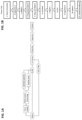
- FIG. 1A depicts a schematic of an exemplary process for manufacturing a T cell therapeutic composition in accord with the provided methods.
- a tumor sample is obtained from a patient for identification and generation of peptides for use in co-culturing methods with antigen presenting cells (APCs) presenting the peptides and autologous antigen T cells obtained from the same subject.
- APCs antigen presenting cells
- a population of T cells from the patient e.g. containing tumor infiltrating lymphocytes (TIL) or peripheral blood lymphocytes (PBL) is stimulated under conditions to expand the cells, prior to coculture with antigen presenting cells that have been contacted or exposed to peptide neoepitopes for presentation on a major histocompability complex.
- TIL tumor infiltrating lymphocytes
- PBL peripheral blood lymphocytes
- the provided methods offer advantages compared to existing methods for producing and expanding TILs because the provided methods involve steps to enrich for tumor reactive cells, such as by the co-culturing step with peptide-presenting APCs followed by selection of reactive T cell clones that have upregulated one or more T cell activation marker.
- the initial small population of tumor reactive T cells expanded from the biological sample e.g. tumor
- the biological sample e.g. tumor
- the initial small population of tumor reactive T cells expanded from the biological sample are enriched for cells that are or likely to be tumor reactive cells before a subsequent second expansion step, thereby promoting preservation and expansion of cells of interest and limiting expansion of bystander T cells that are not reactive to a tumor antigen and/or that may include cells that exhibit inhibitory activity ( FIG. 2A ).
- the provided methods are carried out to maximize the numbers of tumor reactive cells that may be collected, for example by co-culturing all of the cells propagated after the first expansion with peptide-presenting APCs, and then by selecting from among all of the bulk cells after the co-culturing for cells positive for the one or more activation markers before the subsequent second expansion.
- all steps of the method are carried out in a closed system.
- the provided methods include one or more features that provide for or relate to an improved, more efficient and/or more robust process for producing a tumor-reactive T cell therapeutic composition ex vivo.
- the disclosure relates to methods that provide advantages over available methods for producing a TIL therapeutic cell composition. Such advantages include, for example, reduced cost, streamlining, improved enrichment of tumor-reactive T cells in the therapeutic composition, and increased efficacy of the therapeutic composition, including among different subjects and tumor conditions.
- the population of T cells is obtained from a biological sample known to contain T cells.
- the population of T cells is enriched from a biological sample from a subject, in particular a human subject.
- the biological sample can be any sample containing a bulk population of T cells.
- the biological sample is or includes peripheral blood mononuclear cells.
- the biological sample is a peripheral blood or serum sample.
- the biological sample is a lymph node sample.
- the biological sample is a tumor sample.
- the bulk T cells can include tumor-infiltrating T cells (TILs).
- the subject is a human subject.
- the subject is a subject having a cancer, viral infection, bacterial infection, or is a subject with an inflammatory condition.
- the subject has a cancer.
- the starting source of cells in the method can be tumor fragments (e.g. 1-8 mm diameter fragments) or can be a single cell suspension preparation from enzymatic digestion of tumor fragments. It is found herein that, while certain sources may be superior for some tumor types, both fragments and single cell suspensions can support T cell expansion and enrichment of tumor-reactive T cells.
- the tumor cell source can be chosen depending on the tumor type or cancer, such as to optimize or increase expansion and enrichment of tumor-reactive T cells from the tumor.
- the cancer is a melanoma and the starting population of lymphocytes are tumor fragments, such as from a resected tumor.
- the cancer is a colorectal cancer and the starting population of lymphocytes is a single cell suspension obtained by enzymatic digestion, e.g. collagenase, of tumor fragments.
- the methods include a step of co-culturing initially expanded T cells with autologous antigen presenting cells that have been loaded with peptide.
- Findings herein demonstrate that relatively low concentrations of peptide or a peptide pool (containing a plurality of peptides, e.g. 2, 3, 4, 5, 6, 7, 8, 9, 10, 20, 30, 40, 50, 60, 70, 80, 90 or 100 more, or any value between any of the foregoing), such where each individual peptide is less than 20 ng/mL, and even as low as 0.1 ng/mL, can lead to an increase in activation of T cell during the culture.
- the co-culturing step in the provided methods include a ratio of tumor-derived cells containing T cells to autologous APCs (e.g. dendritic cells) of at or about 1:5 to at or about 5:1, such as 1:3 to at or about 3:1, for example as at or about 1:1, and involves loading the APCs with an individual peptide or a pool of peptides.
- APCs e.g. dendritic cells
- the APCs are loaded with a concentration of peptide or peptide pool in which the individual peptide, or individual peptides of the pool of peptides on average, is less than at or about 20 ng/mL, such as from at or about 0.1 ng/mL to at or about 1 ng/mL, for example at or about 0.1 ng/mL.
- Exemplary reactive T cell markers include one or more, such as two, three, four or more of, CD107, CD107a, CD39, CD103, CD137 (4-1BB), CD59, CD90, CD38, CD30, CD154, CD252, CD134 (OX40), CD258, CD256, PD-1, TIM-3 or LAG-3.
- the enrichment or selection for cells positive for one or more such upregulation marker on reactive or activated T cells can be carried out prior to or during one or more steps of the expansion method.
- the provided methods include enrichment or selection for cells positive for one or more upregulation marker on reactive or activated T cells after activation of a population of T cells by the co-culture incubation with peptide-presenting APCs (e.g. DCs).
- the provided methods involve the ex vivo expansion and production of a T cell therapeutic composition, particularly for use in connection with treating cancer.
- the method of manufacturing involves the growth and manipulation of patient cells outside of the body.
- the methods relate to methods for expanding T cells containing an endogenous TCR specific to a tumor-associated antigen (hereinafter "tumor reactive T cells").
- tumor reactive T cells includes T cells that exhibit reactivity to a tumor antigen or that are likely or suspected of being tumor reactive T cells due to upregulation or positive surface expression of a protein expressed on the T cell that are only expressed when the T cells endogenous TCR recognizes a peptide expressed by the APCs, e.g. T cell activation marker.
- the frequency of these cells can be low and in order to expand these cells to a therapeutic dose ex vivo methods for enrichment and expansion are necessary.
- the sample is a tumor sample or a tumor fragment containing tumor infiltrating lymphocytes or TILs.
- the population containing T cells can be directly obtained from a subject (e.g. healthy or cancer subject), such as by selection of T cells or a subset thereof from the biological sample from the subject.
- the biological sample is from a subject that has a tumor and that contains tumor reactive T cells or that has the potential to or that may contain tumor reactive T cells that can be enriched by the provided methods.
- tumor reactive T cells (4) from the third population, separating the APCs to produce a fourth population of T cells containing endogenous TCR that are reactive to peptides present on the APCs; and (5) expanding the fourth population of T cells containing tumor reactive T cells by incubation in the presence of T cell stimulating agents.
- the separating of the T cells in (4) can involve depleting or removing the APCs and/or can include selection of T cells based on upregulation markers on reactive or activated T cells.
- one or more of the steps can be carried out in a closed system using serum free medium.
- tumor reactive T cells (4) from the third population, separating antigen presenting cells from the population of T cells in a closed system; (5) after separating antigen presenting cells, selecting T cells containing endogenous TCR that are reactive to peptides present on the APCs based on upregulation markers on reactive or activated T cells to produce a fourth population of T cells, in which the selecting is in a closed system using serum free medium; and (6) Expanding the fourth population of selected cells in the presence of cytokines in a closed system using serum free medium.
- the provided methods include (1) Enriching a population of lymphocytes obtained from a donor subject to produce a first population of T cells; (2) Stimulating the first population with one or more T-cell stimulating agents of lymphocytes to produce a population of activated T cells wherein the stimulation is performed in a closed system using serum free medium; (3) Co-culturing cells from the second population of T cells in the presence of antigen presenting cells that present one or more (e.g. peptide neoepitopes) on an MHC (MHC-associated non-native peptide), in which the co-culturing produces a third population of cells containing or enriched for T cells reactive to peptides presented on the MHC of the APC (e.g.
- tumor reactive T cells (4) from the third population, selecting T cells containing endogenous TCR that are reactive to peptides present on the APCs based on upregulation markers on reactive or activated T cells to produce a fourth population of T cells, in which the selecting is in a closed system using serum free medium; and (5) Expanding the fourth population of selected cells in the presence of cytokines in a closed system using serum free medium.
- the T-cell stimulating agents include anti-CD3 (e.g. anti-CD3 antibody, such as OKT3), anti-CD28 reagents (e.g. anti-CD28 antibody), such as an anti-CD3 antibody (e.g. OKT3) and an anti-CD28 antibody and/or one or more recombinant cytokine (e.g. IL-2, IL-7, IL-21 and/or IL-15) .
- anti-CD3 e.g. anti-CD3 antibody, such as OKT3
- anti-CD28 antibody e.g. OKT3
- an anti-CD3 antibody e.g. OKT3
- an anti-CD28 antibody and/or one or more recombinant cytokine e.g. IL-2, IL-7, IL-21 and/or IL-15
- the incubation or culture of T cells also is carried out with nutrient containing media so that the cells can survive outside of the body.
- the methods including incubation with recombinant IL-2 alone or in combination with one or more other recombinant cytokines (e.g. IL-7, IL-21 and/or IL-15) and, in some cases, one or more other T cell stimulating agents to provide a primary and secondary (costimulatory) signal to the cells.
- Standard methods for culturing T cells to provide a primary and secondary (costimulatory) signal to the cells involve incubation with T cell stimulating agents provided by anti-CD3 (e.g. OKT3) and anti-CD28 reagents.
- the T cell stimulatory agents include an anti-CD3 antibody (e.g. OKT3) and an anti-CD28 antibody.
- stimulations also include one or more additional recombinant cytokine (e.g. IL-2, IL-7, IL-21 and/or IL-15) and nutrient containing media so that the cells can survive outside of the body.
- the population of T cells are autologous T cells from a subject with a tumor and the source of synthetic peptides are tumor antigenic peptides from a tumor antigen of the same subject.
- cells from the ex vivo coculture are a population of cells (third population) that include tumor reactive T cells that recognize or are activated by a peptide presented on an MHC of an APC in the culture.
- cells from the ex vivo co-culture represent a source of cells that are enriched for tumor reactive T cells.
- IL-2 at least including recombinant IL-2) to produce a second population of T cells containing expanded or stimulated T cells, (3) coculturing the second population containing stimulated T cells in the presence of antigen presenting cells (APCs) that have been contacted or exposed to one or more of the plurality of peptides under conditions in which the APCs present one or more MHC-associated non-native peptide to produce a third population of T cells; and (5) enriching, from the third population of T cells, T cells containing an endogenous TCR that are reactive to peptides present on antigen presenting cells (APCs) to produce a fourth population of T cells.
- APCs antigen presenting cells
- the concentration of the dipeptide form of L-glutamine is from or from about 0.5 mM to 5 mM, 0.5 mM to 4 mM, 0.5 mM to 3 mM, 0.5 mM to 2 mM, 0.5 mM to 1 mM, 1 mM to 5 mM, 1 mM to 4 mM, 1 mM to 3 mM, 1 mM to 2 mM, 2 mM to 5 mM, 2 mM to 4 mM, 2 mM to 3 mM, 3 mM to 5 mM, 3 mM to 4 mM or 4 mM to 5 mM, each inclusive.
- the concentration of the dipeptide form of L-glutamine is or is about 2 mM.
- the cancer-specific cancer neoepitope is determined by identifying or isolating a tumor-associated antigen or peptide sequence thereof from a cancer cell from a subject.
- the cancer cell may be obtained from any bodily sample derived from a patient which contains or is expected to contain tumor or cancer cells.
- the bodily sample may be any tissue sample such as blood, a tissue sample obtained from the primary tumor or from tumor metastases, a lymph node sample or any other sample containing tumor or cancer cells.
- the tumor is a solid tumor.
- solid tumors such as sarcomas and carcinomas, include fibrosarcoma, myxosarcoma, liposarcoma, chondrosarcoma, osteogenic sarcoma, and other sarcomas, synovioma, mesothelioma, Ewing's tumor, leiomyosarcoma, rhabdomyosarcoma, colon carcinoma, lymphoid malignancy, pancreatic cancer, breast cancer (including basal breast carcinoma, ductal carcinoma and lobular breast carcinoma), lung cancers, ovarian cancer, prostate cancer, hepatocellular carcinoma, squamous cell carcinoma, basal cell carcinoma, adenocarcinoma, sweat gland carcinoma, medullary thyroid carcinoma, papillary thyroid carcinoma, pheochromocytomas sebaceous gland carcinoma, papillary carcinoma, papillary adenocarcinomas, medullary carcinoma, bro
- the subject is a subject with a tumor mutational burden (TMB) of less than 8 mutations.
- TMB includes the number of non-synomymous mutations per tumor.
- TMB can be calculated by counting the number of synonymous and non-synonymous mutations across a 0.8- to 1.2-i-negabase (Mb) region, and reporting the result as mutations/Mb.
- TMB can be determined by next generation sequencing (NGS) on tumor tissue samples. In some cases, whole exome sequencing can be used or computational germline status filtering can be used ( Chalmers et al. Genome Med 2017 9:34 ).
- the peptide (P) is a tumor-associated antigen derived from premalignant conditions, such as variants of carcinoma in situ, or vulvar intraepithelial neoplasia, cervical intraepithelial neoplasia, or vaginal intraepithelial neoplasia.
- Neoepitopes are mutant peptides that are recognized by a patient's T cells. These neoepitopes must be presented by a tumor or antigen presenting cell by the MHC complex and then be recognized by a TCR on the T cell.
- the provided methods include a step of calculation of one or more neoepitopes to define neoepitopes that are specific to the tumor and patient. Consequently, it should be recognized that patient and cancer specific neoepitopes can be identified from omics information in an exclusively in silico environment that ultimately predicts potential epitopes that are unique to the patient and tumor type.
- the pool of peptides can include tens to hundreds of individual peptides. In some cases, the pool of peptides includes 2, 3, 4, 5, 6, 7, 8, 9, 10, 11, 12, 13, 14, 15, 16, 17, 18, 19, 20, 30, 40, 50, 60, 70, 80, 90, 100 or more individual peptides, or any value between any of the foregoing.
- the pool of peptides can represent one neo-antigen or can represent several neo-antigens. In some cases, a pool of peptides can include multiple overlapping peptides of the same neo-antigen.
- the antigen may be divided into 7 to 35 amino acid, e.g., 25 amino acid, peptides (P) wherein each peptide (P) contains a unique composition of amino acids; or, the peptides (P) can be overlapping peptide pools wherein an antigen is divided into a set number of 7 to 35 amino acid, e.g., 25 amino acid, peptides (P) that have overlapping sequences.
- each of the peptides of the overlapping pool of an antigen can be offset by a set number of amino acid residues, such as 5, 6, 7, 8, 9, 10, 11, 12, 13, 15 or 15 amino acids.
- Neoepitopes may therefore be identified by considering the type (e.g., deletion, insertion, transversion, transition, translocation) and impact of the mutation (e.g., non-sense, missense, frame shift, etc.), and may as such serve as a content filter through which silent and other non-relevant (e.g., non-expressed) mutations are eliminated.
- identified neoepitopes can be further filtered in silico against an identified patient HLA- type.
- HLA-matching is thought to ensure strong binding of the neoepitopes to the MHC-I complex of nucleated cells and the MHC-II complex of specific antigen presenting cells.
- Targeting both antigen presentation systems is particularly thought to produce a therapeutically effective and durable immune response involving both the cellular and the humoral branches of the immune system. It should also be appreciated that thusly identified HLA-matched neoepitopes can be biochemically validated in vitro.
- modifications to the neoepitopes may be implemented by adding N- and/or C-terminal modifications to the epitope to further increase binding of a synthetic neoepitope to the HLA-type of the patient.
- neoepitopes may be native as identified or further modified to better match a particular HLA-type.
- neoepitopes can be scored/ranked based on allele frequency multiplied by the transcripts per million number to get a likelihood score. This score can then be further augmented using HLA information and calculated or actual binding affinity to the patient' s HLA type.
- neoepitopes are compared against a database that contains known human sequences to so avoid use of a human-identical sequence.
- peptides with cancer neoepitope sequences can be prepared on a solid phase (e.g., using Merrified synthesis), via liquid phase synthesis, or from smaller peptide fragments.
- Peptide epitopes can be obtained by chemical synthesis using a commercially available automated peptide synthesizer.
- the peptides can be synthesized, for example, by using the Fmoc-polyamide mode of solid-phase peptide synthesis which is disclosed by Lu et al (1981).J. Org. Chem. 46,3433 and the references therein.
- peptides can be produced by expression of a recombinant nucleic acid in a suitable host and with a suitable expression system.
- recombinant methods can be used where multiple neoepitopes are on a single peptide chain, such as with spacers between neoepitopes or cleavage sites).
- the T cell stimulatory can include an agent or agents that engage CD3 and/or a costimulatory molecule, such as CD28.
- the T cell stimulatory agent(s) can include an anti-CD3 antibody, such as OKT3, and/or an anti-CD28 agent (presented by APC's or as a soluble antibody).
- the T cells selected from the biological sample i.e. input or first population
- are incubated in the presence of a T cell stimulatory agent(s) such as an anti-CD3 (e.g. OKT3)/anti-CD28 antibody.
- the T cells are incubated with one or more T-cell stimulating agents of lymphocytes, such as but not limited to anti-CD3 antibody (e.g. OKT3) and anti-CD28 (presented by APC's or as soluble antibodies), to produce a second population of T cells that include activated or stimulated T cells.
- one or more recombinant cytokines also are present as additional T cell stimulatory agents during the incubation.
- the incubation with one or more T cell stimulatory agent(s), such as an anti-CD3/anti-CD28 antibody can be continued for a time period sufficient to activate or stimulate the cells.
- the incubation with the T cell stimulatory agent(s), such as an anti-CD3/anti-CD28 antibody is carried out for at or about 1 day, such as generally at or about 2 days, 3 days, 4 days, 5 days, 6 days, 7 days, 8 days, 9 days, 10 days, 11 days, 12 days, or any range of time between any of the foregoing.
- the incubation is carried out for 7-10 days. In some embodiments, the incubation is for at or about 7 days.
- increasing the size of the culture vessel, and hence the number of tumor fragments per vessel may decrease variability as compared to methods involving smaller culture vessels and/or fewer fragments per vessel, such by pooling larger numbers of fragments to minimize inter-tumor variability among fragments.
- the tumor fragments are placed in culture media for stimulation of the cells using any of the conditions described in Subsection I.B.2 below.
- the culture media is a serum-free media containing recombinant cytokine from IL-2, IL-7, IL-15, and/or IL-21, such as recombinant IL-12 or recombinant IL-7 and IL-15.
- the concentration of recombinant cytokine can include any as described.
- the culture media is a serum-free media containing recombinant IL-2, such as from at or about 300 IU/mL to at or about 1000 IU/mL, for example at or about 300 IU/mL.
- the culture media is a serum-free media containing an anti-CD3 antibody and/or CD28 targeting agent (e.g. anti-CD28 antibody) and one or more recombinant cytokines (e.g. IL-2).
- the sample or composition of cells to be separated is incubated with small, magnetizable or magnetically responsive material, such as magnetically responsive particles or microparticles, such as nanoparticles or paramagnetic beads.
- the magnetically responsive material e.g., particle
- a binding partner e.g., an antibody
- a binding partner e.g., an antibody
- Such beads are known and are commercially available from a variety of sources including, in some aspects, Dynabeads ® (Life Technologies, Carlsbad, CA), MACS ® beads (Miltenyi Biotec, San Diego, CA) or Streptamer ® bead reagents (IBA, Germany).
- the sample is placed in a magnetic field, and those cells having magnetically responsive or magnetizable particles attached thereto will be attracted to the magnet and separated from the unlabeled cells. For positive selection, cells that are attracted to the magnet are retained; for negative selection, cells that are not attracted (unlabeled cells) are retained.
- a combination of positive and negative selection is performed during the same selection step, where the positive and negative fractions are retained and further processed or subject to further separation steps.
- separation steps can be based on positive selection, in which the cells having bound the reagents are retained for further use, and/or negative selection, in which the cells having not bound to the antibody or binding partner are retained.
- both fractions are retained for further use.
- negative selection can be particularly useful where no antibody is available that specifically identifies a cell type in a heterogeneous population, such that separation is best carried out based on markers expressed by cells other than the desired population.
- selecting the T cells from the biological sample further includes enriching or selecting for tumor-reactive T cells or T cells that express one or more activation markers associated with tumor-reactive T cells.
- the T cell activation markers include cell surface markers whose expression is upregulated or specific to T cells that have been exposed to antigen and activated. Exemplary markers are described in Section I.D below.
- the provided methods are methods of culturing T cells for manufacture of tumor reactive T cells in which T cells are cultured or incubated in the presence of a T cell stimulatory agent under conditions to expand T cells, such as present in the co-culture.
- the T cell stimulatory agent is or includes an anti-CD3 antibody and anti-CD28 antibody.
- An anti-CD3 antibody can include any antibody directed against or that can specifically bind the CD3 receptor on the surface of T cells, typically human CD3 on human T cells.
- Anti-CD3 antibodies include OKT3, also known as muromonab.
- Anti-CD3 antibodies also include theUHCTI clone, also known as T3 and CD3E.
- Other anti-CD3 antibodies include, for example, otelixizumab, teplizumab, and visilizumab.
- the anti-CD3 antibody can be added as a soluble reagent or bound to a bead. In particular embodiments, the anti-CD3 antibody is soluble.
- the T cell stimulatory agent(s) include an anti-CD3 antibody, which is added to the cell culture medium during the incubation.
- the anti-CD3 antibody is added at a concentration ranging between at or about 0.1 ng/mL and 50 ng/mL, such between at or about 0.5 ng/mL and at or about 50 ng/mL, between at or about 0.5 ng/mL and at or about 30 ng/mL, between at or about 0.5 ng/mL and at or about 15 ng/mL, between at or about 0.5 ng/mL and at or about 5 ng/mL, between at or about 0.5 ng/mL and at or about 1 ng/mL, between at or about 1 ng/mL and at or about 50 ng/mL, between at or about 1 ng/mL and at or about 30 ng/mL, between at or about 1 ng/mL and at or about 15 ng/mL, between at or about 1 ng
- the T cell stimulatory agent(s) includes incubation with an anti-CD3 antibody and incubation with a further agent that specifically binds to CD28 or stimulates or induces a CD28-mediated signal in cells.
- the CD28-mediated signal can be initiated or provided by anti-CD28 antibody or antigen-binding fragment thereof.
- the CD28-mediated signal can be provided by antigen-presenting feeder cells (APCs), such as peripheral blood mononuclear cells (PBMC).
- APCs antigen-presenting feeder cells
- PBMC peripheral blood mononuclear cells
- the T cell stimulatory agent(s) can include adding to the population of T cells feeder cells, such as non-dividing peripheral blood mononuclear cells (PBMC).
- the non-dividing feeder cells can comprise gamma- irradiated PBMC feeder cells.
- the PBMC are irradiated with gamma rays in the range of about 3000 to 3600 rads to prevent cell division.
- the feeder cells are added to culture medium prior to the addition of the populations of T cells.
- the resulting population of cells contains at least about 5, 10, 20, or 40 or more PBMC feeder cells for each T lymphocyte in the initial population to be expanded.
- the ratio of T cells to PBMCs and/or antigen-presenting cells is about 1 to 25, about 1 to 50, about 1 to 100, about 1 to 125, about 1 to 150, about 1 to 175, about 1 to 200, about 1 to 225, about 1 to 250, about 1 to 275, about 1 to 300, about 1 to 325, about 1 to 350, about 1 to 375, about 1 to 400, or about 1 to 500.
- the anti-CD28 antibody is added at a concentration ranging between at or about 1 ng/mL and 1000 ng/mL, between at or about 1 ng/mL and 500 ng/mL, between at or about 1 ng/mL and at or about 100 ng/mL, between at or about 1 ng/mL and at or about 10 ng/mL, between at or about 10 ng/mL and at or about 1000 ng/mL, between at or about 10 ng/mL and at or about 500 ng/mL, between at or about 10 ng/mL and at or about 100 ng/mL, between at or about 100 ng/mL and at or about 1000 ng/mL, between at or about 100 ng/mL and at or about 500 ng/mL or between at or about 500 ng/mL and at or about 1000 ng/mL.
- the recombinant cytokine generally is a recombinant human protein.
- the recombinant cytokine is present in the cell culture medium during the incubation at a concentration of at least or at least about 0.5 IU/mL, at least or at least about 1.0 IU/mL, at least or at least about 5 IU/mL, at least at or about or at or about 10 IU/mL, at least at or about or at or about 100 IU/mL, at least at or about or at or about 1000 IU/mL, at least at or about or at or about 1500 IU/mL, at least at or about or at or about 2000 IU/mL, at least at or about or at or about 2500 IU/mL, at least at or about or at or about 3000 IU/mL, at least at or about or at or about 3500 IU/mL, at least at or about or at or about 4000 IU/mL, at least at or about or at or about 4500
- Recombinant IL-2 can be included in cell culture media during various stages of the provided process.
- recombinant IL-2 can be included in the initial T cell expansion (first expansion), such as to promote TIL outgrowth and allow their proliferation from solid tumor.
- IL-2 also can be included in antigen-presenting cell co-culture as described in Section I.C, such as to allow for peak activation of neo-antigen reactive T prior to their separation or selection.
- recombinant IL-2 can also be included in cultures to expand tumor-reactive T cells during the second expansion phase, such as described in Section I.D.
- recombinant IL-2 is added to the culture medium at a concentration between at or about 10 IU/mL and at or about 1000 IU/mL, such as between at or about 10 IU/mL and at or about 600 IU/mL, between at or about 10 IU/mL and at or about 400 IU/mL, between at or about 10 IU/mL and at or about 200 IU/mL, between at or about 10 IU/mL and at or about 100 IU/mL, between at or about 10 IU/mL and at or about 50 IU/mL, between at or about 50 IU/mL and at or about 1000 IU/mL, between at or about 50 IU/mL and at or about 600 IU/mL, between at or about 50 IU/mL and at or about 400 IU/mL, between at or about 50 IU/mL and at or about 200 IU/mL, between at or about 50 IU/mL and at or about 100 IU/mL, between at or
- recombinant IL-15 can be combined with recombinant IL-7 to provide for activation, survival and/or expansion of tumor-reactive T cells in the provided methods.
- the combination of recombinant IL-7 and IL-15 is an alternative to the use of recombinant IL-2 in the culture, and the culture media does not additionally contain recombinant IL-2.
- the IL-15 is added to the culture medium in an amount between at or about 100 IU/mL and at or about 200 IU/mL. In some embodiments, the IL-15 is added to the culture medium at or about 180 IU/mL.
- the IL-7 is added to the culture medium in an amount between at or about 1000 IU/mL and at or about 2000 IU/mL. In some embodiments, the IL-7 is added to the culture medium at or about 600 IU/mL.
- recombinant IL-21 is added to the culture medium.
- IL-21 is a cytokine that supports a broad range of T cell activation without increasing regulatory T cell signaling.
- IL-21 can support memory cell stabilization, effector function, and proliferation of antigen-experienced T cells.
- IL-21 can induce upregulation of effector molecules in both CD4 and CD8 T cells.
- Recombinant IL-21 is commercially available.
- recombinant IL-21 is GMP grade (e.g. MACS GMP Recombinant Human IL-21, Miltenyi Biotec).
- Recombinant IL-21 can be included in cell culture media during various stages of the provided process.
- recombinant IL-21 can be included in the initial T cell expansion (first expansion), such as to promote TIL outgrowth from solid tumor, including by stabilizing memory T cell activation, function and/or proliferation.
- the presence of IL-21 allows for improved recovery of TIL.
- Recombinant IL-21 also can be included in antigen-presenting cell co-culture as described in Section I.C, such as due to the ability to stimulate expression of T cell activation markers, including expression of activation markers on neo-antigen reactive TIL.
- recombinant IL-21 can also be included in cultures to expand tumor-reactive T cells during the second expansion phase as described in Section I.D, such as to support proliferation and stabilization of memory phenotype.
- the recombinant IL-21 is added to the culture medium at a concentration between at or about 0.5 IU/mL and at or about 20 IU/mL, between at or about 0.5 IU/mL and at or about 15 IU/mL, between at or about 0.5 IU/mL and at or about 10 IU/mL, between at or about 0.5 IU/mL and at or about 5 IU/mL, between at or about 0.5 IU/mL and at or about 2.5 IU/mL, between at or about 0.5 IU/mL and at or about 1 IU/mL, between at or about 1 IU/mL and at or about 20 IU/mL, between at or about 1 IU/mL and at or about 15 IU/mL, between at or about 1 IU/mL and at or about 10 IU/mL, between at or about 1 IU/mL and at or about 5 IU/mL, between at or about 1 IU/mL and at or about 2.5
- the IL-21 is added to the culture medium in an amount between at or about 0.5 IU/mL and at or about 2.5 IU/mL. In some embodiments, the IL-21 is added to the culture medium at or about 1 IU/mL.
- T cell stimulatory agent(s) present during the incubation contains recombinant IL-2.
- one or more other stimulating agent can be included such as one or more other recombinant cytokine from IL-7, IL-15, IL-21 or an anti-CD3 antibody (e.g. OKT-3).
- an anti-CD3 antibody e.g. OKT-3
- the T cell stimulating agent(s) also can include a costimulating agent, such as provided by antigen-presenting feeder cells, such as PBMCs, or a soluble anti-CD28 antibody.
- T cell stimulatory agent(s) present during the incubation contains recombinant IL-2, an anti-CD3 antibody, e.g. OKT-3, and antigen-presenting feeder cells, such as PBMCs.
- T cell stimulatory agent(s) present during the incubation contains recombinant IL-2, recombinant IL-15, recombinant IL-7, an anti-CD3 antibody, e.g. OKT-3, and antigen-presenting feeder cells, such as PBMCs.
- T cell stimulatory agent(s) present during the incubation contains recombinant IL-15 and recombinant IL-7, an anti-CD3 antibody, e.g. OKT-3, and antigen-presenting feeder cells, such as PBMCs.
- T cell stimulatory agent(s) present during the incubation, such as for expansion of cells contains recombinant IL-15 and recombinant IL-7, an anti-CD3 antibody, e.g. OKT-3, and an anti-CD28 antibody.
- the anti-CD3 antibody and/or anti-CD28 antibody are soluble.
- one or both of the anti-CD3 antibody and anti-CD28 antibody are bound to a solid surface, such as a bead (e.g., DYNABEADS ® M-450 CD3/CD28 T Cell Expander).
- the incubation with the T cell stimulatory agent(s) is carried out for at or about 1 day, such as generally at or about 2 days, 3 days, 4 days, 5 days, 6 days, 7 days, 8 days, 9 days, 10 days, 11 days, 12 days, or any range of time between any of the foregoing.
- the incubation with the T cell stimulatory agent(s) is carried out for 7 to 21 days, such as 7 days, 8 days, 9 days, 10 days, 11 days, 12 days, 13 days, 14 days, 15 days, 16 days, 17 days, 18 days, 19 days, 20 days or 21 days, or any value between any of the foregoing.
- the incubation is carried out for 7-14 days.
- the incubation can be carried out under GMP conditions.
- the incubation is in a closed system, which in some aspects may be a closed automated system.
- the culture media containing the T cell stimulatory agent(s) can be a serum-free media.
- the incubation is carried out in a closed automated system and with serum-free media.
- the initial expansion of cells under the one or more stimulatory conditions is in a culture vessel suitable for cell expansion.
- the culture vessel is a gas permeable culture vessel, such as a G-Rex system (e.g. G-Rex 10, G-Rex 10M, G-Rex 100 M/100M-CS or G-Rex 500 M/500M-CS).
- the culture vessel is a microplate, flask, bar or other culture vessel suitable for expansion of cells in a closed system.
- expansion can be carried out in a bioreactor.
- the initial expansion can be carried out using a cell expansion system by transfer of the cells to gas permeable bags, such as in connection with a bioreactor (e.g.
- the bioreactor maintains the temperature at or near 37°C and CO2 levels at or near 5% with a steady air flow at, at about, or at least 0.01 L/min, 0.05 L/min, 0.1 L/min, 0.2 L/min, 0.3 L/min, 0.4 L/min, 0.5 L/min, 1.0 L/min, 1.5 L/min, or 2.0 L/min or greater than 2.0 L/min.
- at least a portion of the culturing is performed with perfusion, such as with a rate of 290 ml/day, 580 ml/day, and/or 1160 ml/day.
- the cells are seeded in an appropriate culture vessel (e.g. gas permeable bag) at a density of from 0.5 ⁇ 10 6 cells/mL to 1.5 x 10 6 cells/mL. In some embodiments, the density is at or about 0.5 x 10 6 cells/mL, 0.75 x 10 6 cells/mL, 1 x 10 6 cells/mL, 1.25 x 10 6 cells/mL or 1.5 x 10 6 cells/mL, or any value between any of the foregoing.
- an appropriate culture vessel e.g. gas permeable bag
- the expansion methods can be carried out under GMP conditions, including in a closed automated system and using serum free medium.
- any one or more of the steps of the method can be carried out in a closed system or under GMP conditions.
- all process operations are performed in a GMP suite.
- a closed system is used for carrying out one or more of the other processing steps of a method for manufacturing, generating or producing a cell therapy.
- one or more or all of the processing steps e.g., isolation, selection and/or enrichment, processing, culturing steps including incubation in connection with expansion of the cells, and formulation steps is carried out using a system, device, or apparatus in an integrated or self-contained system, and/or in an automated or programmable fashion.
- the system or apparatus includes a computer and/or computer program in communication with the system or apparatus, which allows a user to program, control, assess the outcome of, and/or adjust various aspects of the processing, isolation, engineering, and formulation steps.
- compositions formulated for cryopreservation can be stored at low temperatures, such as ultra low temperatures, for example, storage with temperature ranges from -40 °C to -150 °C, such as or about 80 °C ⁇ 6.0 ° C.
- cryopreserved cells are prepared for subsequent steps by thawing.
- the cells can be ready for subsequent culturing with APCs and peptides immediately after thawing following one or more wash steps.
- a plurality of the synthetic peptides are contacted with antigen presenting cells under conditions to present peptides in the context of an MHC molecule and incubated with T cells from a population of T cells for recognition of the peptides presented on the APCs.
- the synthetic peptides are pulsed into autologous or allogeneic APCs that are then co-cultured with patient T cells.
- Antigen presenting cells are used to present these peptides. T cells that recognize these peptides on the surface of the APC can then be isolated, such as by methods described below.
- the tumor reactive T cells are co-cultured with APCs that have been contacted or exposed to present a peptide, e.g. containing a mutated amino acid sequence, such as neoepitope peptides as described above.
- the method may comprise inducing autologous antigen presenting cells (APCs) of the patient to present the mutated amino acid sequence.
- APCs may include any cells which present peptide fragments of proteins in association with major histocompatibility complex (MHC) molecules on their cell surface.
- MHC major histocompatibility complex
- the MHC molecule can be any MHC molecule expressed by the patient including, but not limited to, MHC Class I, MHC Class II, HLA-A, HLA-B, HLA-C, HLA-DM, HLA-DO, HLA-DP, HLA-DQ, and HLA-DR molecules.
- the APCs may include, for example, any one or more of macrophages, DCs, Langerhans cells, B-lymphocytes, and T-cells.
- the APCs are DCs.
- the APCs are B cells.
- the APCs are artificial APCs.
- the APCs include cells that are able to present Class I and Class II restricted molecules.
- B cells and DCs both have the ability to present MHC class I and MHC class II restricted molecules.
- the APC cell sample includes B cells and DCs.
- the APC cell sample is enriched for B cells, such as by selection or isolation from a primary cell sample.
- the APC cell sample is enriched for DCs, such as by selection or isolation from a primary cell sample.
- the APCs express MHC class I and/or MHC class II molecules with a matched HLA from which the source of T cells has been obtained.
- both the APCs and T cells have been isolated from the same subject, i.e. are autologous to the cancer patient.
- the method may comprise inducing autologous antigen presenting cells (APCs) of the patient to present the mutated amino acid sequence.
- APCs autologous antigen presenting cells
- the methods may identify T cells that have antigenic specificity for a mutated amino acid sequence encoded by a cancer-specific mutation that is presented in the context of an MHC molecule expressed by the patient.
- the APCs are cells from a blood or apheresis sample from a subject, such as the patient.
- the APCs include cells present in a peripheral blood mononuclear cell (PBMC) sample.
- PBMC peripheral blood mononuclear cell
- APCs function in a PBMC culture primarily involves monocytes and B cells.
- PBMCs can be used as APCs in the provided methods.
- PBMCs can be obtained using standard methods such as Ficoll-Paque gradient separation.
- the APCs are or include B cells that are isolated from the blood or apheresis sample or from a PBMC sample.
- the concentration representing an individual or single peptide can be at or about 0.00000 1 ⁇ g/mL, at or about 0.00001 ⁇ g/mL, at or about 0.0001 ⁇ g/mL, at or about 0.001 ⁇ g/mL, at or about 0.01 ⁇ g/mL, at or about 0.1 ⁇ g/mL, at or about 1 ⁇ g/mL, or any value between any of the foregoing.
- the peptide concentration, representing the single peptide or pool of peptides can range between at or about 0.0001 ⁇ g/mL and at or about 40 ⁇ g/mL.
- the peptide concentration, representing the single peptide or pool of peptides can range between at or about 0.001 ⁇ g/mL and at or about 40 ⁇ g/mL, at or about 0.001 ⁇ g/mL and at or about 25 ⁇ g/mL, 0.001 ⁇ g/mL and at or about 10 ⁇ g/mL, 0.001 ⁇ g/mL and at or about 5 ⁇ g/mL, 0.001 ⁇ g/mL and at or about 1 ⁇ g/mL , 0.001 ⁇ g/mL and at or about 0.5 ⁇ g/mL, 0.001 ⁇ g/mL and at or about 0.1 ⁇ g/mL, 0.001 ⁇ g/mL and at or about 0.01 ⁇ g/mL, 0.01
- inducing APCs e.g. B cells or monocyte-derived DCs
- inducing APCs comprises introducing a nucleotide sequence encoding the mutated amino acid sequence into the APCs.
- the nucleotide sequence is introduced into the APCs so that the APCs express and display the mutated amino acid sequence, bound to an MHC molecule, on the cell membrane.
- the nucleotide sequence encoding the mutated amino acid may be RNA or DNA. Introducing a nucleotide sequence into APCs may be carried out in any of a variety of different ways.
- the recombinant cytokine generally is a recombinant human protein.
- the recombinant cytokine is present in the cell culture medium during the co-culture at a concentration of at least at or about or at or about 10 IU/mL, at least at or about or at or about 100 IU/mL, at least at or about or at or about 1000 IU/mL, at least at or about or at or about 1500 IU/mL, at least at or about or at or about 2000 IU/mL, at least at or about or at or about 2500 IU/mL, at least at or about or at or about 3000 IU/mL, at least at or about or at or about 3500 IU/mL, at least at or about or at or about 4000 IU/mL, at least at or about or at or about 4500 IU/mL, at least at or about or at or about 5000 IU/mL, at least at or about or at or about 5500 IU/mL, at least
- recombinant IL-2 is present in the cell culture medium.
- recombinant IL-2 is added to the culture medium at a concentration between at or about 10 IU/mL and at or about 1000 IU/mL, such as between at or about 10 IU/mL and at or about 600 IU/mL, between at or about 10 IU/mL and at or about 400 IU/mL, between at or about 10 IU/mL and at or about 200 IU/mL, between at or about 10 IU/mL and at or about 100 IU/mL, between at or about 10 IU/mL and at or about 50 IU/mL, between at or about 50 IU/mL and at or about 1000 IU/mL, between at or about 50 IU/mL and at or about 600 IU/mL, between at or about 50 IU/mL and at or about 400 IU/mL, between at or about 50 IU/mL and at or about 200 IU/mL
- the incubation is carried out with a higher dose IL-2.
- IL-2 is the only recombinant cytokine added to the culture.
- the recombinant IL-2 is added to the culture medium at a concentration between at or about 1000 IU/mL at at or about 8000 IU/mL, such as between at or about 1000 IU/mL and at or about 7000 IU/mL, between at or about 1000 IU/mL and at or about 6000 IU/mL, between at or about 1000 IU/mL and at or about 5000 IU/mL, between at or about 1000 IU/mL and at or about 4000 IU/mL, between at or about 1000 IU/mL and at or about 2000 IU/mL, 2000 IU/mL at at or about 8000 IU/mL, between at or about 2000 IU/mL and at or about 7000 IU/mL, between at or about 2000 IU/mL and at or or or or
- recombinant IL-15 is present in the cell culture medium.
- the recombinant IL-15 is added to the culture medium at a concentration between at or about 10 IU/mL and 500 IU/mL, such as between at or about 10 IU/mL and at or about 400 IU/mL, between at or about 10 IU/mL and at or about 300 IU/mL, between at or about 10 IU/mL and at or about 200 IU/mL, between at or about 10 IU/mL and at or about 100 IU/mL, between at or about 10 IU/mL and at or about 70 IU/mL, between at or about 10 IU/mL and at or about 50 IU/mL, between at or about 10 IU/mL and at or about 30 IU /mL, between at or about 30 IU/mL and 500 IU/mL, between at or about 30 IU/mL and at or about 400 IU/mL, between at or
- the isolation in some aspects includes separation of cells and cell populations based on the cells' expression or expression level of one or more markers, typically cell surface markers, for example, by incubation with an antibody or binding partner that specifically binds to such markers, followed generally by washing steps and separation of cells having bound the antibody or binding partner, from those cells having not bound to the antibody or binding partner.
- markers typically cell surface markers
- the provided methods further involve enrichment or selection of tumor reactive T cells or T cells that are likely or suspected of being tumor reactive T cells.
- enrichment includes selecting or isolating T cells from the co-culture that are surface positive for one or more T cell activation markers associated with tumor reactive T cells.
- T cells selected from the co-culture results in a population of T cells enriched for CD3+ T cells or CD4+ cells and CD8+ cells, that are further positive for one of more of such T cell activation marker.
- such cells include or are enriched for tumor-reactive T cells or T cells associated with tumor-reactive T cells.
- CD3+ T cells, or CD4 + and/or CD8 + populations can be further sorted into sub-populations by positive or negative selection for markers expressed or expressed to a relatively higher degree on tumor-reactive T cells or on T cells having expression of T cell activation markers associated with tumor-reactive T cells.
- the enriched population of cells is cultured under conditions for expansion, such as described in Section I.E.
- positive selection is carried out for one or more T cell activation marker.
- a T cell When a T cell is activated by a target or mutant peptide it begins to express upregulation markers such as, but not limited to, CD107, CD107a, CD39, CD103, CD137 (4-1BB), CD59, CD90, CD38, CD30, CD154, CD252, CD134 (OX40), CD258, CD256, PD-1, TIM-3 and/or LAG-3.
- the upregulation marker is CD107, CD107a, CD39, CD137, CD59, CD90, CD38, or CD103. These markers can then be used to select reactive cells.
- T cell activation markers are those that are upregulated and/or whose expression is specifically detected following antigen stimulation of T cells, such that antigen specific effectors can be identified as a surrogate of an antigen that is activating or stimulating the cells.
- antigen specific effectors can be identified as a surrogate of an antigen that is activating or stimulating the cells.
- human T-cells undergo dynamic functional and phenotypic changes, including upregulated surface expression of multiple activation-associated molecules, such as CD25, CD69, CD38 and others.
- the upregulation of surface molecules provides the opportunity to identify and isolate antigen-specific T-cells, such as tumor-reactive T cells, through antibody binding of the upregulated determinant and subsequent enrichment by flow cytometry, including by methods involving magnetic separation and fluorescence-activated cell sorting (FACS).
- FACS fluorescence-activated cell sorting
- the T cell activation marker is selected for any one or more CD107, CD107a, CD39, CD103, CD137 (4-1BB), CD59, CD90, CD38, CD30, CD154, CD252, CD134, CD258, CD256, PD-1, TIM-3 and/or LAG-3. In some embodiments, the T cell activation marker is selected from any one or more of CD107, CD107a, CD39, CD103, CD59, CD90, CD38, CD30, CD154, CD252, CD134, CD258 and/or CD256. In some embodiments, the T cell activation marker is selected from any one or more of CD107a, CD39, CD103, CD59, CD90 and/or CD38.
- the T cell activation marker is or includes CD107a.
- CD107a is a lysosomal associated protein that is normally found on the T cell Surface. Upon TCR triggering, degranulation of CD8 T cells can occur rapidly, and CD107 and other lysosomal proteins can be transported to the cell membrane to facilitate the release of perforin and granzyme. For example, in some cases CD107 expression can be detected on antigen specific CD8 T cells, such as as early as 30 minutes post-stimulation. ( Betts et al. (2003) J. Immunol. Methods 281:6578 ).
- the T cell activation marker is or includes CD39. In some embodiments, the T cell activation marker is or includes CD103. In some embodiments, the T cell activation marker is or includes CD59. In some embodiments, the T cell activation marker is or includes CD90. In some embodiments, the T cell activation marker is or includes CD38.
- the T cell activation marker is or includes CD137 (41BB). In some embodiments, the T cell activation marker is or includes CD134 (OX40).
- tumor-reactive T cells or T cells associated with tumor-reactive T cells are selected, enriched or isolated based on positive surface expression of at least two or more T cell activation markers, such as at least 3, 4, 5 or 6 T cell activation markers.
- the tumor-reactive T cells or T cells associated with tumor-reactive T cells are selected, enriched or isolated based on positive surface expression of two or more of PD-1, TIM-3, LAG-3, CD137, CD107, CD107a, CD39, CD103, CD59, CD90, CD38, CD30, CD154, CD252, CD134, CD258 and/or CD256.
- tumor-reactive T cells are selected using an MHC tetramer bound to a mutation-associated or tumor-associated peptide.
- the tetramers are prepared using MHC class I or MHC class II algorithms.
- the tetramer is detectably labeled, such as fluorescently labeled.
- the tetramer is HLA-matched to the subject from which the source of biological cells is obtained.
- selection of cells using an MHC tetramer is directly from a cell source, e.g. peripheral blood, for a sample from a subject.
- selection of cells using an MHC tetramer is after selecting or enriching T cells that are surface positive for a T cell activation marker.
- Methods of isolating, selecting and/or enriching for cells can be by any of a variety of methods, such as by positive or negative selection based, such as by using any methods as described in Section I.B above.
- methods can include immunoaffinity-based selections.
- the T cells can be enriched or sorted a variety of ways including, but not limited to, magnetic bead separation, fluorescent cell sorting, and disposable closed cartridge based cell sorters.
- one or more T cell activation markers can be used to select reactive cells using, but not limited to, florescent antibodies, nanoparticles or beads on cell selection equipment, but not limited to, the CliniMACS, Sony FX500 or the Tyto cell sorting systems (Miltenyi).
- the selections produces an enriched population of cells, such as a population of cells enriched for CD3+ T cells or CD4+ cells and CD8+ cells, that are further positive for one of more of such T cell activation marker.
- such cells include or are enriched for tumor-reactive T cells or T cells associated with tumor-reactive T cells.
- the enriched population of cells is used in subsequent processing steps, such as subsequent processing steps involving incubation, stimulation or activation, and/or expansion in accord with one or more steps of any of the provided methods.
- the enriched population of cells are enriched cells from a starting sample as describe above, in which the percentage of cells of a particular phenotype, e.g. tumor-reactive CD3+ T cells or CD3+ T cells surface positive for one or more T cell activation marker, in the enriched population of cells in increased by at least 10%, 20%, 30%, 40%, 50%, 60%, 70%, 80%, 90%, 100%, 500%, 1000%, 5000% or more greater than the percentage of such cells in the starting sample.
- the purity of tumor-reactive CD3+ T cells or CD3+ T cells surface positive for one or more T cell activation marker in the enriched composition i.e. the percentage of cells positive for the selected cell surface marker versus total cells in the population of enriched cells, is at least 90%, 91%, 92%, 93%, 94%, and is generally at least 95%, 96%, 97%, 98%, 99% or greater.
- the T cells from the co-culture, or selected T cells therefrom are further incubated under conditions to expand the cells ex vivo following the co-culture.
- the incubation is carried out in the presence of one or more T cell stimulatory agent(s) under conditions for stimulating the T cells, such as to expand the T cells.
- the T cell stimulatory agent(s) can include any as described in Section B.2 above.
- the T cell stimulatory agent(s) is selected from an agent that initiates TCR/CD3 intracellular signaling and/or an agent that initiates signaling via a costimulatory receptor.
- the agent that initiates TCR/CD3 intracellular signaling is an anti-CD3 antibody, such as OKT3.
- the agent that initiates signaling via a costimulatory receptor comprises peripheral blood mononuclear cells (PBMCs), optionally non-dividing or irradiated PBMCs.
- the agent that initiates signaling via a costimulatory receptor is an anti-CD28 antibody.
- the T cell stimulatory agent(s) is an anti-CD3 antibody and an anti-CD28 antibody that each are soluble.
- the stimulating conditions include one or more agent, e.g., ligand, which turns on or initiates TCR/CD3 intracellular signaling cascade in a T cell and/or a costimulatory signal in a T cell.
- agents can include antibodies, such as those specific for a TCR component, e.g., anti-CD3, and/or costimulatory receptor, e.g. anti-CD28 or anti-4-1BB.
- such agents are added to the culture medium as soluble antibodies.
- such agents are bound to solid support such as a bead.
- the T cell stimulatory agent(s) includes anti-CD3/CD28 conjugated magnetic beads (e.g., DYNABEADS ® M-450 CD3/CD28 T Cell Expander).
- An anti-CD3 antibody can include any antibody directed against or that can specifically bind the CD3 receptor on the surface of T cells, typically human CD3 on human T cells.
- Anti-CD3 antibodies include OKT3, also known as muromonab.
- Anti-CD3 antibodies also include theUHCTI clone, also known as T3 and CD3E.
- Other anti-CD3 antibodies include, for example, otelixizumab, teplizumab, and visilizumab.
- the anti-CD3 antibody can be added as a soluble reagent or bound to a bead. In particular embodiments, the anti-CD3 antibody is soluble.
- the T cell stimulatory agent(s) include an anti-CD3 antibody, which is added to the cell culture medium during the incubation.
- the anti-CD3 antibody is added at a concentration ranging between at or about 0.1 ng/mL and 50 ng/mL, such between at or about 0.5 ng/mL and at or about 50 ng/mL, between at or about 0.5 ng/mL and at or about 30 ng/mL, between at or about 0.5 ng/mL and at or about 15 ng/mL, between at or about 0.5 ng/mL and at or about 5 ng/mL, between at or about 0.5 ng/mL and at or about 1 ng/mL, between at or about 1 ng/mL and at or about 50 ng/mL, between at or about 1 ng/mL and at or about 30 ng/mL, between at or about 1 ng/mL and at or about 15 ng/mL, between at or about 1 ng
- the anti-CD3 antibody is OKT3.
- the cell culture medium comprises about 0.1 ng/mL, about 0.5 ng/mL, about 1 ng/mL, about 2.5 ng/mL, about 5 ng/mL, about 7.5 ng/mL, about 10 ng/mL, about 15 ng/mL, about 20 ng/mL, about 25 ng/mL, about 30 ng/mL, about 35 ng/mL, about 40 ng/mL, about 50 ng/mL, about 60 ng/mL, about 70 ng/mL, about 80 ng/mL, about 90 ng/mL, about 100 ng/mL, about 200 ng/mL, about 500 ng/mL, and about 1 ⁇ g/mL of OKT3 antibody.
- the cell culture medium comprises between 0.1 ng/mL and 1 ng/mL, between 1 ng/mL and 5 ng/mL, between 5 ng/mL and 10 ng/mL, between 10 ng/mL and 20 ng/mL, between 20 ng/mL and 30 ng/mL, between 30 ng/mL and 40 ng/mL, between 40 ng/mL and 50 ng/mL, and between 50 ng/mL and 100 ng/mL of OKT3 antibody.
- the T cell stimulatory agent(s) includes incubation with an anti-CD3 antibody and incubation with a further agent that specifically binds to CD28 or stimulates or induces a CD28-mediated signal in cells.
- the CD28-mediated signal can be initiated or provided by anti-CD28 antibody or antigen-binding fragment thereof.
- the CD28-mediated signal can be provided by antigen-presenting feeder cells (APCs), such as peripheral blood mononuclear cells (PBMC).
- APCs antigen-presenting feeder cells
- PBMC peripheral blood mononuclear cells
- the T cell stimulatory agent(s) can include adding to the population of T cells feeder cells, such as non-dividing peripheral blood mononuclear cells (PBMC).
- the non-dividing feeder cells can comprise gamma- irradiated PBMC feeder cells.
- the PBMC are irradiated with gamma rays in the range of about 3000 to 3600 rads to prevent cell division.
- the feeder cells are added to culture medium prior to the addition of the populations of T cells.
- the resulting population of cells contains at least about 5, 10, 20, or 40 or more PBMC feeder cells for each T lymphocyte in the initial population to be expanded.
- the ratio of T cells to PBMCs and/or antigen-presenting cells is about 1 to 25, about 1 to 50, about 1 to 100, about 1 to 125, about 1 to 150, about 1 to 175, about 1 to 200, about 1 to 225, about 1 to 250, about 1 to 275, about 1 to 300, about 1 to 325, about 1 to 350, about 1 to 375, about 1 to 400, or about 1 to 500.
- the T cell stimulatory agent(s) can include adding adding to the population of cells an anti-CD28 antibody or antigen-binding fragment thereof.
- An anti-CD28 antibody can include any antibody directed against or that can specifically bind the CD28 receptor on the surface of T cells.
- Non-limiting examples of anti-CD28 antibodies include NA/LE (e.g. BD Pharmingen), IM1376 (e.g. Beckman Coulter), or 15E8 (e.g. Miltenyi Biotec).
- the anti-CD28 antibody can be added as a soluble reagent or bound to a bead.
- the anti-CD3 antibody is soluble.
- the anti-CD28 antibody is added at a concentration ranging between at or about 1 ng/mL and 1000 ng/mL, between at or about 1 ng/mL and 500 ng/mL, between at or about 1 ng/mL and at or about 100 ng/mL, between at or about 1 ng/mL and at or about 10 ng/mL, between at or about 10 ng/mL and at or about 1000 ng/mL, between at or about 10 ng/mL and at or about 500 ng/mL, between at or about 10 ng/mL and at or about 100 ng/mL, between at or about 100 ng/mL and at or about 1000 ng/mL, between at or about 100 ng/mL and at or about 500 ng/mL or between at or about 500 ng/mL and at or about 1000 ng/mL.
- the culturing and incubations can occur in the presence of recombinant cytokines.
- the cytokine is added or is exogenous to the culture media.
- the culturing is carried out in the presence of a recombinant cytokine selected from the group consisting of IL-2, IL-15, IL-7 and IL-21.
- the culturing and incubation is carried out in the presence of recombinant IL-2, IL-15 and IL-7.
- the culturing is carried out in the presence of a IL-2.
- the culturing is carried out in the presence of IL-15 and IL-17, which, in some aspects does not additionally include IL-2.
- the recombinant cytokine generally is a recombinant human protein.
- the recombinant cytokine is present in the cell culture medium during the incubation at a concentration of at least or at least about 0.5 IU/mL, at least or at least about 1.0 IU/mL, at least or at least about 5 IU/mL, at least at or about or at or about 10 IU/mL, at least at or about or at or about 100 IU/mL, at least at or about or at or about 1000 IU/mL, at least at or about or at or about 1500 IU/mL, at least at or about or at or about 2000 IU/mL, at least at or about or at or about 2500 IU/mL, at least at or about or at or about 3000 IU/mL, at least at or about or at or about 3500 IU/mL, at least at or about or at or about 4000 IU/mL, at least at or about or at or about 4500
- the cell culture medium comprises between at or about 10 IU/mL and at or about 100 IU/mL, at or about 100 IU/mL and at or about 1000 IU/mL, at or about 1000 and at or about 2000 IU/mL, between at or about 2000 and at or about 3000 IU/mL, between at or about 3000 and 4000 at or about IU/mL, between at or about 4000 and at or about 5000 IU/mL, between at or about 5000 and at or about 6000 IU/mL, between at or about 6000 and at or about 7000 IU/mL, between at or about 7000 and at or about 8000 IU/mL, each inclusive.
- recombinant IL-2 is present in the cell culture medium.
- recombinant IL-2 is added to the culture medium at a concentration between at or about 10 IU/mL and at or about 1000 IU/mL, such as between at or about 10 IU/mL and at or about 600 IU/mL, between at or about 10 IU/mL and at or about 400 IU/mL, between at or about 10 IU/mL and at or about 200 IU/mL, between at or about 10 IU/mL and at or about 100 IU/mL, between at or about 10 IU/mL and at or about 50 IU/mL, between at or about 50 IU/mL and at or about 1000 IU/mL, between at or about 50 IU/mL and at or about 600 IU/mL, between at or about 50 IU/mL and at or about 400 IU/mL, between at or about 50 IU/mL and at or about 200 IU/mL
- the incubation is carried out with a higher dose IL-2.
- IL-2 is the only recombinant cytokine added to the culture.
- the recombinant IL-2 is added to the culture medium at a concentration between at or about 1000 IU/mL at at or about 8000 IU/mL, such as between at or about 1000 IU/mL and at or about 7000 IU/mL, between at or about 1000 IU/mL and at or about 6000 IU/mL, between at or about 1000 IU/mL and at or about 5000 IU/mL, between at or about 1000 IU/mL and at or about 4000 IU/mL, between at or about 1000 IU/mL and at or about 2000 IU/mL, 2000 IU/mL at at or about 8000 IU/mL, between at or about 2000 IU/mL and at or about 7000 IU/mL, between at or about 2000 IU/mL and at or or or or
- recombinant IL-15 is present in the cell culture medium.
- the recombinant IL-15 is added to the culture medium at a concentration between at or about 10 IU/mL and 500 IU/mL, such as between at or about 10 IU/mL and at or about 400 IU/mL, between at or about 10 IU/mL and at or about 300 IU/mL, between at or about 10 IU/mL and at or about 200 IU/mL, between at or about 10 IU/mL and at or about 100 IU/mL, between at or about 10 IU/mL and at or about 70 IU/mL, between at or about 10 IU/mL and at or about 50 IU/mL, between at or about 10 IU/mL and at or about 30 IU /mL, between at or about 30 IU/mL and 500 IU/mL, between at or about 30 IU/mL and at or about 400 IU/mL, between at or
- recombinant IL-7 is added to the culture medium. In some embodiments, the recombinant IL-7 is added to the culture medium at a concentration between at or about 100 IU/mL and at or about 2000 IU/mL, between at or about 100 IU/mL and at or about 1500 IU/mL, between at or about 100 IU/mL and at or about1000 IU/mL, between at or about 100 IU/mL and at or about 800 IU/mL, between at or about 100 IU/mL and at or about 600 IU/mL, between at or about 100 IU/mL and at or about 400 IU/mL, between at or about 100 IU/mL and at or about 200 IU/mL, between at or about 200 IU/mL and at or about 2000 IU/mL, between at or about 200 IU/mL and at or about 1500 IU/mL, between at or about 200 IU/mL and at or about1000 IU/mL, between
- the IL-7 is added to the culture medium in an amount between at or about 1000 IU/mL and at or about 2000 IU/mL. In some embodiments, the IL-7 is added to the culture medium at or about 600 IU/mL.
- recombinant IL-21 is added to the culture medium.
- the recombinant IL-21 is added to the culture medium at a concentration between at or about 0.5 IU/mL and at or about 20 IU/mL, between at or about 0.5 IU/mL and at or about 15 IU/mL, between at or about 0.5 IU/mL and at or about 10 IU/mL, between at or about 0.5 IU/mL and at or about 5 IU/mL, between at or about 0.5 IU/mL and at or about 2.5 IU/mL, between at or about 0.5 IU/mL and at or about 1 IU/mL, between at or about 1 IU/mL and at or about 20 IU/mL, between at or about 1 IU/mL and at or about 15 IU/mL, between at or about 1 IU/mL and at or about 10 IU/mL, between at or about 1 IU/mL and at or about 5
- the IL-21 is added to the culture medium in an amount between at or about 0.5 IU/mL and at or about 2.5 IU/mL. In some embodiments, the IL-21 is added to the culture medium at or about 1 IU/mL.
- the sorted or selected T cells can be expanded under the one or more stimulatory conditions in a culture vessel suitable for cell expansion.
- the culture vessel is a gas permeable culture vessel, such as a G-Rex system (e.g. G-Rex 10, G-Rex 10M, G-Rex 100 M/100M-CS or G-Rex 500 M/500M-CS).
- the culture vessel is a microplate, flask, bar or other culture vessel suitable for expansion of cells in a closed system.
- expansion can be carried out in a bioreactor.
- the composition of expanded T cells is removed from a closed system and placed in and/or connected to a bioreactor for expansion.
- expansion is carried out using a Miltenyi Prodigy bioreactor.
- the cells can be seeded at 0.5-1.5 million cells per mL.
- the cells can be cultured under static or shaking conditions.
- the medium can be added in bolus or on a perfusion schedule.
- the bioreactor maintains the temperature at or near 37°C and CO2 levels at or near 5%.
- the volume of the culture can be maintained at approximately 70 mL to 400 mL.
- the expansion is carried out for 7-14 days such as 7-10 days. In some aspects, expansion results in a 100 million to 3 billion cells after the expansion and/or in a 10 to 1000-fold expansion.
- the expansion methods can be carried out under GMP conditions, including in a closed automated system and using serum free medium.
- any one or more of the steps of the method can be carried out in a closed system or under GMP conditions.
- all process operations are performed in a GMP suite.
- a closed system is used for carrying out one or more of the other processing steps of a method for manufacturing, generating or producing a cell therapy.
- harvesting is carried out within 20 days after initiation of the culturing and/or the enriching of T cells comprising tumor-reactive cells.
- the cells are harvested 7 to 20 days, 7 to 14 days, 7 to 10 days, 10 to 20 days, 10 to 14 days or 14 to 20 days after the initiation of the culturing. It is understood that reference to the number of days is with reference to days in which the cells are present in a culture and do not include time in which the cells from any one or more of the steps may be stored under conditions for cryopreservation.
- compositions containing expanded T cells such as produced by any of the provided methods.
- the compositions contain tumor reactive T cells or T cells containing an endogenous TCR specific to a tumor-associated antigen, e.g. neoantigen.
- compositions of cells that are enriched for tumor reactive T cells or T cells containing an endogenous TCR specific to a tumor-associated antigen, e.g. neoantigen are provided herein.
- the composition comprises about 5-99% tumor-reactive T cells or T cells surface positive for the one or more T cell activation marker, or any percentage of such cells between 5 and 99% inclusive.
- the composition can include an increased or greater percentages of tumor-reactive CD3+ T cells or of CD3+ T cells surface positive for the one or more T cell activation marker relative to total CD3+ T cells or total cells in the composition compared to the percentage of such tumor-reactive CD3+ T cells or of CD3+ T cells surface positive for the one or more T cell activation marker relative to total CD3+ T cells or total cells naturally present in the subject or biological sample from which the cells were isolated.
- the composition comprises more than 80% tumor reactive CD3+ T cells or CD3+ T cells surface positive for one or more activation marker. In some embodiments, the composition comprises more than 90% tumor reactive CD3+ T cells or CD3+ T cells surface positive for one or more activation marker.
- the one or more T cell activation marker can be any as described, such as any one or more of CD107a, CD39, CD103, CD59, CD90 and/or CD38. In such embodiments, the one or more T cell activation marker can be CD134 and/or CD137.
- the tumor reactive CD3+ T cells or CD3+ T cells surface positive for one or more activation marker can be present in the composition in a therapeutically effective amount.
- An effective amount of cells can vary depending on the patient, as well as the type, severity and extent of disease. Thus, a physician can determine what an effective amount is after considering the health of the subject, the extent and severity of disease, and other variables.
- the number of such cells in the composition is a therapeutically effective amount. In some embodiments, the amount is an amount that reduces the severity, the duration and/or the symptoms associated with cancer, viral infection, microbial infection, or septic shock in an animal.
- the composition comprises an amount of tumor reactive CD3+ T cells or CD3+ T cells surface positive for one or more activation marker that is from at or about 10 5 and at or about 10 12 tumor reactive CD3+ T cells or CD3+ T cells surface positive for one or more activation marker, or from at or about 10 5 to at or about 10 8 tumor reactive CD3+ T cells or CD3+ T cells surface positive for one or more activation marker, or from at or about 10 6 and at or about 10 12 tumor reactive CD3+ T cells or CD3+ T cells surface positive for one or more activation marker, or from at or about 10 8 and at or about 10 11 tumor reactive CD3+ T cells or CD3+ T cells surface positive for one or more activation marker, or from at or about 10 9 and at or about 10 10 tumor reactive CD3+ T cells or CD3+ T cells surface positive for one or more activation marker.
- the composition comprises greater than or greater than at or about 10 5 tumor reactive CD3+ T cells or CD3+ T cells surface positive for one or more activation marker, at or about 10 6 tumor reactive CD3+ T cells or CD3+ T cells surface positive for one or more activation marker, at or about 10 7 tumor reactive CD3+ T cells or CD3+ T cells surface positive for one or more activation marker, at or about 10 8 tumor reactive CD3+ T cells or CD3+ T cells surface positive for one or more activation marker, at or about 10 9 tumor reactive CD3+ T cells or CD3+ T cells surface positive for one or more activation marker, at or about10 10 tumor reactive CD3+ T cells or CD3+ T cells surface positive for one or more activation marker, at or about 10 11 tumor reactive CD3+ T cells or CD3+ T cells surface positive for one or more activation marker, or at or about 10 12 tumor reactive CD3+ T cells or CD3+ T cells surface positive for one or more activation marker.
- such an amount can be administered to a subject having a disease or condition, such as to a cancer patient.
- the one or more T cell activation marker can be any as described, such as any one or more of CD107a, CD39, CD103, CD59, CD90 and/or CD38.
- the one or more T cell activation marker is CD134 and/or CD137.
- the composition contains a ratio of CD8+ T cells to CD4+ T cells that is between at or about 1:100 and at or about 100:1, between at or about 1:50 and at or about 50:1, between at or about 1:25 and at or about 25:1, between at or about 1:10 and at or about 10:1, between at or about 1:5 and at or about 5:1, or between at or about 1:2.5 and at or about 2.5:1.
- the volume of the composition is at least or at least about 10 mL, 50 mL, 100 mL, 200 mL, 300 mL, 400 mL or 500 mL, such as is from or from about 10 mL to 500 mL, 10 mL to 200 mL, 10 mL to 100 mL, 10 mL to 50 mL, 50 mL to 500 mL, 50 mL to 200 mL, 50 mL to 100 mL, 100 mL to 500 mL, 100 mL to 200 mL or 200 mL to 500 mL, each inclusive.
- the composition has a cell density of at least or at least about 1 ⁇ 10 5 cells/mL, 5 ⁇ 10 5 cells/mL, 1 ⁇ 10 6 cells/mL, 5 ⁇ 10 6 cells/mL, 1 ⁇ 10 7 cells/mL, 5 ⁇ 10 7 cells/mL or 1 ⁇ 10 8 cells/ mL.
- a pharmaceutically acceptable carrier can include all solvents, dispersion media, coatings, antibacterial and antifungal agents, isotonic and absorption delaying agents, and the like, compatible with pharmaceutical administration (Gennaro, 2000, Remington: The science and practice of pharmacy, Lippincott, Williams & Wilkins, Philadelphia, PA).
- carriers or diluents include, but are not limited to, water, saline, Ringer's solutions, dextrose solution, and 5% human serum albumin. Liposomes and non-aqueous vehicles such as fixed oils may also be used. Supplementary active compounds can also be incorporated into the compositions.
- the pharmaceutical carrier should be one that is suitable for NK cells, such as a saline solution, a dextrose solution or a solution comprising human serum albumin.
- the composition including pharmaceutical composition
- isolation or enrichment of the cells is carried out in a closed or sterile environment, for example, to minimize error, user handling and/or contamination.
- sterility may be readily accomplished, e.g., by filtration through sterile filtration membranes.
- the cryopreserved cells are prepared for administration by thawing.
- the cells can be administered to a subject immediately after thawing.
- the composition is ready-to-use without any further processing.
- the cells are further processed after thawing, such as by resuspension with a pharmaceutically acceptable carrier, incubation with an activating or stimulating agent, or are activated washed and resuspended in a pharmaceutically acceptable buffer prior to administration to a subject.
- compositions and methods relating to the provided therapeutic cell compositions described herein for use in treating diseases or conditions in a subject such as a cancer Such methods and uses include therapeutic methods and uses, for example, involving administration of the therapeutic cells, or compositions containing the same, to a subject having a disease, condition, or disorder.
- the disease or disorder is a tumor or cancer.
- the cells or pharmaceutical composition thereof is administered in an effective amount to effect treatment of the disease or disorder.
- Uses include uses of the cells or pharmaceutical compositions thereof in such methods and treatments, and in the preparation of a medicament in order to carry out such therapeutic methods.
- the methods thereby treat the disease or condition or disorder in the subject.
- the methods of treatment comprise administering an effective amount of a composition containing tumor reactive CD3+ T cells or CD3+ T cells surface, which may include T cells surface positive for one or more activation marker.
- a composition containing tumor reactive CD3+ T cells or CD3+ T cells surface which may include T cells surface positive for one or more activation marker.
- Such compositions can include any as described herein, including compositions produced by the provided methods.
- a subject e.g. autologous is administered from at or about 10 5 to at or about 10 12 CD3+ T cells produced by any of the provided methods, or from at or about 10 5 to at or about 10 8 CD3+ T cells produced by any of the provide methods, or from at or about 10 6 and at or about 10 12 CD3+ T cells produced by any of the provided methods, or from at or about 10 8 and at or about 10 11 CD3+ T cells produced by any of the provided methods, or from at or about 10 9 and at or about 10 10 CD3+ T cells produced by any of the provided methods.
- the therapeutically effective amount for administration comprises greater than or greater than at or about 10 5 CD3+ T cells produced by any of the provided methods, at or about 10 6 CD3+ T cells produced by any of the provided methods, at or about 10 7 CD3+ T cells produced by any of the provided methods, at or about 10 8 CD3+ T cells produced by any of the provided methods, at or about 10 9 CD3+ T cells produced by any of the provided methods, at or about 10 10 CD3+ T cells produced by any of the provided methods, at or about 10 11 CD3+ T cells produced by any of the provided methods, or at or about 10 12 CD3+ T cells produced by any of the provided methods.
- such an amount can be administered to a subject having a disease or condition, such as to a cancer patient.
- the number of T cells are administered are viable T cells.
- the methods of treatment comprise administering an effective amount of a composition containing tumor reactive CD3+ T cells or CD3+ T cells surface positive for one or more activation marker.
- a composition containing tumor reactive CD3+ T cells or CD3+ T cells surface positive for one or more activation marker can include any as described herein, including compositions produced by the provided methods.
- the therapeutically effective amount for administration comprises greater than or greater than at or about 10 5 tumor reactive CD3+ T cells or CD3+ T cells surface positive for one or more activation marker, at or about 10 6 tumor reactive CD3+ T cells or CD3+ T cells surface positive for one or more activation marker, at or about 10 7 tumor reactive CD3+ T cells or CD3+ T cells surface positive for one or more activation marker, at or about 10 8 tumor reactive CD3+ T cells or CD3+ T cells surface positive for one or more activation marker, at or about 10 9 tumor reactive CD3+ T cells or CD3+ T cells surface positive for one or more activation marker, at or about 10 10 tumor reactive CD3+ T cells or CD3+ T cells surface positive for one or more activation marker, at or about 10 11 tumor reactive CD3+ T cells or CD3+ T cells surface positive for one or more activation marker, or at or about 10 12 tumor reactive CD3+ T cells or CD3+ T cells surface positive for one or more activation marker. In some embodiments, such an amount can be administered to
- the amount is administered as a flat dose. In other embodiments, the amount is administered per kilogram body weight of the subject.
- the composition such as produced by any of the provided methods or containing tumor-reactive T cells or T cells surface positive for a T cell activation marker, are administered to an individual soon after expansion according to the provided methods.
- the expanded T cells such as expanded tumor-reactive T cells or T cells surface positive for a T cell activation marker
- are cryopreserved prior to administration such as by methods described above.
- the T cells such as tumor-reactive T cells or T cells surface positive for a T cell activation marker
- Such cryopreserved cells can be thawed prior to the administration.
- the provided compositions such as provided by any of the provided methods or containing tumor-reactive T cells or T cells surface positive for a T cell activation marker, can be administered to a subject by any convenient route including parenteral routes such as subcutaneous, intramuscular, intravenous, and/or epidural routes of administration.
- the compositions such as provided by any of the provided methods or containing tumor-reactive T cells or T cells surface positive for a T cell activation marker may be administered in a single dose. Such administration may be by injection, e.g., intravenous injection. In some embodiments, tumor-reactive T cells or T cells surface positive for a T cell activation marker may be administered in multiple doses. Dosing may be once, twice, three times, four times, five times, six times, or more than six times per year. Dosing may be once a month, once every two weeks, once a week, or once every other day. Administration of such compositions and cells may continue as long as necessary.
- the subject is administered a lymphodepleting therapy prior to the administration of the dose of cells from a provided compositions, such as produced by any of the provided methods or containing tumor-reactive T cells or T cells surface positive for a T cell activation marker.
- the lymphodepleting therapy can include administration of fludarabine and/or cyclophosphamide (the active form being referred to as mafosfamide) and combinations thereof. Such methods are described in Gassner et al. (Cancer Immunol Immunother .
- the fludarabine is administered at a dosage of 10 mg/kg/day, 15 mg/kg/day, 20 mg/kg/day, 25 mg/kg/day, 30 mg/kg/day, 35 mg/kg/day, 40 mg/kg/day, or 45 mg/kg/day, or a dosage amount between a range of any of the foregoing.
- the fludarabine is for 2-7 days, such as for 3-5 days, such as at or about 3 days, at or about 4 days or at or about 5 days.
- the cyclophosphamide is administered at a dosage of 100 mg/m2/day, 150 mg/m2/day, 175 mg/m2/day, 200 mg/m2/day, 225 mg/m2/day, 250 mg/m2/day, 275 mg/m2/day, or 300 mg/m2/day.
- the cyclophosphamide is administered intravenously (i.e., i.v.).
- the cyclophosphamide treatment is for 2-7 days, such as 3-5 days, at or about 3 days, at or about 4 days or at or about 5 days.
- the lymphodepleting therapy is administered prior to the provided cell compositions. In some embodiments, the lymphodepleting therapy is carried out within a week of the administration of the provided cell compositions, such as 5-7 days prior to the administration of the dose of cells.
- compositions described herein can be used in a method for treating hyperproliferative disorders. In a preferred embodiment, they are for use in treating cancers.
- the cancer can be a melanoma, ovarian cancer, cervical cancer, lung cancer, bladder cancer, breast cancer, head and neck cancer, renal cell carcinoma, acute myeloid leukemia, colorectal cancer, and sarcoma.
- the cancer is a cancer with a high mutational burden.
- the cancer is melanoma, lung squamous, lung adenocarcinoma, bladder cancer, lung small cell cancer, esophageal cancer, colorectal cancer, cervical cancer, head and neck cancer, stomach cancer or uterine cancer.
- the cancer is an epithelial cancer.
- the cancer is selected from non-small cell lung cancer (NSCLC), CRC, ovarian cancer, breast cancer, esophageal cancer, gastric cancer, pancreatic cancer, cholangiocarcinoma cancer, endometrial cancer.
- NSCLC non-small cell lung cancer
- CRC CRC
- ovarian cancer breast cancer
- esophageal cancer gastric cancer
- pancreatic cancer cholangiocarcinoma cancer
- endometrial cancer endometrial cancer.
- the breast cancer is HR+/Her2- breast cancer.
- the breast cancer is a triple negative breast cancer (TNBC).
- TNBC triple negative breast cancer
- the breast cancer is a HER2+ breast cancer.
- the subject has a cancer that is is a hematological tumor.
- hematological tumors include leukemias, including acute leukemias (such as 1 lq23- positive acute leukemia, acute lymphocytic leukemia, acute myelocytic leukemia, acute myelogenous leukemia and myeloblastic, promyelocytic, myelomonocytic, monocytic and erythroleukemia), chronic leukemias (such as chronic myelocytic (granulocytic) leukemia, chronic myelogenous leukemia, and chronic lymphocytic leukemia), polycythemia vera, lymphoma, Hodgkin's disease, non-Hodgkin's lymphoma (indolent and high grade forms), multiple myeloma, Waldenstrom's macroglobulinemia, heavy chain disease, myelodysplastic syndrome, hairy cell leukemia and mye
- acute leukemias such
- the subject has a solid tumor cancer.
- solid tumors such as sarcomas and carcinomas, include fibrosarcoma, myxosarcoma, liposarcoma, chondrosarcoma, osteogenic sarcoma, and other sarcomas, synovioma, mesothelioma, Ewing's tumor, leiomyosarcoma, rhabdomyosarcoma, colon carcinoma, lymphoid malignancy, pancreatic cancer, breast cancer (including basal breast carcinoma, ductal carcinoma and lobular breast carcinoma), lung cancers, ovarian cancer, prostate cancer, hepatocellular carcinoma, squamous cell carcinoma, basal cell carcinoma, adenocarcinoma, sweat gland carcinoma, medullary thyroid carcinoma, papillary thyroid carcinoma, pheochromocytomas sebaceous gland carcinoma, papillary carcinoma, papillary adenocarcinomas, medullary carcinoma,
- the cancer is a skin cancer.
- the cancer is a melanoma, such as a cutaneous melanoma.
- the cancer is a merkel cell or metastatic cutaneous squamous cell carcinoma (CSCC).
- the tumor is a carcinoma, which is a cancer that develops from epithelial cells or is a cancer of epithelial origin.
- the cancer arises from epithelial cells which include, but are not limited to, breast cancer, basal cell carcinoma, adenocarcinoma, gastrointestinal cancer, lip cancer, mouth cancer, esophageal cancer, small bowel cancer and stomach cancer, colon cancer, liver cancer, bladder cancer, pancreas cancer, ovary cancer, cervical cancer, lung cancer, breast cancer and skin cancer, such as squamous cell and basal cell cancers, prostate cancer, renal cell carcinoma, and other known cancers that effect epithelial cells throughout the body.
- the subject has a cancer that is a gastrointestinal cancer involving a cancer of the gastrointestinal tract (GI tract), including cancers or the upper or lower digestive tract, or an accessory organ of digestion, such as esophagus, stomach, biliary system, pancreas, small intestine, large intestine, rectum or anus.
- GI tract gastrointestinal tract
- an accessory organ of digestion such as esophagus, stomach, biliary system, pancreas, small intestine, large intestine, rectum or anus.
- the cancer is an espphageal cancer, stomach (gastric) cancer, pancreatic cancer, liver cancer (hepatocellular carcinoma), gallbladder cancer, cancer of the mucosa-associated lymphoid tissue (MALT lymphoma), cancer of the biliary tree, colorectal cancer (including colon cancer, rectum cancer or both), anal cancer, or a gastrointestinal carcinoid tumor.
- the cancer is a colorectal cancer.
- the subject is one whose cancer is refractory to, and or who has relapsed following treatment with, a checkpoint blockade, such as an anti-PD1 or anti-PD-L1 therapy.
- a checkpoint blockade such as an anti-PD1 or anti-PD-L1 therapy.
- the subject is the same subject from with the biological sample was obtained for producing the therapeutic cell composition.
- the provided methods of treatment is an adoptive cell therapy with a therapeutic composition containing T cells autologous to the subject.
- the cell compositions provided herein are autologous to the subject to be treated.
- the starting cells for expansion are isolated directly from a biological sample from the subject as described herein, in some cases including with enrichment for T cells surface positive for one or more T cell activation marker as described, and cultured under conditions for expansion as provided herein.
- the biological sample from the subject is or includes a tumor or lymph node sample and such sample tumor and an amount of such tissue is obtained, such as by resection or biospsy (e.g. core needle biopsy or fine-needle aspiration).
- biospsy e.g. core needle biopsy or fine-needle aspiration
- the cell compositions provided herein are allogenic to the subject to be treated.
- the subject from which the cells are derived or isolated is a healthy subject or is not known to have a disease or conditions, such as a cancer.
- the starting cells for expansion are isolated directly from a biological sample from such a subject as described herein, in some cases including with enrichment for T cells surface positive for one or more T cell activation marker as described, and cultured under conditions for expansion as provided herein.
- the biological sample from the subject is or includes a tumor or lymph node sample and such sample tumor and an amount of such tissue is obtained, such as by resection or biospsy (e.g. core needle biopsy or fine-needle aspiration).
- biospsy e.g. core needle biopsy or fine-needle aspiration
- the provided methods can be carried out with one or more other immunotherapies.
- the immunotherapy is an immune modulating agent that is an immune checkpoint inhibitor.
- the immune checkpoint inhibitor specifically binds a molecule selected from among CD25, PD-1, PD-L1, PD-L2, CTLA-4, LAG-3, TIM-3, 4-1BB, GITR, CD40, CD40L, OX40, OX40L, CXCR2, B7-H3, B7-H4, BTLA, HVEM, CD28, TIGIT and VISTA.
- the immune checkpoint inhibitor is and antibody or antigen-binding fragment, a small molecule or a polypeptide.
- the immune checkpoint inhibitor is selected from among nivolumab, pembrolizumab, pidilizumab, MK-3475, BMS-936559, MPDL3280A, ipilimumab, tremelimumab, IMP31, BMS-986016, urelumab, TRX518, dacetuzumab, lucatumumab, SEQ-CD40, CP-870, CP-893, MED16469, MEDI4736, MOXR0916, AMP-224, and MSB001078C, or is an antigen-binding fragment thereof.
- the provided methods include combination therapy of a cell therapy as described and PD-1 or PD-L1 inhibitors.
- a PD-1 or PD-L1 inhibitor can include binding antibodies, antagonists, or inhibitors (i.e., blockers).
- the PD-I inhibitor is nivolumab (commercially available as OPDIVO from Bristol-Myers Squibb Co.), or biosimilars, antigen-binding fragments, conjugates, or variants thereof.
- Nivolumab is a fully human IgG4 antibody blocking the PD-I receptor.
- the anti-PD-I antibody is an immunoglobulin G4 kappa, anti-(human CD274) antibody.
- Nivolumab is assigned Chemical Abstracts Service (CAS) registry number 946414-94-4 and is also known as 5C4, BMS-936558, l ⁇ tIDX-1106, and ONO-4538. The preparation and properties of nivolumab are described in U.S. Patent No. 8,008,449 and International Patent Publication No. WO 2006/121168 .
- the PD-1 inhibitor comprises pembrolizumab (commercially available as KEYTRUDA from Merck & Co., Inc., Kenilworth, NJ, USA), or antigen-binding fragments, conjugates, or variants thereof.
- Pembrolizumab is assigned CAS registry number 1374853-91-4 and is also known as lambrolizumab, MK-3475, and SCH-900475.
- the properties, uses, and preparation of pembrolizumab are described in International Patent Publication No. WO 2008/156712 A1 , U.S. Patent No. 8,354,509 and U.S. Patent Application Publication Nos. US 2010/0266617 A1 , US 2013/0108651 A1 , and US 2013/0109843 A2 .
- the PD-LI inhibitor is durvalumab, also known as MEDI4736 (which is commercially available from Medimmune, LLC, Gaithersburg, JVIaryland, a subsidiary f AstraZeneca plc.), or antigen-binding fragments, conjugates, or variants thereof.
- the PD-LI inhibitor is an antibody disclosed in U.S. Patent No. 8,779,108 or U.S. Patent Application Publication No. 2013/0034559 .
- the PD-LI inhibitor is avelumab, also known as MSB0010718C (commercially available from Merck KGaA/EMD Serono), or antigen-binding fragments, conjugates, or variants thereof.
- MSB0010718C commercially available from Merck KGaA/EMD Serono
- antigen-binding fragments, conjugates, or variants thereof are described in U.S. Patent Application Publication No. US 2014/0341917 A1 .
- the PD-LI inhibitor is atezolizumab, also known as MPDL3280A or RG7446 (commercially available as TECENTRIQ from Genentech, Inc., a subsidiary of Roche Holding AG, Basel, Switzerland), or antigen-binding fragments, conjugates, or variants thereof.
- the PD-LI inhibitor is an antibody disclosed in U.S. Patent No. 8,217,149 , the disclosure of which is specifically incorporated by reference herein.
- the PD-LI inhibitor is an antibody disclosed in U.S. Patent Application Publication Nos.
- the one or more containers holding the composition may be a single-use vial or a multi-use vial, which, in some cases, may allow for repeat use of the composition.
- the article of manufacture or kit may further comprise a second container comprising a suitable diluent.
- the article of manufacture or kit may further include other materials desirable from a commercial, therapeutic, and user standpoint, including other buffers, diluents, filters, needles, syringes, therapeutic agents and/or package inserts with instructions for use.
- the kit can, optionally, include instructions. Instructions typically include a tangible expression describing the cell composition, optionally, other components included in the kit, and methods for using such. In some embodiments, the instructions indicate methods for using the cell compositions for administration to a subject for treating a disease or condition, such as in accord with any of the provided embodiments. In some embodiments, the instructions are provided as a label or a package insert, which is on or associated with the container. In some embodiments, the instructions may indicate directions for reconstitution and/or use of the composition.
- epitope means a short peptide derived from a protein antigen, wherein the peptide binds to a major histo compatibility complex (MHC) molecule and is recognized in the MHC-bound context by a T cell.
- MHC major histo compatibility complex
- the epitope may bind an MHC class I molecule (e.g., HLA-A1 HLA-A2 or HLA-A3) or an MHC class II molecule.
- allogeneic means a cell or tissue that is removed from one organism and then infused or adoptively transferred into a genetically dissimilar organism of the same species.
- the term encompasses genetically engineered and/or otherwise modified forms of immunoglobulins, such as intrabodies, peptibodies, chimeric antibodies, fully human antibodies, humanized antibodies, and heteroconjugate antibodies, multispecific, e.g., bispecific, antibodies, diabodies, triabodies, and tetrabodies, tandem di-scFv, tandem tri-scFv.
- antibody should be understood to encompass functional antibody fragments thereof.
- the term also encompasses intact or full-length antibodies, including antibodies of any class or sub-class, including IgG and sub-classes thereof, IgM, IgE, IgA, and IgD. Among the provided antibodies are antibody fragments.
- antibody fragment refers to a molecule other than a conventional or intact antibody that comprises a portion of an conventional or intact antibody containing at least a variable region that binds an antigen.
- antibody fragments include but are not limited to Fv, single chain Fvs (sdFvs), Fab, Fab', Fab'-SH, F(ab') 2 ; diabodies; linear antibodies; an single-domain antibodies comprising only the V H region (VHH).
- Binding refers to the participation of a molecule in any attractive interaction with another molecule, resulting in a stable association in which the two molecules are in close proximity to one another. Binding includes, but is not limited to, non-covalent bonds, covalent bonds (such as reversible and irreversible covalent bonds), and includes interactions between molecules such as, but not limited to, proteins, nucleic acids, carbohydrates, lipids, and small molecules, such as chemical compounds including drugs.
- an optionally substituted group means that the group is unsubstituted or is substituted.
- a statement that a cell or population of cells is "positive" for a particular marker refers to the detectable presence on or in the cell of a particular marker, typically a surface marker.
- a surface marker refers to the presence of surface expression as detected by flow cytometry, for example, by staining with an antibody that specifically binds to the marker and detecting said antibody, wherein the staining is detectable by flow cytometry at a level substantially above the staining detected carrying out the same procedure with an isotype-matched control under otherwise identical conditions and/or at a level substantially similar to that for cell known to be positive for the marker, and/or at a level substantially higher than that for a cell known to be negative for the marker.
- treating means slowing, stopping or reversing the disease or disorders progression, as evidenced by decreasing, cessation or elimination of either clinical or diagnostic symptoms, by administration of an immunomodulatory protein or engineered cells of the present invention either alone or in combination with another compound as described herein.
- Treating also means a decrease in the severity of symptoms in an acute or chronic disease or disorder or a decrease in the relapse rate as for example in the case of a relapsing or remitting autoimmune disease course or a decrease in inflammation in the case of an inflammatory aspect of an autoimmune disease.
- Preventing,” “prophylaxis,” or “prevention” of a disease or disorder as used in the context of this invention refers to the administration of an immunomodulatory protein or engineered cells expressing an immunomodulatory protein of the present invention, either alone or in combination with another compound, to prevent the occurrence or onset of a disease or disorder or some or all of the symptoms of a disease or disorder or to lessen the likelihood of the onset of a disease or disorder.
- the terms “treatment” or, “inhibit,” “inhibiting” or “inhibition” of cancer refers to at least one of: a statistically significant decrease in the rate of tumor growth, a cessation of tumor growth, or a reduction in the size, mass, metabolic activity, or volume of the tumor, as measured by standard criteria such as, but not limited to, the Response Evaluation Criteria for Solid Tumors (RECIST), or a statistically significant increase in progression free survival (PFS) or overall survival (OS).
- RECIST Response Evaluation Criteria for Solid Tumors
- PFS progression free survival
- OS overall survival
- an antigen refers to a molecule that can induce an immune response.
- an antigen is a molecule that is capable of being bound by a recognition site on an immune molecule, such as an antibody or T cell receptor if presented by major histocompatibility complex (MHC) molecules.
- An antigen can have one or more epitopes in which each epitope that is part of the antigen can be bound by a recognition site of an antibody or TCR/MHC complex.
- an antigen is capable of inducing a humoral immune response or a cellular immune response leading to the activation of B lymphocytes and/or T lymphocytes.
- tumor-associated antigen or “tumor-specific antigen” refers to a protein or other molecule that is found only on cancer cells and not on normal cells.
- neoantigen refers to an antigen to which the immune system has not previously been exposed, such as one that arises by alteration of host antigens by viral infection, neoplastic transformation, drug metabolism or other manner.
- a neoantigen is an antigen encoded by tumor-specific mutated genes or that is a new antigen that develops in a tumor cell.
- in vivo refers to an event that takes plane in a mammalian subject's body.
- ex vivo refers to an event that takes place on or in a tissue or cells from a mammalian subject but outside of the mammalian subject's body. Typically the event is carried out in an external environment.
- an ex vivo procedure includes any in which an organ, cell or tissue is taken from a subject, typically a living body, for a treatment or procedure and then returned to the subject.
- in vitro refers to an event that takes place in a test system, such as in a laboratory.
- kits are packaged combinations that optionally includes other elements, such as additional reagents and instructions for use of the combination or elements thereof.
- package insert is used to refer to instructions customarily included in commercial packages of therapeutic products, that contain information about the indications, usage, dosage, administration, combination therapy, contraindications and/or warnings concerning the use of such therapeutic products.
- an "article of manufacture” is a product that is made and, in some cases, that can be sold.
- the term can refer to compositions contained in articles of packaging, such as in a container.
- Tumors from patients with colorectal cancer (CRC) or melanoma were processed as described below and resultant infiltrating T-cell populations were analyzed for cell count viability.
- Media was supplemented with either 300 or 6000 IU/mL recombinant IL-2, and also contained gentamicin at 10 ⁇ g/ml, Immune Cell Serum Replacement (ThermoFisher) at between 2 and 5% according to manufacturer's recommendations, and a final concentration of 2.0 mM of a L-alanyl-L-glutamine dipeptide form of glutamine (GlutaMAX Supplement; Thermofisher). Fragment cultures were maintained and monitored visually until a cell count was performed between approximately day 5 and 11 of culture.
- gentamicin 10 ⁇ g/ml
- Immune Cell Serum Replacement ThermoFisher
- GlutaMAX Supplement L-alanyl-L-glutamine dipeptide form of glutamine
- tumors were also minced into fragments 1-8 mm in diameter. Fragments were then homogenized in a closed system using the Miltenyi GentleMACS in the presence or absence of an enzyme to digest the tumor, either an enzyme cocktail from the Miltenyi Tumor Dissociation Kit, human (part 130-095-929) used according to the manufacturer's recommendation, Collagenase I/II blend (Nordmark, Collagenase NB 4G Proved Grade, part: S1746503) or Collagenase IV (Worthington Biomedical part: LS004130) at 1 mg/ml or 5 mg/ml.
- Fragments designated for SCS with homogenization and enzyme digestion were incubated with the enzyme cocktail or collagenase for a total of 60 minutes.
- cell counts and viability assessments were performed on the NC-200 Automated Cell Counter (ChemoMetec).
- FIG. 3A SCS cultures with or without enzymatic digestion yielded more total viable cells (TVC) than was obtained following culture from CRC tumor fragments. Depicted in FIG. 3B the percent of viable cells was similar in cultures generated from fragments or SCS generated by homogenization in the presence of enzymes.
- Tumors were sourced from primary tumors in patients with melanoma and shipped overnight in HypoThermosol at 4 °C. Cells were cultured similarly as described above.
- tumor fragments were homogenized using the Miltenyi GentleMACS in the presence or absence of an enzyme, which included Collagenase IV at a concentration of 1 mg/mL or 5 mg/mL or Collagenase I/II blend at 1 mg/mL or 5 mg/mL (Nordmark, Collagenase NB 4G Proved Grade, part: S 1746503).
- an enzyme included Collagenase IV at a concentration of 1 mg/mL or 5 mg/mL or Collagenase I/II blend at 1 mg/mL or 5 mg/mL (Nordmark, Collagenase NB 4G Proved Grade, part: S 1746503).
- cell count and viability assessments were performed immediately following the generation of SCS using the NC-200 Automated Cell Counter (Chemometec).
- FIG. 4A cultures generated from melanoma tumor fragments yielded more total viable cells than SCS generated by homogenization and dissociation with enzymes.
- FIG. 4B fragment cultures also had a higher percent viable cells than cells in SCS cultures, irrespective of enzymatic homogenization.
- Tumors were processed as described in Example 1 to generate 1-8 mm in diameter fragments or SCS cultures, which were then incubated under conditions to expand T cell populations present within the tumor.
- Cultures were grown under various tested conditions in the presence of recombinant IL-2 as described below in order to assess cellular expansion. Among the conditions that were tested were the effect of the type of culture plate, culture media, and concentration of IL-2 on cell expansion.
- Single cell suspensions obtained by homogenization and enzyme digestion of primary tumors from donor patients with CRC or melanoma, as described in Example 1, were cultured in a conventional 6-well plate or a gas-permeable 24-well culture plate. Where possible, multiple conditions from each donor were initiated and averaged (error bars represent ⁇ standard deviation). For 6-well plates, cells were seeded between 250,000 and 1,000,000 cells/mL and for gas-permeable 24-well plates cells were seeded between 5,000 and 750,000 cells/mL.
- the cells were seeded in either RPMI containing 5% human serum or serum free OpTmizer medium with recombinant IL-2 supplemented at a concentration of IL-2 of 300 IU/mL or 6000 IU/mL.
- the media also contained gentamicin at 10 ⁇ g/ml and a final concentration of 2.0 mM of a L-alanyl-L-glutamine dipeptide form of glutamine (GlutaMAX Supplement; Thermofisher).
- the cells were incubated for up to 31 days, typically 14 to 21 days, wherein 50% of cellular media was exchanged every other day beginning on the 5th day of culture.
- an individual 1-8 mm tumor fragment obtained as described in Example 1 from primary tumors of donor patients with CRC or melanoma, was placed in a well of a gas-permeable 24-well culture plate or a 6-well plate, and cultured in either RPMI containing 5% human serum or serum free OpTmizer medium with recombinant IL-2 supplemented at a concentration of 300 IU/mL or 6000 IU/mL.
- the media also contained gentamicin at 10 ⁇ g/ml, and a final concentration of 2.0 mM of a L-alanyl-L-glutamine dipeptide form of glutamine (GlutaMAX Supplement; Thermofisher).
- the cells were incubated for up to 31 days, such as typically 14 to 21 days, wherein 50% of cellular media was exchanged every other day beginning on the 5th day of culture.
- cell counts were performed approximately every other day using the NC-200 Automated Cell Counter (Chemometec) and samples were collected for fluorescence-activated cell sorting (FACS). After completion of the expansion phase (e.g. day 14 - 31), cells were washed in PBS then cryopreserved in the presence of a cryoprotectant. Cryopreservation was carried out using CoolCell devices (Corning) or the VIA Freeze (GE Healthcare).
- FIGS. 5A and 5B Growth curves of expansion of SCS obtained from tumor fragments from CRC donor patients following expansion culture in different culture vessels are shown in FIGS. 5A and 5B .
- the results shown are from cultures incubated with recombinant IL-2 at both 300 IU/mL or 6000 IU/mL concentrations, and in either media type, but are separated based on the source of starting cells.
- FIGS. 13A-13C Results for expression of activation markers on the surface of CD4 cells are shown in FIGS. 13A-13C , which demonstrates the kinetics of upregulation of markers on CD4+ T cells in the first 48 hours following stimulation with OKT3 compared to the culture in the absence of OKT3 . As shown, all assessed markers were upregulated to some extent during this time course, with the highest percentage of cells being upregulated for markers CD38 ( FIG. 13A ), CD137 ( FIG. 13B ) and CD69 ( FIG. 13C ) during this study.
- T cells were sourced from primary tumors in patients with melanoma or CRC as described in Example 1.
- Cells from the tumors were extracted either as tumor fragments or as a SCS as described in Example 1, and then were assessed for T cell phenotype by flow cytometry.
- tumors were minced into fragments 1 - 8 mm in diameter, then homogenized in the presence or absence of Collagenase IV (Worthington Biomedical part: LS004130) at 1 mg/ml or 5 mg/ml or Collagenase NB4G Proved Grade (Nordmark Biomedicals; Catalog No. S1746503) at 1 mg/mL. After incubation with enzyme for about 90 minutes, cells were immediately stained and analyzed by flow cytometry for T cell markers.
- T cells were sourced from primary tumors in patients with melanoma or CRC as tumor fragments, as described in Example 1. Following 5 days of culture in serum free OpTmizer medium (ThermoFisher) supplemented with 300 IU/mL recombinant IL-2, gentamicin at 10 ⁇ g/ml, Immune Cell Serum Replacement (ThermoFisher) at between 2 and 5% according to manufacturer's recommendations, and a final concentration of 2.0 mM of a L-alanyl-L-glutamine dipeptide form of glutamine (GlutaMAX Supplement; Thermofisher), tumor derived cells were washed with OpTmizer medium before being centrifuged at 300 x g for 5 minutes and suspended at 2 ⁇ 10 6 cells/mL. Cells were then seeded into a conventional 6-well culture plate at 10,000,000 cells/well.
- Purified CD14 (monocyte) cells were counted, the cells were resuspended in DendriMACs (MACS Miltenyi) and seeded at a density of 0.5-2 ⁇ 10 6 cells per mL in the appropriate culture flask.
- GM-CSF (100 ng/mL) and IL-4 (20 ng/mL) were added to cultures to promote differentiation into immature dendritic cells.
- Monocytes were cultured and differentiated for a total of 5 days, with a 50% addition of medium equal to 50% of the starting amount of medium on day 2.
- FIG. 22 depicts CD4+ population purity via flow cytometry of healthy donor T cells activated with OKT3 and stained as described above. Cells were first gated on CD4+, then the population expressing the highest intensity of CD134+ was next gated and the outputs displayed showing CD4+ vs. CD8+ and CD137+ vs. CD134+. These data support the use of these markers for gating a high purity population of tumor infiltrating T cells.
- T cells sourced from primary tumor of patients with ovarian cancer (Sample A), CRC (Sample B) or melanoma (Sample C) were processed from tumor fragments as described in Example 1.
- T cells were then co-cultured with peptide presenting, autologous dendritic cells for 6 hours using methods substantially as described in Example 6.
- autologous DCs were loaded with either a mutant single long peptide (e.g. 25 mer) unique to the patient tumor or a wild-type single long peptide that was not-mutated compared to normal sample from the patient.
- tumor-reactive T cells were enriched by staining the cells for expression of 4-1BB (CD137) and/or OX40 (CD134) and sorting the cells by fluorescence-activated cell sorting (FACS).
- FACS fluorescence-activated cell sorting
- the mutant and wild-type T cell populations were then cultured for 16 hours in media alone or under conditions to stimulate IFN-gamma secretion.
- Unsorted, unenriched T cells (bulk T cells) from the co-culture that had not been sorted based on 41BB and OX40 expression were included as a pre-selection control and were similarly stimulated. Culture supernatant was collected and IFN-gamma secretion levels were determined by ELISA.
- the percentage of T cells in the sorted population expressing a TCR reactive to the peptide neo-epitope was determined by single cell TCR sequencing .
- TCR clonality in the T cell populations also was determined by single cell RNA- sequencing for the TCR-beta and TCR-alpha chains.
- the ability to produce IFNgamma was further increased in the mutant enriched T cell population that was stimulated with the neo-epitope, but no difference was observed in the wild-type enriched T cell population following stimulation in media alone versus stimulation with the neo-epitope conditions. Additionally, the wild-type unenriched T cell population still included some degree of neo-antigen reactive T cells, as evidenced by their upregulation of IFN-gamma secretion compared to media alone. This data indicates that the bulk T cells following coculture contain a neoantigen reactive population, which is enriched by sorting based on expression of 41BB and OX40. Further, the results also demonstrate the specificity of neoantigen enrichment.
- FIG. 24B The TCR clonality of T cells in the unselected population (wild-type enriched T cell population) compared to selected population (mutant enriched T cell population) is shown in FIG. 24C , which shows that the incoming TCR diversity is high the unsorted T cell population and that enrichment of unique TCR clones is achieved in the selected population.
- FIG. 24D demonstrates that the Pre- (bulk) and post-sort cell populations contain CD4 and CD8 cells, indicating that class I and class II reactive cells are present in the enriched population,
- the mutant and wild-type enriched T cell populations, or control bulk T cells, produced from Sample B tumor cells were cultured for 16 hours in media alone or were stimulated in response to a general TCR stimulation using an anti-CD3 antibody (OKT3).
- OKT3 anti-CD3 antibody
- all T cell populations displayed functionality (i.e. IFNg production) after coculture and sorting in response to the general TCR stimulation.
- neoepitope-specific TCRs showed an enrichment of neoantigen "B"specific TCRs in the mutant enriched T cell populations with 71% neoantigen-specific TCRs compared to 42% in the initial bulk T cell population or 17% in the wild-type enriched T cell population ( FIG. 25B ). Compared to the bulk T cells after co-culture, this represents an approximate 1.7-fold enrichment in the tumor-reactive T cells in the sorted T cell population, and an approximate 2.5-fold reduction in tumor-reactive T cells in the non-sorted T cell population.
- the TCR clonality of T cells in the unselected population (wild-type enriched T cell population) compared to selected population (mutant enriched T cell population) is shown in FIG.
- FIG. 25C which shows that the incoming TCR diversity is high in the unsorted T cell population (807 unique TCR clones) and that enrichment of unique TCR clone is achieved in the selected population (64 unique TCR clones).
- FIG. 25D demonstrates that the pre- (bulk) and post-sort cell populations contain CD4 and CD8 cells, indicating that class I and class II reactive cells are present in the enriched population.
- T cells in the mutant and wild-type enriched T cell populations, or control bulk T cells, produced from Sample C tumor cells were assessed for neoepitope-specific TCRs by RNA sequencing and flow cytometry and TCR clonality.
- the results showed an enrichment of neoantigen "C"-specific TCRs in the mutant enriched T cell populations with 33% neoantigen-specific TCRs compared to 5% in the initial bulk T cell population or 4% in the wild-type enriched T cell population ( FIG. 26A ).
- this represents an approximate 7-fold enrichment in the tumor-reactive T cells in the sorted T cell population, and no enrichment in tumor-reactive T cells in the non-sorted T cell population.
Landscapes
- Health & Medical Sciences (AREA)
- Life Sciences & Earth Sciences (AREA)
- Engineering & Computer Science (AREA)
- Immunology (AREA)
- Biomedical Technology (AREA)
- General Health & Medical Sciences (AREA)
- Chemical & Material Sciences (AREA)
- Organic Chemistry (AREA)
- Bioinformatics & Cheminformatics (AREA)
- Zoology (AREA)
- Genetics & Genomics (AREA)
- Biotechnology (AREA)
- Wood Science & Technology (AREA)
- Public Health (AREA)
- Veterinary Medicine (AREA)
- Animal Behavior & Ethology (AREA)
- Epidemiology (AREA)
- Cell Biology (AREA)
- Biochemistry (AREA)
- Hematology (AREA)
- Microbiology (AREA)
- General Engineering & Computer Science (AREA)
- Medicinal Chemistry (AREA)
- Pharmacology & Pharmacy (AREA)
- General Chemical & Material Sciences (AREA)
- Chemical Kinetics & Catalysis (AREA)
- Nuclear Medicine, Radiotherapy & Molecular Imaging (AREA)
- Biophysics (AREA)
- Molecular Biology (AREA)
- Proteomics, Peptides & Aminoacids (AREA)
- Gastroenterology & Hepatology (AREA)
- Toxicology (AREA)
- Micro-Organisms Or Cultivation Processes Thereof (AREA)
- Medicines Containing Material From Animals Or Micro-Organisms (AREA)
- Developmental Biology & Embryology (AREA)
- Virology (AREA)
Applications Claiming Priority (6)
| Application Number | Priority Date | Filing Date | Title |
|---|---|---|---|
| US201962807644P | 2019-02-19 | 2019-02-19 | |
| US201962826974P | 2019-03-29 | 2019-03-29 | |
| US201962941610P | 2019-11-27 | 2019-11-27 | |
| US201962941614P | 2019-11-27 | 2019-11-27 | |
| EP20711461.2A EP3927370B8 (de) | 2019-02-19 | 2020-02-18 | Verfahren zur herstellung autologer t-zellen zur behandlung von krebs und zusammensetzungen davon |
| PCT/US2020/018711 WO2020172202A1 (en) | 2019-02-19 | 2020-02-18 | Methods for producing autologous t cells useful to treat cancers and compositions thereof |
Related Parent Applications (2)
| Application Number | Title | Priority Date | Filing Date |
|---|---|---|---|
| EP20711461.2A Division-Into EP3927370B8 (de) | 2019-02-19 | 2020-02-18 | Verfahren zur herstellung autologer t-zellen zur behandlung von krebs und zusammensetzungen davon |
| EP20711461.2A Division EP3927370B8 (de) | 2019-02-19 | 2020-02-18 | Verfahren zur herstellung autologer t-zellen zur behandlung von krebs und zusammensetzungen davon |
Publications (2)
| Publication Number | Publication Date |
|---|---|
| EP4394032A2 true EP4394032A2 (de) | 2024-07-03 |
| EP4394032A3 EP4394032A3 (de) | 2024-10-02 |
Family
ID=69811963
Family Applications (2)
| Application Number | Title | Priority Date | Filing Date |
|---|---|---|---|
| EP24162638.1A Pending EP4394032A3 (de) | 2019-02-19 | 2020-02-18 | Verfahren zur herstellung autologer t-zellen zur behandlung von krebs und zusammensetzungen davon |
| EP20711461.2A Active EP3927370B8 (de) | 2019-02-19 | 2020-02-18 | Verfahren zur herstellung autologer t-zellen zur behandlung von krebs und zusammensetzungen davon |
Family Applications After (1)
| Application Number | Title | Priority Date | Filing Date |
|---|---|---|---|
| EP20711461.2A Active EP3927370B8 (de) | 2019-02-19 | 2020-02-18 | Verfahren zur herstellung autologer t-zellen zur behandlung von krebs und zusammensetzungen davon |
Country Status (13)
| Country | Link |
|---|---|
| US (1) | US20220135944A1 (de) |
| EP (2) | EP4394032A3 (de) |
| KR (1) | KR20210152464A (de) |
| CN (1) | CN113710270A (de) |
| AU (1) | AU2020226498A1 (de) |
| BR (1) | BR112021016340A2 (de) |
| CA (1) | CA3130578A1 (de) |
| DK (1) | DK3927370T3 (de) |
| ES (1) | ES2980469T3 (de) |
| IL (1) | IL285631A (de) |
| MX (1) | MX2021009967A (de) |
| PT (1) | PT3927370T (de) |
| WO (1) | WO2020172202A1 (de) |
Families Citing this family (12)
| Publication number | Priority date | Publication date | Assignee | Title |
|---|---|---|---|---|
| US20230032934A1 (en) * | 2019-11-27 | 2023-02-02 | Myst Therapeutics, Llc | Method of producing tumor-reactive t cell composition using modulatory agents |
| EP4512828A3 (de) * | 2020-02-27 | 2025-05-14 | Turnstone Biologics Corp. | Verfahren zur ex-vivo-anreicherung und vermehrung von tumorreaktiven t-zellen und verwandten zusammensetzungen davon |
| EP4239059A4 (de) | 2020-11-19 | 2024-05-15 | Suzhou Grit Biotechnology Co., Ltd. | Verfahren zur kultivierung von tumorinfiltrierenden lymphozyten und verwendung davon |
| WO2022135525A1 (zh) * | 2020-12-24 | 2022-06-30 | 苏州沙砾生物科技有限公司 | 肿瘤浸润淋巴细胞的制备方法及其用途 |
| US20250032617A1 (en) * | 2021-10-08 | 2025-01-30 | Baylor College Of Medicine | Transgenic t cell receptors targeting neoantigens for diagnosis, prevention, and/or treatment of hematological cancers |
| CN116426483B (zh) * | 2021-12-30 | 2024-02-23 | 南京紫珑生物科技有限公司 | Cd258蛋白在免疫治疗中的应用 |
| CN118962103B (zh) * | 2022-03-15 | 2025-07-22 | 上海君赛生物科技有限公司 | 肿瘤特异性免疫细胞标志物及其用途 |
| EP4522728A1 (de) | 2022-05-11 | 2025-03-19 | Fundação D. Anna de Sommer Champalimaud e Dr. Carlos Montez Champalimaud - Centro De Investigação Da Fundação Champalimaud | Verfahren zur herstellung und expansion einer immunzellpopulation für krebstherapie, potenztest zur tumorerkennung, biologische impfstoffzubereitung und epitop-target für antikörper |
| JP2025537148A (ja) * | 2022-11-04 | 2025-11-14 | ターンストーン バイオロジクス コーポレイション | ネオ抗原反応性腫瘍浸潤リンパ球を生成するための組成物および方法 |
| WO2024130212A1 (en) | 2022-12-16 | 2024-06-20 | Turnstone Biologics Corp. | Recombinant vaccinia virus encoding one or more natural killer cell and t lymphocyte inhibitors |
| WO2024260318A1 (zh) * | 2023-06-19 | 2024-12-26 | 苏州沙砾生物科技有限公司 | 一种肿瘤特异性细胞的富集方法和应用 |
| WO2025080865A1 (en) | 2023-10-11 | 2025-04-17 | Turnstone Biologics Corp. | Combination of tumor infiltrating lymphocytes (til) and low dose radiation |
Citations (12)
| Publication number | Priority date | Publication date | Assignee | Title |
|---|---|---|---|---|
| WO1998015646A1 (en) | 1996-10-09 | 1998-04-16 | Temple University - Of The Commonwealth System Of Higher Education | Chronic fatigue syndrome diagnosis |
| WO2006121168A1 (en) | 2005-05-09 | 2006-11-16 | Ono Pharmaceutical Co., Ltd. | Human monoclonal antibodies to programmed death 1(pd-1) and methods for treating cancer using anti-pd-1 antibodies alone or in combination with other immunotherapeutics |
| WO2008156712A1 (en) | 2007-06-18 | 2008-12-24 | N. V. Organon | Antibodies to human programmed death receptor pd-1 |
| US20100203056A1 (en) | 2008-12-09 | 2010-08-12 | Genentech, Inc. | Anti-pd-l1 antibodies and their use to enhance t-cell function |
| US20120059670A1 (en) | 2010-05-25 | 2012-03-08 | John Zachary Sanborn | Bambam: parallel comparative analysis of high-throughput sequencing data |
| US20120066001A1 (en) | 2010-05-25 | 2012-03-15 | John Zachary Sanborn | Bambam: Parallel comparative analysis of high-throughput sequencing data |
| US20120244133A1 (en) | 2011-03-22 | 2012-09-27 | The United States of America, as represented by the Secretary, Department of Health and | Methods of growing tumor infiltrating lymphocytes in gas-permeable containers |
| US20130034559A1 (en) | 2009-11-24 | 2013-02-07 | Medlmmune Limited | Targeted Binding Agents Against B7-H1 |
| US8722400B2 (en) | 2004-05-27 | 2014-05-13 | The Trustees Of The University Of Pennsylvania | Artificial antigen presenting cells and uses therefor |
| US20140341917A1 (en) | 2011-11-28 | 2014-11-20 | Merck Patent Gmbh | Anti-pd-l1 antibodies and uses thereof |
| WO2016053338A1 (en) | 2014-10-02 | 2016-04-07 | The United States Of America, As Represented By The Secretary, Department Of Health And Human Services | Methods of isolating t cell receptors having antigenic specificity for a cancer-specific mutation |
| US20170224800A1 (en) | 2014-10-02 | 2017-08-10 | The United States Of America, As Represented By The Secretary, Department Of Health And Human Services | Methods of isolating t cells having antigenic specificity for a cancer-specific mutation |
Family Cites Families (1)
| Publication number | Priority date | Publication date | Assignee | Title |
|---|---|---|---|---|
| CN110612446B (zh) * | 2017-03-31 | 2023-07-04 | 美国卫生和人力服务部 | 分离新抗原-特异性t细胞受体序列的方法 |
-
2020
- 2020-02-18 AU AU2020226498A patent/AU2020226498A1/en active Pending
- 2020-02-18 WO PCT/US2020/018711 patent/WO2020172202A1/en not_active Ceased
- 2020-02-18 KR KR1020217030013A patent/KR20210152464A/ko not_active Ceased
- 2020-02-18 ES ES20711461T patent/ES2980469T3/es active Active
- 2020-02-18 EP EP24162638.1A patent/EP4394032A3/de active Pending
- 2020-02-18 CA CA3130578A patent/CA3130578A1/en active Pending
- 2020-02-18 BR BR112021016340-9A patent/BR112021016340A2/pt unknown
- 2020-02-18 CN CN202080029584.9A patent/CN113710270A/zh active Pending
- 2020-02-18 MX MX2021009967A patent/MX2021009967A/es unknown
- 2020-02-18 DK DK20711461.2T patent/DK3927370T3/da active
- 2020-02-18 PT PT207114612T patent/PT3927370T/pt unknown
- 2020-02-18 EP EP20711461.2A patent/EP3927370B8/de active Active
- 2020-02-18 US US17/431,692 patent/US20220135944A1/en active Pending
-
2021
- 2021-08-15 IL IL285631A patent/IL285631A/en unknown
Patent Citations (25)
| Publication number | Priority date | Publication date | Assignee | Title |
|---|---|---|---|---|
| US5985565A (en) | 1996-10-09 | 1999-11-16 | Temple University-Of The Commonwealth System Of Higher Education | Chronic fatigue syndrome diagnosis |
| WO1998015646A1 (en) | 1996-10-09 | 1998-04-16 | Temple University - Of The Commonwealth System Of Higher Education | Chronic fatigue syndrome diagnosis |
| US20140212446A1 (en) | 2004-05-27 | 2014-07-31 | The Trustees Of The University Of Pennsylvania | Novel Artificial Antigen Presenting Cells and Uses Thereof |
| US8722400B2 (en) | 2004-05-27 | 2014-05-13 | The Trustees Of The University Of Pennsylvania | Artificial antigen presenting cells and uses therefor |
| WO2006121168A1 (en) | 2005-05-09 | 2006-11-16 | Ono Pharmaceutical Co., Ltd. | Human monoclonal antibodies to programmed death 1(pd-1) and methods for treating cancer using anti-pd-1 antibodies alone or in combination with other immunotherapeutics |
| US8008449B2 (en) | 2005-05-09 | 2011-08-30 | Medarex, Inc. | Human monoclonal antibodies to programmed death 1 (PD-1) and methods for treating cancer using anti-PD-1 antibodies alone or in combination with other immunotherapeutics |
| US8354509B2 (en) | 2007-06-18 | 2013-01-15 | Msd Oss B.V. | Antibodies to human programmed death receptor PD-1 |
| WO2008156712A1 (en) | 2007-06-18 | 2008-12-24 | N. V. Organon | Antibodies to human programmed death receptor pd-1 |
| US20100266617A1 (en) | 2007-06-18 | 2010-10-21 | N.V. Organon | Antibodies to human programmed death receptor pd-1 |
| US20130109843A1 (en) | 2007-06-18 | 2013-05-02 | Mds Oss B.V. | Antibodies to human programmed death receptor pd-1 |
| US20130108651A1 (en) | 2007-06-18 | 2013-05-02 | Msd Oss B.V. | Antibodies to human programmed death receptor pd-1 |
| US20140065135A1 (en) | 2008-12-09 | 2014-03-06 | Genentech, Inc. | Methods of using anti-pd-l1 antibodies and their use to enhance t-cell function to treat tumor immunity |
| US20100203056A1 (en) | 2008-12-09 | 2010-08-12 | Genentech, Inc. | Anti-pd-l1 antibodies and their use to enhance t-cell function |
| US20130045200A1 (en) | 2008-12-09 | 2013-02-21 | Genentech, Inc. | Methods of using anti-pd-l1 antibodies and their use to treat infection resulting from t-cell dysfunction |
| US20130045201A1 (en) | 2008-12-09 | 2013-02-21 | Genentech, Inc. | Methods of using anti-pd-l1 antibodies and their use to enhance t-cell function to treat tumor immunity |
| US20130045202A1 (en) | 2008-12-09 | 2013-02-21 | Genentech, Inc. | Anti-pd-l1 antibodies and articles of manufacture |
| US8217149B2 (en) | 2008-12-09 | 2012-07-10 | Genentech, Inc. | Anti-PD-L1 antibodies, compositions and articles of manufacture |
| US8779108B2 (en) | 2009-11-24 | 2014-07-15 | Medimmune, Limited | Targeted binding agents against B7-H1 |
| US20130034559A1 (en) | 2009-11-24 | 2013-02-07 | Medlmmune Limited | Targeted Binding Agents Against B7-H1 |
| US20120066001A1 (en) | 2010-05-25 | 2012-03-15 | John Zachary Sanborn | Bambam: Parallel comparative analysis of high-throughput sequencing data |
| US20120059670A1 (en) | 2010-05-25 | 2012-03-08 | John Zachary Sanborn | Bambam: parallel comparative analysis of high-throughput sequencing data |
| US20120244133A1 (en) | 2011-03-22 | 2012-09-27 | The United States of America, as represented by the Secretary, Department of Health and | Methods of growing tumor infiltrating lymphocytes in gas-permeable containers |
| US20140341917A1 (en) | 2011-11-28 | 2014-11-20 | Merck Patent Gmbh | Anti-pd-l1 antibodies and uses thereof |
| WO2016053338A1 (en) | 2014-10-02 | 2016-04-07 | The United States Of America, As Represented By The Secretary, Department Of Health And Human Services | Methods of isolating t cell receptors having antigenic specificity for a cancer-specific mutation |
| US20170224800A1 (en) | 2014-10-02 | 2017-08-10 | The United States Of America, As Represented By The Secretary, Department Of Health And Human Services | Methods of isolating t cells having antigenic specificity for a cancer-specific mutation |
Non-Patent Citations (26)
| Title |
|---|
| B. JEONGK. M. LEEA. GUTOWSKAY. H. AN, BIOMACROMOLECULES, vol. 3, 2002, pages 865 |
| BETTS ET AL., J. IMMUNOL. METHODS, vol. 281, 2003, pages 6578 |
| BOUHADIR K HLEE K YALSBERG EDAMM K LANDERSON K WMOONEY D J., BIOTECH PROG, vol. 17, 2001, pages 945 |
| BUTLERHIRANO, IMMUNOL REV., vol. 257, no. 1, 2014, pages 10 |
| CANCER IMMUNOL IMMUNOTHER, vol. 60, no. 1, 2011, pages 75 - 85 |
| CHALMERS ET AL., GENOME MED, vol. 9, 2017, pages 34 |
| CHIH-TA LEEPO-HAN KUNGYU-DER LEE, CARBOHYDRATE POLYMERS, vol. 61, 2005, pages 348 |
| DUDLEY ET AL., J CLIN ONCOL, vol. 26, 2008, pages 5233 - 5239 |
| DUDLEY ET AL., J CLIN ONCOL., vol. 23, no. 10, 2005, pages 2346 - 2357 |
| H. GAPPAM. BAUDYSJ. J. KOHS. W. KIMY. H. BAE, TISSUE ENG, vol. 7, 2001, pages 35 |
| LAHIJI ASOHRABI AHUNGERFORD D S ET AL., J BIOMED MATER RES, vol. 51, 2000, pages 586 |
| LU ET AL., J. ORG. CHEM., vol. 46, 1981, pages 3433 |
| M. C. TATED. A. SHEARS. W. HOFFMAND. G. STEINM. C. LAPLACA, BIOMATERIALS, vol. 22, 2001, pages 3145 |
| MCGRANAHAN ET AL., SCIENCE, vol. 351, 2016, pages 1463 - 1469 |
| MURANSKI ET AL., NAT CLIN PRACT ONCOL, vol. 3, no. 12, 2006, pages 668 - 681 |
| NUCLEIC ACIDS RES., vol. 36, 1 July 2008 (2008-07-01), pages W509 - W512 |
| PARKHURST ET AL., CLIN CANCER RES., vol. 23, 2016, pages 2491 - 505 |
| PARKHURST, MARIA R. ET AL.: "Unique neoantigens arise from somatic mutations in patients with gastrointestinal cancers.", CANCER DISCOVERY, vol. 9, no. 1374853-91-4, 2019, pages 1022 - 1035 |
| POWELL ET AL., CLIN. CANCER. RES., 2014 |
| SIMPSON ET AL., JOURNAL OF CLINICAL ONCOLOGY, vol. 35, 2017, pages 9567 - 9567 |
| SMIDSRD OSKJAK-BRAEK G, TRENDS BIOTECH, vol. 8, 1990, pages 71 |
| SUGGS L JMIKOS A G, CELL TRANS, vol. 8, 1999, pages 345 |
| SUH J K FMATTHEW H W T, BIOMATERIALS, vol. 21, 2000, pages 2589 |
| SUHOSHKI ET AL., MOL. THER., vol. 15, 2007, pages 981 - 988 |
| Y. H. BAEB. VERNONC. K. HANS. W. KIM, J. CONTROL. RELEASE, vol. 53, 1998, pages 249 |
| ZHAO ET AL., PLOS COMPUT BIOL, vol. 14, no. 11, 2018, pages e1006457 |
Also Published As
| Publication number | Publication date |
|---|---|
| KR20210152464A (ko) | 2021-12-15 |
| AU2020226498A1 (en) | 2021-10-14 |
| MX2021009967A (es) | 2021-11-12 |
| ES2980469T3 (es) | 2024-10-01 |
| EP3927370B8 (de) | 2024-06-05 |
| EP3927370B1 (de) | 2024-04-24 |
| CA3130578A1 (en) | 2020-08-27 |
| PT3927370T (pt) | 2024-06-03 |
| EP4394032A3 (de) | 2024-10-02 |
| WO2020172202A1 (en) | 2020-08-27 |
| DK3927370T3 (da) | 2024-05-21 |
| BR112021016340A2 (pt) | 2021-10-26 |
| CN113710270A (zh) | 2021-11-26 |
| IL285631A (en) | 2021-09-30 |
| US20220135944A1 (en) | 2022-05-05 |
| EP3927370A1 (de) | 2021-12-29 |
Similar Documents
| Publication | Publication Date | Title |
|---|---|---|
| EP3927370B1 (de) | Verfahren zur herstellung autologer t-zellen zur behandlung von krebs und zusammensetzungen davon | |
| EP3946437B1 (de) | Ex-vivo-verfahren zur herstellung eines t-zell-therapeutikums und zugehörige zusammensetzungen und verfahren | |
| EP4110799B1 (de) | Verfahren zur ex-vivo-anreicherung und vermehrung von tumorreaktiven t-zellen und verwandten zusammensetzungen davon | |
| US20230032934A1 (en) | Method of producing tumor-reactive t cell composition using modulatory agents | |
| HK40066864B (en) | Methods for producing autologous t cells useful to treat cancers and compositions thereof | |
| HK40066864A (en) | Methods for producing autologous t cells useful to treat cancers and compositions thereof | |
| HK40085377A (en) | Methods for ex vivo enrichment and expansion of tumor reactive t cells and related compositions thereof | |
| HK40085377B (en) | Methods for ex vivo enrichment and expansion of tumor reactive t cells and related compositions thereof | |
| WO2024098041A1 (en) | Compositions and methods for generating neo-antigen reactive tumor infiltrating lymphocytes | |
| EP4680269A1 (de) | Zusammensetzung aus ausgewählten tumorinfiltrierenden lymphozyten und zugehörige verfahren zur herstellung und verwendung davon |
Legal Events
| Date | Code | Title | Description |
|---|---|---|---|
| PUAI | Public reference made under article 153(3) epc to a published international application that has entered the european phase |
Free format text: ORIGINAL CODE: 0009012 |
|
| STAA | Information on the status of an ep patent application or granted ep patent |
Free format text: STATUS: THE APPLICATION HAS BEEN PUBLISHED |
|
| AC | Divisional application: reference to earlier application |
Ref document number: 3927370 Country of ref document: EP Kind code of ref document: P |
|
| AK | Designated contracting states |
Kind code of ref document: A2 Designated state(s): AL AT BE BG CH CY CZ DE DK EE ES FI FR GB GR HR HU IE IS IT LI LT LU LV MC MK MT NL NO PL PT RO RS SE SI SK SM TR |
|
| REG | Reference to a national code |
Ref country code: DE Ref legal event code: R079 Free format text: PREVIOUS MAIN CLASS: C12N0005078300 Ipc: A61K0039000000 |
|
| PUAL | Search report despatched |
Free format text: ORIGINAL CODE: 0009013 |
|
| AK | Designated contracting states |
Kind code of ref document: A3 Designated state(s): AL AT BE BG CH CY CZ DE DK EE ES FI FR GB GR HR HU IE IS IT LI LT LU LV MC MK MT NL NO PL PT RO RS SE SI SK SM TR |
|
| RIC1 | Information provided on ipc code assigned before grant |
Ipc: C12N 5/0783 20100101ALI20240826BHEP Ipc: C12N 5/0784 20100101ALI20240826BHEP Ipc: A61P 37/04 20060101ALI20240826BHEP Ipc: A61P 35/00 20060101ALI20240826BHEP Ipc: A61K 39/00 20060101AFI20240826BHEP |
|
| STAA | Information on the status of an ep patent application or granted ep patent |
Free format text: STATUS: REQUEST FOR EXAMINATION WAS MADE |
|
| 17P | Request for examination filed |
Effective date: 20250326 |
|
| STAA | Information on the status of an ep patent application or granted ep patent |
Free format text: STATUS: EXAMINATION IS IN PROGRESS |
|
| 17Q | First examination report despatched |
Effective date: 20251030 |