EP4397973A2 - Vorrichtungen, systeme und verfahren zum nachweis lebensfähiger infektionserreger in einer flüssigkeitsprobe und anfälligkeit von infektionserregern für antiinfektiöse wirkstoffe - Google Patents
Vorrichtungen, systeme und verfahren zum nachweis lebensfähiger infektionserreger in einer flüssigkeitsprobe und anfälligkeit von infektionserregern für antiinfektiöse wirkstoffe Download PDFInfo
- Publication number
- EP4397973A2 EP4397973A2 EP24175686.5A EP24175686A EP4397973A2 EP 4397973 A2 EP4397973 A2 EP 4397973A2 EP 24175686 A EP24175686 A EP 24175686A EP 4397973 A2 EP4397973 A2 EP 4397973A2
- Authority
- EP
- European Patent Office
- Prior art keywords
- solution
- sensing device
- filter
- electrical characteristic
- infectious agent
- Prior art date
- Legal status (The legal status is an assumption and is not a legal conclusion. Google has not performed a legal analysis and makes no representation as to the accuracy of the status listed.)
- Pending
Links
Images






Classifications
-
- C—CHEMISTRY; METALLURGY
- C12—BIOCHEMISTRY; BEER; SPIRITS; WINE; VINEGAR; MICROBIOLOGY; ENZYMOLOGY; MUTATION OR GENETIC ENGINEERING
- C12Q—MEASURING OR TESTING PROCESSES INVOLVING ENZYMES, NUCLEIC ACIDS OR MICROORGANISMS; COMPOSITIONS OR TEST PAPERS THEREFOR; PROCESSES OF PREPARING SUCH COMPOSITIONS; CONDITION-RESPONSIVE CONTROL IN MICROBIOLOGICAL OR ENZYMOLOGICAL PROCESSES
- C12Q1/00—Measuring or testing processes involving enzymes, nucleic acids or microorganisms; Compositions therefor; Processes of preparing such compositions
- C12Q1/02—Measuring or testing processes involving enzymes, nucleic acids or microorganisms; Compositions therefor; Processes of preparing such compositions involving viable microorganisms
- C12Q1/18—Testing for antimicrobial activity of a material
-
- A—HUMAN NECESSITIES
- A61—MEDICAL OR VETERINARY SCIENCE; HYGIENE
- A61B—DIAGNOSIS; SURGERY; IDENTIFICATION
- A61B5/00—Measuring for diagnostic purposes; Identification of persons
- A61B5/145—Measuring characteristics of blood in vivo, e.g. gas concentration or pH-value ; Measuring characteristics of body fluids or tissues, e.g. interstitial fluid or cerebral tissue
- A61B5/14532—Measuring characteristics of blood in vivo, e.g. gas concentration or pH-value ; Measuring characteristics of body fluids or tissues, e.g. interstitial fluid or cerebral tissue for measuring glucose, e.g. by tissue impedance measurement
-
- A—HUMAN NECESSITIES
- A61—MEDICAL OR VETERINARY SCIENCE; HYGIENE
- A61B—DIAGNOSIS; SURGERY; IDENTIFICATION
- A61B5/00—Measuring for diagnostic purposes; Identification of persons
- A61B5/145—Measuring characteristics of blood in vivo, e.g. gas concentration or pH-value ; Measuring characteristics of body fluids or tissues, e.g. interstitial fluid or cerebral tissue
- A61B5/1468—Measuring characteristics of blood in vivo, e.g. gas concentration or pH-value ; Measuring characteristics of body fluids or tissues, e.g. interstitial fluid or cerebral tissue using chemical or electrochemical methods, e.g. by polarographic means
- A61B5/1486—Measuring characteristics of blood in vivo, e.g. gas concentration or pH-value ; Measuring characteristics of body fluids or tissues, e.g. interstitial fluid or cerebral tissue using chemical or electrochemical methods, e.g. by polarographic means using enzyme electrodes, e.g. with immobilised oxidase
-
- C—CHEMISTRY; METALLURGY
- C12—BIOCHEMISTRY; BEER; SPIRITS; WINE; VINEGAR; MICROBIOLOGY; ENZYMOLOGY; MUTATION OR GENETIC ENGINEERING
- C12Q—MEASURING OR TESTING PROCESSES INVOLVING ENZYMES, NUCLEIC ACIDS OR MICROORGANISMS; COMPOSITIONS OR TEST PAPERS THEREFOR; PROCESSES OF PREPARING SUCH COMPOSITIONS; CONDITION-RESPONSIVE CONTROL IN MICROBIOLOGICAL OR ENZYMOLOGICAL PROCESSES
- C12Q1/00—Measuring or testing processes involving enzymes, nucleic acids or microorganisms; Compositions therefor; Processes of preparing such compositions
- C12Q1/001—Enzyme electrodes
- C12Q1/005—Enzyme electrodes involving specific analytes or enzymes
- C12Q1/006—Enzyme electrodes involving specific analytes or enzymes for glucose
-
- C—CHEMISTRY; METALLURGY
- C12—BIOCHEMISTRY; BEER; SPIRITS; WINE; VINEGAR; MICROBIOLOGY; ENZYMOLOGY; MUTATION OR GENETIC ENGINEERING
- C12Q—MEASURING OR TESTING PROCESSES INVOLVING ENZYMES, NUCLEIC ACIDS OR MICROORGANISMS; COMPOSITIONS OR TEST PAPERS THEREFOR; PROCESSES OF PREPARING SUCH COMPOSITIONS; CONDITION-RESPONSIVE CONTROL IN MICROBIOLOGICAL OR ENZYMOLOGICAL PROCESSES
- C12Q1/00—Measuring or testing processes involving enzymes, nucleic acids or microorganisms; Compositions therefor; Processes of preparing such compositions
- C12Q1/02—Measuring or testing processes involving enzymes, nucleic acids or microorganisms; Compositions therefor; Processes of preparing such compositions involving viable microorganisms
- C12Q1/025—Measuring or testing processes involving enzymes, nucleic acids or microorganisms; Compositions therefor; Processes of preparing such compositions involving viable microorganisms for testing or evaluating the effect of chemical or biological compounds, e.g. drugs, cosmetics
-
- C—CHEMISTRY; METALLURGY
- C12—BIOCHEMISTRY; BEER; SPIRITS; WINE; VINEGAR; MICROBIOLOGY; ENZYMOLOGY; MUTATION OR GENETIC ENGINEERING
- C12Q—MEASURING OR TESTING PROCESSES INVOLVING ENZYMES, NUCLEIC ACIDS OR MICROORGANISMS; COMPOSITIONS OR TEST PAPERS THEREFOR; PROCESSES OF PREPARING SUCH COMPOSITIONS; CONDITION-RESPONSIVE CONTROL IN MICROBIOLOGICAL OR ENZYMOLOGICAL PROCESSES
- C12Q1/00—Measuring or testing processes involving enzymes, nucleic acids or microorganisms; Compositions therefor; Processes of preparing such compositions
- C12Q1/02—Measuring or testing processes involving enzymes, nucleic acids or microorganisms; Compositions therefor; Processes of preparing such compositions involving viable microorganisms
- C12Q1/04—Determining presence or kind of microorganism; Use of selective media for testing antibiotics or bacteriocides; Compositions containing a chemical indicator therefor
-
- G—PHYSICS
- G01—MEASURING; TESTING
- G01N—INVESTIGATING OR ANALYSING MATERIALS BY DETERMINING THEIR CHEMICAL OR PHYSICAL PROPERTIES
- G01N27/00—Investigating or analysing materials by the use of electric, electrochemical, or magnetic means
- G01N27/26—Investigating or analysing materials by the use of electric, electrochemical, or magnetic means by investigating electrochemical variables; by using electrolysis or electrophoresis
- G01N27/27—Association of two or more measuring systems or cells, each measuring a different parameter, where the measurement results may be either used independently, the systems or cells being physically associated, or combined to produce a value for a further parameter
-
- G—PHYSICS
- G01—MEASURING; TESTING
- G01N—INVESTIGATING OR ANALYSING MATERIALS BY DETERMINING THEIR CHEMICAL OR PHYSICAL PROPERTIES
- G01N27/00—Investigating or analysing materials by the use of electric, electrochemical, or magnetic means
- G01N27/26—Investigating or analysing materials by the use of electric, electrochemical, or magnetic means by investigating electrochemical variables; by using electrolysis or electrophoresis
- G01N27/28—Electrolytic cell components
- G01N27/30—Electrodes, e.g. test electrodes; Half-cells
- G01N27/327—Biochemical electrodes, e.g. electrical or mechanical details for in vitro measurements
- G01N27/3271—Amperometric enzyme electrodes for analytes in body fluids, e.g. glucose in blood
- G01N27/3273—Devices therefor, e.g. test element readers, circuitry
-
- G—PHYSICS
- G01—MEASURING; TESTING
- G01N—INVESTIGATING OR ANALYSING MATERIALS BY DETERMINING THEIR CHEMICAL OR PHYSICAL PROPERTIES
- G01N27/00—Investigating or analysing materials by the use of electric, electrochemical, or magnetic means
- G01N27/26—Investigating or analysing materials by the use of electric, electrochemical, or magnetic means by investigating electrochemical variables; by using electrolysis or electrophoresis
- G01N27/28—Electrolytic cell components
- G01N27/30—Electrodes, e.g. test electrodes; Half-cells
- G01N27/327—Biochemical electrodes, e.g. electrical or mechanical details for in vitro measurements
- G01N27/3275—Sensing specific biomolecules, e.g. nucleic acid strands, based on an electrode surface reaction
-
- G—PHYSICS
- G01—MEASURING; TESTING
- G01N—INVESTIGATING OR ANALYSING MATERIALS BY DETERMINING THEIR CHEMICAL OR PHYSICAL PROPERTIES
- G01N33/00—Investigating or analysing materials by specific methods not covered by groups G01N1/00 - G01N31/00
- G01N33/48—Biological material, e.g. blood, urine; Haemocytometers
- G01N33/50—Chemical analysis of biological material, e.g. blood, urine; Testing involving biospecific ligand binding methods; Immunological testing
- G01N33/5005—Chemical analysis of biological material, e.g. blood, urine; Testing involving biospecific ligand binding methods; Immunological testing involving human or animal cells
- G01N33/5008—Chemical analysis of biological material, e.g. blood, urine; Testing involving biospecific ligand binding methods; Immunological testing involving human or animal cells for testing or evaluating the effect of chemical or biological compounds, e.g. drugs, cosmetics
- G01N33/5011—Chemical analysis of biological material, e.g. blood, urine; Testing involving biospecific ligand binding methods; Immunological testing involving human or animal cells for testing or evaluating the effect of chemical or biological compounds, e.g. drugs, cosmetics for testing antineoplastic activity
-
- B—PERFORMING OPERATIONS; TRANSPORTING
- B01—PHYSICAL OR CHEMICAL PROCESSES OR APPARATUS IN GENERAL
- B01L—CHEMICAL OR PHYSICAL LABORATORY APPARATUS FOR GENERAL USE
- B01L3/00—Containers or dishes for laboratory use, e.g. laboratory glassware; Droppers
- B01L3/50—Containers for the purpose of retaining a material to be analysed, e.g. test tubes
- B01L3/502—Containers for the purpose of retaining a material to be analysed, e.g. test tubes with fluid transport, e.g. in multi-compartment structures
- B01L3/5027—Containers for the purpose of retaining a material to be analysed, e.g. test tubes with fluid transport, e.g. in multi-compartment structures by integrated microfluidic structures, i.e. dimensions of channels and chambers are such that surface tension forces are important, e.g. lab-on-a-chip
- B01L3/502715—Containers for the purpose of retaining a material to be analysed, e.g. test tubes with fluid transport, e.g. in multi-compartment structures by integrated microfluidic structures, i.e. dimensions of channels and chambers are such that surface tension forces are important, e.g. lab-on-a-chip characterised by interfacing components, e.g. fluidic, electrical, optical or mechanical interfaces
-
- G—PHYSICS
- G01—MEASURING; TESTING
- G01N—INVESTIGATING OR ANALYSING MATERIALS BY DETERMINING THEIR CHEMICAL OR PHYSICAL PROPERTIES
- G01N27/00—Investigating or analysing materials by the use of electric, electrochemical, or magnetic means
- G01N27/26—Investigating or analysing materials by the use of electric, electrochemical, or magnetic means by investigating electrochemical variables; by using electrolysis or electrophoresis
- G01N27/28—Electrolytic cell components
- G01N27/30—Electrodes, e.g. test electrodes; Half-cells
- G01N27/327—Biochemical electrodes, e.g. electrical or mechanical details for in vitro measurements
- G01N27/3271—Amperometric enzyme electrodes for analytes in body fluids, e.g. glucose in blood
- G01N27/3272—Test elements therefor, i.e. disposable laminated substrates with electrodes, reagent and channels
Definitions
- the present disclosure relates generally to in vitro detection of infectious agents and in vitro determination of the susceptibility of infectious agents to anti-infectives. More specifically, the present disclosure relates to devices, systems, and methods to detect viable infectious agents in a fluid sample and determine the susceptibility of such infectious agents to anti-infectives.
- a method of detecting an infectious agent in a fluid sample can include introducing a fluid sample to a surface; exposing the surface to a solution; sampling the solution after exposing the solution to the surface; and detecting a change in an electrical characteristic of a sensing device exposed to the solution sampled corresponding to a presence of the infectious agent in the fluid sample.
- a method of assessing a susceptibility of an infectious agent to an anti-infective can include introducing a fluid sample to a first surface and a second surface; exposing the first surface comprising the infectious agent to a first solution; exposing the second surface comprising the infectious agent to a second solution, wherein at least one of the second surface and the second solution comprises an anti-infective; sampling the first solution after exposing the first solution to the first surface; sampling the second solution after exposing the second solution to the second surface; monitoring a first electrical characteristic of a first sensing device exposed to the first solution sampled; monitoring a second electrical characteristic of a second sensing device exposed to the second solution sampled; and comparing the first electrical characteristic and the second electrical characteristic to assess the susceptibility of the infectious agent to the anti-infective.
- a method of assessing a susceptibility of an infectious agent to an anti-infective includes introducing a fluid sample to a first surface and a second surface; exposing the first surface comprising the infectious agent to a first solution; exposing the second surface comprising the infectious agent to a second solution, wherein at least one of the second surface and the second solution comprises an anti-infective; sampling the first solution after exposing the first surface to the first solution; sampling the second solution after exposing the second surface to the second solution; monitoring a first electrical characteristic of a sensing device exposed to the first solution sampled; monitoring a second electrical characteristic of the sensing device exposed to the second solution sampled; and comparing the first electrical characteristic and the second electrical characteristic to assess the susceptibility of the infectious agent to the anti-infective.
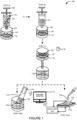
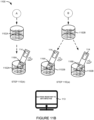
- Figure 1 illustrates an embodiment of a system 100 for detecting an infectious agent 102 in a fluid sample 124.
- the system 100 can comprise a fluid delivery device 106, a filter housing 108 containing a filter 110, a sensing device 116, and a parameter analyzer 114.
- the sensing device 116 can be located on a substrate 112.
- the substrate 112 can be comprised of a polymer or polymeric material, a metal, a ceramic, a semiconductor layer, an oxide layer, an insulator, or a combination thereof.
- the parameter analyzer 114 can be integrated into one device with the sensing device 116.
- the parameter analyzer 114 can be fabricated on the same substrate 112 as the sensing device 116.
- the parameter analyzer 114 can be a standalone unit or device coupled to the sensing device 116. The sensing device will be discussed in more detail in the sections that follow.
- the fluid sample 124 can comprise the infectious agent 102.
- the fluid sample 124 can include a bodily fluid such as blood, serum, plasma, urine, saliva, joint fluid, semen, wound material, spinal fluid, mucus, or a combination thereof.
- the fluid sample 124 can also include an environmental fluid such as liquids sampled from a stream, river, lake, ocean, contamination site, quarantine zone, or emergency area.
- the fluid sample 124 can also be a food sample.
- the infectious agent 102 can be any metabolizing single or multi-cellular organism including a bacteria or fungus.
- the infectious agent 102 can also be a virus or a prion.
- the infectious agent 102 can be a bacteria selected from the genera comprising of, but not limited to, Acinetobacter, Aeromonas, Bacillus, Bacteroides, Citrobacter, Enterobacter, Escherichia, Klebsiella, Morganella, Pandoraea, Proteus, Providencia, Pseudomonas, Ralstonia, Raoultella, Salmonella, Serratia, Shewanella, Shigella, Stenotrophomonas, Streptomyces, Staphylococcus, Enterococcus, Clostridium or any combination thereof.
- the infectious agent 102 can be a fungus selected from the genera comprising of, but not limited to, Candida, Cryptococcus, or any combination thereof.
- the infectious agent 102 can include amoeba.
- the infectious agent 102 can be cancer cells.
- the fluid delivery device 106 can deliver or inject the fluid sample 124 into the filter housing 108 in step 1A.
- the fluid delivery device 106 can be a pump.
- the fluid delivery device 106 can be a hydraulic pump, a pneumatic pump, a syringe pump, or a combination thereof.
- the fluid delivery device 106 can be an injection cartridge, a microfluidic channel, a pipette, a reaction tube, a capillary, a test tube, a combination thereof, or a portion therein.
- the filter housing 108 can be a container or vessel configured to secure or enclose the filter 110.
- the filter housing 108 can be a protective chamber.
- the protective chamber can be an electrically isolated environment.
- the protective chamber can also be a temperature controlled chamber, a light controlled chamber, or a combination thereof.
- the filter 110 can have a filter surface 126.
- the filter 110 can trap or isolate the infectious agent 102 by depositing or delivering the infectious agent 102 on to the filter surface 126.
- the filter surface 126 can be an external surface, an internal surface extending into the filter 110, or a combination thereof.
- the filter 110 can be made of, but is not limited to, cellulose acetate, regenerated cellulose, nylon, polystyrene, polyvinylidene fluoride (PVDF), polyethersulfone (PES), polytetrafluorethylene (PTFE), glass microfiber, or a combination thereof.
- the filter 110 can have filter pores of sequentially smaller pore size.
- the filter 110 can have larger filter pores at the top of the filter and progressively smaller filter pores toward the bottom of the filter.
- the filter 110 can have filter pores of a similar pore size throughout the entire filter.
- the filter surface 126 can be the surface of the pores.
- the filter 110 can be a mesh or matrix structure and the filter surface 126 can be a mesh or matrix surface.
- the filter 110 can comprise, carry, or hold the infectious agent 102 when a fluid sample 124 comprising or carrying an infectious agent 102 is introduced to the filter 110.
- the fluid sample 124 can be introduced to the filter 110 when the fluid sample 124 is poured over or injected through the filter 110.
- the filter 110 can isolate or separate the infectious agent 102 or other molecules or cells from the supernatant of the fluid sample 124.
- the filter housing 108 can have at least one opening which allows fluid or supernatant from the fluid sample 124 to evacuate the filter housing 108.
- step 1A can include the additional step of discarding the fluid or supernatant from the fluid sample 124 through the opening after isolating the infectious agent 102 on the filter surface 126.
- One advantage of the methods and systems 100 disclosed herein is the separation of any suspected or potential infectious agents 102 from the sensing device 116, the parameter analyzer 114, or a combination thereof.
- the filter 110, the filter surface 126, the filter housing 108, or a combination thereof can prevent any suspected or potential infectious agents 102 from contacting any portion of the sensing device 116, the parameter analyzer 114, or a combination thereof.
- the filter 110, the filter surface 126, the filter housing 108, or a combination thereof can trap or isolate any suspected or potential infectious agents 102 on the filter surface 126 or in the filter housing 108.
- a stimulus solution can be added to the fluid sample 124 before introducing the fluid sample 124 to the filter 110.
- the stimulus solution can be a nutrient or growth solution.
- the stimulus solution can be a super nutrient solution.
- the fluid sample 124 can also be pre-filtered in a step before step 1A.
- This pre-filtering step can involve filtering the fluid sample 124 using an additional filter, a microfluidic filter, or a combination thereof to filter out other larger cellular components including blood cells or epithelial cells from the fluid sample 124 when the fluid sample 124 is composed of bodily fluid.
- the same fluid delivery device 106 or another fluid delivery device 106 can also be used to deliver or inject a nutrient solution 130 to the filter housing 108 in step 1B.
- the fluid delivery device 106 can continuously or periodically expose the filter surface 126 to the nutrient solution 130.
- the filter 110 can be heated to a temperature of between 30 °C and 40 °C and allowed to incubate for an incubation period 132 in step 1C.
- the filter 110 can be incubated while in the filter housing 108.
- the filter 110 can be removed from the filter housing 108 prior to incubation.
- the filter 110 can be incubated with the nutrient solution 130.
- the incubation period 132 can range from 15 minutes to over one hour. In other embodiments, the incubation period 132 can be less than 15 minutes.
- the incubation period 132 can be adjusted based on the type of infectious agent 102 suspected in the fluid sample 124, such as the type of bacteria, fungus, virus, or prion.
- the incubation period 132 can also be adjusted based on the suspected amount of the infectious agent 102 present in the fluid sample 124, the amount or volume of the fluid sample 124, or a combination thereof. For example, the incubation period 132 can be increased when the suspected amount of the infectious agent 102 or the volume of the fluid sample 124 is below a threshold amount.
- the filter 110 can be allowed to incubate with the nutrient solution 130 in order to promote the proliferation of the suspected infectious agent 102 on the filter surface 126.
- One advantage of incubating the filter 110 is to increase the sensitivity of the system 100 to small amounts of the suspected infectious agent 102. For example, incubating the filter 110 can allow the system 100 to reduce its level of detection.
- the sample effluent 134 can be analyzed by a sensing device 116.
- the sample effluent 134 can be analyzed by applying or introducing an aliquot of the sample effluent 134 to the sensing device 116 in step 1D(a).
- the sample effluent 134 can be analyzed by inserting a portion of the sensing device 116 directly into the sample effluent 134 in step 1D(b).
- the sample effluent 134 can comprise hydrogen ions (H + ) as a byproduct of bacterial cell metabolism or growth.
- the sample effluent 134 can comprise adenosine triphosphate (ATP), carbon dioxide (CO 2 ), lactic acid, carbonic acid, nitrates (NO 3 - ), or a combination thereof produced by or attributed to an infectious agent 102.
- ATP adenosine triphosphate
- CO 2 carbon dioxide
- NO 3 - nitrates
- the parameter analyzer 114 can be fabricated on the same substrate 112 as the sensing device 116. In other embodiments, the parameter analyzer 114 can be a standalone unit or meter coupled to the sensing device 116. The parameter analyzer 114 can also be connected to or communicatively coupled to a display 113 or display component configured to provide a result of the detection or a read-out of the electrical characteristic of the sensing device 116. In certain embodiments, the parameter analyzer 114 can be a mobile device, a handheld device, a tablet device, or a computing device such as a laptop or desktop computer and the display 113 can be a mobile device display, a handheld device display, a tablet display, or a laptop or desktop monitor.
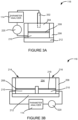
- FIG. 2A illustrates a side cross-sectional view of one embodiment of the sensing device 116.
- the sensing device 116 can be an electrochemical cell comprising an external reference electrode 202, an electrolyte 204 or electrically conducting solution retained by container walls 206, a functionalization layer 208, a conductor layer 210, and a substrate layer 212.
- the sensing device 116 can be connected or coupled to the parameter analyzer 114.
- the parameter analyzer 114 can be coupled to both the external reference electrode 202 and the conductor layer 210.
- the external reference electrode 202 can extend into the electrolyte 204.
- the electrolyte 204 can be the sample effluent 134.
- the electrolyte 204 can comprise portions of the fluid sample 124.
- the conductor layer 210 can be composed of any conducting material which allows an electrical property change to be measured, including, but is not limited to, a voltage change and/or a current change measured through the conductor layer 210, the functionalization layer 208, and the electrolyte 204 to the external reference electrode 202.
- the functionalization layer 208 can be disposed on or cover the conductor layer 210.
- the functionalization layer 208 can comprise silanes, DNA, proteins, antibodies, self-assembled mono layers (SAMs), oxides, buffered hydrogels, PVC, parylene, polyACE, or any other biochemically active materials.
- the functionalization layer 208 can be configured to facilitate the sensing device 116 from interacting with ions, analytes, or other molecules or byproducts in the electrolyte 204.
- the functionalization layer 208 can be a pH-sensitive layer.
- the functionalization layer 208 can comprise hydroxyl groups which can interact with hydrogen ions (H + ) in the electrolyte 204. This interaction can generate a change in the electrical characteristic of the sensing device 116 detected by the parameter analyzer 114. In one embodiment, this interaction can create a measurable change in the electrical characteristic of the sensing device 116 at the interface between the electrolyte 204/functionalization layer 208 or the interface between the electrolyte 204/conductor layer 210.
- the parameter analyzer 114 can be a voltmeter and the voltmeter can detect a voltage change ( ⁇ V) at or near the functionalization layer 208 exposed to the electrolyte 204.
- the voltage change can be determined with respect to the external reference electrode 202 extending into or in contact with the electrolyte 204.
- the functionalization layer 208 and the conductor layer 210 can be considered part of a working or active electrode of the system 100.
- the electrolyte 204, the functionalization layer 208, and the conductor layer 210 can be surrounded by a container wall 206.
- the container wall 206 can be made of an inert or non-conductive material.
- the container wall 206 can hold or retain the electrolyte 204 or be responsible for delivering or introducing the sample effluent 134 to the sensing device 116.
- Figure 2B illustrates a side cross-sectional view of another embodiment of the sensing device 116.
- the sensing device 116 comprises a working electrode 214 and an on-chip reference electrode 216.
- the working electrode 214 and the on-chip reference electrode 216 can be disposed on the same substrate layer 212.
- the substrate layer 212 can be composed of the same material as the substrate layer 212 depicted in Figure 2A .
- the electrolyte 204 can flow over or be exposed to both the working electrode 214 and the on-chip reference electrode 216 simultaneously.
- the working electrode 214 and the on-chip reference electrode 216 can be separated by a container wall 214 or container divide.
- the working electrode 214 can comprise the functionalization layer 208 disposed on or covering the conductor layer 210.
- the functionalization layer 218 can comprise silanes, DNA, proteins, antibodies, oxides, self-assembled mono layers (SAMs), buffered hydrogels, PVC, parylene, polyACE, or any other biochemically active materials.
- Figure 3A illustrates a side cross-sectional view of an embodiment of the sensing device 116 of Figure 2A having a power source 220.
- the power source 220 can apply a DC or AC voltage (usually in the range of +/- 5V) to the conductor layer 210 and the external reference electrode via the functionalization layer 208 and the electrolyte 204. This voltage can also be set to be used as a working point.
- a change in the electrical characteristic e.g., a horizontal shift ( ⁇ V) of the voltage measurement curve
- ⁇ V horizontal shift
- the analytes, ions, molecules, or cellular byproducts can interact with the functionalization layer 208, causing an additional electrical characteristic change, which can also be detected by the parameter analyzer 114.
- a horizontal shift ( ⁇ V) of the voltage measurement curve can occur, as the analytes, ions, molecules, or cellular byproducts interact with the sensing device 116.
- This change can be measured by the parameter analyzer 114.
- the analytes, ions, molecules, or cellular byproducts can interact with the functionalization layer 208, causing an additional electrical characteristic change that can be detected by the parameter analyzer 114.
- the filter housing 108 can have at least one opening which allows fluid or supernatant from the fluid sample 124 to evacuate the filter housing 108.
- step 5A can include the additional step of discarding the fluid or supernatant from the fluid sample 124 through the opening after isolating the infectious agent 102 on the filter surface 126.
- the electrical characteristic can include, but is not limited to, a voltage, an impedance, a current, a capacitance, a resistance, a resonant frequency, a noise level, a level of induction, or a combination thereof measured at or near the sensing device 500.
- the change in the electrical characteristic can include, but is not limited to, a voltage change, an impedance change, a current change, a capacitance change, a resistance change, a change in resonant frequency, a noise level change, an induction change, or a combination thereof measured at or near the sensing device 500.
- the sensing device 500 can be connected to a voltmeter 608 and an ammeter 610 or any other meter or measurement device.
- the voltmeter 608 can have one lead connection wire, such as a copper wire, connected to the working electrode 600 and another lead connection wire connected to the reference electrode 602.
- the ammeter 610 can also have one lead connection wire connected to the reference electrode 602 and another lead connection wire connected to the counter electrode 604.
- the oxidation of glucose to gluconolactone or gluconic acid can be measured.
- glucose dehydrogenase can be used as an enzyme. Additional chemicals can also be added to generate an electrical current that can be measured by the system. The total charge passing through the electrodes can be proportional to the amount of glucose in the solution that has reacted with the enzyme.
- One advantage of the methods and systems 100 disclosed herein is the separation of any suspected or potential infectious agents 102 from the sensing device 800, the parameter analyzer 114, or a combination thereof.
- the filter 110, the filter surface 126, the filter housing 108, or a combination thereof can prevent any suspected or potential infectious agents 102 from contacting any portion of the sensing device 800, the parameter analyzer 114, or a combination thereof.
- the fluid sample 124 can also be pre-filtered in a step before step 8A.
- This pre-filtering step can involve filtering the fluid sample 124 using an additional filter, a microfluidic filter, or a combination thereof to filter out other larger cellular components including blood cells or epithelial cells from the fluid sample 124 when the fluid sample 124 is composed of bodily fluid.
- the incubation period 132 can also be adjusted based on the suspected amount of the infectious agent 102 present in the fluid sample 124, the amount or volume of the fluid sample 124, or a combination thereof. For example, the incubation period 132 can be increased when the suspected amount of the infectious agent 102 or the volume of the fluid sample 124 is below a threshold amount.
- the filter 110 can be allowed to incubate with the nutrient solution 130 in order to promote the proliferation of the suspected infectious agent 102 on the filter surface 126.
- One advantage of incubating the filter 110 is to increase the sensitivity of the system 100 to small amounts of the suspected infectious agent 102. For example, incubating the filter 110 can allow the system 100 to reduce its level of detection.
- the effluent or outflow of the nutrient solution 130 exposed to the filter 110 can be sampled.
- the effluent or outflow of the nutrient solution 130 exposed to the filter 110 can be referred to as the sample effluent 134.
- the sample effluent 134 can comprise a solution characteristic.
- the solution characteristic can refer to one or more attributes of the solution making up the sample effluent 134.
- the solution characteristic can include a concentration of an analyte, molecule, or ion, an absolute number or molecular count of an analyte, molecule, or ion, or a combination thereof.
- the solution characteristic can include a solution temperature.
- the solution characteristic can vary as a result of changes due to the energy use, growth, and metabolism of the infectious agent 102 isolated or trapped by the filter 110.
- the solution characteristic can be a direct or indirect byproduct of a cellular activity undertaken by the infectious agent 102 such as cell metabolism or cell growth.
- the solution characteristic can vary as a result of molecules or ions produced or consumed by the infectious agent 102.
- a change in the solution characteristic can cause a change in the electrical characteristic of the sensing device 800.
- the parameter analyzer 114 can detect a change in an electrical characteristic (see Figure 10 ) of the sensing device 800 exposed to the sample effluent 134.
- the parameter analyzer 114 can include, but is not limited to, any combination of a voltmeter, a multimeter, or an ammeter.
- the parameter analyzer 114 in this embodiment can include the voltage source 908 and the ammeter 910 depicted in Figure 9 ).
- the electrical characteristic can include, but is not limited to, a voltage, a current, a photocurrent, or a combination thereof measured at or near the sensing device 800.
- the change in the electrical characteristic can include, but is not limited to, a voltage change, a current change, a photocurrent change, or a combination thereof measured at or near the sensing device 800.
- the parameter analyzer 114 can be fabricated on the same substrate 112 as the sensing device 800. In other embodiments, the parameter analyzer 114 can be a standalone unit or meter coupled to the sensing device 800. The parameter analyzer 114 can also be connected to or communicatively coupled to the display 113 or display component configured to provide a result of the detection or a read-out of the electrical characteristic of the sensing device 800. In certain embodiments, the parameter analyzer 114 can be a mobile device, a handheld device, a tablet device, or a computing device such as a laptop or desktop computer and the display 113 can be a mobile device display, a handheld device display, a tablet display, or a laptop or desktop monitor.
- the parameter analyzer 114 can display a result indicating the presence of an infectious agent 102 in the fluid sample 124 via the display 113 of the parameter analyzer 114. In another embodiment, the parameter analyzer 114 can wirelessly communicate a result indicating the presence of an infectious agent 102 in the fluid sample 124 to a computing device having the display 113.
- the parameter analyzer 114, a reader, or a combination thereof can detect a change in the electrical characteristic of the sensing device 800 exposed to the sample effluent 134 corresponding to the presence of the infectious agent 102 in the system 100.
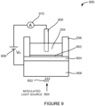
- the electrolyte 204 and the functionalization layer 902 can be surrounded by a container wall 206.
- the container wall 206 can be made of an inert or non-conductive material.
- the container wall 206 can hold or retain the electrolyte 204 or be responsible for delivering or introducing the sample effluent 134 to the sensing device 800.
- the external reference electrode 900 can extend into the electrolyte 204.
- the electrolyte 204 can be the sample effluent 134.
- the electrolyte 204 can comprise portions of the fluid sample 124.
- the voltage source 908 can apply a known potential to the sensing device 800 through the external reference electrode 900.
- This voltage can be, but is not limited to, a DC or AC voltage.
- the external reference electrode 900 can be a silver/silver chloride (Ag/AgCl) electrode.
- the external reference electrode 202 can be a saturated calomel reference electrode (SCE) or a copper-copper (II) sulfate electrode (CSE).
- one advantage of the methods, devices, and systems 100 disclosed herein is the separation of the infectious agent 102 from the components of the system 100 in physical or fluid contact with these external reference electrodes.
- the sensing device 800 can comprise the insulator layer 904 disposed on or covering the semiconductor layer 906.
- the semiconductor layer 906 can be a layer of doped semiconducting material.
- the semiconductor layer 906 can be composed of, but is not limited to, a layer of doped silicon.
- the semiconductor layer 906 can also be made of an organic semiconductor, a carbon nanotube, graphene, an organic conductor such as those derived from polyacetylene, polyaniline, Quinacridone, Poly(3,4-ethylenedioxythiophene) or PEDOT, PEDOT: polystyrene sulfonate (PSS), or a combination thereof.
- the insulator layer 904 (which can also be referred to as an isolator layer) can be a high-k dielectric layer or a material layer having a high dielectric constant (k).
- the insulator layer 904 can comprise aluminum oxide, hafnium oxide, titanium oxide, zirconium oxide, yttrium oxide, tantalum oxide, hafnium silicate, zirconium silicate, silicon nitride, aluminum nitride, hafnium nitride, zirconium nitride, or a combination thereof.
- the insulator layer 904 can comprise aluminum dioxide, hafnium dioxide, zirconium dioxide, or a combination thereof.
- the insulator layer 216 can comprise a silicon dioxide layer.
- the functionalization layer 902 can be disposed on or cover the insulator layer 904.
- the functionalization layer 902 can comprise silanes, DNA, proteins, antibodies, self-assembled mono layers (SAMs), buffered hydrogels, PVC, parylene, polyACE, or any other biochemically active materials.
- the functionalization layer 902 can be configured to facilitate the sensing device 800 from interacting with ions, analytes, or other molecules or byproducts in the electrolyte 204.
- the functionalization layer 902 can be a pH-sensitive layer.
- the functionalization layer 902 can comprise hydroxyl groups which can interact with hydrogen ions (H + ) in the electrolyte 204.
- an insulator layer 904 made of an oxide can be used as the functionalization layer 902.
- An external DC bias voltage can be applied.
- the light source 804 e.g., a modulated light pointer or source
- the light source 804 illuminates the bulk silicon
- light-induced charge carriers can be separated by an internal electric field and a photocurrent can be detected by a peripheral circuit.
- the amplitude of the photocurrent can depend on the local surface potential.
- the voltage source 908 can apply a bias voltage to the electrolyte 204 via the external reference electrode 900.
- This bias voltage can be applied between the semiconductor layer 906 and the reference electrode 900.
- the bias voltage can be set so as to repel electrons from the doped semiconductor layer 906 to form a depletion layer.
- the bias voltage can be set so as to repel the electrons moving to the interface between the semiconductor layer 906 and the insulator layer 904 due to the light 802 directed at the semiconductor layer 906.
- the depletion layer is not formed.
- the photocurrent increases until reaching a limiting value.
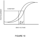
- a change in the solution characteristic of the electrolyte can change the bias voltage needed to maintain the constant photocurrent detected by the ammeter 910.
- hydrogen ions in the solution can interact with the hydroxyl groups of the functionalization layer 902 and generate an additional potential change at the interface. This additional voltage will also cause a shift in the photocurrent, comparable to increasing or decreasing the bias voltage.
- hydrogen ions in the solution can also interact with the insulator layer 904 to generate a potential change at the interface and cause a shift in the photocurrent.
- the light source 804 can be focused so as to direct light 804 of a specific wavelength at a specific portion of the semiconductor layer 906. Moreover, a different light source or the same light source 804 can direct light 804 of a different wavelength at a different portion of the semiconductor layer 906. This allows facile multiplexing by creating a series of different sensing areas on one photocurrent sensor such that different areas of the sensor can be allocated for or dedicated to a different analyte, ion, or molecule.
- Figure 10 illustrates one example of a photocurrent/voltage curve generated based on readings obtained from the voltage source 908 and the ammeter 910 connected to the sensing device 800.
- a change in the solution characteristic of the electrolyte 204 over time or the difference between the solution characteristics of two different electrolytes 204 can be measured by the change in the voltage ( ⁇ V) at a constant photocurrent.
- the infectious agent 102 can be any metabolizing single or multi-cellular organism including a bacteria or fungus.
- the infectious agent 102 can also be a virus or a prion.
- the infectious agent 102 can be a bacteria selected from the genera comprising of, but not limited to, Acinetobacter, Aeromonas, Bacillus, Bacteroides, Citrobacter, Enterobacter, Escherichia, Klebsiella, Morganella, Pandoraea, Proteus, Providencia, Pseudomonas, Ralstonia, Raoultella, Salmonella, Serratia, Shewanella, Shigella, Stenotrophomonas, Streptomyces, Staphylococcus, Enterococcus, Clostridium or any combination thereof.
- the first filter 110A or the second filter 110B can have filter pores of sequentially smaller pore size.
- the first filter 110A or the second filter 110B can have larger filter pores at the top of the filter and progressively smaller filters pores toward the bottom of the filter.
- the first filter 110A or the second filter 110B can have filter pores of a similar pore size throughout the entire filter.
- the first filter surface 126A or the second filter surface 126B can be a surface of the pores.
- the first filter 110A or the second filter 110B can be a mesh or matrix structure and the first filter surface 126A or the second filter surface 126B can be a mesh or matrix surface.
- the first filter 110A can comprise, hold, or carry the infectious agent 102 when a fluid sample 124 comprising or carrying the infectious agent 102 is introduced to the first filter 110A.
- the fluid sample 124 can be introduced to the first filter 110A when the fluid sample 124 is poured over or injected through the first filter 110A.
- the second filter 110B can also comprise, hold, or carry the infectious agent 102 when a fluid sample 124 comprising or carrying the infectious agent 102 is introduced to the second filter 110B.
- the fluid sample 124 can be introduced to the second filter 110B when the fluid sample 124 is poured over or injected through the second filter 110B.
- the first filter 110A or the second filter 110B can isolate or separate the infectious agent 102 or other molecules or cells from the supernatant of the fluid sample 124.
- the first filter 110A or the first filter surface 126A and the second filter 110B or the second filter surface 126B can comprise the anti-infective 104.
- the first filter 110A or the second filter 110B can comprise the anti-infective 104 when the first filter surface 126A or the second filter surface 126B is coated with the anti-infective 104 or exposed to the anti-infective 104 before introducing the fluid sample 124 to the first filter 110A or the second filter 110B.
- the anti-infective 104 can be added or introduced to the first filter 110A or the second filter 110B after exposing the first filter 110A or the second filter 110B to the fluid sample 124.
- the anti-infective 104 can be introduced through an additional solution exposed to the first filter 110A, the second filter 110B, or a combination thereof.
- the anti-infective 104 can be introduced through a nutrient solution 130.
- the chemical compound can include, but is not limited to, sodium azide, bile acids, high sodium chloride, or a combination thereof.
- the anti-infective 104 can also comprise a carbon source other than glucose, such as lactose or mannose, to select for certain bacterial species.
- a bacterial growth inhibitor or stimulator such as urea, citrate, or certain amino acids can also be introduced to the first filter 110A, the second filter 110B, or a combination thereof.
- the effluent or outflow of the nutrient solution 130 and/or the solution of glucose 502 exposed to the first filter 110A or the second filter 110B can be sampled.
- the effluent or outflow of the nutrient solution 130 and/or the solution of glucose 502 exposed to the first filter 110A can be referred to as the first sample effluent 1102A.
- the effluent or outflow of the nutrient solution 130 and/or the solution of glucose 502 exposed to the second filter 110B can be referred to as the second sample effluent 1102B.
- the first sample effluent 1102A can be analyzed by the first sensing device 116A and the second sample effluent 1102B can be analyzed by the second sensing device 116B in step 1 1E(a).
- the first sample effluent 1102A can be analyzed by inserting a portion of the first sensing device 116A directly into the first sample effluent 1102A and the second sample effluent 1102B can be analyzed by inserting a portion of the second sensing device 116B directly into the second sample effluent 1102B.
- the first sample effluent 1102A and the second sample effluent 1102B can each comprise a solution characteristic.
- the solution characteristic can refer to one or more attributes of the solution making up the first sample effluent 1102A, the second sample effluent 1102B, or a combination thereof.
- the solution characteristic can include a concentration of a solute, analyte, or molecule, an absolute number or molecular count of solutes, analytes, or molecules in solution, a solution temperature, or a combination thereof.
- the solution characteristic can vary as a result of ions, molecules, minerals, or nutrients produced by, consumed by, or otherwise attributed to the infectious agent 102 on the first filter surface 126A, the second filter surface 126B, or a combination thereof.
- the first sample effluent 1102A, the second sample effluent 1102B, or a combination thereof can comprise hydrogen ions (H + ) as a byproduct of bacterial cell metabolism or growth.
- a change in the solution characteristic can cause a change in the electrical characteristic of the first sensing device 116A, the second sensing device 116B, or a combination thereof.
- a parameter analyzer 114 can monitor the electrical characteristic of the first sensing device 116A, the second sensing device 116B, or a combination thereof.
- monitoring the second electrical characteristic can involve determining a second voltage change at a second functionalization layer (such as the functionalization layer 208 in Figures 2A, 2B , 3A, or 3B ) of the second sensing device 116B exposed to the second sample effluent 1102B.
- the second voltage change can be with respect to a voltage at a second reference electrode (such as either the external reference electrode 202 of Figures 2A and 3A or the on-chip reference electrode of Figures 2B and 3B ) of the second sensing device 116B exposed to the second sample effluent 1102B.
- the second functionalization layer can cover the working electrode of the second sensing device 116B.
- the parameter analyzer 114 can monitor the electrical characteristic of the first sensing device 116A exposed to the first sample effluent 1102A and the electrical characteristic of the same first sensing device 116A exposed to the second sample effluent 1102B.
- the electrical characteristic of the first sensing device 116A while sampling the first sample effluent 1102A can be referred to as the first electrical characteristic and the electrical characteristic of the first sensing device 116A while sampling the second sample effluent 1102B can be referred to as the second electrical characteristic.
- the first electrical characteristic and the second electrical characteristic can differ when the solution characteristic of the first sample effluent 1102A and the solution characteristic of the second sample effluent differ in their pH.
- the first electrical characteristic can differ from the second electrical characteristic when the solution characteristic of the first sample effluent 1102A differs from the solution characteristic of the second sample effluent 1102B as a result of differences in the temperature of the solution.
- the parameter analyzer 114 or the reader communicatively coupled to the parameter analyzer 114 can assess the susceptibility of the infectious agent 102 as either resistant or non-resistant to the anti-infective 104.
- the second filter 110B or the second filter surface 126B can comprise a set amount of the anti-infective 104.
- the parameter analyzer 114 or a reader communicatively coupled to the parameter analyzer 114 can then assess the susceptibility of the infectious agent 102 as either resistant or non-resistant based on any detected differences in the first electrical characteristic and the second electrical characteristic or any detected differences in the change in the first electrical characteristic and the change in the second electrical characteristic.
- the parameter analyzer 114 or the reader communicatively coupled to the parameter analyzer 114 can assess the level of susceptibility of the infectious agent 102 on a grad ated or tiered scale.
- the parameter analyzer 114 or the reader communicatively coupled to the parameter analyzer 114 can assess the susceptibility of the infectious agent 102 as being resistant, mildly susceptible, or susceptible to the anti-infective 104.
- additional filter surfaces including a third filter surface, can be used which comprise anti-infectives 104 of different concentrations.
- the parameter analyzer 114 can display a result indicating the level of susceptibility of the infectious agent 102 in the fluid sample 124 via the display 113. In another embodiment, the parameter analyzer 114 can wirelessly communicate a result indicating the presence of an infectious agent 102 in the fluid sample 124 to a computing device having the display 113.
- the same first sensing device 500A can be used to analyze the first sample effluent 1102A and the second sample effluent 1102B in step 1 1E(d).
- the first sensing device 500A can be cleaned or recalibrated after each analysis or use.
- the concentration of glucose 502 in the first sample effluent 1102A can differ from the concentration of glucose 502 in the second sample effluent 1102B based on the presence, proliferation, or activity of the infectious agent 102 on the first filter surface 126A, the second filter surface 126B, or a combination thereof.
- the parameter analyzer 114 can be integrated into one device with the first sensing device 500A, the second sensing device 500B, or a combination thereof.
- the parameter analyzer 114 can be fabricated on the same substrate as the first sensing device 500A, the second sensing device 500B, or a combination thereof.
- the parameter analyzer 114 can be a standalone unit or device coupled to the first sensing device 500A, the second sensing device 500B, or a combination thereof.
- the first sample effluent 1102A can be analyzed by the first sensing device 800A and the second sample effluent 1102B can be analyzed by the second sensing device 800B in step 1 1E(e).
- the first sample effluent 1102A can be analyzed by inserting a portion of the first sensing device 800A directly into the first sample effluent 1102A and the second sample effluent 1102B can be analyzed by inserting a portion of the second sensing device 800B directly into the second sample effluent 1102B.
- the solution characteristic can vary as a result of ions, molecules, minerals, or nutrients produced by, consumed by, or otherwise attributed to the infectious agent 102 on the first filter surface 126A, the second filter surface 126B, or a combination thereof.
- the first sample effluent 1102A, the second sample effluent 1102B, or a combination thereof can comprise hydrogen ions (H + ) as a byproduct of bacterial cell metabolism or growth.
- a change in the solution characteristic can cause a change in the electrical characteristic of the first sensing device 800A, the second sensing device 800B, or a combination thereof.
- a parameter analyzer 114 can monitor the electrical characteristic of the first sensing device 800A, the second sensing device 800B, or a combination thereof.
- the parameter analyzer 114 can include voltage source 908 and the ammeter 910 of Figure 9 .
- the parameter analyzer 114 can include a voltmeter, a multimeter, a capacitance analyzer, or a combination thereof.
- the parameter analyzer 114 can be integrated into one device with the first sensing device 800A, the second sensing device 800B, or a combination thereof.
- the parameter analyzer 114 can be fabricated on the same substrate as the first sensing device 800A, the second sensing device 800B, or a combination thereof.
- the parameter analyzer 114 can be a standalone unit or device coupled to the first sensing device 800A, the second sensing device 800B, or a combination thereof.
- the change in the electrical characteristic can include, but is not limited to, a voltage change, an impedance change, a current change, a capacitance change, a resistance change, a change in resonant frequency, a noise level change, an induction change, or a combination thereof measured at or near the first sensing device 800A, the second sensing device 800B, or a combination thereof.
- the parameter analyzer 114 can compare the first electrical characteristic (such as the bias voltage applied to the first sensing device 800A), including a change in the first electrical characteristic (such as a change in the bias voltage needed to maintain a constant photocurrent at the first sensing device 800A), with the second electrical characteristic (such as the bias voltage applied to the second sensing device 800B), including a change in the second electrical characteristic (such as a change in the bias voltage needed to maintain a constant photocurrent at the second sensing device 800B), to assess the susceptibility of the infectious agent 102 to the anti-infective 104.
- the first electrical characteristic such as the bias voltage applied to the first sensing device 800A
- the second electrical characteristic such as the bias voltage applied to the second sensing device 800B
- a change in the second electrical characteristic such as a change in the bias voltage needed to maintain a constant photocurrent at the second sensing device 800B
- the first electrical characteristic can differ from the second electrical characteristic when the solution characteristic of the first sample effluent 1102A differs from the solution characteristic of the second sample effluent 1102B as a result of differences in the concentration or amount of ions, analytes, molecules, minerals, or other solutes present in the sample effluents.
- the first electrical characteristic and the second electrical characteristic can differ when the solution characteristic of the first sample effluent 1102A and the solution characteristic of the second sample effluent differ in their pH.
- the parameter analyzer 114 can assess the susceptibility of the infectious agent 102 to the anti-infective 104 as a binary assessment or a tiered assessment.
- a reader or computing device connected or communicatively coupled to the parameter analyzer 114 can assess the susceptibility of the infectious agent 102 to the anti-infective 104 as a binary assessment or a tiered assessment.
- the parameter analyzer 114 or the reader communicatively coupled to the parameter analyzer 114 can assess the susceptibility of the infectious agent 102 as either resistant or non-resistant to the anti-infective 104.
- the second filter 110B or the second filter surface 126B can comprise a set amount of the anti-infective 104.
- the parameter analyzer 114 or a reader communicatively coupled to the parameter analyzer 114 can then assess the susceptibility of the infectious agent 102 as either resistant or non-resistant based on any detected differences in the first electrical characteristic and the second electrical characteristic or any detected differences in the change in the first electrical characteristic and the change in the second electrical characteristic.
- the parameter analyzer 114 or the reader communicatively coupled to the parameter analyzer 114 can assess the susceptibility of the infectious agent 102 as not resistant to the anti-infective 104 when the parameter analyzer 114 or the reader fails to detect a difference or a statistically significant difference between the first electrical characteristic and the second electrical characteristic or a statistically significant difference between the change in the first electrical characteristic and the change in the second electrical characteristic. More specifically, a statistically significant difference in the electrical characteristic can be a difference exceeding a threshold value.
- the parameter analyzer 114 or the reader communicatively coupled to the parameter analyzer 114 can assess the level of susceptibility of the infectious agent 102 on a tiered scale.
- the parameter analyzer 114 or the reader communicatively coupled to the parameter analyzer 114 can assess the susceptibility of the infectious agent 102 as being resistant, mildly susceptible, or susceptible to the anti-infective 104.
- additional filter surfaces including a third filter surface, can be used which comprise anti-infectives 104 of different concentrations.
- the parameter analyzer 114 can also have or be connected to a display 113 or display component configured to provide a result of the detection or a read-out of the electrical characteristic of the first sensing device 800A, the second sensing device 800B, or a combination thereof.
- the parameter analyzer 114 can be a mobile device, a handheld device, a tablet device, or a computing device such as a laptop or desktop computer and the display 113 can be a mobile device display, a handheld device display, a tablet display, or a laptop or desktop monitor.
- the parameter analyzer 114 can display a result indicating the level of susceptibility of the infectious agent 102 in the fluid sample 124 via the display 113. In another embodiment, the parameter analyzer 114 can wirelessly communicate a result indicating the presence of an infectious agent 102 in the fluid sample 124 to a computing device having the display 113.
- Figure 12 illustrates a method 1200 of detecting an infectious agent 102 in a fluid sample 124.
- the method 1200 can include introducing a fluid sample 124 to a surface, such as the filter surface 126, in a step 1202.
- the filter surface 126 can isolate, trap, or otherwise contain the infectious agent 102 when a fluid sample 124 carrying or comprising the infectious agent 102 is introduced to the filter surface 126.
- the method 1200 can also include exposing the surface to a solution, such as the nutrient solution 130, in a step 1204.
- the method 1200 can further include sampling the solution after exposing the surface to the solution in step 1206.
- Sampling the solution can include sampling the effluent or outflow of the solution, such as the sample effluent 134. Sampling the solution can also involve separating the solution from the surface so the solution is not in fluid communication or contact with the surface, the infectious agent 102 on the surface, or a combination thereof when sampled.
- the method 1200 can also include detecting a change in an electrical characteristic of a sensing device exposed to the solution sampled corresponding to the presence of the infectious agent 102 in the fluid sample 124 in step 1208.
- the sensing device is an electrochemical cell such as the sensing device 116
- detecting the change in the electrical characteristic comprises determining a voltage change at a functionalization layer covering a working electrode of the electrochemical cell.
- the functionalization layer can be exposed to the solution sampled and the voltage change can be determined with respect to a reference electrode, such as the external reference electrode 202 of Figure 2A or the on-chip reference electrode 216 of Figure 2B , also exposed to the solution sampled.
- Figure 13 illustrates a method 1300 of detecting an infectious agent 102 in a fluid sample 124.
- the method 1300 can include introducing a fluid sample 124 to a surface, such as the filter surface 126, in a step 1302.
- the filter surface 126 can isolate, trap, or otherwise contain the infectious agent 102 when a fluid sample 124 carrying or comprising the infectious agent 102 is introduced to the filter surface 126.
- the method 1300 can also include exposing the surface to a solution, such as the nutrient solution 130, in step 1304A.
- the method 1300 can also include adding a known concentration of glucose to the solution or the surface exposed to the solution in a step 1306.
- the method 1300 can further include incubating the solution or surface with the added glucose.
- the method 1300 can also include sampling the solution comprising glucose in step 1308.
- Sampling the solution can include sampling the effluent or outflow of the solution comprising glucose exposed to the surface, such as the sample effluent 504.
- Sampling the solution can also involve separating the solution comprising glucose from the surface so the solution comprising glucose is not in fluid communication or contact with the surface, the infectious agent 102 on the surface, or a combination thereof when sampled.
- the method 1300 can also include detecting a change in an electrical characteristic of a sensing device exposed to the glucose solution sampled corresponding to the presence of the infectious agent 102 in the fluid sample 124 in step 1310.
- the sensing device is a glucose sensor, such as the sensing device 500
- the change in the electrical characteristic is caused by a change in the glucose concentration of the solution sampled.
- Figure 14 illustrates a method 1400 of detecting an infectious agent 102 in a fluid sample 124.
- the method 1400 can include introducing a fluid sample 124 to a surface, such as the filter surface 126, in step 1402.
- the filter surface 126 can isolate, trap, or otherwise contain the infectious agent 102 when a fluid sample 124 carrying or comprising the infectious agent 102 is introduced to the filter surface 126.
- the method 1400 can also include exposing the surface to a solution, such as the nutrient solution 130, in step 1404.
- the method 1400 can further include sampling the solution after exposing the surface to the solution in step 1406.
- Sampling the solution can include sampling the effluent or outflow of the solution, such as the sample effluent 134. Sampling the solution can also involve separating the solution from the surface so the solution is not in fluid communication or contact with the surface, the infectious agent 102 on the surface, or a combination thereof when sampled.
- the method 1400 can also include applying a bias voltage via a reference electrode to a sensing device, such as the sensing device 800, exposed to the solution sampled in step 1408.
- the method 1400 can also include directing light using a modulated light source at a semiconductor layer, such as the semiconductor layer 906 of Figure 9 , of the sensing device in step 1410.
- the method can also include detecting a photocurrent using an ammeter coupled to the sensing device in step 1412.
- the method can further include determining a voltage change at a functionalization layer, such as the functionalization layer 902 of Figure 9 , covering an insulator layer, such as the insulator layer 904, in contact with the semiconductor layer of the sensing device.
- the voltage change, or another change in the electrical characteristic, of the sensing device exposed to the solution sampled can correspond to the presence of an infectious agent 102 in the fluid sample 124.
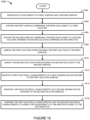
- Figure 15 illustrates a method 1500 for detecting a susceptibility of an infectious agent 102 to one or more anti-infectives 104.
- the method 1500 can include introducing a fluid sample 124 to a first surface, such as the first filter surface 126A, and a second surface, such as the second filter surface 126B, in a step 1502.
- the method 1500 can also include exposing the first surface to a first solution, such as the nutrient solution 130, in a step 1504.
- the first surface can comprise the infectious agent 102 when the infectious agent 102 is present in the fluid sample 124.
- the method 1500 can also include sampling the first solution after exposing the first solution to the first surface in step 1508.
- Sampling the first solution can include sampling the effluent or outflow of the first solution, such as the first sample effluent 1102A.
- sampling the first solution can also involve separating the first solution from the first surface so the first solution is not in fluid communication with the first surface, the infectious agent 102 on the first surface, or a combination thereof when sampled.
- the method 1500 can also include sampling the second solution after exposing the second solution to the second surface in step 1510.
- Sampling the second solution can include sampling the effluent or outflow of the second solution, such as the second sample effluent 1102B.
- sampling the second solution can also involve separating the second solution from the second surface so the second solution is not in fluid communication with the second surface, the infectious agent 102 on the second surface, or a combination thereof when sampled.
- the method 1500 can also include monitoring a first electrical characteristic of a first sensing device, such as the first sensing device 116A, the first sensing device 500A, or the first sensing device 800A, exposed to the first solution sampled in step 1512.
- the method 1500 can also include monitoring a second electrical characteristic of a second sensing device, such as the second sensing device 116B, the second sensing device 500B, or the second sensing device 800B exposed to the second solution sampled in step 1514.
- the method 1500 can further include comparing the first electrical characteristic and the second electrical characteristic to assess the susceptibility of the infectious agent 102 to the anti-infective 104 in step 1516.
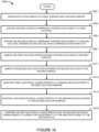
- the method 1600 can also include exposing the second surface to a second solution, such as additional instances of the nutrient solution 130 in a step 1606.
- the second surface can comprise one or more anti-infectives 104 or anti-infectives of differing concentrations.
- the second surface can also comprise the infectious agent 102 when the infectious agent 102 is present in the fluid sample 124.
- the method 1600 can also include sampling the first solution after exposing the first solution to the first surface in step 1608.
- Sampling the first solution can include sampling the effluent or outflow of the first solution, such as the first sample effluent 1102A.
- sampling the first solution can also involve separating the first solution from the first surface so the first solution is not in fluid communication with the first surface, the infectious agent 102 on the first surface, or a combination thereof.
- the method 1600 can also include sampling the second solution after exposing the second solution to the second surface in step 1610.
- Sampling the second solution can include sampling the effluent or outflow of the second solution, such as the second sample effluent 1102B.
- sampling the second solution can also involve separating the second solution from the second surface so the second solution is not in fluid communication with the second surface, the infectious agent 102 on the second surface, or a combination thereof.
- the method 1600 can also include monitoring a first electrical characteristic of a sensing device, such as the first sensing device 116A, the first sensing device 500A, or the first sensing device 800A, exposed to the first solution sampled in step 1612.
- the method 1600 can also include monitoring a second electrical characteristic of the sensing device exposed to the second solution sampled in step 1614.
- the method 1600 can further include comparing the first electrical characteristic and the second electrical characteristic to assess the susceptibility of the infectious agent 102 to the anti-infective 104 in step 1616.
Landscapes
- Health & Medical Sciences (AREA)
- Life Sciences & Earth Sciences (AREA)
- Chemical & Material Sciences (AREA)
- Engineering & Computer Science (AREA)
- Organic Chemistry (AREA)
- Molecular Biology (AREA)
- Physics & Mathematics (AREA)
- Immunology (AREA)
- General Health & Medical Sciences (AREA)
- Zoology (AREA)
- Proteomics, Peptides & Aminoacids (AREA)
- Wood Science & Technology (AREA)
- Biochemistry (AREA)
- Analytical Chemistry (AREA)
- Bioinformatics & Cheminformatics (AREA)
- Microbiology (AREA)
- Biotechnology (AREA)
- Biophysics (AREA)
- Biomedical Technology (AREA)
- Pathology (AREA)
- Toxicology (AREA)
- Genetics & Genomics (AREA)
- General Engineering & Computer Science (AREA)
- General Physics & Mathematics (AREA)
- Hematology (AREA)
- Chemical Kinetics & Catalysis (AREA)
- Urology & Nephrology (AREA)
- Electrochemistry (AREA)
- Medicinal Chemistry (AREA)
- Emergency Medicine (AREA)
- Cell Biology (AREA)
- Tropical Medicine & Parasitology (AREA)
- Food Science & Technology (AREA)
- Animal Behavior & Ethology (AREA)
- Optics & Photonics (AREA)
- Heart & Thoracic Surgery (AREA)
- Medical Informatics (AREA)
- Surgery (AREA)
- Public Health (AREA)
- Veterinary Medicine (AREA)
Applications Claiming Priority (3)
| Application Number | Priority Date | Filing Date | Title |
|---|---|---|---|
| US201662343564P | 2016-05-31 | 2016-05-31 | |
| PCT/US2017/026616 WO2017209839A1 (en) | 2016-05-31 | 2017-04-07 | Detecting viable infectious agents in a fluid sample and susceptibility of infectious agents to anti-infectives |
| EP17807155.1A EP3464551B1 (de) | 2016-05-31 | 2017-04-07 | Nachweis lebensfähiger infektionserreger in einer flüssigen probe und anfälligkeit von infektionserregern gegenüber antiinfektiva |
Related Parent Applications (2)
| Application Number | Title | Priority Date | Filing Date |
|---|---|---|---|
| EP17807155.1A Division EP3464551B1 (de) | 2016-05-31 | 2017-04-07 | Nachweis lebensfähiger infektionserreger in einer flüssigen probe und anfälligkeit von infektionserregern gegenüber antiinfektiva |
| EP17807155.1A Division-Into EP3464551B1 (de) | 2016-05-31 | 2017-04-07 | Nachweis lebensfähiger infektionserreger in einer flüssigen probe und anfälligkeit von infektionserregern gegenüber antiinfektiva |
Publications (2)
| Publication Number | Publication Date |
|---|---|
| EP4397973A2 true EP4397973A2 (de) | 2024-07-10 |
| EP4397973A3 EP4397973A3 (de) | 2024-09-04 |
Family
ID=60421004
Family Applications (2)
| Application Number | Title | Priority Date | Filing Date |
|---|---|---|---|
| EP24175686.5A Pending EP4397973A3 (de) | 2016-05-31 | 2017-04-07 | Vorrichtungen, systeme und verfahren zum nachweis lebensfähiger infektionserreger in einer flüssigkeitsprobe und anfälligkeit von infektionserregern für antiinfektiöse wirkstoffe |
| EP17807155.1A Active EP3464551B1 (de) | 2016-05-31 | 2017-04-07 | Nachweis lebensfähiger infektionserreger in einer flüssigen probe und anfälligkeit von infektionserregern gegenüber antiinfektiva |
Family Applications After (1)
| Application Number | Title | Priority Date | Filing Date |
|---|---|---|---|
| EP17807155.1A Active EP3464551B1 (de) | 2016-05-31 | 2017-04-07 | Nachweis lebensfähiger infektionserreger in einer flüssigen probe und anfälligkeit von infektionserregern gegenüber antiinfektiva |
Country Status (3)
| Country | Link |
|---|---|
| US (4) | US10174356B2 (de) |
| EP (2) | EP4397973A3 (de) |
| WO (1) | WO2017209839A1 (de) |
Families Citing this family (16)
| Publication number | Priority date | Publication date | Assignee | Title |
|---|---|---|---|---|
| EP3071965A1 (de) | 2013-11-21 | 2016-09-28 | Avails Medical, Inc. | Elektrischer biosensor zum nachweis einer substanz in einer körperflüssigkeit sowie verfahren und system dafür |
| CN108350408B (zh) | 2015-08-25 | 2022-12-16 | 阿威尔斯医疗公司 | 用于检测流体样品中有活力的微生物的设备、系统和方法 |
| WO2017132095A1 (en) | 2016-01-25 | 2017-08-03 | Avails Medical, Inc. | Devices, systems and methods for detecting viable infectious agents in a fluid sample using an electrolyte-insulator-semiconductor sensor |
| EP4397973A3 (de) | 2016-05-31 | 2024-09-04 | Avails Medical, Inc. | Vorrichtungen, systeme und verfahren zum nachweis lebensfähiger infektionserreger in einer flüssigkeitsprobe und anfälligkeit von infektionserregern für antiinfektiöse wirkstoffe |
| WO2018111234A1 (en) | 2016-12-13 | 2018-06-21 | Avails Medical, Inc. | DEVICES, SYSTEMS AND METHODS TO DETECT THE PRESENCE OF ß-LACTAM ANTIBIOTIC HYDROLYZING BACTERIA IN A SAMPLE |
| WO2019005296A1 (en) | 2017-06-27 | 2019-01-03 | Avails Medical, Inc. | APPARATUS, SYSTEMS AND METHODS FOR DETERMINING THE SENSITIVITY OF MICROORGANISMS TO ANTI-INFECTIOUS |
| JP7209701B2 (ja) | 2017-10-03 | 2023-01-20 | アベイルズ メディカル,インコーポレイテッド | レドックス反応に基づいて微生物の濃度及び抗感染剤に対する微生物の感受性を決定する装置、システム、及び方法 |
| CN117451472A (zh) | 2017-12-05 | 2024-01-26 | 阿威尔斯医疗公司 | 制备用于下游测试的包含确定浓度的感染原的输出样品 |
| CN112292078B (zh) * | 2018-04-06 | 2024-10-22 | 艾力汇医疗股份有限公司 | 连续葡萄糖监测装置 |
| US11131615B2 (en) | 2018-06-07 | 2021-09-28 | Nanohmics, Inc. | Sensor and methods for detecting and quantifying ions and molecules |
| CA3101880A1 (en) | 2018-06-19 | 2019-12-26 | Avails Medical, Inc. | Devices, systems, and methods for measuring a solution characteristic of a sample comprising microorganisms |
| WO2020117650A1 (en) | 2018-12-03 | 2020-06-11 | Avails Medical, Inc. | Apparatus, systems, and methods for quantifying infectious agents |
| MX2021013604A (es) * | 2019-05-07 | 2022-01-18 | Roshan Water Solutions Incorporated | Dispositivo portatil y sistema para la deteccion rapida de una celula bacteriana en muestras de fluido. |
| CN110180613B (zh) * | 2019-06-27 | 2020-02-14 | 电子科技大学 | 一种基于表面电荷的移液枪 |
| US12071602B2 (en) * | 2021-11-02 | 2024-08-27 | Syracuse University | Inductively coupled capacitor wireless sensor system for rapid antimicrobial susceptibility testing |
| JP7844992B2 (ja) * | 2022-03-31 | 2026-04-14 | コニカミノルタ株式会社 | 電気的特性パラメータ検査装置、電気的特性パラメータ検査方法及びプログラム |
Family Cites Families (101)
| Publication number | Priority date | Publication date | Assignee | Title |
|---|---|---|---|---|
| US3743581A (en) | 1970-10-21 | 1973-07-03 | Bactomatic Inc | Microbiological detection apparatus |
| US4200493A (en) | 1975-01-24 | 1980-04-29 | United States Of America | Microbial detection and enumeration apparatus |
| US4448534A (en) * | 1978-03-30 | 1984-05-15 | American Hospital Corporation | Antibiotic susceptibility testing |
| US4314821A (en) | 1979-04-09 | 1982-02-09 | Minnesota Mining And Manufacturing Company | Sandwich immunoassay using piezoelectric oscillator |
| US4236893A (en) | 1979-04-09 | 1980-12-02 | Minnesota Mining And Manufacturing Company | Method for the assay of classes of antigen-specific antibodies |
| DE3380263D1 (en) | 1982-04-14 | 1989-08-31 | Radiometer Corporate Dev Limit | Microbiological test processes and apparatus |
| US4965193A (en) | 1984-08-06 | 1990-10-23 | Washington Research Foundation | Detection of microbial beta-lactamase |
| US4735906A (en) | 1984-11-28 | 1988-04-05 | Texas A&M University | Sensor having piezoelectric crystal for microgravimetric immunoassays |
| EP0215669A3 (de) | 1985-09-17 | 1989-08-30 | Seiko Instruments Inc. | Vorrichtung und Verfahren zur Analysierung von Biochemikalien, Mikroben und Zellen |
| US4822566A (en) | 1985-11-19 | 1989-04-18 | The Johns Hopkins University | Optimized capacitive sensor for chemical analysis and measurement |
| DK64187A (da) | 1986-02-10 | 1987-08-11 | Terumo Corp | Enzym-foeler og fremgangsmaade til fremstilling af en saadan |
| JPS6366454A (ja) | 1986-09-08 | 1988-03-25 | Terumo Corp | 酵素センサ−およびその製造方法 |
| US4977247A (en) | 1986-02-14 | 1990-12-11 | Genex Corporation | Immobilized protein G variants and the use thereof |
| GB8612221D0 (en) | 1986-05-20 | 1986-06-25 | Amersham Int Plc | Assaying species in sample |
| US5111221A (en) | 1988-05-13 | 1992-05-05 | United States Of America As Represented By The Secretary Of The Navy | Receptor-based sensor |
| US5077210A (en) | 1989-01-13 | 1991-12-31 | Eigler Frances S | Immobilization of active agents on substrates with a silane and heterobifunctional crosslinking agent |
| US5172332A (en) | 1989-12-22 | 1992-12-15 | American Sigma, Inc. | Automatic fluid sampling and monitoring apparatus and method |
| US5182005A (en) | 1990-06-01 | 1993-01-26 | Basf Aktiengesellschaft | Reference electrode for chemical sensors |
| EP0701698B1 (de) | 1993-06-01 | 1997-11-19 | E.I. Du Pont De Nemours And Company | Analyt-reagierende ktp zusammensetzung und verfahren |
| US5821399A (en) | 1993-07-16 | 1998-10-13 | I-Stat Corporation | Automatic test parameters compensation of a real time fluid analysis sensing device |
| DE19512117A1 (de) | 1995-04-04 | 1996-10-10 | Itt Ind Gmbh Deutsche | Meßeinrichtung |
| US5922537A (en) | 1996-11-08 | 1999-07-13 | N.o slashed.AB Immunoassay, Inc. | Nanoparticles biosensor |
| US6391558B1 (en) | 1997-03-18 | 2002-05-21 | Andcare, Inc. | Electrochemical detection of nucleic acid sequences |
| US6548263B1 (en) | 1997-05-29 | 2003-04-15 | Cellomics, Inc. | Miniaturized cell array methods and apparatus for cell-based screening |
| DE19822123C2 (de) | 1997-11-21 | 2003-02-06 | Meinhard Knoll | Verfahren und Vorrichtung zum Nachweis von Analyten |
| IL137260A0 (en) | 1998-02-02 | 2001-07-24 | Signature Bioscience Inc | Method and apparatus for detecting molecular binding events |
| AU2001279134A1 (en) | 2000-07-31 | 2002-02-13 | Maxygen, Inc. | Nucleotide incorporating enzymes |
| DE60144014D1 (de) | 2001-07-19 | 2011-03-24 | Max Planck Gesellschaft | Chemische Sensoren aus Nanopartikel-Dendrimer-Komposit-Materialen |
| EP1442289A2 (de) | 2001-10-10 | 2004-08-04 | Lifescan, Inc. | Elektrochemische zelle |
| US6863792B1 (en) | 2001-10-11 | 2005-03-08 | The Ohio State University | Method of making electrochemical detectors based on iridium oxide |
| US6780307B2 (en) | 2001-10-12 | 2004-08-24 | The United States Of America As Represented By The Secretary Of The Navy | Ion selective electrodes for direct organic drug analysis in saliva, sweat, and surface wipes |
| US20030073071A1 (en) | 2001-10-12 | 2003-04-17 | Jurgen Fritz | Solid state sensing system and method for measuring the binding or hybridization of biomolecules |
| AU2002350427A1 (en) | 2001-11-19 | 2003-06-10 | Cantion A/S | Sensor system with a reference surface mimicking the detection surface but with low ligand binding capacity |
| KR100407822B1 (ko) | 2001-12-04 | 2003-12-01 | 한국전자통신연구원 | 전기화학식 면역 센서와 이를 이용한 생화학 시료검출장치 및 방법 |
| EP1460130B1 (de) | 2001-12-19 | 2007-03-21 | Hitachi High-Technologies Corporation | Potentiometrischer dna-mikroarray, verfahren zu dessen herstellung und verfahren zur nukleinsäureanalyse |
| IL152746A0 (en) | 2002-11-11 | 2003-06-24 | Yissum Res Dev Co | Biosensor for molecules |
| US20040118704A1 (en) | 2002-12-19 | 2004-06-24 | Yi Wang | Analyte test intrument having improved versatility |
| US20120077206A1 (en) | 2003-07-12 | 2012-03-29 | Accelr8 Technology Corporation | Rapid Microbial Detection and Antimicrobial Susceptibility Testing |
| GB0322010D0 (en) | 2003-09-19 | 2003-10-22 | Univ Cambridge Tech | Detection of molecular interactions using field effect transistors |
| US20060246426A1 (en) | 2003-09-26 | 2006-11-02 | Biohesion, Inc. | Recombinant fusion proteins with high affinity binding to gold and applications thereof |
| US8450079B2 (en) | 2003-10-31 | 2013-05-28 | Immunetics, Inc. | Method for detecting bacteria |
| US20070072187A1 (en) | 2003-11-28 | 2007-03-29 | Blok Herman J | Novel methods and apparatus for cell based microarray assays |
| WO2005095938A1 (en) | 2004-04-01 | 2005-10-13 | Nanyang Technological University | Addressable transistor chip for conducting assays |
| US7763426B2 (en) | 2004-11-01 | 2010-07-27 | The Regents Of The University Of California | Probes and methods for detection of Escheridia coli and antibiotic resistance |
| US20060286548A1 (en) | 2005-06-16 | 2006-12-21 | Gregory Liposky | Method of making recombinant human antibodies for use in biosensor technology |
| WO2007035814A2 (en) | 2005-09-20 | 2007-03-29 | Advandx, Inc. | Reagents, methods and kits for classification of fungi and direction of anti-fungal therapy |
| JP5220623B2 (ja) | 2006-03-16 | 2013-06-26 | ユニバーサル ナノセンサー テクノロジーズ インコーポレーテッド | 誘電率検出方法及びシステム |
| US8048626B2 (en) | 2006-07-28 | 2011-11-01 | California Institute Of Technology | Multiplex Q-PCR arrays |
| US20100194409A1 (en) | 2006-08-16 | 2010-08-05 | Agency For Science, Technology And Research | Method of electrically detecting a biological analyte molecule |
| US8349167B2 (en) | 2006-12-14 | 2013-01-08 | Life Technologies Corporation | Methods and apparatus for detecting molecular interactions using FET arrays |
| GB2457851B (en) | 2006-12-14 | 2011-01-05 | Ion Torrent Systems Inc | Methods and apparatus for measuring analytes using large scale fet arrays |
| TWI336782B (en) | 2007-07-05 | 2011-02-01 | Apex Biotechnology Corp | Composite modified electrode trip |
| US8883509B2 (en) * | 2007-07-10 | 2014-11-11 | Ventana Medical Systems, Inc. | Apparatus and method for biological sample processing |
| US20120143027A1 (en) | 2007-12-19 | 2012-06-07 | Kimberly-Clark Worldwide, Inc. | Field effect transistors for detection of nosocomial infection |
| US8836351B2 (en) | 2008-06-10 | 2014-09-16 | University Of Florida Research Foundation, Inc. | Chloride detection |
| US8698226B2 (en) | 2008-07-31 | 2014-04-15 | University Of Connecticut | Semiconductor devices, methods of manufacture thereof and articles comprising the same |
| US20100137143A1 (en) | 2008-10-22 | 2010-06-03 | Ion Torrent Systems Incorporated | Methods and apparatus for measuring analytes |
| US8508100B2 (en) | 2008-11-04 | 2013-08-13 | Samsung Electronics Co., Ltd. | Surface acoustic wave element, surface acoustic wave device and methods for manufacturing the same |
| US20100122904A1 (en) | 2008-11-17 | 2010-05-20 | Board Of Regents, The University Of Texas System | Incorporating cmos integrated circuits in the design of affinity-based biosensor systems |
| KR101101880B1 (ko) | 2008-11-25 | 2012-01-05 | 문우철 | 성교전파성질환 원인균의 탐지, 유전자형 분석 및 항생제 내성 유전자형의 분석용의 dna 칩, 키트, 및 이것을 이용한 탐지 및 유전자형의 분석방법 |
| IT1397120B1 (it) | 2009-02-25 | 2012-12-28 | Alifax Holding S P A | Dispositivo integrato per analisi diagnostiche, e relativo procedimento |
| EP2424971A4 (de) | 2009-05-01 | 2013-07-31 | Univ Utah Res Found | Verfahren und zusammensetzungen zur messung von hochaffinen interaktionen mit kinetischer bildgebung einer einzelmolekülinteraktion (kismi) |
| US20120261274A1 (en) | 2009-05-29 | 2012-10-18 | Life Technologies Corporation | Methods and apparatus for measuring analytes |
| JP2011058900A (ja) | 2009-09-09 | 2011-03-24 | Panasonic Corp | 酸化還元種濃度測定方法 |
| JP2011085038A (ja) | 2009-10-14 | 2011-04-28 | Panasonic Corp | スクロール圧縮機 |
| KR20120120226A (ko) | 2009-12-17 | 2012-11-01 | 메르크 파텐트 게엠베하 | 나노입자의 침착 |
| FR2956868B1 (fr) | 2010-03-01 | 2014-01-10 | Bio Rad Pasteur | Procede rapide de detection d'enzymes et de microorganismes |
| US8987841B2 (en) | 2010-08-09 | 2015-03-24 | Omnivision Technologies, Inc. | Backside stimulated sensor with background current manipulation |
| US8519490B2 (en) | 2010-08-09 | 2013-08-27 | Omnivision Technologies, Inc. | Backside stimulated sensor with background current manipulation |
| NZ610129A (en) | 2010-10-04 | 2014-08-29 | Genapsys Inc | Systems and methods for automated reusable parallel biological reactions |
| WO2012078340A1 (en) | 2010-12-08 | 2012-06-14 | The Board Of Trustees Of The University Of Illinois | Reliable nanofet biosensor process with high-k dielectric |
| US8901678B2 (en) | 2010-12-16 | 2014-12-02 | Chang Gung University | Light-assisted biochemical sensor |
| US9302908B2 (en) | 2010-12-17 | 2016-04-05 | Georgetown University | Systems and process for forming carbon nanotube sensors |
| US9926596B2 (en) | 2011-05-27 | 2018-03-27 | Genapsys, Inc. | Systems and methods for genetic and biological analysis |
| CN105861630B (zh) | 2011-06-22 | 2020-01-21 | 国家医疗保健研究所 | 用于检测样品中存在产生碳青霉烯酶的细菌的方法 |
| US20130089932A1 (en) | 2011-10-10 | 2013-04-11 | Qiang Wu | FET Sensor and Methods for Detecting Melamine |
| US9459234B2 (en) | 2011-10-31 | 2016-10-04 | Taiwan Semiconductor Manufacturing Company, Ltd., (“TSMC”) | CMOS compatible BioFET |
| US9885682B2 (en) | 2011-12-02 | 2018-02-06 | The Johns Hopkins University | Biosensor systems and related methods for detecting analytes in aqueous and biological environments |
| EP2795306B1 (de) | 2011-12-22 | 2022-06-15 | Becton, Dickinson and Company | Verfahren und vorrichtung zur schnellen erkennung infektiöser mikroorganismen |
| EP2984467B1 (de) | 2012-10-18 | 2020-10-14 | Innovaprep LLC | Flüssig-in-flüssig-biopartikel-fraktionierung und -konzentrierung |
| WO2014080292A2 (en) * | 2012-11-22 | 2014-05-30 | Gulak, Glenn | Integrated sensor for the rapid identification of bacteria using isfets |
| US8728844B1 (en) | 2012-12-05 | 2014-05-20 | Taiwan Semiconductor Manufacturing Company, Ltd. | Backside CMOS compatible bioFET with no plasma induced damage |
| CN105143435A (zh) | 2013-02-28 | 2015-12-09 | 远程医疗零距离公司 | 用于评估样品的微生物含量的诊断装置 |
| IL227276A0 (en) | 2013-07-01 | 2014-03-06 | Parasight Ltd | A method and system for preparing a monolayer of cells, particularly suitable for diagnosis |
| EP3071965A1 (de) | 2013-11-21 | 2016-09-28 | Avails Medical, Inc. | Elektrischer biosensor zum nachweis einer substanz in einer körperflüssigkeit sowie verfahren und system dafür |
| US9963733B2 (en) | 2014-06-05 | 2018-05-08 | Avails Medical, Inc. | Devices, systems and methods for detecting viable infectious agents in a fluid sample |
| US20150355129A1 (en) * | 2014-06-05 | 2015-12-10 | Avails Medical, Inc. | Systems and methods for detecting substances in bodily fluids |
| GB201412156D0 (en) | 2014-07-08 | 2014-08-20 | Accunostics Ltd | Analyte concentration measurement |
| EP2998025A1 (de) | 2014-08-08 | 2016-03-23 | Ajay Jain | Flüssigkeitsspendevorrichtung |
| EP3183362B1 (de) | 2014-08-22 | 2021-03-24 | Nanyang Technological University | Elektrochemischer nachweis von mikroorganismen |
| EP3194373B1 (de) | 2014-09-16 | 2021-03-31 | Northeastern University | Verfahren zur überwachung der lebensfähigkeit eines biofilms von pseudomonas aeruginosa basierend auf dem elektrochemischen nachweis von pyocyanin |
| US10465226B2 (en) | 2014-10-16 | 2019-11-05 | The General Hospital Corporation | Devices and methods for target analyte detection in liquid samples |
| US9702847B2 (en) | 2014-12-30 | 2017-07-11 | Avails Medical, Inc. | Systems and methods for detecting a substance in bodily fluid |
| CN108350408B (zh) * | 2015-08-25 | 2022-12-16 | 阿威尔斯医疗公司 | 用于检测流体样品中有活力的微生物的设备、系统和方法 |
| CN105445169B (zh) | 2015-12-21 | 2019-02-05 | 江苏大学 | 一种细菌计数方法 |
| WO2017132095A1 (en) | 2016-01-25 | 2017-08-03 | Avails Medical, Inc. | Devices, systems and methods for detecting viable infectious agents in a fluid sample using an electrolyte-insulator-semiconductor sensor |
| EP4397973A3 (de) | 2016-05-31 | 2024-09-04 | Avails Medical, Inc. | Vorrichtungen, systeme und verfahren zum nachweis lebensfähiger infektionserreger in einer flüssigkeitsprobe und anfälligkeit von infektionserregern für antiinfektiöse wirkstoffe |
| WO2018111234A1 (en) | 2016-12-13 | 2018-06-21 | Avails Medical, Inc. | DEVICES, SYSTEMS AND METHODS TO DETECT THE PRESENCE OF ß-LACTAM ANTIBIOTIC HYDROLYZING BACTERIA IN A SAMPLE |
| CN106755287B (zh) | 2017-02-07 | 2020-05-08 | 盎亿泰地质微生物技术(北京)有限公司 | 一种微生物相对含量检测计数方法 |
| WO2019005296A1 (en) | 2017-06-27 | 2019-01-03 | Avails Medical, Inc. | APPARATUS, SYSTEMS AND METHODS FOR DETERMINING THE SENSITIVITY OF MICROORGANISMS TO ANTI-INFECTIOUS |
| JP7209701B2 (ja) | 2017-10-03 | 2023-01-20 | アベイルズ メディカル,インコーポレイテッド | レドックス反応に基づいて微生物の濃度及び抗感染剤に対する微生物の感受性を決定する装置、システム、及び方法 |
-
2017
- 2017-04-07 EP EP24175686.5A patent/EP4397973A3/de active Pending
- 2017-04-07 EP EP17807155.1A patent/EP3464551B1/de active Active
- 2017-04-07 WO PCT/US2017/026616 patent/WO2017209839A1/en not_active Ceased
- 2017-04-07 US US15/482,307 patent/US10174356B2/en active Active
-
2019
- 2019-01-07 US US16/241,691 patent/US11021732B2/en active Active
-
2021
- 2021-04-28 US US17/242,810 patent/US11913058B2/en active Active
-
2024
- 2024-01-25 US US18/423,077 patent/US12275982B2/en active Active
Also Published As
| Publication number | Publication date |
|---|---|
| EP4397973A3 (de) | 2024-09-04 |
| US20190136290A1 (en) | 2019-05-09 |
| US20240158828A1 (en) | 2024-05-16 |
| EP3464551A1 (de) | 2019-04-10 |
| US20170342459A1 (en) | 2017-11-30 |
| US11913058B2 (en) | 2024-02-27 |
| US12275982B2 (en) | 2025-04-15 |
| US11021732B2 (en) | 2021-06-01 |
| US10174356B2 (en) | 2019-01-08 |
| EP3464551A4 (de) | 2020-04-15 |
| EP3464551B1 (de) | 2024-07-03 |
| US20210246484A1 (en) | 2021-08-12 |
| WO2017209839A1 (en) | 2017-12-07 |
Similar Documents
| Publication | Publication Date | Title |
|---|---|---|
| US12275982B2 (en) | Devices, systems and methods to detect viable infectious agents in a fluid sample and susceptibility of infectious agents to anti-infectives | |
| EP3356511B1 (de) | Verfahren zum nachweis lebensfähiger infektionserreger in einer probenflüssigkeit mithilfe eines elektrolytisolatorhalbleitersensors | |
| US10883135B2 (en) | Devices, systems and methods for detecting viable infectious agents in a fluid sample | |
| US9944969B2 (en) | Devices, systems and methods for detecting viable infectious agents in a fluid sample | |
| US12352714B2 (en) | Devices, systems and methods to detect the presence of ß-lactam antibiotic hydrolyzing bacteria in a sample | |
| JP6998658B2 (ja) | アミノ酸代謝異常の検出のためのデバイス、及びデバイスを使用する方法 | |
| Yu et al. | A new disposable electrode for electrochemical study of leukemia K562 cells and anticancer drug sensitivity test | |
| Liu et al. | Minimum bactericidal concentration of ciprofloxacin to Pseudomonas aeruginosa determined rapidly based on pyocyanin secretion | |
| Ben-Yoav et al. | An electrochemical micro-system for clozapine antipsychotic treatment monitoring | |
| Hsieh et al. | A proton-selective membrane (PSM)-deposited dual-gate ion-sensitive field-effect transistor (DG-ISFET) integrating a microchamber-embedded filter membrane for bacterial enrichment and antimicrobial susceptibility test | |
| CN101858881A (zh) | 一种用于检测液体中青霉素的传感器 | |
| Magar et al. | Nanoelectrochemical biosensors: principles, architectures applications, and future directions | |
| EP3443337B1 (de) | Elektrochemische vorrichtung zur bestimmung ausgewählter quorum sensing-signale | |
| Arcadia et al. | CMOS electrochemical imaging arrays for the detection and classification of microorganisms | |
| CN114585747A (zh) | 测量微生物的抗微生物剂敏感性 | |
| Primiceri et al. | Development of EIS cell chips and their application for cell analysis | |
| US20250154551A1 (en) | Method and system for detecting and identifying a micro-organism contained in a sample | |
| Abbasian et al. | Label-free impedometric antibiogram test | |
| Razumiene et al. | Development of multi-parameter analyser based on electrochemical urea biosensors and electrolyte electrodes for monitoring of hemodialysis patients |
Legal Events
| Date | Code | Title | Description |
|---|---|---|---|
| PUAI | Public reference made under article 153(3) epc to a published international application that has entered the european phase |
Free format text: ORIGINAL CODE: 0009012 |
|
| STAA | Information on the status of an ep patent application or granted ep patent |
Free format text: STATUS: THE APPLICATION HAS BEEN PUBLISHED |
|
| AC | Divisional application: reference to earlier application |
Ref document number: 3464551 Country of ref document: EP Kind code of ref document: P |
|
| AK | Designated contracting states |
Kind code of ref document: A2 Designated state(s): AL AT BE BG CH CY CZ DE DK EE ES FI FR GB GR HR HU IE IS IT LI LT LU LV MC MK MT NL NO PL PT RO RS SE SI SK SM TR |
|
| REG | Reference to a national code |
Ref country code: DE Ref legal event code: R079 Free format text: PREVIOUS MAIN CLASS: G01N0033500000 Ipc: C12M0001340000 |
|
| PUAL | Search report despatched |
Free format text: ORIGINAL CODE: 0009013 |
|
| RAP3 | Party data changed (applicant data changed or rights of an application transferred) |
Owner name: AVAILS MEDICAL, INC. |
|
| AK | Designated contracting states |
Kind code of ref document: A3 Designated state(s): AL AT BE BG CH CY CZ DE DK EE ES FI FR GB GR HR HU IE IS IT LI LT LU LV MC MK MT NL NO PL PT RO RS SE SI SK SM TR |
|
| RIC1 | Information provided on ipc code assigned before grant |
Ipc: C12Q 1/00 20060101ALI20240730BHEP Ipc: C12Q 1/18 20060101ALI20240730BHEP Ipc: A61B 5/00 20060101ALI20240730BHEP Ipc: G01N 33/50 20060101ALI20240730BHEP Ipc: G01N 33/48 20060101ALI20240730BHEP Ipc: C12M 1/42 20060101ALI20240730BHEP Ipc: C12M 1/34 20060101AFI20240730BHEP |
|
| STAA | Information on the status of an ep patent application or granted ep patent |
Free format text: STATUS: REQUEST FOR EXAMINATION WAS MADE |
|
| 17P | Request for examination filed |
Effective date: 20250304 |
|
| STAA | Information on the status of an ep patent application or granted ep patent |
Free format text: STATUS: EXAMINATION IS IN PROGRESS |
|
| 17Q | First examination report despatched |
Effective date: 20260205 |