EP4400840A2 - Zusammensetzungen und verfahren zur erkennung und behandlung von multipler sklerose und anderen demyelinisierenden erkrankungen - Google Patents
Zusammensetzungen und verfahren zur erkennung und behandlung von multipler sklerose und anderen demyelinisierenden erkrankungen Download PDFInfo
- Publication number
- EP4400840A2 EP4400840A2 EP23203909.9A EP23203909A EP4400840A2 EP 4400840 A2 EP4400840 A2 EP 4400840A2 EP 23203909 A EP23203909 A EP 23203909A EP 4400840 A2 EP4400840 A2 EP 4400840A2
- Authority
- EP
- European Patent Office
- Prior art keywords
- microbe
- microbes
- sample
- detecting
- specific antibodies
- Prior art date
- Legal status (The legal status is an assumption and is not a legal conclusion. Google has not performed a legal analysis and makes no representation as to the accuracy of the status listed.)
- Pending
Links
Images









Classifications
-
- A—HUMAN NECESSITIES
- A61—MEDICAL OR VETERINARY SCIENCE; HYGIENE
- A61P—SPECIFIC THERAPEUTIC ACTIVITY OF CHEMICAL COMPOUNDS OR MEDICINAL PREPARATIONS
- A61P33/00—Antiparasitic agents
- A61P33/02—Antiprotozoals, e.g. for leishmaniasis, trichomoniasis, toxoplasmosis
-
- G—PHYSICS
- G01—MEASURING; TESTING
- G01N—INVESTIGATING OR ANALYSING MATERIALS BY DETERMINING THEIR CHEMICAL OR PHYSICAL PROPERTIES
- G01N33/00—Investigating or analysing materials by specific methods not covered by groups G01N1/00 - G01N31/00
- G01N33/48—Biological material, e.g. blood, urine; Haemocytometers
- G01N33/50—Chemical analysis of biological material, e.g. blood, urine; Testing involving biospecific ligand binding methods; Immunological testing
- G01N33/68—Chemical analysis of biological material, e.g. blood, urine; Testing involving biospecific ligand binding methods; Immunological testing involving proteins, peptides or amino acids
- G01N33/6854—Immunoglobulins
-
- A—HUMAN NECESSITIES
- A61—MEDICAL OR VETERINARY SCIENCE; HYGIENE
- A61P—SPECIFIC THERAPEUTIC ACTIVITY OF CHEMICAL COMPOUNDS OR MEDICINAL PREPARATIONS
- A61P31/00—Antiinfectives, i.e. antibiotics, antiseptics, chemotherapeutics
- A61P31/04—Antibacterial agents
-
- A—HUMAN NECESSITIES
- A61—MEDICAL OR VETERINARY SCIENCE; HYGIENE
- A61P—SPECIFIC THERAPEUTIC ACTIVITY OF CHEMICAL COMPOUNDS OR MEDICINAL PREPARATIONS
- A61P33/00—Antiparasitic agents
-
- G—PHYSICS
- G01—MEASURING; TESTING
- G01N—INVESTIGATING OR ANALYSING MATERIALS BY DETERMINING THEIR CHEMICAL OR PHYSICAL PROPERTIES
- G01N33/00—Investigating or analysing materials by specific methods not covered by groups G01N1/00 - G01N31/00
- G01N33/48—Biological material, e.g. blood, urine; Haemocytometers
- G01N33/50—Chemical analysis of biological material, e.g. blood, urine; Testing involving biospecific ligand binding methods; Immunological testing
- G01N33/53—Immunoassay; Biospecific binding assay; Materials therefor
- G01N33/543—Immunoassay; Biospecific binding assay; Materials therefor with an insoluble carrier for immobilising immunochemicals
-
- G—PHYSICS
- G01—MEASURING; TESTING
- G01N—INVESTIGATING OR ANALYSING MATERIALS BY DETERMINING THEIR CHEMICAL OR PHYSICAL PROPERTIES
- G01N33/00—Investigating or analysing materials by specific methods not covered by groups G01N1/00 - G01N31/00
- G01N33/48—Biological material, e.g. blood, urine; Haemocytometers
- G01N33/50—Chemical analysis of biological material, e.g. blood, urine; Testing involving biospecific ligand binding methods; Immunological testing
- G01N33/53—Immunoassay; Biospecific binding assay; Materials therefor
- G01N33/564—Immunoassay; Biospecific binding assay; Materials therefor for pre-existing immune complex or autoimmune disease, i.e. systemic lupus erythematosus, rheumatoid arthritis, multiple sclerosis, rheumatoid factors or complement components C1-C9
-
- G—PHYSICS
- G01—MEASURING; TESTING
- G01N—INVESTIGATING OR ANALYSING MATERIALS BY DETERMINING THEIR CHEMICAL OR PHYSICAL PROPERTIES
- G01N33/00—Investigating or analysing materials by specific methods not covered by groups G01N1/00 - G01N31/00
- G01N33/48—Biological material, e.g. blood, urine; Haemocytometers
- G01N33/50—Chemical analysis of biological material, e.g. blood, urine; Testing involving biospecific ligand binding methods; Immunological testing
- G01N33/53—Immunoassay; Biospecific binding assay; Materials therefor
- G01N33/566—Immunoassay; Biospecific binding assay; Materials therefor using specific carrier or receptor proteins as ligand binding reagents where possible specific carrier or receptor proteins are classified with their target compounds
-
- G—PHYSICS
- G01—MEASURING; TESTING
- G01N—INVESTIGATING OR ANALYSING MATERIALS BY DETERMINING THEIR CHEMICAL OR PHYSICAL PROPERTIES
- G01N33/00—Investigating or analysing materials by specific methods not covered by groups G01N1/00 - G01N31/00
- G01N33/48—Biological material, e.g. blood, urine; Haemocytometers
- G01N33/50—Chemical analysis of biological material, e.g. blood, urine; Testing involving biospecific ligand binding methods; Immunological testing
- G01N33/53—Immunoassay; Biospecific binding assay; Materials therefor
- G01N33/569—Immunoassay; Biospecific binding assay; Materials therefor for microorganisms, e.g. protozoa, bacteria, viruses
-
- G—PHYSICS
- G01—MEASURING; TESTING
- G01N—INVESTIGATING OR ANALYSING MATERIALS BY DETERMINING THEIR CHEMICAL OR PHYSICAL PROPERTIES
- G01N33/00—Investigating or analysing materials by specific methods not covered by groups G01N1/00 - G01N31/00
- G01N33/48—Biological material, e.g. blood, urine; Haemocytometers
- G01N33/50—Chemical analysis of biological material, e.g. blood, urine; Testing involving biospecific ligand binding methods; Immunological testing
- G01N33/53—Immunoassay; Biospecific binding assay; Materials therefor
- G01N33/569—Immunoassay; Biospecific binding assay; Materials therefor for microorganisms, e.g. protozoa, bacteria, viruses
- G01N33/56911—Bacteria
-
- G—PHYSICS
- G01—MEASURING; TESTING
- G01N—INVESTIGATING OR ANALYSING MATERIALS BY DETERMINING THEIR CHEMICAL OR PHYSICAL PROPERTIES
- G01N2800/00—Detection or diagnosis of diseases
- G01N2800/28—Neurological disorders
- G01N2800/285—Demyelinating diseases; Multipel sclerosis
Definitions
- MS Multiple Sclerosis
- CNS central nervous system
- SSPE central nervous system
- JC virus humans
- influenza humans
- Disclosed herein are methods of diagnosing a demyelinating disease in a subject comprising: a) obtaining a sample from the subject, wherein said sample is suspected of containing microbe-specific antibodies; b) incubating the sample with one or more microbe capture agents, wherein the one or more microbe capture agents bind to one or more of the microbe-specific antibodies, and wherein the microbe-specific antibodies are specific to one or more microbes selected from the list in Table 1, Table 2, or Table 8; c) detecting the presence of one or more microbe-specific antibodies in the sample; and d) diagnosing the subject with a demyelinating disease when the one or more microbe-specific antibodies are present in the sample.
- Disclosed herein are methods of detecting a demyelinating disease in a subject comprising: a) obtaining a sample from the subject, wherein said sample is suspected of containing microbe-specific antibodies specific to one or more microbes selected from the list in Table 1, Table 2 or Table 8; b) contacting the sample with one or more microbe capture agents in conditions to allow one or more microbe capture agents bind to one or more of the microbe-specific antibodies to generate a mixed biological sample, wherein each of the one or more microbe capture agents is specific to one or more a microbe-specific antibodies, and wherein the one or more microbe-specific antibodies are specific to one or more microbes selected from the list in Table 1, Table 2 or Table 8; c) incubating said mixed biological sample under conditions such that: said one or more microbe capture agents specifically binds at least one of said microbe-specific antibodies to form a detectable complex; and d) detecting the presence of said detectable complexes, thereby detecting the presence of one or more microbe
- Disclosed herein are methods of diagnosing and treating a demyelinating disease in a subject comprising: a) obtaining a sample from the subject, wherein said sample is suspected of containing microbe-specific antibodies; b) incubating the sample with one or more microbe capture agents, wherein the one or more microbe capture agents bind to one or more of the microbe-specific antibodies, and wherein the microbe-specific antibodies are specific to one or more microbes selected from the list in Table 1, Table 2 or Table 8; c) detecting the presence of one or more microbe-specific antibodies in the sample; d) diagnosing the subject with a demyelinating disease when the one or more microbe-specific antibodies are present in the sample; and e) administering: i) an antibacterial agent when the one or more microbe-specific antibodies detected is a bacteria selected from the list in Table 1, Table 2 or Table 8, ii) an antiviral agent when the one or more microbe-specific antibodies detected is a virus selected from the list in Table 1, Table 2
- Disclosed herein are methods of detecting one or more antibodies in a sample comprising: a) obtaining a sample from a subject, wherein the sample is suspected of containing microbe-specific antibodies specific to one or more microbes selected from the list in Table 1, Table 2 or Table 8; b) incubating the sample with one or more microbe capture agents, wherein the one or more microbe capture agents bind to one or more of the microbe-specific antibodies, and wherein the one or more microbe-specific antibodies are specific to one or more microbes selected from the list in Table 1, Table 2 or Table 8; and c) detecting the presence of one or more microbe-specific antibodies in the sample.
- Disclosed herein are methods of detecting one or more microbes in a sample comprising: a) obtaining a sample from a subject; wherein the sample is suspected of containing one or more microbes selected from the list in Table 1, Table 2 or Table 8; b) incubating the sample with one or more microbe capture agents, wherein the one or more microbe capture agents bind to one or more of the microbes selected from the list in Table 1, Table 2 or Table 8; and c) detecting the presence of one or more microbes selected from the list in Table 1, Table 2 or Table 8 in the sample.
- a) administering an antibacterial agent when the one or more microbe-specific antibodies detected is a bacteria selected from the list in Table 1, Table 2 or Table 8, b) administering an antiviral agent when the one or more microbe-specific antibodies detected is a virus selected from the list in Table 1, Table 2 or Table 8, c) administering an antifungal agent when the one or more microbe-specific antibodies detected is a fungus selected from the list in Table 1, Table 2 or Table 8, or d) administering a combination of a), b), or c) to the subject; wherein the patient is identified as needing an antibacterial agent, antiviral agent, antifungal agent or a combination thereof by i) obtaining a sample from the patient, wherein the sample is suspected of containing microbe-specific antibodies; ii) incubating the sample with one or more microbe capture agents, wherein the one or more microbe capture agents bind to
- Ranges can be expressed herein as from “about” or “approximately” one particular value, and/or to "about” or “approximately” another particular value. When such a range is expressed, a further aspect includes from the one particular value and/or to the other particular value. Similarly, when values are expressed as approximations, by use of the antecedent “about,” or “approximately,” it will be understood that the particular value forms a further aspect. It will be further understood that the endpoints of each of the ranges are significant both in relation to the other endpoint and independently of the other endpoint. It is also understood that there are a number of values disclosed herein and that each value is also herein disclosed as "about” that particular value in addition to the value itself. For example, if the value “10” is disclosed, then “about 10" is also disclosed. It is also understood that each unit between two particular units is also disclosed. For example, if 10 and 15 are disclosed, then 11, 12, 13, and 14 are also disclosed.
- the terms "optional” or “optionally” mean that the subsequently described event or circumstance may or may not occur and that the description includes instances where said event or circumstance occurs and instances where it does not.
- sample is meant a tissue or organ from a subject; a cell (either within a subject, taken directly from a subject, or a cell maintained in culture or from a cultured cell line); a cell lysate (or lysate fraction) or cell extract; or a solution containing one or more molecules derived from a cell or cellular material (e.g. a polypeptide or nucleic acid), which is assayed as described herein.
- a sample may also be any body fluid or excretion (for example, but not limited to, blood, urine, stool, saliva; tears, bile, cerebral spinal fluid) that contains cells or cell components.
- the sample can be taken from the brain, spinal cord, cerebral spinal fluid or blood.
- the term "subject” refers to the target of administration, e.g., a human.
- the subject of the disclosed methods can be a vertebrate, such as a mammal, a fish, a bird, a reptile, or an amphibian.
- the term “subject” also includes domesticated animals (e.g., cats, dogs, etc.), livestock (e.g., cattle, horses, pigs, sheep, goats, etc.), and laboratory animals (e.g., mouse, rabbit, rat, guinea pig, fruit fly, etc.).
- a subject can be a mammal.
- a subject can be a human.
- the term does not denote a particular age or sex. Thus, adult, child, adolescent and newborn subjects, as well as fetuses, whether male or female, are intended to be covered.
- the term "patient” refers to a subject afflicted with a disease or disorder.
- the term “patient” includes human and veterinary subjects.
- the "patient” has been diagnosed with a need for treatment for multiple sclerosis, such as, for example, prior to the administering step.
- the "patient” has been diagnosed with a need for treatment for a demyelinating disease, such as, for example, prior to the administering step.
- abnormal refers to an individual, a sample or a subject that does not have a demyelinating disease or does not have an increased susceptibility of developing a demyelinating disease.
- a human subject with an increased susceptibility for a demyelinating disease can refer to a human subject with an increased likelihood of a subject developing or being clinically diagnosed with a demyelinating disease (e.g., multiple sclerosis, neuromyelitis optics, acute disseminated encephalomyelitis or clinically isolated syndrome).
- a demyelinating disease e.g., multiple sclerosis, neuromyelitis optics, acute disseminated encephalomyelitis or clinically isolated syndrome
- a demyelinating disease e.g., multiple sclerosis, neuromyelitis optics, acute disseminated encephalomyelitis or clinically isolated syndrome
- the term “comprising” can include the aspects “consisting of” and “consisting essentially of.”
- an antibody recognizes and physically interacts with its cognate antigen (for example, a microbe-specific antibody, wherein the microbe-specific antibodies are specific to one or more microbes selected from the list in Table 1, Table 2 or Table 8) and does not significantly recognize and interact with other antigens; such an antibody may be a polyclonal antibody or a monoclonal antibody, which are generated by techniques that are well known in the art.
- probe By “probe,” “primer,” or oligonucleotide is meant a single-stranded DNA or RNA molecule of defined sequence that can base-pair to a second DNA or RNA molecule that contains a complementary sequence (the “target”).
- target a complementary sequence
- the stability of the resulting hybrid depends upon the extent of the base-pairing that occurs.
- the extent of base-pairing is affected by parameters such as the degree of complementarity between the probe and target molecules and the degree of stringency of the hybridization conditions.
- the degree of hybridization stringency is affected by parameters such as temperature, salt concentration, and the concentration of organic molecules such as formamide, and is determined by methods known to one skilled in the art.
- Probes or primers specific for a microbe-specific antibody (wherein the microbe-specific antibodies are specific to one or more microbes selected from the list in Table 1, Table 2 or Table 8) and have at least 80%-90% sequence complementarity, preferably at least 91%-95% sequence complementarity, more preferably at least 96%-99% sequence complementarity, and most preferably 100% sequence complementarity to the region of the microbe-specific antibody to which they hybridize.
- Probes, primers, and oligonucleotides may be detectably-labeled, either radioactively, or non-radioactively, by methods well-known to those skilled in the art.
- Probes, primers, and oligonucleotides are used for methods involving nucleic acid hybridization, such as: nucleic acid sequencing, reverse transcription and/or nucleic acid amplification by the polymerase chain reaction, single stranded conformational polymorphism (SSCP) analysis, restriction fragment polymorphism (RFLP) analysis, Southern hybridization, Northern hybridization, in situ hybridization, electrophoretic mobility shift assay (EMSA).
- SSCP single stranded conformational polymorphism
- RFLP restriction fragment polymorphism
- Southern hybridization Southern hybridization
- Northern hybridization in situ hybridization
- ESA electrophoretic mobility shift assay
- telomere sequence By “specifically hybridizes” is meant that a probe, primer, or oligonucleotide recognizes and physically interacts (that is, base-pairs) with a substantially complementary nucleic acid (for example, a microbe-specific antibody) under high stringency conditions, and does not substantially base pair with other nucleic acids.
- high stringency conditions conditions that allow hybridization comparable with that resulting from the use of a DNA probe of at least 40 nucleotides in length, in a buffer containing 0.5 M NaHPO 4 , pH 7.2, 7% SDS, 1 mM EDTA, and 1% BSA (Fraction V), at a temperature of 65°C, or a buffer containing 48% formamide, 4.8X SSC, 0.2 M Tris-Cl, pH 7.6, 1X Denhardt's solution, 10% dextran sulfate, and 0.1% SDS, at a temperature of 42°C.
- nucleic acid refers to a naturally occurring or synthetic oligonucleotide or polynucleotide, whether DNA or RNA or DNA-RNA hybrid, single-stranded or doublestranded, sense or antisense, which is capable of hybridization to a complementary nucleic acid by Watson-Crick base-pairing.
- Nucleic acids of the invention can also include nucleotide analogs (e.g., BrdU), and non-phosphodiester internucleoside linkages (e.g., peptide nucleic acid (PNA) or thiodiester linkages).
- nucleic acids can include, without limitation, DNA, RNA, cDNA, gDNA, ssDNA, dsDNA or any combination thereof.
- control is a sample from either a normal subject or from tissue that is not demyelinating or demyelinated.
- over-expression means expression greater than the expression detected in normal, non-demyelinting tissue.
- a nucleic acid that is over-expressed may be expressed about 1 standard deviation above normal, or about 2 standard deviations above normal, or about 3 standard deviations above the normal level of expression. Therefore, a nucleic acid that is expressed about 3 standard deviations above a control level of expression is a nucleic acid that is over-expressed.
- treat refers to the medical management of a patient with the intent to cure, ameliorate, stabilize, or prevent a disease, pathological condition, or disorder.
- This term includes active treatment, that is, treatment directed specifically toward the improvement of a disease, pathological condition, or disorder, and also includes causal treatment, that is, treatment directed toward removal of the cause of the associated disease, pathological condition, or disorder.
- this term includes palliative treatment, that is, treatment designed for the relief of symptoms rather than the curing of the disease, pathological condition, or disorder; preventative treatment, that is, treatment directed to minimizing or partially or completely inhibiting the development of the associated disease, pathological condition, or disorder; and supportive treatment, that is, treatment employed to supplement another specific therapy directed toward the improvement of the associated disease, pathological condition, or disorder; and supportive treatment, that is, treatment employed to supplement another specific therapy directed toward the improvement of the associated disease, pathological condition, or disorder.
- the term covers any treatment of a subject, including a mammal (e.g., a human), and includes: (i) preventing the disease from occurring in a subject that can be predisposed to the disease but has not yet been diagnosed as having it; (ii) inhibiting the disease, i.e., arresting its development; or (iii) relieving the disease, i.e., causing regression of the disease.
- a mammal e.g., a human
- prevent refers to precluding, averting, obviating, forestalling, stopping, or hindering something from happening, especially by advance action. It is understood that where reduce, inhibit or prevent are used herein, unless specifically indicated otherwise, the use of the other two words is also expressly disclosed. For example, “prevent” is meant to mean minimize the chance that a subject who has an increased susceptibility for developing a demyelinating disease will develop a demyelinating disease.
- the term "reference,” “reference expression,” “reference sample,” “reference value,” “control,” “control sample” and the like when used in the context of a sample or expression level of one or more microbes refers to a reference standard wherein the reference is expressed at a constant level among different (i.e., not the same tissue, but multiple tissues) tissues, and is unaffected by the experimental conditions, and is indicative of the level in a sample of a predetermined disease status (e.g., not suffering from a demyelinating disease).
- the reference value can be a predetermined standard value or a range of predetermined standard values, representing no illness, or a predetermined type or severity of illness.
- diagnosisd means having been subjected to a physical examination by a person of skill, for example, a physician, and found to have a condition that can be diagnosed or treated by the compounds, compositions, or methods disclosed herein.
- diagnosis with a disorder of demyelinating disease means having been subjected to a examination by a person of skill, for example, a physician, and found to have a condition that can be diagnosed or treated by a compound or composition disclosed herein.
- microbe refers to a microscopic organism (i.e., a living thing that is too small to be seen with the naked eye). Generally, the term “microbe” is used to describe different life forms that can have different sizes and characteristics. For example, a microbe can refer to bacteria, archaea, fungi, protists, viruses and microscopic animals.
- microbe capture agent refers to a molecule that can bind or capture at least one microbe (e.g., directly) or can capture a microbe-specific antibody wherein the microbe-specific antibody is specific to one or more microbes selected from the list in Table 1, Table 2 or Table 8 (e.g., capture or bind at least one microbe indirectly).
- Acute tumefactive MS is an acute tumor-like variant where some patients with demyelinating disease present with large acute lesions, often associated with edema and/or ring enhancement on imaging studies ( Kepes, J. J. Ann Neurol 33, 18-27, (1993 ); and Lucchinetti, C. F. et al. Brain 131, 1759-1775, (2008 )).
- This type of inflammatory demyelinating disease is also called pseudotumoral MS, transitional sclerosis, diffuse myelinoclastic sclerosis, and Marburg variant MS.
- the initial description by Kepes suggested that a few such patients would go on to develop MS.
- CIS Clinically isolated syndrome
- MS multiple sclerosis
- the pathology of multiple sclerosis is well summarized by Lucchinetti ( Vietnamese, B. F., Pirko, I. & Lucchinetti, C. F. Continuum (Minneapolis, Minn.) 19, 901-921, (2013 ): "The pathologic hallmark of multiple sclerosis (MS) is multiple focal areas of myelin loss within the CNS called plaques or lesions....
- Acute active MS lesions are hypercellular demyelinated plaques massively infiltrated by macrophages evenly distributed throughout the lesion forming the classic 'sea of macrophages.' These macrophages contain myelin debris, an indication that they have taken up and degraded the remnants of the destroyed myelin sheaths (i.e., active demyelination)."
- the first step was to obtain RNA sequencing data from MS brain lesions and compare the results against a set of controls or a reference sample. Microbial taxa that are significantly overrepresented in the MS group were then used to prepare a MS microbes or MS microbial families list.
- methods that can be used to assess the intactness of the blood brain barrier can be used to detect and/or correct for a leaky blood brain barrier that can be associated with multiple sclerosis and other demyelinating diseases.
- said method can be used to determine whether antibodies in the CSF of MS patients that are directed to some of the MS microbes of MS microbial families disclosed herein indicate intrathecal antibody synthesis.
- Described herein are sets of microbial reagents that can be used in a set of assays (e.g., serologic). Disclosed herein are assays that can be used to detect antibodies in the CSF of MS patients that are directed against some of the MS microbes or MS microbial families.
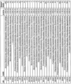
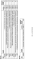
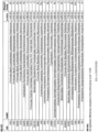
- An exemplary list of MS microbes or MS microbial families is provided in Figure 4 and/or Figure 5 as well as Table 1, Table 2 and/or Table 8.
- results of the methods disclosed herein can be used to direct antimicrobial therapy in patients with MS and related diseases (e.g., acute disseminated encephalomyelitis or neuromyelitis optics) who are developing (i.e., new onset) or experiencing worsening (i.e., relapses of) demyelination.
- MS and related diseases e.g., acute disseminated encephalomyelitis or neuromyelitis optics
- a patient with CSF reactivity to Akkermansia mucinophilia might be treated with intravenous meropenem, predicted to be active, and not with ceftriaxone, predicted to be inactive against this anaerobic bacterium.
- the MS microbes or MS microbial families described herein can also be used to develop specific PCR reagents to detect one or more of the MS microbes or MS microbial families disclosed herein that are causing demyelination.
- primers can be developed and used to detect these MS-causing organisms.
- CSF samples can be used from patients who are early in their course and developing demyelination, before the appearance of specific anti-microbial antibodies and oligoclonal bands.
- Specific PCR primers can be developed and used to detect the MS microbes or MS microbial families disclosed herein in brain tissue, spinal cord, CSF, or blood. A number of primer sets can be multiplexed in a single test. Again, the detection of microbial DNA (or less likely RNA) in the CSF of patients with early demyelination (or those having attacks of established MS) could be used to direct specific antimicrobial treatments.
- the methods disclosed here can be used to detect and treat the putative underlying cause of MS and several related demyelinating disorders including but not limited to neuromyelitis optics (NMO), acute disseminated encephalomyelitis (ADEM), and clinically isolated syndrome (CIS).
- NMO neuromyelitis optics
- ADAM acute disseminated encephalomyelitis
- CIS clinically isolated syndrome
- An advantage of the methods disclosed herein is that they can serve to limit or reverse the damage to the CNS. For instance, a patient with CSF reactivity to Akkermansia mucinophilia might be treated with intravenous meropenem, predicted to be active, and not with ceftriaxone, predicted to be inactive against this anaerobic bacterium.
- Current MS treatments are unreliable and inconsistent where there are no specific factors underlying the choice of treatment, and no reasonable expectations of success or failure.
- Current treatments for acute MS attacks include strategies that are broadly immunosuppressive (e.g., steroids
- Described herein are methods and microbial reagents that can be used alone or in a set of assays (e.g., serologic). Described herein are methods that can be used to detect antibodies in the CSF of MS patients that are directed against some but not all of the MS microbes or MS microbial families described herein.
- Described herein are methods that can be used alone or in combination with a set of assays. Described herein are methods comprising determining a microbial ELISA index (EI) and comparing the results of the EI with the albumin index of the subject. In some aspects, said method can be used to determine whether antibodies in the CSF of MS patients that are directed to one or more of the MS microbes of MS microbial families disclosed herein indicate intrathecal antibody synthesis. Further, said method can be used to identify a subject that may not have been detected or was excluded using any of the other methods of detection described herein.
- EI microbial ELISA index
- the disclosed methods of determining a microbial ELISA index (EI) and comparing the results of the EI of the subject with the albumin index of the subject can be used to improve on the identification of a subject with a demyelinating disease as compared to determining a microbial ELISA index (EI) alone.
- the assays and methods described herein can be used to direct antimicrobial therapy in patients with MS and related diseases (i.e., ADEM, NMO) who are developing (e.g., new onset) or experiencing worsening (e.g., relapses of) demyelination.
- MS and related diseases i.e., ADEM, NMO
- a patient with CSF reactivity to Akkermansia mucinophilia can be treated with intravenous meropenem, predicted to be active, and not with ceftriaxone, predicted to be inactive against this anaerobic bacterium.
- the MS microbes or MS microbial families described herein can also be used to develop specific PCR reagents to detect one or more of the MS microbes or MS microbial families disclosed herein that are causing demyelination.
- Primers can be developed to detect MS-causing organisms.
- the reagents e.g., primers
- the reagents can be applied to the CSF of patients who are early in their course and developing demyelination, before the appearance of specific anti-microbial antibodies and oligoclonal bands. In some aspects, a number of primer sets would be multiplexed in a single test.
- a methods of detection of microbial DNA (or less likely RNA) in the CSF of patients with early demyelination (or those having attacks of established MS) which can be used to direct specific antimicrobial treatments.
- Described herein are methods of detecting and treating the putative underlying cause of MS and several related demyelinating disorders including, but not limited to neuromyelitis optics (NMO), acute disseminated encephalomyelitis (ADEM), and clinically isolated syndrome (CIS).
- NMO neuromyelitis optics
- ADAM acute disseminated encephalomyelitis
- CIS clinically isolated syndrome
- Disclosed here are serologic assays to detect antibodies in the CSF of MS patients that are directed against one or more of the MS microbes or MS microbial families described herein.
- Disclosed herein are methods to direct antimicrobial therapy in patients with MS and related diseases (e.g., ADEM, NMO) who are developing (e.g. new onset) or experiencing worsening (e.g. relapses of) demyelination.
- MS microbes or MS microbial families derived from the deep sequencing data.
- the methods disclosed herein can be useful for making a microbiological diagnosis.
- the detection methods described herein can be performed directly via PCR amplification of a nucleic acid or via direct detection using antibodies or peptides that specifically bind one or more of the disclosed microbes.
- the detection methods described herein can also be performed by detecting antibodies specific to one or more of the microbe-specific antibodies.
- methods comprising contacting a sample with one or more microbe capture agents in conditions to allow one or more microbe capture agents bind to one or more of the microbe-specific antibodies to generate a mixed biological sample, wherein each of the one or more microbe capture agents is specific to one or more a microbe-specific antibodies, and wherein the one or more microbe-specific antibodies are specific to one or more microbes selected from the list in Table 1, Table 2 or Table 8.
- the methods disclosed herein can be useful for making a microbiological diagnosis.
- the methods disclosed herein can also be used to identify a subject with a demyelinating disease that was not identified in any of the other methods described herein.
- the methods disclosed herein can be used to determine the intactness of the blood brain barrier.
- the detection methods described herein can compare a microbial ELISA index (EI) of a subject to an albumin index (AI) of the subject.
- the methods disclosed herein can be performed via ELISA using antibodies that bind or specifically bind to one or more of the disclosed microbes.
- the EI is a measure of antibodies in a sample (e.g., CSF or spinal fluid).
- the EI value can be compared to a normal AI (e.g., 0 to 9) that indicates in intact blood brain barrier.
- Albumin Index CSF albumin (mg/L) / serum albumin (g/L).
- the AI an index of blood-brain barrier (BBB) integrity, adjusted for the serum albumin concentration.
- BBB blood-brain barrier
- the AI is increased in BBB dysfunction.
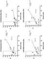
- the EI for a subject for one or more microbes selected from, for example, the list in Table 1, Table 2 and/or Table 8 can be plotted against the AI of the subject. Linear regression analysis can be performed showing the relationship between the EI's for each antigen and BBB intactness (as assessed by the Albumin Index).
- a linear regression line or plot line can be generated and provide an "expected EI" for each bacterial antigen or microbe tested, and can be used to correct for a leaky BBB that may be associated with a subject with MS and other demyelinating diseases.
- the methods disclosed herein can be used alone or in combination with any of the other methods described herein.
- the method can comprise obtaining a sample from the subject.
- the subject can be suspected of having or can be at risk for having a demyelinating disease.
- the method can comprise detecting the presence of one or more microbes in the sample.
- the one or more microbes in the sample can be selected from the list in Table 1, Table 2 or Table 8.
- the presence of the one or more microbes can be detected indirectly by detecting the presence of one or more antibodies in the sample.
- the one or more antibodies can be specific to the one or more microbes in the sample.
- the method can comprise diagnosing the subject with a demyelinating disease. In some aspects, the method can comprise diagnosing the subject with a demyelinating disease when the one or more microbes selected from the list in Table 1 or Table 2 are present in the sample.
- the method of diagnosing a demyelinating disease in a subject can comprising obtaining a sample from the subject.
- the sample can be suspected of containing microbe-specific antibodies.
- the method can comprise incubating the sample with one or more microbe capture agents.
- the one or more microbe capture agents can bind to one or more of the microbe-specific antibodies.
- the microbe-specific antibodies can be specific to one or more microbes selected from the list in Table 1, Table 2 or Table 8.
- the method can comprise detecting the presence of one or more microbe-specific antibodies in the sample.
- the method can comprise diagnosing the subject with a demyelinating disease when the one or more microbe-specific antibodies are present in the sample. In some aspects, the method can further comprise repeating incubating the sample with one or more microbe capture agents step and detecting the presence of one or more microbe-specific antibodies in the sample step using different microbe capture agents. In some aspects, the different microbe capture agents can be specific to the one or more microbe-specific antibodies in the sample. In some aspects, the method can further comprise detecting the presence of one or more microbe-specific antibodies specific to one or more of the microbes listed in Table 1, Table 2 or Table 8. In some aspects, the step of detecting can comprise detecting by microarray. In some aspects, the step of detecting can comprise detecting by ELISA.
- the one or more microbes detected can be from the genus Akkermansia.
- the method of diagnosing can further comprise detecting the presence of one or more microbe-specific antibodies in the sample and determing an ELISA index, comparing the ELISA index to the albumin index.
- an EI ratio can be determinied.
- the EI ratio can be >1.0 indicating more microbe-specific antibodies in the CSF compared to the serum against one or more of the microbes.
- the EI ratio can be ⁇ 1.0, indicating more microbe-specific antibodies in serum than in CSF.
- the method can further comprise determining an ELISA index for one or more microbes, comparing the ELISA index to an AI of the sample by performing a linear regression analysis and generating a plot line.
- the AI can be equal to or less than 9 and the ELISA index can be above the plot line indicating one or more microbe-specific antibodies in the CSF compared to the serum for one or more microbes.
- the method can comprising obtaining a sample from the subject.
- the sample can be suspected of containing microbe-specific antibodies.
- the method can comprise incubating the sample with one or more microbe capture agents.
- the one or more microbe capture agents can bind to one or more of the microbe-specific antibodies.
- the microbe-specific antibodies can be specific to one or more microbes selected from the list in Table 1, Table 2 or Table 8.
- the method can comprise detecting the presence of one or more microbe-specific antibodies in the sample.
- the method can comprise diagnosing the subject with a demyelinating disease.
- the method can comprise diagnosing the subject with a demyelinating disease when the one or more microbe-specific antibodies are present in the sample.
- the method can comprise administering an antibacterial agent when the one or more microbe-specific antibodies detected is a bacteria selected from the list in Table 1, Table 2 or Table 8.
- the method can comprise administering an antiviral agent when the one or more microbe-specific antibodies detected is a virus selected from the list in Table 1, Table 2 or Table 8.
- the method can comprise administering an antifungal agent when the one or more microbe-specific antibodies detected is a fungus selected from the list in Table 1, Table 2 or Table 8.
- the method can comprise administering a combination of antibacterial agent, antiviral agent, or an antifungal agent to the subject.
- the method of diagnosing and treating a demyelinating disease in a subject can further comprise detecting the presence of one or more microbe-specific antibodies in the sample and determing an ELISA index, comparing the ELISA index to the albumin index.
- an EI ratio can be determinied.
- the EI ratio can be >1.0 indicating more microbe-specific antibodies in the CSF compared to the serum against one or more of the microbes.
- the EI ratio can be ⁇ 1.0, indicating more microbe-specific antibodies in serum than in CSF.
- the method can further comprise determining an ELISA index for one or more microbes, comparing the ELISA index to the AI of the subject by performing a linear regression analysis and generating a plot line.
- the AI of the subject can be equal to or less than 9 and the ELISA index can be above the plot line indicating one or more microbe-specific antibodies in the CSF compared to the serum for one or more microbes.
- the method of detecting a demyelinating disease in a subject can comprise obtaining a sample from the subject.
- the sample can be suspected of containing microbe-specific antibodies specific to one or more microbes selected from the list in Table 1, Table 2 or Table 8.
- the method can comprise contacting the sample with one or more microbe capture agents to generate a mixed biological sample.
- each of the one or more microbe capture agents can be specific to one or more a microbe-specific antibodies.
- the one or more microbe-specific antibodies can be specific to one or more microbes selected from the list in Table 1, Table 2 or Table 8.
- the method can comprise incubating the mixed biological sample under conditions such that the one or more microbe capture agents can specifically bind to at least one of the microbe-specific antibodies to form a detectable complex.
- the method can comprise detecting the presence of the detectable complexes, thereby detecting the presence of one or more microbes selected from the list in Table 1, Table 2 or Table 8.
- the presence of one or more microbes selected from the list in Table 1, Table 2 or Table 8 in the mixed biological sample can detect a demyelinating disease in a subject.
- the method can further comprise repeating the contacting the sample step and the incubating the mixed biological step using different microbe capture agents.
- the different microbe capture agents can be specific to the one or more microbe-specific antibodies in the sample.
- the method can further comprise detecting the presence of one or more microbe-specific antibodies specific to one or more of the microbes listed in Table 1, Table 2 or Table 8.
- the method can further comprise determining the quantity of one or more of the microbe-specific antibodies in the sample.
- the step of detecting can comprise detecting by microarray.
- the step of detecting can comprise detecting by ELISA.
- the one or more microbes detected can be from the genus Akkermansia.
- the method can further comprise determing an ELISA index.
- the ELISA index can be compared to or plotted against the albumin index.
- an EI ratio can be determinied.
- the EI ratio can be >1.0 indicating more microbe-specific antibodies in the CSF compared to the serum against one or more of the microbes.
- the EI ratio can be ⁇ 1.0, indicating more microbe-specific antibodies in serum than in CSF.
- the method can further comprise determining an ELISA index for one or more microbes, comparing the ELISA index to the AI of the subject by performing a linear regression analysis and generating a plot line.
- the AI of the subject can be equal to or less than 9 and the ELISA index can be above the plot line indicating one or more microbe-specific antibodies in the CSF compared to the serum for one or more microbes.
- the method can comprise obtaining a sample from the subject.
- the sample can be suspected of containing microbe-specific antibodies specific to one or more microbes selected from the list in Table 1, Table 2 or Table 8.
- the method can comprise contacting the sample with one or more microbe capture agents to generate a mixed biological sample.
- each of the one or more microbe capture agents can be specific to one or more a microbe-specific antibodies.
- the one or more microbe-specific antibodies can be specific to one or more microbes selected from the list in Table 1, Table 2 or Table 8.
- the method can comprise incubating the mixed biological sample under conditions such that: said one or more microbe capture agents can specifically bind to at least one of the microbe-specific antibodies to form a detectable complex.
- the method can comprise detecting the presence of the detectable complexes, thereby detecting the presence of one or more microbes selected from the list in Table 1, Table 2 or Table 8.
- the presence of one or more microbes selected from the list in Table 1, Table 2 or Table 8 in the mixed biological sample can detect a demyelinating disease in a subject.
- the method can comprise administering an antibacterial agent when the one or more microbe-specific antibodies detected is a bacteria selected from the list in Table 1, Table 2 or Table 8.
- the method can comprise administering an antiviral agent when the one or more microbe-specific antibodies detected is a virus selected from the list in Table 1, Table 2 or Table 8. In some aspects, the method can comprise administering an antifungal agent when the one or more microbe-specific antibodies detected is a fungus selected from the list in Table 1, Table 2 or Table 8. In some aspects, the method can comprise administering a combination of antibacterial agent, antiviral agent, or an antifungal agent to the subject. In some aspects, the method can comprise administering an antiparasitic agent. In some aspects, the method can comprise administering a combination of antibacterial agent, antiviral agent, antiparasitic agent or an antifungal agent to the subject.
- the different microbe capture agents can be specific to the one or more microbe-specific antibodies in the sample.
- the method can further comprise detecting the presence of one or more microbe-specific antibodies specific to one or more of the microbes listed in Table 1, Table 2 or Table 8.
- the method can further comprise determining the quantity of one or more of the microbe-specific antibodies in the sample.
- the step of detecting can comprise detecting by microarray.
- the step of detecting can comprise detecting by ELISA.
- the one or more microbes detected can be from the genus Akkermansia.
- the method can further comprise detecting the presence of one or more microbe-specific antibodies in the sample and determing an ELISA index, comparing the ELISA index to the albumin index.
- an EI ratio can be determinied.
- the EI ratio can be >1.0 indicating more microbe-specific antibodies in the CSF compared to the serum against one or more of the microbes.
- the EI ratio can be ⁇ 1.0, indicating more microbe-specific antibodies in serum than in CSF.
- the method can further comprise determining an ELISA index for one or more microbes, comparing the ELISA index to the AI of the subject by performing a linear regression analysis and generating a plot line.
- the AI of the subject can be equal to or less than 9 and the ELISA index can be above the plot line indicating one or more microbe-specific antibodies in the CSF compared to the serum for one or more microbes.
- the method can comprise obtaining a sample from the subject.
- the subject can be suspected of having or can be at risk of having a demyelinating disease.
- the method can comprise detecting the presence of one or more microbes in the sample.
- the one or more microbes can be selected from the list in Table 1, Table 2 or Table 8.
- the method can comprise diagnosing the subject with a demyelinating disease when one or more microbes selected from the list in Table 1, Table 2 or Table 8 are present in the sample.
- the method can comprise obtaining a sample from the subject.
- the sample can be suspected of containing microbe-specific antibodies specific to one or more microbes selected from the list in Table 1, Table 2 or Table 8.
- the method can comprise incubating the sample with one or more microbe capture agents.
- the one or more microbe capture agents can bind to one or more of the microbe-specific antibodies.
- the one or more microbe-specific antibodies can be specific to one or more microbes selected from the list in Table 1, Table 2 or Table 8.
- the method can comprise detecting the presence of one or more microbe-specific antibodies in the sample.
- the detection of the presence of one or more microbe-specific antibodies can be indicative of a demyelinating disease.
- the method can further comprise detecting the presence of one or more microbe-specific antibodies specific to one or more of the microbes listed in Table 1, Table 2 or Table 8.
- the method can further comprise determining the quantity of one or more of the microbe-specific antibodies in the sample.
- the step of detecting can comprise detecting by microarray.
- the step of detecting can comprise detecting by ELISA.
- the one or more microbes detected can be from the genus Akkermansia.
- the method can further comprise determing an ELISA index.
- the method can compare the ELISA index to the albumin index. In some aspects, the method can further comprise detecting the presence of one or more microbe-specific antibodies in the sample and determing an ELISA index, comparing the ELISA index to the albumin index. In some aspects, an EI ratio can be determinied. In some aspects, the EI ratio can be >1.0 indicating more microbe-specific antibodies in the CSF compared to the serum against one or more of the microbes. In some aspects, the EI ratio can be ⁇ 1.0, indicating more microbe-specific antibodies in serum than in CSF.
- the method can further comprise determining an ELISA index for one or more microbes, comparing the ELISA index to the AI of the subject by performing a linear regression analysis and generating a plot line.
- the AI of the subject can be equal to or less than 9 and the ELISA index can be above the plot line indicating one or more microbe-specific antibodies in the CSF compared to the serum for one or more microbes.
- the method can comprise obtaining a sample from a subject.
- the sample can be suspected of containing one or more microbes selected from the list in Table 1, Table 2 or Table 8.
- the method can comprise incubating the sample with one or more microbe capture agents.
- the one or more microbe capture agents can bind to one or more of the microbes selected from the list in Table 1, Table 2 or Table 8.
- the method can comprise detecting the presence of one or more microbes selected from the list in Table 1, Table 2 or Table 8 in the sample.
- the method can further comprise detecting the presence of one or more microbes listed in Table 1, Table 2 or Table 8. In some aspects, the method can further comprise determining the quantity of one or more of the microbes in the sample. In some aspects, detecting the presence of one or more of the microbes is indicative of a demyelinating disease. In some aspects, the step of detecting can comprise detecting by microarray. In some aspects, the step of detecting can comprise detecting by ELISA. In some aspects, the one or more microbes detected can be from the genus Akkermansia. In some aspects, the method can further comprise determing an ELISA index. In some aspects, the method can compare the ELISA index to the albumin index.
- the method can further comprise detecting the presence of one or more microbe-specific antibodies in the sample and determing an ELISA index, comparing the ELISA index to the AI of the subject.
- an EI ratio can be determinied.
- the EI ratio can be >1.0 indicating more microbe-specific antibodies in the CSF compared to the serum against one or more of the microbes.
- the EI ratio can be ⁇ 1.0, indicating more microbe-specific antibodies in serum than in CSF.
- the method can further comprise determining an ELISA index for one or more microbes, comparing the ELISA index to the AI of the subject by performing a linear regression analysis and generating a plot line.
- the AI of the subject can be equal to or less than 9 and the ELISA index can be above the plot line indicating one or more microbe-specific antibodies in the CSF compared to the serum for one or more microbes.
- tissue samples can be obtained from the subject using core needle biopsies.
- the sample can be whole cells or cell organelles. Cells can be collected by scraping the tissue, processing the tissue sample to release individual cells or isolating the cells from a bodily fluid.
- the sample can be fresh tissue, dry tissue, cultured cells or tissue.
- the sample can be unfixed or fixed. Any part of the brain or spinal cord can be obtained and assessed using the methods described herein.
- the sample solution may comprise any number of sources, including, but not limited to, bodily fluids (including, but not limited to, blood, biopsy tissue, CSF). Samples can range from less than a milliliter and can further range in bacterial concentration. Furthermore, the sample can be present in blood, brain tissue, spinal cord tissue or CSF.
- the subject can be suspected of having a demyelinating disease. In any of the methods disclosed herein, the subject can be at risk of having a demyelinating disease. In some aspects, the demyelinating disease can be multiple sclerosis, neuromyelitis optics, acute disseminated encephalomyelitis or clinically isolated syndrome. In some aspects, the subject can be a human. In some aspects, the sample can be brain tissue, cerebral spinal fluid, spinal cord tissue or blood.
- the methods can further comprise administering to the subject an antibacterial agent, an antiviral agent, an antifungal agent or a combination thereof.
- the method can comprise administering an antiparasitic agent.
- the method can comprise administering a combination of antibacterial agent, antiviral agent, antiparasitic agent or an antifungal agent to the subject.
- the antibacterial agent can be administered to the subject when the one or more microbe-specific antibodies or microbes detected can be a bacteria selected from the list in Table 1, Table 2 or Table 8.
- the antiviral agent can be administered to the subject when the one or more microbe-specific antibodies or microbes detected is a virus selected from the list in Table 1, Table 2 or Table 8.
- the antifungal agent can be administered to the subject when the one or more microbe-specific antibodies or microbes detected is a fungus selected from the list in Table 1, Table 2 or Table 8.
- the method can further comprise amplifying one or more nucleic acid sequences in the sample.
- the one or more nucleic acid sequences correspond to one or more of the microbes in Table 1, Table 2 or Table 8.
- the one or more nucleic acid sequences can be amplified using a primer pair that can specifically amplify the one or more nucleic acid sequences.
- the method can further comprise determining whether the expression of the one or more nucleic acid sequences corresponds to the one or more of the microbes in Table 1, Table 2 or Table 8 is overexpressed compared to expression levels of a nucleic acid sequence of the same one or more of the microbes in Table 1, Table 2 or Table 8 in a control.
- the detecting step can comprise detecting by a hybridization reaction.
- the hybridization reaction can further comprise hybridizing the sample to one or more primer sets.
- the hybridization reaction can be a polymerase chain reaction.
- the step of detecting can comprise detecting by expressed sequence tags.
- the step of detecting can comprise detecting by localization.
- the microbe capture agent can be an antibody, peptide, protein or a probe.
- the one or more microbes disclosed herein can be used to bind to one or more of the antibodies from CSF of a subject (for example, a patient that has or is suspected of having of MS.)
- a bacterial antigen can be sonicated at an OD of 0.5 and be coated on the bottom of a single or multi-well plate.
- the bacterial antigen can be from Akkermansia, Atopobium, Lactobacillus, Pseudomonas, Bacteroides and Nitrosospira.
- a positive control In any of the methods or assays or tests described herein, a positive control, a negative control, or both can be further included.
- a positive control include but are not limited to commercial CSF preparations (Randox) and an anti- Pseudomonas antibody (ThermoFisher product #PA1-73116).
- a negative control examples include but are not limited to PBS and IgG-depeted Randox CSF (which lacks all antibodies).
- one or more methods of detection can be used to demonstrate the binding of a specific CSF antibody against one or more of the microbes disclosed herein.
- one or more microbes can be tested (and/or detected) in a subject with or suspected of having MS or other autoimmune disease.
- the method of detection can be an indirect method.
- the indirect method can be ELISA.
- two or more microbes can be tested (and/or detected) using ELISA.
- Methods of treating are methods of treating a patient with multiple sclerosis (MS) or an MS-related disease.
- the method can comprise administering an antibacterial agent, antiviral agent, an antifungal agent, an antiparasitic agent or a combination thereof to the patient.
- the patient can be identified as needing an antibacterial agent, antiviral agent, an antifungal agent, antiparasitic agent or a combination thereof.
- the patient can be identified by: obtaining a sample from the patient, wherein the sample is suspected of containing microbe-specific antibodies; incubating the sample with one or more microbe capture agents, wherein the one or more microbe capture agents bind to one or more of the microbe-specific antibodies, and wherein the microbe-specific antibodies are specific to one or more microbes selected from the list in Table 1, Table 2 or Table 8; and detecting the presence of one or more microbe-specific antibodies in the sample.
- the antibacterial agent can be meropenem or ceftriaxone.
- the MS-related disease can be neuromyelitis optics, acute disseminated encephalomyelitis or clinically isolated syndrome.
- the step of detecting can comprise detecting by microarray. In some aspects, the step of detecting can comprise detecting by ELISA. In some aspects, wherein the one or more microbes detected can be from the genus Akkermansia. In some aspects, the sample can be brain tissue, cerebral spinal fluid, spinal cord tissue or blood.
- the method can further comprise detecting the presence of one or more microbe-specific antibodies specific to one or more microbes are selected from the list in Table 1, Table 2 or Table 8. In some aspects, the method can further comprise determining the quantity of one or more of the microbe-specific antibodies in the sample. In some aspects, the method can further comprise amplifying one or more nucleic acid sequences in the sample. In some aspects, the one or more nucleic acid sequences can correspond to one or more of the microbes in Table 1, Table 2 or Table 8. In some aspects, the one or more nucleic acid sequences can be amplified using a primer pair that can specifically amplify the one or more nucleic acid sequences.
- the method can further comprise determining whether the expression of the one or more nucleic acid sequences corresponding to the one or more of the microbes in Table 1, Table 2 or Table 8 is overexpressed compared to expression levels of a nucleic acid sequence of the same one or more of the microbes in Table 1, Table 2 or Table 8 in a control.
- the step of detecting can comprise detecting by hybridization reaction.
- the hybridization reaction can further comprise hybridizing the sample to one or more primer sets.
- the step of detecting can comprise detecting by a polymerase chain reaction.
- the presence or absence or expression level of one or more microbes or microbe-specific antibodies disclosed herein can be determined directly (e.g., immunoassays, mass spectrometry) or indirectly (e.g., presence or absence of one or more antibodies that are specific to the one or more microbes; determining the mRNA expression of a protein or peptide).
- mass spectrometry examples include but are not limited to ionization sources such as EI, CI, MALDI, ESI, and analysis such as Quad, ion trap, TOF, FT or combinations thereof, spectrometry, isotope ratio mass spectrometry (IRMS), thermal ionization mass spectrometry (TIMS), spark source mass spectrometry, Multiple Reaction Monitoring (MRM) or SRM. Any of these techniques can be carried out in combination with prefractionation or enrichment methods.
- immunoassays include but are not limited to immunoblots, Western blots, Enzyme linked Immunosorbant Assay (ELISA), Enzyme immunoassay (EIA), radioimmune assay. Immunoassay methods use antibodies for detection and determination of levels of an antigen are known in the art. The antibody can be immobilized on a solid support such as a stick, plate, bead, microbead or array.
- RNA expression methods include but are not limited to extraction of cellular mRNA and Northern blotting using labeled probes that hybridize to transcripts encoding all or part of the gene, amplification of mRNA using gene-specific primers, polymerase chain reaction (PCR), and reverse transcriptase-polymerase chain reaction (RT-PCR), followed by quantitative detection of the gene product by a variety of methods; extraction of RNA from cells, followed by labeling, and then used to probe cDNA or olignonucleotides encoding the gene, in situ hybridization; and detection of a reporter gene.
- PCR polymerase chain reaction
- RT-PCR reverse transcriptase-polymerase chain reaction
- Methods to measure protein expression levels include but are not limited to Western blot, immunoblot, ELISA, radioimmunoassay, immunoprecipitation, surface plasmon resonance, chemiluminescence, fluorescent polarization, phosphorescence, immunohistochemical analysis, microcytometry, microarray, microscopy, fluorescence activated cell sorting (FACS), and flow cytometry.
- the methods can also include specific protein property-based assays based including but not limited to enzymatic activity or interaction with other protein partners. Binding assays can also be used, and are well known in the art. For instance, a BIAcore machine can be used to determine the binding constant of a complex between two proteins.
- suitable assays for determining or detecting the binding of one protein to another include, immunoassays, such as ELISA and radioimmunoassays. Determining binding by monitoring the change in the spectroscopic can be used or optical properties of the proteins can be determined via fluorescence, UV absorption, circular dichroism, or nuclear magnetic resonance (NMR). Alternatively, immunoassays using specific antibody can be used to detect the expression on of a particular protein on a tumor cell.
- the disclosed methods also provide for the quantification and/or qualification of susceptibility of a desired treatment based on the identification (or absence of such identification) of one or more microbes. Said information can lead to therapeutic decisions.
- Protein array Disclosed herein are polypeptide or protein arrays.
- the protein arrays can comprise probes including antibodies, aptamers, and other cognate binding ligands specific to a component of the microbe panels disclosed herein. Protein arrays and methods of constructing the protein arrays are well known to one of ordinary skill in the art.
- One type of protein array that can be suitable uses an immobilized "capture antibody.”
- the polypeptides are bound to a solid substrate (e.g., glass) with a treated surface (e.g., aminosilane) or through a biotin-streptavidin conjugation.
- the arrays are then incubated with a solution containing probe that can bind to the capture antibodies in a manner dependent upon time, buffer components, and recognition specificity.
- the probes can then be visualized directly if they have been previously labeled, or can be bound to a secondary labeled reagent (e.g., another antibody).
- a secondary labeled reagent e.g., another antibody
- the amount of probe bound to the capture antibody that is visualized can depend upon the labeling method utilized; generally, a CCD imager or laser scanner that uses filter sets that are appropriate to excite and detect the emissions of the label can be used.
- the imager converts the amount of detected photons into an electronic signal (often an 8-bit or 16-bit scale) that can be analyzed using commercially available software packages.
- the substrate of the array can be organic or inorganic, biological or non-biological or any combination of these materials.
- the substrate can be transparent or translucent.
- materials suitable for use as a substrate in the array include but are not limited to silicon, silica, quartz, glass, controlled pore glass, carbon, alumina, titanium dioxide, germanium, silicon nitride, zeolites, and gallium arsenide; and metals including gold, platinum, aluminum, copper, titanium, and their alloys. Ceramics and polymers can also be used as substrates.
- Suitable polymers include, but are not limited to polystyrene; poly(tetra)fluorethylene; (poly)vinylidenedifluoride; polycarbonate; polymethylmethacrylate; polyvinylethylene; polyethyleneimine; poly(etherether)ketone; polyoxymethylene (POM); polyvinylphenol; polylactides; polymethacrylimide (PM I); polyalkenesulfone (PAS); polyhydroxyethylmethacrylate; polydimethylsiloxane; polyacrylamide; polyimide; co-block-polymers; and Eupergit ® . Photoresists, polymerized Langmuir-Blodgett films, and LIGA structures can also serve as substrates.
- the array can further comprise a coating that can be formed on the substrate or applied to the substrate.
- the substrate can be modified with a coating by using thin-film technology based on either physical vapor deposition (PVD) or plasma-enhanced chemical vapor deposition (PECVD).
- PVD physical vapor deposition
- PECVD plasma-enhanced chemical vapor deposition
- plasma exposure can be used to directly activate the substrate.
- plasma etch procedures can be used to oxidize a polymeric surface (i.e., polystyrene or polyethylene to expose polar functionalities such as hydroxyls, carboxylic acids, aldehydes and the like).
- the coating can comprise a metal film.
- metal films include but are not limited to aluminum, chromium, titanium, nickel stainless steel zinc, lead, iron, magnesium, manganese, cadmium, tungsten, cobalt, and alloys or oxides thereof.
- the metal film can be a noble metal film.
- noble metals that can be used for a coating include, but are not limited to, gold, platinum, silver, copper, and palladium.
- the coating comprises gold or a gold alloy. Electron-beam evaporation can be used to provide a thin coating of gold on the surface.
- the metal film can be from about 50 nm to about 500 nm in thickness.
- the coating can be silicon, silicon oxide, silicon nitride, silicon hydride, indium tin oxide, magnesium oxide, alumina, glass, hydroxylated surfaces, and a polymer.
- the arrays described herein can comprise a collection of addressable elements. Such elements can be spacially addressable, such as arrays contained within microtiter plates or printed on planar surfaces wherein each element can be present at distinct X and Y coordinates. Alternatively, elements can be addressable based on tags, beads, nanoparticles, or physical properties.
- the microarrays can be prepared according to the methods known to one of ordinary skill in the art.
- arrays as used herein can refer to any biologic assay with multiple addressable elements.
- the addressable elements can be polypeptides (e.g., antibodies or fragments thereof) or nucleic acid probes.
- elements refer to any probe (polypeptide or nucleic acid based) that can be bound by a one or more microbes disclosed herein, polypeptide fragment or transcript encoding such polypeptides, as related or associated with any of the microbes disclosed herein.
- Molecules can be, but are not limited to, proteins, polypeptides, peptides, RNA, DNA, lipids, glycosylated molecules, carbohydrates, polypeptides with phosphorylation modifications, and polypeptides with citrulline modifications, aptamers, oxidated molecules, and other molecules.
- addressability refers to the location, position, tags, cleavable tags or markers, identifiers, spectral properties, electrophoretic properties, or other physical properties that enable identification of the element.
- This type of spatial array can generally be synthesized or spotted onto a planar substrate, producing, for example, microarrays, where a large number of different molecules are densely laid out in a small area (e.g., comprising at least about 400 different sequences per cm2, and can be 1000 sequences per cm 2 or as many as 5000 sequences per cm 2 , or more).
- Less dense arrays e.g., ELISA or RIA plates
- ELISA or RIA plates where wells in a plate each contain a distinct probe can comprise from about 96 sequences per plate, up to about 100 sequences per cm 2 , up to the density of a microarray.
- Other spatial arrays utilize fiber optics, where distinct probes can be bound to fibers, which can be formed into a bundle for binding and analysis. Methods for the manufacture and use of spatial arrays of polypeptides are known in the art.
- tags can be cleaved from the element, and subsequently detected to identify the element.
- a set of probes can be synthesized or attached to a set of coded beads, wherein each bead can be linked to a distinct probe, and wherein the beads can be coded in a manner that allows identification of the attached probe.
- flow cytometry can be used for detection of binding. For example, microspheres having fluorescence coding and can identify a particular microsphere.
- the probe can be covalently bound to a "color coded" object.
- a labeled target polypeptide can be detected by flow cytometry, and the coding on the microsphere can be used to identify the bound probe (e.g., immunoglobulin, antigen binding fragments of immunoglobulins, or ligands).
- the array can be an immunoglobulin (e.g., antibody or antigen-binding fragment thereof) array.
- an immunoglobulin array refers to a spatially separated set of discrete molecular entities capable of binding to target polypeptides arranged in a manner that allows identification of the polypeptides contained within the sample.
- the array can comprise one or more of proteins, polypeptides, peptides, RNA, DNA, lipid, glycosylated molecules, polypeptides with phosphorylation modifications, and polypeptides with citrulline modifications, aptamers, and other molecules.
- Antibodies Disclosed herein are isolated antibodies, antibody fragments and antigen-binding fragments thereof, that can specifically bind to one or more of the microbes or microbe-specific antibodies disclosed herein.
- the isolated antibodies, antibody fragments, or antigen-binding fragment thereof can be neutralizing antibodies.
- antibody fragments are portions of a complete antibody.
- a complete antibody refers to an antibody having two complete light chains and two complete heavy chains.
- An antibody fragment lacks all or a portion of one or more of the chains. Examples of antibody fragments include, but are not limited to, half antibodies and fragments of half antibodies.
- a half antibody is composed of a single light chain and a single heavy chain.
- Half antibodies and half antibody fragments can be produced by reducing an antibody or antibody fragment having two light chains and two heavy chains. Such antibody fragments are referred to as reduced antibodies. Reduced antibodies have exposed and reactive sulfhydryl groups. These sulfhydryl groups can be used as reactive chemical groups or coupling of biomolecules to the antibody fragment.
- a preferred half antibody fragment is a F(ab).
- the hinge region of an antibody or antibody fragment is the region where the light chain ends and the heavy chain goes on.
- Antibody fragments for use in the methods disclosed herein can bind antigens (e.g. microbe-specific antibodies or one or more of the microbes described herein).
- the antibody fragment can be specific for an antigen.
- An antibody or antibody fragment is specific for an antigen if it binds with significantly greater affinity to one epitope than to other epitopes.
- the antigen can be any molecule, compound, composition, or portion thereof to which an antibody fragment can bind.
- the antigen can be a microbe-specific antibody or one or more of the microbes described herein.
- An analyte can be any molecule, compound or composition of interest.
- the antibodies or antibody fragments can be tested for their desired activity using the in vitro assays described herein, or by analogous methods.
- the term "monoclonal antibody” as used herein refers to an antibody obtained from a substantially homogeneous population of antibodies, i.e., the individual antibodies within the population are identical except for possible naturally occurring mutations that may be present in a small subset of the antibody molecules. Also disclosed are “chimeric" antibodies in which a portion of the heavy or light chain is identical with or homologous to corresponding sequences in antibodies derived from a particular species or belonging to a particular antibody class or subclass, while the remainder of the chain(s) is identical with or homologous to corresponding sequences in antibodies derived from another species or belonging to another antibody class or subclass, as well as fragments of such antibodies, as long as they exhibit the desired antagonistic activity (See, U.S. Patent No. 4,816,567 and Morrison et al., Proc. Natl. Acad. Sci. USA, 81:6851 6855 (1984 )).
- Monoclonal antibodies can be made using any procedure which produces monoclonal antibodies.
- disclosed monoclonal antibodies can be prepared using hybridoma methods, such as those described by Kohler and Milstein, Nature, 256:495 (1975 ).
- a hybridoma method a mouse or other appropriate host animal is typically immunized with an immunizing agent to elicit lymphocytes that produce or are capable of producing antibodies that will specifically bind to the immunizing agent.
- Monoclonal antibodies may also be made by recombinant DNA methods, such as those described in U.S. Patent No. 4,816,567 (Cabilly et al. ).
- DNA encoding the disclosed monoclonal antibodies can be readily isolated and sequenced using conventional procedures (e.g. , by using oligonucleotide probes that are capable of binding specifically to genes encoding the heavy and light chains of murine antibodies).
- Libraries of antibodies or active antibody fragments can also be generated and screened using phage display techniques, e.g., as described in U.S. Patent No. 5,804,440 to Burton et al. and U.S. Patent No. 6,096,441 to Barbas et al.
- In vitro methods are also suitable for preparing monovalent antibodies.
- Digestion of antibodies to produce fragments thereof, such as an Fv, Fab, Fab', or other antigen binding portion of an antibody can be accomplished using routine techniques known in the art. For example, digestion can be performed using papain. Examples of papain digestion are described in WO 94/29348 published Dec. 22, 1994 and U.S. Patent No. 4,342,566 , the contents of which are hereby incorporated by reference in its entirety for its teaching of papain digestion of antibodies to prepare monovaltent antibodies.
- Papain digestion of antibodies typically produces two identical antigen binding fragments, called Fab fragments, each with a single antigen binding site, and a residual Fc fragment. Pepsin treatment yields a fragment that has two antigen combining sites and is still capable of cross-linking antigen.
- Fragments whether attached to other sequences, can also include insertions, deletions, substitutions, or other selected modifications of particular regions or specific amino acid residues, provided the activity of the antibody or antibody fragment is not significantly altered or impaired compared to the non-modified antibody or antibody fragment. These modifications can provide for some additional property, such as to remove/add amino acids capable of disulfide bonding, to increase its bio-longevity, to alter its secretory characteristics, etc. In any case, the antibody or antibody fragment must possess a bioactive property, such as specific binding to its cognate antigen. Functional or active regions of the antibody or antibody fragment may be identified by mutagenesis of a specific region of the protein, followed by expression and testing of the expressed polypeptide.
- antibody can also refer to a human antibody or a humanized antibody.
- Many non-human antibodies e.g., those derived from mice, rats, or rabbits
- are naturally antigenic in humans and thus can give rise to undesirable immune responses when administered to humans. Therefore, the use of human or humanized antibodies in the methods serves to lessen the chance that an antibody administered to a human will evoke an undesirable immune response.
- Human antibodies can be prepared using any technique. Examples of techniques for human monoclonal antibody production include those described by Cole et al. (Monoclonal Antibodies and Cancer Therapy, Alan R. Liss, p. 77, 1985 ) and by Boemer et al. (J. Immunol., 147(1):86 95, 1991 ). Human antibodies (and fragments thereof) can also be produced using phage display libraries ( Hoogenboom et al., J. Mol. Biol., 227:381, 1991 ; Marks et al., J. Mol. Biol., 222:581, 1991 ).
- Human antibodies can also be obtained from transgenic animals.
- transgenic, mutant mice that are capable of producing a full repertoire of human antibodies, in response to immunization, have been described (see, e.g., Jakobovits et al., Proc. Natl. Acad. Sci. USA, 90:2551-255 (1993 ); Jakobovits et al., Nature, 362:255 258 (1993 ); Bruggermann et al., Year in Immunol., 7:33 (1993 )).
- the homozygous deletion of the antibody heavy chain joining region (J(H)) gene in these chimeric and germline mutant mice results in complete inhibition of endogenous antibody production, and the successful transfer of the human germ line antibody gene array into such germ line mutant mice results in the production of human antibodies upon antigen challenge.
- Antibodies having the desired activity are selected using Env-CD4-co-receptor complexes as described herein.
- human antibodies can be made from memory B cells using a method for Epstein-Barr virus transformation of human B cells.
- a method for Epstein-Barr virus transformation of human B cells See, e.g., Triaggiai et al., An efficient method to make human monoclonal antibodies from memory B cells: potent neutralization of SARS coronavirus, Nat Med. 2004 Aug; 10(8):871-5 . (2004)), which is herein incorporated by reference in its entirety for its teaching of a method to make human monoclonal antibodies from memory B cells).
- memory B cells from a subject who has survived a natural infection are isolated and immortalized with EBV in the presence of irradiated mononuclear cells and a CpG oligonuleotide that acts as a polyclonal activator of memory B cells.
- the memory B cells are cultured and analyzed for the presence of specific antibodies.
- EBV-B cells from the culture producing the antibodies of the desired specificity are then cloned by limiting dilution in the presence of irradiated mononuclear cells, with the addition of CpG 2006 to increase cloning efficiency, and cultured. After culture of the EBV-B cells, monoclonal antibodies can be isolated.
- Such a method offers (1) antibodies that are produced by immortalization of memory B lymphocytes which are stable over a lifetime and can easily be isolated from peripheral blood and (2) the antibodies isolated from a primed natural host who has survived a natural infection, thus eliminating the need for immunization of experimental animals, which may show different susceptibility and, therefore, different immune responses.
- Antibody humanization techniques generally involve the use of recombinant DNA technology to manipulate the DNA sequence encoding one or more polypeptide chains of an antibody molecule.
- a humanized form of a non-human antibody is a chimeric antibody or antibody chain (or a fragment thereof, such as an Fv, Fab, Fab', or other antigen binding portion of an antibody) which contains a portion of an antigen binding site from a non-human (donor) antibody integrated into the framework of a human (recipient) antibody.
- a humanized antibody residues from one or more complementarity determining regions (CDRs) of a recipient (human) antibody molecule are replaced by residues from one or more CDRs of a donor (non-human) antibody molecule that is known to have desired antigen binding characteristics (e.g., a certain level of specificity and affinity for the target antigen).
- CDRs complementarity determining regions
- donor non-human antibody molecule that is known to have desired antigen binding characteristics
- Fv framework (FR) residues of the human antibody are replaced by corresponding non-human residues.
- Humanized antibodies may also contain residues which are found neither in the recipient antibody nor in the imported CDR or framework sequences.
- a humanized antibody has one or more amino acid residues introduced into it from a source which is non-human.
- humanized antibodies are typically human antibodies in which some CDR residues and possibly some FR residues are substituted by residues from analogous sites in rodent antibodies.
- Humanized antibodies generally contain at least a portion of an antibody constant region (Fc), typically that of a human antibody ( Jones et al., Nature, 321:522-525 (1986 ), Reichmann et al., Nature, 332:323 327 (1988 ), and Presta, Curr. Opin. Struct. Biol., 2:593-596 (1992 )).
- Fc antibody constant region
- humanized antibodies can be generated according to the methods of Winter and co-workers ( Jones et al., Nature, 321:522 525 (1986 ), Riechmann et al., Nature, 332:323-327 (1988 ), Verhoeyen et al., Science, 239:1534 1536 (1988 )), by substituting rodent CDRs or CDR sequences for the corresponding sequences of a human antibody.
- Methods that can be used to produce humanized antibodies are also described in U.S. Patent No. 4,816,567 (Cabilly et al. ), U.S. Patent No. 5,565,332 (Hoogenboom et al.
- the broadly neutralizing antibodies to the polypeptides disclosed herein and antibody fragments can also be administered to subjects or subjects as a nucleic acid preparation (e.g., DNA or RNA) that encodes the antibody or antibody fragment, such that the subject's own cells take up the nucleic acid, and produce and secrete the encoded antibody or antibody fragment.
- a nucleic acid preparation e.g., DNA or RNA
- kits comprising one or more probes or primers capable of detecting, amplifying or measuring the presence or expression of one or more microbes disclosed herein. Also disclosed are kits comprising one or more of the microbe capture agents, microbes or microbe-specific antibodies disclosed herein.
- Solid supports comprising one or more primers, probes, polypeptides, or antibodies capable of hybridizing or binding to one or more of the microbes described herein.
- Solid supports are solid state substrates or supports that molecules, such as analytes and analyte binding molecules, can be associated.
- Analytes e.g., calcifying nano-particles and proteins
- analytes can be directly immobilized on solid supports.
- Analyte capture agents e.g., capture compounds
- solid supports can also be immobilized on solid supports.
- microbes or microbe-specific antibodies proteins or nucleic acids
- arrays such as microarrays, RT-PCR (including quantitative RT-PCR), nuclease protection assays and Northern blot analyses can be used.
- the expression levels of one or more of the microbes can be determined using arrays, microarrays, RT-PCR, quantitative RT-PCR, nuclease protection assays or Northern blot analyses.
- An array is a form of solid support.
- An array detector is also a form of solid support to which multiple different capture compounds or detection compounds have been coupled in an array, grid, or other organized pattern.
- Solid-state substrates for use in solid supports can include, for instance, any solid material to which molecules can be coupled.
- materials include but are not limited to acrylamide, agarose, cellulose, nitrocellulose, glass, polystyrene, polyethylene vinyl acetate, polypropylene, polymethacrylate, polyethylene, polyethylene oxide, polysilicates, polycarbonates, teflon, fluorocarbons, nylon, silicon rubber, polyanhydrides, polyglycolic acid, poly lactic acid, polyorthoesters, polypropylfumerate, collagen, glycosaminoglycans, and polyamino acids.
- Solid-state substrates can have any useful form including thin film, membrane, bottles, dishes, fibers, woven fibers, shaped polymers, particles, beads, microparticles, or any combination thereof.
- Solid-state substrates and solid supports can be porous or non-porous.
- An example of a solid-state substrate is a microtiter dish (e.g., a standard 96-well type).
- a multiwell glass slide can also be used. For example, such as one containing one array per well can be used, allowing for greater control of assay reproducibility, increased throughput and sample handling, and ease of automation.
- Different compounds can be used together as a set.
- the set can be used as a mixture of all or subsets of the compounds (e.g., microbe capture agents or microbe-specific antibodies) used separately in separate reactions, or immobilized in an array.
- Compounds used separately or as mixtures can be physically separable through, for example, association with or immobilization on a solid support.
- An array can include a plurality of compounds immobilized at identified or predefined locations on the array. Each predefined location on the array can generally have one type of component (that is, all the components at that location are the same). Each location can have multiple copies of the component. The spatial separation of different components in the array allows separate detection and identification of the polynucleotides or polypeptides disclosed herein.
- each compound can be immobilized in a separate reaction tube or container, or on separate beads or microparticles.
- Different aspects of the disclosed method and use of the gene expression panel or array or diagnostic device can be performed with different components (e.g., different compounds specific for different proteins) immobilized on a solid support.
- Some solid supports can have capture compounds, such as antibodies, attached to a solid-state substrate.
- capture compounds can be specific for calcifying nanoparticles or a protein on calcifying nanoparticles. Captured calcified nanoparticles or proteins can then be detected by binding of a second detection compound, such as an antibody.
- the detection compound can be specific for the same or a different protein on the calcifying nanoparticle.
- Immobilization can be accomplished by attachment, for example, to aminated surfaces, carboxylated surfaces or hydroxylated surfaces using standard immobilization chemistries.
- attachment agents include but are not limited to cyanogen bromide, succinimide, aldehydes, tosyl chloride, avidinbiotin, photocrosslinkable agents, epoxides, maleimides and N-[y-Maleimidobutyryloxy] succinimide ester (GMBS), and a heterobifunctional crosslinker.
- Antibodies can be attached to a substrate by chemically cross-linking a free amino group on the antibody to reactive side groups present within the solid-state substrate.
- Antibodies can be, for example, chemically cross-linked to a substrate that contains free amino, carboxyl, or sulfur groups using glutaraldehyde, carbodiimides, or GMBS, respectively, as cross-linker agents.
- aqueous solutions containing free antibodies can be incubated with the solid-state substrate in the presence of glutaraldehyde or carbodiimide.
- a method for attaching antibodies or other proteins to a solid-state substrate is to functionalize the substrate with an amino- or thiol-silane, and then to activate the functionalized substrate with a homobifunctional cross-linker agent such as (Bis-sulfo-succinimidyl suberate (BS3) or a heterobifunctional cross-linker agent such as GMBS.
- a homobifunctional cross-linker agent such as (Bis-sulfo-succinimidyl suberate (BS3) or a heterobifunctional cross-linker agent such as GMBS.
- BS3 Bis-sulfo-succinimidyl suberate
- GMBS heterobifunctional cross-linker agent
- glass substrates can be chemically functionalized by immersing in a solution of mercaptopropyltrimethoxysilane (1% vol/vol in 95% ethanol pH 5.5) for 1 hour, rinsing in 95% ethanol and heating at
- Thiol-derivatized slides can be activated by immersing in a 0.5 mg/ml solution of GMBS in 1% dimethylformamide, 99% ethanol for 1 hour at room temperature.
- Antibodies or proteins can be added directly to the activated substrate, which can be blocked with solutions containing agents such as 2% bovine serum albumin, and air-dried.
- agents such as 2% bovine serum albumin
- Other standard immobilization chemistries are known by those of ordinary skill in the art.
- Each of the components (e.g., antibodies) immobilized on the solid support can be located in a different predefined region of the solid support.
- Each of the different predefined regions can be physically separated from each other.
- the distance between the different predefined regions of the solid support can be either fixed or variable.
- each of the components can be arranged at fixed distances from each other, while components associated with beads will not be in a fixed spatial relationship.
- the use of multiple solid support units e.g., multiple beads) can result in variable distances.
- Components can be associated or immobilized on a solid support at any density. Components can be immobilized to the solid support at a density exceeding 400 different components per cubic centimeter.
- Arrays of components can have any number of components. For example, an array can have at least 1,000 different components immobilized on the solid support, at least 10,000 different components immobilized on the solid support, at least 100,000 different components immobilized on the solid support, or at least 1,000,000 different components immobilized on the solid support.
- microbes described herein can also be used as markers (i.e., biomarkers) for susceptibility to or presence or progression of a dymyelinating disease.
- markers i.e., biomarkers
- the methods and assays described herein can be performed over time, and the change in the level of the markers assessed. For example, the assays can be performed every 24-72 hours for a period of 6 months to 1 year, and thereafter carried out as needed. Assays can also be completed prior to, during, or after a treatment protocol.
- the microbes or microbe-specific antibodies disclosed herein can be used to profile an individual's risk or progression of a demyelinating disease.
- the terms “differentially expressed” or “differential expression” refers to difference in the level of expression of the biomarkers disclosed herein that can be assayed by measuring the level of expression of the products (e.g., RNA or gene product) of the biomarkers, such as the difference in level of messenger RNA transcript or a portion thereof expressed or of proteins expressed of the biomarkers. In some aspects, this difference is significantly different.
- more than one microbe disclosed herein can be assayed within a given sample. Binding agents specific for different proteins, antibodies, nucleic acids provided herein can be combined within a single assay. Further, multiple primers or probes can be used concurrently. To assist with such assays, specific biomarkers can assist in the specificity of such tests.
- a plurality of detection surfaces can be used.
- each detection surface can have a different specific microbe capture agents. That is, one detection surface may include microbe capture agents comprising antibodies to specific microbial species or genera, and another a different microbe capture agents to a different specific species.
- a plurality of detection surfaces can be used that are fluidically separated from one another; for example, a plurality of detection modules, for example detection channels, wherein one sample can be divided into the detection modules and can then be subjected to different conditions, e.g., different microbe capture agents, for evaluation.
- the plurality of different detection surfaces can have non-specific, or specific microbe capture agents.
- the detection surface(s) can rely on non-specific capture of the microbes or microbe-specific antibodies, but the detection method can rely on specific binding ligands; e.g., microbe-specific antibodies to a specific species or genera of microbe can be used with a fluorescent label.
- simultaneous detection usually relies on different binding ligands containing different labels, while sequential detection can be done using one or more washing steps followed by a different binding ligand with the same label.
- Another aspect of the invention avoids the use of either specific capture or specific labeling.
- the method provides for specific identification of a microbe using spatial separation of the microbes on the detection surface based on detectable or known changes.
- the ability to detect the division of single microorganisms allows identification on the basis of any number of parameters, particularly kinetic parameters, including but not limited to growth rates, assessment of metabolic activity, rate of cell kill with different antibiotics, as well as microorganism morphology, which can include size, shape, and relationships to sibling organisms (e.g., growth into clusters or chains, two-dimensional growth on the surface or three-dimensional growth away from the surface).
- kinetic parameters including but not limited to growth rates, assessment of metabolic activity, rate of cell kill with different antibiotics, as well as microorganism morphology, which can include size, shape, and relationships to sibling organisms (e.g., growth into clusters or chains, two-dimensional growth on the surface or three-dimensional growth away from the surface).
- sibling organisms e.g., growth into clusters or chains, two-dimensional growth on the surface or three-dimensional growth away from the surface.
- single data point analysis may also be done (e.g., increased area associated with an individual microbes on the surface (e
- the invention provides a solid support or a kit for detecting a demyelinating disease in subject.
- the subject can be suspected of having or is at risk for having a demyelinating disease.
- the invention provides a solid support or a kit for detecting one or more antibodies in a sample.
- the detection of the presence of one or more microbe-specific antibodies can be indicative of a demyelinating disease.
- the invention provides a solid support or a kit for detecting one or more microbes in a sample.
- the sample can be from a subject at risk of developing or is suspected of having a demyelinating disease.
- the solid support or kit can include nucleic acid probes, antibodies, microbe-specific antibodies, primers, and/or microbe capture agents that can be useful for determining the presence of one or more microbes selected from the list in Table 1, Table 2 or Table 8.
- the kit can also comprise reagents that can be used for various auxiliary substances so that the kit can be used easily and efficiently, e.g., solvents, wash buffers etc.
- Example 1 Spectrum of Microbial Sequences and a Bacterial Cell Wall Antigen in Primary Demyelination Brain Specimens Obtained from Living Patients
- MS Multiple sclerosis
- MS is an autoimmune disease characterized by multiple lesions in the brain and spinal cord.
- RNA sequencing was used to identify microbial sequences and characterize human gene expression patterns in 30 human brain biopsy specimens. RNAs which aligned to known microbial taxa, were significantly enriched in 10 of 12 primary demyelination (MS) brain specimens compared to a group of 15 epilepsy controls, leading to a list of 29 MS microbes or MS microbial families genera from 11 different phyla. Most of the MS microbes described herein are anaerobic bacteria.
- MS samples with significant microbial RNA enrichment had a distinct set of MS microbes or MS microbial families.
- Bacterial peptidoglycan was demonstrated in brain tissue sections from several MS subjects. Human gene expression analysis showed increased expression of inflammation-related pathways in the MS group. This data shows that demyelinating brain lesions are associated with the presence of microbial RNA sequences and bacterial antigen. This suggests that MS is triggered by the presence of a diverse set of microbes within a lesion.
- Subjects Characteristics of the study subjects are shown in Table 3. Twelve brain biopsy samples were chosen for sequencing from 11 subjects. Biopsies from 10 of these 11 subjects had pathology showing demyelination. Another subject (MS-062) had well established progressive MS. These 11 subjects are designated, for clarity and brevity, as the MS Group. (One subject, MS-021 was biopsied twice over the course of 3 months.) Findings on the brain biopsies were reviewed by a neuropathologist. The disease courses of the MS subjects ranged from a single demyelinating episode to severe fulminant disease (Marburg or tumefactive disease). Clinical information was compiled from electronic records, paper records, and discussions with treating physicians.
- the Control Group consists of tissue taken from 15 subjects with epilepsy who had some brain tissue removed for control of their seizures. Specimens from the MS and OND subjects were white matter while those from the epilepsy control subjects were primarily from cerebral cortex (gray matter).
- RNA Extractions Specimen Preparation, RNA Extractions, and Library Preparation.
- the brain biopsy blocks were obtained. Multiple procedures were used to prevent exogenous contamination of the samples: a dedicated microtome was used with a fresh blade for each specimen, the bench area was cleaned and decontaminated between processing of specimens, the top 50 microns of tissue was discarded, sterile disposable instruments were used to handle the specimens, and the microtome was cleaned with alcohol and bleach between specimens. Five sections of 10 ⁇ m thickness were collected for RNA extractions.
- RNA extraction was performed on the brain biopsies using the Qiagen FFPE RNA Kit, which includes a DNAse step.
- RNA from 12 of these MS brain biopsy specimens passed quality control and have been sequenced on the Illumina HiSeq 2500 platform in two separate runs.
- Ten of the 12 sequenced MS samples are from female subjects.
- One of the female subjects (subject 021) was biopsied twice, 3 months apart.
- human rRNA was physically depleted from the samples using RiboZero (Illumina Catalog # MRZH116).
- RiboZero Illumina Catalog # MRZH116
- FFPE samples were processed and sequenced in parallel using the same equipment and procedures. Two chronic encephalitis and one anoxic brain injury samples were sequenced to serve as other neurologic disease (OND) controls. Two blank specimens, defined as no visible tissue (i.e., paraffin) and ⁇ 10% of the sample's reads mapping to human genome or transcriptome, were sequenced and used in the analysis. The RNA-seq yielded 125 bp paired-end reads.
- the panmicrobial database includes a nonredundant viral database (including complete and partial virus sequences), complete bacterial, archaea, fungal, and protist genomes in GenBank.
- This 11 Gb panmicrobial database contains more than 1.3 million sequence records, each identified by a GenBank identifier (gi), representing 10,654 species.Fischer ( Fischer, K. F. The Panmicrobial Database, ⁇ http://pathogenomics.path.utah.edu/sequences/panmicrobial_nrdb.fasta.zip> (2017 )). Microbial alignments were also performed with Bowtie2 (setting: --end-to-end -phred33). Read-pairs aligning to the same microbial sequence (concordant pairs) were counted and carried forward in the analysis.
- RNA-seq did not yield exactly the same number of HQ pairs in each sample
- the number of reads from a sample that align to each sequence in panmicrobial database were normalized by dividing by the number of reads in high quality pairs (in millions), yielding Pairs Per Million (PPM). These normalized hit rates (HR) were calculated for each microbial taxon and sample. Taxa PPM were aggregated, where possible, and analyzed at the genus and family levels. The data from 2 sequencing runs (Run 1 and Run 2) were combined and analyzed. A Run 2 specific artifact was observed with many more reads aligning to the phylum Proteobacteria than in Run 1.
- Taxa affected by this artifact were excluded from the HR analysis by comparing the HRs of the Run 2 blank (no tissue) specimens to the HRs of the Run 1 control samples. Then taxa with a HR in one or both blanks greater than the mean HR of that taxa in the controls from sequencing Run 1 were excluded.
- the blanks were separate FFPE brain biopsy specimens from enrolled subjects that included no visible tissue.
- MS microbes or MS microbial families were defined as those significantly (q ⁇ 0.05) over-represented in at least one MS sample compared to the set of 15 controls, and with HR > 1.0 PPM. Python 2.7 was used to calculate HR-based significance.
- the sections were incubated with anti-peptidoglycan IgG1 mAb (EMD Millipore product MAB995, Temecula, CA), anti-CD68 (macrophages, ab955, Abeam, Cambridge, MA), anti-lysozyme (macrophages and neutrophils, ab108508, Abcam, Cambridge, MA), or a mouse isotype control Ab (IgG1 isotype control, Invitrogen, Catalog Number MA5-14453) overnight at 4 degrees.
- An additional anti-peptidoglycan monoclonal Ab (mAb 2E9) was provided.
- 2E9 is an IgG3 mAb originally developed in 1994 as a reagent for the detection of intestinal flora-derived bacterial antigen in splenic macrophages ( Kool, J. et al. Journal of Histochemistry & Cytochemistry 42, 1435-1441, (1994 )). This mAb has also been used to show peptidoglycan in brain tissue ( Schrijver, I. A. et al. Brain 124, 1544-1554 (2001 ); and Branton, W. G. et al. Scientific reports 6, 37344, (2016 )).
- the sections were developed with biotinylated anti-mouse IgG Antibody (BA-2000, Vector, California, USA) and streptavidin-peroxidase (S5512, Sigma, St. St. Louis, MO, USA). Imaging was performed on a Zeiss Axioplan microscope.
- Brain specimens used in this study were formalin fixed and paraffin-embedded FFPE.
- Biopsy sites included a mix of white and gray matter in the MS and other neurologic disease (OND) groups.
- the control biopsies were taken primarily from cortex (gray matter).
- Ten of the 12 biopsies from the MS group had neuropathology readings of demyelination consistent with MS.
- One of the subjects (MS-021) was biopsied twice because the first specimen was nondiagnostic (i.e., normal brain).
- Another subject (MS-062) with well-established progressive MS had a biopsy consisting primarily of gray matter that showed perivascular inflammation.
- the 3 subjects in the OND group were initially suspected to have MS based on radiology and clinical findings, but were later reclassified based on the neuropathology of their specimens (i.e. not primary demyelination).
- OND-003 oligoclonal band testing in their record. Among these, 4 were positive and 2 were negative.
- One OND subject (OND-003) also had OCB testing which was positive, despite the ultimate diagnoses of acute disseminated encephalomyelitis (ADEM) and encephalitis, not MS.
- the 11 subjects in the MS group had disease consistent with multiple sclerosis and 6 received disease modifying therapy (Table 3). There is one fewer MS subject than MS samples because one of the subjects (MS-21) had two brain biopsies performed several months apart.
- mappings of read pair alignments for Akkermansia (sample MS-019) and Pseudomonas phage LUZ24like virus (sample MS-053) are shown in Figure 2 .
- the distribution of observed RNA abundances across these genomes are consistent with the expected gene expression.
- the prokaryote ( Akkermansia ) mappings are enriched within the rRNA genes with far fewer mappings to other bacterial genes.
- the observed RNA abundances map to structural genes, as expected in a replicating bacteriophage.
- MS Microbes A list of microbes for each MS brain sample was derived by looking for significant outliers within the dataset (see Methods for details of the analysis). Each specimen was compared to the set of controls, with multiple comparisons adjusted using the False Discovery Rate (q ⁇ 0.05) ( Benjamini, Y. & Hochberg, Y. Journal of the Royal Statistical Society. Series B (Methodological) 57, 289-300 (1995 )). Sequences from 42 microbial families were overrepresented in at least one sample in the MS group. MS microbes included 1 archaeal, 35 bacterial, 2 fungal, and 4 viral families ( Figure 1 ).
- MS microbes or MS microbial families with the greatest number of mapped reads in the entire set of MS samples includes the bacterial genera Nitrosospira, Atopobium, Fusobacterium, and Aggregatibacter, and the fungal genus Ustilago, each with more than 1000 mapped reads.
- the MS microbes listed in Table 6 include 26 bacterial, 2 fungal, and 1 viral genera. The complete list of 84 genera is not shown.
- MS microbes Another way of ranking the MS microbes is by number of specimens significantly increased over controls: Bacteroides and Rubrobacter (5 specimens increased); Ustilago, Lactococcus, Capnocytophaga, Thioalkalivibrio, andAerococcus (4 specimens increased); and Acidothermus and Tolumonas (3 specimens increased).
- the MS microbes or MS microbial families are also listed by subject (sample). (See Table 3. This data is actually specimen-specific, since subject MS-021 was biopsied twice.) Ten of the 12 MS subjects had at least one overrepresented microbial genus. The number of microbial candidates observed in the samples ranged from 0 to 28.
- MS Cluster Analysis of the MS Cluster.
- MS samples 17, 21-2, 55 and 56 had a similar pattern of enrichment of MS microbes at the family level as displayed on the right side of Figure 1 . These samples were compared to the others within the MS group for several technical and clinical parameters.
- the interval between the date of neurologic symptom onset and brain biopsy did not differ between the subgroups.
- RNA sequencing and immunohistochemistry both suggest the presence of microbes in many of the MS brain specimens that differed from the controls.
- sequences mapped to a diverse set of bacterial taxa. The samples did not shared the same bacterial signatures, and no single bacterial taxon was found in all the samples. This suggests that MS disease is associated with the presence of various microbes within a lesion, or that the macrophage infiltration provides a route for bacterial transport to the lesion.
- the data presented here demonstrate a correlation between the presence of microbial macromolecules from taxonomically diverse organisms, primarily bacteria, in MS brain lesions. This is supported by distinct mapping of sequencing reads to some bacterial genomes (e.g., Atopobium, Fusobacterium Akkermansia ) and by immunohistochemical detection of bacterial peptidoglycan within lesions in several of the subjects. The data reported here does not specifically distinguish between living microbes and the remnants of microbes (e.g., nucleic acid, peptidoglycan) which are no longer viable.
- Microbial RNAs were also observed in the control specimens, and this was also supported by immunohistochemical analysis. The reasons for this are not clear, but might have to do with mapping electrodes placed and open procedures performed in the epilepsy controls. These open procedures were required to find, map, and remove the epileptogenic foci. Efforts were made to exclude contamination at points along the pipeline, but the FFPE specimens themselves are necessarily not completely sterile or free of microbial RNA, necessitating the use of an experimentals (MS) vs. controls (epilepsy) study design. Microbial reads in the control specimens might also be telling us something about epileptogenic foci within brain tissue.
- MS microbes are from anaerobic bacterial genera. Many of these might be considered to be commensal bacteria (e.g., Bifidobacterium, Atopobium ) and it is currently unclear if they play a role in MS pathogenesis.
- anaerobic and nonpathogenic bacterial species have also been observed in brain abscesses by sequencing analysis ( Al Masalma, M. et al. Clinical infectious diseases: an official publication of the Infectious Diseases Society of America 54, 202-210, (2012 )). Since many brain abscess specimens do not grow in culture and the microbiology of these lesions is complex, the microbes found within brain abscesses might have some role in the pathologic process. This concept of microbiologic complexity could also apply to MS.
- MS-21 was biopsied twice over the course of several months, acting as its own unintentional control.
- the first biopsy (MS-21-1) was normal while the second (MS-21-2) showed clear-cut demyelination.
- 5 MS microbes were detected in the first (nearly normal) biopsy with relatively few reads mapped ( ⁇ 50 each).
- the second diseased biopsy revealed 28 MS microbes and 4 of these MS microbes each had more than 100 reads mapped. While this data is from a single subject, it does support the hypothesis that demyelinating lesions are associated with more bacteria of multiple types that are significantly different than the controls.
- TLR2 and TLR4 both associated with immunity to bacterial pathogens
- TLR 7 associated with immunity to ssRNA (viruses).
- Other groups have shown an associated between MS and TLRs ( Bustamante, M. F. et al. Ann Neurol 70, 634-645, (2011 ); Hamid, K. M. et al. Egyptian journal of allergy, asthma, and immunology 15, 75-81 (2016 ); Hossain, M. J., Tanasescu, R. & Gran, B.
- TLR4 expression is lower in PBMCs from MS patients compared to controls, whereas we detected higher expression in affected brain tissue ( Hamid, K. M. et al.; and Egyptian journal of allergy, asthma, and immunology 15, 75-81 (2016 ); and White, A. T., Light, A. R., Hughen, R. W., Vanhaitsma, T. A. & Light, K. C. Psychosomatic medicine 74, 46-54, (2012 )).
- MS lesions The source of the microbial sequences and antigen observed in the MS lesions is currently unknown. There are at least two possibilities: 1) hematogenous seeding from a bacteremia, or 2) microbes were brought in by the infiltrating macrophages. If bacteria are in fact seeded into MS lesions from bacteremia, it seems curious that this has not been discovered before. However, many of the MS microbes identified in this study are anaerobic or even unculturable, and it is well established that recovery of microbes from brain abscesses (with a different pathologic appearance than demyelination) is often difficult. Macrophages could be bringing bacterial RNA and antigens into these MS lesions as a result of an autoimmune process.
- macrophages and neutrophils are usually considered to be responders to tissue damage, not initiators, macrophages can actively participate in tissue destruction ( Dragomir, A. C., Laskin, J. D. & Laskin, D. L. Toxicology and applied pharmacology 253, 170-177, (2011 ); and Laskin, D. L., Pilaro, A. M. & Ji, S. Toxicology and applied pharmacology 86, 216-226 (1986 )).
- EAE experimental autoimmune encephalomyelitis
- peptidoglycan works through receptors NOD1, NOD2, and RIP2 and dendritic cells to worsen the disease
- TLR2 and NOD also appear to mediate EAE disease activity in primates ( Visser, L. et al. The American journal of pathology 169, 1671-1685, (2006 )).
- treatment of EAE mice with oral Lactobacillus paracasei improved measures of disease severity ( Libbey, J. E.
- mice Whether any of these interesting findings in mice apply directly to human MS is not clear, but they do demonstrate the importance of peptidoglycan and its sensing molecules to the progression of EAE.
- Example 2 Spectrum of Microbial Sequences Brain Specimens
- EI ELISA indices
- Table 9 shows the summary of the serology data for each group compiled from CSF serologies against 6 MS microbes or microbial families. Table 9 is organized to show qualitative differences in antibody responses between the MS, OND, and control groups. The data was statistically analyzed, confirming that the MS and OND groups are more reactive than the controls for most of the antigens tested. The results provided in Table 8 can be used as a panel type test for directing treatment of patients with active MS.
- Fig. 6 shows EI vs. Albumin Index.
- Fig. 6 shows spinal fluid ELISA indices (EI, a measure of antibodies) for six MS microbial candidate antigens plotted against the albumin index (AI).
- the box shows the normal albumin index region (from 0 to 9) indicating blood-brain barrier (BBB) intactness.
- the displayed line is a linear regression analysis showing the relationship between the EI's for each antigen and BBB intactness (as assessed by the Albumin Index). This line provides an "expected EI" for each bacterial antigen tested, and allowing to correct for the leaky BBB often seen in patients with MS and other demyelinating diseases. Values above the regression line, and particularly those within the normal Albumin Index boxed area, indicate likely intrathecal antibody synthesis (i.e., causality of demyelination).
- AI is an index of BBB integrity, adjusted for the serum albumin concentration. AI is increased in BBB dysfunction with some limitations. For example, LeVine SM. Albumin and multiple sclerosis. BMC Neurol. 2016 Apr 12;16:47. doi: 10.1186/s12883-016-0564-9. PMID: 27067000; PMCID: PMC48287
- the ELISA index can be calculated/determined as follows. Bacterial antigen preparation. Criteria for selection: Prior to harvesting antigen, serial dilutions were conducted in order to obtain various ODs for ELISA optimization. Bacterial strains were obtained from ATCC as freeze-dried, lyophilized cultures. Cultures were rehydrated using the appropriate medium and incubation conditions specified on the product sheets. Organisms were allowed to come out of a lyophilized state, and once within the exponential phase of growth stock cultures were stored at -80°C with a final concentration of 10% glycerol. Broth and plate cultures intended for antigenic harvesting were harvested during the exponential phase or shortly thereafter.
- ELISA optimization Flat bottom 96-well ELISA plates were used and coated with 50 ⁇ l/well of harvested antigen (sonicated bacteria), covered, and rocked overnight at 4°C. The plates were washed with phosphate buffered saline with Tween 20 (PBST) (washes were done with 100 ⁇ l/well of buffer), blocked for one hour at room temperature (RT), and again washed with PBST before applying the appropriate primary antibody, which was rocked at RT for two hours. Following incubation of the primary antibody the plate was washed with PBST, and the appropriate secondary antibody was applied and rocked at RT for one hour.
- PBST phosphate buffered saline with Tween 20
- the antigen coating concentration was then adjusted using ⁇ 0.5, ⁇ 1.0, and ⁇ 2.0 OD. Indeed, the plates were followed with an appropriate secondary antibody with the specified dilutions: anti-mouse IgG (Vector PI-2000, 1:3000), anti-rabbit IgG (Vector PI-1000, 1:3000), and anti-human (Jackson ImmunoResearch, 1:10,000). These optimization steps were done for each bacterial organism.
- Rabbit anti-Pseudomonas Polyclonal Antibody (ThermoFisher Scientific, Rockford, IL) was the optimal primary, anti-bacterial, antibody for the of organisms tested, albeit to varying concentrations for both strongly and weakly positive. While antibodies specific to the organisms tested can be used, the polyclonal antibody used was sufficient in order to recognize characteristics common amongst bacteria, and much more cost effective. Serial dilutions of the antibody that yielded an OD of approximately 1.0 were used as a strong positive control, and those that were just above background served as a weak positive control. Randox also served as a positive control, albeit in order to establish a control of accuracy for the CSF samples. Similarly, depleted Randox served as a negative control and was used to calculate the ELISA index (EI).
- EI ELISA index
- the method comprising comparing EIs against the Albumin Index can be used to show and/or determine intrathecal antibody synthesis (e.g., antibodies against one or more of the microbes disclosed herein, for example, listed in Table 8) in patients with demyelination.
- This method can be used to direct treatments.
- subject 3 shows very high EI values for several different MS candidate antigens (e.g., Atopobium, Akkermansia, and Lactobacillus ) .
- the Albumin Index for subject 3 is also very high ( ⁇ 30), indicating a leaky BBB.
- subject 3's antibody level against Akkermansia is above the expected level (the linear regression line).
- the disclosed method can be used to assess intrathecal production of antibodies (i.e., antibodies made in the brain and spinal cord) that can be usedto prove causality of the demyelination and then direct treatment,
- the most direct method is to determine total IgG concentrations in both serum (typically around 1000 mg/dl) and CSF (typically ⁇ 5), dilute the serum appropriately, and measure the EI for both CSF and serum wherein total IgG concentrations are the same.
- This method referred to as the "direct method" in which the ratio of CSF EI to serum EI is 1.0 or less. Ratios > 1.0 indicate intrathecal antibodies production.
- the Microbial EI vs. Albumin Index method identified several other subjects that had higher than expected antibodies levels in their CSF. These include subject 10 ( Atopobium, Akkermansia, Pseudomonas ), subject 72 ( Akkermansia , Odoribacter ), subject 13 ( Pseudomonas ), and subject 3 ( Akkermansia ) . This method can be used to identify subjects that may otherwise be missed using known methods of determining BBB intactness.
Landscapes
- Health & Medical Sciences (AREA)
- Life Sciences & Earth Sciences (AREA)
- Immunology (AREA)
- Engineering & Computer Science (AREA)
- Chemical & Material Sciences (AREA)
- Hematology (AREA)
- Molecular Biology (AREA)
- Biomedical Technology (AREA)
- Urology & Nephrology (AREA)
- General Health & Medical Sciences (AREA)
- Medicinal Chemistry (AREA)
- General Physics & Mathematics (AREA)
- Microbiology (AREA)
- Cell Biology (AREA)
- Food Science & Technology (AREA)
- Biotechnology (AREA)
- Physics & Mathematics (AREA)
- Analytical Chemistry (AREA)
- Biochemistry (AREA)
- Pathology (AREA)
- Veterinary Medicine (AREA)
- Tropical Medicine & Parasitology (AREA)
- Chemical Kinetics & Catalysis (AREA)
- Animal Behavior & Ethology (AREA)
- Public Health (AREA)
- Pharmacology & Pharmacy (AREA)
- Organic Chemistry (AREA)
- General Chemical & Material Sciences (AREA)
- Nuclear Medicine, Radiotherapy & Molecular Imaging (AREA)
- Virology (AREA)
- Proteomics, Peptides & Aminoacids (AREA)
- Rehabilitation Therapy (AREA)
- Oncology (AREA)
- Rheumatology (AREA)
- Communicable Diseases (AREA)
- Measuring Or Testing Involving Enzymes Or Micro-Organisms (AREA)
- Peptides Or Proteins (AREA)
- Medicines That Contain Protein Lipid Enzymes And Other Medicines (AREA)
- Pharmaceuticals Containing Other Organic And Inorganic Compounds (AREA)
- Medicines Containing Antibodies Or Antigens For Use As Internal Diagnostic Agents (AREA)
Applications Claiming Priority (4)
| Application Number | Priority Date | Filing Date | Title |
|---|---|---|---|
| US201862785377P | 2018-12-27 | 2018-12-27 | |
| US201962875779P | 2019-07-18 | 2019-07-18 | |
| PCT/US2019/068068 WO2020139782A1 (en) | 2018-12-27 | 2019-12-20 | Compositions and methods useful in detecting and treating multiple sclerosis and other demyelinating diseases |
| EP19906536.8A EP3902607A4 (de) | 2018-12-27 | 2019-12-20 | Beim nachweis und zur behandlung von multipler sklerose und anderer demyelinisierender erkrankungen nützliche zusammensetzungen und verfahren |
Related Parent Applications (2)
| Application Number | Title | Priority Date | Filing Date |
|---|---|---|---|
| PCT/US2019/068068 Previously-Filed-Application WO2020139782A1 (en) | 2018-12-27 | 2019-12-20 | Compositions and methods useful in detecting and treating multiple sclerosis and other demyelinating diseases |
| EP19906536.8A Division EP3902607A4 (de) | 2018-12-27 | 2019-12-20 | Beim nachweis und zur behandlung von multipler sklerose und anderer demyelinisierender erkrankungen nützliche zusammensetzungen und verfahren |
Publications (2)
| Publication Number | Publication Date |
|---|---|
| EP4400840A2 true EP4400840A2 (de) | 2024-07-17 |
| EP4400840A3 EP4400840A3 (de) | 2025-02-12 |
Family
ID=71126270
Family Applications (2)
| Application Number | Title | Priority Date | Filing Date |
|---|---|---|---|
| EP23203909.9A Pending EP4400840A3 (de) | 2018-12-27 | 2019-12-20 | Zusammensetzungen und verfahren zur erkennung und behandlung von multipler sklerose und anderen demyelinisierenden erkrankungen |
| EP19906536.8A Withdrawn EP3902607A4 (de) | 2018-12-27 | 2019-12-20 | Beim nachweis und zur behandlung von multipler sklerose und anderer demyelinisierender erkrankungen nützliche zusammensetzungen und verfahren |
Family Applications After (1)
| Application Number | Title | Priority Date | Filing Date |
|---|---|---|---|
| EP19906536.8A Withdrawn EP3902607A4 (de) | 2018-12-27 | 2019-12-20 | Beim nachweis und zur behandlung von multipler sklerose und anderer demyelinisierender erkrankungen nützliche zusammensetzungen und verfahren |
Country Status (4)
| Country | Link |
|---|---|
| US (1) | US20220074951A1 (de) |
| EP (2) | EP4400840A3 (de) |
| JP (1) | JP7682540B2 (de) |
| WO (1) | WO2020139782A1 (de) |
Citations (11)
| Publication number | Priority date | Publication date | Assignee | Title |
|---|---|---|---|---|
| US4342566A (en) | 1980-02-22 | 1982-08-03 | Scripps Clinic & Research Foundation | Solid phase anti-C3 assay for detection of immune complexes |
| US4816567A (en) | 1983-04-08 | 1989-03-28 | Genentech, Inc. | Recombinant immunoglobin preparations |
| WO1994029348A2 (en) | 1993-06-03 | 1994-12-22 | Therapeutic Antibodies Inc. | Production of antibody fragments |
| US5565332A (en) | 1991-09-23 | 1996-10-15 | Medical Research Council | Production of chimeric antibodies - a combinatorial approach |
| US5721367A (en) | 1990-08-29 | 1998-02-24 | Pharming B.V. | Homologous recombination in mammalian cells |
| US5804440A (en) | 1992-09-30 | 1998-09-08 | The Scripps Research Institute | Human neutralizing monoclonal antibodies to human immunodeficiency virus |
| US5837243A (en) | 1995-06-07 | 1998-11-17 | Medarex, Inc. | Therapeutic compounds comprised of anti-Fc receptor antibodies |
| US5939598A (en) | 1990-01-12 | 1999-08-17 | Abgenix, Inc. | Method of making transgenic mice lacking endogenous heavy chains |
| US6096441A (en) | 1997-06-30 | 2000-08-01 | Usinor | Austenoferritic stainless steel having a very low nickel content and a high tensile elongation |
| US6130364A (en) | 1995-03-29 | 2000-10-10 | Abgenix, Inc. | Production of antibodies using Cre-mediated site-specific recombination |
| US6180377B1 (en) | 1993-06-16 | 2001-01-30 | Celltech Therapeutics Limited | Humanized antibodies |
Family Cites Families (5)
| Publication number | Priority date | Publication date | Assignee | Title |
|---|---|---|---|---|
| US6890526B2 (en) * | 1997-05-06 | 2005-05-10 | Vanderbilt University | Methods and reagents for the treatment of multiple sclerosis |
| EP2254574A1 (de) * | 2008-02-26 | 2010-12-01 | Ludwig-Maximilians-Universität München | Beta-lactone als antibakterielle wirkstoffe |
| EP2726867B1 (de) * | 2011-07-01 | 2016-04-06 | Inova Diagnostics, Inc. | Verfahren zur erhöhung der spezifizität von diagnosetests auf autoimmunerkrankungen |
| US10768181B2 (en) * | 2013-12-17 | 2020-09-08 | The Brigham And Women's Hospital, Inc. | Detection of an antibody against a pathogen |
| EP3839072A1 (de) * | 2019-12-17 | 2021-06-23 | Luxia Scientific | Bakterienkombinationen zur vorhersage der aktivität von multipler sklerose |
-
2019
- 2019-12-20 WO PCT/US2019/068068 patent/WO2020139782A1/en not_active Ceased
- 2019-12-20 US US17/418,116 patent/US20220074951A1/en active Pending
- 2019-12-20 EP EP23203909.9A patent/EP4400840A3/de active Pending
- 2019-12-20 EP EP19906536.8A patent/EP3902607A4/de not_active Withdrawn
- 2019-12-20 JP JP2021537182A patent/JP7682540B2/ja active Active
Patent Citations (11)
| Publication number | Priority date | Publication date | Assignee | Title |
|---|---|---|---|---|
| US4342566A (en) | 1980-02-22 | 1982-08-03 | Scripps Clinic & Research Foundation | Solid phase anti-C3 assay for detection of immune complexes |
| US4816567A (en) | 1983-04-08 | 1989-03-28 | Genentech, Inc. | Recombinant immunoglobin preparations |
| US5939598A (en) | 1990-01-12 | 1999-08-17 | Abgenix, Inc. | Method of making transgenic mice lacking endogenous heavy chains |
| US5721367A (en) | 1990-08-29 | 1998-02-24 | Pharming B.V. | Homologous recombination in mammalian cells |
| US5565332A (en) | 1991-09-23 | 1996-10-15 | Medical Research Council | Production of chimeric antibodies - a combinatorial approach |
| US5804440A (en) | 1992-09-30 | 1998-09-08 | The Scripps Research Institute | Human neutralizing monoclonal antibodies to human immunodeficiency virus |
| WO1994029348A2 (en) | 1993-06-03 | 1994-12-22 | Therapeutic Antibodies Inc. | Production of antibody fragments |
| US6180377B1 (en) | 1993-06-16 | 2001-01-30 | Celltech Therapeutics Limited | Humanized antibodies |
| US6130364A (en) | 1995-03-29 | 2000-10-10 | Abgenix, Inc. | Production of antibodies using Cre-mediated site-specific recombination |
| US5837243A (en) | 1995-06-07 | 1998-11-17 | Medarex, Inc. | Therapeutic compounds comprised of anti-Fc receptor antibodies |
| US6096441A (en) | 1997-06-30 | 2000-08-01 | Usinor | Austenoferritic stainless steel having a very low nickel content and a high tensile elongation |
Non-Patent Citations (60)
| Title |
|---|
| "Analysis of the MS Cluster", FOUR MS SAMPLES, vol. 17, pages 21 - 2 |
| ARENTSEN, T. ET AL., MOLECULAR PSYCHIATRY, vol. 22, 2017, pages 257 - 266 |
| ARENTSEN, TKHALID, R.QIAN, YDIAZ HEIJTZR. BRAIN, BEHAVIOR, AND IMMUNITY, vol. 67, 2018, pages 345 - 354 |
| ATKINS, G. ET AL., REV MED VIROL,, 2000, pages 291 - 303 |
| BENJAMINI, YHOCHBERG, Y, JOURNAL OF THE ROYAL STATISTICAL SOCIETY. SERIES B (METHODOLOGICAL, vol. 57, 1995, pages 289 - 300 |
| BOEMER ET AL., J. IMMUNOL., vol. 147, no. 1, 1991, pages 86 - 95 |
| BRANTON, W. G. ET AL., PLOS ONE, vol. 8, 2013, pages 54673 |
| BRANTON, W. G. ET AL., SCIENTIFIC REPORTS, vol. 6, 2016, pages 37344 |
| BRANTON, W. G. ET AL., SCIENTIFIC REPORTS, vol. 6, pages 37344 |
| BREX, P. A. ET AL., N ENGL J MED, vol. 346, 2002, pages 158 - 164 |
| BRUGGERMANN ET AL., YEAR IN IMMUNOL., vol. 7, 1993, pages 33 |
| BUSTAMANTE, M. F. ET AL., ANN NEUROL, vol. 70, 2011, pages 634 - 645 |
| CANTAREL, B. L. ET AL., JOURNAL OF INVESTIGATIVE MEDICINE: THE OFFICIAL PUBLICATION OF THE AMERICAN FEDERATION FOR CLINICAL RESEARCH, vol. 63, 2015, pages 729 - 734 |
| CEYSSENS, P. ET AL., VIROLOGY, vol. 377, 2008, pages 233 - 238 |
| COLE: "Monoclonal Antibodies and Cancer Therapy", 1985, ALAN R. LISS, pages: 77 |
| DE HOON, M. J.IMOTO, S.NOLAN, J.MIYANO, S, BIOINFORMATICS (OXFORD, ENGLAND, vol. 20, 2004, pages 1453 - 1454 |
| DIAZ HEIJTZ, R. ET AL., PROCEEDINGS OF THE NATIONAL ACADEMY OF SCIENCES OF THE UNITED STATES OF AMERICA, vol. 108, 2011, pages 3047 - 3052 |
| DRAGOMIR, A. C.LASKIN, J. D.LASKIN, D. L., TOXICOLOGY AND APPLIED PHARMACOLOGY, vol. 253, 2011, pages 170 - 177 |
| F. AUSUBEL ET AL.: "Current Protocols in Molecular Biology", 1998, JOHN WILEY & SONS |
| FABREGAT, A. ET AL., NUCLEIC ACIDS RES, vol. 44, 2016, pages 481 - 487 |
| FROHMAN, E. M. ET AL., NEUROLOGY, vol. 61, 2003, pages 602 - 611 |
| HAMID, K. M. ET AL., IRANIAN JOURNAL OF ALLERGY, ASTHMA, AND IMMUNOLOGY, vol. 15, 2016, pages 75 - 81 |
| HOOGENBOOM ET AL., J. MOL. BIOL., vol. 222, 1991, pages 581 |
| HOSSAIN, M. J.TANASESCU, RGRAN, B, ONCOTARGET, vol. 6, 2015, pages 35131 - 35132 |
| JAKOBOVITS ET AL., PROC. NATL. ACAD. SCI. USA, vol. 90, 1993, pages 2551 - 255 |
| JAKOBOVITS ET AL.: "362", NATURE, 1993, pages 255 - 258 |
| JANGI, S. ET AL., NATURE COMMUNICATIONS, vol. 7, 2016, pages 12015 |
| JONES ET AL., NATURE, vol. 321, 1986, pages 522 - 525 |
| KEPES, J. J., ANN NEUROL, vol. 33, 1993, pages 18 - 27 |
| KOHLERMILSTEIN: "Nature", vol. 256, 1975, pages: 495 |
| KOOL, J. ET AL., JOURNAL OF HISTOCHEMISTRY & CYTOCHEMISTRY, vol. 42, 1994, pages 1435 - 1441 |
| KURTZKE, J. F, JNEUROVIROL, vol. 6, 2000, pages 134 - 140 |
| KURTZKE, J., PHYS MED REHAB CLIN N AM, vol. 16, 2005, pages 327 - 349 |
| LANGMEAD, BSALZBERG, S. L.: "Fast gapped-read alignment with Bowtie 2", NAT METH, vol. 9, pages 357 - 359, XP002715401, Retrieved from the Internet <URL:http://www.nature.com/nmeth/joumal/v9/n4/abs/nmeth.1923.html> DOI: 10.1038/nmeth.1923 |
| LASKIN, D. L.PILARO, A. M.JI, S, TOXICOLOGY AND APPLIED PHARMACOLOGY, vol. 86, 1986, pages 216 - 226 |
| LEVINE SM: "Albumin and multiple sclerosis", BMC NEUROL, vol. 16, 12 April 2016 (2016-04-12), pages 47 |
| LIBBEY, J. E. ET AL., BENEFICIAL MICROBES, vol. 9, 2018, pages 495 - 513 |
| LOWRY, R., VASSARSTATS: WEBSITE FOR STATISTICAL COMPUTATION, Retrieved from the Internet <URL:http://vassarstats.net> |
| LUCCHINETTI, C. F. ET AL., BRAIN, vol. 131, 2008, pages 1759 - 1775 |
| MASALMA, M. ET AL., CLINICAL INFECTIOUS DISEASES: AN OFFICIAL PUBLICATION OF THE INFECTIOUS DISEASES SOCIETY OF AMERICA, vol. 54, 2012, pages 202 - 210 |
| MEINL, E.: "Concepts of viral pathogesis of MS", CURR OPIN NEUROLOGY, vol. 12, 1999, pages 303 - 307 |
| MORRISON ET AL., PROC. NATL. ACAD. SCI. USA, vol. 81, 1984 |
| MURRAY, J: "Infection as a cause of multiple sclerosis", BMJ, vol. 325, 2002, pages 1128 |
| NATHAN, C., SCIENCE (NEW YORK, N. Y., vol. 350, 2015, pages 161 |
| NYIRENDA, M. H. ET AL., J IMMUNOL, vol. 194, 2015, pages 5761 - 5774 |
| OLEK, M, UPTODATE, 2011 |
| POPESCU, B. F.PIRKO, ILUCCHINETTI, C. F, CONTINUUM (MINNEAPOLIS, MINN., vol. 19, 2013, pages 901 - 921 |
| PRESTA, CURR. OPIN. STRUCT. BIOL., vol. 2, 1992, pages 593 - 596 |
| RIECHMANN ET AL., NATURE, vol. 332, 1988, pages 323 - 327 |
| SCHRIJVER, I. A. ET AL., BRAIN, vol. 124, 2001, pages 1544 - 1554 |
| SHAW, P. J. ET AL., IMMUNITY, vol. 34, 2011, pages 75 - 84 |
| TRAPNELL, C. ET AL., NAT BIOTECH, vol. 28, 2010, pages 511 - 515 |
| TRAPNELL, C. ET AL., NAT BIOTECH, vol. 31, 2013, pages 46 - 53 |
| TRAPNELL, C. ET AL.: "Differential analysis of gene regulation at transcript resolution with RNA-seq", NAT BIOTECH, vol. 31, 2013, pages 46 - 53 |
| TRIAGGIAI ET AL.: "An efficient method to make human monoclonal antibodies from memory B cells: potent neutralization of SARS coronavirus", NAT MED, vol. 10, no. 8, August 2004 (2004-08-01), pages 871 - 5 |
| VERHOEYEN ET AL., SCIENCE, vol. 239, 1988, pages 1534 - 1536 |
| VISSER, L. ET AL., JOURNAL OF IMMUNOLOGY, vol. 174, 2005, pages 808 - 816 |
| VISSER, L. ET AL., THE AMERICAN JOURNAL OFPATHOLOGY, vol. 169, 2006, pages 1671 - 1685 |
| WHITE, A. T.LIGHT, A. R.HUGHEN, R. W.VANHAITSMA, T. A.LIGHT, K. C, PSYCHOSOMATIC MEDICINE, vol. 74, 2012, pages 46 - 54 |
| ZOLLER, M.J., CURR. OPIN. BIOTECHNOL., vol. 3, 1992, pages 348 - 354 |
Also Published As
| Publication number | Publication date |
|---|---|
| JP2022516083A (ja) | 2022-02-24 |
| EP4400840A3 (de) | 2025-02-12 |
| EP3902607A4 (de) | 2023-04-19 |
| EP3902607A1 (de) | 2021-11-03 |
| WO2020139782A1 (en) | 2020-07-02 |
| US20220074951A1 (en) | 2022-03-10 |
| JP7682540B2 (ja) | 2025-05-26 |
Similar Documents
| Publication | Publication Date | Title |
|---|---|---|
| Hale et al. | 3D organoid-derived human glomeruli for personalised podocyte disease modelling and drug screening | |
| Ma et al. | Epsilon toxin–producing Clostridium perfringens colonize the multiple sclerosis gut microbiome overcoming CNS immune privilege | |
| JP6404208B2 (ja) | 特発性肺線維症の予後予測、診断および処置の方法 | |
| Allenspach et al. | Current diagnostics for chronic enteropathies in dogs | |
| KR101787768B1 (ko) | 항-cd40 항체를 사용한 치료에 대한 b-세포 림프종의 반응성 평가를 위한 방법 | |
| US20240409992A1 (en) | Compositions And Methods Useful In Detecting And Diagnosing Multiple Sclerosis | |
| CN103649329A (zh) | 诊断和/或筛选剂及其用途 | |
| CN106755415A (zh) | 用于诊断和预测移植物排斥的生物标志物板 | |
| EP2500435B1 (de) | Nachweis von Antibiotikaresistenz in Mikroorganismen | |
| WO2011112719A1 (en) | Methods and compositions to predict and detect acute rejection | |
| JP2018526362A (ja) | 円形脱毛症の処置のためのバイオマーカー | |
| EP3728638B1 (de) | Diagnose- und therapieverfahren zur behandlung von rheumatoider arthritis (ra) | |
| US20190094223A1 (en) | Infiltrating immune cell proportions predict anti-tnf response in colon biopsies | |
| Kriesel et al. | Spectrum of microbial sequences and a bacterial cell wall antigen in primary demyelination brain specimens obtained from living patients | |
| EP4400840A2 (de) | Zusammensetzungen und verfahren zur erkennung und behandlung von multipler sklerose und anderen demyelinisierenden erkrankungen | |
| KR20200040803A (ko) | 아토피 피부염의 치료 및 치료 선택을 위한 조성물 및 방법 | |
| US20230357856A1 (en) | Methods and compositions for prognosing glioblastoma or breast cancer | |
| US10155008B2 (en) | Use of 15 male fertility related proteins or combination thereof | |
| Kee | Investigating lymphoid-like structures in the pathogenesis of Multiple Sclerosis | |
| CN116411080B (zh) | 一种区分冷、热肿瘤的标志物 | |
| US20240035101A1 (en) | Method for selecting antigenic viral sequences for vaccines and therapeutics | |
| Peacock | Understanding the formation, function and inhibition of multinucleated giant cells in giant cell arteritis | |
| WO2017111129A1 (ja) | 急性リンパ性白血病関連の新規遺伝学的異常及びその利用 | |
| WO1999006592A9 (en) | Separation of nucleic acids by antibodies to halogenated nucleotides | |
| JP2024085108A (ja) | 慢性子宮内膜炎診断薬 |
Legal Events
| Date | Code | Title | Description |
|---|---|---|---|
| PUAI | Public reference made under article 153(3) epc to a published international application that has entered the european phase |
Free format text: ORIGINAL CODE: 0009012 |
|
| STAA | Information on the status of an ep patent application or granted ep patent |
Free format text: STATUS: THE APPLICATION HAS BEEN PUBLISHED |
|
| AC | Divisional application: reference to earlier application |
Ref document number: 3902607 Country of ref document: EP Kind code of ref document: P |
|
| AK | Designated contracting states |
Kind code of ref document: A2 Designated state(s): AL AT BE BG CH CY CZ DE DK EE ES FI FR GB GR HR HU IE IS IT LI LT LU LV MC MK MT NL NO PL PT RO RS SE SI SK SM TR |
|
| PUAL | Search report despatched |
Free format text: ORIGINAL CODE: 0009013 |
|
| AK | Designated contracting states |
Kind code of ref document: A3 Designated state(s): AL AT BE BG CH CY CZ DE DK EE ES FI FR GB GR HR HU IE IS IT LI LT LU LV MC MK MT NL NO PL PT RO RS SE SI SK SM TR |
|
| RIC1 | Information provided on ipc code assigned before grant |
Ipc: G01N 33/543 20060101ALI20250108BHEP Ipc: G01N 33/569 20060101ALI20250108BHEP Ipc: G01N 33/566 20060101ALI20250108BHEP Ipc: A61P 33/02 20060101ALI20250108BHEP Ipc: A61P 33/00 20060101ALI20250108BHEP Ipc: A61P 31/04 20060101ALI20250108BHEP Ipc: G01N 33/564 20060101AFI20250108BHEP |
|
| STAA | Information on the status of an ep patent application or granted ep patent |
Free format text: STATUS: REQUEST FOR EXAMINATION WAS MADE |
|
| 17P | Request for examination filed |
Effective date: 20250811 |
|
| STAA | Information on the status of an ep patent application or granted ep patent |
Free format text: STATUS: EXAMINATION IS IN PROGRESS |
|
| 17Q | First examination report despatched |
Effective date: 20260317 |