EP4403571A1 - Molécule de liaison au récepteur du facteur de croissance épidermique humain et son utilisation - Google Patents
Molécule de liaison au récepteur du facteur de croissance épidermique humain et son utilisation Download PDFInfo
- Publication number
- EP4403571A1 EP4403571A1 EP22827670.5A EP22827670A EP4403571A1 EP 4403571 A1 EP4403571 A1 EP 4403571A1 EP 22827670 A EP22827670 A EP 22827670A EP 4403571 A1 EP4403571 A1 EP 4403571A1
- Authority
- EP
- European Patent Office
- Prior art keywords
- growth factor
- antibody
- factor receptor
- epidermal growth
- null
- Prior art date
- Legal status (The legal status is an assumption and is not a legal conclusion. Google has not performed a legal analysis and makes no representation as to the accuracy of the status listed.)
- Pending
Links
Images

Classifications
-
- C—CHEMISTRY; METALLURGY
- C12—BIOCHEMISTRY; BEER; SPIRITS; WINE; VINEGAR; MICROBIOLOGY; ENZYMOLOGY; MUTATION OR GENETIC ENGINEERING
- C12N—MICROORGANISMS OR ENZYMES; COMPOSITIONS THEREOF; PROPAGATING, PRESERVING, OR MAINTAINING MICROORGANISMS; MUTATION OR GENETIC ENGINEERING; CULTURE MEDIA
- C12N5/00—Undifferentiated human, animal or plant cells, e.g. cell lines; Tissues; Cultivation or maintenance thereof; Culture media therefor
- C12N5/10—Cells modified by introduction of foreign genetic material
-
- C—CHEMISTRY; METALLURGY
- C07—ORGANIC CHEMISTRY
- C07K—PEPTIDES
- C07K16/00—Immunoglobulins [IG], e.g. monoclonal or polyclonal antibodies
- C07K16/18—Immunoglobulins [IG], e.g. monoclonal or polyclonal antibodies against material from animals or humans
- C07K16/28—Immunoglobulins [IG], e.g. monoclonal or polyclonal antibodies against material from animals or humans against receptors, cell surface antigens or cell surface determinants
- C07K16/2863—Immunoglobulins [IG], e.g. monoclonal or polyclonal antibodies against material from animals or humans against receptors, cell surface antigens or cell surface determinants against receptors for growth factors, growth regulators
-
- A—HUMAN NECESSITIES
- A61—MEDICAL OR VETERINARY SCIENCE; HYGIENE
- A61P—SPECIFIC THERAPEUTIC ACTIVITY OF CHEMICAL COMPOUNDS OR MEDICINAL PREPARATIONS
- A61P35/00—Antineoplastic agents
-
- C—CHEMISTRY; METALLURGY
- C12—BIOCHEMISTRY; BEER; SPIRITS; WINE; VINEGAR; MICROBIOLOGY; ENZYMOLOGY; MUTATION OR GENETIC ENGINEERING
- C12N—MICROORGANISMS OR ENZYMES; COMPOSITIONS THEREOF; PROPAGATING, PRESERVING, OR MAINTAINING MICROORGANISMS; MUTATION OR GENETIC ENGINEERING; CULTURE MEDIA
- C12N15/00—Mutation or genetic engineering; DNA or RNA concerning genetic engineering, vectors, e.g. plasmids, or their isolation, preparation or purification; Use of hosts therefor
- C12N15/09—Recombinant DNA-technology
- C12N15/63—Introduction of foreign genetic material using vectors; Vectors; Use of hosts therefor; Regulation of expression
- C12N15/79—Vectors or expression systems specially adapted for eukaryotic hosts
- C12N15/85—Vectors or expression systems specially adapted for eukaryotic hosts for animal cells
-
- C—CHEMISTRY; METALLURGY
- C40—COMBINATORIAL TECHNOLOGY
- C40B—COMBINATORIAL CHEMISTRY; LIBRARIES, e.g. CHEMICAL LIBRARIES
- C40B40/00—Libraries per se, e.g. arrays, mixtures
- C40B40/04—Libraries containing only organic compounds
- C40B40/06—Libraries containing nucleotides or polynucleotides, or derivatives thereof
- C40B40/08—Libraries containing RNA or DNA which encodes proteins, e.g. gene libraries
-
- G—PHYSICS
- G01—MEASURING; TESTING
- G01N—INVESTIGATING OR ANALYSING MATERIALS BY DETERMINING THEIR CHEMICAL OR PHYSICAL PROPERTIES
- G01N33/00—Investigating or analysing materials by specific methods not covered by groups G01N1/00 - G01N31/00
- G01N33/48—Biological material, e.g. blood, urine; Haemocytometers
- G01N33/50—Chemical analysis of biological material, e.g. blood, urine; Testing involving biospecific ligand binding methods; Immunological testing
- G01N33/53—Immunoassay; Biospecific binding assay; Materials therefor
- G01N33/543—Immunoassay; Biospecific binding assay; Materials therefor with an insoluble carrier for immobilising immunochemicals
-
- G—PHYSICS
- G01—MEASURING; TESTING
- G01N—INVESTIGATING OR ANALYSING MATERIALS BY DETERMINING THEIR CHEMICAL OR PHYSICAL PROPERTIES
- G01N33/00—Investigating or analysing materials by specific methods not covered by groups G01N1/00 - G01N31/00
- G01N33/48—Biological material, e.g. blood, urine; Haemocytometers
- G01N33/50—Chemical analysis of biological material, e.g. blood, urine; Testing involving biospecific ligand binding methods; Immunological testing
- G01N33/53—Immunoassay; Biospecific binding assay; Materials therefor
- G01N33/575—Immunoassay; Biospecific binding assay; Materials therefor for cancer
-
- G—PHYSICS
- G01—MEASURING; TESTING
- G01N—INVESTIGATING OR ANALYSING MATERIALS BY DETERMINING THEIR CHEMICAL OR PHYSICAL PROPERTIES
- G01N33/00—Investigating or analysing materials by specific methods not covered by groups G01N1/00 - G01N31/00
- G01N33/48—Biological material, e.g. blood, urine; Haemocytometers
- G01N33/50—Chemical analysis of biological material, e.g. blood, urine; Testing involving biospecific ligand binding methods; Immunological testing
- G01N33/53—Immunoassay; Biospecific binding assay; Materials therefor
- G01N33/575—Immunoassay; Biospecific binding assay; Materials therefor for cancer
- G01N33/5758—Immunoassay; Biospecific binding assay; Materials therefor for cancer involving compounds serving as markers for tumours, cancers or neoplasias, e.g. cellular determinants, receptors, heat shock/stress proteins, A-protein, oligosaccharides or metabolites
-
- G—PHYSICS
- G01—MEASURING; TESTING
- G01N—INVESTIGATING OR ANALYSING MATERIALS BY DETERMINING THEIR CHEMICAL OR PHYSICAL PROPERTIES
- G01N33/00—Investigating or analysing materials by specific methods not covered by groups G01N1/00 - G01N31/00
- G01N33/48—Biological material, e.g. blood, urine; Haemocytometers
- G01N33/50—Chemical analysis of biological material, e.g. blood, urine; Testing involving biospecific ligand binding methods; Immunological testing
- G01N33/53—Immunoassay; Biospecific binding assay; Materials therefor
- G01N33/575—Immunoassay; Biospecific binding assay; Materials therefor for cancer
- G01N33/5758—Immunoassay; Biospecific binding assay; Materials therefor for cancer involving compounds serving as markers for tumours, cancers or neoplasias, e.g. cellular determinants, receptors, heat shock/stress proteins, A-protein, oligosaccharides or metabolites
- G01N33/5759—Immunoassay; Biospecific binding assay; Materials therefor for cancer involving compounds serving as markers for tumours, cancers or neoplasias, e.g. cellular determinants, receptors, heat shock/stress proteins, A-protein, oligosaccharides or metabolites involving compounds localised on the membrane of tumour or cancer cells
-
- G—PHYSICS
- G01—MEASURING; TESTING
- G01N—INVESTIGATING OR ANALYSING MATERIALS BY DETERMINING THEIR CHEMICAL OR PHYSICAL PROPERTIES
- G01N33/00—Investigating or analysing materials by specific methods not covered by groups G01N1/00 - G01N31/00
- G01N33/48—Biological material, e.g. blood, urine; Haemocytometers
- G01N33/50—Chemical analysis of biological material, e.g. blood, urine; Testing involving biospecific ligand binding methods; Immunological testing
- G01N33/68—Chemical analysis of biological material, e.g. blood, urine; Testing involving biospecific ligand binding methods; Immunological testing involving proteins, peptides or amino acids
- G01N33/6872—Intracellular protein regulatory factors and their receptors, e.g. including ion channels
-
- C—CHEMISTRY; METALLURGY
- C07—ORGANIC CHEMISTRY
- C07K—PEPTIDES
- C07K2317/00—Immunoglobulins specific features
- C07K2317/20—Immunoglobulins specific features characterized by taxonomic origin
- C07K2317/22—Immunoglobulins specific features characterized by taxonomic origin from camelids, e.g. camel, llama or dromedary
-
- C—CHEMISTRY; METALLURGY
- C07—ORGANIC CHEMISTRY
- C07K—PEPTIDES
- C07K2317/00—Immunoglobulins specific features
- C07K2317/20—Immunoglobulins specific features characterized by taxonomic origin
- C07K2317/24—Immunoglobulins specific features characterized by taxonomic origin containing regions, domains or residues from different species, e.g. chimeric, humanized or veneered
-
- C—CHEMISTRY; METALLURGY
- C07—ORGANIC CHEMISTRY
- C07K—PEPTIDES
- C07K2317/00—Immunoglobulins specific features
- C07K2317/30—Immunoglobulins specific features characterized by aspects of specificity or valency
- C07K2317/33—Crossreactivity, e.g. for species or epitope, or lack of said crossreactivity
-
- C—CHEMISTRY; METALLURGY
- C07—ORGANIC CHEMISTRY
- C07K—PEPTIDES
- C07K2317/00—Immunoglobulins specific features
- C07K2317/50—Immunoglobulins specific features characterized by immunoglobulin fragments
- C07K2317/52—Constant or Fc region; Isotype
-
- C—CHEMISTRY; METALLURGY
- C07—ORGANIC CHEMISTRY
- C07K—PEPTIDES
- C07K2317/00—Immunoglobulins specific features
- C07K2317/50—Immunoglobulins specific features characterized by immunoglobulin fragments
- C07K2317/56—Immunoglobulins specific features characterized by immunoglobulin fragments variable (Fv) region, i.e. VH and/or VL
- C07K2317/565—Complementarity determining region [CDR]
-
- C—CHEMISTRY; METALLURGY
- C07—ORGANIC CHEMISTRY
- C07K—PEPTIDES
- C07K2317/00—Immunoglobulins specific features
- C07K2317/50—Immunoglobulins specific features characterized by immunoglobulin fragments
- C07K2317/56—Immunoglobulins specific features characterized by immunoglobulin fragments variable (Fv) region, i.e. VH and/or VL
- C07K2317/567—Framework region [FR]
-
- C—CHEMISTRY; METALLURGY
- C07—ORGANIC CHEMISTRY
- C07K—PEPTIDES
- C07K2317/00—Immunoglobulins specific features
- C07K2317/50—Immunoglobulins specific features characterized by immunoglobulin fragments
- C07K2317/56—Immunoglobulins specific features characterized by immunoglobulin fragments variable (Fv) region, i.e. VH and/or VL
- C07K2317/569—Single domain, e.g. dAb, sdAb, VHH, VNAR or nanobody®
-
- C—CHEMISTRY; METALLURGY
- C07—ORGANIC CHEMISTRY
- C07K—PEPTIDES
- C07K2317/00—Immunoglobulins specific features
- C07K2317/70—Immunoglobulins specific features characterized by effect upon binding to a cell or to an antigen
- C07K2317/73—Inducing cell death, e.g. apoptosis, necrosis or inhibition of cell proliferation
-
- C—CHEMISTRY; METALLURGY
- C07—ORGANIC CHEMISTRY
- C07K—PEPTIDES
- C07K2317/00—Immunoglobulins specific features
- C07K2317/90—Immunoglobulins specific features characterized by (pharmaco)kinetic aspects or by stability of the immunoglobulin
- C07K2317/92—Affinity (KD), association rate (Ka), dissociation rate (Kd) or EC50 value
-
- G—PHYSICS
- G01—MEASURING; TESTING
- G01N—INVESTIGATING OR ANALYSING MATERIALS BY DETERMINING THEIR CHEMICAL OR PHYSICAL PROPERTIES
- G01N2333/00—Assays involving biological materials from specific organisms or of a specific nature
- G01N2333/435—Assays involving biological materials from specific organisms or of a specific nature from animals; from humans
- G01N2333/705—Assays involving receptors, cell surface antigens or cell surface determinants
- G01N2333/71—Assays involving receptors, cell surface antigens or cell surface determinants for growth factors; for growth regulators
-
- G—PHYSICS
- G01—MEASURING; TESTING
- G01N—INVESTIGATING OR ANALYSING MATERIALS BY DETERMINING THEIR CHEMICAL OR PHYSICAL PROPERTIES
- G01N2800/00—Detection or diagnosis of diseases
- G01N2800/52—Predicting or monitoring the response to treatment, e.g. for selection of therapy based on assay results in personalised medicine; Prognosis
-
- G—PHYSICS
- G01—MEASURING; TESTING
- G01N—INVESTIGATING OR ANALYSING MATERIALS BY DETERMINING THEIR CHEMICAL OR PHYSICAL PROPERTIES
- G01N2800/00—Detection or diagnosis of diseases
- G01N2800/70—Mechanisms involved in disease identification
- G01N2800/7023—(Hyper)proliferation
- G01N2800/7028—Cancer
Definitions
- the invention relates to the biomedical or biopharmaceutical technology field, more specifically to a human epidermal growth factor receptor (EGFR) - binding molecule and its application.
- EGFR epidermal growth factor receptor
- the invention aims to provide a novel anti-EGFR binding molecule and use thereof.
- SEQ ID NO: 1 is GX 1 X 2 X 3 X 4 X 5 X 6 X 7 X 8 , wherein X 1 is G, X 2 is S, F, G, L or R, X 3 is G, T, V, P, A, S or I, X 4 is F, L or D, X 5 is T, S, E, D, L or I, X 6 is I, D , SorT, X 7 is Q, Y, H or F, and X 8 is A or T.
- SEQ ID NO: 1 is GX 1 X 2 X 3 X 4 X 5 X 6 X 7 X 8 , wherein X 1 is G, X 2 is F or L, X 3 is T, P, or A, X 4 is F or L, X 5 is T, S, E, D, L or I, X 6 is D or T, X 7 is Y, and X 8 is A or T.
- SEQ ID NO: 2 is X 1 X 2 X 3 X 4 X 5 X 6 X 7 X 8 , wherein X 1 is I, L or V, X 2 is H, T, F, A or S, X 3 is Q, G, S, P, T, I or N, X 4 is G, T, D, A, S or Y, X 5 is G, H ,E or N, X 6 is S, E, K, D, G or A, X 7 is T, H, I, K, S or null, and X 8 is S, T, I, P or null.
- SEQ ID NO:3 is X 1 X 2 X 3 X 4 X 5 X 6 X 7 X 8 X 9 X 10 X 11 X 12 X 13 X 14 X 15 X 16 X 17 X 18 X 19 X 20 X 21 X 22 X 23 X 24 X 25 , wherein X 1 is N, S, A or K, X 2 is I, K or A, X 3 is Y, D, L or T, X 4 is H, L, V, S or K, X 5 is P, S, R or T, X 6 is P, F, A, S or D, X 7 is R, P, F, S or I, X 8 is V, D, G or A, X 9 is Y, N, T, R or F, X 10 is P, A, W or null, X 11 is S, H, L or null, X 12 is F, D, N, L or null, X 13 is
- CDR3 comprises the sequence shown in any one of SEQ ID NOs: 22-30, preferably, CDR3 comprises the sequence shown in any one of SEQ ID NOs: 23, 25, 26, 27, or 29.
- the anti human epidermal growth factor receptor single domain antibody is a specific single domain antibody to the membrane proximal region III or IV of the human epidermal growth factor receptor.
- the FR region of the single domain antibody is the FR region of any VHH selected from SEQ ID NOs: 31-82.
- the human epidermal growth factor receptor binding molecule is a monovalent or multivalent single domain antibody, a multispecific single domain antibody, a heavy chain antibody or an antigen binding fragment thereof, an antibody or an antigen binding fragment thereof comprising one, two or more of the anti-EGFR single domain antibodies described herein.
- the multivalent single domain antibody or multispecific single domain antibody connects a plurality of single domain antibodies through a linker.
- the linker consists of 1-15 amino acids selected from G and S.
- the antigen binding fragment of the heavy chain antibody is a single chain heavy chain antibody.
- the heavy chain antibody is a camelid heavy chain antibody or a shark heavy chain antibody.
- the heavy chain antibody further comprises a heavy chain constant region.
- the heavy chain constant region is a constant region of camelid heavy chain antibody, comprising CH2 and CH3.
- the CH2 and CH3 are CH2 and CH3 of human IgG Fc, such as CH2 and CH3 of IgG1 or IgG4.
- the heavy chain constant region is CH2 and CH3 of IgG4, and its amino acid sequence is shown in SEQ ID NO: 83.
- the heavy chain constant region is a constant region of the shark heavy chain antibody, comprising CH1, CH2, CH3, CH4, and CH5.
- the antibody is an antibody comprising the anti-EGFR single domain antibody as the heavy chain variable domain.
- the antibody further comprises a light chain variable domain, a heavy chain constant domain, and a light chain constant domain.
- the antigen binding fragment of the antibody is selected from Fab, F(ab')2, Fv, scFv.
- the binding molecule described in any embodiment of the present description is a chimeric antibody or a fully human antibody; preferably, a fully human antibody.
- the description also provides a polynucleotide, comprising a sequence selected from:
- the fragment is primer.
- the description also provides a nucleic acid construct comprising the polynucleotide described herein.
- the cancer is a human epidermal growth factor receptor related cancer.
- the cancer is selected from the group consisting of: head and neck cancer, breast cancer, bladder cancer, ovarian cancer, renal carcinoma, colon cancer, non-small cell lung cancer, and the like.
- the cancer is a human epidermal growth factor receptor related cancer.
- the cancer is selected from the group consisting of: head and neck cancer, breast cancer, bladder cancer, ovarian cancer, renal carcinoma, colon cancer, non-small cell lung cancer, and the like.
- the kit further comprises a reagent for detecting the binding of human epidermal growth factor receptor to a single domain antibody, an antibody, or an antigen binding fragment thereof.
- the bound reagent is detected by the enzyme-linked immunosorbent assay.
- Human epidermal growth factor receptor binding molecule or "EGFR binding molecule” as used herein is a protein that specifically binds Human epidermal growth factor receptor, including but not limited to antibodies, antigen binding fragments of antibodies, heavy chain antibodies, nano antibodies, micro antibodies, affibodies, target binding regions of receptors, cell adhesion molecules, ligands, enzymes, cytokines, and chemokines.
- immunoglobulins There are five classes of immunoglobulins: IgA, IgD, IgE, IgG and IgM, having heavy chains designated ⁇ , ⁇ , ⁇ , ⁇ and ⁇ , respectively.
- the ⁇ and ⁇ classes are further divided into subclasses on the basis of relatively minor differences in the CH sequence and function, e.g., humans express the following subclasses: IgG1, IgG2A, IgG2B, IgG3, IgG4, IgA1 and IgA2.
- Heavy chain antibody described herein is an antibody derived from camelidae or sharks. Compared with the above 4-chain antibody, the heavy chain antibody lacks light chain and heavy chain constant region 1 (CH1), and only contains two heavy chains composed of variable region (VHH) and other constant regions, wherein the variable region is connected to the constant region through a hinge region like structure. Each heavy chain of camelid heavy chain antibody contains one variable region (VHH) and two constant regions (CH2 and CH3), and each heavy chain of shark heavy chain antibody contains one variable region and five constant regions (CH1-CH5). Antigen binding fragments of heavy chain antibodies include VHH and single chain heavy chain antibodies. Heavy chain antibodies may have CH2 and CH3 of human IgG Fc by fusing with the constant region of human IgG Fc.
- variable region refers to the amino-terminal domains of the heavy or light chain of the antibody.
- variable domains of the heavy chain and light chain may be referred to as "VH” and “VL”, respectively. These domains are generally the most variable parts of the antibody (relative to other antibodies of the same class) and contain the antigen binding sites.
- variable refers to the fact that certain segments of the variable domains differ extensively in sequence among antibodies.
- the V domain mediates antigen binding and defines the specificity of a particular antibody for its particular antigen.
- variability is not evenly distributed across the entire span of the variable domains. Instead, it is concentrated in three segments called hypervariable regions (HVRs) both in the light-chain and the heavy chain variable domains, namely HCDR1, HCDR2 and HCDR3 of heavy chain variable region (CDR1, CDR2 and CDR3 in heavy chain antibodies for short) and LCDR1, LCDR2 and LCDR3 of light chain variable region.
- HVRs hypervariable regions
- variable domains of native heavy and light chains each comprise four FR regions (FR1, FR2, FR3, and FR4), largely adopting a beta-sheet configuration, connected by three HVRs, which form loops connecting, and in some cases forming part of, the beta-sheet structure.
- the HVRs in each chain are held together in close proximity by the FR regions and, with the HVRs from the other chain, contribute to the formation of the antigen binding site of antibodies.
- the structure of the light chain variable region is FR1-LCDR1-FR2-LCDR2-FR3-LCDR3-FR4
- the structure of the heavy chain variable region is FR1-HCDR1-FR2-HCDR2-FR3-HCDR3-FR4.
- the constant domains are not involved directly in the binding of antibody to an antigen, but exhibit various effector functions, such as participation of the antibody in antibody-dependent cellular toxicity.
- Fc region fragment crystallizable region
- Fc domain Fc
- Fc refers to the C-terminal region of the heavy chain of an antibody that mediates the binding of the immunoglobulin to host tissues or factors, including binding to Fc receptors located on various cells of the immune system (e.g., effector cells) or to the first component (Clq) of the classical complement system.
- the Fc region is composed of two identical protein fragments, derived from CH2 and CH3 constant domains of the antibody's two heavy chains; IgM and IgE Fc regions contain three heavy chain constant domains (CH domains 2-4) in each polypeptide chain.
- the human IgG heavy chain Fc region is usually defined to stretch from an amino acid residue at position C226 or P230 to the carboxy-terminus of the heavy chain, wherein the numbering is according to the EU index as in Kabat.
- the Fc region may be a native sequence Fc or a variant Fc.
- F(ab')2 antibody fragments originally were produced as pairs of Fab' fragments which have hinge cysteines between them. Other chemical couplings of antibody fragments are also known.
- the Fc fragment comprises the carboxy-terminal portions of both H chains held together by disulfides.
- the effector functionfunctions of an antibody isantibodies are determined by the sequencesequences in the Fc region, the region which is also recognized by Fc receptors (FcRs) found on somecertain types of cells.
- Fv is the minimum antibody fragment which contains a complete antigen-recognition and -binding site. This fragment consists of a dimer of one heavy- and one light-chain variable region domain in tight, non-covalent association. From the folding of these two domains emanate six hypervariable loops (3 loops each from the H and L chain) that contribute the amino acid residues for antigen binding and confer antigen binding specificity to the antibody. However, even a single variable domain (or half of an Fv comprising only three HVRs specific for an antigen) has the ability to recognize and bind antigen, although at a lower affinity than the entire binding site.
- the modifier "monoclonal” indicates the character of the antibody as being obtained from a substantially homogeneous population of antibodies, and is not to be construed as requiring production of the antibody by any particular method.
- the monoclonal antibodies to be used in accordance with the present invention may be made by a variety of techniques, including, for example, the hybridoma method, phage-display technologies, recombinant DNA methods, and technologies for producing human or humanlike antibodies in animals that have parts or all of the human immunoglobulin loci or genes encoding human immunoglobulin sequences, single-cell sequencing methods.
- the monoclonal antibodies herein specifically include "chimeric" antibodies in which a portion of the heavy and/or light chain is identical with or homologous to corresponding sequences in antibodies derived from a particular species or belonging to a particular antibody class or subclass, while the remainder of the chain(s) is(are) identical with or homologous to corresponding sequences in antibodies derived from another species or belonging to another antibody class or subclass, as well as fragments of such antibodies, so long as they exhibit the desired biological activity.
- “Humanized” forms of non-human (e.g., murine) antibodies are chimeric antibodies that contain minimal sequence derived from non-human immunoglobulin. Therefore, “humanized antibodies” generally refer to non-human antibodies that have had the variable-domain framework regions swapped for sequences found in human antibodies.
- a humanized antibody the entire antibody, except the CDRs, is encoded by a polynucleotide of human origin or is identical to such an antibody except within its CDRs.
- the CDRs some or all of which are encoded by nucleic acids originating in a non-human organism, are grafted into the beta-sheet framework of a human antibody variable region to create an antibody, the specificity of which is determined by the engrafted CDRs.
- the creation of such antibodies are well known in the art, such as using mice with genetically engineered immune systems. In the description, antibodies, single domain antibodies, heavy chain antibodies, etc. all include humanized variants of the antibodies.
- a "human antibody” is an antibody that possesses an amino-acid sequence corresponding to that of an antibody produced by a human and/or has been made using any of the techniques for making human antibodies as disclosed herein. This definition of a human antibody specifically excludes a humanized antibody comprising non-human antigen-binding residues. Human antibodies can be produced using various techniques known in the art, including phage-display libraries.
- the description further provides a single domain antibody, heavy chain antibody, antibody or antigen binding fragment thereof that binds to the same epitope of human epidermal growth factor receptor as any anti-EGFR single domain antibody of the description (For example, human epidermal growth factor receptor membrane proximal region III and IV), that is, a single domain antibody, heavy chain antibody, antibody or antigen binding fragment thereof that can cross-compete with any single domain antibody of the description for binding to human epidermal growth factor receptor.
- a single domain antibody, heavy chain antibody, antibody or antigen binding fragment thereof that binds to the same epitope of human epidermal growth factor receptor as any anti-EGFR single domain antibody of the description (For example, human epidermal growth factor receptor membrane proximal region III and IV), that is, a single domain antibody, heavy chain antibody, antibody or antigen binding fragment thereof that can cross-compete with any single domain antibody of the description for binding to human epidermal growth factor receptor.
- the CDR1 of the anti-EGFR single domain antibody comprises the sequence shown in SEQ ID NO: 1, and SEQ ID NO: 1 is GX 1 X 2 X 3 X 4 X 5 X 6 X 7 X 8 , wherein X 1 is G, X 2 is S, F, G, L or R, X 3 is G, T, V, P, A, S or I, X 4 is F, L or D, X 5 is T, S, E, D, L or I, X 6 is I, D, SorT, X 7 is Q, Y, H or F, and X 8 is A or T.
- X 1 is G
- X 2 is F or L
- X 3 is T
- P or A
- X 4 is F or L
- X 5 is T
- S E, D, L or I
- X 6 is D or T
- X 7 is Y
- X 8 is A or T.
- CDR1 comprises the sequence shown in any one of SEQ ID NOs: 4-12.
- CDR2 comprises the sequence shown in any one of SEQ ID NOs:13-21.
- X 1 is N, S, A or K
- X 2 is I, K or A
- X 3 is Y, D, L or T
- X 4 is H, L, V, S or K
- X 5 is P, S, R or T
- X 6 is P, F, A, S or D
- X 7 is R
- X 8 is V, D, G or A
- X 9 is Y, N, T, R or F
- X 10 is P, A, W or null
- X 11 is S, H, L or null
- X 12 is F, D, N, L or null
- X 13 is Y, V or null
- X 14 is I, G or null
- X 15 is G or null
- X 16 is Y, A or null
- X 17 is G or null
- X 18 is G or null
- X 19 is G or null
- X 20 is E or nu
- the CDR1 of the anti-EGFR single domain antibody comprises the sequence shown in any one of SEQ ID NOs:5,7, 8, 9 or 11.
- CDR2 comprises the sequence shown in any one of SEQ ID NOs: 14, 16,17,18, or 20.
- CDR3 comprises the sequence shown in any one of SEQ ID NOs: 23, 25, 26, 27, or 29.
- the single domain antibody comprises CDR1, CDR2, and CDR3 shown in any one of the following groups a1 to a9: Group CDR1 CDR2 CDR3 a1 4 13 22 a2 5 14 23 a3 6 15 24 a4 7 16 25 a5 8 17 26 a6 9 18 27 a7 10 19 28 a8 11 20 29 a9 12 21 30
- CDR1, CDR2, and CDR3 selected from any of the following groups: a2, a4, a5, a6, or a8.
- FR1 region of the single domain antibody VHH is the FR1 region of any VHH selected from SEQ ID NOs:31-82
- FR2 region of VHH is the FR2 region of any VHH selected from SEQ ID NOs: 31-82
- FR3 region of VHH is the FR3 region of any VHH selected from SEQ ID NOs: 31-82
- FR4 region of VHH is the FR4 region of any VHH selected from SEQ ID NOs: 31-82.
- the FR region of the single domain antibody VHH of the invention is the FR region of any VHH selected from SEQ ID NOs: 31-82. More preferably, the CDR of such antibodies is selected from any of the aforementioned groups a1 to a9. In one or more embodiments, the single domain antibody VHH is as shown in any one of SEQ ID NOs:31-38, 40-46, 57-82.
- the description also comprises the antibody derivatives and analogues.
- “Derivatives” and “analogues” refer to polypeptides that basically maintain the same biological function or activity of the antibody of the present description.
- the derivatives or analogues of the present description may be polypeptides formed from (i) a polypeptide with a substituent group in one or more amino acid residues, or (ii) a polypeptide formed from fusion of a mature polypeptide with another compound, such as a compound that extends the half-life of the polypeptide, such as polyethylene glycol, or (iii) a polypeptide formed by fusing an additional amino acid sequence to this polypeptide sequence (such as a leader sequence or a secretory sequence, or a sequence or prokaryotic sequence used for purifying this polypeptide, or a fusion protein formed with a 6His tag). According to the teaching herein, these derivatives and analogues belong to common sense known to those skilled in the art.
- those skilled in the art may change one or more (for example, 1, 2, 3, 4, 5, 6, 7, 8, 9 or 10 or more) amino acids to the sequence of the description to obtain the variant of the antibody or the functional fragment sequence thereof.
- These variants include (but are not limited to): deletion, insertion, and/or substitution of one or more (usually 1-50, preferably 1-30, more preferably 1-20, and most preferably 1-10) amino acids, and addition of one or more (usually less than 20, preferably less than 10, and more preferably less than 5) amino acids at the C-terminus and/or N-terminus.
- conservative substitution with amino acids with similar or similar properties usually does not change the function of the protein.
- substituting with amino acids having similar properties may be performed in the FR and/or CDR regions of the variable region.
- Amino acid residues available for conservative substitution are well known in the art.
- Such substituted amino acid residues may or may not be encoded by a genetic code.
- adding one or more amino acids to the C-terminus and/or N-terminus usually does not change the function of the protein. They are all considered to be included in the scope of the present description.
- variant forms of the antibody described herein include: homologous sequence, conservative variant, allelic variant, natural mutant, induced mutant, protein encoded by DNA that can hybridize with the coding DNA of the antibody of the description under high or low strictness conditions, and polypeptide or protein obtained by using the antiserum of the antibody of the description.
- the antibody of the description can be prepared by conventional methods in the art, such as hybridoma technology well known in the art.
- the heavy chain antibody of the description can be prepared by conventional methods in the art, such as phage display technology well known in the art.
- the antibodies or heavy chain antibodies of the present description may be expressed in other cell lines.
- Suitable mammalian host cells can be transformed with sequences encoding the antibodies of the present description. Transformation can be carried out using any known method, including, for example, packaging polynucleotides in viruses (or viral vectors) and transducing host cells with the viruses (or vectors). The transformation procedure used depends on the host to be transformed.
- Methods for introducing heterologous polynucleotides into mammalian cells are well known in the art, including dextran mediated transfection, calcium phosphate precipitation, Polybrene mediated transfection, protoplast fusion, electroporation, encapsulation of polynucleotides in liposomes and direct microinjection of DNA into the nucleus.
- the description also provides polynucleotides encoding the above antibody or fragments thereof.
- Polynucleotides encoding heavy chain variable region, light chain variable region, heavy chain, light chain and CDRs are provided.
- the polynucleotide of the description can be in the form of DNAs or RNAs.
- DNAs include cDNAs, genomic DNAs, or synthetic DNAs.
- DNAs can be single stranded DNAs or double stranded DNAs.
- DNAs can be coding or noncoding strand DNAs.
- “strict conditions” refer to: (1) hybridization and elution at lower ionic strength and higher temperature, such as 0.2 ⁇ SSC, 0.1%SDS, 60 °C; or (2) addition of denaturants during hybridization, such as 50% (v/v) formamide, 0.1% calf serum/0.1% Ficoll, 42 °C, etc; or (3) hybridization that occurs only when the identity between two sequences is at least more than 90%, or preferably, more than 95%.
- the polypeptides encoded by hybridizable polynucleotides have the same biological functions and activities as mature polypeptides.
- the nucleotide full-length sequence of the antibody of the description or fragment thereof can usually be obtained by PCR amplification method, recombination method or artificial synthesis method.
- a feasible method is to synthesize relevant sequences by artificial synthesis, especially with short fragment length.
- fragments with very long sequence can be obtained by synthesizing several small fragments first and then connecting them.
- the coding sequence of the heavy chain and the expression tag (such as 6His) can also be fused together to form a fusion protein.
- the relevant sequences can be obtained in large quantities by recombination. They are related sequences usually cloned into vectors, transferred into cells, and then isolated from the proliferated host cells by conventional methods.
- the biomolecules (nucleic acids, proteins, etc.) according to the present description include biomolecules in isolated form.
- the DNA sequence encoding the protein (or fragment thereof, or derivative thereof) of the description can be obtained completely through chemical synthesis. This DNA sequence can then be introduced into various existing DNA molecules (or such as vectors) and cells known in the art. In addition, mutations can also be introduced into the protein sequence of the description through chemical synthesis.
- nucleic acid constructs such as expression vectors and recombinant vectors, comprising the above appropriate DNA sequence and the appropriate promoter or control sequence.
- vectors can be used to transform appropriate host cells to enable them to express proteins.
- Vectors usually contain sequences for plasmid maintenance and for cloning and expressing exogenous nucleotide sequences.
- sequences generally comprises one or more of the following nucleotide sequences: promoter, one or more enhancer sequences, replication origin, transcription termination sequence, complete intronic sequence comprising donor and receptor splice sites, sequence encoding leader sequence for polypeptide secretion, ribosome binding site, polyadenylation sequence, a multi-linker region for inserting nucleic acids encoding antibodies to be expressed and an optional marker element.
- the NK cells are primary NK cells.
- Exemplary T cells include, but are not limited to, peripheral blood T lymphocytes, cytotoxic T cells (CTLs), helper T cells, inhibitory / regulatory T cells ⁇ T cells and T cells with mixed cell populations such as cytokine induced killer cells (CIK) and tumor infiltrating lymphocytes (TIL).
- CTLs cytotoxic T cells
- TIL tumor infiltrating lymphocytes
- the T cells are selected from peripheral blood T lymphocytes and til derived T cells.
- Transformation of host cells with recombinant DNA can be performed using conventional techniques familiar to those skilled in the art.
- the host is a prokaryote such as Escherichia coli
- competent cells that can absorb DNA can be harvested after the exponential growth period and treated with CaCl 2 method. The steps used are well known in the art. Another method is to use MgCl 2 . If necessary, the transformation can also be carried out by electroporation.
- the following DNA transfection methods can be selected: calcium phosphate coprecipitation method, conventional mechanical methods such as microinjection, electroporation, liposome packaging, etc.
- the polypeptide in the above method can be expressed inside the cell, on the cell membrane, or secreted outside the cell.
- the recombinant protein can be separated and purified by various separation methods using its physical, chemical, and other characteristics. These methods are familiar to those skilled in the art. Examples of these methods include but are not limited to: conventional renaturation treatment, treatment with protein precipitant (salting-out method), centrifugation, permeation, ultra-treatment, ultra-centrifugation, molecular sieve chromatography (gel filtration), adsorption chromatography, ion exchange chromatography, high-performance liquid chromatography (HPLC) and other various liquid chromatography technologies and combinations of these methods.
- the condition and disease are cancers, including but not limited to head and neck cancer, breast cancer, bladder cancer, ovarian cancer, renal carcinoma, colon cancer, non-small cell lung cancer, and the like.
- the pharmaceutical composition herein comprises the binding molecules described herein, as well as pharmaceutically acceptable excipients, including but not limited to diluents, vehicles, solubilizers, emulsifiers, preservatives, and/or adjuvants.
- excipients are preferably non-toxic to the recipient at the dose and concentration used.
- excipients include (but are not limited to): saline, buffer, glucose, water, glycerol, ethanol, and their combinations.
- compositions for in vivo administration are usually provided in the form of sterile formulations. Sterilization is achieved by filtration through a sterile filter membrane. When the composition is lyophilized, this method can be used for sterilization before or after lyophilization and rehydration.
- the pharmaceutical composition of the present description may be selected for parenteral delivery.
- Compositions for parenteral administration may be in lyophilized form or stored in solution. For example, it is prepared by conventional methods with normal saline or aqueous solution comprising glucose and other adjuvants. Parenteral compositions are usually placed in containers with sterile access holes, such as intravenous solution strips or vials with plugs that can be pierced by subcutaneous injection needles.
- the description also provides a kit for generating a single dose administration unit.
- the kit of the description can each contain a first container with dried protein and a second container with aqueous formulation.
- a kit comprising single-chamber and multi-chamber pre-filled syringes e.g., liquid syringes and lyophilized syringes.
- the present description also provides a method for treating a patient (especially a patient's EGFR related disease) by administering a binding molecule or a pharmaceutical composition thereof according to any embodiment of the present description.
- a patient especially a patient's EGFR related disease
- object is used interchangeably herein, including any organism, preferably animals, more preferably mammals (such as rats, mice, dogs, cats, rabbits, etc.), and most preferably humans.
- Treatment refers to the subject accepting the treatment scheme described herein to achieve at least one positive treatment effect (for example, a reduced number of cancer cells, a reduced tumor volume, a reduced rate of cancer cells infiltrating into peripheral organs, or a redeuced rate of tumor metastasis or tumor growth).
- the treatment scheme for effectively treating patients can vary according to many factors, such as the patient's disease status, age, weight, and the ability of the therapy to stimulate the subject's anti-cancer response.
- the therapeutically effective amount of the pharmaceutical composition comprising the binding molecule of the present description to be used will depend on, for example, the degree and the target of treatment. Those skilled in the art will understand that the appropriate dose level for treatment will vary in part depending on the molecule delivered, the indication, the route of administration, and the size (body weight, body surface or organ size) and/or condition (age and general health condition) of the patient. In some embodiments, clinicians may titrate the dose and change the route of administration to obtain the optimal therapeutic effect. For example, about 10 micrograms/kg body weight per day to about 50 mg/kg body weight.
- the dosing frequency will depend on the pharmacokinetic parameters of the bound molecules in the formulation used. Clinicians typically administer the compositions until a dosage that achieves the desired effect.
- the composition may therefore be administered as a single dose, or over time as two or more doses (which may or may not contain the same amount of the desired molecule), or as a continuous infusion through an implanted device or catheter.
- binding molecule of the present description can be used in assays due to its high avidity with EGFR, such as binding assays to detect and/or quantify EGFR expressed in tissues or cells. Binding molecules such as single domain antibodies can be used in further studies investigating the function of EGFR in disease.
- the methods for detecting EGFR are roughly as follows: obtaining cell and/or tissue samples; and detecting the level of EGFR in the sample.
- the human epidermal growth factor receptor binding molecule of the invention can be used for diagnostic purposes to detect, diagnose or monitor diseases and / or conditions related to human epidermal growth factor receptor.
- the invention provides a method for detecting the presence of human epidermal growth factor receptor in a sample using a classical immunohistological method known to those skilled in the art.
- Human epidermal growth factor receptor can be detected in vivo or in vitro. Examples of methods suitable for detecting the presence of human epidermal growth factor receptor include ELISA, FACS, RIA, etc.
- binding molecules such as single domain antibodies are usually labeled with detectable labeling groups.
- Suitable labeling groups include (but are not limited to): radioisotopes or radionuclides (e.g., 3H, 14C, 15N, 35S, 90Y, 99Tc, 111In, 125I, 131I), fluorescent groups (e.g., FITC, rhodamine, lanthanide phosphors), enzymatic groups (e.g., horseradish peroxidase, ⁇ -galactosidase, luciferase, alkaline phosphatase), chemiluminescent groups, biotinyl groups, or predetermined polypeptide epitopes recognized by secondary reporters (e.g., leucine zipper pair sequences, binding sites for secondary antibodies, metal binding domains, epitope tags), MRI (magnetic resonance imaging) or CT (computed tomography) contrast agents.
- MRI magnetic resonance imaging
- CT computed tom
- Another aspect of the present description provides a method for detecting the presence of a test molecule that competes with an antibody of the present description to bind EGFR.
- An example of one such assay would involve detecting the amount of free antibody in a solution comprising an amount of EGFR in the presence or absence of the test molecule. An increase in the amount of free antibody (i.e., antibody that does not bind EGFR) will indicate that the test molecule can compete with the antibody for binding to EGFR.
- the antibody is labeled with a labeling group.
- the test molecule is labeled, and the amount of free test molecule is monitored in the presence or absence of the antibody.
- the description also provides a detection kit for detecting EGFR level.
- the kit comprises an antibody that recognizes EGFR protein, a lysis medium for dissolving samples, and general reagents and buffers required for detection, such as various buffers, detection markers, detection substrates, etc.
- the detection kit can be an in vitro diagnostic device.
- the adjuvant GERBU FAMA
- An alpaca was immunized subcutaneously at four sites on the back with an immune amount of 1 mL at each site.
- EGFR.His antigen (Aero, Cat#EGR-H522a)
- EGFR-V3-his tag antigen Aero, Cat#EGI-H52H4
- the gradient diluted serum was added to the ELISA plate for incubation, and then incubated with the anti-llama IgG HRP (Abcam) antibody.
- TMB chromogenic solution was added for development, and the reaction was terminated with 2 M HCl. Then the absorbance value at OD450 nm was detected with a microplate reader.
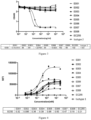
- the titer of Alpaca reached a high level (>512000) after six immunizations.
- a and B in Figure 1 show the binding titers of Alpaca serum before and after immunization with EGFR.His and EGFR-V3-his tag antigens respectively.
- VHH sequences with high binding activity to EGFR were selected and constructed on pCDNA3.4-IgG4 vector to form VHH-IgG4, and then expressed by ExpiCHO TM (Thermo Fisher) expression system. After one week of expression, the supernatant was collected for protein A (GE) purification. Then Nanodrop was used to detect the protein quality, and HPLC was used to detect the protein purity. The purity and yield of the obtained protein met the requirment of subsequent tests.
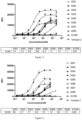
- EGFR his tag antigen EGFR-V3-his tag antigen protein was diluted with PBS to 1 ⁇ g/mL, EGFR-D1D2 (L25-A310, P00533-1 (uniprot) ), EGFR-D4 (F481-S645, P00533-1 (uniprot)) his tag antigen were diluted with PBS to 0.5 ⁇ g/mL, and each well of an ELISA plate was filled with 100 ⁇ L antigen and coated overnight at 4 °C.
- EGFR his tag protein was diluted with PBS to 2 ⁇ g/mL, and 100 ⁇ L antigen was coated overnight at 4 °C. The next day, the plates were washed twice with PBST (added with 0 .5 % TWEEN-20), then blocked with 3%bsa at 37°C for 1.5h, and then washed twice with PBST.
- Human EGF protein, mouse IgG2a FC tag (Aero, Cat#EGF-H525b, 100 ⁇ g) protein was diluted to 10 ⁇ g/ mL.
- the VHH-IgG4 antibody was diluted to 2000nM, and then 4-fold diluted for 8 gradients.
- the dose-response curve was made with Graphprism 8.0 software, and the IC50 was calculated.
- Isotype control 2 is the negative control, and its amino acid sequence is shown in SEQ ID NO:2 in CN106046152A .
- the results are shown in Figure 3 .
- (4) Cross-Species Reactivity Assay ELISA was used to identify the binding of VHH-IgG4 antibody to mouse EGFR, monkey EGFR and human EGFR protein extracellular segments.
- the detection steps were as follows: dilute mouse EGFR protein, his tag protein ( (Acro, Cat#EGR-C52H1), rhesus macaque EGFR protein, His Tag (Aero, Cat#EGR-C52H1 ) to 1 ⁇ g/mL, followed by 100 ⁇ L per well was added to 96 well enzyme labeled plate and coated overnight at 4 °C; The next day, the plates were washed twice with PBST (added with 0.5 % TWEEN-20), then blocked with 3% BSA at 37°C for 1.5h, and then washed twice with PBST.
- the VHH-IgG4 antibody was diluted to 50nM, add 100 ⁇ L to each well, incubate at 37 °C for 1H; The plates were then washed three times with PBST, and then 100 ⁇ L HRP labeled Goat anti human IgG Fc antibody (1:10000,Bethyl) and incubated at 37°Cfor another 40min. The plates were then washed four times with PBST according to 100 ⁇ L/ well, add TMB chromogenic solution, and react for 3 ⁇ 5min; According to 100 ⁇ L/ well, 3N hydrochloric acid was added to stop the reaction. After the reaction was terminated, OD450/650 was selected on the Tecan microplate reader for detection. The results are shown in Table 3 below.
- the isotype control 1 in the figure is a negative control, and its amino acid sequence is shown in SEQ ID NO. 2 in CN 106146653A .
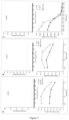
- SKOV3 and Aspc-1 tumor cells expressing EGFR were respectively plated in 96 well plates, with 3 ⁇ 10 5 cells per well, and then the tumor cells were incubated with gradient diluted VHH-IgG4 antibody. After incubation on ice for half an hour, anti human IgG PE (Jackson Immuno Research, Code: 109-117-008, Lot:145501) was added for incubation, and then CytoFLEX flow cytometry was used for detection. The EC50 of the antibody was calculated by fitting the curve. The results are shown in Figure 5 and Figure 6 , respectively.
- MPA Membrane Proteome Array
- This trial was mainly completed by the clinical trial testing center under Shanghai cell therapy group.
- the purpose of this study is to evaluate whether several preferred antibodies cross react with 12 or 34 kinds of normal human frozen tissues (each tissue is derived from 3 different individuals) by using the immunohistochemical staining method of streptavidin combined with biotin.
- the process is as follows: select 12 or 34 kinds of tissues to make frozen sections, dry them at room temperature and fix them with acetone. Blocking was performed using reagent A and reagent B of the endogenous biotin blocking Kit (Sangon, E674001).
- Biotin labeled antibody samples were incubated for 30min, and horseradish peroxidase labeled streptavidin (Abeam, ab7403) was added to incubate for 15min after washing. DAB color development, hematoxylin counterstaining, neutral plastic sealing, natural air drying and microscopic examination.
- the positive control was anti EGFR antibody (Biotin)(Abcam), and the negative control was biotin labeled IgG isotype control.
- Table 4 shows the results of tissue cross reaction between 9 EGFR targeting VHH-IgG4 antibodies and 12 normal human tissues.
- the 9 EGFR nanoantibodies and the purchased commercial EGFR antibodies have obvious localization and typical positive reaction on tonsil, squamous epithelium of cervix, skin and placenta.
- the cross reaction range of E008 antibody was relatively minimal and weak;
- other antibodies also cross react with liver, kidney, gallbladder and other tissues to varying degrees.
- E007 antibody has the widest and strongest tissue cross reaction.
- Table 5 shows the tissue cross reaction results of three EGFR targeting VHH-IgG4 antibodies with 32 normal human tissues. The results of cross reaction of three antibody tissues were almost the same, without much difference. The positive reaction was localized in endothelial cells or squamous epithelial cells with positive control antibody reaction. E008 showed less tissue cross reactivity than the other two antibodies.
- the CDR sequences of 9 VHH antibodies are shown in Table 6, and the amino acid sequences of 9 VHH antibodies and EC255 control antibody are shown in Table 7.
- Table 6 Summary of VHH antibody CDR sequences Antibody number CDR1 SEQ ID NO: CDR2 SEQ ID NO: CDR3 SEQ ID NO: E001 GSGFTIQA 4 IHQGGST 13 NWPPLRV 22 E002 GFTFSDYA 5 ITGTGETS 14 SIYHPPRDY 23 E003 GGVFSSYA 6 ITSGHST 15 NRSPYTPGN 24 E004 GFPFETYA 7 IFPDGKHT 16 AKDLPFFATPSFYIGY 25 E005 GFAFSDYT 8 ITSAGDII 17 NALVSASVRASDY 26 E006 GFTFDTYT 9 LATTGGK P 18 KATSRSPVNPHNY 27 E007 GFSFSDHT 10 ITPAGDII 19 NALVSAAVRASDY 28 E008 GLPLLDYA 11 ISISEGST 20 AADKTDIGFWLLVG
- the CDR region transplantation method was used to humanize the four VHH antibodies E002, E003, E005, E006, E008 that bind EGFR antigen.
- IgBLAST http://www.ncbi.nlm.nih.gov/igblast/
- Germline with high homology with E002, E003, E005, E006 and E008 was selected as the template.
- the modified antibody sequences of each antibody are shown in Table 8 below.
- the effect of the modified VHH antibody on human EGFR was determined by surface plasmon resonance (SPR) Binding kinetics and affinity of his antigen.
- Table 9 Summary of amino acid sequence of antibody after humanized transformation Antibody number Amino acid sequence SEQ ID NO: E002.1 40 E002.2 41 E002.3 42 E002.4 43 E002.5 44 E002.6 45 E002.7 46 E003.1 47 E003.2 48 E003.3 49 E003.4 50 E003.5 51 E003.6 52 E003.7 53 E003.8 54 E003.9 55 E003.10 56 E005.1 57 E005.2 58 E005.3 59 E005.4 60 E005.5 61 E005.6 62 E005.7 63 E005.8 64 E006.1 65 E006.2 66 E006.3 67 E006.4 68 E006.5 69 E006.6 70 E006.7 71 E006.8 72 E008.1 73 E008.2 74 E008.3 75 E008.4 76 E008.5 77 E008.6 78 E008.7 79 E008.8 80 E008.9 81 E008.10 82 Table 9: summary of protein affinity test results Number
Landscapes
- Health & Medical Sciences (AREA)
- Life Sciences & Earth Sciences (AREA)
- Chemical & Material Sciences (AREA)
- Engineering & Computer Science (AREA)
- Immunology (AREA)
- Molecular Biology (AREA)
- Biomedical Technology (AREA)
- General Health & Medical Sciences (AREA)
- Organic Chemistry (AREA)
- Hematology (AREA)
- Urology & Nephrology (AREA)
- Biochemistry (AREA)
- Medicinal Chemistry (AREA)
- Genetics & Genomics (AREA)
- Biotechnology (AREA)
- Cell Biology (AREA)
- Microbiology (AREA)
- Physics & Mathematics (AREA)
- Analytical Chemistry (AREA)
- Food Science & Technology (AREA)
- General Physics & Mathematics (AREA)
- Pathology (AREA)
- Proteomics, Peptides & Aminoacids (AREA)
- Biophysics (AREA)
- Bioinformatics & Cheminformatics (AREA)
- Wood Science & Technology (AREA)
- Zoology (AREA)
- General Engineering & Computer Science (AREA)
- General Chemical & Material Sciences (AREA)
- Chemical Kinetics & Catalysis (AREA)
- Public Health (AREA)
- Nuclear Medicine, Radiotherapy & Molecular Imaging (AREA)
- Pharmacology & Pharmacy (AREA)
- Animal Behavior & Ethology (AREA)
- Veterinary Medicine (AREA)
- Plant Pathology (AREA)
- Peptides Or Proteins (AREA)
- Micro-Organisms Or Cultivation Processes Thereof (AREA)
- Medicines Containing Antibodies Or Antigens For Use As Internal Diagnostic Agents (AREA)
- Hospice & Palliative Care (AREA)
Applications Claiming Priority (2)
| Application Number | Priority Date | Filing Date | Title |
|---|---|---|---|
| CN202110705939.8A CN115521377B (zh) | 2021-06-24 | 2021-06-24 | 人表皮生长因子受体结合分子及其应用 |
| PCT/CN2022/100888 WO2022268178A1 (fr) | 2021-06-24 | 2022-06-23 | Molécule de liaison au récepteur du facteur de croissance épidermique humain et son utilisation |
Publications (2)
| Publication Number | Publication Date |
|---|---|
| EP4403571A1 true EP4403571A1 (fr) | 2024-07-24 |
| EP4403571A4 EP4403571A4 (fr) | 2026-01-07 |
Family
ID=84544126
Family Applications (1)
| Application Number | Title | Priority Date | Filing Date |
|---|---|---|---|
| EP22827670.5A Pending EP4403571A4 (fr) | 2021-06-24 | 2022-06-23 | Molécule de liaison au récepteur du facteur de croissance épidermique humain et son utilisation |
Country Status (7)
| Country | Link |
|---|---|
| US (1) | US20250215092A1 (fr) |
| EP (1) | EP4403571A4 (fr) |
| JP (1) | JP2024547016A (fr) |
| CN (2) | CN115521377B (fr) |
| AU (1) | AU2022298843A1 (fr) |
| CA (1) | CA3241487A1 (fr) |
| WO (1) | WO2022268178A1 (fr) |
Families Citing this family (1)
| Publication number | Priority date | Publication date | Assignee | Title |
|---|---|---|---|---|
| CN117659194B (zh) * | 2023-11-30 | 2024-08-13 | 科弈(浙江)药业科技有限公司 | 靶向egfr的纳米抗体、药物偶联物及其用途 |
Family Cites Families (10)
| Publication number | Priority date | Publication date | Assignee | Title |
|---|---|---|---|---|
| PT1687338E (pt) * | 2003-11-07 | 2011-01-20 | Ablynx Nv | Anticorpos de domínio único vhh de camelídeos direccionados para o receptor do factor de crescimento epidérmico e suas utilizações |
| WO2007042289A2 (fr) * | 2005-10-11 | 2007-04-19 | Ablynx N.V. | Nanobodies™ et polypeptides diriges contre l'egfr et l'igf-1r |
| CA2588106A1 (fr) * | 2007-05-18 | 2008-11-18 | Nrc - Institute For Biological Sciences | Anticorps a domaine unique et anticorps a chaines lourdes ciblant le recepteur du facteur de croissance epidermique et utilisations connexes |
| US20090269343A1 (en) * | 2008-04-11 | 2009-10-29 | Duke University | Dual Specific Immunotoxin for Brain Tumor Therapy |
| WO2015177349A1 (fr) * | 2014-05-23 | 2015-11-26 | INSERM (Institut National de la Santé et de la Recherche Médicale) | Anticorps à domaine conformationnel unique dirigés contre egfr et utilisations correspondantes |
| CN106046152A (zh) | 2016-07-07 | 2016-10-26 | 南昌大学 | 特异性识别组氨酸标签的纳米抗体 |
| CN106146653A (zh) | 2016-07-07 | 2016-11-23 | 南昌大学 | 针对组氨酸标签的单域重链抗体 |
| CN112552402B (zh) * | 2018-09-27 | 2022-08-12 | 暨南大学 | 一种抗人egfr的纳米抗体与应用 |
| CN110964110B (zh) * | 2019-12-24 | 2021-09-03 | 北京纽安博生物技术有限公司 | 抗EGFR人源化的单域抗体、Fc融合蛋白、重链Fab蛋白及其应用 |
| CN111171151B (zh) * | 2019-12-26 | 2021-09-14 | 佛山汉腾生物科技有限公司 | 抗egfr纳米抗体及其应用 |
-
2021
- 2021-06-24 CN CN202110705939.8A patent/CN115521377B/zh active Active
- 2021-06-24 CN CN202410818362.5A patent/CN118562006A/zh active Pending
-
2022
- 2022-06-23 EP EP22827670.5A patent/EP4403571A4/fr active Pending
- 2022-06-23 CA CA3241487A patent/CA3241487A1/fr active Pending
- 2022-06-23 US US18/709,212 patent/US20250215092A1/en active Pending
- 2022-06-23 WO PCT/CN2022/100888 patent/WO2022268178A1/fr not_active Ceased
- 2022-06-23 AU AU2022298843A patent/AU2022298843A1/en active Pending
- 2022-06-23 JP JP2024536307A patent/JP2024547016A/ja active Pending
Also Published As
| Publication number | Publication date |
|---|---|
| CN115521377A (zh) | 2022-12-27 |
| JP2024547016A (ja) | 2024-12-26 |
| WO2022268178A1 (fr) | 2022-12-29 |
| CA3241487A1 (fr) | 2022-12-29 |
| CN118562006A (zh) | 2024-08-30 |
| AU2022298843A1 (en) | 2024-07-25 |
| CN115521377B (zh) | 2024-08-09 |
| US20250215092A1 (en) | 2025-07-03 |
| EP4403571A4 (fr) | 2026-01-07 |
Similar Documents
| Publication | Publication Date | Title |
|---|---|---|
| CN114685655B (zh) | Pd-1结合分子及其应用 | |
| WO2019141268A1 (fr) | Anticorps anti-4-1bb, fragment de liaison à l'antigène de celui-ci et utilisation médicale associée | |
| CN116554317B (zh) | Muc1结合分子及其应用 | |
| EP4269442A1 (fr) | Molécule de liaison à la mésothéline et son utilisation | |
| CN115785268A (zh) | 抗cd47抗体及其用途 | |
| US11685778B2 (en) | Anti-human LAG-3 monoclonal antibody and use thereof | |
| CN114685666B (zh) | 抗间皮素纳米抗体及其应用 | |
| EP4410834A1 (fr) | Molécule de liaison à ctla-4 et son utilisation | |
| CN114685667B (zh) | 间皮素结合分子及其应用 | |
| US20250215092A1 (en) | Human Epidermal Growth Factor Receptor Binding Molecule and Use Thereof | |
| CN121005782A (zh) | ActRIIB结合分子及其应用 | |
| WO2025228370A1 (fr) | Protéine de liaison à l'antigène neutralisant le virus respiratoire syncytial et son utilisation | |
| WO2024222409A1 (fr) | Anticorps du récepteur de la prolactine et son utilisation | |
| CN121181695A (zh) | ActRIIB抗原结合蛋白及其应用 | |
| CN119431584A (zh) | Muc1结合分子及其应用 | |
| CN119638839A (zh) | 间皮素结合分子及其应用 | |
| CN117843793A (zh) | 抗间皮素抗体、抗原结合片段及其用途 |
Legal Events
| Date | Code | Title | Description |
|---|---|---|---|
| STAA | Information on the status of an ep patent application or granted ep patent |
Free format text: STATUS: THE INTERNATIONAL PUBLICATION HAS BEEN MADE |
|
| PUAI | Public reference made under article 153(3) epc to a published international application that has entered the european phase |
Free format text: ORIGINAL CODE: 0009012 |
|
| STAA | Information on the status of an ep patent application or granted ep patent |
Free format text: STATUS: REQUEST FOR EXAMINATION WAS MADE |
|
| 17P | Request for examination filed |
Effective date: 20240422 |
|
| AK | Designated contracting states |
Kind code of ref document: A1 Designated state(s): AL AT BE BG CH CY CZ DE DK EE ES FI FR GB GR HR HU IE IS IT LI LT LU LV MC MK MT NL NO PL PT RO RS SE SI SK SM TR |
|
| DAV | Request for validation of the european patent (deleted) | ||
| DAX | Request for extension of the european patent (deleted) | ||
| RIC1 | Information provided on ipc code assigned before grant |
Ipc: C07K 16/28 20060101AFI20250915BHEP Ipc: C12N 15/13 20060101ALI20250915BHEP Ipc: C12N 15/63 20060101ALI20250915BHEP Ipc: C12N 5/10 20060101ALI20250915BHEP Ipc: A61K 39/395 20060101ALI20250915BHEP Ipc: A61P 35/00 20060101ALI20250915BHEP Ipc: G01N 33/53 20060101ALI20250915BHEP |
|
| A4 | Supplementary search report drawn up and despatched |
Effective date: 20251210 |
|
| RIC1 | Information provided on ipc code assigned before grant |
Ipc: C07K 16/28 20060101AFI20251204BHEP Ipc: C12N 15/13 20060101ALI20251204BHEP Ipc: C12N 15/63 20060101ALI20251204BHEP Ipc: C12N 5/10 20060101ALI20251204BHEP Ipc: A61K 39/395 20060101ALI20251204BHEP Ipc: A61P 35/00 20060101ALI20251204BHEP Ipc: G01N 33/53 20060101ALI20251204BHEP |