EP4407319A1 - Système pour déterminer une concentration de fibrinogène fonctionnel - Google Patents
Système pour déterminer une concentration de fibrinogène fonctionnel Download PDFInfo
- Publication number
- EP4407319A1 EP4407319A1 EP23153713.5A EP23153713A EP4407319A1 EP 4407319 A1 EP4407319 A1 EP 4407319A1 EP 23153713 A EP23153713 A EP 23153713A EP 4407319 A1 EP4407319 A1 EP 4407319A1
- Authority
- EP
- European Patent Office
- Prior art keywords
- features
- signal
- training
- blood
- plasma sample
- Prior art date
- Legal status (The legal status is an assumption and is not a legal conclusion. Google has not performed a legal analysis and makes no representation as to the accuracy of the status listed.)
- Withdrawn
Links
Images




Classifications
-
- G—PHYSICS
- G01—MEASURING; TESTING
- G01N—INVESTIGATING OR ANALYSING MATERIALS BY DETERMINING THEIR CHEMICAL OR PHYSICAL PROPERTIES
- G01N33/00—Investigating or analysing materials by specific methods not covered by groups G01N1/00 - G01N31/00
- G01N33/48—Biological material, e.g. blood, urine; Haemocytometers
- G01N33/50—Chemical analysis of biological material, e.g. blood, urine; Testing involving biospecific ligand binding methods; Immunological testing
- G01N33/86—Chemical analysis of biological material, e.g. blood, urine; Testing involving biospecific ligand binding methods; Immunological testing involving blood coagulating time or factors, or their receptors
-
- G—PHYSICS
- G01—MEASURING; TESTING
- G01N—INVESTIGATING OR ANALYSING MATERIALS BY DETERMINING THEIR CHEMICAL OR PHYSICAL PROPERTIES
- G01N2333/00—Assays involving biological materials from specific organisms or of a specific nature
- G01N2333/435—Assays involving biological materials from specific organisms or of a specific nature from animals; from humans
- G01N2333/745—Assays involving non-enzymic blood coagulation factors
- G01N2333/75—Fibrin; Fibrinogen
Definitions
- the invention relates to systems for determining a functional fibrinogen concentration from a patient's coagulating blood or blood plasma sample and for training a supervised learning model.
- the invention further relates to computer-implemented methods of determining a functional fibrinogen concentration from a patient's coagulating blood or blood plasma sample and of training a supervised learning model.
- the invention also relates to one or more computer program products enabling a computer system to perform such methods.
- the current gold (clinical) standard for the quantitative measurement of the fibrinogen concentration is the Clauss assay: a central laboratory test that is based on the clotting time of a test plasma sample when triggered with an excess of thrombin. To derive the fibrinogen concentration in the test plasma, its clotting time is compared to the clotting times of a series of reference samples with increasing, known fibrinogen concentrations.
- the Clauss method is precise and robust in the sense that it is insensitive to many inhibiting substances (such as anticoagulants and fibrin degradation products). PoC Clauss tests are not available.
- PT Prothrombin Time
- the PT derived Fibrinogen assay is widely used as it comes 'for free' with each PT measurement; it is a second analysis of the transparency over time waveform that is produced by the assay (the first is the determination of the PT; the time at which the plasma sample has reached a predefined transparency).
- the PT test (together with its standardized form, the International Normalized Ratio (INR)) is the second most performed in vitro diagnostic test worldwide (after glucose) and is also available as a PoC test.
- ILR International Normalized Ratio
- the PT derived Fibrinogen assay in its current form is not considered to be accurate. Especially samples from patients taking anticoagulants are known to give erroneous results, but samples from patients with a prolonged PT in general, or with high fibrinogen levels, as well. Thus, the PT derived Fibrinogen assay is not sufficiently accurate and the Clauss assay cannot be used as PoC test.
- a system for determining a functional fibrinogen concentration from a patient's coagulating blood or blood plasma sample comprises at least one processor configured to obtain a signal representing physical or biochemical properties of the developing fibrin network during coagulation of the patient's blood or blood plasma sample over time, the signal being determined from the patient's coagulating blood or blood plasma sample, determine the functional fibrinogen concentration based on one or more values of one or more features of the signal with the help of a learned function which maps values of the one or more features to functional fibrinogen concentrations, and output the functional fibrinogen concentration.
- the one or more features include at least one feature selected from a first set of features and/or multiple features selected from a larger set of features, the larger set of features comprising the first set of features and a second set of features, the first set of features including a feature related to a maximum value of the gradient of the signal and a feature related to the maximum of the second derivative of the signal, and the second set of features including a feature related to a difference between the maximum and the minimum of the signal and a feature related to the minimum of the second derivative of the signal.
- a functional fibrinogen concentration with sufficient accuracy is obtained by not (only) analyzing the change in optical density, but by analyzing one or more other features of the full clotting signal (e.g. as measured by the transparency of the plasma sample over time) that is produced by the assay.
- the signal may, for example, represent transparency of the patient's coagulating blood or blood plasma sample over time, dielectric impedance of the patient's coagulating blood or blood plasma sample over time, or viscoelastic properties of the patient's coagulating blood or blood plasma sample over time, or a combination thereof.
- the system may comprise a measurement component configured to receive the patient's coagulating blood or blood plasma sample and obtain the signal by performing measurements on the patient's coagulating blood or blood plasma sample.
- the system may be a handheld device, for example.
- the at least one processor may be configured to use a trained supervised learning model to determine the functional fibrinogen concentration based on the one or more values of the one or more features of the signal with the help of the learned function, the trained supervised learning model having learned the function based on a set of training examples.
- a trained supervised learning model may be used to determine the functional fibrinogen concentration based on the one or more values of the one or more features of the signal with the help of the learned function, the trained supervised learning model having learned the function based on a set of training examples.
- the supervised learning model may comprise a neural network and/or a linear regression model, for example.
- Each of the training examples may comprise one or more values of the one or more features determined from a training signal and a measured fibrinogen concentration associated with the training signal.
- the one or more values of the one or more features of the signal, determined from the patient's coagulating blood or blood plasma sample, may then be input into the trained supervised learning model.
- each of the training examples may comprise a training signal and a measured fibrinogen concentration associated with the training signal.
- the signal determined from the patient's coagulating blood or blood plasma sample, may then be input into the trained supervised learning model.
- the supervised learning model does not need to be trained to determine the one or more values of the one or more features and as a result, training the supervised learning model requires less training examples.
- Self-supervised learning methods may be used in conjunction with conventional supervised, e.g. deep, learning methods.
- the supervised learning model (or part of the model) may be pre-trained on a task that does not require annotation and the pre-training does in itself not produce a model that solves the task at hand.
- self-supervised learning may be used to learn the one or more features to be extracted from the signal.
- the at least one processor may be configured to determine the functional fibrinogen concentration by using a first trained supervised learning model to determine a first functional fibrinogen concentration based on the one or more values of the one or more features of the signal, using a second trained supervised learning model to determine a second functional fibrinogen concentration based on the one or more values of the one or more features of the signal, and selecting the first functional fibrinogen concentration or the second functional fibrinogen concentration in dependence on the first functional fibrinogen concentration.
- the first trained supervised learning model may comprise a neural network and the second supervised learning model may comprise a linear regression model, for example.
- the neural network concentration may be selected by default except when this concentration exceeds a certain threshold, e.g. 6 g/L, in which case the linear regression concentration is selected instead. This may result in more accurate fibrinogen concentration determination.
- the at least one processor may be configured to determine a prothrombin-time value related to the prothrombin time of the patient's coagulating blood or blood plasma sample and output a warning along with the determined functional fibrinogen concentration or an error instead of a functional fibrinogen concentration in dependency of the prothrombin-time value.
- the correlation of the functional fibrinogen concentrations determined in the manner described above with functional fibrinogen concentrations determined with the Clauss test was found to be weak for certain samples.
- a computer-implemented method of training a supervised learning model comprises obtaining a set of multiple training examples, each of the training examples comprising a fibrinogen concentration measured from a training coagulating blood or blood plasma sample and at least one of a training signal and one or more values of one or more features of the training signal, the training signal representing physical or biochemical properties of the developing fibrin network during coagulation of the training blood or blood plasma sample over time, the training signal being determined from the training coagulating blood or blood plasma sample, and providing the set of training examples to a supervised learning model to make the supervised learning model learn a function which maps values of the one or more features to functional fibrinogen concentrations.
- the one or more features include at least one feature selected from a first set of features and/or multiple features selected from a larger set of features, the larger set of features comprising the first set of features and a second set of features, the first set of features including a feature related to a maximum value of the gradient of the signal and a feature related to the maximum of the second derivative of the signal, and the second set of features including a feature related to a difference between the maximum and the minimum of the signal and a feature related to the minimum of the second derivative of the signal,.
- the method may be performed by software running on a programmable device. This software may be provided as a computer program product.
- the set of training examples may be obtained by obtaining a plurality of training signals and corresponding measured fibrinogen concentrations, analyzing each respective training signal of the training signals to determine the one or more values of the one or more features of the respective training signal from the respective training signal, and including, in the set of training examples, for each respective training signal of the training signals, a training example comprising the one or more values of the one or more features of the respective training signal and the measured fibrinogen concentration associated with the respective training signal.
- the supervised learning model may comprise a neural network and/or a linear regression model, for example.
- the method may comprise determining, for each training example of the set of training examples, a prothrombin-time value related to the prothrombin time of the training coagulating blood or blood plasma sample, and omitting each training example with a prothrombin-time value exceeding a threshold from the set of training examples.
- a prothrombin-time value related to the prothrombin time of the training coagulating blood or blood plasma sample.
- a system for training a supervised learning model comprises at least one processor configured to obtain a set of multiple training examples, each of the training examples comprising a fibrinogen concentration measured from a training coagulating blood or blood plasma sample and at least one of a training signal and one or more values of one or more features of the training signal, the training signal representing physical or biochemical properties of the developing fibrin network during coagulation of the training blood or blood plasma sample over time, the training signal being determined from the training coagulating blood or blood plasma sample, and providing the set of training examples to a supervised learning model to make the supervised learning model learn a function which maps values of the one or more features to functional fibrinogen concentrations.
- the one or more features include at least one feature selected from a first set of features and/or multiple features selected from a larger set of features, the larger set of features comprising the first set of features and a second set of features, the first set of features including a feature related to a maximum value of the gradient of the signal and a feature related to the maximum of the second derivative of the signal, and the second set of features including a feature related to a difference between the maximum and the minimum of the signal and a feature related to the minimum of the second derivative of the signal.
- a computer-implemented method of determining a functional fibrinogen concentration from a patient's coagulating blood or blood plasma sample comprises obtaining a signal representing physical or biochemical properties of the developing fibrin network during coagulation of the patient's blood or blood plasma sample over time, the signal being determined from the patient's coagulating blood or blood plasma sample, determining the functional fibrinogen concentration based on one or more values of one or more features of the signal with the help of a learned function which maps values of the one or more features to functional fibrinogen concentrations, and outputting the functional fibrinogen concentration.
- the one or more features include at least one feature selected from a first set of features and/or multiple features selected from a larger set of features, the larger set of features comprising the first set of features and a second set of features, the first set of features including a feature related to a maximum value of the gradient of the signal and a feature related to the maximum of the second derivative of the signal, and the second set of features including a feature related to a difference between the maximum and the minimum of the signal and a feature related to the minimum of the second derivative of the signal.
- the method may be performed by software running on a programmable device. This software may be provided as a computer program product.
- a computer program for carrying out the methods described herein, as well as a non-transitory computer readable storage-medium storing the computer program are provided.
- a computer program may, for example, be downloaded by or uploaded to an existing device or be stored upon manufacturing of these systems.
- a non-transitory computer-readable storage medium stores at least a first software code portion, the first software code portion, when executed or processed by a computer, being configured to perform executable operations for training a supervised learning model.
- These executable operations comprise obtaining a set of multiple training examples, each of the training examples comprising a fibrinogen concentration measured from a training coagulating blood or blood plasma sample and at least one of a training signal and one or more values of one or more features of the training signal, the training signal representing physical or biochemical properties of the developing fibrin network during coagulation of the training blood or blood plasma sample over time, the training signal being determined from the training coagulating blood or blood plasma sample, and providing the set of training examples to a supervised learning model to make the supervised learning model learn a function which maps values of the one or more features to functional fibrinogen concentrations.
- the one or more features include at least one feature selected from a first set of features and/or multiple features selected from a larger set of features, the larger set of features comprising the first set of features and a second set of features, the first set of features including a feature related to a maximum value of the gradient of the signal and a feature related to the maximum of the second derivative of the signal, and the second set of features including a feature related to a difference between the maximum and the minimum of the signal and a feature related to the minimum of the second derivative of the signal.
- a non-transitory computer-readable storage medium stores at least a second software code portion, the second software code portion, when executed or processed by a computer, being configured to perform executable operations for determining a functional fibrinogen concentration from a patient's coagulating blood or blood plasma sample.
- These executable operations comprise obtaining a signal representing physical or biochemical properties of the developing fibrin network during coagulation of the patient's blood or blood plasma sample over time, the signal being determined from the patient's coagulating blood or blood plasma sample, determining the functional fibrinogen concentration based on one or more values of one or more features of the signal with the help of a learned function which maps values of the one or more features to functional fibrinogen concentrations, and outputting the functional fibrinogen concentration.
- the one or more features include at least one feature selected from a first set of features and/or multiple features selected from a larger set of features, the larger set of features comprising the first set of features and a second set of features, the first set of features including a feature related to a maximum value of the gradient of the signal and a feature related to the maximum of the second derivative of the signal, and the second set of features including a feature related to a difference between the maximum and the minimum of the signal and a feature related to the minimum of the second derivative of the signal.
- aspects of the present invention may be embodied as a device, a method or a computer program product. Accordingly, aspects of the present invention may take the form of an entirely hardware embodiment, an entirely software embodiment (including firmware, resident software, micro-code, etc.) or an embodiment combining software and hardware aspects that may all generally be referred to herein as a "circuit", "module” or “system.” Functions described in this disclosure may be implemented as an algorithm executed by a processor/microprocessor of a computer. Furthermore, aspects of the present invention may take the form of a computer program product embodied in one or more computer readable medium(s) having computer readable program code embodied, e.g., stored, thereon.
- the computer readable medium may be a computer readable signal medium or a computer readable storage medium.
- a computer readable storage medium may be, for example, but not limited to, an electronic, magnetic, optical, electromagnetic, infrared, or semiconductor system, apparatus, or device, or any suitable combination of the foregoing.
- a computer readable storage medium may include, but are not limited to, the following: an electrical connection having one or more wires, a portable computer diskette, a hard disk, a random access memory (RAM), a read-only memory (ROM), an erasable programmable read-only memory (EPROM or Flash memory), an optical fiber, a portable compact disc read-only memory (CD-ROM), an optical storage device, a magnetic storage device, or any suitable combination of the foregoing.
- a computer readable storage medium may be any tangible medium that can contain, or store, a program for use by or in connection with an instruction execution system, apparatus, or device.
- a computer readable signal medium may include a propagated data signal with computer readable program code embodied therein, for example, in baseband or as part of a carrier wave. Such a propagated signal may take any of a variety of forms, including, but not limited to, electro-magnetic, optical, or any suitable combination thereof.
- a computer readable signal medium may be any computer readable medium that is not a computer readable storage medium and that can communicate, propagate, or transport a program for use by or in connection with an instruction execution system, apparatus, or device.
- Program code embodied on a computer readable medium may be transmitted using any appropriate medium, including but not limited to wireless, wireline, optical fiber, cable, RF, etc., or any suitable combination of the foregoing.
- Computer program code for carrying out operations for aspects of the present invention may be written in any combination of one or more programming languages, including an object oriented programming language such as Java(TM), Smalltalk, C++ or the like and conventional procedural programming languages, such as the "C" programming language or similar programming languages.
- the program code may execute entirely on the user's computer, partly on the user's computer, as a stand-alone software package, partly on the user's computer and partly on a remote computer, or entirely on the remote computer or server.
- the remote computer may be connected to the user's computer through any type of network, including a local area network (LAN) or a wide area network (WAN), or the connection may be made to an external computer (for example, through the Internet using an Internet Service Provider).
- LAN local area network
- WAN wide area network
- Internet Service Provider an Internet Service Provider
- These computer program instructions may be provided to a processor, in particular a microprocessor or a central processing unit (CPU), of a general purpose computer, special purpose computer, or other programmable data processing apparatus to produce a machine, such that the instructions, which execute via the processor of the computer, other programmable data processing apparatus, or other devices create means for implementing the functions/acts specified in the flowchart and/or block diagram block or blocks.
- a processor in particular a microprocessor or a central processing unit (CPU), of a general purpose computer, special purpose computer, or other programmable data processing apparatus to produce a machine, such that the instructions, which execute via the processor of the computer, other programmable data processing apparatus, or other devices create means for implementing the functions/acts specified in the flowchart and/or block diagram block or blocks.
- These computer program instructions may also be stored in a computer readable medium that can direct a computer, other programmable data processing apparatus, or other devices to function in a particular manner, such that the instructions stored in the computer readable medium produce an article of manufacture including instructions which implement the function/act specified in the flowchart and/or block diagram block or blocks.
- the computer program instructions may also be loaded onto a computer, other programmable data processing apparatus, or other devices to cause a series of operational steps to be performed on the computer, other programmable apparatus or other devices to produce a computer implemented process such that the instructions which execute on the computer or other programmable apparatus provide processes for implementing the functions/acts specified in the flowchart and/or block diagram block or blocks.
- each block in the flowchart or block diagrams may represent a module, segment, or portion of code, which comprises one or more executable instructions for implementing the specified logical function(s).
- Fig. 1 shows an embodiment of the system for determining a functional fibrinogen concentration from a patient's coagulating blood or blood plasma sample 11.
- the system is a handheld device 1.
- the handheld device 1 comprises a measurement component 2, a processor 5, a memory 7, and a display 9.
- the processor 5 is configured to obtain a signal representing physical or biochemical properties of the developing fibrin network during coagulation of the patient's blood or blood plasma sample over time.
- the handheld device 1 comprises a measurement component 2 configured to receive the patient's coagulating blood or blood plasma sample 11 and obtain the signal by performing measurements on the patient's coagulating blood or blood plasma sample 11.
- the measurements are not performed by the device that determines the functional fibrinogen concentration but by another device.
- the processor 5 is further configured to determine the functional fibrinogen concentration based on one or more values of one or more features of the signal with the help of a learned function which maps values of the one or more features to functional fibrinogen concentrations.
- the one or more features include at least one feature selected from a first set of features and/or multiple features selected from a larger set of features.
- the larger set of features comprises the first set of features and a second set of features.
- the first set of features includes a feature b) related to a maximum value of the gradient of the signal and a feature d) related to the maximum of the second derivative of the signal.
- the second set of features includes a feature a) related to a difference between the maximum and the minimum of the signal and a feature e) related to the minimum of the second derivative of the signal.
- the one or more features may also include other features, e.g. features c), f), and g). These features are shown in Table 1 below along with examples of these features.
- the processor 5 is further configured to output the functional fibrinogen concentration, e.g. via the display 9.
- the above-mentioned learned function is learned on a (e.g. desktop) computer 31.
- the computer 31 comprises a processor 35, a receiver 33, a transmitter 34, and a memory 37.
- the learned function is transferred from the memory 37 of the computer 31 to the memory 7 of the handheld device 1.
- the processor 35 may, for example, be configured to perform steps 121 and 123 of Fig. 4 .
- the above-mentioned signal may represent transparency of the patient's coagulating blood or blood plasma sample over time and/or dielectric impedance of the patient's coagulating blood or blood plasma sample over time and/or viscoelastic properties of the patient's coagulating blood or blood plasma sample over time, for example.
- Fig. 2 shows example of features of a first type of signal, a signal that represent transparency of the patient's coagulating blood or blood plasma sample over time.
- Fig. 2 shows a visualization of transparency signal features, i.e. characteristics of the transparency over time waveform. These characteristics, such as maximum transparency and maximum gradient of the transparency over time, are measured for a specific blood sample and may be used as input to a numerical algorithm that subsequently provides an estimation for the fibrinogen concentration in the sample.
- the numerical algorithm may be derived from existing data analysis approaches, such as neural network training and linear regression, applied to combinations of measured waveforms and fibrinogen concentrations (e.g., measured via the Clauss method).
- Fig. 2 shows the transparency signal, the transparency gradient (the 1 st derivative of transparency), and the 2 nd derivative of transparency.
- Feature 21 is determined from the transparency signal.
- Feature 21, minimum transparency is an example of feature a) of Table 1. Since the maximum transparency is always 100%, the minimum transparency relates to the difference between the maximum and the minimum of the signal.
- Features 22 and 23 are determined from the transparency gradient.
- Feature 22 maximum absolute transparency gradient, is an example of feature b) of Table 1.
- Feature 23 time of maximum absolute transparency gradient, is an example of feature c) of Table 1.
- Features 24-27 are determined from the 2 nd derivative of transparency.
- Feature 24, maximum 2 nd derivative of transparency, is an example of feature d) of Table 1.
- Feature 25 time of maximum of 2 nd derivative of transparency, is an example of feature f) of Table 1.
- Feature 26 time of minimum of 2 nd derivative of transparency, is an example of feature g of Table 1.
- the handheld device 1 comprises one processor 5.
- the handheld device 1 comprises multiple processors.
- the processor 5 of the handheld device 1 may be a general-purpose processor, e.g. from e.g. from ARM or Qualcomm, or an application-specific processor.
- the processor 5 of the handheld device 1 may run a Unix-based operating system for example.
- the memory 7 may comprise one or more memory units.
- the memory 7 may comprise solid-state memory, for example.
- the handheld device 1 may comprise other components typical for a handheld device such as a battery.
- the computer 31 comprises one processor 35.
- the computer 31 comprises multiple processors.
- the processor 35 of the computer 31 may be a general-purpose processor, e.g. from Intel or AMD, or an application-specific processor.
- the processor 35 of the computer 31 may run a Windows or Unix-based operating system for example.
- the memory 37 may comprise one or more memory units.
- the memory 37 may comprise one or more hard disks and/or solid-state memory, for example.
- the memory 37 may be used to store an operating system, applications and application data, for example.
- the receiver 33 and the transmitter 34 may use one or more wired and/or wireless communication technologies such as Ethernet and/or Wi-Fi (IEEE 802.11), for example.
- wired and/or wireless communication technologies such as Ethernet and/or Wi-Fi (IEEE 802.11), for example.
- multiple receivers and/or multiple transmitters are used instead of a single receiver and a single transmitter.
- a separate receiver and a separate transmitter are used.
- the receiver 33 and the transmitter 34 are combined into a transceiver.
- the computer 31 may comprise other components typical for a computer such as a power connector.
- the invention may be implemented using a computer program running on one or more processors.
- the system is a handheld device.
- the system is another type of device, e.g. a desktop computer.
- FIG. 3 A first embodiment of a computer-implemented method of determining a functional fibrinogen concentration from a patient's coagulating blood or blood plasma sample is shown in Fig. 3 .
- the method may be performed by the handheld device of Fig. 1 , for example.
- a step 101 comprises obtaining a signal representing physical or biochemical properties of the developing fibrin network during coagulation of the patient's blood or blood plasma sample over time.
- the signal is or has been determined from the patient's coagulating blood or blood plasma sample.
- a step 103 comprises determining the functional fibrinogen concentration based on one or more values of one or more features of the signal with the help of a learned function which maps values of the one or more features to functional fibrinogen concentrations.
- the one or more features include at least one feature selected from a first set of features and/or multiple features selected from a larger set of features.
- the larger set of features comprises the first set of features and a second set of features.
- the first set of features includes a feature b) related to a maximum value of the gradient of the signal and a feature d) related to the maximum of the second derivative of the signal.
- the second set of features includes a feature a) related to a difference between the maximum and the minimum of the signal and a feature e) related to the minimum of the second derivative of the signal.
- the one or more features may also include other features, e.g. features c), f), and g). These features are shown in Table 1.
- a step 105 comprises outputting the functional fibrinogen concentration, e.g. on a display. Additionally, one or more steps of one or more of the embodiments of Figs. 4-5 and 9-11 may be added to the embodiment of Fig. 3 .
- FIG. 4 A second embodiment of the computer-implemented method of determining a functional fibrinogen concentration from a patient's coagulating blood or blood plasma sample is shown in Fig. 4 .
- the second embodiment of Fig. 4 is an extension of the first embodiment of Fig. 3.
- Fig. 4 also shows a first embodiment of the computer-implemented method of training a supervised learning model.
- a step 121 comprises obtaining a set of multiple training examples.
- Each of the training examples comprises a fibrinogen concentration measured from a training coagulating blood or blood plasma sample (e.g. by using the Clauss test) and at least one of a training signal and one or more values of one or more features of the training signal.
- the training signal represents physical or biochemical properties of the developing fibrin network during coagulation of the training blood or blood plasma sample over time.
- the training signal is or has been determined from the training coagulating blood or blood plasma sample.
- the one or more features of the training signal are the same as the one or more features of the signal, which are used in step 103 for determining the functional fibrinogen concentration.
- a step 123 comprises providing the set of training examples obtained in step 121 to a supervised learning model to make the supervised learning model learn a function which maps values of the one or more features to functional fibrinogen concentrations.
- This supervised learning model is used in step 103.
- the supervised learning model may comprise a neural network and/or a linear regression model, for example.
- step 123 may comprise applying linear regression or neural network learning to the combination of some or all of the numerical features shown in Fig. 2 and the measured Fibrinogen concentrations. The chosen numerical algorithm and its optimized parameter set may then be stored.
- step 103 of Fig. 3 is implemented by a step 131.
- Step 131 comprises using the trained supervised learning model trained in step 123 to determine the functional fibrinogen concentration based on the one or more values of the one or more features of the signal, i.e. the new blood (plasma) sample, with the help of the learned function.
- Step 131 may comprise applying the chosen numerical algorithm and its optimized parameter set, as stored in step 123.
- Steps 121 and 123 which form the training phase, may be performed by the computer 31 of Fig. 1 , for example.
- Steps 101, 131, and 105 which form the inference phase, may be performed by the handheld device of Fig. 1 , for example.
- one or more steps of one or more of the embodiments of Figs. 5 and 8-11 may be added to the embodiment of Fig. 4 .
- no supervised learning model is used in the inference phase.
- a lookup table may be used in step 103.
- the learned function may be encoded in this lookup table.
- the values of the lookup table may be learned using a supervised learning model, e.g. a linear regression model.
- FIG. 5 A third embodiment of a computer-implemented method of determining a functional fibrinogen concentration from a patient's coagulating blood or blood plasma sample is shown in Fig. 5 .
- the third embodiment of Fig. 5 is an extension of the second embodiment of Fig. 4.
- Fig. 5 also shows a second embodiment of the computer-implemented method of training a supervised learning model.
- the second embodiment of Fig. 5 is an extension of the first embodiment of Fig. 4 .
- step 121 of Fig. 4 has been implemented by steps 151, 153, and 155
- step 123 of Fig. 4 has been implemented by a step 157
- step 131 of Fig. 4 has been implemented by a step 163
- a step 161 is performed between steps 101 and 163.
- a step 151 comprises obtaining a plurality of training signals and corresponding measured fibrinogen concentrations.
- a step 153 comprises analyzing each respective training signal of the training signals to determine the one or more values of the one or more features of the respective training signal from the respective training signal.
- step 151 may comprises collecting measured fibrinogen concentrations and transparency over time waveforms (in blood after activation by e.g., a Tissue Factor containing solution) and step 153 may comprise extracting the waveform features shown in Fig. 2 .
- a step 155 comprises including, in the set of training examples, for each respective training signal of the training signals, a training example comprising the one or more values of the one or more features of the respective training signal and the measured fibrinogen concentration associated with the respective training signal.
- Step 157 comprises providing the set of training examples obtained in step 155 to a supervised learning model to make the supervised learning model learn a function which maps values of the one or more features to functional fibrinogen concentrations.
- the supervised learning model may comprise a linear regression algorithm applied to features a), b), d), and e) of Table 1, for example.
- the following values may be learned/used in the linear regression algorithm:
- the supervised learning model may be a 3-layer neural network in which features a), b), d), and e) of Table 1 are used as inputs, for example.
- This neural network may comprise:
- the model inputs may be normalized to zero mean and unit standard deviation in step 153 or 155 and scaled by the same constants in step 161 or 163.
- the training may, for example, result in the following numerical values (weights and biases) of the trained neural network:
- the determination of the fibrinogen concentration may be further optimized by other choices for the neural network architecture and settings.
- Step 101 comprises obtaining a signal representing physical or biochemical properties of the developing fibrin network during coagulation of the patient's blood or blood plasma sample over time.
- Step 161 comprises determining one or more values of the one or more features of the signal obtained in step 101.
- the transparency over time waveform may be measured (in blood after activation by e.g., a Tissue Factor containing solution) for the new blood (plasma) sample in step 101 and the same waveform features extracted in step 153 may be extracted in step 161.
- Step 163 comprises inputting the one or more values determined in step 161 into the supervised learning model trained in step 157.
- step 161 comprises pre-processing the input for the trained supervised learning model.
- the output of the trained supervised learning model is the functional fibrinogen concentration.
- Steps 121 and 123 which form the training phase, may be performed by the computer 31 of Fig. 1 , for example.
- Steps 101, 161, 163, and 105 which form the inference phase, may be performed by the handheld device of Fig. 1 , for example.
- one or more steps of one or more of the embodiments of Figs. 8-11 may be added to the embodiment of Fig. 5 .
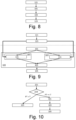
- Fig. 6 is a block diagram of the supervised learning model used in the embodiments of Fig. 5 .
- training example are fed to a supervised learning model 59.
- Each training example comprises an input-output pair.
- This input-output pair comprises training input 55 and corresponding training output 57.
- training signals 53 are analyzed in step 153 of Fig. 5 to determined training input 55.
- the training input 55 comprises the one or the one or more values of the one or more features of the training signals.
- the training output 57 comprises the corresponding measured fibrinogen concentrations 57.
- the training input 55 and the training output 57 are included in the training examples in step 155 of Fig. 5 .
- input 75 is fed into the supervised learning model 59, which then provides output 77 as a result.
- the input 75 comprises one or more values of the one or more features of a signal 73, as determined in step 161 of Fig. 5 , which are then input in the trained supervised learning model 59 in step 163 of Fig. 5 .
- the output 77 comprises the determined functional fibrogen concentration.
- Fig. 7 is a block diagram of a supervised learning model used in an alternative embodiment.
- the training signals 53 and the signal 73 are not pre-processed but fed/input directly into a supervised learning model 89.
- the training examples comprise the training signals 53 and the corresponding measured fibrinogen concentrations (training output 57).
- the output 77 comprises the determined functional fibrogen concentration.
- the supervised learning model 89 needs to be trained to determine the one or more values of the one or more features of the training signal 53 and of the signal 73.
- the training of supervised learning model 89 requires more training examples than the training of supervised learning model 59.
- FIG. 8 A third embodiment of the computer-implemented method of training a supervised learning model is shown in Fig. 8 .
- the method may be performed by the computer 31 of Fig. 1 , for example.
- the third embodiment of Fig. 8 is an extension of the first embodiment of Fig. 4 .
- steps 181 and 183 are performed between steps 121 and 123 of Fig. 4 .
- Step 181 comprises determining, for each training example of the set of training examples, a prothrombin-time value related to the prothrombin time of the training coagulating blood or blood plasma sample.
- Step 183 comprises omitting each training example with a prothrombin-time value exceeding a threshold, e.g. 60 seconds, from the set of training examples. Additionally, one or more steps of one or more of the embodiments of Figs. 5 and 9 may be added to the embodiment of Fig. 8 .
- FIG. 9 A fourth embodiment of a computer-implemented method of determining a functional fibrinogen concentration from a patient's coagulating blood or blood plasma sample is shown in Fig. 9 .
- the fourth embodiment of Fig. 9 is an extension of the second embodiment of Fig. 4 .
- Fig. 9 also shows a fourth embodiment of the computer-implemented method of training a supervised learning model, which is an extension of the first embodiment of Fig. 4 .
- step 103 of Fig. 4 is implemented by steps 201, 203, and 205.
- Step 201 comprises using a first trained supervised learning model to determine a first functional fibrinogen concentration based on one or more values of one or more features of the signal.
- Step 203 comprises using a second trained supervised learning model to determine a second functional fibrinogen concentration based on one or more values of one or more features of the signal.
- the values of the same set of features may be used by, e.g. input into, both supervised learning models or the values of a different (possibly overlapping) set of features may be used by, e.g. input into, the different supervised learning models.
- the first trained supervised learning model may comprise a neural network and the second supervised learning model may comprise a linear regression model, for example.
- Step 205 comprises selecting the first functional fibrinogen concentration determined in step 201 or the second functional fibrinogen concentration determined in step 203 in dependence on the first functional fibrinogen concentration.
- a neural network prediction may be selected by default, except when the prediction exceeds 6 g/L, in which case a linear regression prediction is selected instead.
- Steps 121 and 123 which form the training phase, may be performed by the computer 31 of Fig. 1 , for example.
- Steps 101, 201,203, 205, and 105 which form the inference phase, may be performed by the handheld device of Fig. 1 , for example.
- one or more steps of one or more of the embodiments of Figs. 5 , 8 , and 10-11 may be added to the embodiment of Fig. 9 .
- FIG. 10 A fifth embodiment of a computer-implemented method of determining a functional fibrinogen concentration from a patient's coagulating blood or blood plasma sample is shown in Fig. 10 .
- the fifth embodiment of Fig. 10 is an extension of the first embodiment of Fig. 3 .
- the method may be performed by the handheld device of Fig. 1 , for example.
- a step 221 comprises determining a prothrombin-time value related to the prothrombin time of the patient's coagulating blood or blood plasma sample.
- a step 223 comprises checking the prothrombin-time value determined in step 221 and performing step 225 or step 101 in dependency of the prothrombin-time value. For example, step 225 may be performed if the prothrombin-time value exceeds a threshold T, e.g. 60 seconds, and step 101 may be performed the prothrombin-time value does not exceed threshold T.
- a threshold T e.g. 60 seconds
- Step 225 comprises outputting an error instead of a functional fibrinogen concentration.
- Steps 101-105 have been described in relation to Fig. 3 .
- Step 101 comprises obtaining the signal.
- Step 103 comprises determining the functional fibrinogen concentration.
- Step 105 comprises outputting the functional fibrinogen concentration determined in step 103. Additionally, one or more steps of one or more of the embodiments of Figs. 5 and 9 may be added to the embodiment of Fig. 10 .
- FIG. 11 A sixth embodiment of a computer-implemented method of determining a functional fibrinogen concentration from a patient's coagulating blood or blood plasma sample is shown in Fig. 11 .
- the sixth embodiment of Fig. 11 is an extension of the first embodiment of Fig. 3 .
- the method may be performed by the handheld device of Fig. 1 , for example.
- Step 101 comprises obtaining a signal representing physical or biochemical properties of the developing fibrin network during coagulation of the patient's blood or blood plasma sample over time.
- Step 103 comprises determining the functional fibrinogen concentration based on one or more values of one or more features of the signal obtained in step 101 with the help of a learned function which maps values of the one or more features to functional fibrinogen concentrations.
- Steps 105 and 221 are performed after step 103 has been performed.
- Step 105 comprises outputting the functional fibrinogen concentration, e.g. on a display.
- Step 221 comprises determining a prothrombin-time value related to the prothrombin time of the patient's coagulating blood or blood plasma sample.
- step 223 comprises checking the prothrombin-time value determined in step 221 and performing a step 235 in dependency of the prothrombin-time value determined in step 221.
- step 235 may be performed if the prothrombin-time value exceeds a threshold T, e.g. 60 seconds.
- Step 235 comprises output a warning along with the determined functional fibrinogen concentration output in step 105.
- one or more steps of one or more of the embodiments of Figs. 5 and 9 may be added to the embodiment of Fig. 11 .
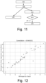
- Fig. 12 shows the correlations between measured fibrinogen concentrations and estimations by linear regression.
- Fig. 13 shows the correlations between measured fibrinogen concentrations and estimations by a neural network.
- Fig. 14 shows the correlations between measured fibrinogen concentrations and estimations by the combination of linear regression and a neural network.
- Figs. 12-14 plot the determined fibrinogen concentrations by linear regression or neural network versus the values as determined/measured with the Clauss method.
- the linear regression approach achieves a correlation coefficient R of 0.94, while the neural network approach achieves an R of 0.95 on the available data (43 samples).
- the combination of linear regression and a neural network achieves an R of 0.97.
- the evaluation is the result of K-fold cross-validation, where K is in this case equal to the total number of samples (43).
- Figs. 12-14 shows the results when features a), b), d), and e) of Table 1 are used as inputs.
- Table 2 shows the results when other features and other combinations of features are used with the afore-mentioned 43 training samples.
- Table 2 shows the correlation coefficients per combination of features for direct correlation between the (single) feature and Fibrinogen level, and between the output of linear regression / the neural network and Fibrinogen.
- the results indicate feature a (feature related to difference between the maximum and the minimum of the signal) to be the strongest feature, feature e (feature related to minimum of the second derivative of the signal) the least strong of the features a, b, d, and e, and features b and d (feature related to maximum value of the gradient of the signal and feature related to maximum of the second derivative of the signal) about equally strong.
- Table 2 Correlation coefficient between estimated and measured Fibrinogen level Features Direct correlation Correlation under linear regression Correlation of neural network output Correlation of combined model Feature a 0.92 0.90 0.63 0.63 Feature b 0.95 0.94 0.91 0.93 Feature c 0.03 Feature d 0.94 0.93 0.85 0.86 Feature e 0.86 0.84 0.77 0.81 Feature f 0.03 Feature g 0.03 a&b 0.96 0.93 0.96 a&d 0.96 0.96 0.97 a&e 0.97 0.93 0.95 b&d 0.94 0.92 0.92 b&e 0.96 0.87 0.90 d&e 0.96 0.87 0.91 a&b&d 0.96 0.95 0.97 a&b&e 0.95 0.94 0.96 a&d&e 0.95 0.93 0.95 b&d&e 0.95 0.89 0.92 a&b&d&e 0.94 0.95 0.97
- Table 3 shows the improved results when a sample with a PT value exceeding a threshold (the outlier) is omitted from the set of training examples, as described in relation to Fig. 8 .
- Table 3 Results with outlier removed Features Direct correlation Correlation under linear regression Correlation of neural network output Correlation of combined model Feature a 0.92 0.91 0.49 0.49 Feature b 0.98 0.98 0.92 0.94 Feature c 0.20 Feature d 0.98 0.98 0.92 0.93 Feature e 0.92 0.92 0.86 0.86 Feature f 0.19 Feature g 0.19 a & b 0.98 0.97 0.98 a & d 0.98 0.96 0.98 a & e 0.98 0.96 0.97 b & d 0.98 0.94 0.97 b & e 0.98 0.93 0.96 d & e 0.99 0.94 0.97 a & b & d 0.98 0.97 0.98 a & b & e 0.98 0.96 0.98 a & d
- Fig. 15 depicts a block diagram illustrating an exemplary data processing system that may perform the method as described with reference to Figs. 3-5 and 8-11 .
- the data processing system 300 may include at least one processor 302 coupled to memory elements 304 through a system bus 306. As such, the data processing system may store program code within memory elements 304. Further, the processor 302 may execute the program code accessed from the memory elements 304 via a system bus 306. In one aspect, the data processing system may be implemented as a computer that is suitable for storing and/or executing program code. It should be appreciated, however, that the data processing system 300 may be implemented in the form of any system including a processor and a memory that is capable of performing the functions described within this specification.
- the memory elements 304 may include one or more physical memory devices such as, for example, local memory 308 and one or more bulk storage devices 310.
- the local memory may refer to random access memory or other non-persistent memory device(s) generally used during actual execution of the program code.
- a bulk storage device may be implemented as a hard drive or other persistent data storage device.
- the processing system 300 may also include one or more cache memories (not shown) that provide temporary storage of at least some program code in order to reduce the number of times program code must be retrieved from the bulk storage device 310 during execution.
- I/O devices depicted as an input device 312 and an output device 314 optionally can be coupled to the data processing system.
- input devices may include, but are not limited to, a keyboard, a pointing device such as a mouse, or the like.
- output devices may include, but are not limited to, a monitor or a display, speakers, or the like.
- Input and/or output devices may be coupled to the data processing system either directly or through intervening I/O controllers.
- the input and the output devices may be implemented as a combined input/output device (illustrated in Fig. 15 with a dashed line surrounding the input device 312 and the output device 314).
- a combined device is a touch sensitive display, also sometimes referred to as a "touch screen display” or simply "touch screen”.
- input to the device may be provided by a movement of a physical object, such as e.g. a stylus or a finger of a user, on or near the touch screen display.
- a network adapter 316 may also be coupled to the data processing system to enable it to become coupled to other systems, computer systems, remote network devices, and/or remote storage devices through intervening private or public networks.
- the network adapter may comprise a data receiver for receiving data that is transmitted by said systems, devices and/or networks to the data processing system 300, and a data transmitter for transmitting data from the data processing system 300 to said systems, devices and/or networks.
- Modems, cable modems, and Ethernet cards are examples of different types of network adapter that may be used with the data processing system 300.
- the memory elements 304 may store an application 318.
- the application 318 may be stored in the local memory 308, he one or more bulk storage devices 310, or separate from the local memory and the bulk storage devices.
- the data processing system 300 may further execute an operating system (not shown in Fig. 15 ) that can facilitate execution of the application 318.
- the application 318 being implemented in the form of executable program code, can be executed by the data processing system 300, e.g., by the processor 302. Responsive to executing the application, the data processing system 300 may be configured to perform one or more operations or method steps described herein.
- Various embodiments of the invention may be implemented as a program product for use with a computer system, where the program(s) of the program product define functions of the embodiments (including the methods described herein).
- the program(s) can be contained on a variety of non-transitory computer-readable storage media, where, as used herein, the expression "non-transitory computer readable storage media" comprises all computer-readable media, with the sole exception being a transitory, propagating signal.
- the program(s) can be contained on a variety of transitory computer-readable storage media.
- Illustrative computer-readable storage media include, but are not limited to: (i) non-writable storage media (e.g., read-only memory devices within a computer such as CD-ROM disks readable by a CD-ROM drive, ROM chips or any type of solid-state non-volatile semiconductor memory) on which information is permanently stored; and (ii) writable storage media (e.g., flash memory, floppy disks within a diskette drive or hard-disk drive or any type of solid-state random-access semiconductor memory) on which alterable information is stored.
- the computer program may be run on the processor 302 described herein.
Landscapes
- Health & Medical Sciences (AREA)
- Life Sciences & Earth Sciences (AREA)
- Engineering & Computer Science (AREA)
- Hematology (AREA)
- Biomedical Technology (AREA)
- Chemical & Material Sciences (AREA)
- Immunology (AREA)
- Molecular Biology (AREA)
- Urology & Nephrology (AREA)
- Physics & Mathematics (AREA)
- General Health & Medical Sciences (AREA)
- General Physics & Mathematics (AREA)
- Food Science & Technology (AREA)
- Medicinal Chemistry (AREA)
- Analytical Chemistry (AREA)
- Biochemistry (AREA)
- Biotechnology (AREA)
- Pathology (AREA)
- Microbiology (AREA)
- Cell Biology (AREA)
- Investigating Or Analysing Biological Materials (AREA)
- Biophysics (AREA)
- Data Mining & Analysis (AREA)
- Medical Informatics (AREA)
- Public Health (AREA)
- Software Systems (AREA)
- Databases & Information Systems (AREA)
- Epidemiology (AREA)
- Evolutionary Computation (AREA)
- Bioethics (AREA)
- Artificial Intelligence (AREA)
- Computer Vision & Pattern Recognition (AREA)
- Bioinformatics & Cheminformatics (AREA)
- Bioinformatics & Computational Biology (AREA)
- Evolutionary Biology (AREA)
- Spectroscopy & Molecular Physics (AREA)
- Theoretical Computer Science (AREA)
- Ecology (AREA)
Priority Applications (4)
| Application Number | Priority Date | Filing Date | Title |
|---|---|---|---|
| EP23153713.5A EP4407319A1 (fr) | 2023-01-27 | 2023-01-27 | Système pour déterminer une concentration de fibrinogène fonctionnel |
| PCT/EP2024/051646 WO2024156749A1 (fr) | 2023-01-27 | 2024-01-24 | Système de détermination d'une concentration fonctionnelle de fibrinogène |
| EP24701928.4A EP4655596A1 (fr) | 2023-01-27 | 2024-01-24 | Système de détermination d'une concentration fonctionnelle de fibrinogène |
| US19/151,058 US20260118372A1 (en) | 2023-01-27 | 2024-01-24 | System for Determining a Functional Fibrinogen Concentration |
Applications Claiming Priority (1)
| Application Number | Priority Date | Filing Date | Title |
|---|---|---|---|
| EP23153713.5A EP4407319A1 (fr) | 2023-01-27 | 2023-01-27 | Système pour déterminer une concentration de fibrinogène fonctionnel |
Publications (1)
| Publication Number | Publication Date |
|---|---|
| EP4407319A1 true EP4407319A1 (fr) | 2024-07-31 |
Family
ID=85132843
Family Applications (2)
| Application Number | Title | Priority Date | Filing Date |
|---|---|---|---|
| EP23153713.5A Withdrawn EP4407319A1 (fr) | 2023-01-27 | 2023-01-27 | Système pour déterminer une concentration de fibrinogène fonctionnel |
| EP24701928.4A Pending EP4655596A1 (fr) | 2023-01-27 | 2024-01-24 | Système de détermination d'une concentration fonctionnelle de fibrinogène |
Family Applications After (1)
| Application Number | Title | Priority Date | Filing Date |
|---|---|---|---|
| EP24701928.4A Pending EP4655596A1 (fr) | 2023-01-27 | 2024-01-24 | Système de détermination d'une concentration fonctionnelle de fibrinogène |
Country Status (3)
| Country | Link |
|---|---|
| US (1) | US20260118372A1 (fr) |
| EP (2) | EP4407319A1 (fr) |
| WO (1) | WO2024156749A1 (fr) |
Citations (1)
| Publication number | Priority date | Publication date | Assignee | Title |
|---|---|---|---|---|
| EP3396384A1 (fr) * | 2017-04-24 | 2018-10-31 | Sysmex Corporation | Procédé et appareil pour déterminer un échantillon sanguin |
-
2023
- 2023-01-27 EP EP23153713.5A patent/EP4407319A1/fr not_active Withdrawn
-
2024
- 2024-01-24 WO PCT/EP2024/051646 patent/WO2024156749A1/fr not_active Ceased
- 2024-01-24 US US19/151,058 patent/US20260118372A1/en active Pending
- 2024-01-24 EP EP24701928.4A patent/EP4655596A1/fr active Pending
Patent Citations (1)
| Publication number | Priority date | Publication date | Assignee | Title |
|---|---|---|---|---|
| EP3396384A1 (fr) * | 2017-04-24 | 2018-10-31 | Sysmex Corporation | Procédé et appareil pour déterminer un échantillon sanguin |
Non-Patent Citations (3)
| Title |
|---|
| K DOUBLEDAY ET AL: "Clot Signature Curves and the ACL Advance TM", 1 November 2001 (2001-11-01), XP055661048, Retrieved from the Internet <URL:https://de.werfen.com/~/media/il%20shared/docs/hemostasis/literature/100%20%20%20clot%20signature%20curves%20and%20the%20acl%20advance.pdf> [retrieved on 20200123] * |
| P. O. SEVENET ET AL: "Clot waveform analysis: Where do we stand in 2017?", INTERNATIONAL JOURNAL OF LABORATORY HEMATOLOGY, vol. 39, no. 6, 6 September 2017 (2017-09-06), GB; US, pages 561 - 568, XP055708297, ISSN: 1751-5521, DOI: 10.1111/ijlh.12724 * |
| SUZUKI ATSUO ET AL: "Clot waveform analysis in Clauss fibrinogen assay contributes to classification of fibrinogen disorders", THROMBOSIS RESEARCH, vol. 174, 2018, pages 98 - 103, XP085587322, ISSN: 0049-3848, DOI: 10.1016/J.THROMRES.2018.12.018 * |
Also Published As
| Publication number | Publication date |
|---|---|
| US20260118372A1 (en) | 2026-04-30 |
| WO2024156749A1 (fr) | 2024-08-02 |
| EP4655596A1 (fr) | 2025-12-03 |
Similar Documents
| Publication | Publication Date | Title |
|---|---|---|
| Christensen et al. | Precision and accuracy of point‐of‐care testing coagulometers used for self‐testing and self‐management of oral anticoagulation therapy | |
| US7925461B2 (en) | Quality control system, analyzer, and quality control method | |
| CN105466927B (zh) | 一种比浊法异常反应曲线的识别、修正及报警方法 | |
| EP0178910B1 (fr) | Méthodes pour la surveillance de la coagulation du sang | |
| CA2843157C (fr) | Extrapolation de donnees de capteur interpolees pour augmenter un debit echantillon | |
| CN109270256B (zh) | 用于确定凝血结果的技术 | |
| CN112014335B (zh) | 基于比浊法测量分析物的方法、样本分析仪及存储介质 | |
| EP3971910B1 (fr) | Procédé de prédiction de marqueurs caractéristiques pour au moins un échantillon médical et/ou un patient | |
| CN108572250A (zh) | 确定分析物浓度的方法 | |
| CN114496243B (zh) | 数据处理方法、装置、存储介质及电子设备 | |
| CN108614017B (zh) | 一种凝血酶原时间检测方法和装置 | |
| EP4239078A1 (fr) | Procédé de détection d'une réaction de coagulation sanguine | |
| CN111326255A (zh) | 一种基于大数据的血糖浓度校正方法、存储介质及系统 | |
| Wei et al. | A Comparison Study Between Point‐of‐Care Testing Systems and Central Laboratory for Determining Blood Glucose in Venous Blood | |
| Westgard et al. | Intelligent Quality Management 2 with IntraSpect™ technology for quality control of GEM® Premier™ 5000 blood gas analyzers—A novel application of the patient sample as its own control | |
| EP4407319A1 (fr) | Système pour déterminer une concentration de fibrinogène fonctionnel | |
| CN120044227B (zh) | 一种临床检验系统及方法 | |
| Refaai et al. | Performance of the microINR point-of-care system: a multicenter clinical trial | |
| SE524574C2 (sv) | Metod för signalbehandling för voltammetri | |
| WO2004053476A1 (fr) | Procede simplifie de traitement des signaux applique en voltametrie | |
| US20240138770A1 (en) | System and method for detecting sensor compression of continuous glucose monitoring (cgm) sensors | |
| CN109030801B (zh) | 一种临床样本自动生化分析仪 | |
| Salmond et al. | Comparison of point-of-care device DiaSpect against the HemoCue and laboratory analyser in an ICU population | |
| US10605759B2 (en) | Verifying operation of a meter | |
| CN112964673A (zh) | 特定蛋白反应曲线的异常识别方法及识别装置 |
Legal Events
| Date | Code | Title | Description |
|---|---|---|---|
| PUAI | Public reference made under article 153(3) epc to a published international application that has entered the european phase |
Free format text: ORIGINAL CODE: 0009012 |
|
| STAA | Information on the status of an ep patent application or granted ep patent |
Free format text: STATUS: THE APPLICATION HAS BEEN PUBLISHED |
|
| AK | Designated contracting states |
Kind code of ref document: A1 Designated state(s): AL AT BE BG CH CY CZ DE DK EE ES FI FR GB GR HR HU IE IS IT LI LT LU LV MC ME MK MT NL NO PL PT RO RS SE SI SK SM TR |
|
| STAA | Information on the status of an ep patent application or granted ep patent |
Free format text: STATUS: THE APPLICATION IS DEEMED TO BE WITHDRAWN |
|
| 18D | Application deemed to be withdrawn |
Effective date: 20250201 |