EP4410323A1 - Geflochtene struktur mit drahtartigen elementen aus mindestens einer membranschicht aus plazentagewebe - Google Patents
Geflochtene struktur mit drahtartigen elementen aus mindestens einer membranschicht aus plazentagewebe Download PDFInfo
- Publication number
- EP4410323A1 EP4410323A1 EP24155948.3A EP24155948A EP4410323A1 EP 4410323 A1 EP4410323 A1 EP 4410323A1 EP 24155948 A EP24155948 A EP 24155948A EP 4410323 A1 EP4410323 A1 EP 4410323A1
- Authority
- EP
- European Patent Office
- Prior art keywords
- braided structure
- membrane layer
- wire
- membrane
- placental tissues
- Prior art date
- Legal status (The legal status is an assumption and is not a legal conclusion. Google has not performed a legal analysis and makes no representation as to the accuracy of the status listed.)
- Pending
Links
- 239000012528 membrane Substances 0.000 title claims abstract description 165
- 210000005059 placental tissue Anatomy 0.000 title claims abstract description 137
- 238000001035 drying Methods 0.000 claims abstract description 102
- 238000000034 method Methods 0.000 claims abstract description 66
- 210000004379 membrane Anatomy 0.000 claims description 167
- 210000001691 amnion Anatomy 0.000 claims description 70
- 210000003041 ligament Anatomy 0.000 claims description 23
- 238000011282 treatment Methods 0.000 claims description 23
- 210000001136 chorion Anatomy 0.000 claims description 17
- XLYOFNOQVPJJNP-UHFFFAOYSA-N water Substances O XLYOFNOQVPJJNP-UHFFFAOYSA-N 0.000 claims description 17
- 230000008569 process Effects 0.000 claims description 15
- 238000004519 manufacturing process Methods 0.000 claims description 14
- 210000002435 tendon Anatomy 0.000 claims description 14
- 208000012287 Prolapse Diseases 0.000 claims description 13
- 206010019909 Hernia Diseases 0.000 claims description 12
- 230000001954 sterilising effect Effects 0.000 claims description 11
- 239000000463 material Substances 0.000 claims description 10
- 230000015572 biosynthetic process Effects 0.000 claims description 7
- 210000003205 muscle Anatomy 0.000 claims description 5
- 206010046543 Urinary incontinence Diseases 0.000 claims description 4
- 239000013078 crystal Substances 0.000 claims description 3
- 238000001356 surgical procedure Methods 0.000 abstract description 3
- 238000002560 therapeutic procedure Methods 0.000 abstract 1
- MHAJPDPJQMAIIY-UHFFFAOYSA-N Hydrogen peroxide Chemical group OO MHAJPDPJQMAIIY-UHFFFAOYSA-N 0.000 description 28
- 238000005728 strengthening Methods 0.000 description 23
- LFQSCWFLJHTTHZ-UHFFFAOYSA-N Ethanol Chemical group CCO LFQSCWFLJHTTHZ-UHFFFAOYSA-N 0.000 description 20
- 238000004108 freeze drying Methods 0.000 description 18
- 239000003795 chemical substances by application Substances 0.000 description 17
- 210000003903 pelvic floor Anatomy 0.000 description 16
- HEMHJVSKTPXQMS-UHFFFAOYSA-M Sodium hydroxide Chemical compound [OH-].[Na+] HEMHJVSKTPXQMS-UHFFFAOYSA-M 0.000 description 15
- 238000009954 braiding Methods 0.000 description 14
- 230000008439 repair process Effects 0.000 description 14
- 210000001519 tissue Anatomy 0.000 description 14
- 210000005070 sphincter Anatomy 0.000 description 13
- 230000002485 urinary effect Effects 0.000 description 13
- 208000025865 Ulcer Diseases 0.000 description 12
- 231100000397 ulcer Toxicity 0.000 description 12
- 238000005520 cutting process Methods 0.000 description 10
- 210000001951 dura mater Anatomy 0.000 description 10
- 239000008213 purified water Substances 0.000 description 10
- 210000000845 cartilage Anatomy 0.000 description 9
- 210000005036 nerve Anatomy 0.000 description 9
- 210000000513 rotator cuff Anatomy 0.000 description 9
- 239000000243 solution Substances 0.000 description 9
- 238000004659 sterilization and disinfection Methods 0.000 description 9
- 230000035876 healing Effects 0.000 description 7
- 230000002779 inactivation Effects 0.000 description 7
- 206010072170 Skin wound Diseases 0.000 description 6
- 206010052428 Wound Diseases 0.000 description 6
- 208000027418 Wounds and injury Diseases 0.000 description 6
- 239000000872 buffer Substances 0.000 description 6
- 239000000126 substance Substances 0.000 description 6
- AZUYLZMQTIKGSC-UHFFFAOYSA-N 1-[6-[4-(5-chloro-6-methyl-1H-indazol-4-yl)-5-methyl-3-(1-methylindazol-5-yl)pyrazol-1-yl]-2-azaspiro[3.3]heptan-2-yl]prop-2-en-1-one Chemical compound ClC=1C(=C2C=NNC2=CC=1C)C=1C(=NN(C=1C)C1CC2(CN(C2)C(C=C)=O)C1)C=1C=C2C=NN(C2=CC=1)C AZUYLZMQTIKGSC-UHFFFAOYSA-N 0.000 description 5
- 238000007710 freezing Methods 0.000 description 5
- 230000008014 freezing Effects 0.000 description 5
- 206010021639 Incontinence Diseases 0.000 description 4
- 241000124008 Mammalia Species 0.000 description 4
- 210000000109 fascia lata Anatomy 0.000 description 4
- 210000003754 fetus Anatomy 0.000 description 4
- 230000000926 neurological effect Effects 0.000 description 4
- 210000000056 organ Anatomy 0.000 description 4
- 230000007170 pathology Effects 0.000 description 4
- 238000002360 preparation method Methods 0.000 description 4
- 230000005855 radiation Effects 0.000 description 4
- 229910001220 stainless steel Inorganic materials 0.000 description 4
- 239000010935 stainless steel Substances 0.000 description 4
- 230000001225 therapeutic effect Effects 0.000 description 4
- 238000005406 washing Methods 0.000 description 4
- 241001465754 Metazoa Species 0.000 description 3
- 230000009471 action Effects 0.000 description 3
- 239000007864 aqueous solution Substances 0.000 description 3
- 210000002469 basement membrane Anatomy 0.000 description 3
- 230000008901 benefit Effects 0.000 description 3
- 230000035606 childbirth Effects 0.000 description 3
- 230000000052 comparative effect Effects 0.000 description 3
- 230000003328 fibroblastic effect Effects 0.000 description 3
- 238000002513 implantation Methods 0.000 description 3
- 230000006872 improvement Effects 0.000 description 3
- 239000007788 liquid Substances 0.000 description 3
- 238000006386 neutralization reaction Methods 0.000 description 3
- 210000002826 placenta Anatomy 0.000 description 3
- -1 polypropylene Polymers 0.000 description 3
- 230000002787 reinforcement Effects 0.000 description 3
- 241000894006 Bacteria Species 0.000 description 2
- 102000008186 Collagen Human genes 0.000 description 2
- 108010035532 Collagen Proteins 0.000 description 2
- 239000004743 Polypropylene Substances 0.000 description 2
- 241000700605 Viruses Species 0.000 description 2
- 238000013019 agitation Methods 0.000 description 2
- 238000007605 air drying Methods 0.000 description 2
- 210000004381 amniotic fluid Anatomy 0.000 description 2
- 239000000560 biocompatible material Substances 0.000 description 2
- 239000012620 biological material Substances 0.000 description 2
- 210000004027 cell Anatomy 0.000 description 2
- 229920001436 collagen Polymers 0.000 description 2
- 210000004207 dermis Anatomy 0.000 description 2
- 210000002219 extraembryonic membrane Anatomy 0.000 description 2
- 239000007943 implant Substances 0.000 description 2
- 210000001161 mammalian embryo Anatomy 0.000 description 2
- 230000000399 orthopedic effect Effects 0.000 description 2
- 150000002978 peroxides Chemical class 0.000 description 2
- 229920001155 polypropylene Polymers 0.000 description 2
- 230000035935 pregnancy Effects 0.000 description 2
- 102000004169 proteins and genes Human genes 0.000 description 2
- 238000012797 qualification Methods 0.000 description 2
- 230000003014 reinforcing effect Effects 0.000 description 2
- 239000007787 solid Substances 0.000 description 2
- 229920002994 synthetic fiber Polymers 0.000 description 2
- 238000010257 thawing Methods 0.000 description 2
- 210000004291 uterus Anatomy 0.000 description 2
- 230000002792 vascular Effects 0.000 description 2
- 230000003612 virological effect Effects 0.000 description 2
- 238000009941 weaving Methods 0.000 description 2
- RKDVKSZUMVYZHH-UHFFFAOYSA-N 1,4-dioxane-2,5-dione Chemical compound O=C1COC(=O)CO1 RKDVKSZUMVYZHH-UHFFFAOYSA-N 0.000 description 1
- 206010000050 Abdominal adhesions Diseases 0.000 description 1
- 241000598436 Human T-cell lymphotropic virus Species 0.000 description 1
- 239000004677 Nylon Substances 0.000 description 1
- 229920000954 Polyglycolide Polymers 0.000 description 1
- 102000016611 Proteoglycans Human genes 0.000 description 1
- 108010067787 Proteoglycans Proteins 0.000 description 1
- 241000589886 Treponema Species 0.000 description 1
- 230000003187 abdominal effect Effects 0.000 description 1
- 125000000217 alkyl group Chemical group 0.000 description 1
- 239000012237 artificial material Substances 0.000 description 1
- 230000000712 assembly Effects 0.000 description 1
- 238000000429 assembly Methods 0.000 description 1
- 244000052616 bacterial pathogen Species 0.000 description 1
- 230000017531 blood circulation Effects 0.000 description 1
- 239000007853 buffer solution Substances 0.000 description 1
- 230000003139 buffering effect Effects 0.000 description 1
- 238000004113 cell culture Methods 0.000 description 1
- 150000001875 compounds Chemical class 0.000 description 1
- 235000014510 cooky Nutrition 0.000 description 1
- 238000009945 crocheting Methods 0.000 description 1
- 230000009089 cytolysis Effects 0.000 description 1
- 210000003785 decidua Anatomy 0.000 description 1
- 238000005202 decontamination Methods 0.000 description 1
- 230000003588 decontaminative effect Effects 0.000 description 1
- 238000003795 desorption Methods 0.000 description 1
- 230000000694 effects Effects 0.000 description 1
- 230000008030 elimination Effects 0.000 description 1
- 238000003379 elimination reaction Methods 0.000 description 1
- 210000004696 endometrium Anatomy 0.000 description 1
- 210000002919 epithelial cell Anatomy 0.000 description 1
- 210000005081 epithelial layer Anatomy 0.000 description 1
- 210000002744 extracellular matrix Anatomy 0.000 description 1
- 210000002950 fibroblast Anatomy 0.000 description 1
- 125000000524 functional group Chemical group 0.000 description 1
- 239000007789 gas Substances 0.000 description 1
- 239000003102 growth factor Substances 0.000 description 1
- 210000003128 head Anatomy 0.000 description 1
- 230000036541 health Effects 0.000 description 1
- 208000002672 hepatitis B Diseases 0.000 description 1
- 125000004435 hydrogen atom Chemical group [H]* 0.000 description 1
- 238000009434 installation Methods 0.000 description 1
- 230000001788 irregular Effects 0.000 description 1
- 238000009940 knitting Methods 0.000 description 1
- JJTUDXZGHPGLLC-UHFFFAOYSA-N lactide Chemical compound CC1OC(=O)C(C)OC1=O JJTUDXZGHPGLLC-UHFFFAOYSA-N 0.000 description 1
- 238000002357 laparoscopic surgery Methods 0.000 description 1
- 210000000867 larynx Anatomy 0.000 description 1
- 230000008774 maternal effect Effects 0.000 description 1
- 230000002906 microbiologic effect Effects 0.000 description 1
- 210000000214 mouth Anatomy 0.000 description 1
- 210000004877 mucosa Anatomy 0.000 description 1
- 210000003739 neck Anatomy 0.000 description 1
- 244000309711 non-enveloped viruses Species 0.000 description 1
- 229920001778 nylon Polymers 0.000 description 1
- 239000011368 organic material Substances 0.000 description 1
- 230000003204 osmotic effect Effects 0.000 description 1
- 125000004430 oxygen atom Chemical group O* 0.000 description 1
- 238000010979 pH adjustment Methods 0.000 description 1
- 244000045947 parasite Species 0.000 description 1
- 230000001717 pathogenic effect Effects 0.000 description 1
- 208000013823 pelvic organ prolapse Diseases 0.000 description 1
- 230000003169 placental effect Effects 0.000 description 1
- 229920002463 poly(p-dioxanone) polymer Polymers 0.000 description 1
- 239000000622 polydioxanone Substances 0.000 description 1
- 239000004633 polyglycolic acid Substances 0.000 description 1
- 238000003825 pressing Methods 0.000 description 1
- 210000000664 rectum Anatomy 0.000 description 1
- 210000001139 rectus abdominis Anatomy 0.000 description 1
- 230000010076 replication Effects 0.000 description 1
- 238000000926 separation method Methods 0.000 description 1
- 238000009958 sewing Methods 0.000 description 1
- 239000004334 sorbic acid Substances 0.000 description 1
- 210000002784 stomach Anatomy 0.000 description 1
- 238000003860 storage Methods 0.000 description 1
- 238000000859 sublimation Methods 0.000 description 1
- 230000008022 sublimation Effects 0.000 description 1
- 208000006379 syphilis Diseases 0.000 description 1
- 230000009897 systematic effect Effects 0.000 description 1
- 238000004448 titration Methods 0.000 description 1
- 210000002993 trophoblast Anatomy 0.000 description 1
- 230000001573 trophoblastic effect Effects 0.000 description 1
- 238000011144 upstream manufacturing Methods 0.000 description 1
- 238000002678 vaginoplasty Methods 0.000 description 1
- 230000000007 visual effect Effects 0.000 description 1
Images


Classifications
-
- A—HUMAN NECESSITIES
- A61—MEDICAL OR VETERINARY SCIENCE; HYGIENE
- A61L—METHODS OR APPARATUS FOR STERILISING MATERIALS OR OBJECTS IN GENERAL; DISINFECTION, STERILISATION OR DEODORISATION OF AIR; CHEMICAL ASPECTS OF BANDAGES, DRESSINGS, ABSORBENT PADS OR SURGICAL ARTICLES; MATERIALS FOR BANDAGES, DRESSINGS, ABSORBENT PADS OR SURGICAL ARTICLES
- A61L27/00—Materials for grafts or prostheses or for coating grafts or prostheses
- A61L27/36—Materials for grafts or prostheses or for coating grafts or prostheses containing ingredients of undetermined constitution or reaction products thereof, e.g. transplant tissue, natural bone, extracellular matrix
- A61L27/3604—Materials for grafts or prostheses or for coating grafts or prostheses containing ingredients of undetermined constitution or reaction products thereof, e.g. transplant tissue, natural bone, extracellular matrix characterised by the human or animal origin of the biological material, e.g. hair, fascia, fish scales, silk, shellac, pericardium, pleura, renal tissue, amniotic membrane, parenchymal tissue, fetal tissue, muscle tissue, fat tissue, enamel
-
- A—HUMAN NECESSITIES
- A61—MEDICAL OR VETERINARY SCIENCE; HYGIENE
- A61L—METHODS OR APPARATUS FOR STERILISING MATERIALS OR OBJECTS IN GENERAL; DISINFECTION, STERILISATION OR DEODORISATION OF AIR; CHEMICAL ASPECTS OF BANDAGES, DRESSINGS, ABSORBENT PADS OR SURGICAL ARTICLES; MATERIALS FOR BANDAGES, DRESSINGS, ABSORBENT PADS OR SURGICAL ARTICLES
- A61L27/00—Materials for grafts or prostheses or for coating grafts or prostheses
- A61L27/36—Materials for grafts or prostheses or for coating grafts or prostheses containing ingredients of undetermined constitution or reaction products thereof, e.g. transplant tissue, natural bone, extracellular matrix
- A61L27/3641—Materials for grafts or prostheses or for coating grafts or prostheses containing ingredients of undetermined constitution or reaction products thereof, e.g. transplant tissue, natural bone, extracellular matrix characterised by the site of application in the body
- A61L27/3645—Connective tissue
-
- A—HUMAN NECESSITIES
- A61—MEDICAL OR VETERINARY SCIENCE; HYGIENE
- A61L—METHODS OR APPARATUS FOR STERILISING MATERIALS OR OBJECTS IN GENERAL; DISINFECTION, STERILISATION OR DEODORISATION OF AIR; CHEMICAL ASPECTS OF BANDAGES, DRESSINGS, ABSORBENT PADS OR SURGICAL ARTICLES; MATERIALS FOR BANDAGES, DRESSINGS, ABSORBENT PADS OR SURGICAL ARTICLES
- A61L27/00—Materials for grafts or prostheses or for coating grafts or prostheses
- A61L27/36—Materials for grafts or prostheses or for coating grafts or prostheses containing ingredients of undetermined constitution or reaction products thereof, e.g. transplant tissue, natural bone, extracellular matrix
- A61L27/3641—Materials for grafts or prostheses or for coating grafts or prostheses containing ingredients of undetermined constitution or reaction products thereof, e.g. transplant tissue, natural bone, extracellular matrix characterised by the site of application in the body
- A61L27/3645—Connective tissue
- A61L27/3662—Ligaments, tendons
-
- A—HUMAN NECESSITIES
- A61—MEDICAL OR VETERINARY SCIENCE; HYGIENE
- A61L—METHODS OR APPARATUS FOR STERILISING MATERIALS OR OBJECTS IN GENERAL; DISINFECTION, STERILISATION OR DEODORISATION OF AIR; CHEMICAL ASPECTS OF BANDAGES, DRESSINGS, ABSORBENT PADS OR SURGICAL ARTICLES; MATERIALS FOR BANDAGES, DRESSINGS, ABSORBENT PADS OR SURGICAL ARTICLES
- A61L27/00—Materials for grafts or prostheses or for coating grafts or prostheses
- A61L27/36—Materials for grafts or prostheses or for coating grafts or prostheses containing ingredients of undetermined constitution or reaction products thereof, e.g. transplant tissue, natural bone, extracellular matrix
- A61L27/3641—Materials for grafts or prostheses or for coating grafts or prostheses containing ingredients of undetermined constitution or reaction products thereof, e.g. transplant tissue, natural bone, extracellular matrix characterised by the site of application in the body
- A61L27/3679—Hollow organs, e.g. bladder, esophagus, urether, uterus, intestine
-
- A—HUMAN NECESSITIES
- A61—MEDICAL OR VETERINARY SCIENCE; HYGIENE
- A61L—METHODS OR APPARATUS FOR STERILISING MATERIALS OR OBJECTS IN GENERAL; DISINFECTION, STERILISATION OR DEODORISATION OF AIR; CHEMICAL ASPECTS OF BANDAGES, DRESSINGS, ABSORBENT PADS OR SURGICAL ARTICLES; MATERIALS FOR BANDAGES, DRESSINGS, ABSORBENT PADS OR SURGICAL ARTICLES
- A61L27/00—Materials for grafts or prostheses or for coating grafts or prostheses
- A61L27/50—Materials characterised by their function or physical properties, e.g. injectable or lubricating compositions, shape-memory materials, surface modified materials
-
- A—HUMAN NECESSITIES
- A61—MEDICAL OR VETERINARY SCIENCE; HYGIENE
- A61L—METHODS OR APPARATUS FOR STERILISING MATERIALS OR OBJECTS IN GENERAL; DISINFECTION, STERILISATION OR DEODORISATION OF AIR; CHEMICAL ASPECTS OF BANDAGES, DRESSINGS, ABSORBENT PADS OR SURGICAL ARTICLES; MATERIALS FOR BANDAGES, DRESSINGS, ABSORBENT PADS OR SURGICAL ARTICLES
- A61L2430/00—Materials or treatment for tissue regeneration
- A61L2430/06—Materials or treatment for tissue regeneration for cartilage reconstruction, e.g. meniscus
-
- A—HUMAN NECESSITIES
- A61—MEDICAL OR VETERINARY SCIENCE; HYGIENE
- A61L—METHODS OR APPARATUS FOR STERILISING MATERIALS OR OBJECTS IN GENERAL; DISINFECTION, STERILISATION OR DEODORISATION OF AIR; CHEMICAL ASPECTS OF BANDAGES, DRESSINGS, ABSORBENT PADS OR SURGICAL ARTICLES; MATERIALS FOR BANDAGES, DRESSINGS, ABSORBENT PADS OR SURGICAL ARTICLES
- A61L2430/00—Materials or treatment for tissue regeneration
- A61L2430/10—Materials or treatment for tissue regeneration for reconstruction of tendons or ligaments
-
- A—HUMAN NECESSITIES
- A61—MEDICAL OR VETERINARY SCIENCE; HYGIENE
- A61L—METHODS OR APPARATUS FOR STERILISING MATERIALS OR OBJECTS IN GENERAL; DISINFECTION, STERILISATION OR DEODORISATION OF AIR; CHEMICAL ASPECTS OF BANDAGES, DRESSINGS, ABSORBENT PADS OR SURGICAL ARTICLES; MATERIALS FOR BANDAGES, DRESSINGS, ABSORBENT PADS OR SURGICAL ARTICLES
- A61L2430/00—Materials or treatment for tissue regeneration
- A61L2430/22—Materials or treatment for tissue regeneration for reconstruction of hollow organs, e.g. bladder, esophagus, urether, uterus
-
- A—HUMAN NECESSITIES
- A61—MEDICAL OR VETERINARY SCIENCE; HYGIENE
- A61L—METHODS OR APPARATUS FOR STERILISING MATERIALS OR OBJECTS IN GENERAL; DISINFECTION, STERILISATION OR DEODORISATION OF AIR; CHEMICAL ASPECTS OF BANDAGES, DRESSINGS, ABSORBENT PADS OR SURGICAL ARTICLES; MATERIALS FOR BANDAGES, DRESSINGS, ABSORBENT PADS OR SURGICAL ARTICLES
- A61L2430/00—Materials or treatment for tissue regeneration
- A61L2430/30—Materials or treatment for tissue regeneration for muscle reconstruction
-
- A—HUMAN NECESSITIES
- A61—MEDICAL OR VETERINARY SCIENCE; HYGIENE
- A61L—METHODS OR APPARATUS FOR STERILISING MATERIALS OR OBJECTS IN GENERAL; DISINFECTION, STERILISATION OR DEODORISATION OF AIR; CHEMICAL ASPECTS OF BANDAGES, DRESSINGS, ABSORBENT PADS OR SURGICAL ARTICLES; MATERIALS FOR BANDAGES, DRESSINGS, ABSORBENT PADS OR SURGICAL ARTICLES
- A61L2430/00—Materials or treatment for tissue regeneration
- A61L2430/34—Materials or treatment for tissue regeneration for soft tissue reconstruction
Definitions
- the present invention relates to the field of tissue engineering, more particularly it relates to a method for preparing braided structures comprising wire-type elements consisting of at least one membrane layer derived from mammalian placental tissues, these braided structures being suitable for serve as a graft in surgery.
- the chorioamniotic membrane which separates the fetus from the mother's endometrium in mammals, includes the amniotic membrane, or amnion, and the chorion membrane, or chorion. These two membranes are connected by a spongy tissue membrane, also called spongy layer, made up, among other things, of collagen and proteoglycans, the spongy tissue membrane comprising protein bridges, attached on either side to the amnion on the one hand, and to the chorion, on the other hand.
- a spongy tissue membrane also called spongy layer, made up, among other things, of collagen and proteoglycans, the spongy tissue membrane comprising protein bridges, attached on either side to the amnion on the one hand, and to the chorion, on the other hand.
- the amniotic membrane is the innermost layer of the chorioamniotic membrane. Its role is to protect the fetus and maintain the amniotic fluid around it.
- This amniotic membrane is a very thin, transparent tissue which has several layers, namely an epithelial layer, a basement membrane, a compact layer and a fibroblastic layer.
- the chorionic membrane is the outermost layer of the chorioamniotic membrane, the one that is immediately in contact with the wall of the maternal uterus (the decidua).
- This chorionic membrane is a thick and fibrous tissue, also having several layers, namely a reticular layer, a basement membrane and a trophoblastic layer.
- Amniotic membrane obtained from the placenta after childbirth, is a tissue used for over a hundred years in the treatment of burns and wounds. Indeed, as early as 1910, Davies used fetal membranes on both burns and ulcerated tissues. Trelford and Trelford-Sauder report that in 1935 authors published clinical applications of amniotic membrane in vaginoplasty, conjunctival reconstruction, treatment of burns or wounds, and treatments relating to intra-abdominal adhesion. It was in 1940 that De Roetth first used a fetal membrane in reconstructive ophthalmology to treat conjunctival changes in patients. Trelford et al. also report that in 1952 Douglas used amnion to treat extensive wounds.
- the amniotic membrane is rich in collagen and various growth factors, and has properties participating in the healing process.
- Tissue engineering techniques are particularly used for the formation of suburethral strips.
- the applicant has developed a braided structure comprising wire-type elements consisting of at least one membrane layer derived from placental tissues making it possible to overcome these drawbacks.
- Tissue engineering production of braided structures is known for different applications, particularly for the formation of vascular structures.
- the patent application WO2008/061261 A2 discloses the production of biological threads synthesized from sheets of extracellular matrices obtained by cell culture, in particular by fibroblasts, and in particular their uses as suture threads as well as their braiding.
- the publication Grémare A, et al, Biofabrication, August 22, 2022 ; 14(4) discloses the manufacture of threads made of amniotic membrane.
- the amniotic membranes are dried using a conventional drying process at room temperature.
- the threads obtained are woven for the manufacture of vascular grafts.
- the structures obtained are therefore logically impermeable and not very deformable so as to be able to contain the blood flow.
- the applicant has developed a technique which, by varying the water content of the braided structure, resolves all the problems known from the prior art.
- the applicant has also developed a process which stands out from the prior art in that it includes a step drying by desiccation making it possible to resolve all the technical problems of the prior art.
- the braided structure thus obtained has both a thickness and greater flexibility allowing in particular the absence of marking during folding, while maintaining a resistance suitable for its use in surgery, in particular as a graft in urology or orthopedics.
- the structure according to the present invention could also be used as a pelvic floor reinforcement implant, in particular in the context of the treatment of pelvic organ prolapse.
- the braided structure can be surgically implanted whether via the peritoneal or abdominal route (upper route) but also via the lower route.
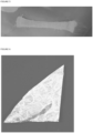
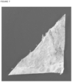
- FIG. 1 and Figure 2 illustrate braided structures using a loom comprising wire-type elements consisting of at least one membrane layer derived from placental tissues obtained by drying in the open air, without a drying step by desiccation. These images demonstrate the irregular appearance, more difficult to handle and implant, of the braided structure obtained with an air drying step.
- FIG. 3 illustrates a comparison of a membrane layer membrane layer from placental tissues dried by desiccation (image A), by freeze-drying (image B), or by drying in the open air (image C).
- FIG. 4 illustrates a braided structure on a loom obtained according to the method according to the invention.
- FIG. 5 illustrates a braided structure obtained according to the method according to the invention.
- FIG. 6 illustrates a membrane layer from placental tissues dried by desiccation and folded on itself along its diagonal.
- FIG. 7 illustrates a membrane layer from placental tissues dried by freeze-drying and folded on itself along its diagonal.
- the drying step by desiccation is carried out on the braided structure comprising wire-type elements consisting of at least one membrane layer derived from placental tissues obtained in step e).
- the expression “at least one” means “one or more” and may be replaced by the latter without distinction.
- membrane layer derived from placental tissues means one of the tissue layers constituting the placenta.
- At least one membrane layer derived from placental tissues comprises amniotic membrane, chorionic membrane, spongy tissue membrane and/or chorioamniotic membrane, each of these layers may or may not include the physiological substructures constituting the amnion and/or chorion.
- At least one membrane layer derived from placental tissues available in step a) is of animal or human origin.
- At least one membrane layer derived from placental tissues available in step a) is of human origin.
- At least one membrane layer derived from placental tissues available in step a) comprises all or part of the amniotic membrane and/or all or part of the chorionic membrane.
- the membrane layer derived from placental tissues available in step a) is a human amniotic membrane.
- amniotic membrane or “amnion” means the tissue envelope which develops around the embryo, then the fetus, in the mammal during pregnancy. Its role is to protect the developing organism by maintaining amniotic fluid around it. It is attached to the second membrane which is the chorion.
- At least one membrane layer derived from placental tissues available in step a) comprises at least one of the physiological sublayers of the amniotic membrane chosen from the group comprising the epithelial cell layer, the basement membrane , the compact layer, the fibroblastic layer and/or the spongy layer.
- At least one membrane layer from placental tissues available in step a) comprises the amniotic membrane including its spongy layer.
- At least one membrane layer derived from placental tissues available in step a) consists of the amniotic membrane comprising its spongy layer.
- chorionic membrane or "chorion” means the tissue envelope located between the amnion and the uterine wall which develops around the embryo, then the fetus, in the mammal during the pregnancy.
- At least one membrane layer derived from placental tissues available in step a) comprises at least one of the physiological sublayers of the chorionic membrane chosen from the group comprising the reticular layer, the basal membrane and /or the trophoblast cell layer.
- chorioamniotic membrane means the entire amnion and chorion, which have not been separated naturally or artificially, these being connected by protein bridges.
- spongy tissue membrane or “spongy layer” means a layer located between the amnion and the chorion.
- the membrane layer derived from placental tissues available in step a) is prepared according to the protocol as described in the application WO2017140914A1 .
- the membrane layer derived from placental tissues according to the present invention is obtained after delivery from a properly informed and consenting donor in accordance with the requirements of the European directives and the Bioethics Law. This delivery may have taken place by cesarean section or vaginally.
- any one of the methods according to the invention comprises a preliminary step of separating the membrane layer(s) originating from placental tissues.
- At least one membrane layer derived from placental tissues available in step a) is previously frozen.
- the placental tissue is previously placed in a sterile box, frozen and transported at a temperature of -20°C, then stored at -80°C.
- the placental tissue is transported at +4°C.
- the placental tissue was not initially frozen, the cutting and separation of the membrane layer(s) from placental tissue are carried out after transport at +4° vs.
- At least one membrane layer derived from placental tissues is stored at -80°C.
- the placenta not separated from its physiological tissues can be frozen for storage at a temperature of -80°C for a period of up to 2 years.
- At least one membrane layer derived from placental tissues in the frozen state is then washed in purified water or remains in a bath of purified water.
- This step ensures both the thawing of the membrane layer(s) originating from placental tissues if necessary up to room temperature, and the lysis of the cells present in the layer(s). membrane(s) from placental tissues by osmotic pressure effect.
- At least one membrane layer from placental tissues available in step a) is viro-inactivated.
- any one of the methods according to the invention comprises a step a') of viro-inactivation of the membrane layer(s) originating from placental tissues following the step has).
- viral-inactivation means a technique which makes it possible to greatly or completely, and definitively, reduce the capacity of action of viruses. These, defined as inactivated, lose their pathogenic and replication capacities by a decrease in their population of 4 log during residual titrations which follow one or two independent chemical steps, whether on enveloped or non-enveloped viruses, DNA or RNA. This step makes it possible in particular to have a product free of microbiological agents such as bacteria, viruses, parasites.
- the viro-inactivation step a') comprises a viro-inactivation step i) comprising the application to the membrane layer(s) originating from placental tissues of a washing and/or the stay of the latter in a bath, composed of a first viro-inactivating agent.
- the first virus-inactivating agent is ethanol.
- the first virus-inactivating agent is ethanol with an alcohol content of 50% to 80% (v/v) (50% ⁇ alcohol content ⁇ 80%).
- the first virus-inactivating agent is ethanol with an alcohol content of 70% (v/v).
- step i) of viro-inactivation is carried out for a duration ranging from 30 minutes to 120 minutes (30 min ⁇ duration ⁇ 120 min).
- step i) of viro-inactivation is carried out for a duration ranging from 45 minutes to 100 minutes (45 min ⁇ duration ⁇ 100 min).
- step i) of viro-inactivation is carried out for a period of approximately 60 minutes.
- step i) of viro-inactivation is carried out by treating the membrane layer(s) derived from placental tissues with ethanol at an alcohol content of 70% (v/v) for a duration of approximately 60 minutes.
- step i) is followed by a step i') of washing the membrane layer from placental tissues with purified water or a stay in a bath of purified water.
- step a') of viro inactivation comprises a second step ii) of viro inactivation consecutive to step i) or i') comprising the application to the membrane layer(s). ) from placental tissue from a wash and/or the stay of the latter in a bath, composed of a second viro-inactivation agent.
- the second viro inactivation agent is hydrogen peroxide.
- the second viro-inactivation agent is hydrogen peroxide in a form chosen from an aqueous solution and a gas.
- the second viro-inactivation agent is hydrogen peroxide in the form of an aqueous solution in a concentration ranging from 1% to 30% (w/v) (1% ⁇ concentration w/ v ⁇ 30%).
- the second viro-inactivation agent is hydrogen peroxide in the form of an aqueous solution in a concentration ranging from 3% to 10% (w/v) (3% ⁇ concentration w/ v ⁇ 10%).
- step ii) of viro-inactivation is carried out for a duration ranging from 30 minutes to 120 minutes (30 min ⁇ duration ⁇ 120 min).
- step ii) of viro-inactivation is carried out for a duration ranging from 45 to 100 minutes (45 min ⁇ duration ⁇ 100 min).
- step ii) of viro-inactivation is carried out for a period of approximately 60 minutes.
- the method is characterized in that the second viro-inactivation step ii) of the membrane layer(s) derived from placental tissues comprises two sub-steps of treatment with the second viro-inactivation agent chosen from the peroxide family.
- peroxides means any chemical compound containing a functional group of general formula R-O-O-R' (two neighboring oxygen atoms bonded together) where R and R' are hydrogen atoms or radicals. any alkyls.
- step a') of viro inactivation comprises a third step iii) consecutive to step ii) comprising at least one neutralization step, by application to the membrane layer(s). from placental tissue from washing and/or a stay of the latter in a bath, composed of at least one basic buffer.
- the third step iii) comprises two baths carried out in a basic buffer containing diluted sodium hydroxide and whose pH is adjusted to a value in a range from 7 to 10 (7 ⁇ pH ⁇ 10).
- the third step iii) comprises two baths carried out in a basic buffer containing diluted sodium hydroxide and whose pH is adjusted to a value in a range from 8 to 9 (8 ⁇ pH ⁇ 9).
- the third step iii) comprises two baths carried out in a basic buffer containing diluted sodium hydroxide and whose pH is adjusted to a value 8.5.
- step a') of viro inactivation comprises a final step iv) comprising the application to the membrane layer(s) originating from placental tissues of a wash and/or or a stay in a bath, composed of at least one buffer solution ensuring physiological rebalancing.
- the last step iv) comprises two baths carried out in PBS solution.
- the steps of thawing, viro-inactivation, washing, pH adjustment and buffering are carried out at room temperature.
- any one of the methods according to the invention further comprises a final step of sterilizing the braided structure comprising wire-type elements consisting of at least one membrane layer obtained from placental tissues.
- Sterilization can be carried out by any method conventionally known to those skilled in the art.
- sterilization is carried out by irradiation.
- sterilization is carried out by irradiation with gamma radiation.
- sterilization is carried out by irradiation with gamma radiation at a dose within a range of 25 to 35 kGrays (25 kGrays ⁇ dose ⁇ 35 kGrays).
- the membrane layer(s) derived from placental tissues are sterile.
- the term “sterile” means a structure free of germs, in its natural state or after sterilization.
- Sterilization can be carried out by any of the methods conventionally used by those skilled in the art.
- the sterilization is carried out under the conditions described above.
- “desiccation” means the elimination of water contained in a substance, using heat, vacuum and/or a hygroscopic material.
- positive temperature means a temperature greater than or equal to 0°C (0°C ⁇ temperature).
- the temperature ranges used are the temperature ranges which do not allow the formation of water crystals.
- vacuum desiccations at negative temperatures such as cryodesiccation more commonly called freeze-drying, are excluded from the definition of desiccation according to the present invention.
- lyophilization means a technique aimed at drying a previously frozen product by sublimation. More precisely, the liquid to be removed from the product is first transformed into a solid (ice) by freezing; then by primary desiccation, under vacuum, the solid is sublimated; finally through secondary desiccation, the water molecules on the surface of the product are extracted by desorption.
- the drying step by desiccation comprises the application of heat on the one hand and vacuum and/or a hygroscopic material on the other hand.
- the desiccation drying step includes the application of heat and vacuum.
- the drying step by desiccation does not include freezing and/or deep freezing of the placental and/or umbilical structure.
- the minimum temperature is chosen so as to avoid the formation of water crystals depending on the pressure applied.
- the drying step by desiccation is carried out by gradually increasing the temperature from at least 0°C (0°C ⁇ temperature).
- the drying step by desiccation is carried out by gradually increasing the temperature from at least 2°C (2°C ⁇ temperature).
- the drying step by desiccation is carried out by gradually increasing the temperature from at least 4°C (4°C ⁇ temperature).
- the drying step by desiccation is carried out by gradually raising the temperature to 35°C.
- the drying step by desiccation is carried out by gradually increasing the temperature from at least 0°C to 35°C (0°C ⁇ temperature ⁇ 35°C).
- the drying step by desiccation is carried out by gradually increasing the temperature in a range ranging from 4°C to 35°C (4°C ⁇ temperature ⁇ 35°C).
- the drying step by desiccation is carried out by gradually increasing the temperature in steps of 1 to 10°C (1°C ⁇ temperature ⁇ 10°C).
- the drying step by desiccation is carried out by gradually increasing the temperature by 5°C.
- the drying step by desiccation is carried out by gradually increasing the temperature in stages of 1 to 10°C.
- the drying step by desiccation is carried out by gradually increasing the temperature in stages of 2 to 8°C.
- the drying step by desiccation is carried out by gradually increasing the temperature in stages of 3 to 7°C.
- the drying step by desiccation is carried out by gradually increasing the temperature in steps of 5°C.
- the drying step by desiccation is carried out by gradually increasing the temperature by 5°C.
- the drying step by desiccation is carried out for a period ranging from 1 hour to 12 hours.
- the drying step by desiccation is carried out for a period ranging from 2 hours to 10 hours.
- the drying step by desiccation is carried out for a period ranging from 3 hours to 7 hours.
- the drying step by desiccation is carried out for a period ranging from 4 hours to 6 hours.
- the drying step by desiccation is carried out under pressures ranging from 50 to 900 microbars (50 microbars ⁇ pressures ⁇ 900 microbars).
- the drying step by desiccation is carried out under pressures ranging from 100 to 800 microbars (100 microbars ⁇ pressures ⁇ 800 microbars).
- the drying step by desiccation is carried out under pressures ranging from 400 to 600 microbars (400 microbars ⁇ pressures ⁇ 600 microbars).
- the drying step by desiccation is carried out under pressures of approximately 600 microbars.
- the drying step by desiccation is carried out under pressures of approximately 400 microbars.
- the drying step by desiccation is carried out by gradually raising the temperature to 35°C and under pressures ranging from 50 to 900 micro-bars (50 micro-bars ⁇ pressures ⁇ 900 microbars).
- the drying step by desiccation is carried out by progressively raising the temperature from at least 0°C to 50°C and under pressures ranging from 50 to 900 microbars. (50 micro-bars ⁇ pressures ⁇ 900 micro-bars).
- the drying step by desiccation is carried out by gradually raising the temperature by at least 2°C to 45°C and under pressures ranging from 50 to 900 microbars. (50 micro-bars ⁇ pressures ⁇ 900 micro-bars).
- the drying step by desiccation is carried out by gradually raising the temperature by at least 4°C to 40°C and under pressures in a range from 50 to 900 microbars. (50 micro-bars ⁇ pressures ⁇ 900 micro-bars).
- the drying step by desiccation is carried out by gradually raising the temperature from at least 8°C to 35°C and under pressures ranging from 50 to 900 microbars. (50 micro-bars ⁇ pressures ⁇ 900 micro-bars).
- the drying step by desiccation is carried out by progressively raising the temperature from at least 0°C to 35°C and under pressures in a range of 50 to 900 microbars. (50 micro-bars ⁇ pressures ⁇ 900 micro-bars).
- the drying step by desiccation is carried out by progressively raising the temperature from at least 0°C to 35°C (0°C ⁇ temperature ⁇ 35°C) and under pressures included in a range from 50 to 900 micro-bars (50 micro-bars ⁇ pressures ⁇ 900 micro-bars).
- the drying step by desiccation is carried out by progressively raising the temperature in a range ranging from 4°C to 35°C (4°C ⁇ temperature ⁇ 35°C) and under pressures included in a range ranging from 50 to 900 micro-bars (50 micro-bars ⁇ pressures ⁇ 900 micro-bars).
- the drying step by desiccation is carried out by gradually raising the temperature to 35°C and under pressures ranging from 100 to 800 micro-bars (100 micro-bars ⁇ pressures ⁇ 800 microbars).
- the drying step by desiccation is carried out by progressively raising the temperature from at least 0°C to 50°C and under pressures within a range of 100 to 800 micro-bars (100 micro-bars ⁇ pressures ⁇ 800 micro-bars).
- the drying step by desiccation is carried out by progressively raising the temperature by at least 2°C to 45°C and under pressures ranging from 100 to 800 microbars. (100 micro-bars ⁇ pressures ⁇ 800 micro-bars).
- the drying step by desiccation is carried out by gradually raising the temperature by at least 4°C to 40°C and under pressures in a range of 100 to 800 microbars. (100 micro-bars ⁇ pressures ⁇ 800 micro-bars).
- the drying step by desiccation is carried out by gradually raising the temperature from at least 8°C to 35°C and under pressures ranging from 100 to 800 microbars. (100 micro-bars ⁇ pressures ⁇ 800 micro-bars).
- the drying step by desiccation is carried out by progressively raising the temperature from at least 0°C to 35°C and under pressures in a range of 100 to 800 microbars. (100 micro-bars ⁇ pressures ⁇ 800 micro-bars).
- the drying step by desiccation is carried out by progressively raising the temperature from at least 0°C to 35°C (0°C ⁇ temperature ⁇ 35°C) and under pressures included in a range from 100 to 800 micro-bars (100 micro-bars ⁇ pressures ⁇ 800 micro-bars).
- the drying step by desiccation is carried out by progressively raising the temperature in a range ranging from 4°C to 35°C (4°C ⁇ temperature ⁇ 35°C) and under pressures included in an interval ranging from 100 to 800 micro-bars (100 micro-bars ⁇ pressures ⁇ 800 micro-bars).
- the drying step by desiccation is carried out by gradually raising the temperature to 35°C and under pressures ranging from 400 to 600 micro-bars (400 micro-bars ⁇ pressures ⁇ 600 microbars).
- the drying step by desiccation is carried out by progressively raising the temperature from at least 0°C to 50°C and under pressures ranging from 400 to 600 microbars. (400 micro-bars ⁇ pressures ⁇ 600 micro-bars).
- the drying step by desiccation is carried out by gradually raising the temperature by at least 2°C to 45°C and under pressures in a range of 400 to 600 microbars. (400 micro-bars ⁇ pressures ⁇ 600 micro-bars).
- the drying step by desiccation is carried out by gradually raising the temperature by at least 4°C to 40°C and under pressures ranging from 400 to 600 microbars. (400 micro-bars ⁇ pressures ⁇ 600 micro-bars).
- the drying step by desiccation is carried out by progressively raising the temperature from at least 8°C to 35°C and under pressures ranging from 400 to 600 microbars. (400 micro-bars ⁇ pressures ⁇ 600 micro-bars).
- the drying step by desiccation is carried out by gradually raising the temperature from at least 0°C to 35°C and under pressures in a range of 400 to 600 microbars. (400 micro-bars ⁇ pressures ⁇ 600 micro-bars).
- the drying step by desiccation is carried out by progressively raising the temperature from at least 0°C to 35°C (0°C ⁇ temperature ⁇ 35°C) and under pressures included in a range from 400 to 600 micro-bars (400 micro-bars ⁇ pressures ⁇ 600 micro-bars).
- the drying step by desiccation is carried out by progressively raising the temperature in a range ranging from 4°C to 35°C (4°C ⁇ temperature ⁇ 35°C) and under pressures included in an interval ranging from 400 to 600 micro-bars (400 micro-bars ⁇ pressures ⁇ 600 micro-bars).
- the membrane layer(s) originating from placental tissues after step a) and/or contained in the wire-type elements obtained in step c) and/or contained in the braided structure after step e) to which the desiccation drying step is applied have a humidity level before said desiccation drying step of at least 50%.
- the membrane layer(s) originating from placental tissues after step a) and/or contained in the wire-type elements obtained in step c) and/or contained in the braided structure after step e) to which the desiccation drying step is applied has a humidity level before said desiccation drying step of at least 60%.
- the membrane layer(s) originating from placental tissues after step a) and/or contained in the wire-type elements obtained in step c) and/or contained in the braided structure after step e) to which the desiccation drying step is applied has a humidity level before said desiccation drying step of at least 70%.
- the membrane layer(s) originating from placental tissues after step a) and/or contained in the wire-type elements obtained in step c) and/or contained in the braided structure after step e) on which the drying step by desiccation is applied has a humidity level before said drying step by desiccation of at least 80 %.
- the membrane layer(s) originating from placental tissues after step a) and/or contained in the wire-type elements obtained in step c) and/or contained in the braided structure after step e) to which the desiccation drying step is applied has a humidity level before said desiccation drying step of at least 90%.
- the step of cutting the membrane layer(s) from placental tissues can be carried out by any means known to those skilled in the art.
- the cutting step is carried out using a scalpel, a scissor, a cutting wheel, an automated cutting device and/or a cookie cutter .
- the cutting step is carried out in such a way that it makes it possible to obtain a flaire type structure made up of at least one membrane layer coming from placental tissues.
- wire-type element means any object having the appearance of a wire and/or a strip and/or a ribbon.
- the wire-type elements have a width and/or diameter within a range of 10 ⁇ m to 10 cm (10 ⁇ m ⁇ width and/or diameter ⁇ 10 cm).
- the wire-type elements have a width and/or diameter within a range of 100 ⁇ m to 1 cm (100 ⁇ m ⁇ width and/or diameter ⁇ 1 cm).
- the wire-type elements have a width and/or diameter within a range of 0.5 mm to 5 mm (0.5 mm ⁇ width and/or diameter ⁇ 5 mm).
- the wire-type elements have a length ranging from 0.5 mm to 10 m (0.5 mm ⁇ length ⁇ 10 mm).
- the wire-type elements have a length ranging from 5 cm to 1 m (5 cm ⁇ length ⁇ 1 m).
- the wire-type elements have a length ranging from 10 cm to 50 cm (10 cm ⁇ length ⁇ 50 cm.
- braiding or “braided structure” is meant the interlacing or weaving of at least two wire-type elements.
- the creation of the braiding can be chosen from all the patterns known for weaving, knitting, knotting, braiding, sewing, crocheting and/or scoubidou-style knotting.
- the braiding is carried out with at least one wire-type element consisting of at least one membrane layer derived from placental tissues according to the invention and at least one wire-type element not obtained by the method according to the invention. invention.
- wire-type element not obtained by the process according to the invention means any wire-type element obtained by a process different from that according to the present invention and/or from a material different from a membrane layer derived from placental tissues.
- wire-type elements not obtained by the process according to the invention have the advantage of allowing more complex assemblies and/or providing different structural properties and/or strengths and/or flexibility.
- the wire-type elements not obtained by the method according to the invention are made up of natural and/or artificial materials.
- At least one wire-type element not obtained by the method according to the invention is made of a biocompatible material.
- At least one wire-type element not obtained by the method according to the invention is a suture thread.
- the suture thread is chosen from the group of nylon, polybutester, polypropylene and/or silk threads.
- the suture is an absorbable suture.
- the absorbable suture thread is chosen from the group of glycolide and lactide threads, polyglytone, poliglecarpone, polyglactin, lactomer, polyglycolic acid, glycomer, polyglyconate or polydioxanone.
- suture thread has the advantage of having a biocompatible material that can be easily removed from the woven structure before application to the patient, without damaging said structure.
- absorbable suture allows the woven structure to be used directly without having to remove it from the annular structure without risk to the patient.
- any one of the methods according to the invention comprises the removal of wire-type elements not obtained by the method according to the invention subsequent to the step of obtaining a braided structure comprising elements of wire type consisting of at least one membrane layer from placental tissues.
- all of the wire-type elements used in the braiding consist of one or more membrane layer(s) originating from placental tissues according to the invention.
- the braiding is carried out using a loom.
- the braiding is carried out with at least two wire-type elements.
- the braiding is carried out with at least four wire-type elements.
- the present invention also relates to a braided structure comprising wire-type elements consisting of at least one membrane layer derived from placental tissues, said braided structure having a humidity level greater than or equal to 13%.
- said braided structure has a humidity level greater than or equal to 20%.
- said braided structure has a humidity level greater than or equal to 23%.
- said braided structure has a humidity level within a range of 13 to 40% (13% ⁇ humidity level ⁇ 40%).
- said braided structure has a humidity level in a range ranging from 15 to 35% (15% ⁇ humidity level ⁇ 35%).
- said braided structure has a humidity level within a range of 20 to 27% (20% ⁇ humidity level ⁇ 27%).
- the humidity level is considered before any rehydration of the braided structure and/or before any implantation in a subject.
- said braided structure has a thickness in a range ranging from 0.1 mm to 5 mm (0.1 mm ⁇ thickness ⁇ 5 mm).
- said braided structure has a thickness ranging from 0.5 mm to 3 mm (0.5 mm ⁇ thickness ⁇ 3 mm).
- said braided structure has a thickness ranging from 1 mm to 2 mm (1 mm ⁇ thickness ⁇ 2 mm).
- the braided structure comprising wire-type elements consisting of at least one membrane layer derived from placental tissues according to the invention is obtained by a process comprising at least step of drying by desiccation.
- the braided structure comprising wire-type elements consisting of at least one membrane layer derived from placental tissues according to the invention is not obtained by drying in the open air.
- the braided structure comprising wire-type elements consisting of at least one membrane layer derived from placental tissues according to the invention is not obtained by drying by freeze-drying.
- the braided structure comprising wire-type elements consisting of at least one membrane layer originating from placental tissues only comprises wire-type elements consisting of one or more membrane layer(s) ( s) derived from placental tissues.
- the braided structure according to the present invention is of animal or human origin.
- the braided structure according to the present invention is of human origin.
- the wire-type elements of the braided structure according to the present invention comprise all or part of the amniotic membrane and/or the chorionic membrane.
- the wire-type elements of the braided structure according to the present invention are made of human amniotic membrane.
- the wire-type elements of the braided structure according to the present invention comprise the amniotic membrane comprising its spongy layer.
- the wire-type elements of the braided structure according to the present invention comprise are made up of amniotic membrane comprising its spongy layer.
- the present invention also relates to an implantable device comprising at least one braided structure according to the invention.
- the present invention also relates to an implantable device comprising at least one braided structure obtained by any of the methods according to the invention.
- the implantable device is a suburethral sling.
- the present invention also relates to a therapeutic or surgical method of strengthening the urinary sphincter, reinforcing the pelvic floor, treatment of amniotic membrane ulcers and/or prolapse, tendon and/or ligament repair, ligament replacement, nerve sleeving repair, cartilage repair, dura mater replacement, submembrane replacement mucosa, strengthening the rotator cuff, the amniotic membrane, ulcers and/or skin wounds, and/or improving healing and/or sutures comprising a step of implantation in a subject of a braided structure comprising wire-type elements consisting of at least one membrane layer derived from placental tissues according to the invention.
- the present invention also relates to a therapeutic or surgical method for strengthening the urinary sphincter and/or the pelvic floor, for treating amniotic membrane ulcers, hernias and/or prolapses, for tendon and/or ligament repair, for ligament replacement. , nerve repair by sleeving, cartilage repair, dura mater replacement, submucosal membrane replacement, strengthening of the rotator cuff, amniotic membrane, ulcers and/or skin wounds, and/ or improvement of healing and/or sutures comprising a step of implanting in a subject a braided structure obtained by any of the methods according to the invention.
- the present invention also relates to a therapeutic or surgical method for strengthening the urinary sphincter and/or the pelvic floor, for treating amniotic membrane ulcers, hernias and/or prolapses, for tendon and/or ligament repair, for ligament replacement. , nerve repair by sleeving, cartilage repair, dura mater replacement, submucosal membrane replacement, strengthening of the rotator cuff, amniotic membrane, ulcers and/or skin wounds, and/ or improvement of healing and/or sutures comprising a step of implantation in a subject of an implantable device according to the invention.
- the subject is human or animal.
- the braided structure comprising wire-type elements consisting of at least one membrane layer derived from placental tissues according to the invention can be used as an allograft material chosen from the group consisting of ligament graft, fascia lata, tendon, dura mater, submucosal membrane, nerve sleeve, ligaments, cartilage, rotator cuff, and/or as agents for treating prolapse, hernias, wounds, ulcers , amniotic membrane, and/or skin wounds, and/or as an agent for improving sutures and/or healing, and/or for repairing amniotic membrane ulcers, for repairing tendons, cartilage and/or ligaments and/or for nerve repair by sleeving, and/or for the replacement of ligaments, dura mater, submucosal membranes, and/or for strengthening the rotator cuff, urinary sphincter and/or the pelvic floor.
- the braided structure comprising wire-type elements consisting of at least one membrane layer derived from placental tissues according to the invention can be used for strengthening the urinary sphincter.
- the braided structure comprising wire-type elements consisting of at least one membrane layer derived from placental tissues according to the invention can be used for strengthening the urinary sphincter in neurological incontinence pathologies.
- the braided structure comprising wire-type elements consisting of at least one membrane layer derived from placental tissues according to the invention can be used for strengthening the pelvic floor.
- the braided structure comprising wire-type elements consisting of at least one membrane layer derived from placental tissues according to the invention can be used for reinforcing the pelvic floor by the upper route and/or by the lower route.
- the braided structure comprising wire-type elements consisting of at least one membrane layer derived from placental tissues according to the invention can be used for strengthening the pelvic floor in the treatment of prolapses and/or hernias of the pelvic organs.
- the lower route is the transvaginal route.
- the upper route is the peritoneal and/or trans-abdominal route and/or is carried out by laparoscopy.
- the pelvic organs include the bladder, uterus and/or rectum.
- the braided structure obtained by any of the methods according to the invention can be used as an allograft material chosen from the group consisting of the graft of ligament, fascia lata, tendon, dura mater. , submucosal membrane, nerve sleeve, ligaments, cartilage, rotator cuff, and/or as agents for treating prolapse, hernias, wounds, ulcers, amniotic membrane, and/or skin wounds, and/or as an agent for improving sutures and/or healing, and/or for repairing amniotic membrane ulcers, for repairing tendons, cartilages and/or ligaments and/or for nerve repair by sleeving, and/or for the replacement of ligaments, dura mater, submucosal membranes, and/or for strengthening the rotator cuff, urinary sphincter and/or pelvic floor.
- allograft material chosen from the group consisting of the graft of ligament, fascia lata, tendon
- the braided structure obtained by any of the methods according to the invention can be used for strengthening the urinary sphincter.
- the braided structure obtained by any of the methods according to the invention can be used for strengthening the urinary sphincter in neurological incontinence pathologies.
- the braided structure obtained by any of the methods according to the invention can be used for strengthening the pelvic floor.
- the braided structure obtained by any one of the methods according to the invention can be used for strengthening the pelvic floor by the upper route and/or by the lower route.
- the braided structure obtained by any of the methods according to the invention can be used for strengthening the pelvic floor in the treatment of prolapses and/or hernias of the pelvic organs.
- the implantable device according to the invention can be used as an allograft material chosen from the group consisting of the graft of ligament, fascia lata, tendon, dura mater, submucosal membrane, nerve sleeve, ligaments, cartilage, rotator cuff, and/or as agents for treating prolapse, hernias, wounds, ulcers, amniotic membrane, and/or skin wounds, and/or as an agent for improvement of sutures and/or healing, and/or for repair of amniotic membrane ulcers, for repair of tendons, cartilages and/or ligaments and/or for nerve repair by sleeving, and/or for the replacement of ligaments, dura mater, submucosal membranes, and/or for strengthening the rotator cuff, urinary sphincter and/or pelvic floor.
- the implantable device according to the invention can be used for strengthening the urinary sphincter.
- the implantable device according to the invention can be used for strengthening the urinary sphincter in neurological incontinence pathologies.
- the implantable device according to the invention can be used for strengthening the pelvic floor.
- the implantable device according to the invention can be used for strengthening the pelvic floor via the upper route and/or via the lower route.
- the implantable device according to the invention can be used for strengthening the pelvic floor in the treatment of prolapses and/or hernias of the pelvic organs.
- the present invention also relates to a braided structure obtained by any of the methods according to the invention for its use in the treatment of urinary incontinence, a prolapse and/or a hernia, a tear and/or or a rupture of a ligament, muscle and/or tendon.
- the present invention also relates to an implantable device according to the invention for its use in the treatment of urinary incontinence, prolapse and/or hernia, tear and/or rupture of a ligament, muscle and/or tendon.
- the present invention also relates to a braided structure comprising wire-type elements consisting of at least one membrane layer derived from placental tissues according to the invention for its use in the treatment of urinary incontinence, prolapse and/or a hernia, tear and/or rupture of a ligament, muscle and/or tendon.
- a properly informed and consenting donor in accordance with the requirements of the Declaration of Helsinki offers as a donation the placental tissue resulting from childbirth.
- the placental tissue is collected in the delivery room. It is frozen at a temperature of -20°C.
- amniotic membrane In a sterile room, the amniotic membrane is isolated and cleaned.
- amniotic membrane undergoes a succession of baths ensuring its viral inactivation. Gentle agitation at approximately 30 rpm of the liquid medium is applied during each bath.
- the amniotic membrane is placed in a bath of purified water at room temperature for approximately 3 hours.
- amniotic membrane is transferred to a decontaminating bath composed of 70% v/v ethanol at room temperature for approximately 1 hour.
- a wash is carried out in purified water for approximately 15 minutes at room temperature to remove the ethanol.
- amniotic membrane is transferred to a bath composed of 30% w/v hydrogen peroxide at room temperature for approximately 15 minutes.
- amniotic membrane is transferred to a decontaminating bath composed of 3% w/v hydrogen peroxide at room temperature for approximately 1 hour.
- the chemical action applied to the amniotic membrane is neutralized in two baths containing sodium hydroxide diluted around a pH of 8.5.
- the neutralization baths are carried out at room temperature for approximately 15 minutes.
- the amniotic membrane is transferred to two successive baths of PBS buffer at room temperature for approximately 15 minutes in order to ensure its physiological rebalancing.
- amniotic membrane is transferred to a final bath in purified water, at room temperature, for 30 minutes.
- the amniotic membrane obtained according to this chemical treatment is disinfected and viro-inactivated.
- the amniotic membrane obtained is cut using a scalpel.
- the wire-type elements constituting the warp (strands placed lengthwise in the loom) are cut into strips approximately 3 to 4 mm wide and approximately 30 cm long.
- the wire-type elements constituting the weft (or strand of fillings, arranged perpendicularly in the loom) are cut in the same way into strips 2 to 3 mm wide without being shortened lengthwise.
- wire-type elements made up of at least one membrane layer from placental tissues thus obtained are then subjected to a braiding step according to the classic method of braiding on a loom and as presented on the Figure 4 .
- the dissected braided structure obtained is packaged flat in sterile conditions.
- a final step of sterilization of the dissected braided structure is carried out by exposing it to gamma radiation between 25 and 32 kGrays.
- a properly informed and consenting donor in accordance with the requirements of the Declaration of Helsinki offers as a donation the placental tissue resulting from childbirth.
- the placental tissue is collected in the delivery room. It is frozen at a temperature of -20°C.
- amniotic membrane In a sterile room, the amniotic membrane is isolated and cleaned.
- amniotic membrane undergoes a succession of baths ensuring its viral inactivation. Gentle agitation at approximately 30 rpm of the liquid medium is applied during each bath.
- the amniotic membrane is placed in a bath of purified water at room temperature for approximately 3 hours.
- amniotic membrane is transferred to a decontaminating bath composed of 70% v/v ethanol at room temperature for approximately 1 hour.
- a wash is carried out in purified water for approximately 15 minutes at room temperature to remove the ethanol.
- amniotic membrane is transferred to a bath composed of 30% w/v hydrogen peroxide at room temperature for approximately 15 minutes.
- amniotic membrane is transferred to a decontaminating bath composed of 3% w/v hydrogen peroxide at room temperature for approximately 1 hour.
- the chemical action applied to the amniotic membrane is neutralized in two baths containing sodium hydroxide diluted around a pH of 8.5.
- the neutralization baths are carried out at room temperature for approximately 15 minutes.
- the amniotic membrane is transferred to two successive baths of PBS buffer at room temperature for approximately 15 minutes in order to ensure its physiological rebalancing.
- amniotic membrane is transferred to a final bath in purified water, at room temperature, for 30 minutes.
- the amniotic membrane obtained according to this chemical treatment is disinfected and viro-inactivated.
- the amniotic membrane obtained is cut using a scalpel.
- the wire-type elements constituting the warp (strands placed lengthwise in the loom) are cut into strips approximately 3 to 4 mm wide and approximately 30 cm long.
- the wire-type elements constituting the weft (or strand of fillings, arranged perpendicularly in the loom) are cut in the same way into strips 2 to 3 mm wide without being shortened lengthwise.
- wire-type elements made up of at least one membrane layer from placental tissues thus obtained are then subjected to a braiding step according to the classic method of braiding on a loom and as presented on the Figure 4 .
- the dissected braided structure obtained is packaged flat in sterile conditions.
- a final step of sterilization of the dissected braided structure is carried out by exposing it to gamma radiation between 25 and 32 kGrays.
- Example 1b Comparative example of manufacturing a braided structure comprising wire-type elements consisting of at least one membrane layer from placental tissues before undergoing a drying step in the open air.
- Example 2a Preparation of a membrane layer from placental tissues dried by desiccation.
- a membrane layer derived from placental tissues is prepared according to the protocol as described in Example 1a) until before the step of cutting the membrane layer derived from placental tissues into wire-type elements.
- the structure obtained is photographed.
- Example 2a bis Preparation of a membrane layer from placental tissues dried by desiccation.
- a membrane layer derived from placental tissues is prepared according to the protocol as described in Example 1a) until before the step of cutting the membrane layer derived from placental tissues into wire-type elements.
- the structure obtained is photographed.
- Example 2b Comparative example of preparation of a membrane layer from placental tissues dried by freeze-drying.
- a membrane layer derived from placental tissues is prepared according to the protocol as described in Example 1a) until before the step of cutting the membrane layer derived from placental tissues into wire-type elements.
- Example 2c Comparative example of preparation of a membrane layer from placental tissues dried by drying in the open air.
- a membrane layer derived from placental tissues is prepared according to the protocol as described in Example 1a) until before the step of cutting the membrane layer derived from placental tissues into wire-type elements.
- the membrane layer is dried at room temperature for 1 to 2 hours then photographed.
- Example 3 Visual comparison between a membrane layer from placental tissues dried by different methods.
- Example 4 Comparison of the flexibility between a membrane layer derived from placental tissues dried by at least one drying step by desiccation and a membrane layer derived from placental tissues dried by at least one freeze-drying step.
- the membrane layer having undergone a drying step by desiccation is therefore more flexible and less brittle than the amniotic membrane having undergone a freeze-drying step.
- Example 5 Comparison of the average thickness between a membrane layer derived from placental tissues dried by at least one drying step by desiccation and a membrane layer derived from placental tissues dried by at least one freeze-drying step,
- the membrane layers obtained according to the process as described in Example 2a and 2b are cut using a scalpel into wire-type elements, more precisely into strips, with a width of approximately 0.02 mm.
- the strips are placed vertically on a slide and held with fine stainless steel pliers, the size of which is previously measured using a caliper.
- the assembly is placed under a microscope (Nikon, Eclipse E200) with a total magnification of x40 between the objective and the eyepiece. Photographs are taken for 4 strips of membrane layers having undergone a drying step by desiccation according to the protocol as described in Example 2a and for 4 strips of membrane layer having undergone a drying step by lyophilization according to the protocol as described. in example 2b.
- the thickness of the strips and the stainless steel pliers are measured with a caliper on the printed photographs.
- Thickness r e She mm Thickness on there photo mm ⁇ Width r e She of there pliers mm Width of there pliers on there photo mm
- amniotic membrane having undergone a drying step by desiccation is therefore almost 50% thicker than the amniotic membrane having undergone a freeze-drying step.
- Example 6 Comparison of the average humidity level between a membrane layer derived from placental tissues dried by different methods.
- the cup is positioned in the desiccator and tared.
- the moisture value of the product is calculated by the desiccator.
- the membrane layer desiccated according to the method of the invention has a residual humidity almost twice that of the membrane layer having undergone a freeze-drying step and is more than twice higher than that of the air-dried membrane layer. free.
Landscapes
- Health & Medical Sciences (AREA)
- Life Sciences & Earth Sciences (AREA)
- Chemical & Material Sciences (AREA)
- Biomedical Technology (AREA)
- Engineering & Computer Science (AREA)
- Oral & Maxillofacial Surgery (AREA)
- Epidemiology (AREA)
- Transplantation (AREA)
- Animal Behavior & Ethology (AREA)
- General Health & Medical Sciences (AREA)
- Public Health (AREA)
- Veterinary Medicine (AREA)
- Medicinal Chemistry (AREA)
- Dermatology (AREA)
- Chemical Kinetics & Catalysis (AREA)
- Botany (AREA)
- Vascular Medicine (AREA)
- Urology & Nephrology (AREA)
- Orthopedic Medicine & Surgery (AREA)
- Rehabilitation Therapy (AREA)
- Rheumatology (AREA)
- Molecular Biology (AREA)
- Zoology (AREA)
- Medicines Containing Material From Animals Or Micro-Organisms (AREA)
Applications Claiming Priority (2)
| Application Number | Priority Date | Filing Date | Title |
|---|---|---|---|
| FR2301113A FR3145490A1 (fr) | 2023-02-06 | 2023-02-06 | Structure tressée comprenant des éléments de type filaire constitués d’au moins une couche membranaire issue de tissus placentaires |
| FR2400351 | 2024-01-15 |
Publications (1)
| Publication Number | Publication Date |
|---|---|
| EP4410323A1 true EP4410323A1 (de) | 2024-08-07 |
Family
ID=89768351
Family Applications (2)
| Application Number | Title | Priority Date | Filing Date |
|---|---|---|---|
| EP24155948.3A Pending EP4410323A1 (de) | 2023-02-06 | 2024-02-06 | Geflochtene struktur mit drahtartigen elementen aus mindestens einer membranschicht aus plazentagewebe |
| EP24155952.5A Pending EP4410324A1 (de) | 2023-02-06 | 2024-02-06 | Ausgetrocknete plazenta- und/oder nabelstruktur |
Family Applications After (1)
| Application Number | Title | Priority Date | Filing Date |
|---|---|---|---|
| EP24155952.5A Pending EP4410324A1 (de) | 2023-02-06 | 2024-02-06 | Ausgetrocknete plazenta- und/oder nabelstruktur |
Country Status (1)
| Country | Link |
|---|---|
| EP (2) | EP4410323A1 (de) |
Citations (4)
| Publication number | Priority date | Publication date | Assignee | Title |
|---|---|---|---|---|
| US20080046095A1 (en) * | 2006-08-17 | 2008-02-21 | Surgical Biologics, Inc. | Placental Tissue Grafts and Improved Methods of Preparing and Using the Same |
| WO2008061261A2 (en) | 2006-11-17 | 2008-05-22 | Cytograft Tissue Engineering, Inc. | Preparation and use of cell-synthesized threads |
| WO2017140914A1 (fr) | 2016-02-18 | 2017-08-24 | Tbf Genie Tissulaire (Tbf) | Procede de preparation d'un materiau d'allogreffe, produit obtenu, et utilisations de celui-ci |
| WO2021234138A1 (fr) | 2020-05-21 | 2021-11-25 | Tbf Genie Tissulaire (Tbf) | Bandelette sous-uretrale constituee de materiau biologique issu de cordon ombilical |
-
2024
- 2024-02-06 EP EP24155948.3A patent/EP4410323A1/de active Pending
- 2024-02-06 EP EP24155952.5A patent/EP4410324A1/de active Pending
Patent Citations (4)
| Publication number | Priority date | Publication date | Assignee | Title |
|---|---|---|---|---|
| US20080046095A1 (en) * | 2006-08-17 | 2008-02-21 | Surgical Biologics, Inc. | Placental Tissue Grafts and Improved Methods of Preparing and Using the Same |
| WO2008061261A2 (en) | 2006-11-17 | 2008-05-22 | Cytograft Tissue Engineering, Inc. | Preparation and use of cell-synthesized threads |
| WO2017140914A1 (fr) | 2016-02-18 | 2017-08-24 | Tbf Genie Tissulaire (Tbf) | Procede de preparation d'un materiau d'allogreffe, produit obtenu, et utilisations de celui-ci |
| WO2021234138A1 (fr) | 2020-05-21 | 2021-11-25 | Tbf Genie Tissulaire (Tbf) | Bandelette sous-uretrale constituee de materiau biologique issu de cordon ombilical |
Non-Patent Citations (3)
| Title |
|---|
| DEVAL ET AL., J GYNECOL OBSTET BIOL REPROD, vol. 31, 2002, pages 131 - 143 |
| GRÉMARE A ET AL., BIOFABRICATION, vol. 14, no. 4, 22 August 2022 (2022-08-22) |
| GRÉMARE AGATHE ET AL: "Development of a vascular substitute produced by weaving yarn made from human amniotic membrane", vol. 14, no. 4, 1 October 2022 (2022-10-01), UK, pages 045010, XP093107411, ISSN: 1758-5082, Retrieved from the Internet <URL:https://iopscience.iop.org/article/10.1088/1758-5090/ac84ae/pdf> [retrieved on 20231130], DOI: 10.1088/1758-5090/ac84ae * |
Also Published As
| Publication number | Publication date |
|---|---|
| EP4410324A1 (de) | 2024-08-07 |
Similar Documents
| Publication | Publication Date | Title |
|---|---|---|
| JP5993387B2 (ja) | コラーゲンバイオ繊維、ならびにその調製方法および使用 | |
| DE69725592T2 (de) | Verfahren zur gewinnung einer gereignigten matrix auf kollagenbasis aus submukosa gewebe | |
| EP3416696B1 (de) | Verfahren zur herstellung eines allotransplantationsmaterials, erhaltenes produkt und dessen verwendung | |
| KR101313033B1 (ko) | 시트형 조성물 | |
| WO2013144727A2 (en) | A scaffold material graft for wound care and/or other tissue healing applications | |
| US20080213335A1 (en) | Graft prosthesis, materials and methods | |
| EP4153725B1 (de) | Suburethrale schlinge aus biologischem material aus nabelschnur | |
| CN114767938A (zh) | 用于盆腔脏器脱垂治疗的脱细胞异体真皮悬吊补片 | |
| EP4410323A1 (de) | Geflochtene struktur mit drahtartigen elementen aus mindestens einer membranschicht aus plazentagewebe | |
| EP3534980B1 (de) | Implantierbare führung zur geweberegeneration | |
| FR3145490A1 (fr) | Structure tressée comprenant des éléments de type filaire constitués d’au moins une couche membranaire issue de tissus placentaires | |
| EP1543846B1 (de) | Bioresorbierbares vernetztes Hydrogel bestehend aus Albumin | |
| WO2006136870A1 (fr) | Hydrogel bioresorbable | |
| CN119587766B (zh) | 一种动物小肠黏膜下层脱细胞基质防粘连膜及其制备方法和用途 | |
| EP4710960A1 (de) | Implantierbare vorrichtung aus nabelschnurwand | |
| Khangembam et al. | Regeneration of Full Thickness Skin Defects Using Decellularized Fish Swim Bladder Scaffolds Developed by Sapindus Mukorossi Fruit Pericarp Extract. |
Legal Events
| Date | Code | Title | Description |
|---|---|---|---|
| PUAI | Public reference made under article 153(3) epc to a published international application that has entered the european phase |
Free format text: ORIGINAL CODE: 0009012 |
|
| STAA | Information on the status of an ep patent application or granted ep patent |
Free format text: STATUS: THE APPLICATION HAS BEEN PUBLISHED |
|
| AK | Designated contracting states |
Kind code of ref document: A1 Designated state(s): AL AT BE BG CH CY CZ DE DK EE ES FI FR GB GR HR HU IE IS IT LI LT LU LV MC ME MK MT NL NO PL PT RO RS SE SI SK SM TR |
|
| STAA | Information on the status of an ep patent application or granted ep patent |
Free format text: STATUS: REQUEST FOR EXAMINATION WAS MADE |
|
| 17P | Request for examination filed |
Effective date: 20250207 |