EP4417709A1 - Mirna-basierter diagnostischer test zum nachweis eines risikos, hauttumore gegen den hintergrund schwerer hautläsionen zu entwickeln - Google Patents
Mirna-basierter diagnostischer test zum nachweis eines risikos, hauttumore gegen den hintergrund schwerer hautläsionen zu entwickeln Download PDFInfo
- Publication number
- EP4417709A1 EP4417709A1 EP23156564.9A EP23156564A EP4417709A1 EP 4417709 A1 EP4417709 A1 EP 4417709A1 EP 23156564 A EP23156564 A EP 23156564A EP 4417709 A1 EP4417709 A1 EP 4417709A1
- Authority
- EP
- European Patent Office
- Prior art keywords
- mir
- subject
- mirna
- squamous cell
- cell carcinoma
- Prior art date
- Legal status (The legal status is an assumption and is not a legal conclusion. Google has not performed a legal analysis and makes no representation as to the accuracy of the status listed.)
- Pending
Links
- 108091070501 miRNA Proteins 0.000 title claims abstract description 119
- 238000003556 assay Methods 0.000 title description 22
- 238000001514 detection method Methods 0.000 title description 16
- 206010040882 skin lesion Diseases 0.000 title description 3
- 231100000444 skin lesion Toxicity 0.000 title description 3
- 206010029098 Neoplasm skin Diseases 0.000 title 1
- 208000000453 Skin Neoplasms Diseases 0.000 title 1
- 206010041823 squamous cell carcinoma Diseases 0.000 claims abstract description 125
- 238000000034 method Methods 0.000 claims abstract description 99
- 230000014509 gene expression Effects 0.000 claims abstract description 71
- 108091058104 miR-187 stem-loop Proteins 0.000 claims abstract description 33
- 208000037265 diseases, disorders, signs and symptoms Diseases 0.000 claims abstract description 31
- 108091030503 miR-1260 stem-loop Proteins 0.000 claims abstract description 30
- 108091046552 miR-1260a stem-loop Proteins 0.000 claims abstract description 30
- 108091029203 miR-301 stem-loop Proteins 0.000 claims abstract description 30
- 108091026505 miR-301a stem-loop Proteins 0.000 claims abstract description 30
- 239000000203 mixture Substances 0.000 claims abstract description 30
- 108091070404 miR-27b stem-loop Proteins 0.000 claims abstract description 29
- 108091027986 miR-27b-1 stem-loop Proteins 0.000 claims abstract description 29
- 108091032408 miR-27b-2 stem-loop Proteins 0.000 claims abstract description 29
- 108091024480 miR-301a-1 stem-loop Proteins 0.000 claims abstract description 29
- 230000029663 wound healing Effects 0.000 claims abstract description 26
- 208000035475 disorder Diseases 0.000 claims abstract description 24
- 108010017377 Collagen Type VII Proteins 0.000 claims abstract description 16
- 102000004510 Collagen Type VII Human genes 0.000 claims abstract description 16
- 230000035772 mutation Effects 0.000 claims abstract description 16
- 101150056204 COL7A1 gene Proteins 0.000 claims abstract description 15
- 208000010975 Dystrophic epidermolysis bullosa Diseases 0.000 claims abstract description 15
- 101100496573 Homo sapiens COL7A1 gene Proteins 0.000 claims abstract description 15
- 208000004298 epidermolysis bullosa dystrophica Diseases 0.000 claims abstract description 15
- 238000011282 treatment Methods 0.000 claims abstract description 15
- 230000004064 dysfunction Effects 0.000 claims abstract description 14
- 239000002679 microRNA Substances 0.000 claims abstract description 10
- 206010052428 Wound Diseases 0.000 claims description 52
- 208000027418 Wounds and injury Diseases 0.000 claims description 51
- 208000037845 Cutaneous squamous cell carcinoma Diseases 0.000 claims description 33
- 108091027558 IsomiR Proteins 0.000 claims description 33
- 238000003753 real-time PCR Methods 0.000 claims description 32
- 239000002243 precursor Substances 0.000 claims description 26
- 108091032973 (ribonucleotides)n+m Proteins 0.000 claims description 19
- 238000012163 sequencing technique Methods 0.000 claims description 19
- 201000010106 skin squamous cell carcinoma Diseases 0.000 claims description 19
- -1 miR-198-3p Proteins 0.000 claims description 18
- 108091044988 miR-125a stem-loop Proteins 0.000 claims description 13
- 108020005075 5S Ribosomal RNA Proteins 0.000 claims description 12
- 108091035155 miR-10a stem-loop Proteins 0.000 claims description 12
- 108091084882 miR-10a-1 stem-loop Proteins 0.000 claims description 12
- 108091047189 miR-29c stem-loop Proteins 0.000 claims description 11
- 108091079151 miR-29c-1 stem-loop Proteins 0.000 claims description 11
- 239000003153 chemical reaction reagent Substances 0.000 claims description 10
- 238000004590 computer program Methods 0.000 claims description 8
- 210000001808 exosome Anatomy 0.000 claims description 8
- 238000011528 liquid biopsy Methods 0.000 claims description 8
- 239000000872 buffer Substances 0.000 claims description 7
- 230000007423 decrease Effects 0.000 claims description 7
- 108091039097 miR-193b stem-loop Proteins 0.000 claims description 7
- 108091053494 miR-22 stem-loop Proteins 0.000 claims description 7
- 108091056126 miR-769 stem-loop Proteins 0.000 claims description 7
- 108091023525 miR-95 stem-loop Proteins 0.000 claims description 7
- 238000002560 therapeutic procedure Methods 0.000 claims description 7
- 108091088205 miR-10a-2 stem-loop Proteins 0.000 claims description 6
- 108091071567 miR-1307 stem-loop Proteins 0.000 claims description 6
- 108091055145 miR-342 stem-loop Proteins 0.000 claims description 6
- 108091073613 miR-941-1 stem-loop Proteins 0.000 claims description 6
- 108091039878 miR-941-2 stem-loop Proteins 0.000 claims description 6
- 108091035558 miR-941-3 stem-loop Proteins 0.000 claims description 6
- 108091031187 miR-941-4 stem-loop Proteins 0.000 claims description 6
- 108091085604 miR-941-5 stem-loop Proteins 0.000 claims description 6
- 238000002360 preparation method Methods 0.000 claims description 6
- 238000000338 in vitro Methods 0.000 claims description 5
- 230000002265 prevention Effects 0.000 claims description 5
- 206010028980 Neoplasm Diseases 0.000 description 122
- 239000000523 sample Substances 0.000 description 77
- 201000000744 recessive dystrophic epidermolysis bullosa Diseases 0.000 description 44
- 230000003902 lesion Effects 0.000 description 32
- 210000004027 cell Anatomy 0.000 description 21
- 201000011510 cancer Diseases 0.000 description 15
- 230000001684 chronic effect Effects 0.000 description 15
- 238000012360 testing method Methods 0.000 description 14
- 238000012549 training Methods 0.000 description 13
- 210000001519 tissue Anatomy 0.000 description 12
- 238000012179 MicroRNA sequencing Methods 0.000 description 11
- 230000000694 effects Effects 0.000 description 11
- 230000003278 mimic effect Effects 0.000 description 11
- 239000002773 nucleotide Substances 0.000 description 11
- 125000003729 nucleotide group Chemical group 0.000 description 11
- 238000001574 biopsy Methods 0.000 description 10
- 238000004458 analytical method Methods 0.000 description 9
- 238000010606 normalization Methods 0.000 description 9
- 239000000126 substance Substances 0.000 description 9
- 238000012706 support-vector machine Methods 0.000 description 9
- 210000004881 tumor cell Anatomy 0.000 description 9
- 108700011259 MicroRNAs Proteins 0.000 description 8
- 230000008859 change Effects 0.000 description 8
- 238000011161 development Methods 0.000 description 8
- 230000018109 developmental process Effects 0.000 description 8
- 238000005070 sampling Methods 0.000 description 8
- 210000003491 skin Anatomy 0.000 description 8
- 201000010099 disease Diseases 0.000 description 7
- 238000012544 monitoring process Methods 0.000 description 7
- 230000001105 regulatory effect Effects 0.000 description 7
- 238000001356 surgical procedure Methods 0.000 description 7
- 239000000090 biomarker Substances 0.000 description 6
- 238000003759 clinical diagnosis Methods 0.000 description 6
- 230000000295 complement effect Effects 0.000 description 6
- 201000010536 head and neck cancer Diseases 0.000 description 6
- 208000014829 head and neck neoplasm Diseases 0.000 description 6
- 229920000915 polyvinyl chloride Polymers 0.000 description 6
- 238000000513 principal component analysis Methods 0.000 description 6
- 238000012216 screening Methods 0.000 description 6
- 230000008685 targeting Effects 0.000 description 6
- 206010061534 Oesophageal squamous cell carcinoma Diseases 0.000 description 5
- 208000036765 Squamous cell carcinoma of the esophagus Diseases 0.000 description 5
- 238000002405 diagnostic procedure Methods 0.000 description 5
- 208000007276 esophageal squamous cell carcinoma Diseases 0.000 description 5
- 238000011156 evaluation Methods 0.000 description 5
- 238000010801 machine learning Methods 0.000 description 5
- 230000003211 malignant effect Effects 0.000 description 5
- 238000013507 mapping Methods 0.000 description 5
- 210000004877 mucosa Anatomy 0.000 description 5
- 230000001855 preneoplastic effect Effects 0.000 description 5
- 238000011179 visual inspection Methods 0.000 description 5
- 206010014989 Epidermolysis bullosa Diseases 0.000 description 4
- 238000002790 cross-validation Methods 0.000 description 4
- 230000006870 function Effects 0.000 description 4
- 206010073071 hepatocellular carcinoma Diseases 0.000 description 4
- 230000036210 malignancy Effects 0.000 description 4
- 108091007431 miR-29 Proteins 0.000 description 4
- 108090000623 proteins and genes Proteins 0.000 description 4
- 102000004169 proteins and genes Human genes 0.000 description 4
- 102000040650 (ribonucleotides)n+m Human genes 0.000 description 3
- 108020004414 DNA Proteins 0.000 description 3
- 206010058467 Lung neoplasm malignant Diseases 0.000 description 3
- 206010027476 Metastases Diseases 0.000 description 3
- 102000014160 PTEN Phosphohydrolase Human genes 0.000 description 3
- 108010011536 PTEN Phosphohydrolase Proteins 0.000 description 3
- 238000011529 RT qPCR Methods 0.000 description 3
- 102000001742 Tumor Suppressor Proteins Human genes 0.000 description 3
- 108010040002 Tumor Suppressor Proteins Proteins 0.000 description 3
- 238000013459 approach Methods 0.000 description 3
- 238000002512 chemotherapy Methods 0.000 description 3
- 238000009826 distribution Methods 0.000 description 3
- 230000007705 epithelial mesenchymal transition Effects 0.000 description 3
- 238000002474 experimental method Methods 0.000 description 3
- 210000002950 fibroblast Anatomy 0.000 description 3
- 230000003176 fibrotic effect Effects 0.000 description 3
- 238000009396 hybridization Methods 0.000 description 3
- 230000001771 impaired effect Effects 0.000 description 3
- 230000002757 inflammatory effect Effects 0.000 description 3
- 210000002510 keratinocyte Anatomy 0.000 description 3
- 201000005202 lung cancer Diseases 0.000 description 3
- 208000020816 lung neoplasm Diseases 0.000 description 3
- 239000003550 marker Substances 0.000 description 3
- 230000009401 metastasis Effects 0.000 description 3
- 108091064399 miR-10b stem-loop Proteins 0.000 description 3
- 238000007481 next generation sequencing Methods 0.000 description 3
- 230000000306 recurrent effect Effects 0.000 description 3
- 230000037390 scarring Effects 0.000 description 3
- 210000002966 serum Anatomy 0.000 description 3
- 238000012546 transfer Methods 0.000 description 3
- 108091032955 Bacterial small RNA Proteins 0.000 description 2
- 208000023514 Barrett esophagus Diseases 0.000 description 2
- 208000023665 Barrett oesophagus Diseases 0.000 description 2
- 102100024335 Collagen alpha-1(VII) chain Human genes 0.000 description 2
- 102000053602 DNA Human genes 0.000 description 2
- 102000010834 Extracellular Matrix Proteins Human genes 0.000 description 2
- 108010037362 Extracellular Matrix Proteins Proteins 0.000 description 2
- WSFSSNUMVMOOMR-UHFFFAOYSA-N Formaldehyde Chemical compound O=C WSFSSNUMVMOOMR-UHFFFAOYSA-N 0.000 description 2
- 101000909498 Homo sapiens Collagen alpha-1(VII) chain Proteins 0.000 description 2
- 108091065168 Homo sapiens miR-29c stem-loop Proteins 0.000 description 2
- 108091007424 MIR27B Proteins 0.000 description 2
- 108091030146 MiRBase Proteins 0.000 description 2
- 241000699670 Mus sp. Species 0.000 description 2
- 238000000636 Northern blotting Methods 0.000 description 2
- 229920000297 Rayon Polymers 0.000 description 2
- 102000004887 Transforming Growth Factor beta Human genes 0.000 description 2
- 108090001012 Transforming Growth Factor beta Proteins 0.000 description 2
- 238000002679 ablation Methods 0.000 description 2
- 230000005856 abnormality Effects 0.000 description 2
- 230000004075 alteration Effects 0.000 description 2
- 210000001124 body fluid Anatomy 0.000 description 2
- 239000010839 body fluid Substances 0.000 description 2
- 230000004663 cell proliferation Effects 0.000 description 2
- 238000013145 classification model Methods 0.000 description 2
- 239000002299 complementary DNA Substances 0.000 description 2
- 230000001143 conditioned effect Effects 0.000 description 2
- 230000002596 correlated effect Effects 0.000 description 2
- 230000000875 corresponding effect Effects 0.000 description 2
- 238000003066 decision tree Methods 0.000 description 2
- 230000003831 deregulation Effects 0.000 description 2
- 238000003745 diagnosis Methods 0.000 description 2
- 238000013399 early diagnosis Methods 0.000 description 2
- 238000010195 expression analysis Methods 0.000 description 2
- 210000002744 extracellular matrix Anatomy 0.000 description 2
- 230000035876 healing Effects 0.000 description 2
- 238000007490 hematoxylin and eosin (H&E) staining Methods 0.000 description 2
- 210000005260 human cell Anatomy 0.000 description 2
- 238000007901 in situ hybridization Methods 0.000 description 2
- 208000014674 injury Diseases 0.000 description 2
- 230000000670 limiting effect Effects 0.000 description 2
- 239000000463 material Substances 0.000 description 2
- 239000011159 matrix material Substances 0.000 description 2
- 230000007246 mechanism Effects 0.000 description 2
- 108091008057 miR-10 Proteins 0.000 description 2
- 238000010208 microarray analysis Methods 0.000 description 2
- 210000000056 organ Anatomy 0.000 description 2
- 230000037361 pathway Effects 0.000 description 2
- 238000007781 pre-processing Methods 0.000 description 2
- 238000012545 processing Methods 0.000 description 2
- 230000035755 proliferation Effects 0.000 description 2
- 238000007388 punch biopsy Methods 0.000 description 2
- 230000002829 reductive effect Effects 0.000 description 2
- 230000011664 signaling Effects 0.000 description 2
- 238000007390 skin biopsy Methods 0.000 description 2
- CCEKAJIANROZEO-UHFFFAOYSA-N sulfluramid Chemical group CCNS(=O)(=O)C(F)(F)C(F)(F)C(F)(F)C(F)(F)C(F)(F)C(F)(F)C(F)(F)C(F)(F)F CCEKAJIANROZEO-UHFFFAOYSA-N 0.000 description 2
- ZRKFYGHZFMAOKI-QMGMOQQFSA-N tgfbeta Chemical compound C([C@H](NC(=O)[C@H](C(C)C)NC(=O)CNC(=O)[C@H](CCC(O)=O)NC(=O)[C@H](CCCNC(N)=N)NC(=O)[C@H](CC(N)=O)NC(=O)[C@H](CC(C)C)NC(=O)[C@H]([C@@H](C)O)NC(=O)[C@H](CCC(O)=O)NC(=O)[C@H]([C@@H](C)O)NC(=O)[C@H](CC(C)C)NC(=O)CNC(=O)[C@H](C)NC(=O)[C@H](CO)NC(=O)[C@H](CCC(N)=O)NC(=O)[C@@H](NC(=O)[C@H](C)NC(=O)[C@H](C)NC(=O)[C@@H](NC(=O)[C@H](CC(C)C)NC(=O)[C@@H](N)CCSC)C(C)C)[C@@H](C)CC)C(=O)N[C@@H]([C@@H](C)O)C(=O)N[C@@H](C(C)C)C(=O)N[C@@H](CC=1C=CC=CC=1)C(=O)N[C@@H](C)C(=O)N1[C@@H](CCC1)C(=O)N[C@@H]([C@@H](C)O)C(=O)N[C@@H](CC(N)=O)C(=O)N[C@@H](CCC(O)=O)C(=O)N[C@@H](C)C(=O)N[C@@H](CC=1C=CC=CC=1)C(=O)N[C@@H](CCCNC(N)=N)C(=O)N[C@@H](C)C(=O)N[C@@H](CC(C)C)C(=O)N1[C@@H](CCC1)C(=O)N1[C@@H](CCC1)C(=O)N[C@@H](CCCNC(N)=N)C(=O)N[C@@H](CCC(O)=O)C(=O)N[C@@H](CCCNC(N)=N)C(=O)N[C@@H](CO)C(=O)N[C@@H](CCCNC(N)=N)C(=O)N[C@@H](CC(C)C)C(=O)N[C@@H](CC(C)C)C(O)=O)C1=CC=C(O)C=C1 ZRKFYGHZFMAOKI-QMGMOQQFSA-N 0.000 description 2
- 230000001225 therapeutic effect Effects 0.000 description 2
- 230000009466 transformation Effects 0.000 description 2
- 238000013519 translation Methods 0.000 description 2
- 230000008733 trauma Effects 0.000 description 2
- 230000005751 tumor progression Effects 0.000 description 2
- 108020005345 3' Untranslated Regions Proteins 0.000 description 1
- 241000894006 Bacteria Species 0.000 description 1
- 108010006654 Bleomycin Proteins 0.000 description 1
- 206010006187 Breast cancer Diseases 0.000 description 1
- 208000026310 Breast neoplasm Diseases 0.000 description 1
- 208000025721 COVID-19 Diseases 0.000 description 1
- 208000005623 Carcinogenesis Diseases 0.000 description 1
- 201000009030 Carcinoma Diseases 0.000 description 1
- 208000005443 Circulating Neoplastic Cells Diseases 0.000 description 1
- 102100027995 Collagenase 3 Human genes 0.000 description 1
- 108050005238 Collagenase 3 Proteins 0.000 description 1
- 241000711573 Coronaviridae Species 0.000 description 1
- 102000004127 Cytokines Human genes 0.000 description 1
- 108090000695 Cytokines Proteins 0.000 description 1
- 206010056340 Diabetic ulcer Diseases 0.000 description 1
- 208000000461 Esophageal Neoplasms Diseases 0.000 description 1
- 206010016654 Fibrosis Diseases 0.000 description 1
- 101000588302 Homo sapiens Nuclear factor erythroid 2-related factor 2 Proteins 0.000 description 1
- 101000928408 Homo sapiens Protein diaphanous homolog 2 Proteins 0.000 description 1
- 101000851007 Homo sapiens Vascular endothelial growth factor receptor 2 Proteins 0.000 description 1
- 108091044862 Homo sapiens miR-1260a stem-loop Proteins 0.000 description 1
- 108091067635 Homo sapiens miR-187 stem-loop Proteins 0.000 description 1
- 108091069018 Homo sapiens miR-27b stem-loop Proteins 0.000 description 1
- 108091065454 Homo sapiens miR-301a stem-loop Proteins 0.000 description 1
- 102000043136 MAP kinase family Human genes 0.000 description 1
- 108091054455 MAP kinase family Proteins 0.000 description 1
- 206010064912 Malignant transformation Diseases 0.000 description 1
- 102100030417 Matrilysin Human genes 0.000 description 1
- 108090000855 Matrilysin Proteins 0.000 description 1
- 108091027766 Mir-143 Proteins 0.000 description 1
- 206010061309 Neoplasm progression Diseases 0.000 description 1
- 102100031701 Nuclear factor erythroid 2-related factor 2 Human genes 0.000 description 1
- 101710163270 Nuclease Proteins 0.000 description 1
- 206010030155 Oesophageal carcinoma Diseases 0.000 description 1
- 238000009004 PCR Kit Methods 0.000 description 1
- 229930040373 Paraformaldehyde Natural products 0.000 description 1
- 102100037765 Periostin Human genes 0.000 description 1
- 101710199268 Periostin Proteins 0.000 description 1
- 102100023087 Protein S100-A4 Human genes 0.000 description 1
- 102100036469 Protein diaphanous homolog 2 Human genes 0.000 description 1
- 102000044126 RNA-Binding Proteins Human genes 0.000 description 1
- 108700020471 RNA-Binding Proteins Proteins 0.000 description 1
- 238000003559 RNA-seq method Methods 0.000 description 1
- 208000007660 Residual Neoplasm Diseases 0.000 description 1
- 208000037847 SARS-CoV-2-infection Diseases 0.000 description 1
- 102000012479 Serine Proteases Human genes 0.000 description 1
- 108010022999 Serine Proteases Proteins 0.000 description 1
- 206010040851 Skin fragility Diseases 0.000 description 1
- 102000015661 Solute Carrier Organic Anion Transporter Family Member 1B3 Human genes 0.000 description 1
- 108010063829 Solute Carrier Organic Anion Transporter Family Member 1B3 Proteins 0.000 description 1
- 230000006044 T cell activation Effects 0.000 description 1
- 108091036066 Three prime untranslated region Proteins 0.000 description 1
- 102000040945 Transcription factor Human genes 0.000 description 1
- 108091023040 Transcription factor Proteins 0.000 description 1
- 102000004243 Tubulin Human genes 0.000 description 1
- 108090000704 Tubulin Proteins 0.000 description 1
- 102000005789 Vascular Endothelial Growth Factors Human genes 0.000 description 1
- 108010019530 Vascular Endothelial Growth Factors Proteins 0.000 description 1
- 102100033177 Vascular endothelial growth factor receptor 2 Human genes 0.000 description 1
- 108020000999 Viral RNA Proteins 0.000 description 1
- 230000001594 aberrant effect Effects 0.000 description 1
- 238000009825 accumulation Methods 0.000 description 1
- 208000009621 actinic keratosis Diseases 0.000 description 1
- 230000006978 adaptation Effects 0.000 description 1
- 230000002411 adverse Effects 0.000 description 1
- 230000004931 aggregating effect Effects 0.000 description 1
- 230000003321 amplification Effects 0.000 description 1
- 230000003510 anti-fibrotic effect Effects 0.000 description 1
- 230000000845 anti-microbial effect Effects 0.000 description 1
- 230000000118 anti-neoplastic effect Effects 0.000 description 1
- 239000004599 antimicrobial Substances 0.000 description 1
- 230000001640 apoptogenic effect Effects 0.000 description 1
- 230000001580 bacterial effect Effects 0.000 description 1
- 230000037365 barrier function of the epidermis Effects 0.000 description 1
- 230000006399 behavior Effects 0.000 description 1
- 230000008901 benefit Effects 0.000 description 1
- 230000033228 biological regulation Effects 0.000 description 1
- 229960001561 bleomycin Drugs 0.000 description 1
- OYVAGSVQBOHSSS-UAPAGMARSA-O bleomycin A2 Chemical compound N([C@H](C(=O)N[C@H](C)[C@@H](O)[C@H](C)C(=O)N[C@@H]([C@H](O)C)C(=O)NCCC=1SC=C(N=1)C=1SC=C(N=1)C(=O)NCCC[S+](C)C)[C@@H](O[C@H]1[C@H]([C@@H](O)[C@H](O)[C@H](CO)O1)O[C@@H]1[C@H]([C@@H](OC(N)=O)[C@H](O)[C@@H](CO)O1)O)C=1N=CNC=1)C(=O)C1=NC([C@H](CC(N)=O)NC[C@H](N)C(N)=O)=NC(N)=C1C OYVAGSVQBOHSSS-UAPAGMARSA-O 0.000 description 1
- 210000000601 blood cell Anatomy 0.000 description 1
- LWQQLNNNIPYSNX-UROSTWAQSA-N calcipotriol Chemical compound C1([C@H](O)/C=C/[C@@H](C)[C@@H]2[C@]3(CCCC(/[C@@H]3CC2)=C\C=C\2C([C@@H](O)C[C@H](O)C/2)=C)C)CC1 LWQQLNNNIPYSNX-UROSTWAQSA-N 0.000 description 1
- 229960002882 calcipotriol Drugs 0.000 description 1
- 230000036952 cancer formation Effects 0.000 description 1
- 231100000504 carcinogenesis Toxicity 0.000 description 1
- 230000015556 catabolic process Effects 0.000 description 1
- 238000004113 cell culture Methods 0.000 description 1
- 230000030833 cell death Effects 0.000 description 1
- 230000001413 cellular effect Effects 0.000 description 1
- 230000004640 cellular pathway Effects 0.000 description 1
- 238000006243 chemical reaction Methods 0.000 description 1
- 230000001427 coherent effect Effects 0.000 description 1
- 230000004154 complement system Effects 0.000 description 1
- 238000010668 complexation reaction Methods 0.000 description 1
- 230000001010 compromised effect Effects 0.000 description 1
- 238000002591 computed tomography Methods 0.000 description 1
- 239000003636 conditioned culture medium Substances 0.000 description 1
- 235000009508 confectionery Nutrition 0.000 description 1
- 230000001276 controlling effect Effects 0.000 description 1
- 238000010219 correlation analysis Methods 0.000 description 1
- 239000012297 crystallization seed Substances 0.000 description 1
- 230000001186 cumulative effect Effects 0.000 description 1
- 125000004122 cyclic group Chemical group 0.000 description 1
- 210000004292 cytoskeleton Anatomy 0.000 description 1
- 238000007405 data analysis Methods 0.000 description 1
- 230000034994 death Effects 0.000 description 1
- 231100000517 death Toxicity 0.000 description 1
- 230000003247 decreasing effect Effects 0.000 description 1
- 238000006731 degradation reaction Methods 0.000 description 1
- 238000012217 deletion Methods 0.000 description 1
- 230000037430 deletion Effects 0.000 description 1
- 230000001419 dependent effect Effects 0.000 description 1
- 238000010586 diagram Methods 0.000 description 1
- 230000009977 dual effect Effects 0.000 description 1
- 230000008846 dynamic interplay Effects 0.000 description 1
- 230000008482 dysregulation Effects 0.000 description 1
- 201000004101 esophageal cancer Diseases 0.000 description 1
- 238000000605 extraction Methods 0.000 description 1
- 210000000416 exudates and transudate Anatomy 0.000 description 1
- 230000004761 fibrosis Effects 0.000 description 1
- 239000012530 fluid Substances 0.000 description 1
- 239000012634 fragment Substances 0.000 description 1
- 208000030536 genetic skin disease Diseases 0.000 description 1
- 238000005469 granulation Methods 0.000 description 1
- 230000003179 granulation Effects 0.000 description 1
- 210000003714 granulocyte Anatomy 0.000 description 1
- 230000012010 growth Effects 0.000 description 1
- 231100000844 hepatocellular carcinoma Toxicity 0.000 description 1
- 230000003118 histopathologic effect Effects 0.000 description 1
- 238000003384 imaging method Methods 0.000 description 1
- 238000011065 in-situ storage Methods 0.000 description 1
- 238000010348 incorporation Methods 0.000 description 1
- 230000006698 induction Effects 0.000 description 1
- 238000007689 inspection Methods 0.000 description 1
- 230000003834 intracellular effect Effects 0.000 description 1
- 238000002955 isolation Methods 0.000 description 1
- 238000007477 logistic regression Methods 0.000 description 1
- 210000004072 lung Anatomy 0.000 description 1
- 230000036212 malign transformation Effects 0.000 description 1
- 230000005741 malignant process Effects 0.000 description 1
- 210000004962 mammalian cell Anatomy 0.000 description 1
- 238000005259 measurement Methods 0.000 description 1
- 230000001394 metastastic effect Effects 0.000 description 1
- 206010061289 metastatic neoplasm Diseases 0.000 description 1
- 108091091207 miR-127 stem-loop Proteins 0.000 description 1
- 108091053592 miR-145-1 stem-loop Proteins 0.000 description 1
- 108091056559 miR-145-2 stem-loop Proteins 0.000 description 1
- 108091026495 miR-148b stem-loop Proteins 0.000 description 1
- 108091023683 miR-187-1 stem-loop Proteins 0.000 description 1
- 108091086421 miR-223 stem-loop Proteins 0.000 description 1
- 108091090583 miR-34c stem-loop Proteins 0.000 description 1
- 108091084066 miR-34c-2 stem-loop Proteins 0.000 description 1
- 108091087125 miR-376a stem-loop Proteins 0.000 description 1
- 108091080263 miR-376a-1 stem-loop Proteins 0.000 description 1
- 108091089006 miR-376a-2 stem-loop Proteins 0.000 description 1
- 108091028761 miR-409 stem-loop Proteins 0.000 description 1
- 108091030938 miR-424 stem-loop Proteins 0.000 description 1
- 108091031190 miR-495 stem-loop Proteins 0.000 description 1
- 108091031110 miR-539 stem-loop Proteins 0.000 description 1
- 108091056454 miR-625 stem-loop Proteins 0.000 description 1
- 108091072565 miR-98 stem-loop Proteins 0.000 description 1
- 244000005700 microbiome Species 0.000 description 1
- 230000001617 migratory effect Effects 0.000 description 1
- 230000001338 necrotic effect Effects 0.000 description 1
- 210000000440 neutrophil Anatomy 0.000 description 1
- 210000004882 non-tumor cell Anatomy 0.000 description 1
- 230000037311 normal skin Effects 0.000 description 1
- 238000003199 nucleic acid amplification method Methods 0.000 description 1
- 102000039446 nucleic acids Human genes 0.000 description 1
- 108020004707 nucleic acids Proteins 0.000 description 1
- 150000007523 nucleic acids Chemical class 0.000 description 1
- 238000005457 optimization Methods 0.000 description 1
- 239000012188 paraffin wax Substances 0.000 description 1
- 229920002866 paraformaldehyde Polymers 0.000 description 1
- 230000036961 partial effect Effects 0.000 description 1
- 210000001539 phagocyte Anatomy 0.000 description 1
- 238000010837 poor prognosis Methods 0.000 description 1
- 230000001124 posttranscriptional effect Effects 0.000 description 1
- 230000002028 premature Effects 0.000 description 1
- 230000002206 pro-fibrotic effect Effects 0.000 description 1
- 238000004393 prognosis Methods 0.000 description 1
- 230000001737 promoting effect Effects 0.000 description 1
- 238000003908 quality control method Methods 0.000 description 1
- 238000011002 quantification Methods 0.000 description 1
- 239000002096 quantum dot Substances 0.000 description 1
- 238000007634 remodeling Methods 0.000 description 1
- 238000003757 reverse transcription PCR Methods 0.000 description 1
- 230000002441 reversible effect Effects 0.000 description 1
- 108020004418 ribosomal RNA Proteins 0.000 description 1
- 238000000926 separation method Methods 0.000 description 1
- 210000001626 skin fibroblast Anatomy 0.000 description 1
- 238000010186 staining Methods 0.000 description 1
- 238000010561 standard procedure Methods 0.000 description 1
- 239000007858 starting material Substances 0.000 description 1
- 238000007619 statistical method Methods 0.000 description 1
- 238000000528 statistical test Methods 0.000 description 1
- 210000000130 stem cell Anatomy 0.000 description 1
- 238000006467 substitution reaction Methods 0.000 description 1
- 208000024891 symptom Diseases 0.000 description 1
- 238000000844 transformation Methods 0.000 description 1
- 230000001960 triggered effect Effects 0.000 description 1
- 238000009966 trimming Methods 0.000 description 1
- 239000000107 tumor biomarker Substances 0.000 description 1
- 230000005748 tumor development Effects 0.000 description 1
- 239000000439 tumor marker Substances 0.000 description 1
- 239000013598 vector Substances 0.000 description 1
- 230000000007 visual effect Effects 0.000 description 1
- 238000012447 xenograft mouse model Methods 0.000 description 1
Images




Classifications
-
- C—CHEMISTRY; METALLURGY
- C12—BIOCHEMISTRY; BEER; SPIRITS; WINE; VINEGAR; MICROBIOLOGY; ENZYMOLOGY; MUTATION OR GENETIC ENGINEERING
- C12Q—MEASURING OR TESTING PROCESSES INVOLVING ENZYMES, NUCLEIC ACIDS OR MICROORGANISMS; COMPOSITIONS OR TEST PAPERS THEREFOR; PROCESSES OF PREPARING SUCH COMPOSITIONS; CONDITION-RESPONSIVE CONTROL IN MICROBIOLOGICAL OR ENZYMOLOGICAL PROCESSES
- C12Q1/00—Measuring or testing processes involving enzymes, nucleic acids or microorganisms; Compositions therefor; Processes of preparing such compositions
- C12Q1/68—Measuring or testing processes involving enzymes, nucleic acids or microorganisms; Compositions therefor; Processes of preparing such compositions involving nucleic acids
- C12Q1/6876—Nucleic acid products used in the analysis of nucleic acids, e.g. primers or probes
- C12Q1/6883—Nucleic acid products used in the analysis of nucleic acids, e.g. primers or probes for diseases caused by alterations of genetic material
- C12Q1/6886—Nucleic acid products used in the analysis of nucleic acids, e.g. primers or probes for diseases caused by alterations of genetic material for cancer
-
- C—CHEMISTRY; METALLURGY
- C12—BIOCHEMISTRY; BEER; SPIRITS; WINE; VINEGAR; MICROBIOLOGY; ENZYMOLOGY; MUTATION OR GENETIC ENGINEERING
- C12Q—MEASURING OR TESTING PROCESSES INVOLVING ENZYMES, NUCLEIC ACIDS OR MICROORGANISMS; COMPOSITIONS OR TEST PAPERS THEREFOR; PROCESSES OF PREPARING SUCH COMPOSITIONS; CONDITION-RESPONSIVE CONTROL IN MICROBIOLOGICAL OR ENZYMOLOGICAL PROCESSES
- C12Q2600/00—Oligonucleotides characterized by their use
- C12Q2600/112—Disease subtyping, staging or classification
-
- C—CHEMISTRY; METALLURGY
- C12—BIOCHEMISTRY; BEER; SPIRITS; WINE; VINEGAR; MICROBIOLOGY; ENZYMOLOGY; MUTATION OR GENETIC ENGINEERING
- C12Q—MEASURING OR TESTING PROCESSES INVOLVING ENZYMES, NUCLEIC ACIDS OR MICROORGANISMS; COMPOSITIONS OR TEST PAPERS THEREFOR; PROCESSES OF PREPARING SUCH COMPOSITIONS; CONDITION-RESPONSIVE CONTROL IN MICROBIOLOGICAL OR ENZYMOLOGICAL PROCESSES
- C12Q2600/00—Oligonucleotides characterized by their use
- C12Q2600/118—Prognosis of disease development
-
- C—CHEMISTRY; METALLURGY
- C12—BIOCHEMISTRY; BEER; SPIRITS; WINE; VINEGAR; MICROBIOLOGY; ENZYMOLOGY; MUTATION OR GENETIC ENGINEERING
- C12Q—MEASURING OR TESTING PROCESSES INVOLVING ENZYMES, NUCLEIC ACIDS OR MICROORGANISMS; COMPOSITIONS OR TEST PAPERS THEREFOR; PROCESSES OF PREPARING SUCH COMPOSITIONS; CONDITION-RESPONSIVE CONTROL IN MICROBIOLOGICAL OR ENZYMOLOGICAL PROCESSES
- C12Q2600/00—Oligonucleotides characterized by their use
- C12Q2600/158—Expression markers
Definitions
- the present invention relates to a method for diagnosing the risk of a subject to suffer from squamous cell carcinoma (SCC), comprising detecting the expression level of the miR-187-3p molecule, in a sample obtained from said subject.
- the method according to the present invention further comprises detecting the expression level of at least one of the miR-27b-3p, miR-301a-3p, or miR-1260a, in said sample obtained from said subject.
- a changed expression level of said at least one miRNA, as compared to healthy donors, further indicates an increased risk of said subject to suffer from squamous cell carcinoma.
- the subject to be diagnosed may suffer from a dysfunction or loss of the structural extracellular matrix protein collagen type VII, for example caused by mutations within the COL7A1 gene, wound healing disorders, and preferably from a severe recessive subtype of dystrophic epidermolysis bullosa (RDEB).
- kits and diagnostic compositions as well as to an improved treatment of SCC, in particular in a background of RDEB.
- the disruption of the epidermal barrier, presenting as blisters and wounds, is the defining characteristic of the severe recessive subtype of dystrophic epidermolysis bullosa (RDEB), a genetic skin disease caused by mutations within the COL7A1 gene, resulting in dysfunctional or loss of the structural extracellular matrix protein collagen type VII (C7) and accounting for 11% of all EB cases (Has et al. 2020, Kim et al. 2018). While the majority of blisters heal within a few weeks, about one in four causes more severe problems due to their cyclic recurrence or by evolving into long-standing chronic open wounds (Solis et al. 2021).
- RDEB dystrophic epidermolysis bullosa
- impaired wound healing is considered to be a consequence of the lack of C7, which in turn leads to an altered migratory behavior of fibroblasts and keratinocytes, matrix remodeling, accumulation of mutations, bacterial challenge and an overall, excessive inflammatory milieu (Woodley et al. 2021, Wimmer et al. 2020, Nyström et al. 2013, Nyström et al. 2015, Cho et al. 2018, Nyström et al. 2018, Phillips et al. 2020, Tartaglia G, Cao Q, Padron ZM, South AP. Impaired Wound Healing, Fibrosis, and Cancer: The Paradigm of Recessive Dystrophic Epidermolysis Bullosa. Int J Mol Sci.
- tumors arising in individuals with RDEB are particularly aggressive, as they show a high recurrence rate and early metastatic spread, accounting for a mortality rate of 87 % by the age of 45 upon SCC development (Fine et al. 2009).
- US20120059048A1 describes diagnostic methods relating to detecting miRNA-205, thereby diagnosing an aggressive form of squamous cell carcinoma.
- Chacon et al. proposed periostin as a serum marker for RDEB to monitor treatment success of anti-fibrotic therapies (Chacón-Solano et al. 2008), whereas MMP-7 was characteristically expressed at the invasive edge of RDEB-SCCs and was thus discussed as a therapeutic target or biomarker for SCC progression. (Kivisaari et al. 2008). The same group also found MMP-13 to be highly expressed by RDEB-SCCs and also SerpinA1 expression was shown to correlate with SCC progression (Kivisaari et al. 2008, Farshchian et al. 2011).
- RDEB-SCCs but not non-malignant keratinocytes were found to specifically express the cancer-type transcript of the solute carrier organic anion transporter family member 1B3 protein (Ct-SLCO 1B3).
- Ct-SLCO 1B3 protein solute carrier organic anion transporter family member 1B3 protein
- EP2794881B1 describes miRNA for treating head and neck cancer and relates to the diagnostic and therapeutic uses of a miRNA-323, miRNA-342, miRNA-326, miRNA-371, miRNA-3157 and/or miRNA-345 molecule in a disease and condition associated with a squamous cell carcinoma such as head and neck cancer or a preneoplastic mucosal change.
- Wimmer et al. disclose that cutaneous squamous cell carcinomas (cSCC) are the primary cause of premature deaths in patients suffering from the rare skin-fragility disorder recessive dystrophic epidermolysis bullosa (RDEB), which is in marked contrast to the rarely metastasizing nature of these carcinomas in the general population.
- cSCC cutaneous squamous cell carcinomas
- RDEB rare skin-fragility disorder recessive dystrophic epidermolysis bullosa
- miRNAs were identified as dysregulated in cSCCs compared to control skin.
- the metastasis-linked miR-10b which was significantly upregulated in primary cell cultures and in archival biopsies.
- Analysis ofmiR-10b downstream effects identified a novel putative target of miR-10b, the actin- and tubulin cytoskeleton-associated protein DIAPH2. This is said to provide an important avenue for future development of novel therapies targeting this metastasis-linked miRNA.
- the miRNAs as identified do not overlap with the ones of the present invention.
- a further object of the present invention is the development of non-invasive tests that allow for a regular testing in the patients without invasive sample collection and thus high compliance.
- the problem of the present invention is solved by providing a method for diagnosing the risk of a subject to suffer from squamous cell carcinoma (SCC), comprising detecting the expression level of the miR-187-3p molecule, an isomiR, or a precursor thereof in a sample obtained from said subject, wherein a changed expression level of said miRNA, as compared to healthy donors, indicates an increased risk of said subject to suffer from squamous cell carcinoma.
- SCC squamous cell carcinoma
- a decrease of the expression level of miR-301a-3p and/or miR-1260a when compared to healthy donors indicates an increased risk of said subject to suffer from squamous cell carcinoma
- an increase of the expression level of miR-187-3p and/or miR-27b-3p when compared to healthy donors indicates an increased risk of said subject to suffer from squamous cell carcinoma
- the sample as used in the methods according to the present invention comprises a liquid biopsy and/or swap sample, such as a wound swab sample, from said subject.
- the subject may suffer from a dysfunction or loss of the structural extracellular matrix protein collagen type VII, for example caused by mutations within the COL7A1 gene, wound healing disorders, and preferably from a severe recessive subtype of dystrophic epidermolysis bullosa (RDEB).
- RDEB dystrophic epidermolysis bullosa
- the problem of the present invention is further solved by providing a diagnostic composition
- a diagnostic composition comprising a sample obtained from a subject at risk to suffer from squamous cell carcinoma (SCC) comprising miR-187-3p molecule, an isomiR, or a precursor thereof, preferably further comprising at least one of miR-27b-3p, miR-301a-3p, or miR-1260a molecule, an isomiR, or a precursor thereof.
- SCC squamous cell carcinoma
- the diagnostic composition according to the present invention wherein the sample comprises a liquid biopsy and/or swap sample, such as a wound swab sample, from said subject. Further preferred is the diagnostic composition according to the present invention, wherein the subject suffers from a dysfunction or loss of the structural extracellular matrix protein collagen type VII, for example caused by mutations within the COL7A1 gene, wound healing disorders, and preferably from a severe recessive subtype of dystrophic epidermolysis bullosa (RDEB).
- RDEB dystrophic epidermolysis bullosa
- the problem of the present invention is further solved by providing a kit for in vitro assessment of the risk that a subject suffers from squamous cell carcinoma (SCC), comprising buffers and reagents to perform a method according to the present invention, optionally together with instructions for performing said method.
- SCC squamous cell carcinoma
- the present invention also encompasses the use of the kit according to the present invention for diagnosing a subject to suffer from SCC, in particular cutaneous squamous cell carcinoma (cSCC), in particular according to a method according to the present invention.
- cSCC cutaneous squamous cell carcinoma
- the problem of the present invention is also solved by providing a computer program for identifying a subject's risk to suffer from SCC, in particular cutaneous squamous cell carcinoma (cSCC), in particular as determined according to a method according to the present invention, wherein said program is preferably implemented to run on a computer.
- SCC cutaneous squamous cell carcinoma
- the problem of the present invention is also solved by providing a method for preventing or treating squamous cell carcinoma (SCC), in particular cSCC, in a subject, comprising the steps of performing method according to to the present invention, to determine if the subject has an increased probability of suffering or is suffering from squamous cell carcinoma (SCC), in particular cSCC, and selecting an appropriate prevention or therapy for the subject based on the determination.
- SCC squamous cell carcinoma
- cSCC squamous cell carcinoma
- the object of the present invention is solved by a method for diagnosing the risk of a subject to suffer from squamous cell carcinoma (SCC), comprising detecting the expression level of the miR-187-3p molecule, an isomiR, or a precursor thereof in a sample obtained from said subject, wherein a changed expression level of said miRNA, as compared to healthy donors, indicates an increased risk of said subject to suffer from squamous cell carcinoma.
- SCC squamous cell carcinoma
- the method according to the present invention further comprises detecting the expression level of at least one of the miR-27b-3p, miR-301a-3p, or miR-1260a, an isomiR, or a precursor thereof in said sample obtained from said subject, wherein a changed expression level of said at least one miRNA, as compared to healthy donors, further indicates an increased risk of said subject to suffer from squamous cell carcinoma.
- miRNA Several micro-RNAs (miRNA) play an important role in the generation and spread of cancer and tumors.
- the present inventors were able to obtain miRNAs from tumor swabs, and to characterize those that allow to identify tumorous tissue. An early diagnosis of the tumors that are otherwise very difficult to detect is important in order to still allow for an effective surgical removal.
- miR-187-3p a recently identified cancer-related microRNA, was previously reported to play promoting or suppressive roles in different malignancies, such as HCC. miR-187-3p is significantly down-regulated in HCC, and is correlated with adverse clinical features and poor prognosis of HCC patients.
- Dou C Liu Z, Xu M, Jia Y, Wang Y, Li Q, Yang W, Zheng X, Tu K, Liu Q. miR-187-3p inhibits the metastasis and epithelial-mesenchymal transition of hepatocellular carcinoma by targeting S100A4. Cancer Lett. 2016 Oct 28;381(2):380-90. doi: 10.1016/j.canlet.2016.08.011. Epub 2016 Aug 17. PMID: 27544906 ).
- PMID: 28852310; PMCID: PMC5558114 describe nine microRNAs (miR-424-5p, miR-127-3p, miR-98-5p, miR-187-3p, miR-495-3p, miR-34c-5p, miR-223-5p, miR-539-5p, miR-376a-3p, miR-409-3p) that were expressed at higher levels in post-ablation neosquamous mucosa than in matched proximal squamous and healthy squamous mucosa. These microRNAs were also more highly expressed in Barrett's esophagus mucosa than matched proximal squamous and squamous mucosa from controls.
- MiR-27b-3p exerts tumor suppressor effects in esophageal squamous cell carcinoma by targeting Nrf2. Hum Cell. 2020 Jul;33(3):641-651. doi: 10.1007/s13577-020-00329-7. Epub 2020 May 17. PMID: 32419118 ) disclose that miR-27b-3p has been reported to function as tumor suppressor in several tumors, including breast cancer and lung cancer. Also, miR-27b-3p has been identified to be significantly down-regulated in esophageal cancer.
- MicroRNA (MiR)-301a-3p regulates the proliferation of esophageal squamous cells via targeting PTEN. Bioengineered. 2020 Dec; 11(1):972-983. doi: 10.1080/21655979.2020.1814658. PMID: 32970954; PMCID: PMC8291791 ) explore the mechanism of miR-301a-3p regulating the proliferation of esophageal squamous cell carcinoma (ESCC) cells. Compared with adjacent normal esophageal tissues, the relative level of miR-301a-3p/U6 was elevated in ESCC tissues. MiR-301a-3p could facilitate ESCC cell proliferation. And miR-301a-3p directly bind to PTEN 3'-UTR and negatively regulated PTEN protein expression.
- ESCC esophageal squamous cell carcinoma
- RNA-sequences of the most preferred miRNA molecules as used in the invention are:
- miRNA shall mean to include a miRNA molecule, a miRNA equivalent, a miRNA mimic or a miRNA isomiR, or a mimic or a suitable isomiR or a miRNA source or a precursor thereof.
- a preferred equivalent is an isomiR or a mimic.
- a miRNA molecule or an equivalent or a mimic or an isomiR thereof may be a synthetic or natural or recombinant or mature or part of a mature miRNA or a human miRNA or derived from a human miRNA as further defined in the part dedicated to the general definitions.
- a human miRNA molecule is a miRNA molecule which is found in a human cell, tissue, organ or body fluids (i.e. endogenous human miRNA molecule).
- a human miRNA molecule may also be a human miRNA molecule derived from an endogenous human miRNA molecule by substitution, deletion and/or addition of a nucleotide.
- a miRNA molecule or an equivalent or a mimic thereof may be a single stranded or double stranded RNA molecule.
- a miRNA molecule or an equivalent, or a mimic thereof is from 6 to 30 nucleotides in length, preferably 12 to 30 nucleotides in length, preferably 15 to 28 nucleotides in length, more preferably said molecule has a length of at least 6, 7, 8, 9, 10, 11, 12, 13, 14, 15, 16, 17, 18, 19, 20, 21, 22, 23, 24, 25, 26, 27, 28, 29, 30 nucleotides or more.
- a miRNA molecule or an equivalent or a mimic or isomiR thereof that is from 15 to 28 nucleotides in length and more preferably comprises at least 6 of the 7 nucleotides present in the seed sequence, i.e. the actual human derived sequence of the miRNAs as described herein.
- a preferred miRNA-xx molecule or equivalent or mimic or isomiR thereof comprises at least 6 of the 7 nucleotides present in the seed sequence identified as any one of SEQ ID NOs: 1 to 4, and more preferably has a length of at least 6, 7, 8, 9, 10, 11, 12, 13, 14, 15, 16, 17, 18, 19, 20, 21, 22, 23, 24, 25, 26, 27, 28, 29, 30 nucleotides or more.
- a source of a miRNA molecule or of an equivalent or a mimic or an isomiR thereof may be a single stranded optionally within a hairpin like structure, a double stranded RNA or a partially double stranded RNA or may comprise three strands, an example of which is described in WO2008/10558 .
- partially double stranded refers to double stranded structures that also comprise single stranded structures at the 5'and/or at the 3' end. It may occur when each strand of a miRNA molecule does not have the same length.
- such partial double stranded miRNA molecule may have less than 75% double stranded structure and more than 25% single stranded structure, or less than 50% double stranded structure and more than 50% single stranded structure, or more preferably less than 25%, 20 % or 15% double stranded structure and more than 75%, 80%, 85% single stranded structure.
- a source of a miRNA molecule or of an equivalent or a mimic or an isomiR thereof is a DNA molecule encoding a precursor of a miRNA molecule or of an equivalent or a mimic or an isomiR thereof.
- the invention encompasses the use of a DNA molecule encoding a precursor of a miRNA molecule that has at least 70% identity with said sequence as identified in SEQ ID Nos 1 to 4. Preferably, the identity is at least 75%, 80%, 85%, 90%, 95%, 97%, 98%, 99% or 100%.
- squamous cell carcinoma or "SCC” shall include all different types of cancer that begin in squamous cells, such as head and neck cancer or a preneoplastic mucosal change, and particularly relate to cutaneous squamous cell carcinoma (cSCC) in a subject that suffers from a dysfunction or loss of the structural extracellular matrix protein collagen type VII, for example caused by mutations within the COL7A1 gene, wound healing disorders, or from a severe recessive subtype of dystrophic epidermolysis bullosa (RDEB).
- cSCC cutaneous squamous cell carcinoma
- the risk of a subject to suffer from squamous cell carcinoma is diagnosed.
- the risk as diagnosed according to the present invention is based on the assessment of the relative risk as determined through detecting the expression level of at least one miRNA as described herein in a sample and comparing it to a control, such as the expression level in healthy donors.
- the invention further comprises detecting the expression level of miR-29c-5p as a stable reference miRNA and/or 5S-rRNA as a small reference RNA as a control.
- a change of the expression level indicates a change in the risk compared to the control
- a decrease of the expression level of miR-301a-3p and/or miR-1260a when compared to healthy donors indicates an increased risk of said subject to suffer from squamous cell carcinoma
- an increase of the expression level of miR-187-3p and/or miR-27b-3p when compared to healthy donors indicates an increased risk of said subject to suffer from squamous cell carcinoma.
- a method according to the present invention further comprising detecting the expression level of miR-29c-5p as a stable reference miRNA and/or 5S-rRNA as a small reference RNA as a control.
- the invention also encompassed a method according to the present invention, further comprising detecting the expression level of at least one of the miRNA molecules, an isomiR, or a precursor thereof selected from the group consisting of miR-769-3p, miR-22-5p, miR125a-3p, miR-198-3p, miR-95-3p, miR-193b-3p, miR-342-3p, miR-125a-5p2, miR-125a-3p4, miR-941, miR-125a-5p5, miR-10a-5p, let-7e-5p6, miR-34a-5p7, miR-193b-5p8, miR-125a-3p9, miR-1307-5p, miR-34a-5p10, miR-10a-3p, and miR-125a-5p.
- miR-769-3p miR-22-5p
- miR125a-3p miR-198-3p
- miR-95-3p miR-193b-3p
- miR-342-3p miR-
- miRNAs will advantageously add further relevance to the panel or set of miRNAs as above, i.e., miR-187-3p, and at least one of miR-27b-3p, miR-301a-3p and/or miR-1260a.
- an important aspect of the invention is the non-invasive nature of obtaining the sample.
- the sample comprises a liquid biopsy and/or swap sample, such as a wound swab sample, from said subject.
- the method of the invention nevertheless also encompasses a pre-conditioned sample, such as to obtain the exosome preparation of the sample before the assay.
- Another important aspect of the invention relates to the fact that the subject to be diagnosed suffers from a dysfunction or loss of the structural extracellular matrix protein collagen type VII, for example caused by mutations within the COL7A1 gene, wound healing disorders, and preferably from a severe recessive subtype of dystrophic epidermolysis bullosa (RDEB).
- RDEB dystrophic epidermolysis bullosa
- Another important aspect of the invention relates to a method according to the present invention, further comprising the (actual) step of diagnosing the subject to suffer from SCC in particular cutaneous squamous cell carcinoma (cSCC).
- SCC cutaneous squamous cell carcinoma
- cSCC cutaneous squamous cell carcinoma
- Any suitable method can be used in the method according to the present invention, in order to detect the expression level of said miRNA(s).
- miRNA detection methods There are a number of well-established miRNA detection methods that can be exploited depending on the comprehensiveness of the study (individual miRNA versus multiplex analysis), the availability of the sample and the location and intracellular concentration of miRNAs (see, for example, Yaylak B, Akgül B. Experimental MicroRNA Detection Methods. Methods Mol Biol. 2022;2257:33-55. doi: 10.1007/978-1-0716-1170-8_2. PMID: 34432272 ).
- miRNA expression levels can be studied by several methods: microarray analysis, real-time PCR, Northern blots, miRNA sequencing, in situ hybridization, and solution hybridization, and the most prominent examples are miRNA sequencing, solution hybridization assays and quantitative polymerase chain reaction (qPCR).
- Another important aspect of the invention relates to a method according to the present invention, wherein the changed expression level of said miRNA, as compared to healthy donors, shows a significant (FDR ⁇ 0.05) differential abundance or expression level (min. 2-fold) in said sample vs healthy control. This can be determined as described herein, for example using a computer program or app.
- Another important aspect of the invention relates to a method according to the present invention, wherein the sample from the subject is obtained from said subject either before, during or after a treatment of said subject against wound healing disorders, RDEB and/or SCC, in particular cSCC.
- This relates to a monitoring of the effect of a treatment of the SCC (such as a chemotherapy) and/or shows the effectiveness of a surgical removal of the SCC cells from the subject or lesion.
- an important aspect of the invention relates to a diagnostic composition
- a diagnostic composition comprising a sample obtained from a subject at risk to suffer from squamous cell carcinoma (SCC) comprising miR-187-3p molecule, an isomiR, or a precursor thereof.
- the diagnostic composition according to the present invention further comprises at least one of miR-27b-3p, miR-301a-3p, or miR-1260a molecule, an isomiR
- the diagnostic composition according to the present invention further comprises miR-29c-5p as a stable reference miRNA and/or 5S-rRNA as a small reference RNA as a control.
- the diagnostic composition according to the present invention can be suitably prepared by obtaining a sample form a subject, and optionally admixing the sample with suitable additional components for the diagnostic assay.
- the diagnostic composition of the invention may also comprise pre-conditioned components, such as an exosome preparation of the sample as taken from the subject.
- the diagnostic composition according to the present invention further comprises at least one of miRNA molecules, an isomiR, or a precursor thereof selected from the group consisting of miR-769-3p, miR-22-5p, miR125a-3p, miR-198-3p, miR-95-3p, miR-193b-3p, miR-342-3p, miR-125a-5p2, miR-125a-3p4, miR-941, miR-125a-5p5, miR-10a-5p, let-7e-5p6, miR-34a-5p7, miR-193b-5p8, miR-125a-3p9, miR-1307-5p, miR-34a-5p10, miR-10a-3p, and miR-125a-5p.
- miRNA molecules an isomiR, or a precursor thereof selected from the group consisting of miR-769-3p, miR-22-5p, miR125a-3p, miR-198-3p, miR-95-3p, miR-193b-3p, miR
- the sample comprises a liquid biopsy and/or swap sample, such as a wound swab sample, from said subject.
- the diagnostic composition according to the present invention is derived from a subject suffering from a dysfunction or loss of the structural extracellular matrix protein collagen type VII, for example caused by mutations within the COL7A1 gene, wound healing disorders, and preferably from a severe recessive subtype of dystrophic epidermolysis bullosa (RDEB).
- RDEB dystrophic epidermolysis bullosa
- Another important aspect of the invention relates to a diagnostic composition according to the present invention, wherein the sample from the subject was obtained from said subject either before, during or after a treatment of said subject against wound healing disorders, RDEB and/or SCC, in particular cSCC.
- These samples relate to a monitoring of the effect of a treatment of the SCC (such as a chemotherapy) and/or shows the effectiveness of a surgical removal of the SCC cells from the subject or lesion.
- kits for in vitro assessment of the risk that a subject suffers from squamous cell carcinoma comprising buffers and reagents to perform a method according to the present invention as described above and herein, optionally together with instructions for performing said method.
- kits according to the present invention wherein said kit comprises suitable buffers and reagents to perform quantitative polymerase chain reaction (qPCR), microarray analysis, real-time PCR, Northern blots, in situ hybridization, miRNA sequencing, and solution hybridization, and/or the kit further comprising reagents needed to obtain the exosome preparation of the sample.
- qPCR quantitative polymerase chain reaction
- kits according to the present invention for diagnosing a subject to suffer from SCC, in particular cutaneous squamous cell carcinoma (cSCC), in particular according to a method according to the present invention as described herein.
- SCC cutaneous squamous cell carcinoma
- Another important aspect of the invention relates to a computer program for identifying a subject's risk to suffer from SCC, in particular cutaneous squamous cell carcinoma (cSCC), in particular as determined according to a method according to the present invention, wherein said program is preferably implemented to run on a computer.
- SCC cutaneous squamous cell carcinoma
- Preferred is the computer program according to the present invention, which is a Shiny app.
- Another important aspect of the invention relates to a method, such as a screening method, for identification of a substance capable of preventing, treating, reverting and/or delaying a condition or disease associated with a squamous cell carcinoma such as head and neck cancer or a preneoplastic mucosal change in a subject, the method comprising the steps of:
- Yet another important aspect of the invention relates to a method of preventing or treating squamous cell carcinoma (SCC), in particular cSCC, in a subject, comprising the steps of performing method according to the present invention to determine if the subject has an increased probability of suffering or is suffering from squamous cell carcinoma (SCC), in particular cSCC, and selecting an appropriate prevention or therapy for the subject based on the determination, such as a chemotherapy and/or a surgical removal of the SCC cells from the subject or lesion, or identifying of a substance capable of preventing, treating, reverting and/or delaying a condition or disease associated with a squamous cell carcinoma according to the present invention, and selecting an appropriate prevention or therapy for the subject based on the substance as identified.
- SCC squamous cell carcinoma
- cSCC squamous cell carcinoma
- the method according to the present invention wherein the subject suffers from a dysfunction or loss of the structural extracellular matrix protein collagen type VII, for example caused by mutations within the COL7A1 gene, wound healing disorders, and preferably from a severe recessive subtype of dystrophic epidermolysis bullosa (RDEB).
- RDEB dystrophic epidermolysis bullosa
- the method according to the present invention wherein the sample from the subject was obtained from said subject either before, during or after a treatment of said subject against wound healing disorders, RDEB and/or SCC, in particular cSCC, again relating to a monitoring of the effect of a treatment of the SCC.
- an embodiment of the method for diagnosing a disease or condition associated with a squamous cell carcinoma such as head and neck cancer or a preneoplastic mucosal change in a subject comprises the steps of: (a) determining the expression level of miR-187-3p molecule, an isomiR, or a precursor thereof in a sample obtained from a subject at risk to suffer from squamous cell carcinoma, and optionally further determining the expression level of at least one of miR-27b-3p, miR-301a-3p, or miR-1260a molecule, an isomiR, or a precursor thereof; (b) comparing the expression level of said molecule as defined in (a) with a reference value for the expression level of said molecule, the reference value preferably being the average value for the expression level of said molecule in a healthy subject, and (C) optionally, concluding on squamous cell carcinoma such as head and neck cancer or a preneo
- the expression level of said miRNA is determined indirectly by quantifying the amount of the nucleotide sequence, for example as described above. Further preferred, the expression level is determined ex vivo in a sample obtained from the subject.
- Micro-RNAs have been shown to hold this potential in the context of several malignancies, and their dysregulation has been associated with tumor development and progression at various levels, affecting tumor hallmarks like the induction of epithelial-to-mesenchymal transition (EMT), or conferring stemness-associated characteristics (Calin et al. 2019, Calin et al. 2006, Rupaimoole et al. 2016, Wimmer et al. 2020, Hanahan et al. 2011).
- EMT epithelial-to-mesenchymal transition
- Tumor-specific expression of miRNAs and their active or passive release into the extracellular matrix (ECM) and circulation upon cell death or via microvesicles underlines their attractiveness to be exploited as biomarkers, as they can be extracted from well accessible sources, and particularly from cutaneous wounds. Further, their stability and relative abundance renders them an interesting alternative to e.g. cell-free DNA or circulating tumor cells (Schwarzenbach et al. 2014).
- the inventors aimed to identify a miRNA signature indicative of malignancy in patients with RDEB.
- the inventors extracted miRNAs from cutaneous wounds, which were designated either as tumor lesions, based on a positive histopathology report, or otherwise as "controls".
- the samples were collected from skin lesions using a non-invasive swabbing method established for such an application for the first time.
- State-of-the-art prediction models were trained using a miRNA-seq dataset generated from isolated miRNA and the resulting signature and models were tested on a large lesion from a patient who underwent extensive surgery to remove suspicious lesions with indistinct tumor boundaries.
- a wound map was created that included more than twenty sites with images for visual inspection, swab specimen for miRNA-based tumor prediction, and tissue biopsies to confirm clinical diagnosis.
- a head-to-head comparison between visual inspection performed by trained dermatologists and miRNA-based models suggests that the inventors' proposed novel non-invasive method has the potential to serve as useful diagnostic support in monitoring large chronic wound areas that are prone to developing tumors for early tumor detection in patients at high risk.
- the feasibility of transferring the swab/miRNA-based tumor prediction model from miRNA-seq to a simple and fast qPCR assay was assessed and a concept for a clinical path forward is presented.
- SCCs arise frequently and at particularly young age (Tartaglia et al., 2021, PMID: 34065916, Tang et al., PMID: 33849616).
- surveillance is recommended to start at an age of 10 years and screening should take place every six to twelve months, after the first SCC has arisen even every three month. (Mellerio et al. 2015).
- Patients usually develop several primary tumors that frequently resemble nonmalignant lesions, thus, visual inspection requires a lot of experience in order to identify clinically suspicious lesions for further histopathologic examination by punch biopsy.
- biopsies represent an additional burden for the patients, as wounds often do not heal satisfactorily.
- biopsies cannot cover a suspicious area to the full extent. Therefore, a non-invasive tool such as swabbing would facilitate pinpoint identification of malignant tissue, as multiple samples can be collected without further harming patients.
- Profiling these samples will support physicians in deciding where additional biopsies should be taken, particularly if regions can be identified that were not clearly suspicious. (Mellerio et al. 2015, Condorelli et al. 2019, PMID: 31739489).
- the inventors' method offers the possibility of detecting malignant transformations in large wounds and skin areas and reducing the burden to patients.
- miRNAs as biomarkers have gained attention as they are abundant in a variety of body fluids and have high stability, owing to their incorporation into microvesicles or complexation with RNA-binding proteins, which protects them from nuclease degradation (Schwarzenbach et al. 2014, Cortez et al. 2011). Both, active and passive release by apoptotic or necrotic events from tumor cells, contribute to changes in circulating miRNA levels that may indicate an altered physiological status. (Schwarzenbach et al. 2014, Mitchell et al. 2008).
- miRNAs as biomarkers have recently been demonstrated in landmark studies. In Japan, screening of more than 10,000 serum samples from patients with malignant diseases resulted in a diagnostic test for early detection of 13 different types of cancer (Matsuzaki et al. 2020). An effort in developing a miRNA panel for lung cancer screening based on a retrospective analysis of the randomized Multicenter Italian Lung Detection (MILD) trial has meanwhile been further advanced to a large prospective trial (BioMILD) with important implications on personalizing screening intervals (Sozzi et al. 2014, Pastorino et al. 2022).
- MILD randomized Multicenter Italian Lung Detection
- miRNAs Alterations in miRNA profiles are frequently associated with various disease states, including tumorigenesis, in which miRNAs play an important biological and clinical role by regulating gene expression and thereby tuning many important cellular pathways (Bartel et al. 2004, Calin et al. 2002, Croce et al. 2009, Peng et al. 2016). Recently, an aberrant miRNome was also confirmed in RDEB tumors (Wimmer et al. 2020). Despite the inventors' previous report on abnormalities in miRNA expression of RDEB-SCC, the inventors could not find a convergent pattern matching the inventors' swab-derived data, neither in tumor cells nor in tumor cell-derived microvesicles.
- TME tumor microenvironment
- miR-187 is involved in controlling cytokine expression in phagocytes in an II,-10 dependent manner, and that miR-301a is triggered in fibroblasts by multiple inflammatory stimuli (Rossato et al. 2012, Wang et al. 2020). Taken together, this supports the reasonable conjecture that tumor-associated changes in cellular composition and adaptations in molecular programs contribute to the observed swab-based miRNA signature.
- This invention provides the development of a minimally-invasive diagnostic test to complement current standards of care in monitoring chronics wounds in the particularly challenging setting of ulcerating and scarring skin of RDEB patients and ultimately aims to support early detection of life-threatening tumors.
- the inventors investigated the potential of miRNAs to be used as diagnostic markers for spatially resolved identification of SCCs by systematically profiling > 50 skin lesions sampled from 12 RDEB-patients.
- the inventors' results illustrate that a small set of only four miRNAs derived from training tree- and rule-based classification models was sufficient to distinguish SCC from non-cancerous wounds with > 90% accuracy and that tumor lesions could be detected before macroscopic visibility.
- the inventors report a hitherto unexplored technique of swab-based miRNA extraction from RDEB lesions with the potential to delineate tumors from a background of ulcerating and fibrotic scarring EB skin.
- the inventors propose a combination of four signature miRNAs able to predict tumor samples, outperforming a single miRNA predictor, chosen based on recent data showing deregulation of miR-10 family members in RDEB tumors (Wimmer et al. 2020).
- the inventors propose a simple, non-invasive sampling technique to detect RDEB-SCCs at an early stage that facilitates the concomitant analysis of numerous suspect skin areas, without inflicting significant additional trauma and pain to patients.
- the present invention relates to the following items:
- MiRNA sequencing library preparation was conducted using the QIAseq miRNA library kit (#331502, Qiagen, Hilden, Germany) for Illumina next generation sequencing and QIAseq miRNA NGS 48 Index kit (#331595, Qiagen, Hilden, Germany) following the manufacturer's instructions. Briefly, 5 ⁇ L RNA were used as starting material. Library quality was assessed by Fragment Analyzer 5200 (Agilent, Santa Clara, US) and RT-PCR (Library Quant Kit KK4827, Roche, Basel, Switzerland). Sequencing was performed on Illumina NextSeq550 or NextSeq2000 instruments (> 75 cycles, single end reads, Illumina, San Diego, US) at the Vienna Biocenter (VBC) core facility.
- QIAseq miRNA library kit #331502, Qiagen, Hilden, Germany
- MiRNAs were normalized to compensate for differences in sequencing depth and library composition by either EdgeR's TMM to provide log2 transformed counts per million (EdgeR's cpm function) for the selection of stable reference miRNAs, PCA and signature miRNA boxplots, or by applying DESeq2 library-size normalization (Robinson et al. 2010, Love et al. 2014).
- PCA was conducted on centered, unscaled EdgeR normalized count data using R packages FactoMineR and factoextra (Lê et al. 2008, Love et al. 2014).
- Low count miRNAs were removed keeping miRNAs with more than 5 reads in at least 80% of samples per condition which were labeled as stably "detected" miRNAs.
- SRMs were defined by calculating the significance in differential miRNA abundance between tumor and control samples applying three complementary widely used methods offered by EdgeR's glmQLFit, limma's voom and DESeq2 (Robinson et al. 2010, Law et al. 2014, Love et al. 2014). The top three miRNAs of an intersection of miRNAs sorted by decreasing significance values were finally chosen as SRMs.
- Raw count data was normalized to SRMs by log2 transformation of all miRNAs and subsequent subtraction of the mean of the three SRMs from individual miRNAs.
- SRM normalized miRNA count data was min-max scaled to accommodate differences in scale between miRNA-seq and qPCR data and promote a transfer of prediction models between the different assays.
- Parameter tuning was performed using caret's model implementation of XGBoost (xgbTree), glmnet (glmnet), SVM (svmLinear) all configured with AUROC as metric for finding optimal model parameters.
- Tune-lengths defining the grid search space were set depending on model complexity to either 4 for XGBoost and 5 for glmnet and SVM.
- the restricted set of miRNAs was acquired by choosing the top 2 miRNAs from a list of most important miRNAs provided by caret's varImp function for XGBoost based on a sharp decline in a variable importance plot or the top 3 in case of continuous decline in variable importance for glmnet and SVM.
- kTSP represents a parameter-free model with integrated feature selection as well as an inherent within-sample normalization procedure by considering informative miRNAs in relation to another one
- model training was carried out with raw count data resulting in a parsimonious model ab initio.
- Model selection was based on the AUROC metric averaged over all 30 x 10-fold test-sets left out during model training for performance evaluation.
- ROC curves were generated for bootstrapped samples drawing from all samples with replacement by using caret's createResample algorithm and plotted as overlay to illustrate differences in mode performance.
- Tissue biopsies were fixed in buffered 4% paraformaldehyde and further processed for formalin fixed and paraffin embedding for subsequent hematoxylin and eosin staining as described in detail elsewhere (Wimmer et al. 2020). Imaging was conducted on a light microscope (Leica Microsystems, Wetzlar, Germany, City, Country) with a digital camera (Nikon, Minato, Japan) and processed with Nikon Imaging Software.
- LNA locked-nucleic-acid
- ⁇ Cq were calculated following the procedure proposed by Livak and Schmittgen by subtracting the mean of both reference miRNAs, 5S-rRNA and hsa-miR-29c-5p (Livak et al. 2001). Normalized qPCR data was inverted by multiplication with negative one to enforce higher values to represent higher abundance and min-max scaled before they were further processed in correlation analysis and used as input for tumor prediction models.
- Swabs are a suitable non-invasive method to sample cutaneous miRNAs with informative tumor-associated profiles.
- swabs have been previously used to analyze proteins in diabetic ulcers (Schmohl et al. 2012), to collect bacteria from various sites (Guttmann-Gruber et al. 2018) and are currently used for the detection of viral RNA to diagnose SARS-CoV-2 infection (Bruce et al. 2020), to the inventors' knowledge they have not yet been utilized to analyze skin-derived miRNAs.
- miRNAs will be detectable amongst wound-related molecules, since miRNAs are associated with the three major components: complexed with proteins like argonauts, or present within cells or exosomes (Zauner et al. 2022).
- Table 1 Sample composition In total 61 swab specimens were obtained from 12 different patients of both sexes with a similar average age at sampling. control tumor # of patients 8 4 # of samples 34 27 female 23 25 male 11 2 ⁇ age> 27.8 34.8
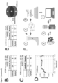
- Illumina next generation sequencing of miRNA libraries showed a length distribution of sequence reads that indicated the presence of a 20-24 bp fraction, which is in accordance with the expected size distribution of miRNAs ( Fig. 1C ). Further examination of the read annotation composition demonstrated that the majority of 20-24 bp reads could be mapped to canonical miRBase (v22) reference sequences ( Fig. 1D ).
- sequencing results showed a highly diverse set of miRNAs with on average 552 ⁇ 171 (mean ⁇ sd) different miRNAs detected per swab sample.
- PCA principal component analysis
- the miRNA-seq dataset initially comprising 27 tumor and 34 control samples and over 1000 detected miRNAs was processed as outlined in Fig. 2A .
- low quality samples, defined as such containing no reads mapping to mature miRNAs were dropped ( Fig. 2A ).
- miRNAs with very low read numbers ( ⁇ 5 reads) were dropped to remove noisy signals.
- a reduced sample set of 26 tumor and 32 control samples with a total of 268 robustly detected miRNAs finally served to train and test the tumor prediction models.
- the data were used either directly as raw counts or alternatively with prior normalization relative to the three most stable reference miRNAs (SRMs) in combination with min-max scaling to compensate for differences in sequencing depth between the samples.
- SRMs most stable reference miRNAs
- miRNA abundance between tumor and control samples were examined with three different standard statistical methods implemented in the Bioconductor packages edgeR, DESeq2 and limma, and the top ranking three miRNAs (miR-29c-5p, miR-625-3p and miR-148b-3p, Fig. 2B ) from the intersection of all three methods were selected based on high p-values, indicative for non-significant differences between the conditions (Robinson et al. 2010, Love et al. 2014, Law et al. 2014). Within-sample normalization of miRNAs to the mean of log2-transformed SRMs provided a reasonably balanced dataset compared to raw-data.
- K-top scoring pairs a rule-based, parameter-free classifier, utilizes a set (k) of paired measurements (miRNAs) comparing their abundance with each other (e.g. miR-A > miR-B).
- miRNAs paired measurements
- miR-A > miR-B paired measurements
- XGBoost Extreme gradient boosting
- RNAs n-dimensional space
- glmnet a classifier based on logistic regression was applied, which offers an integrated regularization to minimize overfitting effects (Zou et al. 2005).
- the learning algorithms were allowed to use all 268 miRNAs as features for their prediction models, which is referred herein as "full” models.
- the inventors performed a second round of training to provide "sparse" models, with the exception of the kTSP algorithm, which automatically searched for a parsimonious model choosing a set of two miRNA pairs combining three miRNAs (miR-1260a, miR-301a-3p, miR-187-3p) without requiring further optimization.
- the inventors opted to limit the features to the three top and thus most important miRNAs (glmnet: miR-769-3p, miR-22-5p, miR125a-3p; SVM: miR-198-3p, miR-95-3p, miR-193b-3p).
- AUROC receiver operating characteristic curve
- AUROC data is presented as mean ⁇ s.e.m. of all 30 repeats x 10-fold performance evaluations.
- RDEB patients are burdened with extensive, long-standing chronic wounds, which require close monitoring as tumors tend to arise in those areas.
- the inventors used the opportunity of a case study of a 37-year old RDEB patient with a history of multiple, recurrent SCCs within a large chronic wound covering about 80% of her left arm. The patient had undergone multiple surgeries as well as several courses of electrochemotherapy (Bleomycin) due to recurrent, incompletely resected and newly evolving tumors at this site.
- XGBoost provides a probability of a sample being a tumor given the abundance of its signature miRNAs ( Fig. 4D ). Both models were able to predict tumor samples with high accuracy (kTSP: 91.3%, XGBoost: 100%, Fig. 4E-F ), outcompeting the ability of four trained dermatologists to label suspicious areas correctly ( Fig. 4G-J ) based on visual inspection of photographs of the wound with one rater knowing the patient and the respective wound, while the other three did not ( Fig. 4A ). Notably, visual assessment and categorization based on two-dimensional images lacks information that supports clinical decision making, including three-dimensional cues, clinical data on the (patient-specific) course of the lesions, or accompanying symptoms.
- the inventors' sequencing approach provided unbiased insights into the miRNA profiles of wound and tumor swabs. Despite the fact that sequencing is becoming more affordable, the costs and also hands-on time spent preparing the sequencing library are still limiting its use in a diagnostic assay. Ever since the recent Coronavirus pandemic, qPCR has become even more readily available to labs and as services to clinical hospitals. Therefore, the inventors considered a transfer of the inventors' sequence-based findings to a more cost-effective qPCR system with short turnaround times.
- the inventors decided in advance to apply a traditional normalization strategy from relative quantification in semi-qPCR experiments to compensate for differences in sequencing depth in the inventors' data (Livak et al. 2001).
- ⁇ Cq values of the four signature miRNAs correlated with log2 transformed, TMM normalized sequencing counts ( Fig. 5A ). All four signature miRNAs and the reference miRNA were detectable in the qPCR assay and three out of the four signature miRNAs demonstrated significant (p ⁇ 0.02) correlation between sequencing counts and qPCR ⁇ Cq values. Because miR-1260a lacks coherent abundance between the two assays and thus affects one of two voting rules of the kTSP model, the inventors relaxed the majority vote rule so that one of two votes was sufficient to predict a tumor sample. The prediction accuracy of the kTSP model was slightly higher (80.9%) compared to XGBoost (75.2%) ( Fig. 5B, C ).
- the inventors also specified the procedure and required materials necessary to perform the assay as outlined in Fig. 5F . Although routine labs would typically have all required equipment available, a lack of access to bioinformatic resources to interpret the data and run the prediction models might pose a limitation. Therefore, the inventors also developed a Shiny app to demonstrate that the analysis in principle can be carried out on any standard browser without technical specialists.
Landscapes
- Chemical & Material Sciences (AREA)
- Life Sciences & Earth Sciences (AREA)
- Health & Medical Sciences (AREA)
- Organic Chemistry (AREA)
- Proteomics, Peptides & Aminoacids (AREA)
- Engineering & Computer Science (AREA)
- Immunology (AREA)
- Pathology (AREA)
- Analytical Chemistry (AREA)
- Zoology (AREA)
- Genetics & Genomics (AREA)
- Wood Science & Technology (AREA)
- Physics & Mathematics (AREA)
- Biotechnology (AREA)
- Microbiology (AREA)
- Molecular Biology (AREA)
- Hospice & Palliative Care (AREA)
- Biophysics (AREA)
- Oncology (AREA)
- Biochemistry (AREA)
- Bioinformatics & Cheminformatics (AREA)
- General Engineering & Computer Science (AREA)
- General Health & Medical Sciences (AREA)
- Measuring Or Testing Involving Enzymes Or Micro-Organisms (AREA)
Priority Applications (2)
| Application Number | Priority Date | Filing Date | Title |
|---|---|---|---|
| EP23156564.9A EP4417709A1 (de) | 2023-02-14 | 2023-02-14 | Mirna-basierter diagnostischer test zum nachweis eines risikos, hauttumore gegen den hintergrund schwerer hautläsionen zu entwickeln |
| PCT/EP2024/053027 WO2024170372A1 (en) | 2023-02-14 | 2024-02-07 | Mirna-based diagnostic assay for the detection of a risk to develop skin tumors against the background of severe skin lesions |
Applications Claiming Priority (1)
| Application Number | Priority Date | Filing Date | Title |
|---|---|---|---|
| EP23156564.9A EP4417709A1 (de) | 2023-02-14 | 2023-02-14 | Mirna-basierter diagnostischer test zum nachweis eines risikos, hauttumore gegen den hintergrund schwerer hautläsionen zu entwickeln |
Publications (1)
| Publication Number | Publication Date |
|---|---|
| EP4417709A1 true EP4417709A1 (de) | 2024-08-21 |
Family
ID=85239094
Family Applications (1)
| Application Number | Title | Priority Date | Filing Date |
|---|---|---|---|
| EP23156564.9A Pending EP4417709A1 (de) | 2023-02-14 | 2023-02-14 | Mirna-basierter diagnostischer test zum nachweis eines risikos, hauttumore gegen den hintergrund schwerer hautläsionen zu entwickeln |
Country Status (2)
| Country | Link |
|---|---|
| EP (1) | EP4417709A1 (de) |
| WO (1) | WO2024170372A1 (de) |
Citations (6)
| Publication number | Priority date | Publication date | Assignee | Title |
|---|---|---|---|---|
| WO2008010558A1 (en) | 2006-07-20 | 2008-01-24 | Panasonic Corporation | Ultrasonic probe |
| US20120059048A1 (en) | 2009-04-23 | 2012-03-08 | Northwestern University | METHODS FOR DIAGNOSING AND TREATING SQUAMOUS CELL CARCINOMA UTILIZING miRNA-205 AND INHIBITORS THEREOF |
| EP2794881B1 (de) | 2011-12-22 | 2018-06-27 | InteRNA Technologies B.V. | Mirna zur behandlung von kopf-hals-karzinom |
| US20180327853A1 (en) * | 2015-11-05 | 2018-11-15 | Arphion Diagnostics | Process for the indentification of patients at risk for oscc |
| US20190284554A1 (en) * | 2016-03-07 | 2019-09-19 | The United States Of America, As Represented By The Secretary, Dept. Of Health And Human Services | Micrornas and methods of their use |
| EP4015653A1 (de) * | 2020-12-16 | 2022-06-22 | Universita' degli studi di Brescia | Verwendung von mirnas als diagnostische und prognostische biomarker eines oralen schuppenzellkarzinoms |
Family Cites Families (1)
| Publication number | Priority date | Publication date | Assignee | Title |
|---|---|---|---|---|
| WO2013063544A1 (en) | 2011-10-27 | 2013-05-02 | Asuragen, Inc. | Mirnas as diagnostic biomarkers to distinguish benign from malignant thyroid tumors |
-
2023
- 2023-02-14 EP EP23156564.9A patent/EP4417709A1/de active Pending
-
2024
- 2024-02-07 WO PCT/EP2024/053027 patent/WO2024170372A1/en not_active Ceased
Patent Citations (6)
| Publication number | Priority date | Publication date | Assignee | Title |
|---|---|---|---|---|
| WO2008010558A1 (en) | 2006-07-20 | 2008-01-24 | Panasonic Corporation | Ultrasonic probe |
| US20120059048A1 (en) | 2009-04-23 | 2012-03-08 | Northwestern University | METHODS FOR DIAGNOSING AND TREATING SQUAMOUS CELL CARCINOMA UTILIZING miRNA-205 AND INHIBITORS THEREOF |
| EP2794881B1 (de) | 2011-12-22 | 2018-06-27 | InteRNA Technologies B.V. | Mirna zur behandlung von kopf-hals-karzinom |
| US20180327853A1 (en) * | 2015-11-05 | 2018-11-15 | Arphion Diagnostics | Process for the indentification of patients at risk for oscc |
| US20190284554A1 (en) * | 2016-03-07 | 2019-09-19 | The United States Of America, As Represented By The Secretary, Dept. Of Health And Human Services | Micrornas and methods of their use |
| EP4015653A1 (de) * | 2020-12-16 | 2022-06-22 | Universita' degli studi di Brescia | Verwendung von mirnas als diagnostische und prognostische biomarker eines oralen schuppenzellkarzinoms |
Non-Patent Citations (88)
| Title |
|---|
| AFSARI BFERTIG EJGEMAN DMARCHIONNI L: "switchBox: an R package for k-Top Scoring Pairs classifier development", BIOINFORMATICS, vol. 31, no. 2, 26 September 2014 (2014-09-26), pages 273 - 4 |
| APARICIO-PUERTA ELEBRON RRUEDA AGOMEZ-MARTIN CGIANNOUKAKOS SJASPEZ D ET AL.: "Nucleic Acids Res.", vol. 47, 2019, OXFORD ACADEMIC, article "sRNAbench and sRNAtoolbox 2019: intuitive fast small RNA profiling and differential expression", pages: W530 - 5 |
| BARTEL DP: "MicroRNAs: genomics, biogenesis, mechanism, and function", CELL, vol. 116, no. 2, 23 January 2004 (2004-01-23), pages 281 - 97, XP002359089, DOI: 10.1016/S0092-8674(04)00045-5 |
| BOERI MPASTORINO USOZZI G: "Role of microRNAs in lung cancer: microRNA signatures in cancer prognosis", CANCER J, vol. 18, no. 3, May 2012 (2012-05-01), pages 268 - 74, XP008175873, DOI: 10.1097/PPO.0b013e318258b743 |
| BRUCE EA ET AL.: "Direct RT-qPCR detection of SARS-CoV-2 RNA from patient nasopharyngeal swabs without an RNA extraction step", PLOS BIOL, vol. 18, no. 10, 2 October 2020 (2020-10-02), pages e3000896, XP055781605, DOI: 10.1371/journal.pbio.3000896 |
| CALIN GACROCE CM: "MicroRNA signatures in human cancers", NAT REV CANCER, vol. 6, no. 11, November 2006 (2006-11-01), pages 857 - 66, XP002473506, DOI: 10.1038/nrc1997 |
| CHACON-SOLANO E ET AL.: "Fibroblast activation and abnormal extracellular matrix remodelling as common hallmarks in three cancer-prone genodermatoses", BR J DERMATOL, vol. 181, no. 3, 15 April 2019 (2019-04-15), pages 512 - 522 |
| CHEN TIANQIGUESTRIN CARLOS: "XGBoost: A Scalable Tree Boosting System KDD '16", PROCEEDINGS OF THE 22ND ACM SIGKDD INTERNATIONAL CONFERENCE ON KNOWLEDGE DISCOVERY AND DATA MININGAUGUST, 2016, pages 785 - 794, XP058631191, Retrieved from the Internet <URL:https://doi.org/10.1145/2939672.2939785> DOI: 10.1145/2939672.2939785 |
| CHO RJ ET AL.: "APOBEC mutation drives early-onset squamous cell carcinomas in recessive dystrophic epidermolysis bullosa", SCI TRANSL MED, vol. 10, no. 455, 22 August 2018 (2018-08-22) |
| CIANFARANI FZAMBRUNO GCASTIGLIA DODORISIO T: "Pathomechanisms of Altered Wound Healing in Recessive Dystrophic Epidermolysis Bullosa", AM J PATHOL, vol. 187, no. 7, 29 April 2017 (2017-04-29), pages 1445 - 1453 |
| CONDORELLI A G ET AL: "MicroRNA-145-5p regulates fibrotic features of recessive dystrophic epidermolysis bullosa skin fibroblasts", BRITISH JOURNAL OF DERMATOLOGY, vol. 181, no. 5, November 2019 (2019-11-01), pages 1017 - 1027, XP009545247, ISSN: 0007-0963 * |
| CONDORELLI AGDELLAMBRA ELOGLI EZAMBRUNO GCASTIGLIA D: "Epidermolysis Bullosa-Associated Squamous Cell Carcinoma: From Pathogenesis to Therapeutic Perspectives", INT J MOL SCI, vol. 20, no. 22, 14 November 2019 (2019-11-14), pages 5707 |
| CONDORELLI ET AL.: "MicroRNA-145-5p regulates fibrotic features of recessive dystrophic epidermolysis bullosa skin fibroblasts", BR J DERMATOL, vol. 181, no. 5, 20 October 2019 (2019-10-20), pages 1017 - 1027, XP071160549, DOI: 10.1111/bjd.17840 |
| CROCE CM: "Causes and consequences of microRNA dysregulation in cancer", NAT REV GENET., vol. 10, no. 10, pages 704 - 14, XP055032479, DOI: 10.1038/nrg2634 |
| CUI MWANG HYAO XZHANG DXIE YCUI RZHANG X: "Circulating MicroRNAs in Cancer: Potential and Challenge", FRONT GENET, vol. 10, 18 July 2019 (2019-07-18), pages 626 |
| DING L ET AL.: "Promising therapeutic role of miR-27b in tumor", TUMOUR BIOL, vol. 39, no. 3, March 2017 (2017-03-01) |
| DOU CHANGWEI ET AL: "miR-187-3p inhibits the metastasis and epithelial-mesenchymal transition of hepatocellular carcinoma by targeting S100A4", CANCER LETTERS, NEW YORK, NY, US, vol. 381, no. 2, 17 August 2016 (2016-08-17), pages 380 - 390, XP029735873, ISSN: 0304-3835, DOI: 10.1016/J.CANLET.2016.08.011 * |
| DOU CLIU ZXU MJIA YWANG YLI QYANG WZHENG XTU KLIU Q: "miR-187-3p inhibits the metastasis and epithelial-mesenchymal transition of hepatocellular carcinoma by targeting S100A4", CANCER LETT., vol. 381, no. 2, 17 August 2016 (2016-08-17), pages 380 - 90, XP029735873, DOI: 10.1016/j.canlet.2016.08.011 |
| FANG H ET AL.: "Proinflammatory role of blister fluid-derived exosomes in bullous pemphigoid", J PATHOL, vol. 245, no. 1, 2 April 2018 (2018-04-02), pages 114 - 125 |
| FARSHCHIAN, MEHDI ET AL.: "Serpin peptidase inhibitor clade A member 1 (SerpinA1) is a novel biomarker for progression of cutaneous squamous cell carcinoma.", THE AMERICAN JOURNAL OF PATHOLOGY, vol. 179, no. 3, 2011, pages 1110 - 9, XP055332550, DOI: 10.1016/j.ajpath.2011.05.012 |
| FINE JDJOHNSON LBWEINER MLI KPSUCHINDRAN C: "Epidermolysis bullosa and the risk of life-threatening cancers: the National EB Registry experience, 1986-2006", J AM ACAD DERMATOL., vol. 60, no. 2, 20 November 2008 (2008-11-20), pages 203 - 11, XP025892245, DOI: 10.1016/j.jaad.2008.09.035 |
| FORTUNATO O ET AL.: "Circulating mir-320a promotes immunosuppressive macrophages M2 phenotype associated with lung cancer risk", INT J CANCER, vol. 144, no. 11, 1 June 2019 (2019-06-01), pages 2746 - 2761, XP071290737, DOI: 10.1002/ijc.31988 |
| FUENTES I ET AL.: "Reduced Microbial Diversity Is a Feature of Recessive Dystrophic Epidermolysis Bullosa-Involved Skin and Wounds", J INVEST DERMATOL, vol. 138, no. 11, 16 May 2018 (2018-05-16), pages 2492 - 2495 |
| G. A. CALIN ET AL.: "Frequent deletions and down-regulation of micro- RNA genes miR15 and miR16 at 13q14 in chronic lymphocytic leukemia", PNAS, vol. 99, no. 24, November 2002 (2002-11-01), pages 15524 - 15529, XP002982123, DOI: 10.1073/pnas.242606799 |
| GHAFOUR AA ET AL.: "High expression level of miR-1260 family in the peripheral blood of patients with ovarian carcinoma", J OVARIAN RES, vol. 14, no. 1, 10 October 2021 (2021-10-10), pages 131 |
| GRUBER C ET AL.: "The design and optimization of RNA trans-splicing molecules for skin cancer therapy", MOL ONCOL, vol. 7, no. 6, 19 August 2013 (2013-08-19), pages 1056 - 68, XP055099110, DOI: 10.1016/j.molonc.2013.08.005 |
| GUO J ET AL.: "miR-301a-3p induced by endoplasmic reticulum stress mediates the occurrence and transmission of trastuzumab resistance in HER2-positive gastric cancer", CELL DEATH DIS, vol. 12, no. 7, 13 July 2021 (2021-07-13), pages 696 |
| GUTTMANN-GRUBER, C. ET AL.: "Low-dose calcipotriol can elicit wound closure, anti-microbial, and anti-neoplastic effects in epidermolysis bullosa keratinocytes", SCI REP, vol. 8, no. 1, 2018, pages 13430 |
| HAN M ET AL.: "MiR-27b-3p exerts tumor suppressor effects in esophageal squamous cell carcinoma by targeting Nrf2", HUM CELL, vol. 33, no. 3, 17 May 2020 (2020-05-17), pages 641 - 651, XP037179833, DOI: 10.1007/s13577-020-00329-7 |
| HANAHAN DWEINBERG RA: "Hallmarks of cancer: the next generation", CELL, vol. 144, no. 5, 4 March 2011 (2011-03-04), pages 646 - 74, XP028185429, DOI: 10.1016/j.cell.2011.02.013 |
| HAS C ET AL.: "Consensus reclassification of inherited epidermolysis bullosa and other disorders with skin fragility", BR J DERMATOL, vol. 183, no. 4, 11 March 2020 (2020-03-11), pages 614 - 627, XP071159281, DOI: 10.1111/bjd.18921 |
| HONG LZ ET AL.: "Systematic evaluation of multiple qPCR platforms, NanoString and miRNA-Seq for microRNA biomarker discovery in human biofluids", SCI REP, vol. 11, no. 1, 24 February 2021 (2021-02-24), pages 4435 |
| HUANG MWCHEN CWLIN WCKE SWTSAI CF: "SVM and SVM Ensembles in Breast Cancer Prediction", PLOS ONE, vol. 12, no. 1, 6 January 2017 (2017-01-06), pages e0161501 |
| J. MATSUZAKIT. OCHIYA: "Circulating microRNAs: Next-generation Cancer Detection", KEIO J MED, May 2020 (2020-05-01) |
| KIM MLI MINTONG-WHEELER LRATRAN KMARUCCI DMURRELL DF: "Epidemiology and Outcome of Squamous Cell Carcinoma in Epidermolysis Bullosa in Australia and New Zealand", ACTA DERM VENEREOL, vol. 98, no. 1, 12 January 2018 (2018-01-12), pages 70 - 76 |
| KIVISAARI AK ET AL.: "Transformation-specific matrix metalloproteinases (MMP)-7 and MMP-13 are expressed by tumour cells in epidermolysis bullosa-associated squamous cell carcinomas", BR J DERMATOL, vol. 158, no. 4, 16 February 2008 (2008-02-16), pages 778 - 85 |
| KOSHIZUKA KEIICHI ET AL: "Involvement of aberrantly expressed microRNAs in the pathogenesis of head and neck squamous cell carcinoma", CANCER METASTASIS, KLUWER ACADEMIC PUBLISHERS, DORDRECHT, NL, vol. 36, no. 3, 23 August 2017 (2017-08-23), pages 525 - 545, XP036327996, ISSN: 0167-7659, [retrieved on 20170823], DOI: 10.1007/S10555-017-9692-Y * |
| KUHN, M: "Building Predictive Models in R Using the caret Package", JOURNAL OF STATISTICAL SOFTWARE, vol. 28, no. 5, pages 1 - 26, Retrieved from the Internet <URL:https://doi.org/10.18637/jss.v028.i05> |
| L. WANGM. HANX. LIN. ZHANGH. CHENG: "Review of Classification Methods on Unbalanced Data Sets", IEEE ACCESS, vol. 9, 2021, pages 64606 - 64628, XP011851628, DOI: 10.1109/ACCESS.2021.3074243 |
| LAW CWCHEN YSHI WSMYTH GK: "voom: Precision weights unlock linear model analysis tools for RNA-seq read counts", GENOME BIOL, vol. 15, no. 2, 3 February 2014 (2014-02-03), pages R29, XP021177342, DOI: 10.1186/gb-2014-15-2-r29 |
| LE SJOSSE JHUSSON F: "FactoMineR: A Package for Multivariate Analysis", JOURNAL OF STATISTICAL SOFTWARE, vol. 25, no. 1, 2008, pages 1 - 18 |
| LETTNER, T. ET AL.: "Increased levels of matrix metalloproteinase-9 and interleukin-8 in blister fluids of dystrophic and junctional epidermolysis bullosa patients", J EUR ACAD DERMATOL VENEREOL, vol. 29, no. 2, 2015, pages 396 - 8 |
| LIU CHUNG-JI ET AL: "Plasma miR-187* is a potential biomarker for oral carcinoma", CLINICAL ORAL INVESTIGATIONS, SPRINGER, BERLIN, DE, vol. 21, no. 4, 20 June 2016 (2016-06-20), pages 1131 - 1138, XP036216175, ISSN: 1432-6981, [retrieved on 20160620], DOI: 10.1007/S00784-016-1887-Z * |
| LIVAKKJSCHMITTGEN TD: "Analysis of relative gene expression data using real-time quantitative PCR and the 2(-Delta Delta C(T)) Method", METHODS, vol. 25, no. 4, pages 402 - 8 |
| LOVE MIHUBER WANDERS S: "Moderated estimation of fold change and dispersion for RNA-seq data with DESeq2", GENOME BIOL, vol. 15, no. 12, 2014, pages 550, XP021210395, DOI: 10.1186/s13059-014-0550-8 |
| M. A. CORTEZ ET AL.: "MicroRNAs in body fluids-the mix of hormones and biomarkers", NAT REV CLIN ONCOL, vol. 8, no. 8, June 2011 (2011-06-01), pages 467 - 477, XP055209761, DOI: 10.1038/nrclinonc.2011.76 |
| MANDUJANO-TINOCO EAGARCIA-VENZOR AMUNOZ-GALINDO LLIZARRAGA-SANCHEZ FOROZCO ACHAVEZ-GUTIERREZ EKROTZSCH ESALGADO RMMELENDEZ-ZAJGLA : "miRNA expression profile in multicellular breast cancer spheroids", BIOCHIM BIOPHYS ACTA MOL CELL RES, vol. 1864, no. 10, 31 May 2017 (2017-05-31), pages 1642 - 1655, XP085184290, DOI: 10.1016/j.bbamcr.2017.05.023 |
| MELLERIO JE ET AL.: "Management of cutaneous squamous cell carcinoma in patients with epidermolysis bullosa: best clinical practice guidelines", BR J DERMATOL, vol. 174, no. 1, 7 November 2015 (2015-11-07), pages 56 - 67, XP071093399, DOI: 10.1111/bjd.14104 |
| MENSAH, M. ET AL.: "MicroRNA Based Liquid Biopsy: The Experience of the Plasma miRNA Signature Classifier (MSC) for Lung Cancer Screening", JOURNAL OF VISUALIZED EXPERIMENTS : JOVE, no. 128, 2017, pages 56326, Retrieved from the Internet <URL:https://doi.org/10.3791/56326> |
| NG YI-ZHEN ET AL: "Fibroblast-Derived Dermal Matrix Drives Development of Aggressive Cutaneous Squamous Cell Carcinoma in Patients with Recessive Dystrophic Epidermolysis Bullosa", CANCER RESEARCH, vol. 72, no. 14, 15 July 2012 (2012-07-15), US, pages 3522 - 3534, XP093061240, ISSN: 0008-5472, Retrieved from the Internet <URL:https://aacrjournals.org/cancerres/article-pdf/72/14/3522/2668871/3522.pdf> DOI: 10.1158/0008-5472.CAN-11-2996 * |
| NYSTROM A ET AL.: "Collagen VII plays a dual role in wound healing", J CLIN INVEST, vol. 123, no. 8, 8 July 2013 (2013-07-08), pages 3498 - 509 |
| NYSTROM A ET AL.: "Impaired lymphoid extracellular matrix impedes antibacterial immunity in epidermolysis bullosa", PROC NATL ACAD SCI U S A., vol. 115, no. 4, 5 January 2018 (2018-01-05), pages E705 - E714 |
| NYSTROM A ET AL.: "Losartan ameliorates dystrophic epidermolysis bullosa and uncovers new disease mechanisms", EMBO MOL MED, vol. 7, no. 9, September 2015 (2015-09-01), pages 1211 - 28, XP055596211, DOI: 10.15252/emmm.201505061 |
| OPITZ DMACLIN R: "Popular Ensemble Methods: An Empirical Study", JOURNAL OF ARTIFICIAL INTELLIGENCE RESEARCH, vol. 11, 1999, pages 169 - 198 |
| P. S. MITCHELL ET AL.: "Circulating microRNAs as stable blood-based markers for cancer detection", PROC NATL ACAD SCI USA, vol. 105, no. 30, July 2008 (2008-07-01), pages 10513 - 10518 |
| PASTORINO U ET AL.: "Baseline computed tomography screening and blood microRNA predict lung cancer risk and define adequate intervals in the BioMILD trial", ANN ONCOL, vol. 33, no. 4, 25 January 2022 (2022-01-25), pages 395 - 405 |
| PHILLIPS T ET AL.: "Aberrant recruitment of leukocytes defines poor wound healing in patients with recessive dystrophic epidermolysis bullosa", J DERMATOL SCI, vol. 100, no. 3, 17 October 2020 (2020-10-17), pages 209 - 216, XP086418651, DOI: 10.1016/j.jdermsci.2020.10.009 |
| PRITCHARD CC ET AL.: "Blood cell origin of circulating microRNAs: a cautionary note for cancer biomarker studies", CANCER PREV RES (PHILA, vol. 5, no. 3, 12 December 2011 (2011-12-12), pages 492 - 497, XP055198618, DOI: 10.1158/1940-6207.CAPR-11-0370 |
| R CORE TEAM: "R: A language and environment for statistical computing", R FOUNDATION FOR STATISTICAL COMPUTING, VIENNA, AUSTRIA, 2020, Retrieved from the Internet <URL:https://www.R-project.org/.> |
| RIIHILA P ET AL.: "Tumour-cell-derived complement components C1r and C1s promote growth of cutaneous squamous cell carcinoma", BR J DERMATOL, vol. 182, no. 3, 28 July 2019 (2019-07-28), pages 658 - 670 |
| ROBINSON MDOSHLACK A: "A scaling normalization method for differential expression analysis of RNA-seq data", GENOME BIOL, vol. 11, no. 3, 2 March 2010 (2010-03-02), pages R25, XP021070870 |
| RODRIGUEZ-GALAN A ET AL.: "MiRNA post-transcriptional modification dynamics in T cell activation", ISCIENCE, vol. 24, no. 6, 12 May 2021 (2021-05-12), pages 102530 |
| ROSSATO M ET AL.: "II,-10-induced microRNA-187 negatively regulates TNF-a, IL-6, and IL-12p40 production in TLR4-stimulated monocytes", PROC NATL ACAD SCI U S A., vol. 109, no. 45, 15 October 2012 (2012-10-15), pages E3101 - 10 |
| RSTUDIO TEAM, RSTUDIO: INTEGRATED DEVELOPMENT FOR R. RSTUDIO, PBC, BOSTON, MA, 2020, Retrieved from the Internet <URL:http://www.rstudio.com/.> |
| RUPAIMOOLE RCALIN GALOPEZ-BERESTEIN GSOOD AK: "miRNA Deregulation in Cancer Cells and the Tumor Microenvironment", CANCER DISCOV, vol. 6, no. 3, 10 February 2016 (2016-02-10), pages 235 - 46 |
| SCHMOHL, M. ET AL.: "Superficial wound swabbing: a novel method of sampling and processing wound fluid for subsequent immunoassay analysis in diabetic foot ulcerations", DIABETES CARE, vol. 35, no. 11, 2012, pages 2113 - 20 |
| SCHWARZENBACH HNISHIDA NCALIN GAPANTEL K: "Clinical relevance of circulating cell-free microRNAs in cancer", NAT REV CLIN ONCOL, vol. 11, no. 3, 4 February 2014 (2014-02-04), pages 145 - 56, XP055209756, DOI: 10.1038/nrclinonc.2014.5 |
| SHEN SJSONG YREN XYXU YLZHOU YDLIANG ZYSUN Q: "MicroRNA-27b-3p Promotes Tumor Progression and Metastasis by Inhibiting Peroxisome Proliferator-Activated Receptor Gamma in Triple-Negative Breast Cancer", FRONT ONCOL, vol. 10, 5 August 2020 (2020-08-05), pages 1371 |
| SOLIS DCTENG CGORELL ESBARRIGA MNAZAROFF JLI SLU YBRUCKNER AMARINKOVICH MPTANG JY: "Classification of 2 distinct wound types in recessive dystrophic epidermolysis bullosa: A retrospective and cohort natural history study", J AM ACAD DERMATOL, vol. 85, no. 5, 4 September 2020 (2020-09-04), pages 1296 - 1298, XP086825066, DOI: 10.1016/j.jaad.2020.08.118 |
| SOZZI G ET AL.: "Clinical utility of a plasma-based miRNA signature classifier within computed tomography lung cancer screening: a correlative MILD trial study", J CLIN ONCOL, vol. 32, no. 8, 13 January 2014 (2014-01-13), pages 768 - 73 |
| SREEDHARAN L ET AL.: "MicroRNA profile in neosquamous esophageal mucosa following ablation of Barrett's esophagus", WORLD J GASTROENTEROL, vol. 23, no. 30, 14 August 2017 (2017-08-14), pages 5508 - 5518 |
| SUN Y: "Extracellular Vesicles as Biomarkers for the Detection of a Tumor Marker Gene in Epidermolysis Bullosa-Associated Squamous Cell Carcinoma", J INVEST DERMATOL, vol. 138, no. 5, 2 December 2017 (2017-12-02), pages 1197 - 1200 |
| TARTAGLIA GCAO QPADRON ZMSOUTH AP: "Impaired Wound Healing, Fibrosis, and Cancer: The Paradigm of Recessive Dystrophic Epidermolysis Bullosa", INT J MOL SCI, vol. 22, no. 10, 12 May 2021 (2021-05-12), pages 5104 |
| TONCIC RJPETKOVIC MSUSIC SMCEOVIC RARGENZIANO G: "Use of dermatoscopy in the detection of squamous cell carcinoma in a patient with recessive dystrophic epidermolysis bullosa", DERMATOL PRACT CONCEPT, vol. 8, no. 3, 31 July 2018 (2018-07-31), pages 227 - 230 |
| TORRI A ET AL.: "Extracellular MicroRNA Signature of Human Helper T Cell Subsets in Health and Autoimmunity", J BIOL CHEM., vol. 292, no. 7, 11 January 2017 (2017-01-11), pages 2903 - 2915 |
| VANDEN OEVER ET AL.: "miR-29 Regulates Type VII Collagen in Recessive Dystrophic Epidermolysis Bullosa", J INVEST DERMATOL, vol. 136, no. 10, October 2016 (2016-10-01), pages 2013 - 2021 |
| WANG J ET AL.: "miR-301a Suppression within Fibroblasts Limits the Progression of Fibrosis through the TSC1/mTOR Pathway", MOL THER NUCLEIC ACIDS, vol. 21, 26 May 2020 (2020-05-26), pages 217 - 228 |
| WIMMER ET AL.: "A cancer stem cell-like phenotype is associated with miR-10b expression in aggressive squamous cell carcinomas", CELL COMMUN SIGNAL, vol. 18, no. 1, 10 April 2020 (2020-04-10), pages 61 |
| WIMMER, MONIKA ET AL.: "A cancer stem cell-like phenotype is associated with miR-10b expression in aggressive squamous cell carcinomas", CELL COMMUNICATION AND SIGNALING: CCS, vol. 18, 10 April 2020 (2020-04-10) |
| WOODLEY DTCOGAN JMOSALLAEI DYIM KCHEN M: "Characterization of mutant type VII collagens underlying the inversa subtype of recessive dystrophic epidermolysis bullosa", J DERMATOL SCI, vol. 104, no. 2, 23 September 2021 (2021-09-23), pages 104 - 111, XP086880216, DOI: 10.1016/j.jdermsci.2021.09.006 |
| Y. PENGC. M. CROCE: "The role of MicroRNAs in human cancer", SIGNAL TRANSDUCTION AND TARGETED THERAPY, vol. 1, no. 1, January 2016 (2016-01-01) |
| YAGER, D.R.R.A. KULINAL.A. GILMAN: "Wound fluids: a window into the wound environment?", INT J LOW EXTREM WOUNDS, vol. 6, no. 4, 2007, pages 262 - 72, XP009160609, DOI: 10.1177/1534734607307035 |
| YAYLAK BAKGUL B: "Experimental MicroRNA Detection Methods", METHODS MOL BIOL, vol. 2257, 2022, pages 33 - 55 |
| YOUN, Y. J. ET AL.: "Neutrophil-derived trail is a proinflammatory subtype of neutrophil-derived extracellular vesicles", THERANOSTICS, vol. 11, no. 6, 2021, pages 2770 - 2787, Retrieved from the Internet <URL:https://doi.org/10.7150/thno.51756> |
| ZAUNER RWIMMER MATZMUELLER SPROELL JNIKLAS NABLINGER MREISENBERGER MLETTNER TBAUER J WWALLY V: "Biomarker discovery in rare malignancies: development of a miRNA signature for RDEB-cSCC. manuscript in preparation for publication", BIOMARKER RES, 2022 |
| ZAUNER RWIMMER MDORFER SABLINGER MKOLLER UPINON HOFBAUER JGUTTMANN-GRUBER CBAUER JWWALLY V: "Transcriptome-Guided Drug Repurposing for Aggressive SCCs", INT J MOL SCI, vol. 23, no. 2, 17 January 2022 (2022-01-17), pages 1007 |
| ZHANG NLIU JF: "MicroRNA (MiR)-301a-3p regulates the proliferation of esophageal squamous cells via targeting PTEN", BIOENGINEERED, vol. 11, no. 1, pages 972 - 983 |
| ZOU HHASTIE T: "Regularization and variable selection via the elastic net", JOURNAL OF THE ROYAL STATISTICAL SOCIETY: SERIES B (STATISTICAL METHODOLOGY, vol. 67, 2005, pages 301 - 20, XP055849418, DOI: 10.1111/j.1467-9868.2005.00503.x |
Also Published As
| Publication number | Publication date |
|---|---|
| WO2024170372A1 (en) | 2024-08-22 |
Similar Documents
| Publication | Publication Date | Title |
|---|---|---|
| Riester et al. | Combination of a novel gene expression signature with a clinical nomogram improves the prediction of survival in high-risk bladder cancer | |
| Wild et al. | Gene expression profiling of progressive papillary noninvasive carcinomas of the urinary bladder | |
| Lee et al. | Prediction of recurrence-free survival in postoperative non–small cell lung cancer patients by using an integrated model of clinical information and gene expression | |
| Mannucci et al. | Stool and blood biomarkers for colorectal cancer management: an update on screening and disease monitoring | |
| Schuetz et al. | Progression-specific genes identified by expression profiling of matched ductal carcinomas in situ and invasive breast tumors, combining laser capture microdissection and oligonucleotide microarray analysis | |
| Segura et al. | Melanoma MicroRNA signature predicts post-recurrence survival | |
| Bansal et al. | Feasibility of microRNAs as biomarkers for Barrett's esophagus progression: a pilot cross-sectional, phase 2 biomarker study | |
| US20140162887A1 (en) | Methods of using gene expression signatures to select a method of treatment, predict prognosis, survival, and/or predict response to treatment | |
| Mittempergher et al. | A gene signature for late distant metastasis in breast cancer identifies a potential mechanism of late recurrences | |
| Mastracci et al. | Genomic alterations in lobular neoplasia: a microarray comparative genomic hybridization signature for early neoplastic proliferationin the breast | |
| JP7354099B2 (ja) | マイクロrna遺伝子発現を使用して黒色腫を診断し、病期分類し、モニタリングする方法 | |
| WO2018001295A1 (zh) | 分子标志物、内参基因及其应用、检测试剂盒以及检测模型的构建方法 | |
| Zhang et al. | Circulating microRNA 216 as a marker for the early identification of severe acute pancreatitis | |
| JP2020503850A (ja) | 腫瘍抑制foxo活性を酸化ストレスから区別する方法 | |
| JP2014516531A (ja) | 肺癌に対するバイオマーカー | |
| Yang et al. | SnoRNAs are involved in the progression of ulcerative colitis and colorectal cancer | |
| Kawaguchi et al. | Gene Expression Signature–Based Prognostic Risk Score in Patients with Primary Central Nervous System Lymphoma | |
| Stricker et al. | Validation of a prognostic multi-gene signature in high-risk neuroblastoma using the high throughput digital NanoString nCounter™ system | |
| Villemin et al. | A cell-to-patient machine learning transfer approach uncovers novel basal-like breast cancer prognostic markers amongst alternative splice variants | |
| CA2684897A1 (en) | Prostate cancer survival and recurrence | |
| Jandova et al. | Sporadic early-onset colon cancer expresses unique molecular features | |
| He et al. | Seven-microRNA panel for lung adenocarcinoma early diagnosis in patients presenting with ground-glass nodules | |
| CN111187840A (zh) | 一种用于早期乳腺癌诊断的生物标志物 | |
| AU2009337963B2 (en) | Prognosis of breast cancer patients by monitoring the expression of two genes | |
| Xu et al. | Evidence that dysplasia related microRNAs in Barrett’s esophagus target PD-L1 expression and contribute to the development of esophageal adenocarcinoma |
Legal Events
| Date | Code | Title | Description |
|---|---|---|---|
| PUAI | Public reference made under article 153(3) epc to a published international application that has entered the european phase |
Free format text: ORIGINAL CODE: 0009012 |
|
| STAA | Information on the status of an ep patent application or granted ep patent |
Free format text: STATUS: THE APPLICATION HAS BEEN PUBLISHED |
|
| AK | Designated contracting states |
Kind code of ref document: A1 Designated state(s): AL AT BE BG CH CY CZ DE DK EE ES FI FR GB GR HR HU IE IS IT LI LT LU LV MC ME MK MT NL NO PL PT RO RS SE SI SK SM TR |
|
| STAA | Information on the status of an ep patent application or granted ep patent |
Free format text: STATUS: REQUEST FOR EXAMINATION WAS MADE |
|
| 17P | Request for examination filed |
Effective date: 20241204 |
|
| RBV | Designated contracting states (corrected) |
Designated state(s): AL AT BE BG CH CY CZ DE DK EE ES FI FR GB GR HR HU IE IS IT LI LT LU LV MC ME MK MT NL NO PL PT RO RS SE SI SK SM TR |