EP4420638A2 - Transkathetervorrichtung und minimal invasives verfahren zum einziehen und einstellen des blutflusses durch ein blutgefäss - Google Patents
Transkathetervorrichtung und minimal invasives verfahren zum einziehen und einstellen des blutflusses durch ein blutgefäss Download PDFInfo
- Publication number
- EP4420638A2 EP4420638A2 EP24187796.8A EP24187796A EP4420638A2 EP 4420638 A2 EP4420638 A2 EP 4420638A2 EP 24187796 A EP24187796 A EP 24187796A EP 4420638 A2 EP4420638 A2 EP 4420638A2
- Authority
- EP
- European Patent Office
- Prior art keywords
- pulmonary artery
- flow restrictor
- membrane
- frame
- restrictor device
- Prior art date
- Legal status (The legal status is an assumption and is not a legal conclusion. Google has not performed a legal analysis and makes no representation as to the accuracy of the status listed.)
- Pending
Links
Images


Classifications
-
- A—HUMAN NECESSITIES
- A61—MEDICAL OR VETERINARY SCIENCE; HYGIENE
- A61B—DIAGNOSIS; SURGERY; IDENTIFICATION
- A61B17/00—Surgical instruments, devices or methods
- A61B17/12—Surgical instruments, devices or methods for ligaturing or otherwise compressing tubular parts of the body, e.g. blood vessels or umbilical cord
- A61B17/12022—Occluding by internal devices, e.g. balloons or releasable wires
- A61B17/12027—Type of occlusion
- A61B17/12036—Type of occlusion partial occlusion
-
- A—HUMAN NECESSITIES
- A61—MEDICAL OR VETERINARY SCIENCE; HYGIENE
- A61B—DIAGNOSIS; SURGERY; IDENTIFICATION
- A61B17/00—Surgical instruments, devices or methods
- A61B17/12—Surgical instruments, devices or methods for ligaturing or otherwise compressing tubular parts of the body, e.g. blood vessels or umbilical cord
- A61B17/12022—Occluding by internal devices, e.g. balloons or releasable wires
- A61B17/12099—Occluding by internal devices, e.g. balloons or releasable wires characterised by the location of the occluder
- A61B17/12109—Occluding by internal devices, e.g. balloons or releasable wires characterised by the location of the occluder in a blood vessel
-
- A—HUMAN NECESSITIES
- A61—MEDICAL OR VETERINARY SCIENCE; HYGIENE
- A61B—DIAGNOSIS; SURGERY; IDENTIFICATION
- A61B17/00—Surgical instruments, devices or methods
- A61B17/12—Surgical instruments, devices or methods for ligaturing or otherwise compressing tubular parts of the body, e.g. blood vessels or umbilical cord
- A61B17/12022—Occluding by internal devices, e.g. balloons or releasable wires
- A61B17/12131—Occluding by internal devices, e.g. balloons or releasable wires characterised by the type of occluding device
- A61B17/12168—Occluding by internal devices, e.g. balloons or releasable wires characterised by the type of occluding device having a mesh structure
- A61B17/12172—Occluding by internal devices, e.g. balloons or releasable wires characterised by the type of occluding device having a mesh structure having a pre-set deployed three-dimensional shape
-
- A—HUMAN NECESSITIES
- A61—MEDICAL OR VETERINARY SCIENCE; HYGIENE
- A61B—DIAGNOSIS; SURGERY; IDENTIFICATION
- A61B17/00—Surgical instruments, devices or methods
- A61B17/12—Surgical instruments, devices or methods for ligaturing or otherwise compressing tubular parts of the body, e.g. blood vessels or umbilical cord
- A61B17/12022—Occluding by internal devices, e.g. balloons or releasable wires
- A61B17/12131—Occluding by internal devices, e.g. balloons or releasable wires characterised by the type of occluding device
- A61B17/12168—Occluding by internal devices, e.g. balloons or releasable wires characterised by the type of occluding device having a mesh structure
- A61B17/12177—Occluding by internal devices, e.g. balloons or releasable wires characterised by the type of occluding device having a mesh structure comprising additional materials, e.g. thrombogenic, having filaments, having fibers or being coated
-
- A—HUMAN NECESSITIES
- A61—MEDICAL OR VETERINARY SCIENCE; HYGIENE
- A61F—FILTERS IMPLANTABLE INTO BLOOD VESSELS; PROSTHESES; DEVICES PROVIDING PATENCY TO, OR PREVENTING COLLAPSING OF, TUBULAR STRUCTURES OF THE BODY, e.g. STENTS; ORTHOPAEDIC, NURSING OR CONTRACEPTIVE DEVICES; FOMENTATION; TREATMENT OR PROTECTION OF EYES OR EARS; BANDAGES, DRESSINGS OR ABSORBENT PADS; FIRST-AID KITS
- A61F2/00—Filters implantable into blood vessels; Prostheses, i.e. artificial substitutes or replacements for parts of the body; Appliances for connecting them with the body; Devices providing patency to, or preventing collapsing of, tubular structures of the body, e.g. stents
- A61F2/82—Devices providing patency to, or preventing collapsing of, tubular structures of the body, e.g. stents
-
- A—HUMAN NECESSITIES
- A61—MEDICAL OR VETERINARY SCIENCE; HYGIENE
- A61M—DEVICES FOR INTRODUCING MEDIA INTO, OR ONTO, THE BODY; DEVICES FOR TRANSDUCING BODY MEDIA OR FOR TAKING MEDIA FROM THE BODY; DEVICES FOR PRODUCING OR ENDING SLEEP OR STUPOR
- A61M29/00—Dilators with or without means for introducing media, e.g. remedies
- A61M29/02—Dilators made of swellable material
-
- A—HUMAN NECESSITIES
- A61—MEDICAL OR VETERINARY SCIENCE; HYGIENE
- A61B—DIAGNOSIS; SURGERY; IDENTIFICATION
- A61B17/00—Surgical instruments, devices or methods
- A61B17/12—Surgical instruments, devices or methods for ligaturing or otherwise compressing tubular parts of the body, e.g. blood vessels or umbilical cord
- A61B17/12022—Occluding by internal devices, e.g. balloons or releasable wires
- A61B2017/1205—Introduction devices
- A61B2017/12054—Details concerning the detachment of the occluding device from the introduction device
-
- A—HUMAN NECESSITIES
- A61—MEDICAL OR VETERINARY SCIENCE; HYGIENE
- A61F—FILTERS IMPLANTABLE INTO BLOOD VESSELS; PROSTHESES; DEVICES PROVIDING PATENCY TO, OR PREVENTING COLLAPSING OF, TUBULAR STRUCTURES OF THE BODY, e.g. STENTS; ORTHOPAEDIC, NURSING OR CONTRACEPTIVE DEVICES; FOMENTATION; TREATMENT OR PROTECTION OF EYES OR EARS; BANDAGES, DRESSINGS OR ABSORBENT PADS; FIRST-AID KITS
- A61F2/00—Filters implantable into blood vessels; Prostheses, i.e. artificial substitutes or replacements for parts of the body; Appliances for connecting them with the body; Devices providing patency to, or preventing collapsing of, tubular structures of the body, e.g. stents
- A61F2/02—Prostheses implantable into the body
- A61F2/04—Hollow or tubular parts of organs, e.g. bladders, tracheae, bronchi or bile ducts
- A61F2/06—Blood vessels
- A61F2002/068—Modifying the blood flow model, e.g. by diffuser or deflector
Definitions
- This invention relates to a pulmonary artery restriction device and a minimally invasive method for constricting and adjusting blood flow through a blood vessel.
- Congenital heart disease refers to the various malformations of the heart and surrounding vessels that occur prior to birth. Almost 1% of children are born with some form of significant CHD. CHD remains a leading cause of infant death in the United States. Many infants with CHD develop exuberant blood flow to their lungs that, if left untreated, may result in overwhelming congestive heart failure and death. For example, in ventricular septal defect (VSD), the most common type of CHD occurring in about 37% of cases, holes in the septum allows oxygen rich blood entering into the left ventricle from the lungs to leak out to the right ventricle and escape out the pulmonary artery instead of out the aorta and to the rest of the body.
- VSD ventricular septal defect
- the pulmonary artery band results in distortion of the pulmonary arteries which can negatively impact future surgical intervention.
- the pulmonary artery band can only be adjusted through additional surgery.
- placement of a surgical shunt or conduit may result in distortion of the pulmonary arteries.
- Such artificial connections have the potential for thrombosis, distortion and occlusion, which may have fatal consequences. See also U.S. Patent No. 6,638,257 and WO 2015/114471 both incorporated herein by this reference.
- a pulmonary artery flow restrictor system comprising a funnel shaped membrane with a proximal base and a restrictive distal opening which is stretchable to larger sizes and a self-expanding frame attached to the proximal base of the membrane for securing the membrane within the pulmonary artery.
- the frame includes arms extending upward over the membrane distal opening.
- the funnel shaped membrane may be made of a polymer, for example, polytetrafluoroethylene (ePTFE).
- the frame may be made of a shape memory alloy, for example, Nitinol.
- the frame may include a stent like structure with a series of spaced upper and lower apexes.
- the spaced lower apexes are secured to the proximal base of the membrane.
- the frame further includes a plurality of bent anchoring arms extending upwardly over the membrane distal opening each formed from members extending from adjacent frame upper apexes. The arms may cross above the membrane distal opening.
- the pulmonary artery flow restrictor may further include one or more flexible lines connected to the frame for collapsing the frame.
- the pulmonary artery flow restrictor system may further include a transcatheter device for delivering the membrane and frame into the pulmonary artery.
- the transcatheter device includes an inner lumen about a guide wire and a retractable lumen retractable relative to the inner lumen.
- the frame may be removably attached to the inner lumen, using, for example, pins attached to the inner lumen and frame eyelets receiving said pins therethrough.
- a pulmonary artery flow restrictor comprising a membrane including a restrictive opening.
- the membrane is made of a material which is irreversibly stretchable to larger sizes by a balloon catheter to vary the size of the restrictive opening.
- a self-expanding frame is attached to the membrane for securing the membrane within the pulmonary artery.
- the frame includes arms extending upward over the membrane restrictive opening.
- the membrane is preferably made of a polymer such as polytetrafluoroethylene (ePTFE).
- One or more embodiments of the transcatheter device and minimally invasive method for constricting and adjusting blood flow through a blood vessel of this invention provides a minimally invasive surgery (MIS) device which can be implanted within the main pulmonary artery in order to elevate flow resistance to palliate the patient's CHD symptoms and move toward systemic flow.
- the device includes a self-expanding metal frame or skeleton which constrains the device radially and axially attached to a balloon-expandable membrane, also referred to herein as a pulmonary artery resistor, that interacts with the blood flow and provides flow constriction.
- the pulmonary artery resistor facilitates MIS intervention for modification of flow resistance.
- the device may be deployed minimally invasively via catheter by a cardiac surgeon.
- the pulmonary artery resistor may provide the highest level of flow resistance possible.
- the highest level resistance would palliate patients with the most severe CHD symptoms, such as multiple and large septal tears the highest spectrum CHD cases.
- the surgeon may use a balloon catheter of the desired size to incrementally expand the size of the resistor in situ.
- An adequate sized balloon is chosen and delivered over a guidewire to a location inside the resistor. The balloon is then dilated to expand the resistor. The balloon is then deflated and finally removed.
- This cycle ensures that the adjustment is performed quickly since a fully expanded balloon in the middle of the main pulmonary artery (Main PA) may cause an acute spike in pulmonary pressure and stress on the heart.
- Evaluation of the sufficiency of the resistance change provided by the pulmonary artery resistor is obtained after the dilation balloon has performed the expansion task and is fully removed from the patient, while the guidewire and femoral introducer catheter remain in place.
- a surgeon may incrementally alter the resistance further by stepping up the size of the balloon and repeating the dilation procedure.
- the expansion cycles continue until a satisfactory pulmonary arterial pressure is reached while adequate oxygenated blood flow to the aorta is also satisfactory.
- MIS procedures to reduce resistance may be periodically necessary to maintain desired apportionment of blood flow between the lungs and the remainder of the body as the patient grows.
- the minimally invasive procedure to correct the flow resistance should reduce or eliminate post-surgery recovery time in an intensive care unit and hospital stay duration when compared to conventional pulmonary artery banding which requires open surgery.
- the pulmonary artery resistor incorporates the flow constriction geometry necessary to create the desired flow without distorting the pulmonary artery wall.
- the frame of the resistor may be a tubular closed or open celled lattice made out of a shape memory alloy such as nitinol.
- the frame may be plated in gold or other radiopaque material or alloy to improve visibility inside the vessel.
- the frame may include eyelets with inserted radiopaque rivets that enhance visibility for the surgeon.
- the frame preferably includes at least two crossing arms, which preferably extend beyond its tubular radius.
- the arms may include slots for improved adhesion to the vasculature walls.
- the distal ends of the arms may feature tips that have an increased surface area and a backwards bend, essentially forming feet that reduce the contact pressure against the vessel wall.
- the arms may be tied together in the center to prevent the frame from collapsing under high pressure.
- the resistor membrane may be a 2D annular shape or an annular shape stretched along the third axis forming a conical tube or funnel.
- the membrane may be made of an inelastic material that retains its new shape after deformation by the balloon as described above.
- the material may be woven or otherwise structured in such a way that the membrane preferentially stretches in the radial direction while minimizing foreshortening.
- the material may be an expanded polytetrafluoroethylene (ePTFE) or similar type material.
- the membrane may be attached to the frame chemically such as an adhesive bond, mechanically such as stitching, hook and loop, thermal bonding or related technology.
- the membrane may be extruded, wrapped, or otherwise formed separately from the frame and then attached in a post process.
- the membrane may also be formed directly on the frame via sputtering, wrapping, or other means.
- the expandable membrane may be attached on the proximal side of the device such that in the conical configuration the narrow end of the funnel extends distally down the center of the frame.
- the resistor may be deployed through a transcatheter procedure by which the device is collapsed between an inner guidewire lumen and outer sheath lumen.
- there may include two or more proximally located eyelets on the frame which may extend axially beyond the main body of the frame to interact with pins, hooks, recesses, or some other attachment mechanism attached to the guidewire lumen.
- the mechanism constrains the axial and rotational motion of the frame such that any motion of the attachment device translates into corresponding motion by the frame.
- This link may be used for ejection of the device out the distal end of the deployment catheter, for axially repositioning or rotating a semi-deployed device, or for re-sheathing a semi-deployed device.
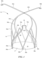
- Fig. 1 shows an example of a pulmonary artery flow resistor 10 including funnel shaped membrane 12 and self-expanding frame 14.
- Membrane 12 has proximal base 16 having a wide opening 17 (e.g., with a diameter of between 10 and 18 mm) and a distal narrow opening or spout 18 (e.g., having a diameter of between 0.5 and 6 mm).
- frame 14 is secured to the base 16 of the membrane.
- base 16 was 14 mm in diameter and restricted opening was 2 mm in diameter.
- the membrane functions to resist blood flow through the pulmonary artery 20, Fig. 2 since, when the flow resistor is in place in the pulmonary artery 20, blood is restricted to only flow out distal narrow opening 18 of membrane 12 since base 16 is urged against the inner wall of the artery.
- Frame 14 functions to retain membrane 12 in place in the pulmonary artery.
- membrane 12 is made of polytetrafluoroethylene (ePTFE) or similar type polymer material typically between 0.3 thousandths of an inch and 3 thousandths of an inch thick. Other polymers may be used.
- ePTFE polytetrafluoroethylene
- Frame 14 may be made of a shape memory alloy such as Nitinol. Frame 14 may further include inwardly bent arms 22a and 22b extending upward over membrane 12 and crossing above restricted opening 18 as shown in Figs. 1 and 2 . The arms may each extend into a branch of the pulmonary artery as shown. The arms 22a, 22b have a large surface area to reduce pressure on the pulmonary artery wall and function to anchor membrane 12 in place against the normal blood flow.
- a shape memory alloy such as Nitinol.
- Frame 14 may further include inwardly bent arms 22a and 22b extending upward over membrane 12 and crossing above restricted opening 18 as shown in Figs. 1 and 2 .
- the arms may each extend into a branch of the pulmonary artery as shown.
- the arms 22a, 22b have a large surface area to reduce pressure on the pulmonary artery wall and function to anchor membrane 12 in place against the normal blood flow.
- frame 14 is a stent like in structure and being self-expanding it expands the membrane 12 and maintains contact with pulmonary artery wall even as the pulmonary artery grows in size.
- frame 14 includes circumferential lower spaced apexes 30a, 30b, 30c and the like and upper spaced apexes 32a, 32b, 32c, and the like each between adjacent lower apexes.
- the lower apexes may be secured to the proximal base 16 of membrane 12.
- Arms 22a, 22b may include members extending from select upper apexes of the frame. So, for example, arm 22b includes member 34a extending from apex 32a and member 34b extending from adjacent apex 32b.
- Crossing arms 22a, 22b may be include downwardly bent distal eyelet tips 36a, 36b, respectively.
- lower apexes 30a, 30b, 30c, and the like may include eyelets 38a, 38b, and 38c, and the like, respectively.
- Eyelet 38a and the eyelet directly across from it may be slightly enlarged and constitute deployment eyelets which fit over deployment pins associated with a deployment device.
- the other eyelets e.g., 38b, 38c, and the like
- the entire frame including the arms may be formed by cutting a single thin tube of Nitinol which is then expanded on a mandrel and then heat treated so that it naturally retains this expanded shape.
- Radiopaque stripes 15a, 15b may be included (printed on or adhered to) on membrane 12 to enable visualization of the membrane during deployment into the pulmonary artery.
- Frame 14 may include a radiopaque coating to enable visualization of the frame during deployment into the pulmonary artery.
- the flow resistor reduces the effective diameter of the pulmonary artery to 2 mm, a diameter which can be changed by using a balloon catheter to expand the flow resistor.
- the flow resistor may provide a maximum pressure drop of 40-50 mm Hg in the pulmonary artery and resist the blood flow rate to a maximum of 1.5-2 L/min. If the membrane is fully expanded, the flow resistor would produce no pressure drop and no flow rate reduction. In testing, the flow restrictor shown in Fig.
- the membrane 1 could be collapsed down into a 2mm diameter package and then expanded such that base 16 and frame 14 were 13 mm-14 mm in diameter (or equal to the vessel diameter).
- the height of the membrane may be 8-16mm.
- the frame may be 5-15mm (from upper apex to lower apex) and the arms may be 15-30mm long.
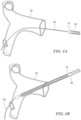
- the system may further include transcatheter device 60, Fig. 3 for delivering the membrane and the frame into the pulmonary artery.
- the delivery device preferably includes inner braided guide wire lumen about guide wire 65 and retractile lumen 66 (e.g., 3-5Fr) which is retractable relative to inner lumen 62 and flared at its tip (to, e.g., 5-9Fr) forming a garage section.
- the frame is releasably attached to the inner lumen, for example, by placing opposing deployment eyelets on pins 68a, 68b. Thus the frame and the membrane are collapsed and reside between lumen 66 and lumen 62.
- An outer support sheath 70 may also be provided.
- Braided guide wire lumen 62 may be thinned out just before a garage section giving maximum space between the garage section and guide wire lumen 62 in order to fit the restrictor device.
- the tip of the transcatheter device and the deployment pins 68a, 68b are attached to the thinned portion of guide wire lumen 62.
- the outer most lumen 70 serves to reduce friction for the retractile lumen 66 as it retracts relative to outer lumen 70.
- Outer lumen 70 terminates before the garage section giving space for the translation.
- the retractile sheath 66 slides along the lubricious outer lumen inner liner rather than along the vessel walls.
- Fig. 4A shows catheter 60 following the guide wire 20 to the artery.
- the catheter continues past the valve to just beyond the split in the pulmonary artery.
- the lumen 66 is pulled back to reveal the arms of the restrictor frame.
- the lumen 66 is pulled back more until the arms begin to separate and in Fig. 4E the catheter is gradually pushed forward keeping the outer sheath back.
- the inner lumen 62 is then advanced, Fig. 4F until the deployment pins reach the end of the sheath and in Fig. 4G the lumen 66 is pulled back allowing the frame and the membrane to spring into position.
- the catheter and the guide wire are removed leaving the flow restrictor in place.
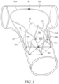
- Fig. 5 shows a design where one or more flexible lines (e.g., sutures) 80 are connected to frame 14 for collapsing the frame and removing the frame and the membrane 12 from the pulmonary artery 20.
- a suture 80a, 80b, 80c are each secured to the lower apexes 38a, 38b, 38c, respectively of frame 14 to form a web with a hook, loop, ball, knot, or other feature 82 tying the other end of all the sutures together in the center.
- This feature 82 may be snared by a standard transcatheter snare.
- arms 22a, 22b may be flared members in a light bulb shape as shown with two or three arms intersecting in the center where they are sutured together as shown as 84.
- the suture keeps the arms in place and acts as a hinge allowing the arms to collapse into the outer lumen during deployment or during retrieval.
- the light bulb shape of the arms fit into the T of the artery.
- a snare may be guided through an outer lumen to grab onto feature 82 and then pulled back into the outer lumen whereupon frame 14 collapses by the force on sutures 80.
Landscapes
- Health & Medical Sciences (AREA)
- Life Sciences & Earth Sciences (AREA)
- Surgery (AREA)
- Engineering & Computer Science (AREA)
- Biomedical Technology (AREA)
- Animal Behavior & Ethology (AREA)
- Veterinary Medicine (AREA)
- Vascular Medicine (AREA)
- Public Health (AREA)
- Heart & Thoracic Surgery (AREA)
- General Health & Medical Sciences (AREA)
- Molecular Biology (AREA)
- Medical Informatics (AREA)
- Nuclear Medicine, Radiotherapy & Molecular Imaging (AREA)
- Reproductive Health (AREA)
- Anesthesiology (AREA)
- Hematology (AREA)
- Cardiology (AREA)
- Oral & Maxillofacial Surgery (AREA)
- Transplantation (AREA)
- Surgical Instruments (AREA)
- Prostheses (AREA)
Applications Claiming Priority (3)
| Application Number | Priority Date | Filing Date | Title |
|---|---|---|---|
| US201662289402P | 2016-02-01 | 2016-02-01 | |
| PCT/US2017/015826 WO2017136341A1 (en) | 2016-02-01 | 2017-01-31 | Transcatheter device and minimally invasive method for constricting and adjusting blood flow through a blood vessel |
| EP17748015.9A EP3410982A4 (de) | 2016-02-01 | 2017-01-31 | Transkathetervorrichtung und minimalinvasives verfahren zur verengung und anpassung des blutflusses durch ein blutgefäss |
Related Parent Applications (1)
| Application Number | Title | Priority Date | Filing Date |
|---|---|---|---|
| EP17748015.9A Division EP3410982A4 (de) | 2016-02-01 | 2017-01-31 | Transkathetervorrichtung und minimalinvasives verfahren zur verengung und anpassung des blutflusses durch ein blutgefäss |
Publications (2)
| Publication Number | Publication Date |
|---|---|
| EP4420638A2 true EP4420638A2 (de) | 2024-08-28 |
| EP4420638A3 EP4420638A3 (de) | 2024-11-13 |
Family
ID=59385933
Family Applications (2)
| Application Number | Title | Priority Date | Filing Date |
|---|---|---|---|
| EP24187796.8A Pending EP4420638A3 (de) | 2016-02-01 | 2017-01-31 | Transkathetervorrichtung und minimal invasives verfahren zum einziehen und einstellen des blutflusses durch ein blutgefäss |
| EP17748015.9A Withdrawn EP3410982A4 (de) | 2016-02-01 | 2017-01-31 | Transkathetervorrichtung und minimalinvasives verfahren zur verengung und anpassung des blutflusses durch ein blutgefäss |
Family Applications After (1)
| Application Number | Title | Priority Date | Filing Date |
|---|---|---|---|
| EP17748015.9A Withdrawn EP3410982A4 (de) | 2016-02-01 | 2017-01-31 | Transkathetervorrichtung und minimalinvasives verfahren zur verengung und anpassung des blutflusses durch ein blutgefäss |
Country Status (3)
| Country | Link |
|---|---|
| US (3) | US10568634B2 (de) |
| EP (2) | EP4420638A3 (de) |
| WO (1) | WO2017136341A1 (de) |
Families Citing this family (10)
| Publication number | Priority date | Publication date | Assignee | Title |
|---|---|---|---|---|
| US10667931B2 (en) * | 2014-07-20 | 2020-06-02 | Restore Medical Ltd. | Pulmonary artery implant apparatus and methods of use thereof |
| US10568634B2 (en) | 2016-02-01 | 2020-02-25 | Vivonics, Inc. | Transcatheter device and minimally invasive method for constricting and adjusting blood flow through a blood vessel |
| US11771434B2 (en) | 2016-09-28 | 2023-10-03 | Restore Medical Ltd. | Artery medical apparatus and methods of use thereof |
| IL271184B2 (en) | 2017-06-05 | 2024-04-01 | Restore Medical Ltd | Fixed-length, double-walled stent-like device and methods of using the same |
| US11963864B2 (en) * | 2019-01-11 | 2024-04-23 | Varun Shetty | Method and system for reducing pulmonary flow |
| USD956220S1 (en) | 2020-05-01 | 2022-06-28 | Tau-Pnu Medical Co., Ltd. | Transcatheter device |
| JP2024517070A (ja) * | 2021-04-02 | 2024-04-19 | スターライト・カーディオバスキュラー・インコーポレーテッド | 血流制限器 |
| WO2023133475A2 (en) * | 2022-01-05 | 2023-07-13 | Rl3T Llc | Adaptive and radially expanding speculum |
| US11883030B2 (en) | 2022-04-29 | 2024-01-30 | inQB8 Medical Technologies, LLC | Systems, devices, and methods for controllably and selectively occluding, restricting, and diverting flow within a patient's vasculature |
| AU2023262477A1 (en) | 2022-04-29 | 2024-12-05 | Relief Cardiovascular, Inc. | Systems, devices, and methods for controllably and selectively occluding, restricting, and diverting flow within a patient's vasculature |
Citations (4)
| Publication number | Priority date | Publication date | Assignee | Title |
|---|---|---|---|---|
| US5662711A (en) | 1995-06-07 | 1997-09-02 | Douglas; William | Flow adjustable artery shunt |
| US6638257B2 (en) | 2002-03-01 | 2003-10-28 | Aga Medical Corporation | Intravascular flow restrictor |
| US20140236211A1 (en) | 2013-02-18 | 2014-08-21 | King Abdullah International Medical Research Center | Minimally invasive pulmonary artery band |
| WO2015114471A1 (en) | 2014-01-31 | 2015-08-06 | Jacques Seguin | Apparatus using an anchored balloon for treating pulmonary arterial hypertension |
Family Cites Families (13)
| Publication number | Priority date | Publication date | Assignee | Title |
|---|---|---|---|---|
| US6120534A (en) | 1997-10-29 | 2000-09-19 | Ruiz; Carlos E. | Endoluminal prosthesis having adjustable constriction |
| DE60317474T2 (de) * | 2002-03-05 | 2008-10-02 | Salviac Ltd. | System aus embolischem filter und rückziehschlinge |
| US20070293808A1 (en) * | 2006-04-27 | 2007-12-20 | Williams Michael S | Renal blood flow augmentation for congestive heart failure treatment |
| US20090287290A1 (en) | 2008-01-24 | 2009-11-19 | Medtronic, Inc. | Delivery Systems and Methods of Implantation for Prosthetic Heart Valves |
| US8876854B2 (en) * | 2008-04-03 | 2014-11-04 | Cook Medical Technologies Llc | Implant release mechanism |
| DE202009018984U1 (de) | 2008-07-15 | 2015-01-29 | St. Jude Medical, Inc. | Tasche zur Verwendung in einer Herzklappenprothese |
| US9649480B2 (en) | 2012-07-06 | 2017-05-16 | Corvia Medical, Inc. | Devices and methods of treating or ameliorating diastolic heart failure through pulmonary valve intervention |
| WO2014022124A1 (en) | 2012-07-28 | 2014-02-06 | Tendyne Holdings, Inc. | Improved multi-component designs for heart valve retrieval device, sealing structures and stent assembly |
| US8926690B2 (en) | 2012-08-13 | 2015-01-06 | Medtronic, Inc. | Heart valve prosthesis |
| US20140262884A1 (en) * | 2013-03-14 | 2014-09-18 | Apothecary Products, Inc. | Medicine storage arrangements and methods of assembly and use |
| US9636116B2 (en) * | 2013-06-14 | 2017-05-02 | Artventive Medical Group, Inc. | Implantable luminal devices |
| US10568634B2 (en) | 2016-02-01 | 2020-02-25 | Vivonics, Inc. | Transcatheter device and minimally invasive method for constricting and adjusting blood flow through a blood vessel |
| EP3243451B1 (de) * | 2016-05-12 | 2020-08-05 | IntelliStent AG | Vorrichtung zur einstellbaren einschränkung von intravaskulärem strom |
-
2017
- 2017-01-31 US US15/420,772 patent/US10568634B2/en active Active
- 2017-01-31 EP EP24187796.8A patent/EP4420638A3/de active Pending
- 2017-01-31 EP EP17748015.9A patent/EP3410982A4/de not_active Withdrawn
- 2017-01-31 WO PCT/US2017/015826 patent/WO2017136341A1/en not_active Ceased
-
2019
- 2019-12-27 US US16/728,028 patent/US11278289B2/en active Active
-
2022
- 2022-02-16 US US17/672,893 patent/US11980369B2/en active Active
Patent Citations (4)
| Publication number | Priority date | Publication date | Assignee | Title |
|---|---|---|---|---|
| US5662711A (en) | 1995-06-07 | 1997-09-02 | Douglas; William | Flow adjustable artery shunt |
| US6638257B2 (en) | 2002-03-01 | 2003-10-28 | Aga Medical Corporation | Intravascular flow restrictor |
| US20140236211A1 (en) | 2013-02-18 | 2014-08-21 | King Abdullah International Medical Research Center | Minimally invasive pulmonary artery band |
| WO2015114471A1 (en) | 2014-01-31 | 2015-08-06 | Jacques Seguin | Apparatus using an anchored balloon for treating pulmonary arterial hypertension |
Also Published As
| Publication number | Publication date |
|---|---|
| EP4420638A3 (de) | 2024-11-13 |
| US20170215885A1 (en) | 2017-08-03 |
| US11980369B2 (en) | 2024-05-14 |
| US20220183696A1 (en) | 2022-06-16 |
| US20200146683A1 (en) | 2020-05-14 |
| WO2017136341A1 (en) | 2017-08-10 |
| EP3410982A1 (de) | 2018-12-12 |
| US10568634B2 (en) | 2020-02-25 |
| EP3410982A4 (de) | 2019-10-30 |
| US11278289B2 (en) | 2022-03-22 |
Similar Documents
| Publication | Publication Date | Title |
|---|---|---|
| US11980369B2 (en) | Transcatheter device and minimally invasive method for constricting and adjusting blood flow through a blood vessel | |
| US20210315558A1 (en) | Devices and methods for delivery of expandable prostheses | |
| JP5759949B2 (ja) | プロテーゼの経皮的送達のための送達ツール | |
| JP7042259B2 (ja) | ステントグラフトを半径方向に収縮するための送達システム | |
| AU2007329243B2 (en) | System and method for transapical delivery of an annulus anchored self-expanding valve | |
| CN103124537B (zh) | 无支架支撑结构 | |
| JP2023052763A (ja) | 束縛可能なステントグラフト、送達システムおよび使用方法 | |
| JP2025023967A (ja) | 心臓弁ドッキング・コイルとシステム | |
| CN113143542B (zh) | 主动脉瓣闭锁不全修复设备及方法 | |
| US20070168013A1 (en) | Vascular graft and deployment system | |
| JP2019072511A (ja) | 反転臨時弁シース | |
| AU2016215197A1 (en) | Expandable epicardial pads and devices and methods for their delivery | |
| US12588909B2 (en) | Transcatheter device and minimally invasive method for constricting and adjusting blood flow through a blood vessel | |
| JP2008541865A (ja) | 経腔的送達用の非円筒形人工弁システム | |
| US20240122711A1 (en) | System and method for reducing tricuspid regurgitation | |
| CN114948333A (zh) | 带有压力释放端口的支架移植物假体 | |
| WO2026026756A1 (zh) | 腔内植入系统 |
Legal Events
| Date | Code | Title | Description |
|---|---|---|---|
| PUAI | Public reference made under article 153(3) epc to a published international application that has entered the european phase |
Free format text: ORIGINAL CODE: 0009012 |
|
| STAA | Information on the status of an ep patent application or granted ep patent |
Free format text: STATUS: THE APPLICATION HAS BEEN PUBLISHED |
|
| AC | Divisional application: reference to earlier application |
Ref document number: 3410982 Country of ref document: EP Kind code of ref document: P |
|
| AK | Designated contracting states |
Kind code of ref document: A2 Designated state(s): AL AT BE BG CH CY CZ DE DK EE ES FI FR GB GR HR HU IE IS IT LI LT LU LV MC MK MT NL NO PL PT RO RS SE SI SK SM TR |
|
| REG | Reference to a national code |
Ref country code: DE Ref legal event code: R079 Free format text: PREVIOUS MAIN CLASS: A61F0002440000 Ipc: A61F0002000000 |
|
| PUAL | Search report despatched |
Free format text: ORIGINAL CODE: 0009013 |
|
| AK | Designated contracting states |
Kind code of ref document: A3 Designated state(s): AL AT BE BG CH CY CZ DE DK EE ES FI FR GB GR HR HU IE IS IT LI LT LU LV MC MK MT NL NO PL PT RO RS SE SI SK SM TR |
|
| RIC1 | Information provided on ipc code assigned before grant |
Ipc: A61F 2/44 20060101ALI20241010BHEP Ipc: A61F 2/38 20060101ALI20241010BHEP Ipc: A61F 2/30 20060101ALI20241010BHEP Ipc: A61F 2/28 20060101ALI20241010BHEP Ipc: A61F 2/24 20060101ALI20241010BHEP Ipc: A61F 2/06 20130101ALI20241010BHEP Ipc: A61F 2/00 20060101AFI20241010BHEP |
|
| STAA | Information on the status of an ep patent application or granted ep patent |
Free format text: STATUS: REQUEST FOR EXAMINATION WAS MADE |
|
| 17P | Request for examination filed |
Effective date: 20250120 |
|
| GRAP | Despatch of communication of intention to grant a patent |
Free format text: ORIGINAL CODE: EPIDOSNIGR1 |
|
| STAA | Information on the status of an ep patent application or granted ep patent |
Free format text: STATUS: GRANT OF PATENT IS INTENDED |
|
| INTG | Intention to grant announced |
Effective date: 20260220 |