EP4432301A1 - Verfahren zum trainieren eines künstlichen neuronalen netzwerks mit bereitstellung des ergebnisses einer pathologieprobe und computersystem zur durchführung davon - Google Patents
Verfahren zum trainieren eines künstlichen neuronalen netzwerks mit bereitstellung des ergebnisses einer pathologieprobe und computersystem zur durchführung davon Download PDFInfo
- Publication number
- EP4432301A1 EP4432301A1 EP22907881.1A EP22907881A EP4432301A1 EP 4432301 A1 EP4432301 A1 EP 4432301A1 EP 22907881 A EP22907881 A EP 22907881A EP 4432301 A1 EP4432301 A1 EP 4432301A1
- Authority
- EP
- European Patent Office
- Prior art keywords
- training
- pathology
- neural network
- image
- stain
- Prior art date
- Legal status (The legal status is an assumption and is not a legal conclusion. Google has not performed a legal analysis and makes no representation as to the accuracy of the status listed.)
- Pending
Links
Images




Classifications
-
- G—PHYSICS
- G06—COMPUTING OR CALCULATING; COUNTING
- G06V—IMAGE OR VIDEO RECOGNITION OR UNDERSTANDING
- G06V10/00—Arrangements for image or video recognition or understanding
- G06V10/70—Arrangements for image or video recognition or understanding using pattern recognition or machine learning
- G06V10/77—Processing image or video features in feature spaces; using data integration or data reduction, e.g. principal component analysis [PCA] or independent component analysis [ICA] or self-organising maps [SOM]; Blind source separation
- G06V10/774—Generating sets of training patterns; Bootstrap methods, e.g. bagging or boosting
-
- G—PHYSICS
- G06—COMPUTING OR CALCULATING; COUNTING
- G06N—COMPUTING ARRANGEMENTS BASED ON SPECIFIC COMPUTATIONAL MODELS
- G06N3/00—Computing arrangements based on biological models
- G06N3/02—Neural networks
- G06N3/08—Learning methods
-
- G—PHYSICS
- G06—COMPUTING OR CALCULATING; COUNTING
- G06N—COMPUTING ARRANGEMENTS BASED ON SPECIFIC COMPUTATIONAL MODELS
- G06N3/00—Computing arrangements based on biological models
- G06N3/02—Neural networks
- G06N3/04—Architecture, e.g. interconnection topology
-
- G—PHYSICS
- G06—COMPUTING OR CALCULATING; COUNTING
- G06N—COMPUTING ARRANGEMENTS BASED ON SPECIFIC COMPUTATIONAL MODELS
- G06N3/00—Computing arrangements based on biological models
- G06N3/02—Neural networks
- G06N3/08—Learning methods
- G06N3/084—Backpropagation, e.g. using gradient descent
-
- G—PHYSICS
- G06—COMPUTING OR CALCULATING; COUNTING
- G06T—IMAGE DATA PROCESSING OR GENERATION, IN GENERAL
- G06T7/00—Image analysis
-
- G—PHYSICS
- G06—COMPUTING OR CALCULATING; COUNTING
- G06T—IMAGE DATA PROCESSING OR GENERATION, IN GENERAL
- G06T7/00—Image analysis
- G06T7/0002—Inspection of images, e.g. flaw detection
- G06T7/0012—Biomedical image inspection
-
- G—PHYSICS
- G06—COMPUTING OR CALCULATING; COUNTING
- G06T—IMAGE DATA PROCESSING OR GENERATION, IN GENERAL
- G06T7/00—Image analysis
- G06T7/90—Determination of colour characteristics
-
- G—PHYSICS
- G06—COMPUTING OR CALCULATING; COUNTING
- G06V—IMAGE OR VIDEO RECOGNITION OR UNDERSTANDING
- G06V10/00—Arrangements for image or video recognition or understanding
- G06V10/70—Arrangements for image or video recognition or understanding using pattern recognition or machine learning
- G06V10/82—Arrangements for image or video recognition or understanding using pattern recognition or machine learning using neural networks
-
- G—PHYSICS
- G06—COMPUTING OR CALCULATING; COUNTING
- G06V—IMAGE OR VIDEO RECOGNITION OR UNDERSTANDING
- G06V20/00—Scenes; Scene-specific elements
- G06V20/60—Type of objects
- G06V20/69—Microscopic objects, e.g. biological cells or cellular parts
-
- G—PHYSICS
- G16—INFORMATION AND COMMUNICATION TECHNOLOGY [ICT] SPECIALLY ADAPTED FOR SPECIFIC APPLICATION FIELDS
- G16H—HEALTHCARE INFORMATICS, i.e. INFORMATION AND COMMUNICATION TECHNOLOGY [ICT] SPECIALLY ADAPTED FOR THE HANDLING OR PROCESSING OF MEDICAL OR HEALTHCARE DATA
- G16H30/00—ICT specially adapted for the handling or processing of medical images
- G16H30/40—ICT specially adapted for the handling or processing of medical images for processing medical images, e.g. editing
-
- G—PHYSICS
- G16—INFORMATION AND COMMUNICATION TECHNOLOGY [ICT] SPECIALLY ADAPTED FOR SPECIFIC APPLICATION FIELDS
- G16H—HEALTHCARE INFORMATICS, i.e. INFORMATION AND COMMUNICATION TECHNOLOGY [ICT] SPECIALLY ADAPTED FOR THE HANDLING OR PROCESSING OF MEDICAL OR HEALTHCARE DATA
- G16H50/00—ICT specially adapted for medical diagnosis, medical simulation or medical data mining; ICT specially adapted for detecting, monitoring or modelling epidemics or pandemics
- G16H50/20—ICT specially adapted for medical diagnosis, medical simulation or medical data mining; ICT specially adapted for detecting, monitoring or modelling epidemics or pandemics for computer-aided diagnosis, e.g. based on medical expert systems
-
- A—HUMAN NECESSITIES
- A61—MEDICAL OR VETERINARY SCIENCE; HYGIENE
- A61B—DIAGNOSIS; SURGERY; IDENTIFICATION
- A61B10/00—Instruments for taking body samples for diagnostic purposes; Other methods or instruments for diagnosis, e.g. for vaccination diagnosis, sex determination or ovulation-period determination; Throat striking implements
-
- A—HUMAN NECESSITIES
- A61—MEDICAL OR VETERINARY SCIENCE; HYGIENE
- A61B—DIAGNOSIS; SURGERY; IDENTIFICATION
- A61B5/00—Measuring for diagnostic purposes; Identification of persons
-
- G—PHYSICS
- G06—COMPUTING OR CALCULATING; COUNTING
- G06T—IMAGE DATA PROCESSING OR GENERATION, IN GENERAL
- G06T2207/00—Indexing scheme for image analysis or image enhancement
- G06T2207/10—Image acquisition modality
- G06T2207/10024—Color image
-
- G—PHYSICS
- G06—COMPUTING OR CALCULATING; COUNTING
- G06T—IMAGE DATA PROCESSING OR GENERATION, IN GENERAL
- G06T2207/00—Indexing scheme for image analysis or image enhancement
- G06T2207/10—Image acquisition modality
- G06T2207/10056—Microscopic image
-
- G—PHYSICS
- G06—COMPUTING OR CALCULATING; COUNTING
- G06T—IMAGE DATA PROCESSING OR GENERATION, IN GENERAL
- G06T2207/00—Indexing scheme for image analysis or image enhancement
- G06T2207/20—Special algorithmic details
- G06T2207/20081—Training; Learning
-
- G—PHYSICS
- G06—COMPUTING OR CALCULATING; COUNTING
- G06T—IMAGE DATA PROCESSING OR GENERATION, IN GENERAL
- G06T2207/00—Indexing scheme for image analysis or image enhancement
- G06T2207/20—Special algorithmic details
- G06T2207/20084—Artificial neural networks [ANN]
-
- G—PHYSICS
- G06—COMPUTING OR CALCULATING; COUNTING
- G06T—IMAGE DATA PROCESSING OR GENERATION, IN GENERAL
- G06T2207/00—Indexing scheme for image analysis or image enhancement
- G06T2207/30—Subject of image; Context of image processing
- G06T2207/30004—Biomedical image processing
- G06T2207/30024—Cell structures in vitro; Tissue sections in vitro
-
- G—PHYSICS
- G06—COMPUTING OR CALCULATING; COUNTING
- G06T—IMAGE DATA PROCESSING OR GENERATION, IN GENERAL
- G06T2207/00—Indexing scheme for image analysis or image enhancement
- G06T2207/30—Subject of image; Context of image processing
- G06T2207/30004—Biomedical image processing
- G06T2207/30096—Tumor; Lesion
-
- G—PHYSICS
- G06—COMPUTING OR CALCULATING; COUNTING
- G06V—IMAGE OR VIDEO RECOGNITION OR UNDERSTANDING
- G06V10/00—Arrangements for image or video recognition or understanding
- G06V10/70—Arrangements for image or video recognition or understanding using pattern recognition or machine learning
- G06V10/764—Arrangements for image or video recognition or understanding using pattern recognition or machine learning using classification, e.g. of video objects
-
- G—PHYSICS
- G06—COMPUTING OR CALCULATING; COUNTING
- G06V—IMAGE OR VIDEO RECOGNITION OR UNDERSTANDING
- G06V2201/00—Indexing scheme relating to image or video recognition or understanding
- G06V2201/03—Recognition of patterns in medical or anatomical images
Definitions
- the present disclosure relates to a method of training an artificial neural network which provides a determination result of a pathology specimen and a computing system performing the same. More specifically, the present disclosure relates to a method which is capable of perform determination on stained pathology images through various staining techniques and a computing system performing the same by extracting only parts stained with a specific staining agent from a pathology slide stained with a mixture of various types of stains and training an artificial neural network.
- TEE tumor microenvironment
- Hematoxylin-eosin staining is a method of staining the cell nucleus and stroma area with hematoxylin and eosin in purple and pink, respectively, and is the most basic pathology diagnosis method used by pathologists to identify morphological characteristics of cells and tissues of specimens through light microscopy.
- Immunohistochemistry (IHC) staining is a method which is widely used to measure an expression level of a biomarker such as a specific protein in tissues, and a target biomarker present in tissues based on antigen-antibody binding can be stained with brown diaminobenzidine (DAB) dye to determine its expression level, and hematoxyline staining is used as a counterstain.
- DAB brown diaminobenzidine
- Cocktail staining or multiplex IHC staining methods in which multiple biomarkers are stained simultaneously to measure expression levels and analyze positional relationships among them, are also in the practical application stage, where staining of various colors such as red or blue in addition to brown is used, and hematoxylin staining is still used as a counterstain.
- WSI whole-slide image
- an advanced automated pathology diagnosis system such as automatically measuring the level of expression of a biomarker
- a pathologist When developing a method to detect the lesion by analyzing H&E-stained WSI using image analysis deep learning technology, a pathologist with expertise in the cancer lesion reviews the H&E stained WSI used in the development of deep learning technology and performs area annotation for the lesion. This data building process is equally necessary for the development of methods for detecting lesions by analyzing different types of stained WSIs. In other words, in order to develop a method for detecting lesions from WSI of a target stain other than H&E staining, a pathologist must review stained WSI and annotate the lesion area.
- WSIs generated from various IHC-stained or cocktail-stained slides may have different color distribution patterns according to the staining method, there is a concern that detection performance will decrease significantly when deep learning technology for detecting cancer lesions generated from a specific stained WSI is applied to a different type of stained WSI.
- targets to be stained through the IHC staining method may be newly created as disease research progresses, and therefore, it is not realistic to utilize all existing IHC-stained and cocktail-stained images for technology development.
- a technical object to be achieved by the present disclosure is a universal method and system that is capable of detecting lesions caused by diseases such as cancer by analyzing pathology images generated from pathology slides produced by staining with various staining methods.
- Another object is to provide a universal lesion detection method and system to detect lesions caused by cancer by analyzing pathology images generated from pathology slides produced by staining specific targets with DAB and other colors and staining cell nuclei with hematoxylin, regardless of the type of target.
- the stain to be extracted may be a hematoxylin stain.
- a part of the first to M-th original pathology images and another part of the first to M-th original pathology images may be pathology images stained with different mixed stains.
- the extracting of the part stained by the stain to be extracted from the m-th original pathology image to generate the m-th pathology image for training corresponding to the m-th original pathology image may include converting a signal intensity for each channel in a color space representing the m-th original pathology image into optical density, and converting the optical density into staining intensity according to a predetermined correlation formula.
- a method of providing a determination result for a predetermined determination target pathology specimen through an artificial neural network trained by the artificial neural network training method including acquiring, by a computing system, a determination target pathology image stained with a mixed stain in which the stain to be extracted and one or more stains excluding the stain to be extracted are mixed, generating, by the computing system, an extraction image corresponding to the the determination target pathology image by extracting a part stained by the stain to be extracted from the determination target pathology image, and outputting, by the computing system, a determination result for the determination target pathology specimen by the artificial neural network based on the extraction image.
- At least a part of the determination target image and a part of the first to M-th original pathology images may be pathology images stained with different mixed stains.
- a lesion area caused by a predetermined disease may be annotated in the m-th original pathology image
- the generating of the m-th training data based on the m-th original pathology image may include generating the m-th training data by annotating the lesion area annotated in the m-th original pathology image to the m-th pathology image for training.
- a computer program which is installed in a data processing device and recorded on a non-transitory medium for performing the method as described above.
- a non-transitory computer-readable recording medium on which a computer program for performing the method as described above is recorded.
- the stain to be extracted may be a hematoxylin stain.
- a part of the first to M-th original pathology images and another part of the first to M-th original pathology images may be pathology images stained with different mixed stains.
- the extracting of the part stained by the stain to be extracted from the m-th original pathology image to generate the m-th pathology image for training corresponding to the m-th original pathology image may include converting a signal intensity for each channel in a color space representing the m-th original pathology image into optical density, and converting the optical density into staining intensity according to a predetermined correlation formula.
- a determination result providing system for a pathology specimen including a processor, and a memory in which a computer program is stored, wherein the computer program is configured to, when executed by the processor, cause the determination result providing system to perform a method of providing a determination result for the pathology specimen through an artificial neural network trained by the method according to claim 1, and the method of providing the determination result includes acquiring, by the determination result providing system, a determination target pathology image stained with a mixed stain in which the stain to be extracted and one or more stains excluding the stain to be extracted are mixed, generating, by the determination result providing system, an extraction image corresponding to the the determination target pathology image by extracting a part stained by the stain to be extracted from the determination target pathology image, andoutputting, by the determination result providing system, a determination result for the determination target pathology specimen by the artificial neural network based on the extraction image.
- At least a part of the determination target image and a part of the first to M-th original pathology images may be pathology images stained with different mixed stains.
- first and second may be used to describe various components, but the components should not be limited by the terms. Terms such as first and second do not indicate a particular order and are used only for the purpose of distinguishing one component from another component.
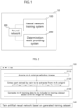
- FIG. 1 is a schematic diagram illustrating an environment in which an artificial neural network training method and a determination result providing method for a pathology specimen are performed according to the technical idea of the present disclosure.
- the artificial neural network training method may be performed by a neural network training system 100, and the determination result providing method for a pathology specimen according to one embodiment of the present disclosure may be performed by a determination result providing system for a pathology specimen (200; hereinafter, referred to as a "determination result providing system").
- the neural network training system 100 may train an artificial neural network 300 to detect lesions included in the pathology specimen, output diagnostic information for the pathology specimen, or provide prognostic information and/or response information to treatment methods, and the determination result providing system 200 may use the trained artificial neural network 300 to make various determinations about a target specimen (for example, determination on lesion detection, presence or absence of development of a disease, prognosis, treatment methods, etc.).
- the neural network training system 100 and/or the determination result providing system 200 may be a computing system which is a data processing device having computing power to implement the technical idea of the present disclosure, and may generally include a computing device such as a personal computer or mobile terminal as well as a server which is a data processing device which is accessible by a client through a network.
- the neural network training system 100 and/or the determination result providing system 200 may be implemented with any one physical device, but an average expert in the art to which the present disclosure pertains may easily deduce that the neural network training system 100 and/or the determination result providing system 200 can be implemented according to the technical idea of the present disclosure by organically combining a plurality of physical devices as needed.
- the neural network training system 100 may train the neural network 300 based on training data generated from a large number of pathology specimens.
- the pathology specimens may be biopsies collected from various organs of the human body or biological tissues excised through surgery.
- the neural network training system 100 may generate individual training data using a digital pathology image of the pathology specimen, and input it into an input layer of the neural network 300 to train the neural network 300.
- the pathology image may be a digital slide image of the pathology specimen or a part of the digital slide image.
- a slide of the pathology specimen may be a sliced portion of the pathology specimen.
- the digital slide image of the pathology specimen may be generated by slicing the pathology specimen to produce a glass slide, staining the glass slide with a predetermined mixed stain, and digitizing it.
- the pathology image may be a pathology slide image obtained by producing a slide of the pathology specimen and staining it with a predetermined mixed stain (staining reagent), or an image obtained by dividing the stained pathology slide image into a predetermined size.
- the pathology image may be a pathology image obtained by staining the slide of the pathology specimen with a mixed stain in which multiple individual stains (staining reagents) are mixed.
- the pathology image may be a whole slide image or a part of the whole slide image (for example, a patch image obtained by dividing the whole slide image into a predetermined size).
- the mixed stain may be a stain that is a mixture of a predetermined stain to be extracted and one or more stains excluding the stain to be extracted.
- the stain to be extracted may be a hematoxylin stain (hereinafter, referred to as 'H-stain'), and hereinafter, the case where the stain to be extracted is the H-stain will be described as an example.
- 'H-stain' hematoxylin stain
- the technical idea of the present disclosure is not limited thereto, and various types of stains may be determined as the stain to be extracted.
- the mixed stain may be a hematoxylin-eosin stain (H&E stain), a mixed stain for the IHC staining method in which hematoxylin staining is a counterstain, or a mixed stain for a DAB staining method in which hematoxylin staining is a counterstain, and in addition, it may be various cocktail stains including the hematoxylin stain.
- H&E stain hematoxylin-eosin stain
- IHC staining method in which hematoxylin staining is a counterstain
- a mixed stain for a DAB staining method in which hematoxylin staining is a counterstain
- it may be various cocktail stains including the hematoxylin stain.
- the neural network 300 may be an artificial neural network trained to output a probability value for whether a given disease is developed or whether a lesion is included.
- the neural network 300 may output a numerical value, i.e., a probability value, indicating a determination result for the target specimen (for example, the possibility of developing the disease) based on data input through the input layer.
- the artificial neural network 300 may be a patch-level classification neural network.
- the patch-level neural network may be a neural network which receives patch-level images as input and outputs values for classifying the patch.
- the artificial neural network 300 may be a pixel-level classification neural network.
- the pixel-level classification neural network may be a neural network which outputs a value for classifying each pixel included in an image.
- the artificial neural network is a neural network artificially constructed based on operating principles of human neurons, and includes a multilayer perceptron model, and it may refer to a set of information expressing a series of design details which define an artificial neural network.
- the artificial neural network 300 may be a convolutional neural network or include a convolutional neural network.
- the trained neural network 300 may be stored in the determination result providing system 200, and the determination result providing system 200 may make a determination about a predetermined diagnosis target specimen using the trained artificial neural network.
- the neural network training system 100 and/or the determination result providing system 200 may be implemented in the form of a subsystem of a predetermined parent system 10.
- the parent system 10 may be a server.
- the server 10 refers to a data processing device having computing power to implement the technical idea of the present disclosure, and In general, an average expert in the art to which the present disclosure pertains may easily deduce that not only data processing devices which is accessible by clients through a network, but also any device which can perform a specific service, such as a personal computer or mobile terminal, can be defined as the server.
- the neural network training system 100 and the determination result providing system 200 may be implemented separately from each other.
- FIG. 2 is a flowchart for illustrating a neural network training method according to one embodiment of the present disclosure.
- the neural network training system 100 may acquire an m-th original pathology image (here, N is a natural number greater than or equal to 2) (S110).
- the neural network training system 100 may receive the m-th original pathology image corresponding to a predetermined pathology specimen from an external terminal, and may also obtain the m-th original pathology image corresponding to the pathology specimen from a memory device which stores the m-th original pathology image in advance.
- the m-th pathology image may be a pathology image stained with a mixed stain in which a predetermined stain to be extracted (for example, hematoxylin stain) and one or more stains excluding the stain to be extracted are mixed.
- a predetermined stain to be extracted for example, hematoxylin stain
- one or more stains excluding the stain to be extracted are mixed.
- the m-th pathology image may be labeled with predetermined information.
- the labeled information may vary depending on the purpose of the artificial neural network 300.
- the information labeled in the m-th pathology image may be the presence or absence of a disease.
- a lesion area caused by a predetermined disease may be annotated.
- the m-th original pathology image may be tagged with annotation information representing the area of the lesion included in the m-th original pathology image.
- FIG. 3A is a diagram showing an example of the original pathology image
- FIG. 3B is a diagram showing a lesion area annotated to the pathology image of FIG. 3A .
- the area marked by the solid line represents the annotated lesion area.
- the neural network training system 100 may extract a part stained by the stain to be extracted (for example, hematoxylin stain) from the m-th original pathology image to generate an m-th pathology image for training (S120).
- a part stained by the stain to be extracted for example, hematoxylin stain
- the neural network training system 100 may perform color deconvolution to generate the m-th pathology image for training corresponding to the m-th original pathology image.
- FIG. 4 is an example illustrating a process of generating the m-th pathology image for training corresponding to the m-th original pathology image according to one embodiment of the present disclosure.
- the neural network training system 100 may convert a signal intensity for each channel in a color space representing the m-th original pathology image into optical density (S121). Thereafter, the neural network training system 100 can convert the optical density into staining intensity according to a predetermined correlation formula (S122).
- FIG. 3C is an example of an image extracted from the original pathology image shown in FIG. 3A of a part stained by the stain to be extracted (for example, hematoxylin stain).
- the neural network training system 100 may generate the m-th training data to be included in the training dataset based on the m-th pathology image for training (S130). More specifically, the neural network training system 100 may generate the m-th training data by tagging the m-th pathology image for training with the information labeled in the m-th original image.
- the m-th original image may have a lesion area annotated as shown in FIG. 3b .
- the generated m-th pathology image for training may be tagged with the annotation information of the m-th original image, an example of which is shown in FIG. 3D .
- FIG. 3D shows training data generated from the original pathology image tagged with the annotation information as shown in FIG. 3B .
- the neural network training system 100 may set the same as a label for the m-th training data.
- the neural network training system 100 may train the neural network 300 by inputting the generated training dataset into the input layer of the neural network 300 (S140 of FIG. 2 ). Since the deep learning technique of inputting training data tagged with correct answer information into the artificial neural network 300 and training the artificial neural network 300 through methods such as gradient descent and backpropagation is widely known, detailed description thereof will be omitted.
- M pathology images used to construct the training dataset may be stained with two or more different mixed stains.
- some of the first to M-th original pathology images and other parts of the first to M-th original pathology images may be pathology images stained with different mixed stains.
- two or more mixed stains used to stain the M pathology images may commonly include the stain to be extracted.
- the first to 50th pathology images may be pathology images stained by the H&E staining method
- the 51st to 100th pathology images may be pathology images stained by the IHC staining method (counterstain is H-staining)
- the 101st to 150th pathology images may be pathology images stained by the DAB staining method (counterstain is H-staining).
- the M pathology images may be pathology images stained by four (4) or more different mixed stains.
- the neural network training method extracts only the part stained by the stain to be extracted from the original pathology image and uses it as the training data of the artificial neural network. Therefore, through the neural network training method according to the technical idea of the present disclosure, there is an effect of capable of effectively performing training using pathology images produced by various staining methods using the stain to be extracted.
- the neural network training method has an effect of effectively training an artificial neural network that is able to make determination about a lesion or disease using images stained by different methods.
- multiple images used as the training data are not pathology images stained by the same method, there is an effect of capable of using them as training data that can train one artificial neural network (for example, 300) by the neural network training method.
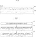
- FIG. 5 is a flowchart illustrating an example of a determination result providing method for a pathology specimen according to one embodiment of the present disclosure.
- the determination result providing method for a pathology specimen according to FIG. 5 may be performed by the determination result providing system 200, and the determination result providing system 200 may store the artificial neural network 300 trained by the neural network training system 100.
- the determination result providing system 200 may acquire a determination target pathology image of a predetermined determination target pathology specimen (S210).
- the determination target pathology image may be a pathology slide image obtained by staining a slide of the determination target pathology specimen with a mixed stain in which the stain to be extracted (for example, hematoxylin stain) and one or more stains excluding the stain to be extracted (for example, hematoxylin stain) are mixed.
- the determination result providing system 200 may extract the part stained by the stain to be extracted from the determination target pathology image and generate an extraction image corresponding to the determination target pathology image (S220). Since a process of generating input data corresponding to the determination target pathology image of the determination target specimen is very similar to the process as described above, separate explanation will be omitted.
- the determination result providing system 200 may input the input data to the artificial neural network 300, and based on a result output by the artificial neural network, output a determination result for the determination target pathology specimen (S230).
- the staining method used to stain at least a part of the original pathology images used to generate the training data and the staining method used to stain the determination target pathology image may be different.
- at least a part of the determination target image and a part of the images used to train the artificial neural network 300 may be pathology images stained with different mixed stains.
- all or at least a part of the images used to train the artificial neural network 300 may be pathology images stained by the H&E staining method, but the determination target image may be an image stained by the IHC method.
- the staining method used to stain the determination target image may be different from a method of staining any of the images used to train the artificial neural network 300.
- the staining method used to stain the determination target image is the DAB method, but all images used to train the artificial neural network 300 may be images stained using the H&E staining method.
- FIG. 6 is a diagram illustrating a schematic configuration of the artificial neural network training system 100 according to one embodiment of the present disclosure
- FIG. 7 is a diagram illustrating a schematic configuration of the determination result providing system 200 according to one embodiment of the present disclosure.
- the artificial neural network training system 100 and the determination result providing system 200 may mean a logical configuration with hardware resources and/or software required to implement the technical idea of the present disclosure, and does not necessarily mean a single physical component or a single device.
- the artificial neural network training system 100 and the determination result providing system 200 may mean a logical combination of hardware and/or software provided to implement the technical idea of the present disclosure and, if necessary, may be implemented as a set of logical configurations to implement the technical idea of the present disclosure by being installed on devices separated from each other and performing their respective functions.
- the artificial neural network training system 100 and the determination result providing system 200 may mean a set of configurations that are implemented separately for each function or role to implement the technical idea of the present disclosure.
- Each configuration of the artificial neural network training system 100 and the determination result providing system 200 may be located on different physical devices or on the same physical device. Further, according to implementation examples, combinations of software and/or hardware that constitute respective components of the artificial neural network training system 100 and the determination result providing system 200 are also located in different physical devices, and the configurations located in the different physical devices may be organically combined with each other to implement each module.
- module in this specification may mean a functional and structural combination of hardware for carrying out the technical idea of the present disclosure and software for driving the hardware.
- the module may mean a logical unit of a predetermined code and hardware resources for executing the predetermined code and does not necessarily mean a physically connected code or a single type of hardware.
- the artificial neural network training system 100 may include a storage module 110, an acquisition module 120, a generation module 130, and a training module 140. According to embodiments of the present disclosure, some of the above-described components may not necessarily correspond to the components essential for the implementation of the present disclosure, and of course, the artificial neural network training system 100 may include more components than these.
- the artificial neural network training system 100 may further include a communication module (not shown) for communicating with an external device, and a control module (not shown) for controlling the components and resources of the artificial neural network training system 100.
- the storage module 110 may store an artificial neural network 40 to be trained.
- the acquisition module 120 may acquire M original pathology images stained with a mixed stain in which the predetermined stain to be extracted and one or more stains excluding the stain to be extracted are mixed.
- the generation module 130 may generate individual training data based on each original pathology image, and may construct the training dataset including a plurality pieces of individual training data.
- the training module 140 may train the artificial neural network 300 based on the training dataset.
- the determination result providing system 200 may include a storage module 210, an acquisition module 220, a generation module 230, and a determination module 240. According to embodiments of the present disclosure, some of the above-described components may not necessarily correspond to the components essential to implementation of the present disclosure, and of course, the determination result providing system 200 may include more components according to embodiments.
- the determination result providing system 200 may further include a communication module (not shown) to communicate with a three-axis vibration sensor 20, and a control module (not shown) to control the components and resources of the determination result providing system 200.
- the storage module 210 may store the trained artificial neural network 40.
- the acquisition module 220 may acquire the determination target pathology image obtained by staining the predetermined determination target pathology specimen with the mixed stain in which one or more stains excluding the stain to be extracted are mixed.
- the generation module 230 may generate the input data based on the determination target pathology image.
- the determination module 240 may input the input data to the artificial neural network 300, and make determination on the determination target specimen based on a prediction value output from the artificial neural network 300.
- the artificial neural network training system 100 and the determination result providing system 200 may include a processor and a memory to store a program executed by the processor.
- the processor may include a single-core CPU or a multi-core CPU.
- the memory may include high-speed random access memory and may include non-volatile memory, such as one or more magnetic disk storage devices, flash memory devices, or other non-volatile solid-state memory devices. Access to memory by the processor and other components may be controlled by a memory controller.
- the method according to the embodiment of the present disclosure may be implemented in the form of computer-readable program instructions and stored in a non-transitory computer-readable recording medium, and a control program and its target program according to an embodiment of the present disclosure may also be stored in a non-transitory computer-readable recording medium.
- a non-transitory computer-readable recording medium includes any type of recording device in which data that can be read by a computer system is stored.
- the program instructions recorded on the recording medium may be specially designed and configured for the present disclosure or may be known and available to a person skilled in the field of software.
- non-transitory computer-readable recording media examples include magnetic media such as hard disks, floppy disks, and magnetic tapes, optical media such as CD-ROMs and DVDs, magneto-optical media such as floptical disks, and hardware devices specially configured to store and execute program instructions, such as ROMs, RAMs, flash memory, and the like.
- non-transitory computer-readable recording media may be distributed across networked computer systems, where computer-readable codes may be stored and executed in a distributed manner.
- program instructions include machine language code, such as those generated by a compiler, as well as high-level language code that can be executed by a device that processes information electronically using an interpreter such as a computer.
- the hardware devices described above may be configured to operate as one or more software modules to perform the operations of the present disclosure, and vice versa.
- the present disclosure may be used for a training method of an artificial neural network which provides a determination result for a pathology specimen, and a computing system which performs the same.
Landscapes
- Engineering & Computer Science (AREA)
- Health & Medical Sciences (AREA)
- Theoretical Computer Science (AREA)
- Physics & Mathematics (AREA)
- General Health & Medical Sciences (AREA)
- General Physics & Mathematics (AREA)
- Medical Informatics (AREA)
- Biomedical Technology (AREA)
- Evolutionary Computation (AREA)
- Computer Vision & Pattern Recognition (AREA)
- Software Systems (AREA)
- Artificial Intelligence (AREA)
- Computing Systems (AREA)
- Data Mining & Analysis (AREA)
- Life Sciences & Earth Sciences (AREA)
- Public Health (AREA)
- Molecular Biology (AREA)
- Biophysics (AREA)
- Nuclear Medicine, Radiotherapy & Molecular Imaging (AREA)
- Radiology & Medical Imaging (AREA)
- General Engineering & Computer Science (AREA)
- Mathematical Physics (AREA)
- Computational Linguistics (AREA)
- Epidemiology (AREA)
- Primary Health Care (AREA)
- Databases & Information Systems (AREA)
- Multimedia (AREA)
- Quality & Reliability (AREA)
- Pathology (AREA)
- Image Analysis (AREA)
- Image Processing (AREA)
- Bioinformatics & Computational Biology (AREA)
- Biotechnology (AREA)
- Evolutionary Biology (AREA)
- Spectroscopy & Molecular Physics (AREA)
- Bioinformatics & Cheminformatics (AREA)
- Bioethics (AREA)
- Medical Treatment And Welfare Office Work (AREA)
- Investigating Or Analysing Biological Materials (AREA)
Applications Claiming Priority (2)
| Application Number | Priority Date | Filing Date | Title |
|---|---|---|---|
| KR1020210177378A KR102485414B1 (ko) | 2021-12-13 | 2021-12-13 | 병리 검체에 대한 판단 결과를 제공하는 인공 뉴럴 네트워크의 학습 방법, 및 이를 수행하는 컴퓨팅 시스템 |
| PCT/KR2022/020180 WO2023113414A1 (ko) | 2021-12-13 | 2022-12-13 | 병리 검체에 대한 판단 결과를 제공하는 인공 뉴럴 네트워크의 학습 방법, 및 이를 수행하는 컴퓨팅 시스템 |
Publications (2)
| Publication Number | Publication Date |
|---|---|
| EP4432301A1 true EP4432301A1 (de) | 2024-09-18 |
| EP4432301A4 EP4432301A4 (de) | 2025-07-16 |
Family
ID=84923930
Family Applications (1)
| Application Number | Title | Priority Date | Filing Date |
|---|---|---|---|
| EP22907881.1A Pending EP4432301A4 (de) | 2021-12-13 | 2022-12-13 | Verfahren zum trainieren eines künstlichen neuronalen netzwerks mit bereitstellung des ergebnisses einer pathologieprobe und computersystem zur durchführung davon |
Country Status (6)
| Country | Link |
|---|---|
| US (1) | US20240331364A1 (de) |
| EP (1) | EP4432301A4 (de) |
| JP (1) | JP2024546649A (de) |
| KR (1) | KR102485414B1 (de) |
| CN (1) | CN118414673A (de) |
| WO (1) | WO2023113414A1 (de) |
Families Citing this family (1)
| Publication number | Priority date | Publication date | Assignee | Title |
|---|---|---|---|---|
| KR20250156974A (ko) * | 2024-04-26 | 2025-11-04 | 사회복지법인 삼성생명공익재단 | 딥러닝 모델을 이용한 종양 미세환경 예측 방법 및 분석장치 |
Family Cites Families (9)
| Publication number | Priority date | Publication date | Assignee | Title |
|---|---|---|---|---|
| US9002077B2 (en) * | 2009-08-10 | 2015-04-07 | Cambridge Research & Instrumentation, Inc. | Visualization of stained samples |
| EP2601513B1 (de) * | 2010-08-05 | 2014-05-14 | Cambridge Research & Instrumentation, Inc. | Verbesserte visuelle untersuchung von proben |
| JP5344073B2 (ja) * | 2012-09-14 | 2013-11-20 | 日本電気株式会社 | 病理組織画像撮影システム、病理組織画像撮影方法、および病理組織画像撮影プログラム |
| CA2944829C (en) * | 2014-05-23 | 2022-10-25 | Ting Chen | Systems and methods for detection of biological structures and/or patterns in images |
| JP6710373B2 (ja) * | 2017-03-30 | 2020-06-17 | 国立研究開発法人産業技術総合研究所 | 超音波画像診断支援方法、およびシステム |
| EP3721373B1 (de) * | 2017-12-07 | 2025-03-05 | Ventana Medical Systems, Inc. | Tieenlernsysteme und verfahren zur gemeinsamen zell- und regionklassifizierung auf biologischen bildern |
| JP7217589B2 (ja) * | 2018-02-27 | 2023-02-03 | シスメックス株式会社 | 画像解析方法、画像解析装置、プログラム、学習済み深層学習アルゴリズムの製造方法および学習済み深層学習アルゴリズム |
| CN112868024B (zh) * | 2018-10-15 | 2025-11-25 | 文塔纳医疗系统公司 | 用于细胞分类的系统和方法 |
| KR102246319B1 (ko) * | 2021-01-07 | 2021-05-03 | 주식회사 딥바이오 | 병리 검체에 대한 판단 결과를 제공하는 인공 뉴럴 네트워크의 학습 방법, 및 이를 수행하는 컴퓨팅 시스템 |
-
2021
- 2021-12-13 KR KR1020210177378A patent/KR102485414B1/ko active Active
-
2022
- 2022-12-13 WO PCT/KR2022/020180 patent/WO2023113414A1/ko not_active Ceased
- 2022-12-13 JP JP2024533277A patent/JP2024546649A/ja active Pending
- 2022-12-13 CN CN202280082765.7A patent/CN118414673A/zh active Pending
- 2022-12-13 EP EP22907881.1A patent/EP4432301A4/de active Pending
-
2024
- 2024-06-12 US US18/741,765 patent/US20240331364A1/en active Pending
Also Published As
| Publication number | Publication date |
|---|---|
| JP2024546649A (ja) | 2024-12-26 |
| KR102485414B1 (ko) | 2023-01-06 |
| WO2023113414A1 (ko) | 2023-06-22 |
| CN118414673A (zh) | 2024-07-30 |
| EP4432301A4 (de) | 2025-07-16 |
| US20240331364A1 (en) | 2024-10-03 |
Similar Documents
| Publication | Publication Date | Title |
|---|---|---|
| US12293821B2 (en) | Computational pathology approach for retrospective analysis of tissue-based companion diagnostic driven clinical trial studies | |
| JP7406745B2 (ja) | コンピュータ検出方法のための電子画像を処理するためのシステムおよび方法 | |
| CN111448584B (zh) | 计算肿瘤空间和标记间异质性的方法 | |
| JP7742610B2 (ja) | 疾患の算出査定のために電子画像を処理するためのシステムおよび方法 | |
| US12525040B2 (en) | Machine learning models for cell localization and classification learned using repel coding | |
| JP2021506022A (ja) | 生体画像における連帯的細胞および領域分類のための深層学習システムならびに方法 | |
| CN114207675A (zh) | 用于数字病理学的用于处理图像以针对所处理的图像制备载片的系统和方法 | |
| US20240281653A1 (en) | Method for training artificial neural network providing determination result of pathological specimen, and computing system for performing same | |
| US20250046069A1 (en) | Label-free virtual immunohistochemical staining of tissue using deep learning | |
| JP7743536B2 (ja) | 免疫組織化学(ihc)の合成画像への組織化学的染色画像の変換 | |
| US12597137B2 (en) | Digital synthesis of histological stains using multiplexed immunofluorescence imaging | |
| CN112703531A (zh) | 生成组织图像的注释数据 | |
| EP4481747A1 (de) | Verfahren zum trainieren eines maschinenlernmodells zur analyse immunhistochemisch gefärbter bilder und computersystem zur durchführung davon | |
| Selcuk et al. | Automated HER2 scoring in breast cancer images using deep learning and pyramid sampling | |
| US20230162485A1 (en) | Digital analysis of preanalytical factors in tissues used for histological staining | |
| US20240331364A1 (en) | Method for training artificial neural network providing determination result of pathology specimen, and computing system performing same | |
| KR102682730B1 (ko) | 병리 진단 케이스의 대표 병변 이미지 생성 방법 및 이를 수행하는 컴퓨팅 시스템 | |
| CN119563190A (zh) | 生成基于推理的虚拟染色图像注释的方法 | |
| US20240212146A1 (en) | Method and apparatus for analyzing pathological slide images |
Legal Events
| Date | Code | Title | Description |
|---|---|---|---|
| STAA | Information on the status of an ep patent application or granted ep patent |
Free format text: STATUS: THE INTERNATIONAL PUBLICATION HAS BEEN MADE |
|
| PUAI | Public reference made under article 153(3) epc to a published international application that has entered the european phase |
Free format text: ORIGINAL CODE: 0009012 |
|
| STAA | Information on the status of an ep patent application or granted ep patent |
Free format text: STATUS: REQUEST FOR EXAMINATION WAS MADE |
|
| 17P | Request for examination filed |
Effective date: 20240610 |
|
| AK | Designated contracting states |
Kind code of ref document: A1 Designated state(s): AL AT BE BG CH CY CZ DE DK EE ES FI FR GB GR HR HU IE IS IT LI LT LU LV MC ME MK MT NL NO PL PT RO RS SE SI SK SM TR |
|
| DAV | Request for validation of the european patent (deleted) | ||
| DAX | Request for extension of the european patent (deleted) | ||
| REG | Reference to a national code |
Ref country code: DE Ref legal event code: R079 Free format text: PREVIOUS MAIN CLASS: G16H0050700000 Ipc: G06T0007110000 |
|
| A4 | Supplementary search report drawn up and despatched |
Effective date: 20250616 |
|
| RIC1 | Information provided on ipc code assigned before grant |
Ipc: G06T 7/11 20170101AFI20250610BHEP Ipc: G06T 7/00 20170101ALI20250610BHEP Ipc: G16H 50/70 20180101ALI20250610BHEP Ipc: G16H 50/50 20180101ALI20250610BHEP Ipc: G16H 50/20 20180101ALI20250610BHEP Ipc: G16B 40/20 20190101ALI20250610BHEP Ipc: G16H 30/20 20180101ALI20250610BHEP Ipc: G16H 30/40 20180101ALI20250610BHEP Ipc: G16H 70/60 20180101ALI20250610BHEP Ipc: G16B 45/00 20190101ALI20250610BHEP |
|
| STAA | Information on the status of an ep patent application or granted ep patent |
Free format text: STATUS: THE APPLICATION IS DEEMED TO BE WITHDRAWN |