EP4434516A2 - Modulare plattform für gezielte therapeutika - Google Patents
Modulare plattform für gezielte therapeutika Download PDFInfo
- Publication number
- EP4434516A2 EP4434516A2 EP24193705.1A EP24193705A EP4434516A2 EP 4434516 A2 EP4434516 A2 EP 4434516A2 EP 24193705 A EP24193705 A EP 24193705A EP 4434516 A2 EP4434516 A2 EP 4434516A2
- Authority
- EP
- European Patent Office
- Prior art keywords
- antibody
- immunoparticle
- diseases
- lipid
- cells
- Prior art date
- Legal status (The legal status is an assumption and is not a legal conclusion. Google has not performed a legal analysis and makes no representation as to the accuracy of the status listed.)
- Pending
Links
Images




Classifications
-
- C—CHEMISTRY; METALLURGY
- C07—ORGANIC CHEMISTRY
- C07K—PEPTIDES
- C07K16/00—Immunoglobulins [IG], e.g. monoclonal or polyclonal antibodies
- C07K16/18—Immunoglobulins [IG], e.g. monoclonal or polyclonal antibodies against material from animals or humans
- C07K16/28—Immunoglobulins [IG], e.g. monoclonal or polyclonal antibodies against material from animals or humans against receptors, cell surface antigens or cell surface determinants
- C07K16/30—Immunoglobulins [IG], e.g. monoclonal or polyclonal antibodies against material from animals or humans against receptors, cell surface antigens or cell surface determinants from tumour cells
-
- A—HUMAN NECESSITIES
- A61—MEDICAL OR VETERINARY SCIENCE; HYGIENE
- A61K—PREPARATIONS FOR MEDICAL, DENTAL OR TOILETRY PURPOSES
- A61K47/00—Medicinal preparations characterised by the non-active ingredients used, e.g. carriers or inert additives; Targeting or modifying agents chemically bound to the active ingredient
- A61K47/50—Medicinal preparations characterised by the non-active ingredients used, e.g. carriers or inert additives; Targeting or modifying agents chemically bound to the active ingredient the non-active ingredient being chemically bound to the active ingredient, e.g. polymer-drug conjugates
- A61K47/51—Medicinal preparations characterised by the non-active ingredients used, e.g. carriers or inert additives; Targeting or modifying agents chemically bound to the active ingredient the non-active ingredient being chemically bound to the active ingredient, e.g. polymer-drug conjugates the non-active ingredient being a modifying agent
- A61K47/54—Medicinal preparations characterised by the non-active ingredients used, e.g. carriers or inert additives; Targeting or modifying agents chemically bound to the active ingredient the non-active ingredient being chemically bound to the active ingredient, e.g. polymer-drug conjugates the non-active ingredient being a modifying agent the modifying agent being an organic compound
- A61K47/543—Lipids, e.g. triglycerides; Polyamines, e.g. spermine or spermidine
-
- A—HUMAN NECESSITIES
- A61—MEDICAL OR VETERINARY SCIENCE; HYGIENE
- A61K—PREPARATIONS FOR MEDICAL, DENTAL OR TOILETRY PURPOSES
- A61K9/00—Medicinal preparations characterised by special physical form
- A61K9/10—Dispersions; Emulsions
- A61K9/127—Synthetic bilayered vehicles, e.g. liposomes or liposomes with cholesterol as the only non-phosphatidyl surfactant
-
- A—HUMAN NECESSITIES
- A61—MEDICAL OR VETERINARY SCIENCE; HYGIENE
- A61P—SPECIFIC THERAPEUTIC ACTIVITY OF CHEMICAL COMPOUNDS OR MEDICINAL PREPARATIONS
- A61P35/00—Antineoplastic agents
-
- C—CHEMISTRY; METALLURGY
- C07—ORGANIC CHEMISTRY
- C07K—PEPTIDES
- C07K16/00—Immunoglobulins [IG], e.g. monoclonal or polyclonal antibodies
- C07K16/18—Immunoglobulins [IG], e.g. monoclonal or polyclonal antibodies against material from animals or humans
- C07K16/28—Immunoglobulins [IG], e.g. monoclonal or polyclonal antibodies against material from animals or humans against receptors, cell surface antigens or cell surface determinants
- C07K16/2803—Immunoglobulins [IG], e.g. monoclonal or polyclonal antibodies against material from animals or humans against receptors, cell surface antigens or cell surface determinants against the immunoglobulin superfamily
- C07K16/2809—Immunoglobulins [IG], e.g. monoclonal or polyclonal antibodies against material from animals or humans against receptors, cell surface antigens or cell surface determinants against the immunoglobulin superfamily against the T-cell receptor (TcR)-CD3 complex
-
- C—CHEMISTRY; METALLURGY
- C07—ORGANIC CHEMISTRY
- C07K—PEPTIDES
- C07K16/00—Immunoglobulins [IG], e.g. monoclonal or polyclonal antibodies
- C07K16/18—Immunoglobulins [IG], e.g. monoclonal or polyclonal antibodies against material from animals or humans
- C07K16/28—Immunoglobulins [IG], e.g. monoclonal or polyclonal antibodies against material from animals or humans against receptors, cell surface antigens or cell surface determinants
- C07K16/2803—Immunoglobulins [IG], e.g. monoclonal or polyclonal antibodies against material from animals or humans against receptors, cell surface antigens or cell surface determinants against the immunoglobulin superfamily
- C07K16/2812—Immunoglobulins [IG], e.g. monoclonal or polyclonal antibodies against material from animals or humans against receptors, cell surface antigens or cell surface determinants against the immunoglobulin superfamily against CD4
-
- C—CHEMISTRY; METALLURGY
- C07—ORGANIC CHEMISTRY
- C07K—PEPTIDES
- C07K16/00—Immunoglobulins [IG], e.g. monoclonal or polyclonal antibodies
- C07K16/18—Immunoglobulins [IG], e.g. monoclonal or polyclonal antibodies against material from animals or humans
- C07K16/28—Immunoglobulins [IG], e.g. monoclonal or polyclonal antibodies against material from animals or humans against receptors, cell surface antigens or cell surface determinants
- C07K16/2803—Immunoglobulins [IG], e.g. monoclonal or polyclonal antibodies against material from animals or humans against receptors, cell surface antigens or cell surface determinants against the immunoglobulin superfamily
- C07K16/2815—Immunoglobulins [IG], e.g. monoclonal or polyclonal antibodies against material from animals or humans against receptors, cell surface antigens or cell surface determinants against the immunoglobulin superfamily against CD8
-
- C—CHEMISTRY; METALLURGY
- C07—ORGANIC CHEMISTRY
- C07K—PEPTIDES
- C07K16/00—Immunoglobulins [IG], e.g. monoclonal or polyclonal antibodies
- C07K16/18—Immunoglobulins [IG], e.g. monoclonal or polyclonal antibodies against material from animals or humans
- C07K16/28—Immunoglobulins [IG], e.g. monoclonal or polyclonal antibodies against material from animals or humans against receptors, cell surface antigens or cell surface determinants
- C07K16/2839—Immunoglobulins [IG], e.g. monoclonal or polyclonal antibodies against material from animals or humans against receptors, cell surface antigens or cell surface determinants against the integrin superfamily
- C07K16/2845—Immunoglobulins [IG], e.g. monoclonal or polyclonal antibodies against material from animals or humans against receptors, cell surface antigens or cell surface determinants against the integrin superfamily against integrin beta2-subunit-containing molecules, e.g. CD11, CD18
-
- C—CHEMISTRY; METALLURGY
- C07—ORGANIC CHEMISTRY
- C07K—PEPTIDES
- C07K16/00—Immunoglobulins [IG], e.g. monoclonal or polyclonal antibodies
- C07K16/18—Immunoglobulins [IG], e.g. monoclonal or polyclonal antibodies against material from animals or humans
- C07K16/28—Immunoglobulins [IG], e.g. monoclonal or polyclonal antibodies against material from animals or humans against receptors, cell surface antigens or cell surface determinants
- C07K16/2866—Immunoglobulins [IG], e.g. monoclonal or polyclonal antibodies against material from animals or humans against receptors, cell surface antigens or cell surface determinants against receptors for cytokines, lymphokines, interferons
-
- C—CHEMISTRY; METALLURGY
- C07—ORGANIC CHEMISTRY
- C07K—PEPTIDES
- C07K16/00—Immunoglobulins [IG], e.g. monoclonal or polyclonal antibodies
- C07K16/18—Immunoglobulins [IG], e.g. monoclonal or polyclonal antibodies against material from animals or humans
- C07K16/28—Immunoglobulins [IG], e.g. monoclonal or polyclonal antibodies against material from animals or humans against receptors, cell surface antigens or cell surface determinants
- C07K16/2896—Immunoglobulins [IG], e.g. monoclonal or polyclonal antibodies against material from animals or humans against receptors, cell surface antigens or cell surface determinants against molecules with a "CD"-designation, not provided for elsewhere
-
- C—CHEMISTRY; METALLURGY
- C07—ORGANIC CHEMISTRY
- C07K—PEPTIDES
- C07K16/00—Immunoglobulins [IG], e.g. monoclonal or polyclonal antibodies
- C07K16/42—Immunoglobulins [IG], e.g. monoclonal or polyclonal antibodies against immunoglobulins
- C07K16/4208—Immunoglobulins [IG], e.g. monoclonal or polyclonal antibodies against immunoglobulins against an idiotypic determinant on Ig
- C07K16/4241—Immunoglobulins [IG], e.g. monoclonal or polyclonal antibodies against immunoglobulins against an idiotypic determinant on Ig against anti-human or anti-animal Ig
-
- C—CHEMISTRY; METALLURGY
- C12—BIOCHEMISTRY; BEER; SPIRITS; WINE; VINEGAR; MICROBIOLOGY; ENZYMOLOGY; MUTATION OR GENETIC ENGINEERING
- C12N—MICROORGANISMS OR ENZYMES; COMPOSITIONS THEREOF; PROPAGATING, PRESERVING, OR MAINTAINING MICROORGANISMS; MUTATION OR GENETIC ENGINEERING; CULTURE MEDIA
- C12N15/00—Mutation or genetic engineering; DNA or RNA concerning genetic engineering, vectors, e.g. plasmids, or their isolation, preparation or purification; Use of hosts therefor
- C12N15/09—Recombinant DNA-technology
- C12N15/11—DNA or RNA fragments; Modified forms thereof; Non-coding nucleic acids having a biological activity
-
- C—CHEMISTRY; METALLURGY
- C12—BIOCHEMISTRY; BEER; SPIRITS; WINE; VINEGAR; MICROBIOLOGY; ENZYMOLOGY; MUTATION OR GENETIC ENGINEERING
- C12N—MICROORGANISMS OR ENZYMES; COMPOSITIONS THEREOF; PROPAGATING, PRESERVING, OR MAINTAINING MICROORGANISMS; MUTATION OR GENETIC ENGINEERING; CULTURE MEDIA
- C12N15/00—Mutation or genetic engineering; DNA or RNA concerning genetic engineering, vectors, e.g. plasmids, or their isolation, preparation or purification; Use of hosts therefor
- C12N15/09—Recombinant DNA-technology
- C12N15/11—DNA or RNA fragments; Modified forms thereof; Non-coding nucleic acids having a biological activity
- C12N15/111—General methods applicable to biologically active non-coding nucleic acids
-
- C—CHEMISTRY; METALLURGY
- C12—BIOCHEMISTRY; BEER; SPIRITS; WINE; VINEGAR; MICROBIOLOGY; ENZYMOLOGY; MUTATION OR GENETIC ENGINEERING
- C12N—MICROORGANISMS OR ENZYMES; COMPOSITIONS THEREOF; PROPAGATING, PRESERVING, OR MAINTAINING MICROORGANISMS; MUTATION OR GENETIC ENGINEERING; CULTURE MEDIA
- C12N2320/00—Applications; Uses
- C12N2320/30—Special therapeutic applications
- C12N2320/32—Special delivery means, e.g. tissue-specific
Definitions
- the present invention in some embodiments thereof, relates immunoparticles and methods of generating and using same.
- RNA interference RNA interference
- siRNA-based therapeutics to knock down disease-causing genes.
- Clinical studies using lipid based nanoparticles (LNPs) that carry siRNA payloads or GalNAc-conjugated siRNAs that are taken up by the asialoglycoprotein receptor on hepatocytes are showing impressive and durable gene knockdown in the liver in advance phase trials with little toxicity.
- designing clinically useful strategies for delivering siRNAs to tissues outside the liver has proven difficult. Most LNPs get trapped in the liver and other siRNA conjugates have so far not worked well. A flexible platform for cell-specific siRNA uptake would be ideal.
- Targeted uptake to the diseased tissue would have the advantage of reduced bystander cell toxicity and require lower drug doses.
- the wide availability of monoclonal antibodies that bind with high affinity and selectivity to virtually any cell-specific receptor make them desirable components of a targeted delivery platform that could potentially be used to deliver siRNAs or other macromolecules to a wide range of cell types.
- antibody-mediated delivery and gene knockdown has been demonstrated for specific cell types using antibody fusion proteins and conjugated LNPs in animal models, these methods require a lot of optimization for each cell type (1-5).
- a flexible platform that can be nimbly applied to target multiple tissues by substituting one antibody for another has so far not been possible because conventional methods of chemical conjugation to antibodies for constructing targeting delivery systems are inefficient and require careful optimization for each targeted carrier for functionality.
- lipidated secondary antibody According to one aspect of the present invention there is provided a lipidated secondary antibody.
- an isolated polynucleotide encoding the lipidated secondary antibody described herein.
- a cell expressing the isolated polynucleotide described herein.
- a pharmaceutical composition comprising a lipidated polypeptide immunocomplexed with a primary antibody, wherein the secondary antibody is non-covalently attached to an outer surface of a particle via the lipidated portion of the lipidated secondary antibody and wherein the particle is loaded with a pharmaceutical agent.
- a method of delivering a pharmaceutical agent to a subject in need thereof comprising administering to the subject the pharmaceutical composition described herein, thereby delivering the pharmaceutical agent to the subject.
- an immunoparticle comprising a lipidated polypeptide non-covalently attached to an outer surface of a particle via the lipidated portion of the lipidated polypeptide.
- kits for in-vivo delivery of a pharmaceutical agent comprising the immunoparticle described herein and instructions for effecting immunocomplexation of the lipidated polypeptide to a primary antibody.
- the secondary antibody is attached to a lipidated peptide.
- the lipidated secondary antibody comprises the sequence as set forth in SEQ ID NO: 13.
- the secondary antibody is a monoclonal antibody.
- the N terminus of the secondary antibody is lipidated.
- the lipidated secondary antibody further comprises a detectable moiety.
- the lipidated secondary antibody further comprises an affinity tag.
- the monoclonal secondary antibody is a humanized monoclonal secondary antibody.
- the lipidated secondary antibody comprises an antigen recognition domain capable of specifically binding a humanized or human primary antibody.
- the cell is a bacterial cell.
- the lipidated polypeptide is the lipidated secondary antibody described herein.
- the lipidated polypeptide is protein A or protein G.
- the lipidated polypeptide is the lipidated secondary antibody described herein.
- the lipidated polypeptide is Protein A or Protein G.
- the polypeptide is immunocomplexed with a humanized or human primary antibody.
- the immunoparticle is loaded with a pharmaceutical agent.
- the pharmaceutical agent is a diagnostic agent.
- the pharmaceutical agent is a therapeutic agent.
- the therapeutic agent is a polynucleotide agent.
- the administering is systemically administering.
- the primary antibody comprises an Fc region.
- the secondary antibody is an antibody fragment and optionally wherein the fragment is selected from the group consisting of a Fab, F(ab) 2 an ScFv and a sdFv.
- the primary antibody and the secondary antibody are of different antibody classes or antibody isotypes.
- the primary antibody and the secondary antibody are selected from the group consisting of IgG1, IgG2 and IgG4.
- the primary antibody is an IgG1 and the secondary antibody is an IgG2 or IgG4.
- the primary antibody is selected from the group consisting of anti-CD44, anti-CD34, anti-Ly6C, anti-CD3, anti-CD4, anti-CD25 and anti-intergrin beta 7.
- the primary antibody comprises a plurality of primary antibodies which bind distinct targets.
- the primary antibody comprises an antigen recognition region which binds a tissue specific antigen.
- the primary antibody comprises an antigen recognition region which binds a cancer specific antigen.
- the particle is selected from the group consisting of a polymeric particle, a microcapsule, a liposome, a microsphere, a microemulsion, a nanoparticle, a nanocapsule, a nanosphere and a nanocage.
- the particle comprises a charged external surface.
- the particle comprises a neutral external surface.
- the particle is lipid-based particle.
- the lipids of the lipid based particle comprise cationic lipids.
- the cationic lipid is selected from the group consisting of 1,2-Dilauroyl-sn-Glicero-3-Phosphoethanolamine (DLPE) and 1,2-Dilauroyl-sn-Glicero-3-Glycerol (DLPG), dioleoyl-1,2-diacyl-3-trimethylammonium-propane (DOTAP, at 18:1; 14:0; 16:0, 18:0) and N-[1-(2,3-dioleyloxy)propyl]-N,N,N-trimethlylammonium chloride (DOTMA); dimethyldioctadecylammonium (DDAB); 1,2-dilauroyl-sn-glycero-3-ethylphosphocholine (Ethyl PC, at 12:0; 14:0; 16:0; 18:0; 18:1; 16:0-18:1); 1,2-di-(9Z-oct
- DLPE 1,2-Dilauroy
- the lipids comprise a neutral lipid.
- the neutral lipid comprises phosphatidylethanolamine or dioleilphosphatidylethanolamine (DOPE).
- DOPE dioleilphosphatidylethanolamine
- the lipids comprise anionic phospholipids.
- the anionic phospholipids are selected from the group consisting of phosphatidylserine, phosphatidic acid, phosphatidylcholine and phosphatidyl glycerol.
- the lipidated portion of the secondary antibody comprises the first two amino acids encoded by the E. coli NlpA gene or the first six amino acids encoded by the E. coli NlpA gene.
- the sequence is an inner membrane lipoprotein or fragment thereof selected from the group consisting of: AraH, MglC, MalF, MalG, Mal C, MalD, RbsC, RbsC, ArtM, ArtQ, GliP, ProW, HisM, HisQ, LivH, LivM, LivA, Liv E,Dpp B, DppC, OppB,AmiC, AmiD, BtuC, FhuB, FecC, FecD,FecR, FepD, NikB, NikC, CysT, CysW, UgpA, UgpE, PstA, PstC, PotB, PotC,PotH, PotI, ModB, NosY, PhnM, LacY, SecY, TolC, Dsb,B, DsbD, TonB, TatC, CheY, TraB, Exb D, ExbB and Aas.
- the present invention in some embodiments thereof, relates to immunoparticles and methods of generating and using same.
- mAbs monoclonal antibodies
- siRNAs small interfering
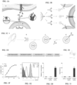
- siRNA-loaded lipid nanoparticles are coated with oriented, targeting antibodies which are non-covalently bound to a membrane-anchored lipoprotein that recognizes their Fc domain. Unlike chemically conjugated antibodies, these oriented antibodies maintain their high affinity and the LNPs avoid scavenging by Fc receptors on macrophages. Notably, each mAb can be utilized for targeting with almost no need for calibration or optimization.
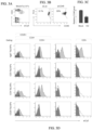
- Intravenously injected anti-Ly6C-coated LNP encapsulating TNF siRNAs were taken up selectively by Ly6C+ monocytes and activated tissue macrophages, suppressed TNF- ⁇ expression in the colon and ameliorated inflammatory bowel disease symptoms in a DSS-induced colitis mouse model ( Figures 4A-H ).
- Anti-CD29-coated LNPs entrapping Polo-like kinase 1 ( PLK1 ) siRNAs prolonged survival of mice bearing Mantle Cell Lymphoma xenografts ( Figures 7A-C ).
- Adapting this flexible platform for human use will enable personal therapy for cancer patients in which the targeted antibody can be adjusted according to each patient's cancer cell surface marker expression.
- the platform could also allow the use of more than one targeting antibody to reduce the chance of cancer resistance due to receptor down-modulation.
- lipidated secondary antibody a lipidated secondary antibody.
- antibody as used in this invention includes intact molecules as well as functional fragments thereof.
- secondary antibody refers to an antibody which binds to conserved regions of a primary antibody.
- the secondary antibody has a specificity for the antibody species and optionally isotype of the primary antibody.
- Secondary antibodies can bind parts of whole IgG (heavy and light chains, H+L), or only the Fab or Fc region, or only the gamma chain. Secondary antibodies also exist that are specific for IgM heavy chains ( ⁇ or Fc5 ⁇ ), or the ⁇ or ⁇ light chains common to all immunoglobulins (IgG, IgA, IgD, IgE and IgM).
- the secondary antibody is a monoclonal antibody (as further described herein below), for example a humanized monoclonal antibody.
- the secondary antibody can belong to any antibody class (e.g., IgG, IgA, IgD, IgE and IgM) or isotype. According to a specific embodiment, the secondary antibody is selected from the group consisting of IgG1, IgG2, IgG3 and IgG4.
- the secondary antibodies may be provided as intact antibodies (e.g., whole IgG) or as divalent F(ab') 2 fragments and monovalent Fab fragments, though other forms of antibody fragments, as described herein below can be used.
- antibody fragment refers to a functional fragment of an antibody (such as Fab, F(ab')2, Fv, scFv, dsFv, or single domain molecules such as VH and VL) that is capable of binding to an epitope of an antigen.
- the antibody or antibody fragment comprises a constant region.
- Suitable antibody fragments for practicing some embodiments of the invention include a complementarity-determining region (CDR) of an immunoglobulin light chain (referred to herein as “light chain”), a complementarity-determining region of an immunoglobulin heavy chain (referred to herein as “heavy chain”), a variable region of a light chain, a variable region of a heavy chain, a light chain, a heavy chain, an Fd fragment, and antibody fragments comprising essentially whole variable regions of both light and heavy chains such as a Fv, a single chain Fv (scFv), a disulfide-stabilized Fv (dsFv), an Fab, an Fab', and an F(ab')2.
- CDR complementarity-determining region
- light chain referred to herein as "light chain”
- heavy chain a complementarity-determining region of an immunoglobulin heavy chain
- variable region of a light chain a variable region of a heavy chain
- a light chain a variable region of a
- the fragment is a scFv.
- the secondary antibody may be an antibody fragment that binds to the Fc constant region of Rat IgG2a antibodies. In another embodiment, the secondary antibody may be an antibody fragment that binds to the Fc constant region of human antibodies (for example IgG antibodies).
- the secondary antibody (or fragment thereof, such as the scFv) should have sufficient affinity to avoid exchange with serum IgG e.g. having a Kd between10 -10 M to 10 -8 M.
- the secondary antibody of this aspect of the present invention is lipidated.
- the polypeptide sequence of the secondary antibody contains an N-terminal sequence which is derived from a leader peptide recognized by a bacterial lipidation system. After removal of the leader peptide during export through the inner membrane, the mature polypeptide contains the N-terminal sequence of the leader peptide (e.g. CDQSSS - SEQ ID NO: 13) which is targeted by the lipidation system, resulting in lipid-acylation of the cysteine). It will be appreciated that in this embodiment, there is no linker sequence between the antibody itself and the lipidated portion of the antibody - rather the antibody itself is directly lipidated.
- the signal sequence is part of an inner membrane bacterial (e.g. E.coli ) lipoprotein.
- NlpA new lipoprotein A
- the first six amino acid of NlpA can be used as an N terminal anchor (CDQSSS: SEQ ID NO: 13).
- Other examples of anchors that may find use with the invention include lipoproteins, Pullulanase of K. pneumoniae, which has the CDNSSS (SEQ ID NO: 17) mature lipoprotein anchor, phage encoded celB, and E. coli acrE (envC).
- the secondary antibody is attached to a lipidated peptide.
- the lipidated peptide of this aspect of the present invention may be attached to the C or N terminus of the secondary antibody. In one embodiment, it is attached to the N terminus of the secondary antibody.
- the lipidated peptide may be attached directly to the secondary antibody or by way of a linker peptide.
- the lipidated antibody of this aspect of the present invention may further comprise additional elements to aid in purification and/or identification.
- Table 1 provides non-limiting examples of identifiable moieties and purification moieties which can be conjugated to the secondary antibody of the invention.
- a polynucleotide sequence encoding the elements described above is preferably ligated into a nucleic acid construct suitable for host cell expression.
- a nucleic acid construct includes a promoter sequence for directing transcription of the polynucleotide sequence in the cell in a constitutive or inducible manner.
- the nucleic acid construct (also referred to herein as an "expression vector") of some embodiments of the invention includes additional sequences which render this vector suitable for replication and integration in prokaryotes, eukaryotes, or preferably both (e.g., shuttle vectors).
- a typical cloning vectors may also contain a transcription and translation initiation sequence, transcription and translation terminator and a polyadenylation signal.
- such constructs will typically include a 5' LTR, a tRNA binding site, a packaging signal, an origin of second-strand DNA synthesis, and a 3' LTR or a portion thereof.
- the nucleic acid construct of some embodiments of the invention typically includes a signal sequence for secretion of the fusion protein from a host cell in which it is placed.
- Eukaryotic promoters typically contain two types of recognition sequences, the TATA box and upstream promoter elements.
- the TATA box located 25-30 base pairs upstream of the transcription initiation site, is thought to be involved in directing RNA polymerase to begin RNA synthesis.
- the other upstream promoter elements determine the rate at which transcription is initiated.
- promoters contemplated by the present invention include, but are not limited to polyoma, Simian Virus 40 (SV40), adenovirus, retroviruses, hepatitis-B virus and cytomegalovirus promoters.
- the promoter is a bacterial promoter.
- the promoter utilized by the nucleic acid construct of some embodiments of the invention is active in the specific cell population transformed.
- cell type-specific and/or tissue-specific promoters include promoters such as albumin that is liver specific [ Pinkert et al., (1987) Genes Dev. 1:268-277 ], lymphoid specific promoters [ Calame et al., (1988) Adv. Immunol. 43:235-275 ]; in particular promoters of T-cell receptors [ Winoto et al., (1989) EMBO J. 8:729-733 ] and immunoglobulins; [ Banerji et al.
- neuron-specific promoters such as the neurofilament promoter [ Byrne et al. (1989) Proc. Natl. Acad. Sci. USA 86:5473-5477 ], pancreas-specific promoters [ Edlunch et al. (1985) Science 230:912-916 ] or mammary gland-specific promoters such as the milk whey promoter ( U.S. Pat. No. 4,873,316 and European Application Publication No. 264,166 ).
- Enhancer elements can stimulate transcription up to 1,000 fold from linked homologous or heterologous promoters. Enhancers are active when placed downstream or upstream from the transcription initiation site. Many enhancer elements derived from viruses have a broad host range and are active in a variety of tissues. For example, the SV40 early gene enhancer is suitable for many cell types. Other enhancer/promoter combinations that are suitable for some embodiments of the invention include those derived from polyoma virus, human or murine cytomegalovirus (CMV), the long term repeat from various retroviruses such as murine leukemia virus, murine or Rous sarcoma virus and HIV. See, Enhancers and Eukaryotic Expression, Cold Spring Harbor Press, Cold Spring Harbor, N.Y. 1983 , which is incorporated herein by reference.
- CMV cytomegalovirus
- the promoter is preferably positioned approximately the same distance from the heterologous transcription start site as it is from the transcription start site in its natural setting. As is known in the art, however, some variation in this distance can be accommodated without loss of promoter function.
- Polyadenylation sequences can also be added to the expression vector in order to increase the efficiency of mRNA translation.
- Two distinct sequence elements are required for accurate and efficient polyadenylation: GU or U rich sequences located downstream from the polyadenylation site and a highly conserved sequence of six nucleotides, AAUAAA, located 11-30 nucleotides upstream.
- Termination and polyadenylation signals that are suitable for some embodiments of the invention include those derived from SV40.
- the expression vector of some embodiments of the invention may typically contain other specialized elements intended to increase the level of expression of cloned nucleic acids or to facilitate the identification of cells that carry the recombinant DNA.
- a number of animal viruses contain DNA sequences that promote the extra chromosomal replication of the viral genome in permissive cell types. Plasmids bearing these viral replicons are replicated episomally as long as the appropriate factors are provided by genes either carried on the plasmid or with the genome of the host cell.
- the vector may or may not include a eukaryotic replicon. If a eukaryotic replicon is present, then the vector is amplifiable in eukaryotic cells using the appropriate selectable marker. If the vector does not comprise a eukaryotic replicon, no episomal amplification is possible. Instead, the recombinant DNA integrates into the genome of the engineered cell, where the promoter directs expression of the desired nucleic acid.
- the expression vector of some embodiments of the invention can further include additional polynucleotide sequences that allow, for example, the translation of several proteins from a single mRNA such as an internal ribosome entry site (IRES) and sequences for genomic integration of the promoter-chimeric polypeptide.
- IRES internal ribosome entry site
- the individual elements comprised in the expression vector can be arranged in a variety of configurations.
- enhancer elements, promoters and the like, and even the polynucleotide sequence(s) encoding the fusion protein can be arranged in a "head-to-tail" configuration, may be present as an inverted complement, or in a complementary configuration, as an anti-parallel strand. While such variety of configuration is more likely to occur with non-coding elements of the expression vector, alternative configurations of the coding sequence within the expression vector are also envisioned.
- mammalian expression vectors include, but are not limited to, pcDNA3, pcDNA3.1(+/-), pGL3, pZeoSV2(+/-), pSecTag2, pDisplay, pEF/myc/cyto, pCMV/myc/cyto, pCR3.1, pSinRep5, DH26S, DHBB, pNMT1, pNMT41, pNMT81, which are available from Invitrogen, pCI which is available from Promega, pMbac, pPbac, pBK-RSV and pBK-CMV which are available from Strategene, pTRES which is available from Clontech, and their derivatives.
- Expression vectors containing regulatory elements from eukaryotic viruses such as retroviruses can be also used.
- SV40 vectors include pSVT7 and pMT2.
- Vectors derived from bovine papilloma virus include pBV-1MTHA, and vectors derived from Epstein Bar virus include pHEBO, and p2O5.
- exemplary vectors include pMSG, pAV009/A + , pMTO10/A + , pMAMneo-5, baculovirus pDSVE, and any other vector allowing expression of proteins under the direction of the SV-40 early promoter, SV-40 later promoter, metallothionein promoter, murine mammary tumor virus promoter, Rous sarcoma virus promoter, polyhedrin promoter, or other promoters shown effective for expression in eukaryotic cells.
- viruses are very specialized infectious agents that have evolved, in many cases, to elude host defense mechanisms.
- viruses infect and propagate in specific cell types.
- the targeting specificity of viral vectors utilizes its natural specificity to specifically target predetermined cell types and thereby introduce a recombinant gene into the infected cell.
- the type of vector used by some embodiments of the invention will depend on the cell type transformed. The ability to select suitable vectors according to the cell type transformed is well within the capabilities of the ordinary skilled artisan and as such no general description of selection consideration is provided herein.
- Recombinant viral vectors are useful for in vivo expression of the fusion protein since they offer advantages such as lateral infection and targeting specificity.
- Lateral infection is inherent in the life cycle of, for example, retrovirus and is the process by which a single infected cell produces many progeny virions that bud off and infect neighboring cells. The result is that a large area becomes rapidly infected, most of which was not initially infected by the original viral particles. This is in contrast to vertical-type of infection in which the infectious agent spreads only through daughter progeny.
- Viral vectors can also be produced that are unable to spread laterally. This characteristic can be useful if the desired purpose is to introduce a specified gene into only a localized number of targeted cells.
- nucleic acids by viral infection offers several advantages over other methods such as lipofection and electroporation, since higher transfection efficiency can be obtained due to the infectious nature of viruses.
- nucleic acid transfer techniques include transfection with viral or non-viral constructs, such as adenovirus, lentivirus, Herpes simplex I virus, or adeno-associated virus (AAV) and lipid-based systems.
- viral or non-viral constructs such as adenovirus, lentivirus, Herpes simplex I virus, or adeno-associated virus (AAV) and lipid-based systems.
- Useful lipids for lipid-mediated transfer of the gene are, for example, DOTMA, DOPE, and DC-Chol [ Tonkinson et al., Cancer Investigation, 14(1): 54-65 (1996 )].
- the most preferred constructs for use in gene therapy are viruses, most preferably adenoviruses, AAV, lentiviruses, or retroviruses.
- a viral construct such as a retroviral construct includes at least one transcriptional promoter/enhancer or locus-defining element(s), or other elements that control gene expression by other means such as alternate splicing, nuclear RNA export, or post-translational modification of messenger.
- Such vector constructs also include a packaging signal, long terminal repeats (LTRs) or portions thereof, and positive and negative strand primer binding sites appropriate to the virus used, unless it is already present in the viral construct.
- LTRs long terminal repeats
- such a construct typically includes a signal sequence for secretion of the peptide from a host cell in which it is placed.
- the signal sequence for this purpose is a mammalian signal sequence or the signal sequence of the polypeptide variants of some embodiments of the invention.
- the construct may also include a signal that directs polyadenylation, as well as one or more restriction sites and a translation termination sequence.
- a signal that directs polyadenylation will typically include a 5' LTR, a tRNA binding site, a packaging signal, an origin of second-strand DNA synthesis, and a 3' LTR or a portion thereof.
- Other vectors can be used that are non-viral, such as cationic lipids, polylysine, and dendrimers.
- the expression construct of some embodiments of the invention can also include sequences engineered to enhance stability, production, purification, yield or toxicity of the expressed peptide.
- the expression of a fusion protein or a cleavable fusion protein comprising the secondary antibody attached to the lipidated peptide and a heterologous protein can be engineered.
- Such a fusion protein can be designed so that the fusion protein can be readily isolated by affinity chromatography; e.g., by immobilization on a column specific for the heterologous protein.
- the "fusion” protein can be released from the chromatographic column by treatment with an appropriate enzyme or agent that disrupts the cleavage site [e.g., see Booth et al. (1988) Immunol. Lett. 19:65-70 ; and Gardella et al., (1990) J. Biol. Chem. 265:15854-15859 ].
- prokaryotic or eukaryotic cells can be used as host-expression systems to express the polypeptides of some embodiments of the invention.
- host-expression systems include, but are not limited to, microorganisms, such as bacteria transformed with a recombinant bacteriophage DNA, plasmid DNA or cosmid DNA expression vector containing the coding sequence; yeast transformed with recombinant yeast expression vectors containing the coding sequence; plant cell systems infected with recombinant virus expression vectors (e.g., cauliflower mosaic virus, CaMV; tobacco mosaic virus, TMV) or transformed with recombinant plasmid expression vectors, such as Ti plasmid, containing the coding sequence.
- Mammalian expression systems can also be used to express the polypeptides of some embodiments of the invention.
- bacterial constructs include the pET series of E. coli expression vectors [ Studier et al. (1990) Methods in Enzymol. 185:60-89 ).
- Additional bacterial systems contemplated by the present invention include but are not limited to Lactoccocus lactis, Pseudomonas, Streptomyces, coryneform bacteria, and halophilic bacteria.
- yeast a number of vectors containing constitutive or inducible promoters can be used, as disclosed in U.S. Pat. Application No: 5,932,447 .
- vectors can be used which promote integration of foreign DNA sequences into the yeast chromosome.
- the expression of the coding sequence can be driven by a number of promoters.
- viral promoters such as the 35S RNA and 19S RNA promoters of CaMV [ Brisson et al. (1984) Nature 310:511-514 ], or the coat protein promoter to TMV [ Takamatsu et al. (1987) EMBO J. 3:17-311 ] can be used.
- plant promoters such as the small subunit of RUBISCO [ Coruzzi et al. (1984) EMBO J.
- the secondary antibody Since the secondary antibody is lipidated, it is inserted into the membranes of the expressing cells.
- the membrane fraction may be isolated (e.g. by centrifugation) and the lipidated secondary antibody may be extracted from the membranes using detergent before optionally being further purified (e.g. using Nickel affinity chromatography).
- lipidated secondary antibody described herein may be contacted with a particle to generate an immunoparticle.
- lipidated proteins other than the above described lipidated secondary antibodies
- Such proteins include Protein A, Protein G, Protein L, Protein Z, Protein LG, Protein LA and Protein AG.
- additional proteins that may be lipidated and used to generate the immunoparticles of the present invention are described in Lombardi et al., Discovery Today,Volume 20, Number 10, Pages 1271-1283, October 201 ; Braisted et al., Proc. Natl. Acad. Sci. USA,Vol. 93, pp. 5688-5692, June 1996 ; and Y. J. Jeong et al., Peptides 31 (2010) 202-206 , the contents of which are incorporated herein by reference.
- the contacting is effected for a length of time (e.g. 6-72 hours) and under conditions (e.g. temperature) that allow the lipidated portion of the protein (e.g. secondary antibody) to insert into the immunoparticle.
- a length of time e.g. 6-72 hours
- conditions e.g. temperature
- the lipidated protein e.g. secondary antibody
- the lipidated protein is thus non-covalently attached to the particle via its lipidated portion.
- the lipidated protein (e.g. secondary antibody) of the invention couples on the outer surface of the particle. Measures are taken to couple the protein (e.g. antibody) without significantly affecting its functionality in binding the primary antibody (i.e., more than 80 %, 90 % or 95% of the secondary antibodies on the particle are available for binding the primary antibody) and the particle's loadability or loading with the pharmaceutical agent.
- the lipidated protein (e.g. secondary antibody) is coupled to the outer surface of the particle and not via the primary antibody.
- the lipidated protein (e.g. secondary antibody) is a linker for the primary antibody i.e., the lipidated protein (e.g. secondary antibody) links the primary antibody to the particle.
- the binding of the secondary antibody is not via the CDRs of the secondary antibody e.g., via the conserved regions e.g., the Fc region.
- the binding of the secondary antibody to the particles surface may be via a linker as further described herein.
- immunoparticle refers to a particle which typically serves as a drug carrier to which an antibody has been coupled on a surface thereof.
- particles refers to nano to micro structures which are not biological cells.
- the particle may be a synthetic carrier, gel or other object or material having an external surface which is capable of being loadable with (e.g., encapsulating) a pharmaceutical agent.
- the particle may be either polymeric or non-polymeric preparations.
- Exemplary particles that may be used according to this aspect of the present invention include, but are not limited to polymeric particles, microcapsules, liposomes, microspheres, microemulsions, nanoparticles, nanocapsules, nano-spheres, nano-liposomes, nano-emulsions and nanotubes.
- the particle is a biological particle - e.g. an erythrocyte or a cell ghost.
- the particle is a non-biological particle - i.e. not a cell.
- the particles are nanoparticles.
- nanoparticle refers to a particle or particles having an intermediate size between individual atoms and macroscopic bulk solids.
- nanoparticle has a characteristic size (e.g., diameter for generally spherical nanoparticles, or length for generally elongated nanoparticles) in the sub-micrometer range, e.g., from about 1 nm to about 500 nm, or from about 1 nm to about 200 nm, or of the order of 10 nm, e.g., from about 1 nm to about 100 nm.
- the nanoparticles may be of any shape, including, without limitation, elongated particle shapes, such as nanowires, or irregular shapes, in addition to more regular shapes, such as generally spherical, hexagonal and cubic nanoparticles. According to one embodiment, the nanoparticles are generally spherical.
- the particles of this aspect of the present invention may have a charged surface (i.e., positively charged or negatively charged) or a neutral surface.
- Agents which are used to fabricate the particles may be selected according to the desired charge required on the outer surface of the particles.
- the particles may be fabricated from negatively charged lipids (i.e. anionic phospholipids) such as described herein below.
- the particles may be fabricated from positively charged lipids (i.e. cationic phospholipids), such as described herein below.
- positively charged lipids i.e. cationic phospholipids
- non charged particles are also contemplated by the present invention.
- Such particles may be fabricated from neutral lipids such as phosphatidylethanolamine or dioleilphosphatidylethanolamine (DOPE).
- neutral lipids such as phosphatidylethanolamine or dioleilphosphatidylethanolamine (DOPE).
- lipids may be used to fabricate the particles of the present invention, including a mixture of more than one cationic lipid, a mixture of more than one anionic lipid, a mixture of more than one neutral lipid, a mixture of at least one cationic lipid and at least one anionic lipid, a mixture of at least one cationic lipid and at least one neutral lipid, a mixture of at least one anionic lipid and at least one neutral lipid and additional combinations of the above.
- polymer-lipid based formulations may be used.
- Polymers typically used as lipid modifiers include, without being limited thereto: polyethylene glycol (PEG), polysialic acid, polylactic (also termed polylactide), polyglycolic acid (also termed polyglycolide), apolylactie-polyglycolic acid' polyvinyl alcohol, polyvinylpyrrolidone, polymethoxazoline, polyethyloxazoline, polyllydroxyetlyloxazolille, solyhydroxypryloxazoline, polyaspartarllide, polyhydroxypropyl methacrylamide, polymethacrylamide, polydimethylacrylamide, polyvinylmethylether, polyhydroxyethyl acrylate, derivatized celluloses such as hydroxymethylcellulose or hydroxyethylcellulose.
- PEG polyethylene glycol
- polysialic acid polylactic
- polyglycolic acid also termed polyglycolide
- the polymers may be employed as homopolymers or as block or random copolymers.
- the particles may also include other components.
- other components includes, without being limited thereto, fatty alcohols, fatty acids, and/or cholesterol esters or any other pharmaceutically acceptable excipients which may affect the surface charge, the membrane fluidity and assist in the incorporation of the biologically active lipid into the lipid assembly.
- sterols include cholesterol, cholesterol hemisuccinate, cholesterol sulfate, or any other derivatives of cholesterol.
- Preferred lipid assemblies according the invention include either those which form a micelle (typically when the assembly is absent from a lipid matrix) or those which form a liposome (typically, when a lipid matrix is present).
- the particle is a lipid-based nanoparticle.
- the core of the particle may be hydrophilic or hydrophobic.
- the core of the lipid-based nanoparticle may comprise some lipids, such that it is not fully hydrophilic.
- the particle is a liposome.
- liposomes include any synthetic (i.e., not naturally occurring) structure composed of lipid bilayers, which enclose a volume. Liposomes include emulsions, foams, micelles, insoluble monolayers, liquid crystals, phospholipid dispersions, lamellar layers and the like. The liposomes may be prepared by any of the known methods in the art [ Monkkonen, J. et al., 1994, J. Drug Target, 2:299-308 ; Monkkonen, J. et al., 1993, Calcif.
- the liposomes may be unilamellar or may be multilamellar. Unilamellar liposomes may be preferred in some instances as they represent a larger surface area per lipid mass. Suitable liposomes in accordance with the invention are preferably non-toxic.
- the liposomes may be fabricated from a single phospholipid or mixtures of phospholipids.
- the liposomes may also comprise other lipid materials such as cholesterol.
- acidic phospho- or sphingo- or other synthetic-lipids may be used.
- the lipids have a high partition coefficient into lipid bilayers and a low desorption rate from the lipid assembly.
- Exemplary phospholipids that may be used for fabricating liposomes with a negative electrical surface potential include, but are not limited to phosphatidylserine, phosphatidic acid, phosphatidylcholine and phosphatidyl glycerol.
- sphingolipids such as cerebroside sulfate, and various gangliosides.
- lipids derivatized into lipopolymers are those based on phosphatidyl ethanolamine (PE), usually distearylphosphatidylethanolamine (DSPE).
- PE phosphatidyl ethanolamine
- DSPE distearylphosphatidylethanolamine
- the lipid phase of the liposome may comprise a physiologically acceptable liposome forming lipid or a combination of physiologically acceptable liposome forming lipids for medical or veterinarian applications.
- Liposome-forming lipids are typically those having a glycerol backbone wherein at least one of the hydrofoil groups is substituted with an acyl chain, a phosphate group, a combination or derivatives of same and may contain a chemically reactive group (such as an as amine imine, acids ester, aldelhyde or alcohol) at the headgroup.
- the acyl chain is between 12 to about 24 carbon atoms in length, and has varying degrees of saturation being fully, partially or non-hydrogenated lipids.
- the lipid matrix may be of natural source, semi-synthetic or fully synthetic lipid, and neutral, negatively or positively charged.
- the lipid phase comprises phospholipids.
- the phospholipids may be a glycerophospholipid.
- glycerophospholipid include, without being limited thereto, phosphatidylglycerol (PG) including dimyristoyl phosphatidylglycerol (DMPG); phosphatidylcholine (PC), including egg yolk phosphatidylcholine and dimyristoyl phosphatidylcholine (DMPC), phosphatidic acid (PA), phosphatidylinositol (PI), phosphatidylserine (PS) and sphingomyelin (SM) and derivatives of the same.
- PG phosphatidylglycerol
- DMPG dimyristoyl phosphatidylglycerol
- PC phosphatidylcholine
- PA phosphatidic acid
- PI phosphatidylinositol
- PS phosphatidylserine
- SM sphingomyelin
- lipid matrix employed according to the invention includes cationic lipids (monocationic or polycationic lipids).
- Cationic lipids typically consist of a lipophilic moiety, such as a sterol or the same glycerol backbone to which two acyl or two alkyl, or one acyl and one alkyl chain contribute the hydrophobic region of the amphipathic molecule, to form a lipid having an overall net positive charge.
- polycationic lipids include a similar lipoplilic moiety as with the mono cationic lipids, to which spermine or spermidine is attached. These include' without being limited thereto, N-[2-[[2,5-bis[3 - aminopropyl)amino]-1- oxopentyl]amino ]ethyl]N,N dimethul-2,3 bis (1-oXo- 9-octadecenyl) oXy];-1 propanaminium (DOSPA), and ceramide carbamoyl spermine (CCS).
- DOSPA -1 propanaminium
- CCS ceramide carbamoyl spermine
- the cationic lipids may be used alone, in combination with cholesterol, with neutral phospholipids or other known lipid assembly components.
- the cationic lipids may form part of a derivatized phospholipids such as the neutral lipid dioleoylphosphatidyl ethanolamine (DOPE) derivatized with polylysine to form a cationic lipopolymer.
- DOPE neutral lipid dioleoylphosphatidyl ethanolamine
- the diameter of the liposomes used preferably ranges from 50-200 nM and more preferably from 20-100 nM.
- Homogenizers which may be conveniently used include microfluidizers (produced by Microfluidics of Boston, MA, USA) or microfluidic micro mixer (Precision NanoSystems, Vancouver, BC, Canada).
- microfluidizers produced by Microfluidics of Boston, MA, USA
- microfluidic micro mixer Precision NanoSystems, Vancouver, BC, Canada.
- liposomes are recirculated through a standard emulsion homogenizer until selected liposomes sizes are observed.
- the particle size distribution can be monitored by conventional laser beam particle size discrimination.
- Extrusion of liposomes through a small-pore polycarbonate membrane or an asymmetric ceramic membrane is an effective method for reducing liposome sizes to a relatively well defined size distribution.
- the suspension is cycled through the membrane one or more times until the desired liposome size distribution is achieved.
- the liposomes may be extruded through successively smaller pore membranes to achieve a gradual reduction in liposome size.
- the particle is a nanoparticle.
- nanoparticles are less than 100 nm in diameter and can be spherical, non-spherical, or polymeric particles.
- the polymer used for fabricating nanoparticles is biocompatible and biodegradable, such as poly(DL-lactide-co-glycolide) polymer (PLGA).
- additional polymers which may be used for fabricating the nanoparticles include, but are not limited to, PLA (polylactic acid), and their copolymers, polyanhydrides, polyalkyl-cyanoacrylates (such as polyisobutylcyanoacrylate), polyethyleneglycols, polyethyleneoxides and their derivatives, chitosan, albumin, gelatin and the like.
- PLA polylactic acid
- polyanhydrides polyanhydrides
- polyalkyl-cyanoacrylates such as polyisobutylcyanoacrylate
- polyethyleneglycols polyethyleneoxides and their derivatives
- chitosan albumin, gelatin and the like.
- the particles of the present invention may be modified. According modified to enhance their circulatory half-life (e.g. by PEGylation) to reduce their clearance, to prolong their scavenging time-frame and to allow antibody binding.
- the PEG which is incorporated into the particles may be characterized by of any of various combinations of chemical composition and/or molecular weight, depending on the application and purpose.
- an immunoparticle may be packed in a container and identified as a universal kit for in-vivo delivery of a pharmaceutical agent.
- the kit may further comprise instructions for performing immunocomplexation of the immunoparticles with the primary antibody.
- Drugs or therapeutic agents that may be loaded into the particles include but are not limited to anticancer agent (e.g., chemotherapy, radioisotopes, immunotherapy), antibiotic, enzyme, antioxidant, lipid intake inhibitor, hormone, anti-inflammatory, steroid, vasodilator, angiotensin converting enzyme inhibitor, angiotensin receptor antagonist, inhibitor for smooth muscle cell growth and migration, platelet aggregation inhibitor, anticoagulant, inhibitor for release of chemical mediator, promoter or inhibitor for endothelial cell growth, aldose reductase inhibitor, inhibitor for mesangium cell growth, lipoxygenase inhibitor, immunosuppressive, immunostimulant, antiviral agent, Maillard reaction suppressor, amyloidosis inhibitor, nitric oxide synthetic inhibitor, AGEs (Advanced glycation endproducts) inhibitor, radical scavenger, protein, peptide; glycosaminoglycan and derivatives thereof; and oligosaccharide, polysaccharide, and derivatives thereof.
- the particles are loaded with a diagnostic agent.
- Exemplary diagnostic drugs include in vivo diagnostics such as an X ray contrast medium, a diagnostic agent for ultrasound, an isotope-labeled agent for diagnosis by nuclear medicine, and an agent for diagnosis by nuclear magnetic resonance.
- Loading of the particle with the pharmaceutical agent can be effected concomitant with, or following particle assembly.
- the nucleic acid agent of interest when the pharmaceutical agent is a nucleic acid, e.g., DNA, RNA, siRNA, plasmid DNA, short-hairpin RNA, small temporal RNA (stRNA), microRNA (miRNA), RNA mimetics, or heterochromatic siRNA, the nucleic acid agent of interest has a charged backbone that prevents efficient encapsulation in the lipid particle.
- the nucleic acid agent of interest may be condensed with a cationic polymer, e.g., PEI, polyamine spermidine, and spermine, or cationic peptide, e.g., protamine and polylysine, prior to encapsulation in the lipid particle.
- the agent is not condensed with a cationic polymer.
- the agent of interest is encapsulated in the lipid particle in the following manner.
- the immunoparticle is provided lyophilized.
- the agent of interest is in an aqueous solution.
- the agent of interest in aqueous solution is utilized to rehydrate the lyophilized lipid particle.
- the agent of interest is encapsulated in the rehydrated lipid particle.
- more than one agent of interest may be delivered by the immunoparticles (e.g., lipid-based particle) of this aspect of the present invention.
- two or more agents may be delivered, where both (or all) the agents are hydrophilic.
- two or more agents may be delivered, where both (or all) the agents are hydrophobic.
- two cargo agents of interest may be delivered by the immunoparticles (e.g., lipid-based particle).
- One cargo agent may be hydrophobic and the other hydrophilic.
- the hydrophobic agent may be added to the lipid particle during formation of the lipid particle.
- the hydrophobic agent associates with the lipid portion of the lipid particle.
- the hydrophilic agent is added in the aqueous solution rehydrating the lyophilized lipid particle.
- a condensed siRNA is encapsulated in a liposome and a drug that is poorly soluble in aqueous solution is associated with the lipid portion of the lipid particle.
- “poorly soluble in aqueous solution” refers to a composition that is less that 10% soluble in water.
- lipid: pharmaceutical agent ratio that is efficacious is contemplated by this invention.
- Preferred lipid: pharmaceutical agent molar ratios include about 2:1 to about 30:1, about 5:1 to about 100:1, about 10:1 to about 40:1, about 15:1 to about 25:1.
- the fusion protein: siRNA weight ratio is about 1:20, 1:30, 1:36 or even 1:50.
- the preferred loading efficiency of pharmaceutical agent is a percent encapsulated pharmaceutical agent of about 50%, about 60%, about 70% or greater. In one embodiment, the loading efficiency for a hydrophilic agent is a range from 50-100%.
- the preferred loading efficiency of pharmaceutical agent associated with the lipid portion of the lipid particle e.g., a pharmaceutical agent poorly soluble in aqueous solution, is a percent loaded pharmaceutical agent of about 50%, about 60%, about 70%, about 80%, about 90%, about 100%. In one embodiment, the loading efficiency for a hydrophobic agent in the lipid layer is a range from 80-100%.
- loading refers to encapsulating or absorbing.
- encapsulated refers to the pharmaceutical agent being distributed in the interior portion of the particles.
- the pharmaceutical agents are homogenously distributed. Homogeneous distribution of a pharmaceutical agent in polymer particles is known as a matrix encapsulation. However, due to the manufacturing process it is foreseen that minor amounts of the pharmaceutical agent may also be present on the outside of the particle and/or mixed with the polymer making up the shell of the particle.
- absorbed refers to binding of the pharmaceutical agent to the outer surface of the particle.
- the desired amount of the drug loaded in the particle varies depending on the type of the drug. However, it is preferable that the drug can be loaded in the particle at a high loading efficiency.
- the present invention contemplates contacting the immunoparticle with a primary antibody.
- primary antibody refers to an antibody (or antibody fragment as defined herein) which specifically recognizes an antigenic target of interest (e.g., a protein, peptide, carbohydrate, or other small molecule) and is typically unconjugated (unlabelled).
- an antigenic target of interest e.g., a protein, peptide, carbohydrate, or other small molecule
- Primary antibodies that recognize and bind with high affinity and specificity to unique epitopes across a broad spectrum of biomolecules are available as high specificity (e.g., 1 ⁇ M to 0.5 nM) monoclonal antibodies and/or as polyclonal antibodies.
- the primary and/or the secondary antibody is a monoclonal antibody.
- the primary antibody comprises an antigen recognition domain which binds a tissue or tumor specific antigen.
- tissue specific antigen refers to a heterogenetic antigen with organ or tissue specificity.
- a tumor (or cancer) specific antigen refers to an antigenic substance produced in tumor cells, i.e., it triggers an immune response in the host. Tumor antigens are useful in identifying tumor cells and are potential candidates for use in cancer therapy. The term also encompasses tumor associated antigens.
- the antigen recognized by the primary antibody is a cell-surface antigen.
- the antigen recognized by the primary antibodies is CD44, CD34, Ly6C, CD3, CD4, CD25, CD29 and/or Itgb7.
- the primary antibody refers to a plurality of primary antibodies that bind different targets e.g., 2, 3 or 4 distinct targets.
- one target may be a tissue specific antigen while the other(s) can be a tumor specific antigen or vise a versa.
- all the primary antibodies bind tissue (cell) specific antigens.
- all the primary antibodies bind tumor specific antigens.
- the primary antibody is a monoclonal antibody. According to a specific embodiment, the primary antibody is a bispecific antibody.
- the primary antibody is conjugated to a pharmaceutical agent.
- immunocomplexation of the primary antibody with the secondary antibody refers to antibody (i.e., secondary antibody)-antigen (i.e., primary antibody)-based interaction.
- Antibody-antigen binding is a non-covalent, reversible interaction (specific binding is typically in the 1 ⁇ M- 0.1 nM range), which fully maintains the functionality of the primary antibody in binding its epitope.
- the immunocomplexation reaction is effected ex-vivo.
- the present inventors also contemplate immunocomplexation of particular polypeptides (described herein above) to the FC portion of the primary antibody.
- the immunocomplexation is also typically a non-covalent reversible interaction, which fully maintains the functionality of the primary antibody in binding its epitope.
- the weight ratio of secondary antibody: primary antibody is typically 1:1, although other ratios such as 1:2, 2:1, 1:3, 3:1 are also contemplated.
- Conditions for performing immunocomplexation are well known in the art and require physiological conditions and avoid high salt concentrations and extremes of pH which disrupt antigen-antibody binding by weakening electrostatic interactions and/or hydrogen bonds.
- Antibody fragments according to some embodiments of the invention can be prepared by proteolytic hydrolysis of the antibody or by expression in E. coli or mammalian cells (e.g. Chinese hamster ovary cell culture or other protein expression systems) of DNA encoding the fragment.
- Antibody fragments can be obtained by pepsin or papain digestion of whole antibodies by conventional methods.
- antibody fragments can be produced by enzymatic cleavage of antibodies with pepsin to provide a 5S fragment denoted F(ab')2.
- This fragment can be further cleaved using a thiol reducing agent, and optionally a blocking group for the sulfhydryl groups resulting from cleavage of disulfide linkages, to produce 3.5S Fab' monovalent fragments.
- a thiol reducing agent optionally a blocking group for the sulfhydryl groups resulting from cleavage of disulfide linkages
- an enzymatic cleavage using pepsin produces two monovalent Fab' fragments and an Fc fragment directly.
- cleaving antibodies such as separation of heavy chains to form monovalent light-heavy chain fragments, further cleavage of fragments, or other enzymatic, chemical, or genetic techniques may also be used, so long as the fragments bind to the antigen that is recognized by the intact antibody.
- Fv fragments comprise an association of VH and VL chains. This association may be noncovalent, as described in Inbar et al. [Proc. Nat'l Acad. Sci. USA 69:2659-62 (19720 ]. Alternatively, the variable chains can be linked by an intermolecular disulfide bond or cross-linked by chemicals such as glutaraldehyde. Preferably, the Fv fragments comprise VH and VL chains connected by a peptide linker.
- sFv single-chain antigen binding proteins
- the structural gene is inserted into an expression vector, which is subsequently introduced into a host cell such as E. coli.
- the recombinant host cells synthesize a single polypeptide chain with a linker peptide bridging the two V domains.
- Methods for producing sFvs are described, for example, by [ Whitlow and Filpula, Methods 2: 97-105 (1991 ); Bird et al., Science 242:423-426 (1988 ); Pack et al., Bio/Technology 11:1271-77 (1993 ); and U.S. Pat. No. 4,946,778 , which is hereby incorporated by reference in its entirety.
- CDR peptides (“minimal recognition units") can be obtained by constructing genes encoding the CDR of an antibody of interest. Such genes are prepared, for example, by using the polymerase chain reaction to synthesize the variable region from RNA of antibody-producing cells. See, for example, Larrick and Fry [Methods, 2: 106-10 (1991 )].
- Humanized forms of non-human (e.g., murine) antibodies are chimeric molecules of immunoglobulins, immunoglobulin chains or fragments thereof (such as Fv, Fab, Fab', F(ab').sub.2 or other antigen-binding subsequences of antibodies) which contain minimal sequence derived from non-human immunoglobulin.
- Humanized antibodies include human immunoglobulins (recipient antibody) in which residues form a complementary determining region (CDR) of the recipient are replaced by residues from a CDR of a non-human species (donor antibody) such as mouse, rat or rabbit having the desired specificity, affinity and capacity.
- CDR complementary determining region
- donor antibody such as mouse, rat or rabbit having the desired specificity, affinity and capacity.
- Fv framework residues of the human immunoglobulin are replaced by corresponding non-human residues.
- Humanized antibodies may also comprise residues which are found neither in the recipient antibody nor in the imported CDR or framework sequences.
- the humanized antibody will comprise substantially all of at least one, and typically two, variable domains, in which all or substantially all of the CDR regions correspond to those of a non-human immunoglobulin and all or substantially all of the FR regions are those of a human immunoglobulin consensus sequence.
- the humanized antibody optimally also will comprise at least a portion of an immunoglobulin constant region (Fc), typically that of a human immunoglobulin [ Jones et al., Nature, 321:522-525 (1986 ); Riechmann et al., Nature, 332:323-329 (1988 ); and Presta, Curr. Op. Struct. Biol., 2:593-596 (1992 )].
- Fc immunoglobulin constant region
- a humanized antibody has one or more amino acid residues introduced into it from a source which is non-human. These non-human amino acid residues are often referred to as import residues, which are typically taken from an import variable domain. Humanization can be essentially performed following the method of Winter and co-workers [ Jones et al., Nature, 321:522-525 (1986 ); Riechmann et al., Nature 332:323-327 (1988 ); Verhoeyen et al., Science, 239:1534-1536 (1988 )], by substituting rodent CDRs or CDR sequences for the corresponding sequences of a human antibody.
- humanized antibodies are chimeric antibodies ( U.S. Pat. No. 4,816,567 ), wherein substantially less than an intact human variable domain has been substituted by the corresponding sequence from a non-human species.
- humanized antibodies are typically human antibodies in which some CDR residues and possibly some FR residues are substituted by residues from analogous sites in rodent antibodies.
- Human antibodies can also be produced using various techniques known in the art, including phage display libraries [ Hoogenboom and Winter, J. Mol. Biol., 227:381 (1991 ); Marks et al., J. Mol. Biol., 222:581 (1991 )].
- the techniques of Cole et al. and Boerner et al. are also available for the preparation of human monoclonal antibodies ( Cole et al., Monoclonal Antibodies and Cancer Therapy, Alan R. Liss, p. 77 (1985 ) and Boerner et al., J. Immunol., 147(1):86-95 (1991 )].
- human antibodies can be made by introduction of human immunoglobulin loci into transgenic animals, e.g., mice in which the endogenous immunoglobulin genes have been partially or completely inactivated. Upon challenge, human antibody production is observed, which closely resembles that seen in humans in all respects, including gene rearrangement, assembly, and antibody repertoire. This approach is described, for example, in U.S. Pat. Nos.
- immunoparticles of the present invention are typically used in pharmaceutical applications, they are generated non-immunogenic in the subject administered therewith.
- the secondary antibody and optionally the primary antibody are humanized.
- Methods of humanizing antibodies are provided hereinabove.
- the humanized monoclonal secondary antibody is isolated.
- isolated refers to retrieved from the human body and optionally further purified. Isolated can be a purified preparation which includes at least 90 % antibody of interest (e.g., primary or secondary) and less than 10 % other immunoglobulins.
- the primary antibody is a humanized antibody.
- the primary and the secondary antibody are purified recombinant monoclonal antibodies (or mixtures thereof)
- the particles of the present invention may be administered to the subject per se or as part of a pharmaceutical composition in order to treat a disease.
- a pharmaceutical composition refers to a preparation of the particles encapsulating the active ingredients described herein with other chemical components such as physiologically suitable carriers and excipients.
- the particles can be used to treat any disease depending on the therapeutic agent that is loaded therein.
- the immunoparticles are loaded with an siRNA to TNFalpha, they may be used to treat immune related diseases such as colitis and IBD.
- the primary antibody which is used for this application is Ly6C.
- IBD gene targets for IBD include but are not limited to Cyclin D1, IL-6, IL-2, RORgt. Additional primary antibodies for IBD include but are not limited to Itbg7, CCR9 and CCR7.
- the immunoparticles are loaded with an siRNA to PLK1, they may be used to treat cancer, such as lymphoma.
- cancer such as lymphoma.
- Other gene targets for cancer may include but are not limited to eIF3c, PD-L1 and VEGF.
- the primary antibody which is used for this application is CD29, Her2, EGFR (for targeting cancer cells).
- the platform can be used to target not only cancer cells, but also to manipulate other cell populations that support cancer progression such as macrophages and Tregs using Ly6C or GARP, respectively.
- additional cancers which may be treated using the particles of the present invention include, but are not limited to adrenocortical carcinoma, hereditary; bladder cancer; breast cancer; breast cancer, ductal; breast cancer, invasive intraductal; breast cancer, sporadic; breast cancer, susceptibility to; breast cancer, type 4; breast cancer, type 4; breast cancer-1; breast cancer-3; breast-ovarian cancer; triple negative breast cancer, Burkitt's lymphoma; cervical carcinoma; colorectal adenoma; colorectal cancer; colorectal cancer, hereditary nonpolyposis, type 1; colorectal cancer, hereditary nonpolyposis, type 2; colorectal cancer, hereditary nonpolyposis, type 3; colorectal cancer, hereditary nonpolyposis, type 6; colorectal cancer, hereditary nonpolyposis, type 7; dermatofibrosarcoma protube
- Inflammatory diseases include, but are not limited to, chronic inflammatory diseases and acute inflammatory diseases.
- hypersensitivity examples include, but are not limited to, Type I hypersensitivity, Type II hypersensitivity, Type III hypersensitivity, Type IV hypersensitivity, immediate hypersensitivity, antibody mediated hypersensitivity, immune complex mediated hypersensitivity, T lymphocyte mediated hypersensitivity and DTH.
- Type I or immediate hypersensitivity such as asthma.
- Type II hypersensitivity include, but are not limited to, rheumatoid diseases, rheumatoid autoimmune diseases, rheumatoid arthritis ( Krenn V. et al., Histol Histopathol 2000 Jul;15 (3):791 ), spondylitis, ankylosing spondylitis ( Jan Voswinkel et al., Arthritis Res 2001; 3 (3): 189 ), systemic diseases, systemic autoimmune diseases, systemic lupus erythematosus ( Erikson J. et al., Immunol Res 1998;17 (1-2):49 ), sclerosis, systemic sclerosis ( Renaudineau Y. et al., Clin Diagn Lab Immunol.
- vasculitises necrotizing small vessel vasculitises, microscopic polyangiitis, Churg and Strauss syndrome, glomerulonephritis, pauci-immune focal necrotizing glomerulonephritis, crescentic glomerulonephritis ( Noel LH. Ann Med Interne (Paris). 2000 May;151 (3):178 ); antiphospholipid syndrome ( Flamholz R. et al., J Clin Apheresis 1999;14 (4):171 ); heart failure, agonist-like beta-adrenoceptor antibodies in heart failure ( Wallukat G. et al., Am J Cardiol.
- Type IV or T cell mediated hypersensitivity include, but are not limited to, rheumatoid diseases, rheumatoid arthritis ( Tisch R, McDevitt HO. Proc Natl Acad Sci USA 1994 Jan 18;91 (2):437 ), systemic diseases, systemic autoimmune diseases, systemic lupus erythematosus ( Datta SK., Lupus 1998;7 (9):591 ), glandular diseases, glandular autoimmune diseases, pancreatic diseases, pancreatic autoimmune diseases, Type 1 diabetes ( Castano L. and Eisenbarth GS. Ann. Rev. Immunol. 8:647 ); thyroid diseases, autoimmune thyroid diseases, Graves' disease ( Sakata S.
- delayed type hypersensitivity examples include, but are not limited to, contact dermatitis and drug eruption.
- T lymphocyte mediating hypersensitivity examples include, but are not limited to, helper T lymphocytes and cytotoxic T lymphocytes.
- helper T lymphocyte-mediated hypersensitivity examples include, but are not limited to, T h 1 lymphocyte mediated hypersensitivity and T h 2 lymphocyte mediated hypersensitivity.
- cardiovascular diseases include, but are not limited to, cardiovascular diseases, rheumatoid diseases, glandular diseases, gastrointestinal diseases, cutaneous diseases, hepatic diseases, neurological diseases, muscular diseases, nephric diseases, diseases related to reproduction, connective tissue diseases and systemic diseases.
- autoimmune cardiovascular diseases include, but are not limited to atherosclerosis ( Matsuura E. et al., Lupus. 1998;7 Suppl 2:S135 ), myocardial infarction ( Vaarala O. Lupus. 1998;7 Suppl 2:S132 ), thrombosis ( Tincani A. et al., Lupus 1998;7 Suppl 2:S107-9 ), Wegener's granulomatosis, Takayasu's arteritis, Kawasaki syndrome ( Praprotnik S. et al., Wien Klin Klin Klin Klinschr 2000 Aug 25;112 (15-16):660 ), anti-factor VIII autoimmune disease ( Lacroix-Desmazes S.
- autoimmune rheumatoid diseases include, but are not limited to rheumatoid arthritis ( Krenn V. et al., Histol Histopathol 2000 Jul;15 (3):791 ; Tisch R, McDevitt HO. Proc Natl Acad Sci units S A 1994 Jan 18;91 (2):437 ) and ankylosing spondylitis ( Jan Voswinkel et al., Arthritis Res 2001; 3 (3): 189 ).
- autoimmune glandular diseases include, but are not limited to, pancreatic disease, Type I diabetes, thyroid disease, Graves' disease, thyroiditis, spontaneous autoimmune thyroiditis, Hashimoto's thyroiditis, idiopathic myxedema, ovarian autoimmunity, autoimmune anti-sperm infertility, autoimmune prostatitis and Type I autoimmune polyglandular syndrome.
- diseases include, but are not limited to autoimmune diseases of the pancreas, Type 1 diabetes ( Castano L. and Eisenbarth GS. Ann. Rev. Immunol. 8:647 ; Zimmet P. Diabetes Res Clin Pract 1996 Oct;34 Suppl:S125 ), autoimmune thyroid diseases, Graves' disease ( Orgiazzi J.
- autoimmune gastrointestinal diseases include, but are not limited to, chronic inflammatory intestinal diseases ( Garcia Herola A. et al., Gastroenterol Hepatol. 2000 Jan;23 (1):16 ), celiac disease ( Landau YE. and Shoenfeld Y. Harefuah 2000 Jan 16;138 (2):122 ), colitis, ileitis and Crohn's disease.
- autoimmune cutaneous diseases include, but are not limited to, autoimmune bullous skin diseases, such as, but are not limited to, pemphigus vulgaris, bullous pemphigoid and pemphigus foliaceus.
- autoimmune hepatic diseases include, but are not limited to, hepatitis, autoimmune chronic active hepatitis ( Franco A. et al., Clin Immunol Immunopathol 1990 Mar;54 (3):382 ), primary biliary cirrhosis ( Jones DE. Clin Sci (Colch) 1996 Nov;91 (5):551 ; Strassburg CP. et al., Eur J Gastroenterol Hepatol. 1999 Jun;11 (6):595 ) and autoimmune hepatitis ( Manns MP. J Hepatol 2000 Aug;33 (2):326 ).
- autoimmune neurological diseases include, but are not limited to, multiple sclerosis ( Cross AH. et al., J Neuroimmunol 2001 Jan 1;112 (1-2):1 ), Alzheimer's disease ( Oron L. et al., J Neural Transm Suppl. 1997;49:77 ), myasthenia gravis ( Infante AJ. And Kraig E, Int Rev Immunol 1999;18 (1-2):83 ; Oshima M. et al., Eur J Immunol 1990 Dec;20 (12):2563 ), neuropathies, motor neuropathies ( Kornberg AJ. J Clin Neurosci.
- autoimmune muscular diseases include, but are not limited to, myositis, autoimmune myositis and primary Sjogren's syndrome ( Feist E. et al., Int Arch Allergy Immunol 2000 Sep;123 (1):92 ) and smooth muscle autoimmune disease ( Zauli D. et al., Biomed Pharmacother 1999 Jun;53 (5-6):234 ).
- autoimmune nephric diseases include, but are not limited to, nephritis and autoimmune interstitial nephritis ( Kelly CJ. J Am Soc Nephrol 1990 Aug;1 (2):140 ).
- autoimmune diseases related to reproduction include, but are not limited to, repeated fetal loss ( Tincani A. et al., Lupus 1998;7 Suppl 2:S107-9 ).
- autoimmune connective tissue diseases include, but are not limited to, ear diseases, autoimmune ear diseases ( Yoo TJ. et al., Cell Immunol 1994 Aug;157 (1):249 ) and autoimmune diseases of the inner ear ( Gloddek B. et al., Ann N Y Acad Sci 1997 Dec 29;830:266 ).
- autoimmune systemic diseases include, but are not limited to, systemic lupus erythematosus ( Erikson J. et al., Immunol Res 1998;17 (1-2):49 ) and systemic sclerosis ( Renaudineau Y. et al., Clin Diagn Lab Immunol. 1999 Mar;6 (2):156 ); Chan OT. et al., Immunol Rev 1999 Jun;169:107 ).
- infectious diseases include, but are not limited to, chronic infectious diseases, subacute infectious diseases, acute infectious diseases, viral diseases, bacterial diseases, protozoan diseases, parasitic diseases, fungal diseases, mycoplasma diseases and prion diseases.
- diseases associated with transplantation of a graft include, but are not limited to, graft rejection, chronic graft rejection, subacute graft rejection, hyperacute graft rejection, acute graft rejection and graft versus host disease.
- allergic diseases include, but are not limited to, asthma, hives, urticaria, pollen allergy, dust mite allergy, venom allergy, cosmetics allergy, latex allergy, chemical allergy, drug allergy, insect bite allergy, animal dander allergy, stinging plant allergy, poison ivy allergy and food allergy.
- the pharmaceutical composition comprises an immunoparticle comprising a monoclonal secondary antibody (e.g., humanized) immunocomplexed with a primary antibody, wherein the monoclonal secondary antibody is coupled on an outer surface of a particle via a lipidated peptide and wherein the particle is loaded with a pharmaceutical agent.
- a monoclonal secondary antibody e.g., humanized
- the pharmaceutical agent is a therapeutic agent, as further described herein above.
- the purpose of the pharmaceutical composition is to facilitate administration of the active ingredients to the subject.
- active ingredient refers to the pharmaceutical agents.
- physiologically acceptable carrier and “pharmaceutically acceptable carrier” which may be interchangeably used refer to a carrier or a diluent that does not cause significant irritation to the subject and does not abrogate the biological activity and properties of the administered active ingredients.
- An adjuvant is included under these phrases.
- excipient refers to an inert substance added to the pharmaceutical composition to further facilitate administration of an active ingredient of the present invention.
- excipients include calcium carbonate, calcium phosphate, various sugars and types of starch, cellulose derivatives, gelatin, vegetable oils and polyethylene glycols.
- the pharmaceutical composition may advantageously take the form of a foam or a gel.
- Suitable routes of administration include any of various suitable systemic and/or local routes of administration.
- Suitable routes of administration may, for example, include the inhalation, oral, buccal, rectal, transmucosal, topical, transdermal, intradermal, transnasal, intestinal and/or parenteral routes; the intramuscular, subcutaneous and/or intramedullary injection routes; the intrathecal, direct intraventricular, intravenous, intraperitoneal, intranasal, and/or intraocular injection routes.
- the pharmaceutical composition may be manufactured by processes well known in the art, e.g., by means of conventional mixing, dissolving, granulating, dragee-making, levigating, emulsifying, encapsulating, entrapping or lyophilizing processes.
- compositions for use in accordance with the present invention thus may be formulated in conventional manner using one or more physiologically acceptable carriers comprising excipients and auxiliaries, which facilitate processing of the active ingredients into preparations which, can be used pharmaceutically. Proper formulation is dependent upon the route of administration chosen.
- the active ingredients of the pharmaceutical composition may be formulated in aqueous solutions, preferably in physiologically compatible buffers such as Hank's solution, Ringer's solution, or physiological salt buffer.
- penetrants appropriate to the barrier to be permeated are used in the formulation.
- penetrants are generally known in the art.
- the pharmaceutical composition can be formulated readily by combining the active ingredients with pharmaceutically acceptable carriers well known in the art.
- Such carriers enable the pharmaceutical composition to be formulated as tablets, pills, dragees, capsules, liquids, gels, syrups, slurries, suspensions, and the like, for oral ingestion by a patient.
- Pharmacological preparations for oral use can be made using a solid excipient, optionally grinding the resulting mixture, and processing the mixture of granules, after adding suitable auxiliaries if desired, to obtain tablets or dragee cores.
- Suitable excipients are, in particular, fillers such as sugars, including lactose, sucrose, mannitol, or sorbitol; cellulose preparations such as, for example, maize starch, wheat starch, rice starch, potato starch, gelatin, gum tragacanth, methyl cellulose, hydroxypropylmethyl-cellulose, sodium carbomethylcellulose; and/or physiologically acceptable polymers such as polyvinylpyrrolidone (PVP).
- disintegrating agents may be added, such as cross-linked polyvinyl pyrrolidone, agar, or alginic acid or a salt thereof such as sodium alginate.
- Dragee cores are provided with suitable coatings.
- suitable coatings For this purpose, concentrated sugar solutions may be used which may optionally contain gum arabic, talc, polyvinyl pyrrolidone, carbopol gel, polyethylene glycol, titanium dioxide, lacquer solutions and suitable organic solvents or solvent mixtures.
- Dyestuffs or pigments may be added to the tablets or dragee coatings for identification or to characterize different combinations of active ingredient doses.
- compositions which can be used orally, include push-fit capsules made of gelatin as well as soft, sealed capsules made of gelatin and a plasticizer, such as glycerol or sorbitol.
- the push-fit capsules may contain the active ingredients in admixture with filler such as lactose, binders such as starches, lubricants such as talc or magnesium stearate and, optionally, stabilizers.
- the active ingredients may be dissolved or suspended in suitable liquids, such as fatty oils, liquid paraffin, or liquid polyethylene glycols.
- stabilizers may be added. All formulations for oral administration should be in dosages suitable for the chosen route of administration.
- compositions may take the form of tablets or lozenges formulated in conventional manner.
- the active ingredients for use according to the present invention can be delivered in the form of an aerosol/spray presentation from a pressurized pack or a nebulizer with the use of a suitable propellant, e.g., a fluorochlorohydrocarbon such as dichlorodifluoromethane, trichlorofluoromethane, dichlorotetrafluoroethane; carbon dioxide; or a volatile hydrocarbon such as butane, propane, isobutane, or mixtures thereof.
- a suitable propellant e.g., a fluorochlorohydrocarbon such as dichlorodifluoromethane, trichlorofluoromethane, dichlorotetrafluoroethane
- carbon dioxide or a volatile hydrocarbon such as butane, propane, isobutane, or mixtures thereof.
- the dosage unit may be determined by providing a valve to deliver a metered amount.
- the pharmaceutical composition may be formulated for parenteral administration, e.g., by bolus injection or continuous infusion.
- Formulations for injection may be presented in unit dosage form, e.g., in ampoules or in multidose containers with optionally, an added preservative.
- the compositions may be suspensions, solutions or emulsions in oily or aqueous vehicles, and may contain formulatory agents such as suspending, stabilizing and/or dispersing agents.
- a pharmaceutical composition for parenteral administration may include an aqueous solution of the active ingredients in water-soluble form.
- suspensions of the active ingredients may be prepared as appropriate oily or water based injection suspensions.
- Suitable lipophilic solvents or vehicles include fatty oils such as sesame oil, or synthetic fatty acids esters such as ethyl oleate, triglycerides or liposomes.
- Aqueous injection suspensions may contain substances, which increase the viscosity of the suspension, such as sodium carboxymethyl cellulose, sorbitol or dextran.
- the suspension may also contain suitable stabilizers or agents which increase the solubility of the active ingredients to allow for the preparation of highly concentrated solutions.
- the active ingredients may be in powder form for constitution with a suitable vehicle, e.g., sterile, pyrogen-free water based solution, before use.
- a suitable vehicle e.g., sterile, pyrogen-free water based solution
- the pharmaceutical composition may also be formulated in rectal compositions such as suppositories or retention enemas, using, e.g., conventional suppository bases such as cocoa butter or other glycerides.
- the pharmaceutical composition should contain the active ingredients in an amount effective to achieve disease treatment.
- the therapeutically effective amount or dose can be estimated initially from in vitro and cell culture assays - e.g. lysosomal enzyme comprising particles may be tested for in-vitro activity in plasma or in other plasma mimicking environments.
- a dose can be formulated in animal models to achieve a desired tissue concentration or titer. Such information can be used to more accurately determine useful doses in humans.
- Toxicity and therapeutic efficacy of the active ingredients described herein can be determined by standard pharmaceutical procedures in vitro, in cell cultures or experimental animals.
- the data obtained from these in vitro and cell culture assays and animal studies can be used in formulating a range of dosage for use in human.
- the dosage may vary depending upon the dosage form employed and the route of administration utilized.
- the exact formulation, route of administration and dosage can be chosen by the individual physician in view of the patient's condition. (See e.g., Fingl, et al., 1975, in "The Pharmacological Basis of Therapeutics", Ch. 1 p.1 ).
- Dosage amount and interval may be adjusted individually to provide plasma or tissue levels of the active ingredients which are sufficient to achieve the desired therapeutic effect (minimal effective concentration, MEC).
- MEC minimum effective concentration
- the MEC will vary for each preparation, but can be estimated from in vitro data. Dosages necessary to achieve the MEC will depend on individual characteristics and route of administration. Detection assays can be used to determine plasma concentrations.
- dosing can be of a single or a plurality of administrations, with course of treatment lasting from several days to several weeks or until cure is effected or diminution of the disease state is achieved.
- the amount of the composition to be administered will be dependent on the subject being treated, the severity of the affliction, the manner of administration, the judgment of the prescribing physician, etc.
- compositions of the present invention may, if desired, be presented in a pack or dispenser device, such as an FDA approved kit, which may contain one or more unit dosage forms containing the active ingredients.
- the pack may, for example, comprise metal or plastic foil, such as a blister pack.
- the pack or dispenser device may be accompanied by instructions for administration.
- the pack or dispenser may also be accommodated by a notice associated with the container in a form prescribed by a governmental agency regulating the manufacture, use or sale of pharmaceuticals, which notice is reflective of approval by the agency of the form of the compositions or human or veterinary administration.
- Such notice for example, may be of labeling approved by the U.S. Food and Drug Administration for prescription drugs or of an approved product insert.
- the immunoparticles of the present invention may be used to deliver a pharmaceutical agent to a subject in need thereof. Both therapeutic and clinical applications are contemplated herein.
- Subjects who may be treated according to the methods described herein are typically mammalian subjects, e.g. human.
- compositions, method or structure may include additional ingredients, steps and/or parts, but only if the additional ingredients, steps and/or parts do not materially alter the basic and novel characteristics of the claimed composition, method or structure.
- a compound or “at least one compound” may include a plurality of compounds, including mixtures thereof.
- range format is merely for convenience and brevity and should not be construed as an inflexible limitation on the scope of the invention. Accordingly, the description of a range should be considered to have specifically disclosed all the possible subranges as well as individual numerical values within that range. For example, description of a range such as from 1 to 6 should be considered to have specifically disclosed subranges such as from 1 to 3, from 1 to 4, from 1 to 5, from 2 to 4, from 2 to 6, from 3 to 6 etc., as well as individual numbers within that range, for example, 1, 2, 3, 4, 5, and 6. This applies regardless of the breadth of the range.
- method refers to manners, means, techniques and procedures for accomplishing a given task including, but not limited to, those manners, means, techniques and procedures either known to, or readily developed from known manners, means, techniques and procedures by practitioners of the chemical, pharmacological, biological, biochemical and medical arts.
- treating includes abrogating, substantially inhibiting, slowing or reversing the progression of a condition, substantially ameliorating clinical or aesthetical symptoms of a condition or substantially preventing the appearance of clinical or aesthetical symptoms of a condition.
- E. coli Rosetta BL21 (DE3) cells were transformed with secondary scFv expression vector.
- OD600nm 2.5, 1mM IPTG was added for 3hours at 30°C shaking at 250 RPM.
- Periplasmic proteins were extracted according to a standard protocols (12)and secondary scFv was purified using HisTrap HP columns (GE Healthcare Life Science) according to the supplier's recommendations.
- ASSET was purified from the membrane fraction and solubilized in 20mM TRIS(HCL) (PH 8) buffer with 1% Triton TM X-100 (Sigma-Aldrich), followed by buffer exchange to 1.4% Octyl glucoside (Omat, Israel) as described previously (7, 8).
- ASSET was purified using HisTrap HP columns (GE Healthcare Life Science). For micelle creation, 250nM cholesterol (Avanti Polar Lipids, USA) was added and ASSET was stored at -80°C, for.
- Binding affinity of purified mutated (m)ASSET to RIg antibodies was determined by Surface Plasmon Resonance (SPR) with a Biacore T-200 instrument.
- mASSET dilutions (0 to 30 nM) in HEPES-buffered saline (HBS) were flowed over immobilized RIg on a CM5 sensor chip (about 1000 RU immobilized IgGs).
- EDC/NHS immobilization was at a flow rate of 20 ⁇ l/min.
- Dissociation and association constants K d / a ) were analyzed using Biacore evaluation software 4.1.
- ELISA plated were coated O.N with 5 ⁇ g/ml Rig (BioXcell, clone FIB504) or BSA, for control. After blocking for 2 hours at 37°C with 3% skim milk in PBS, secondary scFv or ASSET were added in serial dilutions for 1 hour at RT. For detection mouse anti-His (Roche) was used followed by anti-mouse HRP. To test ASSET LNPs functionality, the same protocol was followed with an additional stage of LNPs pre-incubation with 0.2% Triton TM X-100 (Sigma-Aldrich) for 20 min at 37°C.
- TsiLNPs and ccTsiLNPs RIg functionality were coated O.N with 5 ⁇ g/ml recombinant mouse CD34 (Sino Biolgical Inc,PA). After blocking for 2h at 37°C with 3% skim milk, TsiLNPs or ccTsiLNPs, pre-incubated with 0.2% Triton TM X-100 (Sigma-Aldrich), were added in serial dilutions for 1hour at room temperature (RT). anti-Rat HRP was used for detection. TMB solution (Millipore, USA) was used as a substrate and stopped by 2 M H 2 SO 4 . Results were obtained by a plate reader (Biotek) at OD 450nm.
- siRNAs against CD45 and luciferase were synthesized at IDT (Coralville, Iowa) using standard phosphoramidite chemistry.
- LNPs were prepared by using microfluidic micro mixture (Precision NanoSystems, Vancouver, BC) as previously disclosed (13). Cholesterol (Chol), DSPC, and DSPE PEG-Mal were purchased (Avanti Polar Lipids, USA). Dlin-MC3-DMA (MC3) was synthesized according to previously described method ( 13 ).
- lipid mixtures MC3, DSPC, Cholesterol, DMG-PEG, and DSPE-PEG at 50:10.5:38:1.4:0.1 mol ratio
- three volumes of siRNA (1:16 w/w siRNA to lipid) containing acetate buffer solutions were mixed by using dual syringe pump (model S200, kD Scientific, Holliston, MA) to drive the solutions through the micro mixer at a combined flow rate of 2 mL/min (0.5 mL/min for ethanol and 1.5 mL/min for aqueous buffer).
- the resultant mixture was dialyzed against phosphate buffered saline (PBS) (pH 7.4) for 16 hours to remove ethanol.
- PBS phosphate buffered saline
- Cy5-labeled particles 10% Cy5-labeled nontargeted siRNA was used.
- carboxyl LNPs harboring functional group for conjugation DSPE-PEG carboxyl was added to the lipid mixture (MC3, DSPC, Cholesterol, DMG-PEG, and DSPE-PEG carboxyl at 50:10:38:1.5:0.5 mol ratio).
- LNPs were measured by dynamic light scattering using Malvern nano-ZS Zetasizer (Malvern Instruments Ltd., Worcestershire, UK). Size measurements were performed in PBS buffer.
- a drop of aqueous solution containing LNPs or ASSET LNPs was placed on the carbon-coated copper grid and dried.
- the morphology of LNPs was analyzed by a JEOL 1200 EX (Japan) transmission electron microscope.
- TsiLNPs were purified from possible free ASSET using dialysis with 1megaDalton cutoff (Biolab LTD, Israel). ASSET incorporation was measured via mCherry fluorescence and by functional ELISA.
- RIg was added to ASSET LNPs for 30 min incubation (1:1, RIg:ASSET weight ratio). Possible Free RIg was removed using dialysis with 1megaDalton cutoff. Complex components were verified using western blot analysis.
- Detection of ASSET was done by using anti-His tag antibody (Roche) followed by HRP-conjugated goat anti-mouse antibody. RIg was detected by HRP-conjugated goat anti-Rat antibody. After optimizing RIg:ASSET ratio, no purification stages were required as all the RIg was incorporated.
- siRNA encapsulation efficiency was determined by agarose gel electrophoresis as previously described ( 13 ). Briefly, the encapsulation efficiency was determined by comparing siRNA staining by Ethidium bromide between LNPs and ASSET LNPs in the presence and absence of 0.2% Triton TM X-100 (Sigma-Aldrich).
- Quant-iT TM RiboGreen TM RNA assay (Life Technology, CA, USA) was used as previously described (13). 2 ⁇ L of LNPs or dilutions of siRNA at known concentrations were diluted into a final volume of 100 ⁇ L of TE buffer (10mM Tris-HCl, 20mM EDTA) and 0.5% Triton TM X-100 (Sigma-Aldrich) in a 96-well fluorescent plate (Costar TM , Corning TM , NY, USA). The plate remained for 10 minutes at 40°C to allow particles permeabilization.
- TE buffer 10mM Tris-HCl, 20mM EDTA
- Triton TM X-100 Sigma-Aldrich
- 293 cells were co-transfected with a Fcgr2a (CD32) expression plasmid and GFP reporter using lipofectamine 2000 (Thermo Fisher Scientific) standard protocol. 48 hours after transfection, cells were harvest and subjected to 1 ⁇ g of siCy5 loaded LNPs, conjugated to anti-CD34 (ccTsiLNPs) or 1 ⁇ g of siCy5 entrapped with TsiLNPs, self-assembled with anti-CD34 RIg. As a control, cells were also subjected to 1ug siCy5 loaded naked LNPs (LNPs). Cy5 and GFP fluorescence was analyzed by flow cytometer (BD FACSCaliber).
- Cy5 levels were calculated from GFP high cells, representing highly transfected cells and GFP negative cells, representing untransfected cells.
- the score for interaction was calculated as the ratio of geometric mean Cy5 fluorescence of GFP high and GFP low cell population.
- ASSET LNPs encapsulating siCy5 were incubated for 10 min with increasing amounts of an anti-LFA1 antibody (clone M17/4) RIg (0-5:1, RIg:ASSET mole ratio). Total amount of 1 ⁇ g siCy5 loaded TsiLNPs were subjected to Tk1 cells for 15 min at 4°C in DMEM (Biological Industries, Israel) supplemented with 10% serum. LNP binding was detected in flow cytometry.
- TsiLNPs In Vivo Silencing using anti CD4 RIg
- mice 10 week old C57BL6/J mice were obtained from the Animal Breeding Center, Tel Aviv University (Tel Aviv, Israel). All animal protocols were approved by the Tel Aviv Institutional Animal Care and Use Committee. Mice were maintained and treated according to the National Institutes of Health guidelines. TsiLNPs, self-assembled with anti-CD4 RIg containing siRNA against CD45 or luciferase were injected intravenously on day 1 and day 3 (3mg/Kg). On day 5, inguinal lymph nodes were isolated and minced to make a single-cell suspension. Cells were washed twice with PBS followed by passing through a 70 ⁇ m cell strainer.
- Cells were then washed with PBS containing 1% fetal bovine serum and incubated with labeled anti-CD4, CD3 and CD45 antibodies (Biolegend, USA) for 30 min at 4°C. Cells were washed and analyzed on a Becton Dickinson FACScalibur flow cytometer with CellQuest software (Becton Dickinson, Franklin Lakes, NJ). Data analysis was performed using FlowJo software (Tree Star, Inc., OR, USA).
- TsiLNPs In vivo Binding using anti CD4 RIg
- mice 10 week old C57BL6/J mice were injected intravenously with siCy5 loaded TsiLNPs, self-assembled with different RIg: anti -Itgb 7 (BioXcell, USA), anti-CD3 (BioXcell, USA), anti-CD4 (BioXcell, USA) and anti-CD25 (R&D Systems, USA). 1.5 hours after injection, blood was collected in heparin-coated collection tubes, and the leukocytes were isolated by density centrifugation using ficoll paque plus (GE Healthcare).
- RAW 264.7 cells (ATCC, TIB-71) (60% confluence) in 24-well were treated with 0.5 ⁇ g of siTNF ⁇ or siLuc entrapped in LNPs. After 16 hours cells were washed and activated with 2.5ng/ml IFNY (Peprotech, USA). After 48h, cells were harvested and mRNA was isolated using EZ-RNA (Biological Industries, Israel), and cDNA was prepared using a cDNA synthesis kit (Quanta Biosciences) mouse GAPDH was used as the endogenous control.
- Primer sequences GAPDH FW: 5' TTG TGG AAG GGC TCA TGA CC 3' (SEQ ID NO: 9); GAPDH Rev: 5' TCT TCT GGG TGG CAG TGA TG 3' (SEQ ID NO: 10); TNF ⁇ FW: 5' GCA CCA CCA TCA AGG ACT CAA 3' (SEQ ID NO: 11); TNF ⁇ Rev: 5' TCG AGG CTC CAG TGA ATT CG 3' (SEQ ID NO: 12).
- mice were sacrificed and the length of the entire colon was removed from cecum to anus and measured. Small segments of the colon were taken for histology histopathology and immunohistochemistry evaluations.
- cytokines were extracted from the colon using a homogenizer and measured using IL-6 and TNF ⁇ ELISA kits (R&D System, USA).
- a recombinant lipoprotein (named ASSET, A nchored S econdary s cFv E nabling T argeting) was designed for expression in E. coli that is composed of two functional domains - an N-terminal signal sequence and short CDQSSS (SEQ ID NO: 13) peptide NlpA motif that undergoes lipidation, fused to an scFv of a monoclonal antibody (clone RG7/1.30) (6) that binds to the Fc constant region of Rat IgG2a antibodies (RIg) ( Figures 5A-B ).
- the amino acid sequence of the VH domain of the antibody is set forth in SEQ ID NO: 14.
- amino acid sequence of the VL domain (kappa) of the antibody is set forth in SEQ ID NO: 15
- the linker sequence of the antibody is set forth in SEQ ID NO: 16 (GSAGGGGSGGGGSGGGGS).
- This lipidation strategy previously used for displaying proteins anchored to the inner membrane of E. coli (7, 8), allows purified recombinant ASSET to be inserted into any lipid vesicle by post insertion ( Figures 1A , C and D)(7, 8).
- An mCherry domain and His-tag were added to the C-terminus of the ASSET construct to enable the tracking and the uptake of ASSET-coated LNPs into cells and for the purification of the recombinant fusion protein, respectively.
- ASSET was expressed and purified from bacteria with high yield (30 mg/L), reasonable purity after a single step His-tag-based purification (-47.5%) ( Figure 1E ) and with minimal endotoxin contamination (1.57 EU/Kg).
- Purified ASSET specifically bound RIg with a K d of ⁇ 10nM ( Figure 1F ) and pre-bound to cells ( Figure 1G ).
- Purified mutated (m)ASSET composed of a fusion protein containing a mutated NlpA motif, specifically bound to Rat IgG2a (RIg) with K d ⁇ 22.7 nM as measured by surface plasmon resonance (SPR) ( Figure 1J ).
- TsiLNPs targeted LNPs containing siRNA, which are referred to herein as "TsiLNPs”
- ASSET and RIg were sequentially added in equimolar amounts to previously described siRNA-loaded LNPs ( 1 ).
- the carriers' components were tested by Western blot and LNPs purification was followed by high cutoff (1 MDa) Dialysis ( Figure 2A ).
- ASSET was shown to mediate siLNPs coating by RIg with high efficiency of ⁇ 98%. This is in contrast to the inefficiency of carbodiimide-mediated chemical conjugation ( ⁇ 0.5%) ( Figure 6D and Table 2, herein below).
- RIg bound to the LNP by ASSET maintained its high affinity (Kd - 0.24 nM), compared to RIg chemically conjugated to LNPs (ccTsiLNPs), which only bound with Kd -1.08nM, a 4.5-fold loss in activity, as assayed by ELISA ( Figure 6E and Table 2).
- TsiLNPs bind to cells bearing the targeted receptor.
- ASSET-containing LNPs incubated with an LFA-1 RIg were selectively taken up by the LFA-1+ mouse T cell lymphoma cell line TK1.
- incubation of TK1 cells with ASSET LNPs without any antibody ( Figure 2B ) or in the absence of ASSET ( Figure 6C ) did not result in LNPs binding.
- particle uptake was tested in mouse macrophage RAW264.7 cells. RAW264.7 efficiently took up TsiLNPs having a CD44 RIg, detected by both mCherry and siCy5, but not TsiLNPs incubated with irrelevant CD34 RIg ( Figure 2C ).
- TsiLNPs are selectively taken up and cause gene knockdown in targeted cells in vivo after intravenous injection. They chose as targets ordinarily hard-to-transfect lymphocytes, which were previously targeted using ccTsiLNPs ( 1 ).
- CD4 TsiLNPs encapsulating Cy5-conjugated siRNAs administration exhibit specific uptake by blood CD4+ lymphocytes ( Figure 3A ).
- CD4 TsiLNPs incorporating CD45 siRNAs were injected iv and 5 days later CD45 expression was measured in inguinal lymph node CD4+ lymphocytes by flow cytometry ( Figure 3B ).
- CD4 TsiLNPs were uniformly taken up by CD4+ T cells, but not by CD4-CD3+ T lymphocytes or CD3- cells. Itgb7 TsiLNPs were taken up by a subset of both CD3+ and CD3- cells, as expected based on its expression by both T and B lymphocytes. CD25 TsiLNP uptake was restricted to the small population of CD25+ CD4+ T cells. Thus, simply switching the RIg used for targeting, directs LNP in vivo uptake into cell surface receptor-defined subsets of lymphocytes with precision.
- TsiLNPs were tested. Because secretion of TNF ⁇ by intestinal macrophages plays a major role in Inflammatory Bowel Disease (IBD), reducing TNF ⁇ secretion in the gut should provide protection in an animal model of IBD (9).
- An RIg to Ly6C (a marker of mouse circulating monocytes and inflammatory tissue macrophages (10)) was used for targeting.
- Ly6C TsiLNPs could knock down TNF, the gene encoding TNFoc, in the RAW 267 mouse macrophage cell line in vitro ( Figure 4A ). Indeed TNF mRNA, assessed by qRT-PCR relative to GAPDH, was reduced to 5.8%.
- Ly6C or isotype control antibody TsiLNPs encapsulating TNF ⁇ siRNAs were administered intravenously 3, 5 and 7 days after giving DSS. TNF ⁇ knockdown and colitis severity were assessed on day 8. Ly6C-TsiLNPs reduced TNF ⁇ protein in the large intestine by ⁇ 3-fold ( Figure 4D ), even below its level in non-inflamed gut. IL-6 expression in the gut, an indicator of inflammation, was also drastically reduced to its baseline levels in healthy mice ( Figure 4E ). DSS colitis leads to profound weight loss, a shortening of the inflamed colon and erythema, swelling and inflammatory infiltration of the colon.
- ⁇ CD29 or isotype control antibody TsiLNP encapsulating PLK1 siRNAs were administered intravenously every 3 days beginning 5 days after tumor cell injection ( Figure 7C ).
- Mock-treated mice and mice that were treated with TsiLNP assembled with isotype control RIg had median survival of 24 and 28 days, respectively, while mice treated with ⁇ CD29 TsiLNP showed prolong survival for a median of 46 days.
Landscapes
- Health & Medical Sciences (AREA)
- Chemical & Material Sciences (AREA)
- Immunology (AREA)
- Organic Chemistry (AREA)
- Life Sciences & Earth Sciences (AREA)
- Genetics & Genomics (AREA)
- General Health & Medical Sciences (AREA)
- Molecular Biology (AREA)
- Medicinal Chemistry (AREA)
- Biophysics (AREA)
- Biochemistry (AREA)
- Proteomics, Peptides & Aminoacids (AREA)
- Engineering & Computer Science (AREA)
- Biomedical Technology (AREA)
- Bioinformatics & Cheminformatics (AREA)
- General Engineering & Computer Science (AREA)
- Wood Science & Technology (AREA)
- Biotechnology (AREA)
- Zoology (AREA)
- Veterinary Medicine (AREA)
- Public Health (AREA)
- Pharmacology & Pharmacy (AREA)
- Animal Behavior & Ethology (AREA)
- Physics & Mathematics (AREA)
- Microbiology (AREA)
- Plant Pathology (AREA)
- Epidemiology (AREA)
- General Chemical & Material Sciences (AREA)
- Nuclear Medicine, Radiotherapy & Molecular Imaging (AREA)
- Chemical Kinetics & Catalysis (AREA)
- Dispersion Chemistry (AREA)
- Cell Biology (AREA)
- Peptides Or Proteins (AREA)
- Medicines Containing Antibodies Or Antigens For Use As Internal Diagnostic Agents (AREA)
Applications Claiming Priority (3)
| Application Number | Priority Date | Filing Date | Title |
|---|---|---|---|
| US201662363417P | 2016-07-18 | 2016-07-18 | |
| PCT/IB2017/054334 WO2018015881A1 (en) | 2016-07-18 | 2017-07-18 | Modular platform for targeted therapeutics |
| EP17830577.7A EP3484517B1 (de) | 2016-07-18 | 2017-07-18 | Modulare plattform für gezielte therapeutika |
Related Parent Applications (2)
| Application Number | Title | Priority Date | Filing Date |
|---|---|---|---|
| EP17830577.7A Division EP3484517B1 (de) | 2016-07-18 | 2017-07-18 | Modulare plattform für gezielte therapeutika |
| EP17830577.7A Division-Into EP3484517B1 (de) | 2016-07-18 | 2017-07-18 | Modulare plattform für gezielte therapeutika |
Publications (2)
| Publication Number | Publication Date |
|---|---|
| EP4434516A2 true EP4434516A2 (de) | 2024-09-25 |
| EP4434516A3 EP4434516A3 (de) | 2025-02-26 |
Family
ID=60993296
Family Applications (2)
| Application Number | Title | Priority Date | Filing Date |
|---|---|---|---|
| EP24193705.1A Pending EP4434516A3 (de) | 2016-07-18 | 2017-07-18 | Modulare plattform für gezielte therapeutika |
| EP17830577.7A Active EP3484517B1 (de) | 2016-07-18 | 2017-07-18 | Modulare plattform für gezielte therapeutika |
Family Applications After (1)
| Application Number | Title | Priority Date | Filing Date |
|---|---|---|---|
| EP17830577.7A Active EP3484517B1 (de) | 2016-07-18 | 2017-07-18 | Modulare plattform für gezielte therapeutika |
Country Status (4)
| Country | Link |
|---|---|
| US (2) | US11708421B2 (de) |
| EP (2) | EP4434516A3 (de) |
| ES (1) | ES3034904T3 (de) |
| WO (1) | WO2018015881A1 (de) |
Families Citing this family (9)
| Publication number | Priority date | Publication date | Assignee | Title |
|---|---|---|---|---|
| US10543278B2 (en) | 2012-09-12 | 2020-01-28 | Ramot At Tel-Aviv University Ltd. | Immunoparticles and methods of generating and using same |
| US11708421B2 (en) | 2016-07-18 | 2023-07-25 | Ramot At Tel-Aviv University Ltd. | Modular platform for targeted therapeutics |
| US11859172B2 (en) | 2020-01-02 | 2024-01-02 | The Trustees Of Columbia University In The City Of New York | Programmable and portable CRISPR-Cas transcriptional activation in bacteria |
| EP4370134A4 (de) | 2021-07-13 | 2025-08-06 | Technion Res & Dev Foundation | Zusammensetzungen und verfahren zur behandlung eines medizinischen zustands |
| CN120500329A (zh) * | 2022-11-07 | 2025-08-15 | 拉莫特特拉维夫大学有限公司 | 用于治疗剂的靶向递送的自组装脂质纳米颗粒 |
| WO2025021286A1 (en) * | 2023-07-23 | 2025-01-30 | MAX-PLANCK-Gesellschaft zur Förderung der Wissenschaften e.V. | A drug delivery system for increased endosomal escape |
| WO2025087303A1 (en) * | 2023-10-24 | 2025-05-01 | Mote Therapeutics Inc. | Lipid nanoparticle with non-covalent bifunctional binders for active targeting |
| WO2025215636A1 (en) | 2024-04-08 | 2025-10-16 | Yeda Research And Development Co. Ltd. | Anti-defense system polypeptides and uses thereof |
| WO2026009227A1 (en) | 2024-07-04 | 2026-01-08 | Yeda Research And Development Co. Ltd. | Compositions for downregulating zeb2 in macrophages and uses thereof |
Citations (37)
| Publication number | Priority date | Publication date | Assignee | Title |
|---|---|---|---|---|
| US3791932A (en) | 1971-02-10 | 1974-02-12 | Akzona Inc | Process for the demonstration and determination of reaction components having specific binding affinity for each other |
| US3839153A (en) | 1970-12-28 | 1974-10-01 | Akzona Inc | Process for the detection and determination of specific binding proteins and their corresponding bindable substances |
| US3850752A (en) | 1970-11-10 | 1974-11-26 | Akzona Inc | Process for the demonstration and determination of low molecular compounds and of proteins capable of binding these compounds specifically |
| US3850578A (en) | 1973-03-12 | 1974-11-26 | H Mcconnell | Process for assaying for biologically active molecules |
| US3853987A (en) | 1971-09-01 | 1974-12-10 | W Dreyer | Immunological reagent and radioimmuno assay |
| US3867517A (en) | 1971-12-21 | 1975-02-18 | Abbott Lab | Direct radioimmunoassay for antigens and their antibodies |
| US3879262A (en) | 1972-05-11 | 1975-04-22 | Akzona Inc | Detection and determination of haptens |
| US3901654A (en) | 1971-06-21 | 1975-08-26 | Biological Developments | Receptor assays of biologically active compounds employing biologically specific receptors |
| US3935074A (en) | 1973-12-17 | 1976-01-27 | Syva Company | Antibody steric hindrance immunoassay with two antibodies |
| US3984533A (en) | 1975-11-13 | 1976-10-05 | General Electric Company | Electrophoretic method of detecting antigen-antibody reaction |
| US3996345A (en) | 1974-08-12 | 1976-12-07 | Syva Company | Fluorescence quenching with immunological pairs in immunoassays |
| US4034074A (en) | 1974-09-19 | 1977-07-05 | The Board Of Trustees Of Leland Stanford Junior University | Universal reagent 2-site immunoradiometric assay using labelled anti (IgG) |
| US4036945A (en) | 1976-05-03 | 1977-07-19 | The Massachusetts General Hospital | Composition and method for determining the size and location of myocardial infarcts |
| US4098876A (en) | 1976-10-26 | 1978-07-04 | Corning Glass Works | Reverse sandwich immunoassay |
| US4331647A (en) | 1980-03-03 | 1982-05-25 | Goldenberg Milton David | Tumor localization and therapy with labeled antibody fragments specific to tumor-associated markers |
| US4666828A (en) | 1984-08-15 | 1987-05-19 | The General Hospital Corporation | Test for Huntington's disease |
| US4683202A (en) | 1985-03-28 | 1987-07-28 | Cetus Corporation | Process for amplifying nucleic acid sequences |
| EP0264166A1 (de) | 1986-04-09 | 1988-04-20 | Genzyme Corporation | Genetisch transformierte Tiere, die ein gewünschtes Protein in Milch absondern |
| US4801531A (en) | 1985-04-17 | 1989-01-31 | Biotechnology Research Partners, Ltd. | Apo AI/CIII genomic polymorphisms predictive of atherosclerosis |
| US4816567A (en) | 1983-04-08 | 1989-03-28 | Genentech, Inc. | Recombinant immunoglobin preparations |
| US4873316A (en) | 1987-06-23 | 1989-10-10 | Biogen, Inc. | Isolation of exogenous recombinant proteins from the milk of transgenic mammals |
| US4879219A (en) | 1980-09-19 | 1989-11-07 | General Hospital Corporation | Immunoassay utilizing monoclonal high affinity IgM antibodies |
| US4946778A (en) | 1987-09-21 | 1990-08-07 | Genex Corporation | Single polypeptide chain binding molecules |
| US5011771A (en) | 1984-04-12 | 1991-04-30 | The General Hospital Corporation | Multiepitopic immunometric assay |
| US5192659A (en) | 1989-08-25 | 1993-03-09 | Genetype Ag | Intron sequence analysis method for detection of adjacent and remote locus alleles as haplotypes |
| US5272057A (en) | 1988-10-14 | 1993-12-21 | Georgetown University | Method of detecting a predisposition to cancer by the use of restriction fragment length polymorphism of the gene for human poly (ADP-ribose) polymerase |
| US5281521A (en) | 1992-07-20 | 1994-01-25 | The Trustees Of The University Of Pennsylvania | Modified avidin-biotin technique |
| US5464764A (en) | 1989-08-22 | 1995-11-07 | University Of Utah Research Foundation | Positive-negative selection methods and vectors |
| US5545807A (en) | 1988-10-12 | 1996-08-13 | The Babraham Institute | Production of antibodies from transgenic animals |
| US5545806A (en) | 1990-08-29 | 1996-08-13 | Genpharm International, Inc. | Ransgenic non-human animals for producing heterologous antibodies |
| US5569825A (en) | 1990-08-29 | 1996-10-29 | Genpharm International | Transgenic non-human animals capable of producing heterologous antibodies of various isotypes |
| US5625126A (en) | 1990-08-29 | 1997-04-29 | Genpharm International, Inc. | Transgenic non-human animals for producing heterologous antibodies |
| US5633425A (en) | 1990-08-29 | 1997-05-27 | Genpharm International, Inc. | Transgenic non-human animals capable of producing heterologous antibodies |
| US5661016A (en) | 1990-08-29 | 1997-08-26 | Genpharm International Inc. | Transgenic non-human animals capable of producing heterologous antibodies of various isotypes |
| US5932447A (en) | 1994-05-17 | 1999-08-03 | Bristol-Myers Squibb Company | Cloning and expression of a gene encoding bryodin 1 from Bryonia dioica |
| US20070099267A1 (en) | 2000-10-27 | 2007-05-03 | Harvey Barrett R | Combinatorial protein library screening by periplasmic expression |
| WO2014041544A1 (en) | 2012-09-12 | 2014-03-20 | Ramot At Tel-Aviv University Ltd. | Immunoparticles and methods of generating and using same |
Family Cites Families (21)
| Publication number | Priority date | Publication date | Assignee | Title |
|---|---|---|---|---|
| US5530101A (en) | 1988-12-28 | 1996-06-25 | Protein Design Labs, Inc. | Humanized immunoglobulins |
| KR0162259B1 (ko) | 1989-12-05 | 1998-12-01 | 아미 펙터 | 감염성 병변 및 염증성 병변을 검출 및 치료하기 위한 키메라 항체 |
| US5814318A (en) | 1990-08-29 | 1998-09-29 | Genpharm International Inc. | Transgenic non-human animals for producing heterologous antibodies |
| CA2062582C (en) | 1991-03-27 | 1996-03-26 | Tse-Wen Chang | Methods and substances for recruiting therapeutic agents to solid tissues |
| US5994511A (en) | 1997-07-02 | 1999-11-30 | Genentech, Inc. | Anti-IgE antibodies and methods of improving polypeptides |
| US6440736B1 (en) * | 1998-10-16 | 2002-08-27 | U-Bisys B.V. | Altering the properties of cells or of particles with membranes derived from cells by means of lipid-modified proteinaceous molecules |
| GB0117870D0 (en) | 2001-07-21 | 2001-09-12 | Univ Nottingham Trent | Method of diagnosis and kit of parts therefor |
| EP1532162B1 (de) | 2002-06-28 | 2013-08-07 | THE GOVERNMENT OF THE UNITED STATES OF AMERICA, as represented by THE SECRETARY, DEPARTMENT OF HEALTH AND HUMAN SERVICES | Humanisierter anti-tag-72 cc49 zur diagnose und therapie humaner tumore |
| US7611866B2 (en) * | 2002-07-15 | 2009-11-03 | Board Of Regents, The University Of Texas System | Selection of bacterial inner-membrane anchor polypeptides |
| WO2005060457A2 (en) | 2003-12-04 | 2005-07-07 | Pdl Biopharma, Inc. | Treatment of inflammatory bowel diseases with anti-ip-10 antibodies |
| CA2560074A1 (en) * | 2004-03-18 | 2005-11-03 | Board Of Regents, The University Of Texas System | Combinatorial protein library screening by periplasmic expression |
| US7999161B2 (en) | 2005-01-22 | 2011-08-16 | Alexander Oraevsky | Laser-activated nanothermolysis of cells |
| AU2006210724A1 (en) | 2005-02-03 | 2006-08-10 | Antitope Limited | Human antibodies and proteins |
| WO2007145659A1 (en) * | 2005-11-10 | 2007-12-21 | The Regents Of The University Of California | Synthetic ldl as targeted drug delivery vehicle |
| WO2007127272A2 (en) | 2006-04-24 | 2007-11-08 | The Cbr Institute For Biomedical Research | Method of producing immunoliposomes and compositions thereof |
| DK2102239T3 (da) * | 2006-11-30 | 2012-05-29 | Res Dev Foundation | Forbedrede immunoglobulin-biblioteker |
| SI2132230T1 (sl) | 2007-03-22 | 2014-07-31 | Genentech, Inc. | Apoptotska protitelesa, ki veĹľejo membransko-vezan IgE |
| WO2009134866A2 (en) * | 2008-04-29 | 2009-11-05 | The Brigham And Women's Hospital, Inc. | Cell membrane engineering |
| AU2011248607B2 (en) | 2010-04-27 | 2015-02-05 | Ventana Medical Systems, Inc. | Antibody-nanoparticle conjugates and methods for making and using such conjugates |
| US20130064762A1 (en) | 2011-09-13 | 2013-03-14 | The Rockefeller University | Methods of detecting and treating cancers using autoantibodies |
| US11708421B2 (en) * | 2016-07-18 | 2023-07-25 | Ramot At Tel-Aviv University Ltd. | Modular platform for targeted therapeutics |
-
2017
- 2017-07-18 US US16/316,629 patent/US11708421B2/en active Active
- 2017-07-18 WO PCT/IB2017/054334 patent/WO2018015881A1/en not_active Ceased
- 2017-07-18 EP EP24193705.1A patent/EP4434516A3/de active Pending
- 2017-07-18 EP EP17830577.7A patent/EP3484517B1/de active Active
- 2017-07-18 ES ES17830577T patent/ES3034904T3/es active Active
-
2023
- 2023-06-07 US US18/206,645 patent/US20230312745A1/en active Pending
Patent Citations (39)
| Publication number | Priority date | Publication date | Assignee | Title |
|---|---|---|---|---|
| US3850752A (en) | 1970-11-10 | 1974-11-26 | Akzona Inc | Process for the demonstration and determination of low molecular compounds and of proteins capable of binding these compounds specifically |
| US3839153A (en) | 1970-12-28 | 1974-10-01 | Akzona Inc | Process for the detection and determination of specific binding proteins and their corresponding bindable substances |
| US3791932A (en) | 1971-02-10 | 1974-02-12 | Akzona Inc | Process for the demonstration and determination of reaction components having specific binding affinity for each other |
| US3901654A (en) | 1971-06-21 | 1975-08-26 | Biological Developments | Receptor assays of biologically active compounds employing biologically specific receptors |
| US3853987A (en) | 1971-09-01 | 1974-12-10 | W Dreyer | Immunological reagent and radioimmuno assay |
| US3867517A (en) | 1971-12-21 | 1975-02-18 | Abbott Lab | Direct radioimmunoassay for antigens and their antibodies |
| US3879262A (en) | 1972-05-11 | 1975-04-22 | Akzona Inc | Detection and determination of haptens |
| US3850578A (en) | 1973-03-12 | 1974-11-26 | H Mcconnell | Process for assaying for biologically active molecules |
| US3935074A (en) | 1973-12-17 | 1976-01-27 | Syva Company | Antibody steric hindrance immunoassay with two antibodies |
| US3996345A (en) | 1974-08-12 | 1976-12-07 | Syva Company | Fluorescence quenching with immunological pairs in immunoassays |
| US4034074A (en) | 1974-09-19 | 1977-07-05 | The Board Of Trustees Of Leland Stanford Junior University | Universal reagent 2-site immunoradiometric assay using labelled anti (IgG) |
| US3984533A (en) | 1975-11-13 | 1976-10-05 | General Electric Company | Electrophoretic method of detecting antigen-antibody reaction |
| US4036945A (en) | 1976-05-03 | 1977-07-19 | The Massachusetts General Hospital | Composition and method for determining the size and location of myocardial infarcts |
| US4098876A (en) | 1976-10-26 | 1978-07-04 | Corning Glass Works | Reverse sandwich immunoassay |
| US4331647A (en) | 1980-03-03 | 1982-05-25 | Goldenberg Milton David | Tumor localization and therapy with labeled antibody fragments specific to tumor-associated markers |
| US4879219A (en) | 1980-09-19 | 1989-11-07 | General Hospital Corporation | Immunoassay utilizing monoclonal high affinity IgM antibodies |
| US4816567A (en) | 1983-04-08 | 1989-03-28 | Genentech, Inc. | Recombinant immunoglobin preparations |
| US5011771A (en) | 1984-04-12 | 1991-04-30 | The General Hospital Corporation | Multiepitopic immunometric assay |
| US4666828A (en) | 1984-08-15 | 1987-05-19 | The General Hospital Corporation | Test for Huntington's disease |
| US4683202A (en) | 1985-03-28 | 1987-07-28 | Cetus Corporation | Process for amplifying nucleic acid sequences |
| US4683202B1 (de) | 1985-03-28 | 1990-11-27 | Cetus Corp | |
| US4801531A (en) | 1985-04-17 | 1989-01-31 | Biotechnology Research Partners, Ltd. | Apo AI/CIII genomic polymorphisms predictive of atherosclerosis |
| EP0264166A1 (de) | 1986-04-09 | 1988-04-20 | Genzyme Corporation | Genetisch transformierte Tiere, die ein gewünschtes Protein in Milch absondern |
| US4873316A (en) | 1987-06-23 | 1989-10-10 | Biogen, Inc. | Isolation of exogenous recombinant proteins from the milk of transgenic mammals |
| US4946778A (en) | 1987-09-21 | 1990-08-07 | Genex Corporation | Single polypeptide chain binding molecules |
| US5545807A (en) | 1988-10-12 | 1996-08-13 | The Babraham Institute | Production of antibodies from transgenic animals |
| US5272057A (en) | 1988-10-14 | 1993-12-21 | Georgetown University | Method of detecting a predisposition to cancer by the use of restriction fragment length polymorphism of the gene for human poly (ADP-ribose) polymerase |
| US5464764A (en) | 1989-08-22 | 1995-11-07 | University Of Utah Research Foundation | Positive-negative selection methods and vectors |
| US5487992A (en) | 1989-08-22 | 1996-01-30 | University Of Utah Research Foundation | Cells and non-human organisms containing predetermined genomic modifications and positive-negative selection methods and vectors for making same |
| US5192659A (en) | 1989-08-25 | 1993-03-09 | Genetype Ag | Intron sequence analysis method for detection of adjacent and remote locus alleles as haplotypes |
| US5545806A (en) | 1990-08-29 | 1996-08-13 | Genpharm International, Inc. | Ransgenic non-human animals for producing heterologous antibodies |
| US5569825A (en) | 1990-08-29 | 1996-10-29 | Genpharm International | Transgenic non-human animals capable of producing heterologous antibodies of various isotypes |
| US5625126A (en) | 1990-08-29 | 1997-04-29 | Genpharm International, Inc. | Transgenic non-human animals for producing heterologous antibodies |
| US5633425A (en) | 1990-08-29 | 1997-05-27 | Genpharm International, Inc. | Transgenic non-human animals capable of producing heterologous antibodies |
| US5661016A (en) | 1990-08-29 | 1997-08-26 | Genpharm International Inc. | Transgenic non-human animals capable of producing heterologous antibodies of various isotypes |
| US5281521A (en) | 1992-07-20 | 1994-01-25 | The Trustees Of The University Of Pennsylvania | Modified avidin-biotin technique |
| US5932447A (en) | 1994-05-17 | 1999-08-03 | Bristol-Myers Squibb Company | Cloning and expression of a gene encoding bryodin 1 from Bryonia dioica |
| US20070099267A1 (en) | 2000-10-27 | 2007-05-03 | Harvey Barrett R | Combinatorial protein library screening by periplasmic expression |
| WO2014041544A1 (en) | 2012-09-12 | 2014-03-20 | Ramot At Tel-Aviv University Ltd. | Immunoparticles and methods of generating and using same |
Non-Patent Citations (107)
| Title |
|---|
| "Enhancers and Eukaryotic Expression", 1983, COLD SPRING HARBOR PRESS |
| "Genome Analysis: A Laboratory Manual Series", vol. 1-4, 1998, COLD SPRING HARBOR LABORATORY PRESS |
| "Immobilized Cells and Enzymes", 1986, IRL PRESS |
| "Nucleic Acid Hybridization", 1985 |
| "PCR Protocols: A Guide To Methods And Applications", 1990, ACADEMIC PRESS, pages: 1 - 317 |
| "Transcription and Translation", 1984 |
| ALEXANDER RB. ET AL., UROLOGY, vol. 50, no. 6, December 1997 (1997-12-01), pages 893 |
| ANTOINE JC.HONNORAT J., REV NEUROL (PARIS, vol. 156, no. 1, January 2000 (2000-01-01), pages 23 |
| ANTOINE JC.HONNORAT, J. REV NEUROL (PARIS, vol. 156, no. 1, January 2000 (2000-01-01), pages 23 |
| BANERJI ET AL., CELL, 1983, pages 33729 - 740 |
| BIOTECHNIQUES, vol. 4, no. 6, 1986, pages 504 - 512 |
| BOERNER ET AL., J. IMMUNOL., vol. 147, no. 1, 1991, pages 86 - 95 |
| BOOTH ET AL., IMMUNOL. LETT., vol. 19, 1988, pages 65 - 70 |
| BRAISTED ET AL., PROC. NATL. ACAD. SCI. USA, vol. 93, June 1996 (1996-06-01), pages 5688 - 5692 |
| BRALEY-MULLEN H.YU S, J IMMUNOL, vol. 165, no. 12, 15 December 2000 (2000-12-15), pages 7262 |
| BRISSON ET AL., NATURE, vol. 310, 1984, pages 511 - 514 |
| BROGLI ET AL., SCIENCE, vol. 224, 1984, pages 838 - 843 |
| BYRNE ET AL., PROC. NATL. ACAD. SCI. USA, vol. 86, 1989, pages 5473 - 5477 |
| CALAME ET AL., ADV. IMMUNOL., vol. 43, 1988, pages 235 - 275 |
| CAPOROSSI AP. ET AL., VIRAL IMMUNOL, vol. 11, no. 1, 1998, pages 9 |
| CASTANO L.EISENBARTH GS., ANN. REV. IMMUNOL., vol. 8, pages 647 |
| CHAN OT. ET AL., IMMUNOL REV, vol. 169, June 1999 (1999-06-01), pages 107 |
| COLE ET AL., MONOCLONAL ANTIBODIES AND CANCER THERAPY, 1985, pages 77 |
| CORUZZI ET AL., EMBO J., vol. 3, 1984, pages 1671 - 1680 |
| CROSS AH. ET AL., J NEUROIMMUNOL, vol. 112, no. 1-2, 1 January 2001 (2001-01-01), pages 1 |
| CUNHA-NETO E. ET AL., J CLIN INVEST, vol. 98, no. 8, 15 October 1996 (1996-10-15), pages 1709 |
| DIEKMAN AB. ET AL., AM J REPROD IMMUNOL., vol. 43, no. 3, March 2000 (2000-03-01), pages 134 |
| EDLUNCH ET AL., SCIENCE, vol. 230, 1985, pages 912 - 916 |
| EFREMOV DG. ET AL., LEUK LYMPHOMA, vol. 28, no. 3-4, January 1998 (1998-01-01), pages 285 |
| ERIKSON J. ET AL., IMMUNOL RES, vol. 17, no. 1-2, 1998, pages 49 |
| FEIST E. ET AL., INT ARCH ALLERGY IMMUNOL, vol. 123, no. 1, September 2000 (2000-09-01), pages 92 |
| FINGL ET AL., THE PHARMACOLOGICAL BASIS OF THERAPEUTICS, 1975 |
| FISHWILD ET AL., NATURE BIOTECHNOLOGY, vol. 14, 1996, pages 826 - 51 |
| FLAMHOLZ R. ET AL., J CLIN APHERESIS, vol. 14, no. 4, 1999, pages 171 |
| FRANCO A. ET AL., CLIN IMMUNOL IMMUNOPATHOL, vol. 54, no. 3, March 1990 (1990-03-01), pages 382 |
| FRESHNEY: "Culture of Animal Cells - A Manual of Basic Technique", 1994, WILEY-LISS, article "Basic and Clinical Immunology" |
| GARCIA HEROLA A. ET AL., GASTROENTEROL HEPATOL., vol. 23, no. 1, January 2000 (2000-01-01), pages 16 |
| GARDELLA ET AL., J. BIOL. CHEM., vol. 265, 1990, pages 15854 - 15859 |
| GARZA KM. ET AL., J REPROD IMMUNOL, vol. 37, no. 2, February 1998 (1998-02-01), pages 87 |
| GLODDEK B. ET AL., ANN N Y ACAD SCI, vol. 830, 29 December 1997 (1997-12-29), pages 266 |
| GURLEY ET AL., MOL. CELL. BIOL., vol. 6, 1986, pages 559 - 565 |
| HARA T. ET AL., BLOOD, vol. 77, no. 5, 1 March 1991 (1991-03-01), pages 1127 |
| HARA T. ET AL., BLOOD., vol. 77, no. 5, 1 March 1991 (1991-03-01), pages 1127 |
| HIEMSTRA HS. ET AL., PROC NATL ACAD SCI U S A, vol. 98, no. 7, 27 March 2001 (2001-03-27), pages 3988 |
| HIEMSTRA HS. ET AL., PROC NATL ACAD SCI UNITS S A, vol. 98, no. 7, 27 March 2001 (2001-03-27), pages 3988 |
| HOOGENBOOMWINTER, J. MOL. BIOL., vol. 222, 1991, pages 581 |
| INBAR ET AL., PROC. NAT'L ACAD. SCI. USA, vol. 69, pages 2659 - 62 |
| INFANTE AJ.KRAIG E, INT REV IMMUNOL, vol. 18, no. 1-2, 1999, pages 83 |
| JAN VOSWINKEL ET AL., ARTHRITIS RES, vol. 3, no. 3, 2001, pages 189 |
| JONES DE., CLIN SCI (COLCH, vol. 91, no. 5, November 1996 (1996-11-01), pages 551 |
| JONES ET AL., NATURE, vol. 321, 1986, pages 522 - 525 |
| KELLY CJ., J AM SOC NEPHROL, vol. 1, no. 2, August 1990 (1990-08-01), pages 140 |
| KORNBERG AJ., J CLIN NEUROSCI., vol. 7, no. 3, May 2000 (2000-05-01), pages 191 |
| KRENN V. ET AL., HISTOL HISTOPATHOL, vol. 15, no. 3, July 2000 (2000-07-01), pages 791 |
| LACROIX-DESMAZES S. ET AL., SEMIN THROMB HEMOST., vol. 26, no. 2, 2000, pages 157 |
| LOMBARDI ET AL., DISCOVERY TODAY, vol. 20, pages 1271 - 1283 |
| LONBERG ET AL., NATURE, vol. 368, 1994, pages 812 - 859 |
| LONBERGHUSZAR, INTERN. REV. IMMUNOL., vol. 13, 1995, pages 65 - 93 |
| MANNS MP., J HEPATOL, vol. 33, no. 2, August 2000 (2000-08-01), pages 326 |
| MARKS ET AL., BIO/TECHNOLOGY, vol. 10, 1992, pages 779 - 783 |
| MARSHAK ET AL.: "Strategies for Protein Purification and Characterization - A Laboratory Course Manual", 1996, CSHL PRESS |
| MATSUURA E. ET AL., LUPUS, vol. 7, no. 2, 1998, pages 107 - 9 |
| MITSUMA T. NIPPON RINSHO., vol. 57, no. 8, August 1999 (1999-08-01), pages 1759 |
| MITSUMA T., NIPPON RINSHO., vol. 57, no. 8, August 1999 (1999-08-01), pages 1759 |
| MOCCIA F., ANN ITAL MED INT., vol. 14, no. 2, April 1999 (1999-04-01), pages 114 |
| MONKKONEN, J. ET AL., CALCIF. TISSUE INT., vol. 53, 1993, pages 139 - 145 |
| MONKKONEN, J. ET AL., J. DRUG TARGET, vol. 2, 1994, pages 299 - 308 |
| NOBILE-ORAZIO E. ET AL., ELECTROENCEPHALOGR CLIN NEUROPHYSIOL SUPPL, vol. 50, 1999, pages 419 |
| NOBILE-ORAZIO E. ET AL., ELECTROENCEPHALOGR CLIN NEUROPHYSIOL, vol. 50, 1999, pages 419 |
| NOEL LH., ANN MED INTERNE (PARIS, vol. 151, no. 3, May 2000 (2000-05-01), pages 178 |
| ORGIAZZI J., ENDOCRINOL METAB CLIN NORTH AM, vol. 29, no. 2, June 2000 (2000-06-01), pages 339 |
| ORON L. ET AL., J NEURAL TRANSM SUPPL., vol. 49, 1997, pages 77 |
| OSHIMA M. ET AL., EUR J IMMUNOL, vol. 20, no. 12, December 1990 (1990-12-01), pages 2563 |
| PACK ET AL., BIO/TECHNOLOGY, vol. 11, 1993, pages 1271 - 105 |
| PERBAL, B.: "A Practical Guide to Molecular Cloning", 1984, JOHN WILEY & SONS |
| PINKERT ET AL., GENES DEV., vol. 1, 1987, pages 268 - 277 |
| PORTER, R. R., BIOCHEM. J., vol. 73, 1959, pages 119 - 126 |
| PRAPROTNIK S. ET AL., WIEN KLIN WOCHENSCHR, vol. 112, no. 15-16, 25 August 2000 (2000-08-25), pages 660 |
| PRESTA, CURR. OP. STRUCT. BIOL., vol. 2, 1992, pages 593 - 596 |
| RENAUDINEAU Y. ET AL., CLIN DIAGN LAB IMMUNOL., vol. 6, no. 2, March 1999 (1999-03-01), pages 156 |
| RIECHMANN ET AL., NATURE, vol. 332, 1988, pages 323 - 327 |
| SAKATA S. ET AL., MOL CELL ENDOCRINOL, vol. 92, no. 1, March 1993 (1993-03-01), pages 77 |
| SALLAH S. ET AL., ANN HEMATOL, vol. 74, no. 3, March 1997 (1997-03-01), pages 139 |
| SAMBROOK ET AL.: "Molecular Cloning: A laboratory Manual", 1989, COLD SPRINGS HARBOR LABORATORY |
| SEMPLE JW. ET AL., BLOOD, vol. 87, no. 10, 15 May 1996 (1996-05-15), pages 4245 |
| SODERSTROM M. ET AL., J NEUROL NEUROSURG PSYCHIATRY, vol. 57, no. 5, May 1994 (1994-05-01), pages 544 |
| STRASSBURG CP. ET AL., EUR J GASTROENTEROL HEPATOL., vol. 11, no. 6, June 1999 (1999-06-01), pages 595 |
| STUDIER ET AL., METHODS IN ENZYMOL., vol. 185, 1990, pages 60 - 89 |
| TAKAMATSU ET AL., EMBO J., vol. 6, 1987, pages 307 - 311 |
| TAKAMORI M., AM J MED SCI., vol. 319, no. 4, April 2000 (2000-04-01), pages 204 |
| TISCH RMCDEVITT HO., PROC NATL ACAD SCI U S A, vol. 91, no. 2, 18 January 1994 (1994-01-18), pages 437 |
| TISCH RMCDEVITT HO., PROC NATL ACAD SCI UNITS S A, vol. 91, no. 2, 18 January 1994 (1994-01-18), pages 437 |
| TONKINSON ET AL., CANCER INVESTIGATION, vol. 14, no. 1, 1996, pages 54 - 65 |
| TOYODA N. ET AL., NIPPON RINSHO, vol. 57, no. 8, August 1999 (1999-08-01), pages 1810 |
| VERHOEYEN ET AL., SCIENCE, vol. 239, 1988, pages 1534 - 1536 |
| VINCENT A. ET AL., ANN N Y ACAD SCI., vol. 841, 13 May 1998 (1998-05-13), pages 482 |
| W. H. FREEMAN AND CO.: "Selected Methods in Cellular Immunology", 1980 |
| WALLUKAT G. ET AL., AM J CARDIOL., vol. 83, no. 12A, 17 June 1999 (1999-06-17), pages 75 |
| WATSON ET AL.: "Recombinant DNA", SCIENTIFIC AMERICAN BOOKS |
| WHITLOWFILPULA, METHODS, vol. 2, 1991, pages 106 - 105 |
| WINOTO ET AL., EMBO J., vol. 8, 1989, pages 729 - 733 |
| WINTERHALTER MLASIC D D, CHEM PHYS LIPIDS, vol. 64, no. 1-3, September 1993 (1993-09-01), pages 35 - 43 |
| Y. J. JEONG ET AL., PEPTIDES, vol. 31, 2010, pages 202 - 206 |
| YOO TJ. ET AL., CELL IMMUNOL, vol. 157, no. 1, August 1994 (1994-08-01), pages 249 |
| ZAULI D. ET AL., , BIOMED PHARMACOTHER, vol. 53, no. 5-6, June 1999 (1999-06-01), pages 234 |
| ZAULI D. ET AL., BIOMED PHARMACOTHER, vol. 53, no. 5-6, June 1999 (1999-06-01), pages 234 |
| ZIMMET P., DIABETES RES CLIN PRACT, vol. 34, October 1996 (1996-10-01), pages 125 |
Also Published As
| Publication number | Publication date |
|---|---|
| ES3034904T3 (en) | 2025-08-27 |
| EP4434516A3 (de) | 2025-02-26 |
| EP3484517C0 (de) | 2025-07-02 |
| EP3484517A1 (de) | 2019-05-22 |
| EP3484517B1 (de) | 2025-07-02 |
| US11708421B2 (en) | 2023-07-25 |
| EP3484517A4 (de) | 2020-04-15 |
| WO2018015881A1 (en) | 2018-01-25 |
| US20230312745A1 (en) | 2023-10-05 |
| US20190309087A1 (en) | 2019-10-10 |
Similar Documents
| Publication | Publication Date | Title |
|---|---|---|
| US20230312745A1 (en) | Modular platform for targeted therapeutics | |
| JP7660382B2 (ja) | 多様な多重抗原のターゲティングのための汎用的キメラ抗原受容体を発現する免疫細胞および該免疫細胞の製造方法ならびに該免疫細胞の、癌、感染症および自己免疫疾患の治療のための使用 | |
| JP6906653B2 (ja) | 三重特異性結合タンパク質と使用方法 | |
| JP6145595B2 (ja) | バイオ医薬の送達のためのエキソソーム | |
| US20260102510A1 (en) | Conditioning for in vivo immune cell engineering | |
| ES2635316T3 (es) | Anticuerpos contra ROR1 que pueden inducir muerte celular de LLC | |
| CN109906231B (zh) | 用于靶向多种抗原的适配体嵌合抗原受体表达细胞 | |
| CN105392888A (zh) | 使用人源化抗cd19嵌合抗原受体治疗癌症 | |
| WO2019133639A2 (en) | Human antibodies that bind and are internalized by mesothelioma and other cancer cells | |
| AU2014330895A1 (en) | Combination tumor treatment with drug-loaded, bispecific ligand-targeted minicells and interferon-gamma | |
| JP2025535815A (ja) | 脂質組成物および治療剤を送達する方法 | |
| EP4615416A1 (de) | Selbstanordnende lipidnanopartikel zur gezielten abgabe von therapeutika | |
| US9683042B2 (en) | T-cell-specific humanized single fragment antibody delivery vehicle | |
| WO2023200873A2 (en) | Chimeric antigen receptor compositions and methods of using the same | |
| CN116323657B (zh) | 同时靶向PD-L1和TGFβ的双功能分子及其医药用途 | |
| US20250302763A1 (en) | Immune engineering amplification | |
| HK40117558A (en) | Modular platform for targeted therapeutics | |
| KR20210097101A (ko) | 표적화 모이어티-약물 이식된 면역 세포 조성물 및 사용 방법 | |
| EP4153615A1 (de) | Madcam-1-polypeptide und verwendungen davon | |
| US20230357353A1 (en) | MAdCAM-1 POLYPEPTIDES AND USES THEREOF | |
| WO2025252993A1 (en) | Chimeric antigen receptor targeting glioma-associated antigens | |
| WO2021154709A1 (en) | Clathrin-chimeric antibody receptor constructs for immune cell activation therapy in vivo | |
| HK1252925B (en) | Trispecific binding proteins and methods of use |
Legal Events
| Date | Code | Title | Description |
|---|---|---|---|
| PUAI | Public reference made under article 153(3) epc to a published international application that has entered the european phase |
Free format text: ORIGINAL CODE: 0009012 |
|
| STAA | Information on the status of an ep patent application or granted ep patent |
Free format text: STATUS: REQUEST FOR EXAMINATION WAS MADE |
|
| 17P | Request for examination filed |
Effective date: 20240808 |
|
| AC | Divisional application: reference to earlier application |
Ref document number: 3484517 Country of ref document: EP Kind code of ref document: P |
|
| AK | Designated contracting states |
Kind code of ref document: A2 Designated state(s): AL AT BE BG CH CY CZ DE DK EE ES FI FR GB GR HR HU IE IS IT LI LT LU LV MC MK MT NL NO PL PT RO RS SE SI SK SM TR |
|
| REG | Reference to a national code |
Ref country code: DE Ref legal event code: R079 Free format text: PREVIOUS MAIN CLASS: A61K0009127000 Ipc: A61K0039395000 |
|
| PUAL | Search report despatched |
Free format text: ORIGINAL CODE: 0009013 |
|
| AK | Designated contracting states |
Kind code of ref document: A3 Designated state(s): AL AT BE BG CH CY CZ DE DK EE ES FI FR GB GR HR HU IE IS IT LI LT LU LV MC MK MT NL NO PL PT RO RS SE SI SK SM TR |
|
| RIC1 | Information provided on ipc code assigned before grant |
Ipc: C12N 15/11 20060101ALI20250121BHEP Ipc: C07K 16/42 20060101ALI20250121BHEP Ipc: A61K 9/127 20060101ALI20250121BHEP Ipc: C07K 16/30 20060101ALI20250121BHEP Ipc: C07K 16/18 20060101ALI20250121BHEP Ipc: A61P 35/00 20060101ALI20250121BHEP Ipc: A61K 47/24 20060101ALI20250121BHEP Ipc: A61K 39/395 20060101AFI20250121BHEP Ipc: C07K 16/28 20060101ALI20250121BHEP |
|
| REG | Reference to a national code |
Ref country code: HK Ref legal event code: DE Ref document number: 40117558 Country of ref document: HK |
|
| STAA | Information on the status of an ep patent application or granted ep patent |
Free format text: STATUS: EXAMINATION IS IN PROGRESS |