EP4445831A1 - Système et procédé de surveillance de l'écoulement du lait pendant l'allaitement ou l'expression du lait - Google Patents
Système et procédé de surveillance de l'écoulement du lait pendant l'allaitement ou l'expression du lait Download PDFInfo
- Publication number
- EP4445831A1 EP4445831A1 EP23167695.8A EP23167695A EP4445831A1 EP 4445831 A1 EP4445831 A1 EP 4445831A1 EP 23167695 A EP23167695 A EP 23167695A EP 4445831 A1 EP4445831 A1 EP 4445831A1
- Authority
- EP
- European Patent Office
- Prior art keywords
- milk
- breast
- milk flow
- doppler
- light
- Prior art date
- Legal status (The legal status is an assumption and is not a legal conclusion. Google has not performed a legal analysis and makes no representation as to the accuracy of the status listed.)
- Withdrawn
Links
Images


Classifications
-
- A—HUMAN NECESSITIES
- A61—MEDICAL OR VETERINARY SCIENCE; HYGIENE
- A61B—DIAGNOSIS; SURGERY; IDENTIFICATION
- A61B8/00—Diagnosis using ultrasonic, sonic or infrasonic waves
- A61B8/48—Diagnostic techniques
- A61B8/488—Diagnostic techniques involving Doppler signals
-
- A—HUMAN NECESSITIES
- A61—MEDICAL OR VETERINARY SCIENCE; HYGIENE
- A61B—DIAGNOSIS; SURGERY; IDENTIFICATION
- A61B5/00—Measuring for diagnostic purposes; Identification of persons
- A61B5/0059—Measuring for diagnostic purposes; Identification of persons using light, e.g. diagnosis by transillumination, diascopy, fluorescence
- A61B5/0062—Arrangements for scanning
- A61B5/0066—Optical coherence imaging
-
- A—HUMAN NECESSITIES
- A61—MEDICAL OR VETERINARY SCIENCE; HYGIENE
- A61B—DIAGNOSIS; SURGERY; IDENTIFICATION
- A61B5/00—Measuring for diagnostic purposes; Identification of persons
- A61B5/0059—Measuring for diagnostic purposes; Identification of persons using light, e.g. diagnosis by transillumination, diascopy, fluorescence
- A61B5/0073—Measuring for diagnostic purposes; Identification of persons using light, e.g. diagnosis by transillumination, diascopy, fluorescence by tomography, i.e. reconstruction of 3D images from 2D projections
-
- A—HUMAN NECESSITIES
- A61—MEDICAL OR VETERINARY SCIENCE; HYGIENE
- A61B—DIAGNOSIS; SURGERY; IDENTIFICATION
- A61B5/00—Measuring for diagnostic purposes; Identification of persons
- A61B5/0059—Measuring for diagnostic purposes; Identification of persons using light, e.g. diagnosis by transillumination, diascopy, fluorescence
- A61B5/0075—Measuring for diagnostic purposes; Identification of persons using light, e.g. diagnosis by transillumination, diascopy, fluorescence by spectroscopy, i.e. measuring spectra, e.g. Raman spectroscopy, infrared absorption spectroscopy
-
- A—HUMAN NECESSITIES
- A61—MEDICAL OR VETERINARY SCIENCE; HYGIENE
- A61B—DIAGNOSIS; SURGERY; IDENTIFICATION
- A61B5/00—Measuring for diagnostic purposes; Identification of persons
- A61B5/0059—Measuring for diagnostic purposes; Identification of persons using light, e.g. diagnosis by transillumination, diascopy, fluorescence
- A61B5/0082—Measuring for diagnostic purposes; Identification of persons using light, e.g. diagnosis by transillumination, diascopy, fluorescence adapted for particular medical purposes
- A61B5/0091—Measuring for diagnostic purposes; Identification of persons using light, e.g. diagnosis by transillumination, diascopy, fluorescence adapted for particular medical purposes for mammography
-
- A—HUMAN NECESSITIES
- A61—MEDICAL OR VETERINARY SCIENCE; HYGIENE
- A61B—DIAGNOSIS; SURGERY; IDENTIFICATION
- A61B5/00—Measuring for diagnostic purposes; Identification of persons
- A61B5/42—Detecting, measuring or recording for evaluating the gastrointestinal, the endocrine or the exocrine systems
- A61B5/4261—Evaluating exocrine secretion production
- A61B5/4288—Evaluating exocrine secretion production mammary secretions
-
- A—HUMAN NECESSITIES
- A61—MEDICAL OR VETERINARY SCIENCE; HYGIENE
- A61B—DIAGNOSIS; SURGERY; IDENTIFICATION
- A61B5/00—Measuring for diagnostic purposes; Identification of persons
- A61B5/43—Detecting, measuring or recording for evaluating the reproductive systems
- A61B5/4306—Detecting, measuring or recording for evaluating the reproductive systems for evaluating the female reproductive systems, e.g. gynaecological evaluations
- A61B5/4312—Breast evaluation or disorder diagnosis
-
- A—HUMAN NECESSITIES
- A61—MEDICAL OR VETERINARY SCIENCE; HYGIENE
- A61B—DIAGNOSIS; SURGERY; IDENTIFICATION
- A61B5/00—Measuring for diagnostic purposes; Identification of persons
- A61B5/68—Arrangements of detecting, measuring or recording means, e.g. sensors, in relation to patient
- A61B5/6801—Arrangements of detecting, measuring or recording means, e.g. sensors, in relation to patient specially adapted to be attached to or worn on the body surface
- A61B5/6802—Sensor mounted on worn items
- A61B5/6804—Garments; Clothes
- A61B5/6805—Vests, e.g. shirts or gowns
-
- A—HUMAN NECESSITIES
- A61—MEDICAL OR VETERINARY SCIENCE; HYGIENE
- A61B—DIAGNOSIS; SURGERY; IDENTIFICATION
- A61B8/00—Diagnosis using ultrasonic, sonic or infrasonic waves
- A61B8/08—Clinical applications
- A61B8/0825—Clinical applications for diagnosis of the breast, e.g. mammography
-
- A—HUMAN NECESSITIES
- A61—MEDICAL OR VETERINARY SCIENCE; HYGIENE
- A61B—DIAGNOSIS; SURGERY; IDENTIFICATION
- A61B8/00—Diagnosis using ultrasonic, sonic or infrasonic waves
- A61B8/52—Devices using data or image processing specially adapted for diagnosis using ultrasonic, sonic or infrasonic waves
- A61B8/5215—Devices using data or image processing specially adapted for diagnosis using ultrasonic, sonic or infrasonic waves involving processing of medical diagnostic data
- A61B8/5238—Devices using data or image processing specially adapted for diagnosis using ultrasonic, sonic or infrasonic waves involving processing of medical diagnostic data for combining image data of patient, e.g. merging several images from different acquisition modes into one image
- A61B8/5261—Devices using data or image processing specially adapted for diagnosis using ultrasonic, sonic or infrasonic waves involving processing of medical diagnostic data for combining image data of patient, e.g. merging several images from different acquisition modes into one image combining images from different diagnostic modalities, e.g. ultrasound and X-ray
Definitions
- This invention relates to a monitoring system and method for monitoring milk flow during breastfeeding or during milk expression, in particular for determining milk flow quantity.
- Breastfeeding is the most natural way of infant feeding and is associated with many short term and long term medical and neurodevelopmental advantages. It is well known that breastfeeding drastically reduces the risk of sudden infant death syndrome, respiratory tract infections, gastrointestinal tract infections, necrotizing enterocolitis, allergic diseases, inflammatory bowel disease, celiac disease, obesity, childhood leukemia and lymphomia. It also improves neurodevelopmental outcomes. Consequently, the American Academy of Pediatrics strongly recommends mothers to breastfeed for 1 year or longer.
- multiple milk ducts are present, and each of them originates from a specific region containing the milk secreting alveoli.
- a system for estimating milk flow during breastfeeding or milk expression there is provided a system for estimating milk flow during breastfeeding or milk expression.
- the system comprises: a Doppler optical coherence tomography, OCT, imaging apparatus adapted to generate one or more Doppler OCT images of a breast during the expression of milk from the breast; and a processor adapted to: determine a milk flow velocity and a cross-sectional area of a milk duct in the breast from the one or more Doppler OCT images; and estimate a milk flow value based on the determined milk flow velocity and the determined cross-sectional area.
- OCT Doppler optical coherence tomography
- Proposed concepts thus aim to provide schemes, solutions, concepts, designs, methods and system pertaining to estimating milk flow during breastfeeding or milk expression.
- embodiments aim to provide a system for estimating milk flow during breastfeeding or milk expression by using Doppler optical coherence tomography to determine a velocity of a milk flow and a cross-sectional area of a milk duct, and using this information, to estimate a milk flow.
- Doppler optical coherence tomography to determine a velocity of a milk flow and a cross-sectional area of a milk duct, and using this information, to estimate a milk flow.
- the amount of milk that is being expressed and/or that the baby is consuming may be tracked in real-time, thereby providing reassurance and comfort to parents. If milk flow is found to be inadequate, for example, the parents may then be informed so that they can make adjustments.
- A-scans many one-dimensional depth scans (A-scans) may be performed at several locations to create a two-dimensional image (B-scan) or volumetric 3D images (C-scan). Milk flow velocity, direction, and volume in the milk ducts may then be determined by analyzing the Doppler signal of these repeated scans of milk ducts. From these determined parameters, further values may be estimated such as a milk flow value, or potentially an indication of milk duct clogging.
- Doppler OCT imaging can be performed at home using a small system that could, for example, fit into a nipple shield. In this way, a system may be provided that can be easily integrated into the busy workflow of a mom. The imaging and processing may also be performed in real-time so that the parents may be informed of how much milk the mother is expressing or the baby is consuming in real-time. This can therefore provide reassurance to the parents and increase the efficiency of the milk-expressing or breastfeeding workflow.
- milk flow velocity may be determined as well as the cross-sectional area of the milk ducts, and by combining this information, a milk flow value may be estimated.
- the milk flow value may be milk volumetric flow within the ducts or total milk volume expressed.
- the Doppler OCT imaging apparatus may comprise: a light source adapted to emit, towards the breast, a first light, wherein the first light is at least partially coherent and of an infrared wavelength; and at least one detector configured to: determine a first interference signal between backscattered light from the breast tissue and a reference light; and generate an OCT image from the first interference signal.
- Traditional OCT also known as LOCT
- LOCT requires light with partial coherence to work, and the optimum coherence length is determined by the desired imaging depth. If the coherence length is too short, then the desired imaging depth cannot be reached. If the coherence length is too long, however, disturbance from other surfaces or tissue depths may occur.
- Fully coherent light i.e. laser light, may be used however, if using SLD-based and Swept Source laser based OCTs.
- the Doppler OCT imaging apparatus may be configured to generate a plurality of velocity measurements over time, and wherein the processor may be adapted to determine a likelihood of a blockage in the milk duct based on one or more of: a stepped reduction of milk flow velocity; a rate of increase in milk flow velocity, wherein the rate of increase falls below a first threshold; and a rate of decrease in milk flow velocity, wherein the rate of decrease falls below a second threshold.
- the Doppler OCT imaging apparatus may be adapted to generate a 3-dimensional OCT image.
- a 3-dimensional OCT image is commonly referred to as a "C-scan”.
- C-scans 3D Doppler data is provided within the 3D scan data. Therefore, knowledge of the Doppler angle of the flow is not required for deriving absolute flow.
- the wavelength of light used by the Doppler OCT imaging apparatus may be between 700 and 1700nm, preferably between 750 and 1300 nm. This range of wavelengths has been determined to result in an effective penetration depth in breast tissue. Accordingly, accurate information can be derived.
- the Doppler OCT imaging apparatus may be further adapted for balanced detection and comprises at least two light detectors.
- Balanced detection enables noise in the images to be reduced but requires at least two light detectors to work, one for measuring the desired signal and the other for directly measuring the source of the emitted light. This allows the cancelling out of any noise due to the source noise.
- the Doppler OCT imaging apparatus may be further adapted for polarization imaging.
- the system may be further enhanced by employing Doppler OCT enhancement crossed-polarization method, where polarization optics are used to decrease background surface reflections and increase signal to noise ratio (SNR) of the OCT images.
- SNR signal to noise ratio
- the Doppler OCT imaging apparatus may comprise a self-mixing interferometry detector.
- Self-mixing interferometry is an effect where the light emitted by a laser re-enters the laser cavity after being scattered by an object and interferes with the light present in the laser cavity.
- SMI hardware is especially suited to measure relative velocities, and laser sensors making use of SMI are commercially available, such as the Philips laser beetle TM .
- a light source of the Doppler OCT imaging apparatus comprises a swept-laser source, or comprises a broadband source and a spectrometer. Both of these embodiments are suitable for a high-resolution, high-speed Fourier domain (FD) OCT system, thereby keeping measurement times short and avoiding large motion artifacts while imaging ducts in a moving nipple with a high enough resolution.
- FD Fourier domain
- a brassiere type garment comprising any herein disclosed system.
- Integrating the flow sensor arrangement into a brassiere type garment, nipple shield, or a breast pump expression kit provides highly convenient and unobtrusive operation. Accordingly, milk quantity may be determined with no additional steps or systems.
- the system may also be miniaturized by (on-chip) photonic integration to meet form factor requirements.
- a method for estimating milk flow during breastfeeding or milk expression comprises: generating one or more Doppler optical coherence tomography, OCT, images of a breast during the expression of milk from the breast; determining a milk flow velocity and a cross-sectional area of a milk duct in the breast from the one or more Doppler OCT images; and estimating a milk flow value based on the determined milk flow velocity and the determined cross-sectional area.
- OCT Doppler optical coherence tomography
- generating one or more Doppler OCT images of a breast during the expression of milk from the breast may further comprise: emitting, towards the breast, a first light, wherein the first light is at least partially coherent and of an infrared wavelength; determining a first interference signal between backscattered light from the breast tissue and a reference light; and generating an OCT image from the first interference signal.
- the method may further comprise the step of determining one or more of: a stepped reduction of milk flow velocity; a rate of increase in milk flow velocity, wherein the rate of increases falls below a first threshold; and a rate of decrease in milk flow velocity, wherein the rate of decrease falls below a second threshold, to determine a likelihood of a blockage in the milk duct.
- a computer program comprising computer program code means for implementing the method of any herein disclosed method when said program is run on a processing system.
- Implementations in accordance with the present disclosure relate to various techniques, methods, schemes and/or solutions pertaining to estimating milk flow during breastfeeding or milk expression. According to proposed concepts, a number of possible solutions may be implemented separately or jointly. That is, although these possible solutions may be described below separately, two or more of these possible solutions may be implemented in one combination or another.
- Embodiments of the invention aim to provide a system for estimating milk flow during breastfeeding or milk expression. This can be achieved by using a Doppler OCT imaging apparatus to generate images of a breast, and a processor to determine a milk flow velocity and cross-sectional area of a milk duct in order to ultimately estimate a milk flow value.
- Proposed concepts thus aim to utilize the depth penetration imaging capabilities of a Doppler OCT imaging apparatus in order to provide a small system able to monitor milk flow in real-time.
- This small system may then be integrated into a nipple shield, breast pump, or brassiere type garment.
- milk flow velocity may be determined as well as the cross-sectional area of the milk ducts, and by combining this information, a milk flow value may be estimated such as milk volumetric flow within the ducts or total milk volume expressed.
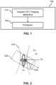
- FIG. 1 there is depicted a block diagram of a system 100 for estimating milk flow during breastfeeding or milk expression according to a proposed embodiment.
- the system 100 comprises a Doppler optical coherence tomography, OCT, imaging apparatus 110 adapted to generate one or more Doppler OCT images of a breast during expression of milk from the breast.
- the system 100 further comprises a processor 130 adapted to determine a milk flow velocity and a cross-sectional area of a milk duct in the breast from the one or more Doppler OCT images, and estimate a milk flow value based on the determined milk flow velocity and the determined cross-sectional area.
- the velocity of milk flow may be determined via the Doppler signal, and a cross-sectional area of one or more milk ducts may be directly determined from the OCT image scans: (axial) A-, (lateral) B-scans, and optionally (lateral) C-scans.
- the Doppler OCT imaging apparatus 110 is adapted to generate a 3-dimensional OCT image.
- a 3-dimensional OCT image is commonly referred to as a "C-scan".
- C-scan 3-dimensional OCT image
- the Doppler OCT imaging apparatus may be adapted to only generate 2-dimensional OCT images (B-scan) or 1-dimensional OCT images (A-scan). In these cases, the processor 130 will need to analyze the images in order to determine a Doppler angle of the milk flow.
- Cross-sectional imaging of the milk ducts by B-scans can be achieved by laterally combining a series of A-scans.
- scans should be taken at a high-speed, i.e. A-scan rate should be substantially higher than pumping rate.
- A-, B-, and C-scans should ideally be performed at a turning point, e.g. when the nipple is most stretched.
- a motion or proximity sensor may be included in the system 100 for triggering the OCT scans.
- the moment of scanning should be synced to the pumping frequency of the breast pump.
- the wavelength of light used by the Doppler OCT imaging apparatus 110 is between 750 and 1300 nm. This range of wavelengths has been determined to result in an effective penetration depth in breast tissue. Accordingly, accurate information can be derived. Any wavelength of light between 700 and 1700nm may be suitable, however.
- the Doppler OCT imaging apparatus 110 may be configured to generate a plurality of velocity measurements over time, and the processor 130 may be adapted to determine a likelihood of a blockage in the milk duct based on one or more of: a stepped reduction of milk flow velocity; a rate of increase in milk flow velocity, wherein the rate of increase falls below a first threshold; and a rate of decrease in milk flow velocity, wherein the rate of decrease falls below a second threshold.
- thresholds provides an efficient way to determine whether the milk duct is clogged or at risk of clogging, either partially, substantially, or fully.
- the thresholds may be predetermined based on generalized data, or may be personalized to each user based on past measurements. This personalization may be performed automatically by the processor 130.
- the processor 130 may also be adapted to generate a map of milk flow levels for one or more regions of the breast. Generating a map of milk flow for different regions may be useful for diagnosing particular problem-areas or blockages.
- a map of milk flow for different regions may be useful for diagnosing particular problem-areas or blockages.
- milk ducts with low flow can be detected early before severe problems develop, because the reduction in milk flow from a localized area of the breast becomes visible.
- the image may for example colour code different flow levels.
- the generated flow map allows personalized guidance to be given on how to position the baby during a next feeding session, reducing the occurrence of blocked milk ducts and discomfort. Thresholds may be applied to the flow levels to enable detection of blocked ducts.
- the processor 130 may be configured to alert the user if a volume or flow of milk that the baby is receiving falls below a threshold value.
- the processor 130 may also be configured to provide guidance on breast feeding based on the estimated milk flow value.
- the processor 130 may also be configured to translate the milk flow value to a drinking or sucking rate.
- system 100 may further be configured to measure a user's health vital signs via their skin.
- Example vital signs that could be measured are vitamin levels, stress levels, nutrition status, etc.
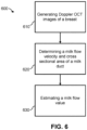
- FIG. 2 there is depicted a diagram showing how volumetric milk flow through a milk duct 220 may be determined.
- both the Doppler angle 230 (the angle between flow direction and optical beam) and the structure of the duct 220 (elliptical axes Ea and Eb) needs to be determined by performing A-scans 210 and B-scans.
- the actual flow velocity can be calculated by taking the vector sum of the Doppler flow of the A-scan 210 (Va) and the B-scan (Vb).
- the average velocity, v, in a milk duct 220 can be defined as the distance, d, the milk in a duct travels in a period of time, t.
- Flow rate, Q can be defined as the volume of fluid passing by a location, P, through an area, A, during a period of time, t.
- the flow rate can be calculated by finding the average flow velocity through the Doppler signal combined with estimating the cross-sectional area of the milk duct 220 using the OCT intensity images.
- the flow velocity in the direction of a probe beam, v z can be calculated.
- the absolute velocity, v abs can then be calculated by estimating the angle between the probe beam 210 and the duct 220.
- CA stands for the cross-sectional area of the duct 220, which can be determined from an OCT image.
- V ⁇ Q dt
- 3D Doppler data is collected and therefore knowledge of the Doppler angle 230 is not required for deriving absolute flow.
- This may be referred to as an en-face plane-based flow method, and in this method, total flow can be derived from the cross-sectional area in a plane bisecting ducts perpendicular to the optical beam.
- the measured velocity vector milk flow and the normal vector of the cross-sectional area may be parallel.
- multiple systems with optical beams passing the ducts can be arranged in a rotational symmetric set-up around the areola area. In this way, duct blockages may be detected. Longitudinal data could be collected and used to follow the lactation process, i.e. monitor a decreased volume of milk expressed or decreased breastfeeding. This information may then be used to guide the mother to phase out breastfeeding in a gentle and gradual way.
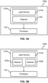
- FIG. 3A there is depicted a block diagrams of a system 300a for estimating milk flow during breastfeeding or milk expression according to a proposed embodiment.
- the Doppler OCT imaging apparatus 310a comprises a light source 315a and a detector 320.
- the light source 315a is adapted to emit, towards the breast, a first light, wherein the first light is at least partially coherent and of an infrared wavelength.
- Traditional OCT also known as LOCT, requires light with partial coherence to work, and the optimum coherence length is determined by the desired imaging depth. If the coherence length is too short, then the desired imaging depth cannot be reached. If the coherence length is too long, however, disturbance from other surfaces or tissue depths may occur. Fully coherent light, i.e. laser light, may be used however, if using SLD-based and Swept Source laser based OCTs.
- the detector 320 is configured to determine a first interference signal between backscattered light from the breast tissue and a reference light.
- a reference light may be produced by light from the light source 315a being reflected back towards the detector 320 by a mirror. Lenses may be placed throughout the system 300a in order to focus or disperse the light where required. The detector 320 then generates a Doppler OCT image from the first interference signal.
- Fig. 3B there is depicted a block diagram of a system 300b for estimating milk flow during breastfeeding or milk expression according to a proposed embodiment.
- the processor 130 is the same as was described in relation to Fig. 1 , however, in this embodiment the Doppler OCT imaging apparatus 310b comprises a light source 315b, which may be substantially the same as light source 315a, and two detectors 320a and 320b.
- the use of at least two light detectors, 320a and 320b, allows the imaging apparatus 320b to be adapted for balanced detection. In another embodiment, there may be a plurality of light detectors.
- one suitable specific implementation of balanced detection in this invention may include the Doppler OCT imaging apparatus 310b further comprising an optical circulator to redirect light from the light source 315a towards a reference and measurement branch, and one first photodiode for cancelling out laser noise for image analysis.
- the apparatus 310b may further include a splitter, such as a 50/50 splitter, to split the light that comes from the reference and measurement branch into a second photodiode and a third photodiode.
- the processor 130 may then compare the light from the light source 315a and the light from the reference and measurement branch in the first photodiode (or second and third photodiode) in order to reduce noise and improve signal-to-noise ratio.
- This implementation may reduce laser intensity noise, but other implementations of balanced detection are possible and suitable for the present invention. It should be noted that for RF balancing of system noise, more hardware in the system 310b (or any other herein disclosed embodiment of the system) may be needed such as beam splitters and polarization controllers.
- the Doppler OCT imaging apparatus 310b may be further adapted for polarization imaging.
- the system 300b may be further enhanced by employing Doppler OCT enhancement crossed-polarization method, where polarization optics are used to decrease background surface reflections and increase signal to noise ratio (SNR) of the OCT images.
- SNR signal to noise ratio
- polarization sensitive OCT systems there are two types.
- the first requires more complex signals processing, due to the required balancing between P and S state of polarization in the polarization controllers.
- Polarization controller optics may be photonics integrated. The latter type, without a polarization controller, would be the more preferred embodiment in the present invention due to compactness and cost considerations.
- QWPs quarter wave plates
- a reference branch in the OCT system is split up into two branches using two reference mirrors. The first branch has QWPs and the second branch does not.
- the second type of implementation of a polarization sensitive OCT system without polarization controllers there is provided one reference branch with a first QWP and one measurement branch with a second QWP.
- the two QWPs are aligned at 45 degrees with respect to one another, so that the two states of polarization in each branch are orthogonal, i.e. cross-polarized.
- Signal processing then consists of gathering co- and cross-polarized images to be separated into individual images. In this way, sample reflectivity, i.e. layered constructions of biological tissue, can be extracted from the images.
- Fig. 3C there is depicted a block diagram of a system 300c for estimating milk flow during breastfeeding or milk expression according to a proposed embodiment.
- the processor 130 is the same as was described in relation to Fig. 1 , however, in this embodiment the Doppler OCT imaging apparatus 310c comprises a self-mixing interferometry, SMI, detector 330 and the light source 315c comprises a swept-laser light source 325.
- SMI Self-mixing interferometry
- SMI hardware such as the SMI detector 330, is especially suited to measure relative velocities, and laser sensors making use of SMI are commercially available, such as the Philips laser beetle TM .
- a swept-laser light source 325 allows for a high-resolution, high-speed Fourier domain (FD) OCT system 310c, thereby keeping measurement times short and avoiding large motion artifacts while imaging milk ducts in a moving nipple with high enough resolution.
- FD Fourier domain
- the light source 315c need not be a swept-laser light source 325.
- the light source may instead be a super luminescent diode, SLD.
- the laser may be moved to probe different locations. This may comprise moving the laser within the system to scan the breast while the system is fixed on the breast, or it may comprise moving the whole system with respect to the breast, for instance, rotating or re-sitting the system. Alternatively, the breast could be moved with respect to the system until, for instance, a feedback signal is provided that informs the user that the position is good. Moving the laser within the system would be the preferred embodiment as this requires less user intervention. Moving the laser to probe different locations would be particularly useful to find the places where the milk flow gives the best signal. Since SLD SMI has the potential to discriminate among different regions, scanning in a Doppler OCT set-up may provide information about the best locations to measure milk flow.
- Fig. 3D there is depicted a block diagram of a system 300d for estimating milk flow during breastfeeding or milk expression according to a proposed embodiment.
- the processor 130 is the same as was described in relation to Fig. 1 , however, in this embodiment the Doppler OCT imaging apparatus 310d comprises a light source 315d which comprises a broadband source 335 and a spectrometer 340.
- the detector 320 may be the same as the detector as was described in relation to Fig. 3A .
- FD Fourier domain
- the spectrometer 340 may be replaced by a grating with a line sensor or a camera.
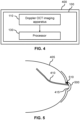
- a simplified block diagram of a nipple shield 400 comprising a system 100 for estimating milk flow during breastfeeding or milk expression according to a proposed embodiment.
- the system 100 is not limited to the system 100 of Fig. 1 , but may be a system according to any of the herein disclosed embodiments.
- the nipple shield 400 may be replaced with a brassiere type garment or a breast pump expression kit, either electric or manual. Integrating the flow sensor arrangement into a brassiere type garment, nipple shield, or a breast pump expression kit provides highly convenient and unobtrusive operation.
- the OCT apparatus should make contact with the nipple area or areola for greater effectiveness, however, this is not essential.
- the OCT apparatus could, for instance, still function hovering above the nipple area or areola. Accordingly, milk quantity may be determined with no additional steps or systems.
- the system may also be miniaturized by (on-chip) photonic integration to meet form factor requirements. Photonic integration contributes to the miniaturization and integration of optical systems.
- the nipple shield 400 may also be replaced by a bottle, storage cup, breast pad, or thermal pad.
- a simplified diagram of a nipple shield 500 on a breast 405 comprising a system 510 for estimating milk flow during breastfeeding or milk expression according to a proposed embodiment.
- the system 510 When integrated into a nipple shield 500 or a brassiere type garment or a breast pump expression kit, the system 510 should be placed so that in use the OCT apparatus is located over an area where the milk ducts 410 are close to the skin. The system 510 may then typically be placed within the nipple shield 500 so that in use they are close to the nipple 415.
- FIG. 6 there is depicted a simplified flow diagram of a method 600 for estimating milk flow during breastfeeding or milk expression according to a proposed embodiment.
- step 610 one or more Doppler OCT images of a breast during the expression of milk from the breast are generated.
- step 620 a milk flow velocity and a cross-sectional area of a milk duct in the breast from the one or more Doppler OCT images are determined.
- step 630 a milk flow value based on the determined milk flow velocity and the determined cross-sectional area is estimated.
- a first light is emitted towards the breast, wherein the first light is at least partially coherent and of an infrared wavelength.
- a first interference signal is determined between backscattered light from the breast tissue and a reference light.
- an OCT image is generated from the first interference signal. Steps 620 and 630 are the same as were described in relation to Fig. 6 .
- step 740 one or more of the following are determined to determine a likelihood of a blockage in the milk duct: a stepped reduction of milk flow velocity; a rate of increase in milk flow velocity, wherein the rate of increases falls below a first threshold; and a rate of decrease in milk flow velocity, wherein the rate of decrease falls below a second threshold.
- any herein disclosed processing does not need to be in real time. Images can for example be stored and analyzed after the breast pumping or breast feeding event, and the output may be given a short time afterwards. However, real time processing is also possible.
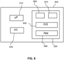
- Fig. 8 illustrates an example of a computer 800 within which one or more parts of an embodiment may be employed.
- Various operations discussed above may utilize the capabilities of the computer 800.
- system functional blocks can run on a single computer or may be distributed over several computers and locations (e.g. connected via internet).
- the computer 800 includes, but is not limited to, PCs, workstations, laptops, PDAs, palm devices, servers, storages, and the like.
- the computer 800 may include one or more processors 810, memory 820 and one or more I/O devices 830 that are communicatively coupled via a local interface (not shown).
- the local interface can be, for example but not limited to, one or more buses or other wired or wireless connections, as is known in the art.
- the local interface may have additional elements, such as controllers, buffers (caches), drivers, repeaters, and receivers, to enable communications. Further, the local interface may include address, control, and/or data connections to enable appropriate communications among the aforementioned components.
- the processor 810 is a hardware device for executing software that can be stored in the memory 820.
- the processor 810 can be virtually any custom made or commercially available processor, a central processing unit (CPU), a digital signal processor (DSP), or an auxiliary processor among several processors associated with the computer 800, and the processor 810 may be a semiconductor based microprocessor (in the form of a microchip) or a microprocessor.
- the memory 820 can include any one or combination of volatile memory elements (e.g., random access memory (RAM), such as dynamic random access memory (DRAM), static random access memory (SRAM), etc.) and non-volatile memory elements (e.g., ROM, erasable programmable read only memory (EPROM), electronically erasable programmable read only memory (EEPROM), programmable read only memory (PROM), tape, compact disc read only memory (CD-ROM), disk, diskette, cartridge, cassette or the like, etc.).
- RAM random access memory
- DRAM dynamic random access memory
- SRAM static random access memory
- non-volatile memory elements e.g., ROM, erasable programmable read only memory (EPROM), electronically erasable programmable read only memory (EEPROM), programmable read only memory (PROM), tape, compact disc read only memory (CD-ROM), disk, diskette, cartridge, cassette or the like, etc.
- the memory 820 may incorporate electronic, magnetic, optical, and/or other types
- the software in the memory 820 may include one or more separate programs, each of which comprises an ordered listing of executable instructions for implementing logical functions.
- the software in the memory 820 includes a suitable operating system (O/S) 850, compiler 860, source code 870, and one or more applications 880 in accordance with exemplary embodiments.
- the application 880 comprises numerous functional components for implementing the features and operations of the exemplary embodiments.
- the application 880 of the computer 800 may represent various applications, computational units, logic, functional units, processes, operations, virtual entities, and/or modules in accordance with exemplary embodiments, but the application 880 is not meant to be a limitation.
- the operating system 850 controls the execution of other computer programs, and provides scheduling, input-output control, file and data management, memory management, and communication control and related services. It is contemplated by the inventors that the application 880 for implementing exemplary embodiments may be applicable on all commercially available operating systems.
- Application 880 may be a source program, executable program (object code), script, or any other entity comprising a set of instructions to be performed.
- a source program then the program is usually translated via a compiler (such as the compiler 860), assembler, interpreter, or the like, which may or may not be included within the memory 820, so as to operate properly in connection with the O/S 850.
- the application 880 can be written as an object oriented programming language, which has classes of data and methods, or a procedure programming language, which has routines, subroutines, and/or functions, for example but not limited to, C, C++, C#, Pascal, Python, BASIC, API calls, HTML, XHTML, XML, ASP scripts, JavaScript, FORTRAN, COBOL, Perl, Java, ADA, .NET, and the like.
- the I/O devices 830 may include input devices such as, for example but not limited to, a mouse, keyboard, scanner, microphone, camera, etc. Furthermore, the I/O devices 830 may also include output devices, for example but not limited to a printer, display, etc. Finally, the I/O devices 830 may further include devices that communicate both inputs and outputs, for instance but not limited to, a NIC or modulator/demodulator (for accessing remote devices, other files, devices, systems, or a network), a radio frequency (RF) or other transceiver, a telephonic interface, a bridge, a router, etc. The I/O devices 830 also include components for communicating over various networks, such as the Internet or intranet.
- a NIC or modulator/demodulator for accessing remote devices, other files, devices, systems, or a network
- RF radio frequency
- the I/O devices 830 also include components for communicating over various networks, such as the Internet or intranet.
- the software in the memory 820 may further include a basic input output system (BIOS) (omitted for simplicity).
- BIOS is a set of essential software routines that initialize and test hardware at start-up, start the O/S 850, and support the transfer of data among the hardware devices.
- the BIOS is stored in some type of read-only-memory, such as ROM, PROM, EPROM, EEPROM or the like, so that the BIOS can be executed when the computer 800 is activated.
- the processor 810 When the computer 800 is in operation, the processor 810 is configured to execute software stored within the memory 820, to communicate data to and from the memory 820, and to generally control operations of the computer 800 pursuant to the software.
- the application 880 and the O/S 850 are read, in whole or in part, by the processor 810, perhaps buffered within the processor 810, and then executed.
- a computer readable medium may be an electronic, magnetic, optical, or other physical device or means that can contain or store a computer program for use by or in connection with a computer related system or method.
- the application 880 can be embodied in any computer-readable medium for use by or in connection with an instruction execution system, apparatus, or device, such as a computer-based system, processor-containing system, or other system that can fetch the instructions from the instruction execution system, apparatus, or device and execute the instructions.
- a "computer-readable medium" can be any means that can store, communicate, propagate, or transport the program for use by or in connection with the instruction execution system, apparatus, or device.
- the computer readable medium can be, for example but not limited to, an electronic, magnetic, optical, electromagnetic, infrared, or semiconductor system, apparatus, device, or propagation medium.
- Figs. 6 and 7 may be implemented in hardware or software, or a mixture of both (for example, as firmware running on a hardware device).
- the functional steps illustrated in the process flowcharts may be performed by suitably programmed physical computing devices, such as one or more central processing units (CPUs) or graphics processing units (GPUs).
- CPUs central processing units
- GPUs graphics processing units
- Each process - and its individual component steps as illustrated in the flowcharts - may be performed by the same or different computing devices.
- a computer-readable storage medium stores a computer program comprising computer program code configured to cause one or more physical computing devices to carry out an encoding or decoding method as described above when the program is run on the one or more physical computing devices.
- Storage media may include volatile and non-volatile computer memory such as RAM, PROM, EPROM, and EEPROM, optical discs (like CD, DVD, BD), magnetic storage media (like hard discs and tapes).
- RAM random access memory
- PROM read-only memory
- EPROM erasable programmable read-only memory
- EEPROM electrically erasable programmable read-only memory
- optical discs like CD, DVD, BD
- magnetic storage media like hard discs and tapes.
- Various storage media may be fixed within a computing device or may be transportable, such that the one or more programs stored thereon can be loaded into a processor.
- the blocks shown in the block diagrams of Figs. 1 , 3A-3D and 4 may be separate physical components, or logical subdivisions of single physical components, or may be all implemented in an integrated manner in one physical component.
- the functions of one block shown in the drawings may be divided between multiple components in an implementation, or the functions of multiple blocks shown in the drawings may be combined in single components in an implementation.
- Hardware components suitable for use in embodiments of the present invention include, but are not limited to, conventional microprocessors, application specific integrated circuits (ASICs), and field-programmable gate arrays (FPGAs).
- One or more blocks may be implemented as a combination of dedicated hardware to perform some functions and one or more programmed microprocessors and associated circuitry to perform other functions.
- a computer program may be stored/distributed on a suitable medium, such as an optical storage medium or a solid-state medium supplied together with or as part of other hardware, but may also be distributed in other forms, such as via the Internet or other wired or wireless telecommunication systems.
- a suitable medium such as an optical storage medium or a solid-state medium supplied together with or as part of other hardware, but may also be distributed in other forms, such as via the Internet or other wired or wireless telecommunication systems.
- each block in the flowchart or block diagrams may represent a module, segment, or portion of instructions, which comprises one or more executable instructions for implementing the specified logical function(s).
- the functions noted in the block may occur out of the order noted in the figures.
- two blocks shown in succession may, in fact, be executed substantially concurrently, or the blocks may sometimes be executed in the reverse order, depending upon the functionality involved.
- each block of the block diagrams and/or flowchart illustration, and combinations of blocks in the block diagrams and/or flowchart illustration can be implemented by special purpose hardware-based systems that perform the specified functions or acts or carry out combinations of special purpose hardware and computer instructions, the architecture, functionality, and operation of possible implementations of systems, methods, and computer program products according to various embodiments of the present invention.
- each block in the flowchart or block diagrams may represent a module, segment, or portion of instructions, which comprises one or more executable instructions for implementing the specified logical function(s).
- the functions noted in the block may occur out of the order noted in the figures.
Landscapes
- Health & Medical Sciences (AREA)
- Life Sciences & Earth Sciences (AREA)
- Engineering & Computer Science (AREA)
- Physics & Mathematics (AREA)
- General Health & Medical Sciences (AREA)
- Pathology (AREA)
- Biomedical Technology (AREA)
- Heart & Thoracic Surgery (AREA)
- Medical Informatics (AREA)
- Molecular Biology (AREA)
- Surgery (AREA)
- Animal Behavior & Ethology (AREA)
- Biophysics (AREA)
- Public Health (AREA)
- Veterinary Medicine (AREA)
- Radiology & Medical Imaging (AREA)
- Nuclear Medicine, Radiotherapy & Molecular Imaging (AREA)
- Gastroenterology & Hepatology (AREA)
- Physiology (AREA)
- Endocrinology (AREA)
- Spectroscopy & Molecular Physics (AREA)
- Computer Vision & Pattern Recognition (AREA)
- Gynecology & Obstetrics (AREA)
- Reproductive Health (AREA)
- Measuring Pulse, Heart Rate, Blood Pressure Or Blood Flow (AREA)
- Investigating Or Analysing Materials By Optical Means (AREA)
Priority Applications (3)
| Application Number | Priority Date | Filing Date | Title |
|---|---|---|---|
| EP23167695.8A EP4445831A1 (fr) | 2023-04-13 | 2023-04-13 | Système et procédé de surveillance de l'écoulement du lait pendant l'allaitement ou l'expression du lait |
| PCT/EP2024/059322 WO2024213478A1 (fr) | 2023-04-13 | 2024-04-05 | Système et procédé de surveillance permettant de surveiller un débit de lait pendant l'allaitement ou l'expression de lait |
| CN202410435504.XA CN118787378A (zh) | 2023-04-13 | 2024-04-11 | 用于监测母乳喂养或乳汁挤出期间的乳汁流量的监测系统和方法 |
Applications Claiming Priority (1)
| Application Number | Priority Date | Filing Date | Title |
|---|---|---|---|
| EP23167695.8A EP4445831A1 (fr) | 2023-04-13 | 2023-04-13 | Système et procédé de surveillance de l'écoulement du lait pendant l'allaitement ou l'expression du lait |
Publications (1)
| Publication Number | Publication Date |
|---|---|
| EP4445831A1 true EP4445831A1 (fr) | 2024-10-16 |
Family
ID=86007760
Family Applications (1)
| Application Number | Title | Priority Date | Filing Date |
|---|---|---|---|
| EP23167695.8A Withdrawn EP4445831A1 (fr) | 2023-04-13 | 2023-04-13 | Système et procédé de surveillance de l'écoulement du lait pendant l'allaitement ou l'expression du lait |
Country Status (3)
| Country | Link |
|---|---|
| EP (1) | EP4445831A1 (fr) |
| CN (1) | CN118787378A (fr) |
| WO (1) | WO2024213478A1 (fr) |
Citations (4)
| Publication number | Priority date | Publication date | Assignee | Title |
|---|---|---|---|---|
| US20090054771A1 (en) | 2004-11-18 | 2009-02-26 | Mamsense Ltd. | Breast milk flow meter apparatus and method |
| US20160120524A1 (en) * | 2013-06-13 | 2016-05-05 | Canon Kabushiki Kaisha | Apparatus and method for supporting biopsy |
| EP3827737A1 (fr) * | 2019-11-26 | 2021-06-02 | Koninklijke Philips N.V. | Système et procédé de surveillance pour surveiller l'écoulement de lait pendant l'allaitement maternel ou l'expression du lait |
| US20210169398A1 (en) * | 2018-08-02 | 2021-06-10 | Coroflo Limited | Microsensor-based Breastfeeding Volume Measurement Device |
Family Cites Families (2)
| Publication number | Priority date | Publication date | Assignee | Title |
|---|---|---|---|---|
| ES1217271Y (es) | 2018-07-24 | 2018-12-05 | Font Caselles Ramon | Aplicador de tampones |
| US20220400946A1 (en) * | 2021-06-17 | 2022-12-22 | Physical Sciences, Inc. | Vivo calibration of doppler flowmetry |
-
2023
- 2023-04-13 EP EP23167695.8A patent/EP4445831A1/fr not_active Withdrawn
-
2024
- 2024-04-05 WO PCT/EP2024/059322 patent/WO2024213478A1/fr not_active Ceased
- 2024-04-11 CN CN202410435504.XA patent/CN118787378A/zh active Pending
Patent Citations (4)
| Publication number | Priority date | Publication date | Assignee | Title |
|---|---|---|---|---|
| US20090054771A1 (en) | 2004-11-18 | 2009-02-26 | Mamsense Ltd. | Breast milk flow meter apparatus and method |
| US20160120524A1 (en) * | 2013-06-13 | 2016-05-05 | Canon Kabushiki Kaisha | Apparatus and method for supporting biopsy |
| US20210169398A1 (en) * | 2018-08-02 | 2021-06-10 | Coroflo Limited | Microsensor-based Breastfeeding Volume Measurement Device |
| EP3827737A1 (fr) * | 2019-11-26 | 2021-06-02 | Koninklijke Philips N.V. | Système et procédé de surveillance pour surveiller l'écoulement de lait pendant l'allaitement maternel ou l'expression du lait |
Non-Patent Citations (2)
| Title |
|---|
| RAMSAY DT ET AL.: "Anatomy of the lactating human breast redefined with ultrasound imaging", JOURNAL OF ANATOMY., vol. 206, no. 6, June 2005 (2005-06-01), pages 525 - 534, XP055063447, DOI: 10.1111/j.1469-7580.2005.00417.x |
| SHARMA U ET AL.: "Long-wavelength optical coherence tomography at 1.7 microns for enhanced imaging depth", OPTICS EXPRESS., vol. 16, no. 24, 2008, pages 19712 - 19723 |
Also Published As
| Publication number | Publication date |
|---|---|
| CN118787378A (zh) | 2024-10-18 |
| WO2024213478A1 (fr) | 2024-10-17 |
Similar Documents
| Publication | Publication Date | Title |
|---|---|---|
| US10588572B2 (en) | Bulk motion subtraction in optical coherence tomography angiography | |
| JP5166889B2 (ja) | 眼底血流量の定量測定装置 | |
| US7869663B2 (en) | Methods, systems and computer program products for analyzing three dimensional data sets obtained from a sample | |
| US7995814B2 (en) | Dynamic motion contrast and transverse flow estimation using optical coherence tomography | |
| US9105123B2 (en) | Non-linear projections of 3-D medical imaging data | |
| US10405793B2 (en) | Systems and methods for in vivo visualization of lymphatic vessels with optical coherence tomography | |
| CN105342568B (zh) | 联合相位和幅值的光学相干造影方法及系统 | |
| TW201100053A (en) | Method and apparatus for quantitative imaging of blood perfusion in living tissue | |
| US10682054B2 (en) | Apparatus for angiographic optical coherence tomography in retina or choroid, and method for diagnosing diseases by using same | |
| US20160066798A1 (en) | Methods and Systems for Determining Hemodynamic Properties of a Tissue | |
| CN107862724B (zh) | 一种改进的微血管血流成像方法 | |
| He et al. | Comparison of hemodialysis arteriovenous fistula blood flow rates measured by Doppler ultrasound and phase-contrast magnetic resonance imaging | |
| US10548483B2 (en) | Image processing apparatus and image processing method | |
| CN111134614A (zh) | 基于oct测量眼球血管中血流的绝对速度的方法及系统 | |
| Pijewska et al. | Computationally effective 2D and 3D fast phase unwrapping algorithms and their applications to Doppler optical coherence tomography | |
| Brandts et al. | Site-specific coupling between vascular wall thickness and function: an observational MRI study of vessel wall thickening and stiffening in hypertension | |
| CN113706567B (zh) | 一种结合血管形态特征的血流成像量化处理方法与装置 | |
| JP2019150345A (ja) | 画像処理装置、画像処理方法及びプログラム | |
| Ullah et al. | M-mode swept source optical coherence tomography for quantification of salt concentration in blood: an in vitro study | |
| EP4445831A1 (fr) | Système et procédé de surveillance de l'écoulement du lait pendant l'allaitement ou l'expression du lait | |
| CN111343906B (zh) | 测量装置、测量方法及计算机可读存储介质 | |
| JP6701005B2 (ja) | 装置および情報処理方法 | |
| CN106073811B (zh) | 使用成像方法来确定流体速度 | |
| Albinsson et al. | A combination of parabolic and grid slope interpolation for 2D tissue displacement estimations | |
| Goncharsky et al. | Artifacts of reconstructed images in inverse problems of ultrasound tomography in models with absorption |
Legal Events
| Date | Code | Title | Description |
|---|---|---|---|
| PUAI | Public reference made under article 153(3) epc to a published international application that has entered the european phase |
Free format text: ORIGINAL CODE: 0009012 |
|
| STAA | Information on the status of an ep patent application or granted ep patent |
Free format text: STATUS: THE APPLICATION HAS BEEN PUBLISHED |
|
| AK | Designated contracting states |
Kind code of ref document: A1 Designated state(s): AL AT BE BG CH CY CZ DE DK EE ES FI FR GB GR HR HU IE IS IT LI LT LU LV MC ME MK MT NL NO PL PT RO RS SE SI SK SM TR |
|
| STAA | Information on the status of an ep patent application or granted ep patent |
Free format text: STATUS: THE APPLICATION IS DEEMED TO BE WITHDRAWN |
|
| 18D | Application deemed to be withdrawn |
Effective date: 20250417 |