EP4454663A1 - Agent thérapeutique ou prophylactique pour la crise cardiaque, la fibrose cardiaque ou l'insuffisance cardiaque, dans lequel htra3 est la cible thérapeutique - Google Patents
Agent thérapeutique ou prophylactique pour la crise cardiaque, la fibrose cardiaque ou l'insuffisance cardiaque, dans lequel htra3 est la cible thérapeutique Download PDFInfo
- Publication number
- EP4454663A1 EP4454663A1 EP22911314.7A EP22911314A EP4454663A1 EP 4454663 A1 EP4454663 A1 EP 4454663A1 EP 22911314 A EP22911314 A EP 22911314A EP 4454663 A1 EP4454663 A1 EP 4454663A1
- Authority
- EP
- European Patent Office
- Prior art keywords
- htra3
- heart failure
- expression
- igfbp7
- cardiac
- Prior art date
- Legal status (The legal status is an assumption and is not a legal conclusion. Google has not performed a legal analysis and makes no representation as to the accuracy of the status listed.)
- Pending
Links
Images















Classifications
-
- A—HUMAN NECESSITIES
- A61—MEDICAL OR VETERINARY SCIENCE; HYGIENE
- A61K—PREPARATIONS FOR MEDICAL, DENTAL OR TOILETRY PURPOSES
- A61K39/00—Medicinal preparations containing antigens or antibodies
- A61K39/0005—Vertebrate antigens
-
- A—HUMAN NECESSITIES
- A01—AGRICULTURE; FORESTRY; ANIMAL HUSBANDRY; HUNTING; TRAPPING; FISHING
- A01K—ANIMAL HUSBANDRY; AVICULTURE; APICULTURE; PISCICULTURE; FISHING; REARING OR BREEDING ANIMALS, NOT OTHERWISE PROVIDED FOR; NEW BREEDS OF ANIMALS
- A01K67/00—Rearing or breeding animals, not otherwise provided for; New or modified breeds of animals
- A01K67/027—New or modified breeds of vertebrates
-
- A—HUMAN NECESSITIES
- A01—AGRICULTURE; FORESTRY; ANIMAL HUSBANDRY; HUNTING; TRAPPING; FISHING
- A01K—ANIMAL HUSBANDRY; AVICULTURE; APICULTURE; PISCICULTURE; FISHING; REARING OR BREEDING ANIMALS, NOT OTHERWISE PROVIDED FOR; NEW BREEDS OF ANIMALS
- A01K67/00—Rearing or breeding animals, not otherwise provided for; New or modified breeds of animals
- A01K67/027—New or modified breeds of vertebrates
- A01K67/0275—Genetically modified vertebrates, e.g. transgenic
-
- A—HUMAN NECESSITIES
- A01—AGRICULTURE; FORESTRY; ANIMAL HUSBANDRY; HUNTING; TRAPPING; FISHING
- A01K—ANIMAL HUSBANDRY; AVICULTURE; APICULTURE; PISCICULTURE; FISHING; REARING OR BREEDING ANIMALS, NOT OTHERWISE PROVIDED FOR; NEW BREEDS OF ANIMALS
- A01K67/00—Rearing or breeding animals, not otherwise provided for; New or modified breeds of animals
- A01K67/027—New or modified breeds of vertebrates
- A01K67/0275—Genetically modified vertebrates, e.g. transgenic
- A01K67/0276—Knock-out vertebrates
-
- A—HUMAN NECESSITIES
- A61—MEDICAL OR VETERINARY SCIENCE; HYGIENE
- A61K—PREPARATIONS FOR MEDICAL, DENTAL OR TOILETRY PURPOSES
- A61K31/00—Medicinal preparations containing organic active ingredients
- A61K31/70—Carbohydrates; Sugars; Derivatives thereof
- A61K31/7088—Compounds having three or more nucleosides or nucleotides
-
- A—HUMAN NECESSITIES
- A61—MEDICAL OR VETERINARY SCIENCE; HYGIENE
- A61K—PREPARATIONS FOR MEDICAL, DENTAL OR TOILETRY PURPOSES
- A61K31/00—Medicinal preparations containing organic active ingredients
- A61K31/70—Carbohydrates; Sugars; Derivatives thereof
- A61K31/7088—Compounds having three or more nucleosides or nucleotides
- A61K31/713—Double-stranded nucleic acids or oligonucleotides
-
- A—HUMAN NECESSITIES
- A61—MEDICAL OR VETERINARY SCIENCE; HYGIENE
- A61K—PREPARATIONS FOR MEDICAL, DENTAL OR TOILETRY PURPOSES
- A61K38/00—Medicinal preparations containing peptides
- A61K38/16—Peptides having more than 20 amino acids; Gastrins; Somatostatins; Melanotropins; Derivatives thereof
- A61K38/17—Peptides having more than 20 amino acids; Gastrins; Somatostatins; Melanotropins; Derivatives thereof from animals; from humans
- A61K38/1703—Peptides having more than 20 amino acids; Gastrins; Somatostatins; Melanotropins; Derivatives thereof from animals; from humans from vertebrates
- A61K38/1709—Peptides having more than 20 amino acids; Gastrins; Somatostatins; Melanotropins; Derivatives thereof from animals; from humans from vertebrates from mammals
-
- A—HUMAN NECESSITIES
- A61—MEDICAL OR VETERINARY SCIENCE; HYGIENE
- A61K—PREPARATIONS FOR MEDICAL, DENTAL OR TOILETRY PURPOSES
- A61K38/00—Medicinal preparations containing peptides
- A61K38/16—Peptides having more than 20 amino acids; Gastrins; Somatostatins; Melanotropins; Derivatives thereof
- A61K38/43—Enzymes; Proenzymes; Derivatives thereof
- A61K38/46—Hydrolases (3)
- A61K38/48—Hydrolases (3) acting on peptide bonds (3.4)
- A61K38/482—Serine endopeptidases (3.4.21)
-
- A—HUMAN NECESSITIES
- A61—MEDICAL OR VETERINARY SCIENCE; HYGIENE
- A61K—PREPARATIONS FOR MEDICAL, DENTAL OR TOILETRY PURPOSES
- A61K39/00—Medicinal preparations containing antigens or antibodies
-
- A—HUMAN NECESSITIES
- A61—MEDICAL OR VETERINARY SCIENCE; HYGIENE
- A61K—PREPARATIONS FOR MEDICAL, DENTAL OR TOILETRY PURPOSES
- A61K45/00—Medicinal preparations containing active ingredients not provided for in groups A61K31/00 - A61K41/00
- A61K45/06—Mixtures of active ingredients without chemical characterisation, e.g. antiphlogistics and cardiaca
-
- A—HUMAN NECESSITIES
- A61—MEDICAL OR VETERINARY SCIENCE; HYGIENE
- A61P—SPECIFIC THERAPEUTIC ACTIVITY OF CHEMICAL COMPOUNDS OR MEDICINAL PREPARATIONS
- A61P37/00—Drugs for immunological or allergic disorders
- A61P37/02—Immunomodulators
- A61P37/04—Immunostimulants
-
- A—HUMAN NECESSITIES
- A61—MEDICAL OR VETERINARY SCIENCE; HYGIENE
- A61P—SPECIFIC THERAPEUTIC ACTIVITY OF CHEMICAL COMPOUNDS OR MEDICINAL PREPARATIONS
- A61P9/00—Drugs for disorders of the cardiovascular system
-
- A—HUMAN NECESSITIES
- A61—MEDICAL OR VETERINARY SCIENCE; HYGIENE
- A61P—SPECIFIC THERAPEUTIC ACTIVITY OF CHEMICAL COMPOUNDS OR MEDICINAL PREPARATIONS
- A61P9/00—Drugs for disorders of the cardiovascular system
- A61P9/04—Inotropic agents, i.e. stimulants of cardiac contraction; Drugs for heart failure
-
- A—HUMAN NECESSITIES
- A61—MEDICAL OR VETERINARY SCIENCE; HYGIENE
- A61P—SPECIFIC THERAPEUTIC ACTIVITY OF CHEMICAL COMPOUNDS OR MEDICINAL PREPARATIONS
- A61P9/00—Drugs for disorders of the cardiovascular system
- A61P9/10—Drugs for disorders of the cardiovascular system for treating ischaemic or atherosclerotic diseases, e.g. antianginal drugs, coronary vasodilators, drugs for myocardial infarction, retinopathy, cerebrovascula insufficiency, renal arteriosclerosis
-
- C—CHEMISTRY; METALLURGY
- C07—ORGANIC CHEMISTRY
- C07K—PEPTIDES
- C07K14/00—Peptides having more than 20 amino acids; Gastrins; Somatostatins; Melanotropins; Derivatives thereof
- C07K14/435—Peptides having more than 20 amino acids; Gastrins; Somatostatins; Melanotropins; Derivatives thereof from animals; from humans
- C07K14/46—Peptides having more than 20 amino acids; Gastrins; Somatostatins; Melanotropins; Derivatives thereof from animals; from humans from vertebrates
- C07K14/47—Peptides having more than 20 amino acids; Gastrins; Somatostatins; Melanotropins; Derivatives thereof from animals; from humans from vertebrates from mammals
- C07K14/4701—Peptides having more than 20 amino acids; Gastrins; Somatostatins; Melanotropins; Derivatives thereof from animals; from humans from vertebrates from mammals not used
- C07K14/4702—Regulators; Modulating activity
-
- C—CHEMISTRY; METALLURGY
- C07—ORGANIC CHEMISTRY
- C07K—PEPTIDES
- C07K14/00—Peptides having more than 20 amino acids; Gastrins; Somatostatins; Melanotropins; Derivatives thereof
- C07K14/435—Peptides having more than 20 amino acids; Gastrins; Somatostatins; Melanotropins; Derivatives thereof from animals; from humans
- C07K14/46—Peptides having more than 20 amino acids; Gastrins; Somatostatins; Melanotropins; Derivatives thereof from animals; from humans from vertebrates
- C07K14/47—Peptides having more than 20 amino acids; Gastrins; Somatostatins; Melanotropins; Derivatives thereof from animals; from humans from vertebrates from mammals
- C07K14/4701—Peptides having more than 20 amino acids; Gastrins; Somatostatins; Melanotropins; Derivatives thereof from animals; from humans from vertebrates from mammals not used
- C07K14/4743—Insulin-like growth factor binding protein
-
- C—CHEMISTRY; METALLURGY
- C07—ORGANIC CHEMISTRY
- C07K—PEPTIDES
- C07K16/00—Immunoglobulins [IG], e.g. monoclonal or polyclonal antibodies
- C07K16/18—Immunoglobulins [IG], e.g. monoclonal or polyclonal antibodies against material from animals or humans
-
- C—CHEMISTRY; METALLURGY
- C12—BIOCHEMISTRY; BEER; SPIRITS; WINE; VINEGAR; MICROBIOLOGY; ENZYMOLOGY; MUTATION OR GENETIC ENGINEERING
- C12N—MICROORGANISMS OR ENZYMES; COMPOSITIONS THEREOF; PROPAGATING, PRESERVING, OR MAINTAINING MICROORGANISMS; MUTATION OR GENETIC ENGINEERING; CULTURE MEDIA
- C12N15/00—Mutation or genetic engineering; DNA or RNA concerning genetic engineering, vectors, e.g. plasmids, or their isolation, preparation or purification; Use of hosts therefor
- C12N15/09—Recombinant DNA-technology
- C12N15/11—DNA or RNA fragments; Modified forms thereof; Non-coding nucleic acids having a biological activity
- C12N15/52—Genes encoding for enzymes or proenzymes
-
- C—CHEMISTRY; METALLURGY
- C12—BIOCHEMISTRY; BEER; SPIRITS; WINE; VINEGAR; MICROBIOLOGY; ENZYMOLOGY; MUTATION OR GENETIC ENGINEERING
- C12N—MICROORGANISMS OR ENZYMES; COMPOSITIONS THEREOF; PROPAGATING, PRESERVING, OR MAINTAINING MICROORGANISMS; MUTATION OR GENETIC ENGINEERING; CULTURE MEDIA
- C12N15/00—Mutation or genetic engineering; DNA or RNA concerning genetic engineering, vectors, e.g. plasmids, or their isolation, preparation or purification; Use of hosts therefor
- C12N15/09—Recombinant DNA-technology
- C12N15/63—Introduction of foreign genetic material using vectors; Vectors; Use of hosts therefor; Regulation of expression
- C12N15/79—Vectors or expression systems specially adapted for eukaryotic hosts
- C12N15/85—Vectors or expression systems specially adapted for eukaryotic hosts for animal cells
- C12N15/8509—Vectors or expression systems specially adapted for eukaryotic hosts for animal cells for producing genetically modified animals, e.g. transgenic
-
- C—CHEMISTRY; METALLURGY
- C12—BIOCHEMISTRY; BEER; SPIRITS; WINE; VINEGAR; MICROBIOLOGY; ENZYMOLOGY; MUTATION OR GENETIC ENGINEERING
- C12N—MICROORGANISMS OR ENZYMES; COMPOSITIONS THEREOF; PROPAGATING, PRESERVING, OR MAINTAINING MICROORGANISMS; MUTATION OR GENETIC ENGINEERING; CULTURE MEDIA
- C12N9/00—Enzymes; Proenzymes; Compositions thereof; Processes for preparing, activating, inhibiting, separating or purifying enzymes
- C12N9/14—Hydrolases (3)
- C12N9/48—Hydrolases (3) acting on peptide bonds (3.4)
- C12N9/50—Proteinases, e.g. Endopeptidases (3.4.21-3.4.25)
- C12N9/64—Proteinases, e.g. Endopeptidases (3.4.21-3.4.25) derived from animal tissue
- C12N9/6421—Proteinases, e.g. Endopeptidases (3.4.21-3.4.25) derived from animal tissue from mammals
- C12N9/6424—Serine endopeptidases (3.4.21)
-
- C—CHEMISTRY; METALLURGY
- C12—BIOCHEMISTRY; BEER; SPIRITS; WINE; VINEGAR; MICROBIOLOGY; ENZYMOLOGY; MUTATION OR GENETIC ENGINEERING
- C12Y—ENZYMES
- C12Y304/00—Hydrolases acting on peptide bonds, i.e. peptidases (3.4)
- C12Y304/21—Serine endopeptidases (3.4.21)
-
- C—CHEMISTRY; METALLURGY
- C40—COMBINATORIAL TECHNOLOGY
- C40B—COMBINATORIAL CHEMISTRY; LIBRARIES, e.g. CHEMICAL LIBRARIES
- C40B30/00—Methods of screening libraries
- C40B30/06—Methods of screening libraries by measuring effects on living organisms, tissues or cells
-
- G—PHYSICS
- G01—MEASURING; TESTING
- G01N—INVESTIGATING OR ANALYSING MATERIALS BY DETERMINING THEIR CHEMICAL OR PHYSICAL PROPERTIES
- G01N33/00—Investigating or analysing materials by specific methods not covered by groups G01N1/00 - G01N31/00
- G01N33/48—Biological material, e.g. blood, urine; Haemocytometers
- G01N33/50—Chemical analysis of biological material, e.g. blood, urine; Testing involving biospecific ligand binding methods; Immunological testing
- G01N33/5005—Chemical analysis of biological material, e.g. blood, urine; Testing involving biospecific ligand binding methods; Immunological testing involving human or animal cells
- G01N33/5008—Chemical analysis of biological material, e.g. blood, urine; Testing involving biospecific ligand binding methods; Immunological testing involving human or animal cells for testing or evaluating the effect of chemical or biological compounds, e.g. drugs, cosmetics
-
- G—PHYSICS
- G01—MEASURING; TESTING
- G01N—INVESTIGATING OR ANALYSING MATERIALS BY DETERMINING THEIR CHEMICAL OR PHYSICAL PROPERTIES
- G01N33/00—Investigating or analysing materials by specific methods not covered by groups G01N1/00 - G01N31/00
- G01N33/48—Biological material, e.g. blood, urine; Haemocytometers
- G01N33/50—Chemical analysis of biological material, e.g. blood, urine; Testing involving biospecific ligand binding methods; Immunological testing
- G01N33/5005—Chemical analysis of biological material, e.g. blood, urine; Testing involving biospecific ligand binding methods; Immunological testing involving human or animal cells
- G01N33/5008—Chemical analysis of biological material, e.g. blood, urine; Testing involving biospecific ligand binding methods; Immunological testing involving human or animal cells for testing or evaluating the effect of chemical or biological compounds, e.g. drugs, cosmetics
- G01N33/5044—Chemical analysis of biological material, e.g. blood, urine; Testing involving biospecific ligand binding methods; Immunological testing involving human or animal cells for testing or evaluating the effect of chemical or biological compounds, e.g. drugs, cosmetics involving specific cell types
- G01N33/5061—Muscle cells
-
- G—PHYSICS
- G01—MEASURING; TESTING
- G01N—INVESTIGATING OR ANALYSING MATERIALS BY DETERMINING THEIR CHEMICAL OR PHYSICAL PROPERTIES
- G01N33/00—Investigating or analysing materials by specific methods not covered by groups G01N1/00 - G01N31/00
- G01N33/48—Biological material, e.g. blood, urine; Haemocytometers
- G01N33/50—Chemical analysis of biological material, e.g. blood, urine; Testing involving biospecific ligand binding methods; Immunological testing
- G01N33/5005—Chemical analysis of biological material, e.g. blood, urine; Testing involving biospecific ligand binding methods; Immunological testing involving human or animal cells
- G01N33/5008—Chemical analysis of biological material, e.g. blood, urine; Testing involving biospecific ligand binding methods; Immunological testing involving human or animal cells for testing or evaluating the effect of chemical or biological compounds, e.g. drugs, cosmetics
- G01N33/5082—Supracellular entities, e.g. tissue, organisms
- G01N33/5088—Supracellular entities, e.g. tissue, organisms of vertebrates
-
- G—PHYSICS
- G01—MEASURING; TESTING
- G01N—INVESTIGATING OR ANALYSING MATERIALS BY DETERMINING THEIR CHEMICAL OR PHYSICAL PROPERTIES
- G01N33/00—Investigating or analysing materials by specific methods not covered by groups G01N1/00 - G01N31/00
- G01N33/48—Biological material, e.g. blood, urine; Haemocytometers
- G01N33/50—Chemical analysis of biological material, e.g. blood, urine; Testing involving biospecific ligand binding methods; Immunological testing
- G01N33/5005—Chemical analysis of biological material, e.g. blood, urine; Testing involving biospecific ligand binding methods; Immunological testing involving human or animal cells
- G01N33/5091—Chemical analysis of biological material, e.g. blood, urine; Testing involving biospecific ligand binding methods; Immunological testing involving human or animal cells for testing the pathological state of an organism
-
- G—PHYSICS
- G01—MEASURING; TESTING
- G01N—INVESTIGATING OR ANALYSING MATERIALS BY DETERMINING THEIR CHEMICAL OR PHYSICAL PROPERTIES
- G01N33/00—Investigating or analysing materials by specific methods not covered by groups G01N1/00 - G01N31/00
- G01N33/48—Biological material, e.g. blood, urine; Haemocytometers
- G01N33/50—Chemical analysis of biological material, e.g. blood, urine; Testing involving biospecific ligand binding methods; Immunological testing
- G01N33/53—Immunoassay; Biospecific binding assay; Materials therefor
- G01N33/573—Immunoassay; Biospecific binding assay; Materials therefor for enzymes or isoenzymes
-
- G—PHYSICS
- G01—MEASURING; TESTING
- G01N—INVESTIGATING OR ANALYSING MATERIALS BY DETERMINING THEIR CHEMICAL OR PHYSICAL PROPERTIES
- G01N33/00—Investigating or analysing materials by specific methods not covered by groups G01N1/00 - G01N31/00
- G01N33/48—Biological material, e.g. blood, urine; Haemocytometers
- G01N33/50—Chemical analysis of biological material, e.g. blood, urine; Testing involving biospecific ligand binding methods; Immunological testing
- G01N33/68—Chemical analysis of biological material, e.g. blood, urine; Testing involving biospecific ligand binding methods; Immunological testing involving proteins, peptides or amino acids
- G01N33/6893—Chemical analysis of biological material, e.g. blood, urine; Testing involving biospecific ligand binding methods; Immunological testing involving proteins, peptides or amino acids related to diseases not provided for elsewhere
-
- A—HUMAN NECESSITIES
- A01—AGRICULTURE; FORESTRY; ANIMAL HUSBANDRY; HUNTING; TRAPPING; FISHING
- A01K—ANIMAL HUSBANDRY; AVICULTURE; APICULTURE; PISCICULTURE; FISHING; REARING OR BREEDING ANIMALS, NOT OTHERWISE PROVIDED FOR; NEW BREEDS OF ANIMALS
- A01K2207/00—Modified animals
- A01K2207/05—Animals modified by non-integrating nucleic acids, e.g. antisense, RNAi, morpholino, episomal vector, for non-therapeutic purpose
-
- A—HUMAN NECESSITIES
- A01—AGRICULTURE; FORESTRY; ANIMAL HUSBANDRY; HUNTING; TRAPPING; FISHING
- A01K—ANIMAL HUSBANDRY; AVICULTURE; APICULTURE; PISCICULTURE; FISHING; REARING OR BREEDING ANIMALS, NOT OTHERWISE PROVIDED FOR; NEW BREEDS OF ANIMALS
- A01K2207/00—Modified animals
- A01K2207/30—Animals modified by surgical methods
-
- A—HUMAN NECESSITIES
- A01—AGRICULTURE; FORESTRY; ANIMAL HUSBANDRY; HUNTING; TRAPPING; FISHING
- A01K—ANIMAL HUSBANDRY; AVICULTURE; APICULTURE; PISCICULTURE; FISHING; REARING OR BREEDING ANIMALS, NOT OTHERWISE PROVIDED FOR; NEW BREEDS OF ANIMALS
- A01K2217/00—Genetically modified animals
- A01K2217/07—Animals genetically altered by homologous recombination
- A01K2217/075—Animals genetically altered by homologous recombination inducing loss of function, i.e. knock out
-
- A—HUMAN NECESSITIES
- A01—AGRICULTURE; FORESTRY; ANIMAL HUSBANDRY; HUNTING; TRAPPING; FISHING
- A01K—ANIMAL HUSBANDRY; AVICULTURE; APICULTURE; PISCICULTURE; FISHING; REARING OR BREEDING ANIMALS, NOT OTHERWISE PROVIDED FOR; NEW BREEDS OF ANIMALS
- A01K2217/00—Genetically modified animals
- A01K2217/20—Animal model comprising regulated expression system
- A01K2217/206—Animal model comprising tissue-specific expression system, e.g. tissue specific expression of transgene, of Cre recombinase
-
- A—HUMAN NECESSITIES
- A01—AGRICULTURE; FORESTRY; ANIMAL HUSBANDRY; HUNTING; TRAPPING; FISHING
- A01K—ANIMAL HUSBANDRY; AVICULTURE; APICULTURE; PISCICULTURE; FISHING; REARING OR BREEDING ANIMALS, NOT OTHERWISE PROVIDED FOR; NEW BREEDS OF ANIMALS
- A01K2227/00—Animals characterised by species
- A01K2227/10—Mammal
- A01K2227/105—Murine
-
- A—HUMAN NECESSITIES
- A01—AGRICULTURE; FORESTRY; ANIMAL HUSBANDRY; HUNTING; TRAPPING; FISHING
- A01K—ANIMAL HUSBANDRY; AVICULTURE; APICULTURE; PISCICULTURE; FISHING; REARING OR BREEDING ANIMALS, NOT OTHERWISE PROVIDED FOR; NEW BREEDS OF ANIMALS
- A01K2267/00—Animals characterised by purpose
- A01K2267/03—Animal model, e.g. for test or diseases
- A01K2267/035—Animal model for multifactorial diseases
- A01K2267/0375—Animal model for cardiovascular diseases
-
- A—HUMAN NECESSITIES
- A61—MEDICAL OR VETERINARY SCIENCE; HYGIENE
- A61K—PREPARATIONS FOR MEDICAL, DENTAL OR TOILETRY PURPOSES
- A61K39/00—Medicinal preparations containing antigens or antibodies
- A61K2039/555—Medicinal preparations containing antigens or antibodies characterised by a specific combination antigen/adjuvant
- A61K2039/55505—Inorganic adjuvants
-
- A—HUMAN NECESSITIES
- A61—MEDICAL OR VETERINARY SCIENCE; HYGIENE
- A61K—PREPARATIONS FOR MEDICAL, DENTAL OR TOILETRY PURPOSES
- A61K39/00—Medicinal preparations containing antigens or antibodies
- A61K2039/57—Medicinal preparations containing antigens or antibodies characterised by the type of response, e.g. Th1, Th2
- A61K2039/575—Medicinal preparations containing antigens or antibodies characterised by the type of response, e.g. Th1, Th2 humoral response
-
- A—HUMAN NECESSITIES
- A61—MEDICAL OR VETERINARY SCIENCE; HYGIENE
- A61K—PREPARATIONS FOR MEDICAL, DENTAL OR TOILETRY PURPOSES
- A61K39/00—Medicinal preparations containing antigens or antibodies
- A61K2039/60—Medicinal preparations containing antigens or antibodies characteristics by the carrier linked to the antigen
- A61K2039/6031—Proteins
- A61K2039/6081—Albumin; Keyhole limpet haemocyanin [KLH]
-
- C—CHEMISTRY; METALLURGY
- C07—ORGANIC CHEMISTRY
- C07K—PEPTIDES
- C07K14/00—Peptides having more than 20 amino acids; Gastrins; Somatostatins; Melanotropins; Derivatives thereof
- C07K14/435—Peptides having more than 20 amino acids; Gastrins; Somatostatins; Melanotropins; Derivatives thereof from animals; from humans
- C07K14/475—Growth factors; Growth regulators
-
- C—CHEMISTRY; METALLURGY
- C12—BIOCHEMISTRY; BEER; SPIRITS; WINE; VINEGAR; MICROBIOLOGY; ENZYMOLOGY; MUTATION OR GENETIC ENGINEERING
- C12N—MICROORGANISMS OR ENZYMES; COMPOSITIONS THEREOF; PROPAGATING, PRESERVING, OR MAINTAINING MICROORGANISMS; MUTATION OR GENETIC ENGINEERING; CULTURE MEDIA
- C12N2750/00—MICROORGANISMS OR ENZYMES; COMPOSITIONS THEREOF; PROPAGATING, PRESERVING, OR MAINTAINING MICROORGANISMS; MUTATION OR GENETIC ENGINEERING; CULTURE MEDIA ssDNA viruses
- C12N2750/00011—Details
- C12N2750/14011—Parvoviridae
- C12N2750/14111—Dependovirus, e.g. adenoassociated viruses
- C12N2750/14141—Use of virus, viral particle or viral elements as a vector
- C12N2750/14143—Use of virus, viral particle or viral elements as a vector viral genome or elements thereof as genetic vector
-
- G—PHYSICS
- G01—MEASURING; TESTING
- G01N—INVESTIGATING OR ANALYSING MATERIALS BY DETERMINING THEIR CHEMICAL OR PHYSICAL PROPERTIES
- G01N2333/00—Assays involving biological materials from specific organisms or of a specific nature
- G01N2333/435—Assays involving biological materials from specific organisms or of a specific nature from animals; from humans
- G01N2333/705—Assays involving receptors, cell surface antigens or cell surface determinants
- G01N2333/71—Assays involving receptors, cell surface antigens or cell surface determinants for growth factors; for growth regulators
-
- G—PHYSICS
- G01—MEASURING; TESTING
- G01N—INVESTIGATING OR ANALYSING MATERIALS BY DETERMINING THEIR CHEMICAL OR PHYSICAL PROPERTIES
- G01N2333/00—Assays involving biological materials from specific organisms or of a specific nature
- G01N2333/90—Enzymes; Proenzymes
- G01N2333/914—Hydrolases (3)
- G01N2333/948—Hydrolases (3) acting on peptide bonds (3.4)
- G01N2333/95—Proteinases, i.e. endopeptidases (3.4.21-3.4.99)
- G01N2333/964—Proteinases, i.e. endopeptidases (3.4.21-3.4.99) derived from animal tissue
- G01N2333/96425—Proteinases, i.e. endopeptidases (3.4.21-3.4.99) derived from animal tissue from mammals
- G01N2333/96427—Proteinases, i.e. endopeptidases (3.4.21-3.4.99) derived from animal tissue from mammals in general
- G01N2333/9643—Proteinases, i.e. endopeptidases (3.4.21-3.4.99) derived from animal tissue from mammals in general with EC number
- G01N2333/96433—Serine endopeptidases (3.4.21)
-
- G—PHYSICS
- G01—MEASURING; TESTING
- G01N—INVESTIGATING OR ANALYSING MATERIALS BY DETERMINING THEIR CHEMICAL OR PHYSICAL PROPERTIES
- G01N2500/00—Screening for compounds of potential therapeutic value
- G01N2500/10—Screening for compounds of potential therapeutic value involving cells
-
- G—PHYSICS
- G01—MEASURING; TESTING
- G01N—INVESTIGATING OR ANALYSING MATERIALS BY DETERMINING THEIR CHEMICAL OR PHYSICAL PROPERTIES
- G01N2800/00—Detection or diagnosis of diseases
- G01N2800/32—Cardiovascular disorders
- G01N2800/324—Coronary artery diseases, e.g. angina pectoris, myocardial infarction
-
- G—PHYSICS
- G01—MEASURING; TESTING
- G01N—INVESTIGATING OR ANALYSING MATERIALS BY DETERMINING THEIR CHEMICAL OR PHYSICAL PROPERTIES
- G01N2800/00—Detection or diagnosis of diseases
- G01N2800/32—Cardiovascular disorders
- G01N2800/325—Heart failure or cardiac arrest, e.g. cardiomyopathy, congestive heart failure
Definitions
- the present invention relates to a therapeutic or preventive agent for myocardial infarction, cardiac fibrosis, or heart failure using Htra3 as a therapeutic target.
- the present invention also relates to an agent for regulating cardiac fibroblast DNA repair mechanisms.
- Tissue fibrosis and organ dysfunction are characteristics of age-related diseases including heart failure (Documents 1 to 5). Pathological stresses on the heart induce fibroblast activation and collagen secretion, leading to cardiac fibrosis (Documents 6 to 8). In contrast, suppression of fibroblast activation can alleviate cardiac fibrosis and ameliorate cardiac function after injury (Document 9), suggesting pathogenicity of fibrosis in heart failure.
- Non-Patent Documents 10 to 14 Inhibition of p53 or senolysis can improve the phenotypes of the heart following injury or aging (Documents 12 and 18), suggesting that senescence-like failing cardiomyocytes cause heart failure (Non-Patent Documents 1 and 2).
- tissue fibrosis and cellular senescence which are characteristics of many age-related diseases, cause the onset and progression of heart failure, but whether there is a common pathway to trigger both tissue fibrosis and heart failure events is unknown.
- An object of the present invention is to provide a therapeutic or preventive agent for myocardial infarction, cardiac fibrosis, or heart failure using Htra3 as a therapeutic target, and an agent for regulating a DNA repair mechanism of cardiac fibroblasts. It is also an object of the present invention to provide a non-human mammalian heart failure model animal, a method for screening for an agent or the like for regulating the DNA repair mechanism of cardiac fibroblasts, a method of diagnosing myocardial infarction, cardiac fibrosis or heart failure, and a method of determining the severity level of myocardial infarction or heart failure.
- the inventors have also found that cardiac fibroblasts strongly interact with different types of cardiac cells, including cardiomyocytes, in particular there are many interactions between cardiac fibroblasts and cardiomyocytes, by intercellular communication maps of the heart using single cell analysis (single-cell RNA sequencing, scRNA-seq).
- the inventors also identified high-temperature requirement A serine peptidase 3 (Htra3) in cardiac fibroblasts as an important regulator of cardiomyocyte homeostasis and cardiac fibrosis by single-cell co-expression network analysis.
- Htra3 serine peptidase 3
- TGF- ⁇ transforming growth factor ⁇
- the present inventors have also found that, the mechanism behind the state observed in knockout mice is that, when the function of Htra3 is lost, the amount of TGF- ⁇ protein, which is normally degraded by Htra3, increases, causing fibroblasts to produce more fibers and harden the heart, and that Nox4 protein, which is induced in cardiomyocytes by TGF- ⁇ , increases oxidative stress and damages DNA, thereby indirectly affecting cardiomyocytes and changing their properties.
- pressure overload downregulates Htra3 expression in cardiac fibroblasts and activates TGF- ⁇ signaling, thereby inducing heart failure not only in cardiac fibrosis but also through accumulation of DNA damage and induction of a secretory phenotype in dysfunctional cardiomyocytes.
- the present inventors have also found that overexpression of Htra3 in the heart inhibits TGF- ⁇ signaling and ameliorates cardiac dysfunction after pressure overload.
- the present inventors have also found that Htra3-controlled induction of spatio-temporal cardiac fibrosis and cardiomyocyte secretory phenotype are specifically observed in the infarct regions after myocardial infarction.
- the present inventors have also found through Integrative analyses of human single cardiomyocyte transcriptome and plasma proteome that IGFBP7, a cytokine downstream of TGF- ⁇ and secreted from failing myocytes, is the most predictable marker of advanced heart failure.
- the present inventors have also found that these findings underscore the role of cardiac fibroblasts in cardiomyocyte homeostasis and regulation of cardiac fibrosis via the Htra3-TGF- ⁇ -IGFBP7 pathway, which is a therapeutic target for heart failure, and that cardiac fibroblasts regulate the development of heart failure via the Htra3-TGF- ⁇ -IGFBP7 axis.
- the present inventors have also found that overexpression of the inflammatory cytokine Igfbp7 in endothelial cells of aged hearts leads to reduced insulin signaling in cardiomyocytes.
- the present inventors have also found that overexpression of Igfbp7 in the heart degraded cardiac function, whereas vaccines targeting Igfbp7 ameliorated cardiac dysfunction in the presence of pressure overload.
- the present inventors have also concluded that pressure overload induces endothelial cell senescence and produces the inflammatory cytokine Igfbp7.
- the present inventors have also found that Igfbp7 suppresses cardiomyocyte metabolism, resulting in development of heart failure, and that vaccine therapy targeting IGFBP7 is useful in treating heart failure.
- the present invention is based on these findings.
- an agent for regulating of the DNA repair mechanism of cardiac fibroblasts and a pharmaceutical composition for the treatment or prevention of myocardial infarction or heart failure can be provided.
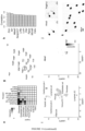
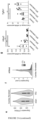
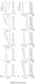
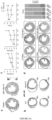
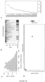
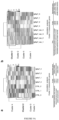
- Fig. 1A illustrates that cardiac fibroblasts Htra3 are identified by single cell network analysis.
- Sorted live non-cardiomyocytes (non-CM) were used for single cell analysis using the 10X Genomics platform.
- Sorted fibroblasts, including antibodies against PDGFR- ⁇ were used for single cell analysis using full-length Smart-seq2.
- b c Ligand-receptor interaction maps of the heart. The ligand and receptor genes were projected onto a two-dimensional map based on their correlation structures. The cell types were annotated by the cell-type specific expression profile of the genes assigned to each module.
- the edge illustrates a correlation (b) or an interaction (c).
- UMAP Uniform manifold approximation and projection
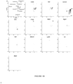
- Fig. 1B illustrates single cell analysis of non-cardiomyocytes in the heart using Chromium (10X Genomics) and the Smart-seq2 platform associated with Fig. 1A .
- UMAP Uniform Manifold Approximation and Projection
- b Heatmap illustrating cell-type specific expression profile of genes including Htra3 (top).
- Violin plot illustrating fibroblast specific expression of Htra3 (bottom).
- c Histogram illustrating the distribution of the number of detected genes. Cells in which 2,000 or more genes were detected were used for subsequent analysis.
- Cell types were determined by graph-based clustering and random forest classifier (left). The origin of the cells was also annotated (right), e Bar chart illustrating cell type distribution in sham surgery (blue) and TAC surgery (red), f Average reduction of the accuracy of each module specific gene (module eigengene, ME) assigned by the random forest classifier (decreasing order of accuracy from top to bottom).
- g tSNE plot illustrating each ME expression.
- Module names were annotated by cell type-specific expression detected by each module network in (H).
- h Co-expression network of each module.
- the size of the dot represents the node centrality.
- i Gene ontology (GO) analysis of each module.
- the most characteristic GO terms in the top 4 ranked annotation clusters in the "functional annotation clustering" function with statistical significance (P ⁇ 0.05) were extracted for each module.
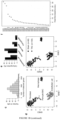
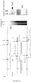
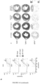
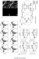
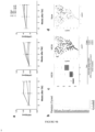
- Fig. 2A illustrates that Htra3 knockout mice exhibit hypertrophy of the heart and cardiomyocytes and are vulnerable to mechanical stress.
- WT wild-type
- KO Htra3 knockout mice
- n 11 (wild-type sham)
- n 15 (wild-type TAC)
- n 10 (Htra3 knockout sham)
- n 15 (Htra3 knockout TAC).
- CTL control subjects
- HF heart failure
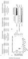
- Fig. 2B illustrates generation of Htra3 knockout mice and single cell analysis showing HTRA3 expression in the human heart, in relation to Fig. 2A .
- Genomic DNA from target ES cells was digested with Bam HI and subjected to southern blot analysis.
- the DIG-labeled probe (from intron 2 illustrated in gray lines) detected a 15kb fragment from the wild type Htra3 allele and a 11kb fragment from the target allele.
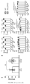
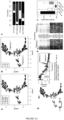
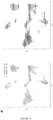
- Fig. 3A illustrates that single-cell analysis of cardiac fibroblasts revealed that Htra3 maintains a quiescent state by TGF- ⁇ inhibition.
- b Trajectory analysis of the UMAP plot of a. Clusters classified by graph-based clustering were displayed in color. The trajectory identified by the Slingshot algorithm is also displayed.
- c Bar chart illustrating the distribution of clusters in b.
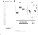
- Fig. 3B illustrates single cell analysis of cardiac fibroblasts and the effect of TGF- ⁇ neutralization in the TAC model, as associated with Fig. 3A .
- a Histogram illustrating the number of genes detected by single cell analysis of cardiac fibroblasts from wild-type and Htra3 knockout mice. Single cell transcriptomes with 3,000 or more genes detected were used for subsequent analysis.
- b Dendrogram illustrating clustered modules assigned using weighted gene correlation network analysis (WGCNA).
- WGCNA weighted gene correlation network analysis
- c Mean decrease in accuracy (in order of decreasing accuracy from top to bottom) of each ME as assigned by the random forest classifier, d Module-specific expression profile on the UMAP plot.
- TGF- ⁇ 1 treatment was used as a positive control for pSmad2/3.
- g Echocardiographic assessment of heart in Htra3 knockout mice following TAC with or without TGF- ⁇ neutralizing antibody treatment (n 3 each). Data pieces are displayed as a mean and SD. *P ⁇ 0.05, **P ⁇ 0.01, ***P ⁇ 0.005, ****P ⁇ 0.001; Significance was determined by two-way ANOVA using Bonferroni's multiple comparison test.
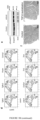
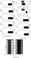
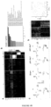
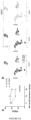
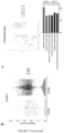
- Fig. 4A illustrates that single-cell analysis of cardiomyocytes revealed that Htra3 prevents DNA damage and induction of failing cardiomyocytes with a secretory phenotype.
- b Trajectory analysis of the UMAP plot of a. Clusters classified by graph-based clustering are displayed in color. The trajectory identified by the Slingshot algorithm is also displayed.
- c Bar chart illustrating the distribution of clusters in b.
- g M2 expression on UMAP plot (left) and its dynamics along pseudotime (top right). The enriched GO terms are also displayed (bottom right). p-values were determined by Fisher's exact test.
- Fig. 4B illustrates single cell analysis of cardiomyocytes from wild-type and Htra3 knockout mice, in relation to Fig. 4A .
- a Histogram illustrating the number of genes detected by single cell analysis of cardiomyocytes of wild type and Htra3 knockout mice. Single cell transcriptomes with 4,000 or more genes detected was used for subsequent analysis.
- b Dendrogram illustrating clustered modules assigned using WGCNAs.
- c Mean decrease in accuracy (in order of decreasing accuracy from top to bottom) of each ME as assigned by the random forest classifier, d Module-specific expression profile on the UMAP plot.
- e Scatter plot illustrating the mean expression profile of single cell analysis of cardiomyocytes isolated from wild-type and Htra3 knockout mice after sham surgery.
- enriched GO terms and associated P-values of the top 300 upregulated and downregulated genes are also displayed.
- Upregulation of genes associated with translation in Htra3 knockout cardiomyocytes suggests a transcriptional signature of enlarged cardiomyocytes in Htra3 knockout mice.
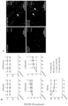
- Fig. 5A illustrates that TGF- ⁇ induced Nox4 activation induces senescent cardiomyocytes and heart failure
- WGA red
- DAPI blue
- Arrows indicate GFP introduced cells
- b Echocardiographic assessment of hearts from TAC-induced Htra3 knockout mice after injection of control or shNox4 AAV9 vectors (n 4 each). Data pieces are displayed as a mean and SD.
- the source data was provided as a source data file.
- i Representative echocardiographic images of wild-type mice injected with either AAV9-control or AAV9-Htra3.
- Fig. 5B shows functional and transcriptional analysis of shNox4-induced and p53 knockout mice in relation to Fig. 5A .
- OE AAV9-Nox4 overexpression
- n 3 (wild-type control)
- n 3 (Htra3 knockout control)
- n 5 (Htra3 knockout Nox4 OE).
- Data pieces are displayed as a mean and SD. **P ⁇ 0.01, ***P ⁇ 0.005, and ****P ⁇ 0.001 Nox4 OE vs.
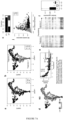
- Fig. 6A illustrates that spatial transcriptomic analysis reveals Htra3 repression-mediated spatial induction of senescent failing cardiomyocytes with secretory phenotype.
- MI myocardial infarction
- Dot plot representing the distribution of cell types predicted based on single cell analysis of cells isolated from mice after sham or MI surgery.
- the dot size represents the average of the prediction scores of each spot in each cluster.
- g Single-cell trajectory analysis of cardiomyocytes isolated from the infarct zone.
- h Expression dynamics of genes characteristic of senescent failing cardiomyocytes along pseudotime.
- i Spatial expression pattern of genes characteristic of senescent cardiomyocytes.
- the dashed circle represents the infarction region.
- Fig. 6B illustrates spatial induction via Htra3 inhibition of failing cardiomyocytes with secretory phenotype by spatial transcriptome analysis and single cell analysis of cardiomyocytes after MI, related to Fig. 6A .
- a Heatmap illustrating expression levels of representative genes of each cluster.
- b Enriched GO terms and enriched P -values associated with representative genes of Cluster 2.
- CM Cardiomyocytes.
- SMC Smooth muscle cells.
- DC dendritic cells, d Violin plots showing Htra3 RNA expression in each cell cluster on sham, day 1 post-MI (pMI), and day 7 pMI.
- pMI post-MI
- pMI post MI
- pMI pMI 1 post MI
- SMC Smooth muscle cells.
- DC dendritic cells
- f Enriched GO terms for characteristic genes of FB2 and FB4 clusters.
- g Violin plots showing total expression levels of Htra3 on sham, post MI (
- Fig. 7A illustrates single-cardiomyocyte RNA-seq and plasma proteome analysis of heart failure patients.
- CTL control subjects
- HF heart failure
- c Bar chart illustrating the distribution of clusters in b.
- e Heatmap illustrating expression levels of selected M2 genes in the trajectory.
- the box plots illustrate the median (centerline), the first and third quartiles (box edge) and the whisker goes from each quartile to the minimum or maximum.
- l Heatmap illustrating expression levels of selected M44 genes in the trajectory.
- the box plots illustrate the median (centerline), the first and third quartiles (box edge) and the whisker goes from each quartile to the minimum or maximum.
- RFU Relative fluorescence unit.
- HF Heart failure.
- HTx Heart transplant.
- Data represents box plots and individual data points. The box plots illustrate the median (centerline), the first and third quartiles (box edge) and the whisker goes from each quartile to the minimum or maximum.
- ROC Receiving-operating-characteristic
- Fig. 7B illustrates single cell and plasma proteome analyses of heart failure patients in relation to Fig. 7A .
- b Dendrogram illustrating clustered modules assigned using WGCNAs.
- d Cross-tabulation table of cardiomyocyte modules obtained from mouse single cell analysis ( Figs. 3A and 7B ) and human single cell analysis ( Figs. 4A and 9B ). The table is colored with -logic (P-value) obtained using a two-tailed Fisher's exact test.
- h Schematic representation of the mechanism by which Htra3 of cardiac fibroblasts prevents cardiomyocyte aging and heart failure induced by TGF- ⁇ signaling.
- Fig. 8A illustrates that Igfbp7 from aged endothelial cells reduces cardiac function.
- b (Left, Center) Results of single cell analysis using WGCNAs and graph-based clustering of cardiomyocytes from mice injected with control vector and AAV9-Igfbp7. (Right) Expression level of Igfbp7 in UMAP.
- Fig. 8B illustrates single cell analysis of cardiomyocytes treated with AAV-Igfbp7 and a control vector, related to Figs. 8Ab and 8Ac .
- Fig. 9A illustrates RNA-seq analysis of human iPSCM treated with IGFBP7.
- the DESeq2 package was used to select the differential expressed genes (DEG).
- DEG differential expressed genes
- the DESeq2 package was used to select the differential expressed genes (DEG).
- Bottom Annotation analysis of Cluster 3 gene.
- Fig. 9B illustrates functional analysis of vaccinated mice and single cell analysis of mouse cardiomyocytes pretreated with IGFBP7 vac#2.
- c (Left) Box plot and (right) UMAP plot color-coded by gene expression of ME38 modules.
- Bottomtom Results of annotation analysis of ME38 gene.
- Fig. 10 illustrates single nuclear RNA-seq analysis in DCM patients and control patients.
- b Results of violin plots of endothelial cells divided by patient status.
- c top) Results of UMAP of cardiomyocytes sorted by patient status.
- Bottom Annotation analysis of characteristic genes in DCM patient cardiomyocytes was compared to control patients.
- an agent for regulating a DNA repair mechanism of cardiac fibroblasts comprising at least one of the above (a), (b), (c), (d), and (e) (in the present specification, it may be referred to as a regulation agent of the present invention).
- High-temperature requirement A serine peptidase 3 (Serine protease HtrA3), which is an active ingredient of the present invention, is a proteinase also called a high-temperature requirement A3 serine protease.
- the serine protease HtrA3 is preferably derived from human or mouse.
- Both the base sequence encoding human serine protease HtrA3 and the base sequence encoding mouse serine protease HtrA3 are based on the base sequence published in the database (https://www.ncbi.nlm.nih.gov/gene) of U.S. National Center for Biotechnology Information (NCBI).
- NCBI National Center for Biotechnology Information
- the human base sequence is based on the base sequence published in NCBI Gene ID: 94031
- the mouse base sequence is based on the base sequence published in NCBI Gene ID: 78558.
- the amino acid sequence of human serine protease HtrA3 and the amino acid sequence of mouse serine protease HtrA3 are both based on the amino acid sequences published in the database of U.S.
- NCBI National Center for Biotechnology Information
- the human amino acid sequence is based on the amino acid sequence published in NCBI Reference Sequence: NP_444272.1
- the mouse amino acid sequence is based on the amino acid sequence published in NCBI Reference Sequence: NP_084403.2.
- the transformed cells of (d) and the antibody or fragment thereof of (e) can be prepared by a method well known to those skilled in the art based on the sequence information.
- a modified base or an artificial base having in vivo stability can also be used as the base constituting the nucleic acid in the present invention.
- the "DNA repair mechanism” is a mechanism for repairing DNA damage (damaged DNA).
- DNA damage means a single-stranded break and/or double-stranded break generated in nuclear DNA
- the DNA repair mechanism means a mechanism for repairing the single-stranded break and/or double-stranded break.
- regulation of a DNA repair mechanism means promoting a DNA repair mechanism, and includes inhibiting suppression of a DNA repair mechanism.
- Cardiac fibroblasts produce TGF- ⁇ , which plays a physiologically important role, such as repairing wounds by promoting fibrosis.
- TGF- ⁇ plays a physiologically important role, such as repairing wounds by promoting fibrosis.
- the action of TGF- ⁇ is too strong in cardiac fibroblasts, accumulation of DNA damage in cardiac fibroblasts and cardiomyocytes, cardiac fibrosis, cardiac fibrosis, myocardial infarction, and heart failure are caused.
- Htra3 which is an active ingredient of the agent for regulating the present invention acts on TGF- ⁇ and decomposes TGF- ⁇ , thereby inhibiting accumulation of DNA damage caused by TGF- ⁇ , cardiac fibrosis, cardiac fibrosis, myocardial infarction, and heart failure.
- a pharmaceutical composition for the treatment or prevention of myocardial infarction, cardiac fibrosis or heart failure, containing the regulation agent of the present invention or at least one of the above (a), (b), (c), (d) and (e) as an active ingredient.
- a method of regulating a DNA repair mechanism of cardiac fibroblasts and a method for treating or preventing myocardial infarction, cardiac fibrosis or heart failure including administering at least one of the above (a), (b), (c), (d), and (e) to a subject in need thereof.
- the heart failure is preferably heart failure with reduced ejection fraction (HFrEF).
- HFrEF heart failure with reduced ejection fraction
- the heart failure with reduced ejection fraction results in poor left ventricular contraction and insufficient ejection, resulting in increased diastolic volume, increased diastolic pressure, and reduced ejection fraction.
- the administration route is not particularly limited as long as a desired therapeutic or preventive effect is obtained, but oral administration or parenteral administration (for example, intravenous administration, subcutaneous administration, or intraperitoneal administration) can be selected.
- oral administration agent examples include granules, powders, tablets (including sugar coating tablets), pills, capsules, syrups, solutions, jellies, emulsions, and suspensions.
- parenteral administration agent an appropriate dosage form can be selected depending on the specific administration route, and examples thereof include injections and suppositories.
- formulations can be formulated using a pharmaceutically acceptable carrier by procedures commonly practiced in the art (for example, a known method described in General Rules for Preparations of the Japanese Pharmacopoeia, 18th edition, or the like).
- Examples of the pharmaceutically acceptable carrier include an excipient, a binder, a diluent, an additive, a fragrance, a buffer, a thickener, a colorant, a stabilizer, an emulsifier, a dispersant, a suspending agent, and a preservative.
- the active ingredient of the present invention can be administered to a subject as a gene therapeutic agent.
- Gene therapy can be performed by methods known to those skilled in the art ( Cannata A, Ali H, Sinagra G, Giacca M. Gene Therapy for the Heart Lessons Learned and Future Perspectives. Circ Res. 2020 May 8;126(10):1394-1414. doi:10.1161/CIRCRESAHA.120.315855. Epub 2020 May 7. PMID:32379579 ), for example, a nucleic acid or a fragment thereof can be administered using a delivery reagent, or an expression vector incorporating a nucleic acid or a fragment thereof can be administered.
- the pharmaceutical composition 1 of the present invention can be a composition containing the nucleic acid of the active ingredient (b) or a fragment thereof (for example, naked DNA) together with a delivery reagent.
- delivery reagents include, but are not limited to, lipophilic reagents, lipofectin, lipofectamine, serfectin, cationic polymers (for example, polylysine), micelles, PEGylated liposomes or nanoparticles, oligonucleotide nanoparticles, and combinations thereof.
- the pharmaceutical composition 1 of the present invention may also be a composition containing an expression vector of the active ingredient (c), optionally together with a delivery reagent.
- the expression vector include, but are not limited to, a viral vector system (for example, adeno-associated viral vectors (AAV vectors), cytomegalovirus vectors (CMV vectors), lentiviral vectors, retroviral vectors, adenoviral vectors, herpesviral vectors, or vaccinia viral vectors), and a non-viral vector system such as a plasmid.
- a viral vector system for example, adeno-associated viral vectors (AAV vectors), cytomegalovirus vectors (CMV vectors), lentiviral vectors, retroviral vectors, adenoviral vectors, herpesviral vectors, or vaccinia viral vectors
- non-viral vector system such as a plasmid.
- the gene therapeutic agent of the present invention can be administered, for example, by intravenous administration, intraperitoneal administration, subcutaneous administration, local administration to the heart, or administration via the coronary artery. It is needless to say that the gene therapeutic agent can also be formulated using a pharmaceutically acceptable carrier by a method usually performed in the art as described above.
- the dose of the active ingredient in the present invention can be determined depending on the type of the active ingredient, the sex, age, body weight, symptom, dosage form, administration route, and the like of the subject to be administered.
- the dose per adult in a case where the active ingredient is administered for the purpose of treating or preventing myocardial infarction, cardiac fibrosis or heart failure, the dose per adult can be determined, for example, in the range of 0.0001 mg to 1000 mg/kg of body weight, but is not limited thereto.
- the active ingredient in the above dosage can be administered once a day or in 2 to 4 divided doses.
- the active ingredient of the present invention can be administered not only to humans in need thereof but also to mammals other than humans (for example, mouse, rat, rabbit, dog, cat, cow, horse, pig, sheep, goat, monkey).
- model animal 1 of the present invention a non-human mammalian heart failure model animal in which expression of serine protease HtrA3 in cardiac fibroblasts is reduced or inhibited
- Animal species of the model animal 1 of the present invention include non-human mammals such as rodents (for example, mice and rats), cows, horses, pigs, monkeys, and dogs.
- the reduction or inhibition of expression of the serine protease HtrA3 in the model animal 1 of the present invention can be carried out by a method known to those skilled in the art, and for example, an animal produced by deleting a sequence portion or a control region of the serine protease HtrA3 gene by genetic manipulation to eliminate the function (knockout) can be used, and examples of such a model animal include knockout mice.
- the HtrA3 gene is knocked out refers to a state in which the expression product of the HtrA3 gene is not expressed at all, or even when expressed, the function of the normal HtrA3 gene product cannot be exhibited, and the function of the HtrA3 gene is deficient, due to modification of the base sequence of the HtrA3 gene or the like.
- the specific mode of deleting the function of the HtrA3 gene is not particularly limited as long as the HtrA3 gene is knocked out, but includes, for example, a mode in which at least a part of the HtrA3 gene is modified or the promoter of the HtrA3 gene is inactivated.
- At least a part of the HtrA3 gene is modified means that a modification causing deletion, substitution, or addition is added to at least a part of the base sequence of the HtrA3 gene.
- a modification causing deletion, substitution, or addition is added to at least a part of the base sequence of the HtrA3 gene.
- one or more mutations among deletion, substitution, and addition may be combined with respect to the HtrA3 gene.
- deletion of at least a part of the base sequence of the HtrA3 gene means that the expression product of the HtrA3 gene is modified so as not to express the function as the HtrA3 protein by deleting a part or all of the HtrA3 gene.
- substitution of at least a part of the base sequence of the HtrA3 gene means that a part or all of the HtrA3 gene is substituted with a separate sequence not related to the HtrA3 gene, thereby modifying the expression product of the HtrA3 gene so that the function of the HtrA3 gene is not expressed.
- "adding" to at least a part of the base sequence of the HtrA3 gene means that a sequence other than the HtrA3 gene is added to the HtrA3 gene, thereby modifying the expression product of the HtrA3 gene so that the function as the HtrA3 protein is not expressed.
- the model animal 1 of the present invention in which the HtrA3 gene is knocked out may be a heterozygote knock-out animal in which either one of the alleles in the HtrA3 gene is knocked out, or may be a homozygote knock-out animal in which both functions of the alleles are knocked out.
- the model animal 1 of the present invention may be either female or male.
- the model animal 1 of the present invention in which the HtrA3 gene is knocked out can be produced by a method known to those skilled in the art.
- a technique include, but are not limited to, a genome editing technique such as a CRISPR/Cas9 method or a transcription activation-like effector nuclease (TALEN) method, and a gene disruption technique by a homologous recombination method.
- TALEN transcription activation-like effector nuclease
- a method for screening for an agent or method of the regulation of DNA repair mechanisms in cardiac fibroblasts including the steps (A) and (B) (which may be referred to herein as Screening Method 1 of the present invention).
- a method for screening for an agent or method of the regulation of DNA repair mechanisms in cardiac fibroblasts including the steps (C) and (D) (sometimes referred to herein as the screening method 2 of the present invention).
- test factor in Screening Methods 1 and 2 of the present invention include, but are not limited to, a low molecular weight compound, a middle molecular weight compound, a peptide, a protein, an antibody, and the like.
- the test factor in order to treat a non-human mammal in which the expression of Htra3 is reduced or inhibited with the test factor, the test factor may be formulated into an optimal dosage form according to the administration route.
- cardiomyocytes or cardiac fibroblasts of human or non-human mammal in which the expression of serine protease HtrA3 is reduced or inhibited can be used, and the cells can be prepared according to the method of the knockout model animal described above.
- the test factor can be determined to be an agent candidate useful for regulating the DNA repair mechanism of cardiac fibroblasts.
- Treating with the test factor in the step (C) of Screening Method 2 of the present invention means, for example, administering the test factor to a non-human mammal (model animal 1 of the present invention).
- the administration route can be appropriately selected according to the type of the test factor and the like.
- the test factor can be determined to be an agent candidate useful for regulating the DNA repair mechanism of cardiac fibroblasts when cardiac dysfunction and/or fibrosis is improved.
- a method of diagnosing myocardial infarction, cardiac fibrosis or heart failure (which may be referred to as Diagnosis Method 1 of the present invention in the present specification) including the step (E).
- the step (E) in a case where the expression level of serine protease HtrA3 in cardiac fibroblasts of a subject is significantly reduced as compared with the expression level of Htra3 in cardiac fibroblasts of a healthy subject, it can be determined that the subject may suffer from myocardial infarction, cardiac fibrosis, or heart failure, that is, there is a risk of suffering from myocardial infarction, cardiac fibrosis, or heart failure.
- Diagnosis Method 1 of the present invention can be performed in vitro. In addition, in Diagnosis Method 1 of the present invention, a method including a determination step by a doctor can be excluded.
- Htra3 is used as a marker for examination of myocardial infarction, cardiac fibrosis or heart failure in Diagnosis Method 1 of the present invention
- use of serine protease HtrA3 or a nucleic acid encoding the same or a fragment thereof as a marker for diagnosing myocardial infarction, cardiac fibrosis or heart failure is provided.
- a pharmaceutical composition for the treatment or prevention of myocardial infarction or heart failure, including at least one of the above (f), (g), (h), and (i).
- a method for treating or preventing myocardial infarction or heart failure containing administering at least one of the above (f), (g), (h), and (i) to a subject in need thereof.
- IGFBP7 Insulin-like growth factor-binding protein 7
- IGFBP7 is a secreted protein also called insulin-like growth factor binding protein 7.
- the IGFBP7 is preferably derived from human or mouse.
- Both the base sequence encoding human insulin-like growth factor binding protein 7 (IGFBP7) and the base sequence encoding mouse IGFBP7 are based on the base sequence published in the database of the U.S. National Center for Biotechnology Information (NCBI) (same as above).
- the human base sequence is based on the base sequence published in NCBI Gene ID: 3490
- the mouse base sequence is based on the base sequence published in NCBI Gene ID: 29817.
- the amino acid sequence of human IGFBP7 and the amino acid sequence of mouse IGFBP7 are both based on the amino acid sequences published in the database of the U.S. National Center for Biotechnology Information (NCBI) (same as above).
- the human amino acid sequence is based on the amino acid sequence published in NCBI Reference Sequence: NP_001544.1
- the mouse amino acid sequence is based on the amino acid sequence published in NCBI Reference Sequence: NP_001152990.2.
- An expression vector containing the antibody (g) or a fragment thereof, a nucleic acid or a fragment thereof encoding the antibody (h) or a fragment thereof, and a nucleic acid or a fragment thereof encoding the antibody (i) or a fragment thereof can be prepared by a method known to those skilled in the art based on this sequence information.
- nucleic acid (f) having a complementary base sequence examples include nucleic acids targeting IGFBP7 such as siRNA, antisense nucleic acid, shRNA, miRNA, gRNA, ribozyme, and nucleic acid aptamer.
- the small interfering RNA is an artificially synthesized small double-stranded RNA used for gene silencing by RNA interference (degradation of mRNA), and is used in the sense of including an siRNA expression vector capable of supplying the double-stranded RNA in vivo.
- the siRNA introduced into the cell binds to an RNA-induced silencing complex (rise). This complex binds to and cleaves mRNA having a sequence complementary to the siRNA, whereby the expression of the gene can be suppressed in a sequence-specific manner.
- the siRNA can be prepared by synthesizing sense strand and antisense strand oligonucleotides with a DNA/RNA automatic synthesizer, respectively, for example, denaturing the sense strand and the antisense strand oligonucleotides at 90 to 95°C for about 1 minute in an appropriate annealing buffer, and then annealing the sense strand and the antisense strand oligonucleotides at 30 to 70°C for about 1 to 8 hours.
- the length of the siRNA is preferably 19 to 27 base pairs, and more preferably 21 to 25 base pairs or 21 to 23 base pairs.
- the siRNA for IGFBP7 can be designed based on its base sequence so as to cause degradation (RNA interference) of mRNA transcribed from the IGFBP7 gene.
- Examples of the siRNA that inhibits the expression of IGFBP7 include siRNA having the above-described mRNA sequence of IGFBP7 as a target sequence.
- the antisense nucleic acid is a nucleic acid complementary to the target sequence.
- the antisense nucleic acid can suppress the expression of the target gene by, for example, inhibition of transcription initiation by triplex formation, inhibition of transcription by hybridizing with a site where an open loop structure is formed locally by RNA polymerase, inhibition of transcription by hybridizing with RNA in the process of synthesis, inhibition of splicing by hybridizing at the junction of an intron and an exon, inhibition of splicing by hybridizing with a spliceosome formation site, inhibition of nucleus-to-cytoplasmic migration by hybridizing with mRNA, inhibition of splicing by hybridizing with a capping site or poly(A) addition site, inhibition of translation initiation by hybridizing with a translation initiation factor binding site, inhibition of translation by hybridizing with a ribosome binding site near the initiation codon, prevention of peptide chain elongation by hybridizing with the translation region or polysome binding site of mRNA, inhibition of gene expression by hybridizing
- the antisense nucleic acid for IGFBP7 refers to, for example, a single-stranded nucleic acid complementary to some base sequences selected from the above-described gene sequence of IGFBP7, the above-described base sequence encoding the amino acid sequence of IGFBP7, and the above-described mRNA sequence of IGFBP7.
- the nucleic acid may be a naturally-derived nucleic acid or an artificial nucleic acid, or may be based on either DNA or RNA.
- the length of the antisense nucleic acid is usually about 15 bases to the same length as the entire length of the mRNA, and is preferably about 15 to about 30 bases.
- the complementarity of the antisense nucleic acid does not necessarily need to be 100%, and may be such that the antisense nucleic acid can complementarily bind to DNA or RNA encoding IGFBP7 in vivo.
- the shRNA for IGFBP7 can be designed based on its base sequence so as to cause degradation (RNA interference) of mRNA transcribed from the IGFBP7 gene.
- Examples of the shRNA that inhibits the expression of IGFBP7 include shRNA having the above-described mRNA sequence of IGFBP7 as a target sequence.
- a miRNA(microRNA) is a functional nucleic acid that is encoded on a genome and finally becomes a microRNA of about 20 bases through a multistage generation process. miRNAs are classified as functional non-coding RNAs (ncRNAs: generic term for RNA that is not translated into protein) and play an important role in life phenomena, regulating the expression of other genes.
- ncRNAs functional non-coding RNAs
- the expression of the IGFBP7 gene can be suppressed by introducing an miRNA having a specific base sequence into a cell by a vector and administering the miRNA to a living body.
- the guide RNA is an RNA molecule used for genome editing technology.
- gRNA specifically recognizes a target sequence, leads binding of Cas9 protein to the target sequence, and enables gene knockout and knock-in.
- the gRNA is used in a sense including single guide RNA (sgRNA).
- sgRNA single guide RNA
- Ribozymes are RNAs having catalytic activity. Although there are ribozymes having various activities, research on ribozymes as enzymes for cleaving RNA has made it possible to design ribozymes for site-specific cleavage of RNA.
- the ribozyme may have a size of 400 nucleotides or more such as M1RNA contained in group I intron type or RNaseP, or may have about 40 nucleotides called hammerhead type or hairpin type.
- Aptamers include nucleic acid aptamers and peptide aptamers.
- the nucleic acid aptamer and the peptide aptamer used in the present invention can be obtained using an in vitro molecular evolution method in which a complex of a library molecule and a target molecule is formed in a test tube and then selected on the basis of affinity, represented by a Systematic Evolution of Ligands by Exponential enrichment (SELEX method), an mRNA display method, or the like.
- the antisense nucleic acid, siRNA, shRNA, miRNA, ribozyme, and nucleic acid aptamer may contain various chemical modifications in order to improve stability and activity.
- the phosphate residue may be substituted with a chemically modified phosphate residue such as phosphorothioate (PS), methyl phosphonate, or phosphorodithionate.
- PS phosphorothioate
- methyl phosphonate methyl phosphonate
- phosphorodithionate phosphorodithionate
- at least a part thereof may be constituted by a nucleic acid analog such as a peptide nucleic acid (PNA).
- PNA peptide nucleic acid
- the antibody against IGFBP7 refers to an antibody that specifically binds to IGFBP7 and inhibits the function of IGFBP7 by binding.
- any of a monoclonal antibody, a polyclonal antibody, a chimeric antibody, a humanized antibody, a human antibody, a mouse antibody, a rat antibody, a camel antibody, an antibody fragment (for example, Fab, Fv, Fab', F(ab') 2 , scFv), and the like may be used as the antibody, and these can be prepared according to a known technique by those skilled in the art.
- the antibody against IGFBP7 can be produced by a known method of producing an antibody or antiserum using the IGFBP7 protein or a part thereof as an antigen.
- the IGFBP7 protein or a part thereof can be prepared by known protein expression methods and purification methods.
- Examples of the IGFBP7 protein include, but are not limited to, human IGFBP7 defined by the sequence information of IGFBP7 described above. IGFBP7 proteins from various organisms may be used as immunogens.
- Antibodies to IGFBP7 that can be used in the present invention can also be produced via phage display (see, e.g., FEBS Letter, 441:20-24 (1998 )).
- a fragment of the IGFBP7 protein includes a peptide having the amino acid sequence of SEQ ID NO: 13 and a peptide having the amino acid sequence of SEQ ID NO: 14.
- IGFBP7 or an epitope having immunogenicity thereof may be administered to a subject to induce antibody production against IGFBP7 in a living body of the subject.
- the pharmaceutical composition 2 of the invention also comprises forms of such vaccine compositions. That is, the vaccine composition of the present invention contains IGFBP7 or an epitope having immunogenicity thereof.
- An epitope that induces a neutralizing antibody can be selected.
- Vaccines include mRNA vaccines, genetically modified protein vaccines and peptide vaccines. Examples of the epitope capable of inducing a neutralizing antibody against IGFBP7 include a peptide having the amino acid sequence of SEQ ID NO: 13 and a peptide having the amino acid sequence of SEQ ID NO: 14.
- IGFBP7 is secreted from the failing cardiomyocytes, and excessive IGFBP7 in the cardiomyocytes causes cardiac dysfunction, myocardial infarction, and heart failure. It is considered that the active ingredients (f), (g), (h), and (i) of the pharmaceutical composition 2 of the present invention inhibit IGFBP7 gene expression or the action of IGFBP7 protein, and treat or prevent cardiac dysfunction, myocardial infarction and heart failure caused by IGFBP7.
- the administration route, dosage form, formulation, and dose of the pharmaceutical composition 2 of the present invention can be carried out according to the description of the pharmaceutical composition 1 of the present invention.
- the pharmaceutical composition 2 of the present invention can be a composition containing the nucleic acid of the active ingredients (f) and (h) or a fragment thereof (for example, naked DNA) together with a delivery reagent.
- the pharmaceutical composition 2 of the present invention may also be a composition containing an expression vector of the active ingredient (i), optionally together with a delivery reagent.
- Formulation of delivery reagents and expression vectors and gene therapy agents can be performed according to the description for pharmaceutical composition 1 of the present invention.
- a non-human mammalian heart failure model animal in which the expression of insulin-like growth factor binding protein 7 (IGFBP7) in cardiac fibroblasts is enhanced (sometimes referred to herein as model animal 2 of the present invention).
- Animal species of the model animal 2 of the present invention include non-human mammals such as rodents (for example, mice and rats), cows, horses, pigs, monkeys, and dogs.
- the enhancement of the expression of the insulin-like growth factor binding protein 7 (IGFBP7) in the model animal 2 of the present invention can be carried out by a method known to those skilled in the art, and for example, an animal produced by overexpressing the IGFBP7 gene by genetic engineering can be used, and examples of such a model animal include overexpressing mice.
- Examples of the method of obtaining the model animal 2 include, but are not limited to, a method of injecting an expression vector in which a target gene is connected to a promoter active in a cell type to be overexpressed into a fertilized egg, gene knock-in to a ROSA26 site, and knock-in of a target gene under a promoter of an endogenous gene.
- a method for screening for an agent or method of the treatment or prevention of myocardial infarction or heart failure including the steps (F) and (G) (which may be referred to as Screening Method 3 of the present invention herein).
- a method for screening for an agent or method of the treatment or prevention of myocardial infarction or heart failure including the steps (H) and (I) (which may be referred to as Screening Method 4 of the present invention herein).
- test factor in the Screening Methods 3 and 4 of the present invention examples include, but are not limited to, a low molecular weight compound, a middle molecular weight compound, a peptide, a protein, an antibody, and the like.
- the test factor in order to treat a non-human mammal in which the expression of IGFBP7 is enhanced with the test factor, the test factor may be formulated into an optimal dosage form according to the administration route.
- the test factor can be determined to be an agent candidate useful for treatment or prevention of myocardial infarction or heart failure.
- Treating with the test factor in the step (H) of Screening Method 4 of the present invention means, for example, administering the test factor to a non-human mammal (model animal 2 of the present invention).
- the administration route can be appropriately selected according to the type of the test factor and the like.
- the test factor can be determined to be an agent candidate useful for the treatment or prevention of myocardial infarction or heart failure.
- a method of diagnosing myocardial infarction or heart failure including the step (J).
- the step (J) in a case where the expression level of IGFBP7 in cardiac fibroblasts, cardiomyocytes, or blood of the subject is significantly increased as compared with the expression level of IGFBP7 in cardiac fibroblasts, cardiomyocytes, or blood of a healthy subject, it can be determined that the subject can suffer from myocardial infarction or heart failure, that is, there is a risk of suffering from myocardial infarction or heart failure.
- the expression level of IGFBP7 in cardiac fibroblasts, cardiomyocytes, or blood can be measured for a biopsy sample or blood sample of a subject using a nucleic acid amplification method such as quantitative PCR.
- Diagnosis Method 2 of the present invention can be performed in vitro. In addition, in Diagnosis Method 2 of the present invention, a method including a determination step by a doctor can be excluded.
- IGFBP7 is used as a marker for diagnosing myocardial infarction or heart failure in Diagnosis Method 2 of the present invention
- the use of IGFBP7 or a nucleic acid encoding the IGFBP7 or a fragment thereof is provided as a marker for diagnosing myocardial infarction or heart failure.
- a method for determining the severity level of myocardial infarction or heart failure (which may be referred to as the method of determining of the present invention in the present specification), including the step (K).
- the step (K) in a case where the expression level of IGFBP7 in cardiac fibroblasts, cardiomyocytes, or blood of the subject is significantly increased as compared with the expression level of IGFBP7 in cardiac fibroblasts, cardiomyocytes, or blood of a healthy subject, it can be determined that the severity level of myocardial infarction or heart failure in the subject is high, that is, there is a risk of exacerbation of myocardial infarction or heart failure.
- the method of determining of the present invention can be performed over time. For example, in a case where the expression level of IGFBP7 in cardiac fibroblasts, cardiomyocytes, or blood of a subject increases over time, it can be determined that the severity level of myocardial infarction or heart failure has deteriorated (or the risk of deterioration has increased). Conversely, when the expression level of IGFBP7 in cardiac fibroblasts, cardiomyocytes, or blood of a subject decreases over time, it can be determined that the severity level of myocardial infarction or heart failure has alleviated (or the risk of deterioration has decreased).
- the expression level of IGFBP7 in cardiac fibroblasts, cardiomyocytes, or blood can be measured in the same manner as in Screening Method 3 of the present invention.
- the method of determining of the present invention can be performed in vitro.
- a method including a determination step by a doctor can be excluded.
- IGFBP7 is used as a marker for determining the severity level of myocardial infarction or heart failure in the method of determining of the present invention
- use of IGFBP7 or a nucleic acid encoding the IGFBP7 or a fragment thereof as a marker for determining the severity level of myocardial infarction or heart failure is provided.
- Htra3 knockout mice were generated by a conventional homologous recombination method using RF8 ES cells, similar to the previous generation of Htra1 knockout mice (Document 55).
- DT-Ap/Neo cassettes were provided by RIKEN Biological Resource Research Center, Japan.
- the targeting vector was designed to delete 424 base pairs from the 5' end of exon 1 of the Htra3 gene.
- Fig. 2Ba To verify successful gene targeting in ES cells, 10 ⁇ g of genomic DNA extracted from ES cells was digested with BamHI and hybridized with an 800-bp DIG-labeled probe derived from a region near intron 2 of the Htra1 gene ( Fig.
- Htra3 knockout mice were maintained as a C57BL/6 N background after 10 or more generations of backcross. Only male mice were used in all experiments. Female and male mice were housed in separate cages with ad libitum access to food and water on a 12 hour light/dark cycle with up to 6 mice per cage in temperature-controlled bibarium without specific pathogens.
- the ambient room temperature was regulated to 73° ⁇ 5°F and the humidity was controlled to 50° ⁇ 10%. All experiments were approved by the University of Tokyo Ethics Committee for Animal Experiments and adhered to the University of Tokyo guidelines for animal experiments. Htra3 knockout mice are available in material transfer agreements.
- TAC surgery was performed on male mice randomly selected from male mice weighing 20 to 23 g at 9 to 11 weeks of age as previously described (Document 56). Mice were anesthetized with 2% isoflurane by inhalation, then approached the aorta with minimal sternotomy and a 7-0 ligature was placed around the vessel using a 26 gauge needle to ensure consistent occlusion. Sham-operated animals that underwent a similar surgical procedure without aortic constriction were used as controls. Surgeons were not informed about the genotypes of the mice. Mice that died within one week after surgery were excluded from analysis. Trans-thoracic echocardiography was performed in conscious mice using the Vevo 2100 imaging system (Visualsonics, Inc.). To minimize data variability, cardiac function was assessed only when the heart rate was 600 to 700 beats per minute. M-mode echocardiography images were acquired from a longitudinal view measuring the size and function of the left ventricle.
- MI Myocardial infarction
- cardiomyocyte separation and collection hearts were minced and enzymatically dissociated using 2 mg/mL type 2 collagenase (Worthington), 1 mg/mL dispase (Roche), and 20 U/mL DNase I (Roche) in a digestion cycle of 40 min total at 37°C. After cardiomyocytes were removed using a 40 ⁇ m cell strainer (Greiner), cells were stained with Zombie Green Fixable Viability Kit (BioLegend) and viable cells were collected by fluorescence-activated cell sorting (FACS) using a FACSJazz cell sorter (BD Biosciences).
- FACS fluorescence-activated cell sorting
- Cardiomyocytes were isolated using Langendorff perfusion from the left ventricular free wall. Enzyme dissociation using Langendorff perfusion was performed using 35 mL of enzyme solution (Type 2 collagenase 1 mg/mL [Worthington], protease type XIV 0.05 mg/mL [Sigma-Aldrich], NaCl 130 mM, KCl 5.4 mM, MgCl 2 0.5 mM, NaH 2 PO 4 0.33 mM, D-Glucose 22 mM, HEPES 25 mM; pH7.4) was preheated to 37°C and run at a rate of 3 mL/min.
- enzyme solution Type 2 collagenase 1 mg/mL [Worthington], protease type XIV 0.05 mg/mL [Sigma-Aldrich], NaCl 130 mM, KCl 5.4 mM, MgCl 2 0.5 mM, NaH 2 PO 4 0.33 mM, D-Gluco
- FBS fetal bovine serum
- the cardiomyocytes were resuspended in medium (NaCl 130 mM, KCl 5.4mM, MgCl 2 0.5mM, NaH 2 PO 4 0.33mM, D-glucose 22mM, HEPES 25 mM, FBS 0.2%; pH 7.4) containing a low concentration of calcium (0.1 mM).
- Rod-shaped live cardiomyocytes (cardiomyocyte viability was ⁇ 80% at all time points) were collected with a 0.2 to 2 ⁇ L pipette (sample volume, 0.5 ⁇ L) immediately after separation from 2 mice and incubated in lysis buffer.
- Single cell cDNA libraries were generated using the Smart-seq2 protocol or Chromium3'v2 chemistry kit (10x Genomics) according to the manufacturer's instructions.
- the distribution of cDNA fragment lengths was evaluated using LabChip GX (Perkin Elmer) and/or TapeStation 2200 (Agilent Technologies) using quantitative real-time polymerase chain reaction (qRT-PCR) CFX96 Real-Time PCR Detection System (Bio-Rad).
- Tnnt2 mRNA forward TCCTGGCAGA GAGGAGGAAG (SEQ ID NO: 1); Tnnt2 mRNA reverse: TGCAGGTCGA ACTTCTCAGC (SEQ ID NO: 2); Actb mRNA forward: CAACTGGGACGACATGGAGA (SEQ ID NO: 3); and Actb mRNA reverse: GCATACAGGGACAGCACAGC (SEQ ID NO: 4).
- a Ct value of 25 was set as the threshold.
- Sequence libraries were subjected to paired-end 51 bp RNA sequences in HiSeq 2500 (Illumina) in rapid mode. The insert size was 345 ⁇ 40 bp (mean ⁇ standard deviation).
- RefSeq transcripts (coded and non-coded) were downloaded from the UCSC Genome Browser (http://genome.ucsc.edu). Reads were mapped to the mouse genome (mm9) using the parameters "-g1-p8mm9-no-coverage-search" using TopHat and Cufflinks (Documents 58 and 59). FPKM was calculated using reads mapped to the nuclear genome (Document 60). Single cell analysis by full-length cDNA library synthesis by Smart-seq2 calculated the detected genes for each cell and generated histograms to set the cutoffs for the genes to be analyzed.
- a signed network was constructed using the WGCNA R package, which was also used for cell type annotation, using all genes expressed at FPKM values greater than or equal to 10 in at least one sample.
- the soft power threshold was analyzed with the "pickSoftThreshold” function and applied to construct a signed network and calculate the module-specific gene expression using the "blockwiseModules" function. Modules with less than 30 genes were merged with the closest large neighboring module. Cytoscape (version 3.7.2) with "edge-weighted force-directed" (Document 61) was used to visualize the weighted co-expression network.
- the correlation coefficients between each gene expression and fibroblast module expression were calculated in Fig. 3Ah and Supplemental Table 2.
- UMAP (Document 62) was used for dimension reduction.
- Graph-based clustering was performed using the "buildSNNGraph” function in scran (Document 63).
- the "randomForest” package in R was used to evaluate the accuracy of the classification.
- the cell type of each cluster was determined by characteristic marker genes such as Myl2, Myl4, Myh6, or Myh7 for cardiomyocytes, Kdr, Fabp4, or Vwf for endothelial cells, Col1a1, Dcn, or Lum for fibroblasts, and their expression profiles were illustrated ( Figs. 1Ba and 2Bf ), and in the LR communication analysis, LR interaction pairs were extracted using a published database (Document 22). Matrix data for cardiac LR communication was described in Supplemental Table 1.
- DAVID Database for annotation, visualization and integrated discovery
- KEGG Kyoto Encyclopedia of Genes and Genomes
- the libraries were prepared according to the Visium Spatial Gene Expression User Guide (CG000239 Rev D, 10X Genomics) and sequenced on a NovaSeq 6000 System (Illumina) using the NovaSeq S4 Reagent Kit (200 cycles, 20027466, Illumina) , at a sequencing depth of approximately 250-400 M read-pairs per sample.
- the sequence was performed using the following read protocol: Read 1, 28 cycles; i7 index read, 10 cycles; i5 index read, 10 cycles; Read 2, 91 cycles.
- Raw FASTQ files and histology images were processed on a Cell Ranger mm10 reference genome "refdata-gex-mm10-2020-A" sample by sample using Space Ranger software (ver1.1.0, 10X Genomics) and are available at https:// cf. 10xgenomics.com/supp/spatial-exp/refdata-gex-mm10-2020-A.tar.gz. Seurat v4 was used for downstream analysis. SCTransform normalization was implemented individually for each dataset and merged them. The merged data were used for dimension reduction and cluster detection. Differentially expressed genes were detected using FindMarkers in Seurat (log2fc.threshold>0.25, p_val_adj ⁇ 0.05). To predict the proportion of cell type in each spot, predict, score was calculated using Seurat's "FindTransferAnchors" and "TransferData” functions. Single cell analysis of cells isolated from mice after sham or MI surgery was used as reference data.
- lysis buffer containing 2 mg/mL type 2 collagenase (Worthington), 1 mg/mL dispase (Roche), and 20 U/mL DNase I (Roche). After 4 cycles of lytic digestion with gentle shaking for a total of 20 min at 37°C, rod-shaped live cardiomyocytes were isolated. Single cell cDNA libraries were generated using the Smart-seq2 protocol. The efficiency of reverse transcription was assessed by examining the Ct value of a control gene (TNNT2) from qRT-PCR using CFX96 Real-Time PCR Detection System (Bio-Rad).
- TNNT2 control gene
- the distribution of cDNA fragment length was evaluated using LabChip GX (Perkin Elmer) and/or TapeStation 2200 (Agilent Technologies). The following primer set was used for qRT-PCR: TNNT2 mRNA forward, AAGTGGGAAG AGGCAGACTGA (SEQ ID NO: 5); TNNT2 mRNA reverse, GTCAATGGCC AGCACCTTC (SEQ ID NO: 6). A Ct value of 25 was set as the threshold. The remaining libraries were sequenced using the HiSeq 2500 System (Illumina). Reads were mapped to the human genome (hg19) with the parameter "-S - m 1 -1 36 -n 2 hg19" using Bowtie (Document 69) as previously described. RPKM values were calculated with reads mapped to the nuclear genome using DEGseq70. A single-cell transcriptome consisting of 2,000 or more detected genes was used for subsequent analysis.
- Seurat v3 (Document 67) was used for single cell analysis of human hearts using a published dataset (GSE121893) (Document 4). Cells with more than 500 expressed genes were retained, and 2,459 cells were obtained from 8 patients (6 patients with heart failure, 2 healthy control subjects) for subsequent analysis. After SCTransform normalization was implemented individually for each dataset, the top 2,000 characteristic genes were selected and used for data integration. The integrated data was used for dimensionality reduction and cluster detection. Marker genes specific for each cluster were used for cell type annotation.
- Htra3 knockout mice were intraperitoneally injected daily with 1.5 mg/kg body weight of isotype IgG1 control antibody (#MAB002, R&D Systems) or anti-TGF ⁇ 1 antibody (#MAB240, R&D Systems) as previously described (Document 71).
- Anti-TGF- ⁇ treatment was initiated 24 hours prior to TAC surgery.
- Cardiac fibroblasts were isolated from C57BL/6 N (Takasugi Experimental Animal Supply Co., Ltd) hearts at day 1 after birth. Isolated cardiac fibroblasts were cultured in Dulbecco's Modified Eagle's Medium supplemented with 10% FBS at 37°C, 5% CO 2 . Plasmid cDNA encoding Htra3 tagged with FLAG at the C-terminus (pCMV-Htra3-flag) was prepared by VectorBuilder Inc., and transfected into these fibroblasts using Lipofectamine 3000 (Thermo Fisher Scientific) according to the manufacturer's instructions. Transfected cells were cultured in media containing 10% FBS serum for 48 hours prior to analysis. Each experiment was repeated at least three times.
- Cardiac tissue, purified cardiomyocytes, purified non-cardiomyocytes, or cultured cardiac fibroblasts were used as samples for western blotting in this study.
- Samples were homogenized and lysed with RIPA buffer containing 10 mM Tris-HCl, 150mM NaCl, 5mM EDTA, 1% Triton X-100, 1% sodium deoxycholate, and 0.1% sodium dodecyl sulfate with protease and phosphatase inhibitor cocktails for 30 min on ice. The lysate was centrifuged at 15,000 ⁇ g for 30 min and the supernatant was used as a whole cell extract.
- Bicinchoninic acid assay (Pierce BCA Protein Assay Kit; Thermo Scientific) was used to measure total protein concentration in the supernatant.
- extracted protein samples were separated on a 5 to 20% Mini-PROTEAN TGX precast gradient gel (Bio-Rad) and transferred to nitrocellulose membranes (BioRad). The membrane was blocked with 5% FBS in which 0.05% Tween was added to Tris buffered saline, incubated with the primary antibody overnight at 4°C, and then horseradish peroxidase-conjugated secondary antibody (Cell Signaling Technology) and ECL Plus (GE Healthcare) were used. Immunoreactive signals were detected using a LAS 4000 analyzer (GE Healthcare).
- the following antibodies were used for western blotting: a rabbit monoclonal anti-pSmad2/3 (Ser456/467) antibody (#8828, Cell Signaling Technology, 1:1000), a rabbit monoclonal anti-Smad3 (phosphorylated S423+S425) antibody (ab52903, Abcam, 1: 1000), a rabbit monoclonal anti-Smad2/3 antibody (#8685, Cell Signaling Technology, 1: 1000), a rabbit monoclonal anti-Smad3 antibody (ab40854, Abcam, 1:1000), a rabbit monoclonal anti-Collagen1 antibody (ab138492, Abcam, 1:2000), a rabbit monoclonal anti-TGF beta1 antibody (ab179695, Abcam, 1: 1000), a rabbit polyclonal anti-TGF beta1 antibody (ab92486, Abcam, 1:1000), a horseradish peroxidase (HRP)-linked rabbit polyclonal anti-DDDDK tag antibody (PM020-7, MBL, 1
- Htra3-flag protein was immunoprecipitated from the lysate of the cells at 4°C using anti-DDDDK-tagged magnetic beads (MBL M185-11).
- Mouse IgG2a magnetic beads (MBL M076-11) were used for isotype control. Immunoprecipitates from fibroblasts without transfection (using the same beads) were also used as negative controls. After immunoprecipitation, each sample was washed and eluted according to the manufacturer's instructions (MBL).
- RNA in situ hybridization was performed using the ISH Reagent Kit (Genostaff) according to the manufacturer's instructions. Tissue sections were deparaffinized with G-Nox and rehydrated in a set of ethanol and phosphate buffered saline (PBS).
- PBS phosphate buffered saline
- the sections were fixed with 10% neutral buffered formalin (10% formalin in PBS) for 30 min at 37°C, washed in distilled water, placed in 0.2 N HCl for 10 min at 37°C, washed with PBS, treated with 4 ⁇ g/mL proteinase K (Wako Pure Chemical Industries, Ltd.) in PBS for 10 min at 37°C, washed with PBS, and placed in Coplin jar containing 1xG-Wash (Genostaff), equal to 1x saline-sodium citrate. Hybridization was performed using probe (250 ng/mL) in G-Hybo-L (Genostaff) for 16 hours at 60°C.
- Sections were washed twice with TBST and incubated with 100 mM NaCl, 50 mM MgCl 2 , 0.1% Tween20, and 100 mM Tris-HCl (pH9.5). The chromogenic reaction was performed overnight in NBT/BCIP solution (Sigma-Aldrich) and washed with PBS. Sections were counterstained with Kernechtrotstain (Muto Pure Chemicals) and mounted with G-Mount (Genostaff). Paired mirror heart sections were used to perform immunostaining of Pdgfra protein and in situ hybridization of Htra3 mRNAin Fig. 1Aj .
- RNAscope system72 Advanced Cell Diagnostics
- probes against human LUM mRNA NM_002345.3, #494761.
- Frozen sections (10 ⁇ m) were fixed in PBS containing 4% paraformaldehyde for 5 minutes at room temperature, dehydrated by continuous immersion in 50%, 70%, and 100% ethanol for 5 minutes at room temperature, and treated with protease for 30 minutes at room temperature.
- the probes were then hybridized for 2 h at 40°C, followed by RNAscope amplification and costaining with Alexa Fluor 488-conjugated wheat germ agglutinin (W11261; Thermo Fisher Scientific; 1:200) and DAPI to detect cell membranes and nuclei. Images were acquired as a Z-stack using an In Cell Analyzer 6000 (GE Healthcare).
- mice were anesthetized by isoflurane inhalation and sacrificed by cervical dislocation. The chest was opened and a 25 gauge needle was inserted into the apex and the heart was flushed with cold PBS. The right atrium was cut to allow evacuation of blood from the heart and mice were briefly perfused through the apex of the heart with cold fixative (4% paraformaldehyde in PBS). Tissues and organs were excised, flushed with fixative, incubated for 12 h at 4°C with gentle rotation with fixative, and finally embedded in paraffin.
- cold fixative 4% paraformaldehyde in PBS
- Paraffin-embedded cardiac tissue was cut into 4 ⁇ m slices using SM2010 R Sliding Microtome (Leica Biosystems).
- SM2010 R Sliding Microtome Leica Biosystems
- paraffin-embedded sections were treated with antigen retrieval solution (Dako) and blocked with 5% normal goat serum followed by overnight incubation with primary antibodies. After washing with PBS, samples were stained with the appropriate secondary antibody (Anti-rabbit IgG-Alexa594, 1:400; Anti-mouse IgG-Alexa647, 1:400) for 1 hour.
- the membranes and nuclei of the cells were counterstained with wheat germ agglutinin-Alexa488 (Thermo Fisher Scientific; 1:200) and DAPI (4',6 diamidino-2 phenylindole; Dojindo, 1:1000). Images were acquired using a LSM 880 META confocal microscope (Zeiss) or a BZ-X700 microscope (Keyence).
- rabbit monoclonal anti-pSmad2/3 (Ser456/467) antibody #8828, Cell Signaling Technology, 1:200
- rabbit monoclonal anti-p21 antibody rabbit monoclonal anti-p21 antibody
- mouse monoclonal anti-yH2A.X (Ser140) antibody #MA1-2022, Thermo Fisher Scientific, 1:200
- rabbit monoclonal anti-PDGFRa antibody #13174, Cell Signaling Technology, 1:200).
- AAV vectors were prepared by VectorBuilder Inc. (https://en.vectorbuilder.com) following established procedures (Document 73). Briefly, AAV vectors of serotypes 2 and 9 were generated in HEK293T cells using triple plasmid co-transfection for packaging. Virus stocks were obtained by CsCl 2 gradient centrifugation. Titration of AAV viral particles was performed by real-time PCR quantification of the number of viral genomes, measured as CMV copy number. The viral preparation had a titer between 1 ⁇ 10 12 and 5 ⁇ 10 12 genomic copies (GC)/mL. The virus was administered in 100 ⁇ L of saline by tail vein injection.
- GC genomic copies
- mice 3 ⁇ 10 11 GC doses of rAAV9-GFP or 3 ⁇ 10 11 GC doses of rAAV9-shNox4 were administered to intact mice 1 week prior to TAC surgery.
- wild type mice were administered 3 ⁇ 10 11 GC doses of rAAV9-GFP or rAAV9-Htra3 1 week after TAC surgery.
- Nox4 overexpression experiment 3 ⁇ 10 11 GC doses of rAAV9-GFP or rAAV9-Nox4 were administered to wild-type mice at 8 weeks of age.
- cardiac fibroblasts isolated from neonatal mice were plated on silicone rubber culture plates coated with 100 mm square gelatin (0.1%) and cultured in serum-free DMEM for 4 hours as previously described (Documents 74 and 75). After 4 hours, the medium was changed to 2% FBS/DMEM and cultured for another 24 hours. Mechanical stretch was introduced to the attached fibroblasts by 10-30% persistent elongation.
- cells were lysed using Smart-seq2 lysis buffer and used for cDNA library construction using Smart-seq2.
- mRNA expression was evaluated by qRT-PCR using a CFX96 Real-time PCR detection system and relative expression levels of target genes were normalized to expression of internal control genes using the comparative Ct method. The following primer sets were used for qRT-PCR.
- RNA expression was evaluated by qRT-PCR using a CFX96 real-time PCR detection system and relative expression levels of target genes were normalized to expression of internal control genes using the comparative Ct method.
- Plasma proteomics data from various stages of heart failure were obtained from Egerstedt et al. and used for proteomic analysis (Document 34).
- the "randomForest" package of R was used to evaluate the accuracy of the classification and identify factors to identify groups.
- Receiver operating characteristic analysis was performed to assess diagnostic and discriminative performance using the "pROC" package of R.
- the DEG was detected using the "FindMarkers" function (log2fc.threshold>0.25 and p_val_adj ⁇ 0.05). Following GO enrichment analysis using DAVID, the DEG was identified based on log 2
- IGFBP7 binds to the IGF-1 receptor and blocks its activation by insulin-like growth factors. Sci Signal. 2012 Dec 18; 5 (255): ra92. doi: 10.1126/scisignal. 2003184. PMID: 23250396 .), the functional region of IGFBP7 was examined, and it was found that the site of 1 to 98 amino acids on the N-terminal side interacts with IGFR1.
- ELISA plates (MaxiSorp Nunc; Thermo Fisher Scientific) were coated with 5 mg/mL of the candidate IGFBP7 peptide in carbonate buffer overnight at 4°C.
- the peptides were conjugated to BSA carrier proteins (Peptide Institute). After blocking with PBS containing 5% skim milk, serum was diluted in a range of 100 to 312,500 fold with blocking buffer. After overnight incubation at 4°C and washing, the plates were incubated with a horseradish peroxidase (HRP)-conjugated antibody specific for mouse IgG (GE Healthcare) for 3 h at room temperature.
- HRP horseradish peroxidase
- the reaction was developed using the peroxidase chromogenic substrate 3,3'-5,5'-tetramethylbenzidine (Sigma-Aldrich) and quenched with 0.5 N sulfuric acid. Absorbance at 450 nm was monitored with a microplate reader (iMARK, Bio-Rad).
- AAV vectors were prepared by VectorBuilder Inc. (https://en.vectorbuilder.com) following established procedures (Document 34). Mice were administered 2 ⁇ 10 12 GC doses of AAV9-CMV-Igfbp7 or 2 ⁇ 10 11 GC doses of EGFP control AAV9.
- cardiomyocytes and non-cardiomyocytes were separated from mice 2 weeks after transverse aortic constriction (TAC) or sham surgery, single cell analysis of the droplet-based encapsulation using the 10X Genomics platform and detailed transcriptional phenotypic analysis by Smart-seq2 profiling (Document 21) were performed ( Fig. 1Aa and Figs. 1Ba to 1Bd ). 1,019 expression profiles of ligand and receptor (LR) from all datasets were used to generate a co-expression network map to reveal the cell-type specific transcription properties of LR in the heart ( Fig. 1Ab ).
- the LR interaction database (Document 22) was then used to reconstruct the LR interaction network map of the heart to reveal the potential comprehensive interactions between the same or different cell types ( Fig. 1Ac , Table S1, and Supplementary Data 1).
- Pathway analysis revealed that specific signaling pathways, such as PI3K-Akt, Rap1, and TGF- ⁇ signaling pathways, were strongly activated in the heart ( Fig. 1Ad ).
- cardiac fibroblasts interacted most strongly with multiple cell types, including cardiomyocytes ( Fig. 1Ae ).
- Pressure overload with TAC increased the cardiac fibroblast population in which extracellular matrix-associated genes such as Ctgf, Tgfb1, and Fbn1 were activated ( Figs. 1Af and 1Be ).
- Single cell co-expression network analysis was performed to identify key gene modules characteristic of each cell type ( Figs. 1Bf to 1Bh ).
- High-temperature requirement A serine peptidase 3 (Htra3) is identified as a molecule located in the center of the network, together with TGF- ⁇ signaling related molecules such as Dcn (decorin) and Islr (Meflin) ( Fig. 1Ag , Fig. 1Bh , and Supplementary Data 2)(Documents 24 and 25).
- Htra3 expression strongly correlated with expression of cardiac fibroblast modules ( Fig. 1Ah ), suggesting that Htra3 defines cardiac fibroblast identity.
- Htra3 has been reported to be specifically expressed in placenta and heart and involved in the development of placenta (Documents 26 and 27) ( Fig. 1Bj ), its role in the heart is not known.
- Cardiac fibroblast-specific Htra3 expression was confirmed by single-cell analysis ( Figs. 1Ai and 1Bb ) and by immunostaining and RNA-in-situ hybridization of Pdgfr- ⁇ , a well-known cardiac fibroblast marker protein ( Fig. 1Aj ).
- Western blot analysis also showed that Htra3 protein is specifically detected in non-cardiomyocytes, including cardiac fibroblasts ( Fig. 1Bk ).
- HTRA3 expression in the human cardiac interstitium was also confirmed ( Fig. 1Ak ).
- Example 2 Cardiac Fibroblast Htra3 governs Cardiac Homeostasis and is Downregulated by Pressure Overload
- Htra3 knockout mice were generated (Supplemental Fig. S2a). Compared with wild-type (WT) control mice, Htra3 knockout mice exhibited cardiac hypertrophy with expansion of cardiomyocyte size even in the absence of pressure overload without changes in blood pressure ( Figs. 2Aa-c , 2Bb, and c ). Additional mild pressure overload with TAC rapidly induced severe heart failure in Htra3 knockout mice but not in wild-type mice ( Fig. 2Ab ). In major organs other than the heart, there were no apparent morphological and histological differences between wild type and Htra3 knockout mice ( Fig. 2Bd ).
- Example 3 Htra3-induced degradation of TGF- ⁇ is essential for the prevention of heart failure and fibrosis
- UMAP Uniform Manifold Approximation and Projection
- Module M1 contains genes involved in innate immunity and Toll-like receptor signaling pathways including Tlr2 and Tlr4 ( Figs. 3Ae and 3Af ), which were shown to be expressed in cardiac fibroblasts (Document 28).
- the activity of the module M1 was strongly suppressed between trajectories 1 and 2 ( Figs. 3Ae to 3Ag and Fig. 3Bd ), both of which were associated with Htra3 downregulation, suggesting that Htra3 is important for maintaining the quiescent state of cardiac fibroblasts (Documents 6 and 29).
- Module M17 was enriched in genes involved in extracellular matrix formation and TGF- ⁇ signaling pathways, and its activity was upregulated to different degrees in both trajectories 1 and 2 ( Fig. 3Ah-j and Fig. 3Bd ). Either pressure overload or Htra3 deletion activated M17 and they synergistically enhanced its activity ( Fig. 3Aj ). Expression of collagen fiber organization genes such as Col1a1 and Col3a1 was activated in both trajectories, whereas expression of TGF- ⁇ signaling molecules such as Tgfb3 and Tgfbr2 and Postn (Document 6), a marker for activated cardiac fibroblasts, was activated only in trajectory 2 ( Fig. 3Ai ). These findings showed that Htra3 essentially inhibited TGF- ⁇ signaling and pressure overload and Htra3 deletion synergistically activated TGF- ⁇ signaling leading to fibroblast activation.
- TGF- ⁇ signaling molecules such as Tgfb3 and Tgfbr2 and Post
- Htra3 was examined how Htra3 inhibits TGF- ⁇ signaling in vitro and in vivo.
- Biochemical analysis using primary cardiac fibroblasts revealed that overexpression of Htra3 reduced the protein levels of both TGF- ⁇ 1 and phosphorylated Smad2/3 (phospho-Smad2/3), a marker of activated TGF- ⁇ signaling ( Fig. 3Ak and Fig. 3Be ).
- Htra3 also binds directly to TGF- ⁇ ( Fig. 3Al ), suggesting that Htra3 inhibits TGF- ⁇ signaling by degrading TGF- ⁇ .
- Example 4 Htra3 repression-induced activation of TGF- ⁇ signaling promotes the induction of senescent failing cardiomyocytes.
- a single cell analysis of cardiomyocytes isolated from wild-type and Htra3 knockout mice at two weeks after TAC or sham surgery was performed.
- Co-expression network analysis and random forest analysis identified modules M1 and M2 as being highly involved in cell classification ( Figs. 4Bb and Fig. 4Bc ).
- Htra3 deletion, TAC surgery, and their combination induced the transition of cardiomyocytes from C1 to C2, C3, and C4, respectively ( Fig. 4Ac ).
- Trajectories 1 and 2 of cardiomyocytes correspond to C1 to C3 transition via C2 and C1 to C4 transition via C2, respectively ( Fig. 4Ab ).
- Module M1 enriched in genes involved in mitochondrial oxidative phosphorylation and DNA repair (Atm, Parp1/2, Rpa2, or the like) ( Fig. 4Ad ) was significantly repressed in both trajectories 1 and 2 ( Figs. 4Ad to 4Af and Fig. 4Bd ), suggesting that activation of TGF- ⁇ signaling represses expression of genes associated with mitochondrial and DNA repair ( Figs. 4Ad and 4Ae ).
- genes involved in oxidative-reduction processes are down-regulated and genes involved in protein synthesis are up-regulated without pressure overload ( Fig. 4Be ), which may lead to cardiomyocyte hypertrophy in Htra3 knockout mice ( Fig. 2Ac ).
- the module M2 containing genes associated with small GTPase signaling (such as Rhob and Rac1), TGF- ⁇ receptor signaling (such as Ltbp4 and Tgfbr2), and p53 signaling (such as Trp53 and Cdkn1a), was specifically activated at C4 via the trajectory 2 ( Fig. 4Ag to 4Ai ).
- small GTPase signaling such as Rhob and Rac1
- TGF- ⁇ receptor signaling such as Ltbp4 and Tgfbr2
- p53 signaling such as Trp53 and Cdkn1a
- M2 also includes various types of secretory factors, such as Bmp1, Cxcl12, or Igfbp7, suggesting that cardiomyocytes expressing M2 display a DNA damage-induced secretion phenotype similar to the senescence associated secretory phenotypes (SASP) ( Fig. 4Al ).
- SASP senescence associated secretory phenotypes
- Nox4-shRNA adeno-associated virus 9 (AAV9) vector that suppresses Nox4 expression was injected ( Fig. 5Aa ). Echocardiography revealed that knockdown of Nox4 rescues cardiac dysfunction in Htra knockout mice after TAC surgery ( Fig. 5Ab and Fig. 5Ba ). Immunostaining and western blot analysis showed that Nox4 suppression significantly reduced DNA damage ( Figs.
- the AAV9-Htra3 vector was then injected to wild-type mice at one week after TAC surgery.
- Overexpression of Htra3 in the heart inhibited TGF- ⁇ signaling and ameliorated cardiac dysfunction and fibrosis ( Figs. 5Af to 5Aj ).
- TGF- ⁇ signaling suppresses DNA repair genes and promotes ROS production via Nox4, thereby inducing accumulation of DNA damage and subsequent activation of p53 signaling, resulting in induction of senescent failing cardiomyocytes characterized by expression of TGF- ⁇ signaling-related molecules and secretory factors.
- Htra3-induced TGF- ⁇ suppression may be a novel therapeutic approach for heart failure.
- Example 6 Spatial Transcriptome demonstrates that Htra3-TGF- ⁇ Axis is Essential for Prevention of Cardiac Fibrosis and Induction of Cardiomyocyte Secretory Phenotype in Infarcted Area after Myocardial Infarction
- Htra3 spatially modulates cardiac remodeling
- Fig. 6Aa Htra3 knockout mice showed severe cardiac remodeling after MI compared with wild-type mice ( Fig. 6Ab ).
- Echocardiographic analysis revealed that Htra3 knockout mice exhibited significant cardiac dilation and systolic dysfunction ( Fig 6Ac ).
- Spatial transcriptome analysis of cardiac tissue from wild-type and Htra3 knockout mice following sham or MI surgery identified specific gene clusters characterized by spatial localization ( Fig.
- MI induced expression of cluster 1 genes enriched in genes expressed in mitochondria (examples: mt-Nd6 and mt-Atp6) in the non-infarct zones of both wild-type and Htra3 knockout mice ( Figs. 6Ad and 6Ae ).
- Gene ontology analysis showed that Cluster 2 was characterized by genes involved in several biological phenomena, such as organization of the extracellular matrix, response to TGF- ⁇ , inflammatory responses, and integrin-mediated signaling pathway ( Fig. 6Bb ).
- Fig. 6Bc Single cell analysis of cells isolated from the infarct zone following MI was then performed ( Fig. 6Bc ) and the spatially arranged spots were deconvoluted using a single cell analysis profile to indicate that fibroblasts were highly enriched in the regions assigned to Cluster 2 ( Fig. 6Af ).
- the fibroblast population was divided into four clusters ( Fig. 6Bc ).
- Fibroblast clusters 2 (FB2) and 4 (FB4) which were increased in the early stages after myocardial infarction, showed lower expression levels of Htra3 ( Figs. 6Bd to 6Bf ), and the total expression levels of Htra3 were decreased in the early stages after MI ( Fig.
- mouse cardiomyocyte M1 ( FIG. 7Bd )
- the module M2 rich in genes involved in mitochondrial electron transport and ATP biosynthesis ( Fig. 7Ad ) was suppressed in both trajectories ( Figs. 7Ad to 7Af ).
- murine cardiomyocytes M2 ( Fig. 7Bd )
- type I interferon signaling IFNAR1, IRF9, JAK1, or the like
- TGF- ⁇ receptor signaling TGFB3 and TGFBR2
- small GTPase signaling (such as RHOA or RAC1)
- DNA damage response CDKN1A, MDM4, RBX1, or the like) were specifically activated in the trajectory 2 ( Figs.
- M1 and M2 showed mutually exclusive relationships in modular activity (Fig. 7Aj).
- M1 also included secretory factors specifically expressed in murine senescent failing cardiomyocytes (example: CXCL12 and IGFBP7) ( FIG. 7Ah and Fig. 4Bg ).
- CXCL12 and IGFBP7 secretory factors specifically expressed in murine senescent failing cardiomyocytes
- Fig. 7Ah and Fig. 4Bg secretory factors specifically expressed in murine senescent failing cardiomyocytes
- M44 Fig. 7Bd
- Fig. 7Bd corresponding to the mouse cardiomyocyte M9 is enriched and activated in genes involved in extracellular matrix organization, particularly in the trajectory 2 ( Fig. 7Ak-m ).
- Fig. 7Bf singlemolecule RNA in situ hybridization
- Example 8 IGFBP7 secreted from Failing Myocytes is Biomarker of Heart Failure Progression
- cytokines secreted from failing cardiomyocytes may be related to the pathogenesis of heart failure
- Igfbp7 was first overexpressed in the heart using the AAV9 vector (Document 0021) and subjected to pressure overload. Echocardiography revealed a significant reduction in cardiac function in Igfbp7 overexpressing mice compared to mice injected with control vector at TAC for 2 weeks ( Fig. 8Aa ).
- Igfbp7 overexpressing mice the two clusters were cardiomyocytes infected with many AAVs and non-infected cardiomyocytes.
- Annotation analysis of the ME1 gene showed that cell populations expressing more Igfbp7, that is, those infected with AAV, were more likely to suggest reduced insulin signaling ( Fig. 8Bd ). This suggests that Igfbp7 has an autocrine effect on cardiomyocytes.
- human recombinant IGFBP7 was added to human iPS cell-derived cardiomyocytes (hiPSCM).
- Bulk RNA-seq analysis revealed that expression of mitochondrial electron transport pathway and complex IV-related genes was downregulated in IGFBP7-treated hiPSCM ( Fig. 9Aa ).
- vaccine-based therapies targeting endogenous proteins have attracted lots of attention for a variety of non-transmissible diseases such as cancer (Document 0022), hypertension (Document 0023), or hyperlipidemia (Document 0024).
- non-transmissible diseases such as cancer (Document 0022), hypertension (Document 0023), or hyperlipidemia (Document 0024).
- two B cell epitopes were selected from putative binding sites with other proteins (Document 0016) based on the amino acid composition and predicted structure of the N-terminal region.
- Vaccines were prepared by combining the synthesized epitope peptides with keyhole limpet hemocyanin (KLH) and administered to mice with aluminum hydroxide as adjuvant.
- KLH keyhole limpet hemocyanin
- Example 10 Vascular Endothelial Cells of Heart of DCM Patients Express IGFBP7
- the expression level of IGFBP7 was significantly upregulated in cardiovascular endothelial cells of patient samples compared to control samples ( Fig. 10b ).
- Single nuclear RNA-seq analysis of endothelial cells using Seurat revealed that IGFBP7 expression was upregulated in DCM patients ( Figs. 8Ah to 8Ai ).
- Annotation analysis of cardiomyocytes from DCM patients showed a decrease in oxidative phosphorylation pathways ( Fig. 10c ).
- TGF- ⁇ signaling also activates Nox4 expression, a major cause of oxidative stress in heart failure (Document 33), promoting ROS production in cardiomyocytes. These events collectively induce accumulation of DNA damage and activation of p53 signaling, resulting in induction of failing cardiomyocytes with expression of TGF- ⁇ signaling-related molecules and secretory factors, similar to senescence in proliferative cells (Documents 2, 38, and 39).
- TGF- ⁇ signaling and DNA damage are involved in many age-related disorders, such as neurodegenerative diseases (Document 40) and cancers (Document 41).
- DNA damage and subsequent cellular senescence activate the expression of TGF- ⁇ signaling-related molecules that play a major role in paracrine senescence (Documents 42, 43, and 44)
- the vicious cycle between TGF- ⁇ signaling and DNA damage as well as the senescence associated disorders may be the pathological basis of heart failure. Since Htra3 expression was downregulated in murine and human cardiac fibroblasts of heart failure and overexpression of Htra3 in the murine heart inhibited TGF- ⁇ signaling and ameliorated cardiac dysfunction and fibrosis ( Figs.
- Htra3-mediated prevention of cardiac fibrosis and the secretory phenotype of cardiomyocytes would represent a novel therapeutic strategy for heart failure in addition to senolytic therapies (Documents 18 and 45). Htra3 expression has been shown to be downregulated as endometrial and ovarian cancer grades increase (Documents 46 and 47), suggesting that Htra3 may also be a target for cancer therapy.
- Integrative analyses of human single cardiomyocyte transcriptome and plasma proteome identified a clinically significant association between failing cardiomyocytes and cytokines including IGFBP7. It has been reported that IGFBP7 expression is upregulated by TGF- ⁇ via smad248 and TGF- ⁇ -induced IGFBP7 expression in the stroma defines a poor prognostic subtype of colorectal cancer (Document 49). IGFBP7 has been reported to be secreted from senescent cells and to have the ability to induce senescence (Document 50), suggesting that IGFBP7 itself may be involved in the development of heart failure.
- Context-dependent SASPs have been reported to affect the tumor microenvironment and immune response (Documents 51 and 52) and may be therapeutic targets (Documents 53 and 54).
- the failing cardiomyocyte-specific secretory phenotype and its secreted cytokine IGFBP7 could be potential therapeutic targets for heart failure.
Landscapes
- Health & Medical Sciences (AREA)
- Life Sciences & Earth Sciences (AREA)
- Chemical & Material Sciences (AREA)
- Engineering & Computer Science (AREA)
- General Health & Medical Sciences (AREA)
- Immunology (AREA)
- Medicinal Chemistry (AREA)
- Biomedical Technology (AREA)
- Molecular Biology (AREA)
- Organic Chemistry (AREA)
- Bioinformatics & Cheminformatics (AREA)
- Animal Behavior & Ethology (AREA)
- Veterinary Medicine (AREA)
- Biochemistry (AREA)
- Genetics & Genomics (AREA)
- Biotechnology (AREA)
- Zoology (AREA)
- Urology & Nephrology (AREA)
- Pharmacology & Pharmacy (AREA)
- Public Health (AREA)
- Microbiology (AREA)
- Hematology (AREA)
- Cell Biology (AREA)
- Epidemiology (AREA)
- Physics & Mathematics (AREA)
- Wood Science & Technology (AREA)
- Proteomics, Peptides & Aminoacids (AREA)
- General Physics & Mathematics (AREA)
- Environmental Sciences (AREA)
- Food Science & Technology (AREA)
- General Engineering & Computer Science (AREA)
- Analytical Chemistry (AREA)
- Pathology (AREA)
- Chemical Kinetics & Catalysis (AREA)
- General Chemical & Material Sciences (AREA)
- Biophysics (AREA)
- Toxicology (AREA)
- Nuclear Medicine, Radiotherapy & Molecular Imaging (AREA)
- Gastroenterology & Hepatology (AREA)
- Cardiology (AREA)
Applications Claiming Priority (2)
| Application Number | Priority Date | Filing Date | Title |
|---|---|---|---|
| US202163292079P | 2021-12-21 | 2021-12-21 | |
| PCT/JP2022/047234 WO2023120612A1 (fr) | 2021-12-21 | 2022-12-21 | Agent thérapeutique ou prophylactique pour la crise cardiaque, la fibrose cardiaque ou l'insuffisance cardiaque, dans lequel htra3 est la cible thérapeutique |
Publications (2)
| Publication Number | Publication Date |
|---|---|
| EP4454663A1 true EP4454663A1 (fr) | 2024-10-30 |
| EP4454663A4 EP4454663A4 (fr) | 2026-04-08 |
Family
ID=86902657
Family Applications (1)
| Application Number | Title | Priority Date | Filing Date |
|---|---|---|---|
| EP22911314.7A Pending EP4454663A4 (fr) | 2021-12-21 | 2022-12-21 | Agent thérapeutique ou prophylactique pour la crise cardiaque, la fibrose cardiaque ou l'insuffisance cardiaque, dans lequel htra3 est la cible thérapeutique |
Country Status (4)
| Country | Link |
|---|---|
| US (1) | US20260000742A1 (fr) |
| EP (1) | EP4454663A4 (fr) |
| JP (1) | JPWO2023120612A1 (fr) |
| WO (1) | WO2023120612A1 (fr) |
Family Cites Families (5)
| Publication number | Priority date | Publication date | Assignee | Title |
|---|---|---|---|---|
| WO2008067065A2 (fr) * | 2006-10-19 | 2008-06-05 | Shiv Srivastava | Procédés, kits et systèmes pour diagnostiquer et pronostiquer le cancer de la prostate en utilisant des biomarqueurs sécrétés |
| CN106908603B (zh) * | 2007-01-25 | 2019-04-02 | 霍夫曼-拉罗奇有限公司 | Igfbp-7在心力衰竭评估中的用途 |
| AU2010231514A1 (en) * | 2009-04-01 | 2011-11-03 | Rappaport Family Institute For Research In The Medical Sciences | A method of regulating proliferation and differentiation of keratinocytes |
| EP2924438B1 (fr) * | 2014-03-26 | 2018-04-18 | Roche Diagnostics GmbH | IGFBP7 permettant de diagnostiquer un dysfonctionnement diastolique |
| US10865412B2 (en) * | 2016-11-04 | 2020-12-15 | Ottawa Heart Institute Research | Therapeutics targeting IGFBP7 for the treatment or prevention of heart failure and metabolic diseases |
-
2022
- 2022-12-21 EP EP22911314.7A patent/EP4454663A4/fr active Pending
- 2022-12-21 JP JP2023569518A patent/JPWO2023120612A1/ja active Pending
- 2022-12-21 US US18/721,482 patent/US20260000742A1/en active Pending
- 2022-12-21 WO PCT/JP2022/047234 patent/WO2023120612A1/fr not_active Ceased
Also Published As
| Publication number | Publication date |
|---|---|
| JPWO2023120612A1 (fr) | 2023-06-29 |
| EP4454663A4 (fr) | 2026-04-08 |
| US20260000742A1 (en) | 2026-01-01 |
| WO2023120612A1 (fr) | 2023-06-29 |
Similar Documents
| Publication | Publication Date | Title |
|---|---|---|
| Ko et al. | Cardiac fibroblasts regulate the development of heart failure via Htra3-TGF-β-IGFBP7 axis | |
| Cui et al. | Nrf1 promotes heart regeneration and repair by regulating proteostasis and redox balance | |
| De Boeck et al. | Glioma-derived IL-33 orchestrates an inflammatory brain tumor microenvironment that accelerates glioma progression | |
| Chen et al. | WWP2 regulates pathological cardiac fibrosis by modulating SMAD2 signaling | |
| Vieira et al. | BRG1-SWI/SNF-dependent regulation of the Wt1 transcriptional landscape mediates epicardial activity during heart development and disease | |
| Cherry et al. | Transfer learning in a biomaterial fibrosis model identifies in vivo senescence heterogeneity and contributions to vascularization and matrix production across species and diverse pathologies | |
| Lan et al. | Inhibition of DYRK1A, via histone modification, promotes cardiomyocyte cell cycle activation and cardiac repair after myocardial infarction | |
| Drummond et al. | Na/K-ATPase signaling regulates collagen synthesis through microRNA-29b-3p in cardiac fibroblasts | |
| Jiang et al. | Mettl3-mediated m6A modification of Fgf16 restricts cardiomyocyte proliferation during heart regeneration | |
| Moulton et al. | PTEN deficiency promotes pathological vascular remodeling of human coronary arteries | |
| Cao et al. | H19/TET1 axis promotes TGF‐β signaling linked to endothelial‐to‐mesenchymal transition | |
| Majumdar et al. | Loss of Sh3gl2/endophilin A1 is a common event in urothelial carcinoma that promotes malignant behavior | |
| Liu et al. | Aging aggravates aortic aneurysm and dissection via miR-1204-MYLK signaling axis in mice | |
| Zhang et al. | Keratin 1 attenuates hypoxic pulmonary artery hypertension by suppressing pulmonary artery media smooth muscle expansion | |
| US10828377B2 (en) | Method for determining presence or absence of suffering from malignant lymphoma or leukemia, and agent for treatment and/or prevention of leukemia | |
| Xia et al. | FoxO3 controls cardiomyocyte proliferation and heart regeneration by regulating Sfrp2 expression in postnatal mice | |
| Meng et al. | Histone methyltransferase ASH1L primes metastases and metabolic reprogramming of macrophages in the bone niche | |
| Lin et al. | Circular RNA circ_0001006 aggravates cardiac hypertrophy via miR-214-3p/PAK6 axis | |
| EP2726869B1 (fr) | Mutations somatiques de l'atrx dans le cancer du cerveau | |
| Qian et al. | Involvement of USP7 in aggravating Kawasaki disease by promoting TGFβ2 signaling mediated endothelial-mesenchymal transition and coronary artery remodeling | |
| EP4454663A1 (fr) | Agent thérapeutique ou prophylactique pour la crise cardiaque, la fibrose cardiaque ou l'insuffisance cardiaque, dans lequel htra3 est la cible thérapeutique | |
| Yang et al. | C2C12 myoblasts differentiate into myofibroblasts via the TGF-β1 signaling pathway mediated by Fibulin2 | |
| Pan et al. | Reducing SULT2B1 promotes the interaction of LncRNAgga3-204 with SMAD4 to inhibit the macrophage inflammatory response and delay atherosclerosis progression | |
| JP2012170335A (ja) | 口腔扁平上皮癌の診断方法及び治療用組成物 | |
| Cai et al. | circTldc1 increases Tldc1 expression by targeting miR-485–5p to promote fibroblast-like synoviocytes proliferation in collagen-induced arthritis |
Legal Events
| Date | Code | Title | Description |
|---|---|---|---|
| STAA | Information on the status of an ep patent application or granted ep patent |
Free format text: STATUS: THE INTERNATIONAL PUBLICATION HAS BEEN MADE |
|
| PUAI | Public reference made under article 153(3) epc to a published international application that has entered the european phase |
Free format text: ORIGINAL CODE: 0009012 |
|
| STAA | Information on the status of an ep patent application or granted ep patent |
Free format text: STATUS: REQUEST FOR EXAMINATION WAS MADE |
|
| 17P | Request for examination filed |
Effective date: 20240717 |
|
| AK | Designated contracting states |
Kind code of ref document: A1 Designated state(s): AL AT BE BG CH CY CZ DE DK EE ES FI FR GB GR HR HU IE IS IT LI LT LU LV MC ME MK MT NL NO PL PT RO RS SE SI SK SM TR |
|
| DAV | Request for validation of the european patent (deleted) | ||
| DAX | Request for extension of the european patent (deleted) | ||
| REG | Reference to a national code |
Ref country code: DE Ref legal event code: R079 Free format text: PREVIOUS MAIN CLASS: A61K0039395000 Ipc: A61K0038480000 |
|
| A4 | Supplementary search report drawn up and despatched |
Effective date: 20260306 |
|
| RIC1 | Information provided on ipc code assigned before grant |
Ipc: A61K 38/48 20060101AFI20260303BHEP Ipc: A61K 31/7088 20060101ALI20260303BHEP Ipc: A61K 39/395 20060101ALI20260303BHEP Ipc: A61P 9/04 20060101ALI20260303BHEP Ipc: A61P 9/10 20060101ALI20260303BHEP Ipc: G01N 33/53 20060101ALI20260303BHEP Ipc: A01K 67/027 20240101ALI20260303BHEP |