EP4459285A2 - Procédés de détection de la présence d'anticorps spécifiques de coronavirus chez un sujet - Google Patents
Procédés de détection de la présence d'anticorps spécifiques de coronavirus chez un sujet Download PDFInfo
- Publication number
- EP4459285A2 EP4459285A2 EP24197643.0A EP24197643A EP4459285A2 EP 4459285 A2 EP4459285 A2 EP 4459285A2 EP 24197643 A EP24197643 A EP 24197643A EP 4459285 A2 EP4459285 A2 EP 4459285A2
- Authority
- EP
- European Patent Office
- Prior art keywords
- cov
- sars
- igg
- antibodies
- particles
- Prior art date
- Legal status (The legal status is an assumption and is not a legal conclusion. Google has not performed a legal analysis and makes no representation as to the accuracy of the status listed.)
- Granted
Links
Images


Classifications
-
- G—PHYSICS
- G01—MEASURING; TESTING
- G01N—INVESTIGATING OR ANALYSING MATERIALS BY DETERMINING THEIR CHEMICAL OR PHYSICAL PROPERTIES
- G01N33/00—Investigating or analysing materials by specific methods not covered by groups G01N1/00 - G01N31/00
- G01N33/48—Biological material, e.g. blood, urine; Haemocytometers
- G01N33/50—Chemical analysis of biological material, e.g. blood, urine; Testing involving biospecific ligand binding methods; Immunological testing
- G01N33/53—Immunoassay; Biospecific binding assay; Materials therefor
- G01N33/569—Immunoassay; Biospecific binding assay; Materials therefor for microorganisms, e.g. protozoa, bacteria, viruses
- G01N33/56983—Viruses
-
- G—PHYSICS
- G01—MEASURING; TESTING
- G01N—INVESTIGATING OR ANALYSING MATERIALS BY DETERMINING THEIR CHEMICAL OR PHYSICAL PROPERTIES
- G01N2333/00—Assays involving biological materials from specific organisms or of a specific nature
- G01N2333/005—Assays involving biological materials from specific organisms or of a specific nature from viruses
- G01N2333/08—RNA viruses
- G01N2333/165—Coronaviridae, e.g. avian infectious bronchitis virus
-
- G—PHYSICS
- G01—MEASURING; TESTING
- G01N—INVESTIGATING OR ANALYSING MATERIALS BY DETERMINING THEIR CHEMICAL OR PHYSICAL PROPERTIES
- G01N2469/00—Immunoassays for the detection of microorganisms
- G01N2469/20—Detection of antibodies in sample from host which are directed against antigens from microorganisms
Definitions
- the present invention is in the field of medicine in particular immunology and virology.
- Coronaviridae is a family of enveloped, positive-sense, single-stranded RNA viruses.
- the viral genome is 26-32 kilobases in length.
- the particles are typically decorated with large ( ⁇ 20 nm), club- or petal-shaped surface projections (the "peplomers” or “spikes”), which in electron micrographs of spherical particles create an image reminiscent of the solar corona.
- peplomers club- or petal-shaped surface projections
- the World Health Organization has named the severe pneumonia caused by this new coronavirus COVID-19 (for Corona Virus Disease 2019, WHO, 2020).
- RT-PCR reverse transcription-polymerase chain reaction
- Serological tests are useful for epidemiological purposes, vaccination research, and, possibly, for assessment of the level of protection toward reinfection.
- Serological assays evaluate the humoral immune response to nucleocapsid (N) or Spike (S) proteins as they have been shown to be the most immunogenic proteins among coronaviruses (Meyer et al., 2014).
- N nucleocapsid
- S Spike
- the development and availability of tests for the detection and quantification of anti-SARS-CoV-2 antibodies in subjects with COVID-19 is of strong diagnostic interest. On an individual basis, they would potentially provide information on the state of protection against reinfection. They would also make it possible to determine a posteriori the level of exposure of the population to the virus, thus constituting an element likely to guide a strategy of containment or lifting of containment.
- Serological tests include lateral flow immunoassays (LFIAs), chemiluminescent assays (CLIAs), bead-based assays, immunometric luminescence, electrochemiluminescence immunoassays, or enzyme-linked immunosorbent assays (ELISA).
- LFIAs lateral flow immunoassays
- CLIAs chemiluminescent assays
- bead-based assays immunometric luminescence
- electrochemiluminescence immunoassays or enzyme-linked immunosorbent assays (ELISA).
- ELISA enzyme-linked immunosorbent assays
- SARS-CoV-2 immunoassays may vary widely according to the time when serum samples were collected, with a higher sensitivity for CLIAs and ELISAs than for LFIAs, whereas the specificity of the different tests is typically higher than 95% (Lisboa Bastos et al., 2020).
- Cai et al. developed a peptide-based luminescent immunoassay to detect anti-protein S IgG and IgM in the 276 sera from confirmed patients. They evaluated the sensitivity of the test at 71.4 % , 57.2% and 81,5% for IgG, IgM and IgM+G respectively.
- EUROIMMUN develops ELISAs using different coronavirus proteins including the Spike protein of SARS-CoV-2 produced in HEK293T cells. Using this test under development, Okba and collaborators have demonstrated cross-reactivity between different coronaviruses.
- the ELISA method has the advantage that it can be used in many diagnostic laboratories.
- the ELISA method requires a large amount of antigenic protein per measuring point, which has an impact on the cost price. It does not allow the simultaneous detection of several antibodies in the same well, which would be useful in the context of an antiviral response against several antigens.
- Adressable Laser Beads ImmunoAssay (ALBIA) method based on the Luminex TM Technology is based on the principle of flow cytometry. It combines the use of fluorescent polystyrene microbeads on which the target antigens are fixed and a double reading by two lasers detecting the signals emitted by the microbeads and a coupled secondary antibody. Using a panel of beads coupled to different antigens and containing a different ratio of red and orange fluorescence, it is possible to multiplex the assays and detect several antibodies in the same well. However, said method has not yet been investigated for diagnosis of coronavirus infection.
- the present invention relates to methods for detecting the presence of coronavirus-specific antibodies in a subject.
- the inventors have developed a multiplex addressable laser bead immunoassay (ALBIA) to detect and quantify IgG Abs against the Spike S1 domain and nucleocapsid N, and a monoplex ALBIA to assay for anti-S1 IgM Abs.
- ABIA multiplex addressable laser bead immunoassay
- Recombinant S1 and N proteins were bound to fluorescent beads (ALBIA-IgG-S1/N). Abs were revealed using class-specific anti-human Ig Abs.
- the performances of the test were analyzed on 575 serum samples including 192 from SARS-CoV-2 polymerase chain reaction-confirmed patients, 13 from seasonal coronaviruses, 70 from different inflammatory/autoimmune diseases, and 300 from healthy donors.
- Anti-S1 IgM were detected by monoplex ALBIA-IgM-S1. Multiplex ALBIA-IgG-S1/N was effective in detecting and quantifying anti-SARS-CoV-2 IgG Abs. Two weeks after first symptoms, sensitivity and specificity were 97.7 and 98.0% (anti-S1), and 100 and 98.7% (anti-N), respectively.
- the first object of the present invention relates to a method for detecting the presence of coronavirus-specific antibodies in a subject comprising the steps of:
- coronavirus has its general meaning in the art and refers to any member of members of the Coronaviridae family.
- Coronavirus is a virus whose genome is plus-stranded RNA of about 27 kb to about 33 kb in length depending on the particular virus.
- the virion RNA has a cap at the 5' end and a poly A tail at the 3' end.
- the length of the RNA makes coronaviruses the largest of the RNA virus genomes.
- coronavirus RNAs encode: (1) an RNA-dependent RNA polymerase; (2) N-protein; (3) three envelope glycoproteins; plus (4) three non-structural proteins.
- the coronavirus particle comprises at least the four canonical structural proteins E (envelope protein), M (membrane protein), N (nucleocapsid protein), and S (spike protein).
- E envelope protein
- M membrane protein
- N membrane protein
- S spike protein
- the S protein is cleaved into 3 chains: Spike protein S1, Spike protein S2 and Spike protein S2'.
- Production of the replicase proteins is initiated by the translation of ORF1a and ORF1ab via a -1 ribosomal frame-shifting mechanism.
- pp1a and pp1ab are further processed by two virally encoded cysteine proteases, the papain-like protease (PLpro) and a 3C-like protease (3CLpro), which is sometimes referred to as main protease (Mpro).
- PLpro papain-like protease
- 3CLpro 3C-like protease
- Coronaviruses infect a variety of mammals and birds. They cause respiratory infections (common), enteric infections (mostly in infants >12 mo.), and possibly neurological syndromes. Coronaviruses are transmitted by aerosols of respiratory secretions.
- Coronaviruses are exemplified by, but not limited to, human enteric coV (ATCC accession # VR-1475), human coV 229E (ATCC accession # VR-740), human coV OC43 (ATCC accession # VR-920), Middle East respiratory syndrome-related coronavirus (MERS-Cov) and SARS-coronavirus (Center for Disease Control), in particular SARS-Cov1 and SARS-Cov2.
- human enteric coV ATCC accession # VR-1475
- human coV 229E ATCC accession # VR-740
- human coV OC43 ATCC accession # VR-920
- Middle East respiratory syndrome-related coronavirus MERS-Cov
- SARS-coronavirus Center for Disease Control
- the subject can be human or any other animal (e.g., birds and mammals) susceptible to coronavirus infection (e.g. domestic animals such as cats and dogs; livestock and farm animals such as horses, cows, pigs, chickens, etc.).
- said subject is a mammal including a non-primate (e.g., a camel, donkey, zebra, cow, pig, horse, goat, sheep, cat, dog, rat, and mouse) and a primate (e.g., a monkey, chimpanzee, and a human).
- the subject is a non-human animal.
- the subject is a farm animal or pet.
- the subject is a human.
- the subject is a human infant. In some embodiments, the subject is a human child. In some embodiments, the subject is a human adult. In some embodiments, the subject is an elderly human. In some embodiments, the subject is a premature human infant.
- the subject can be symptomatic or asymptomatic.
- asymptomatic refers to a subject who experiences no detectable symptoms for the coronavirus infection.
- symptomatic refers to a subject who experiences detectable symptoms of coronavirus infection. Symptoms of coronavirus infection include: fatigue, anosmia, headache, cough, fever, difficulty to breathe.
- sample refers to a biological sample obtained for the purpose of in vitro evaluation.
- Typical biological samples to be used in the method according to the invention are blood samples (e.g. whole blood sample or serum sample).
- said biological liquids comprise blood, plasma, serum, saliva and exsudates.
- the sample is chosen from blood samples, plasma samples, saliva samples, exsudate samples and serum samples.
- the sample is a blood sample, a serum sample or a plasma sample.
- the term "antibody”, “immunoglobulins” or “Igs” has its general meaning in the art and relates to proteins of the immunoglobulin superfamily.
- the immunoglobulins are characterized by a structural domain, i.e., the immunoglobulin domain, having a characteristic immunoglobulin (Ig) fold.
- the term encompasses secretory immunoglobulins.
- Immunoglobulins generally comprise several chains, typically two identical heavy chains and two identical light chains which are linked via disulfide bonds.
- immunoglobulin domains including the VL domain (light chain variable domain), the CL domain (light chain constant domain), the VH domain (heavy chain variable domain) and the CH domains (heavy chain constant domains) CH1, optionally a hinge region, CH2, CH3, and optionally CH4.
- VL domain light chain variable domain
- CL domain light chain constant domain
- VH domain heavy chain variable domain
- CH domains heavy chain constant domains
- the immunoglobulin may be an IgM, IgD, IgG, IgA or IgE.
- the immunoglobulin is an IgG.
- the IgG isotype encompasses four subclasses: the subclasses lgG1, lgG2, lgG3 and lgG4.
- the IgA isotype encompasses 2 subclasses: IgA1 and IgA2 immunoglobulins.
- the term "particle” has its general meaning in the art and refers to a particle from 1 nm to 1000 nm, preferably from 100 to 500 nm and even more preferably from 350 to 450nm in size. In some embodiments, the size of the particle is about 400nm.
- a particle may typically be spherical, though the shape is not limited to that of a sphere and may include other shapes like spheroid, irregular particles, cubes, irregular cubes, and disks. According to the present invention the term “particle” is interchangeable with the term “bead”.
- the particle of the present invention is made of an organic polymer.
- Organic polymers encompass, but are not limited to, polystyrene, poly(vinyl acetate), poly(methylstyrene), poly(acrylamide), poly(acrylonitrile), poly(vinyl chloride), poly(butyl acrylate), poly(acrylic acid), copolymers of styrene and C1-C4alkyl (meth)acrylate, copolymers of styrene and acrylamide, copolymers of styrene and acrylonitrile, copolymers of styrene and vinyl acetate, copolymers of acrylamide and C1-C4 alkyl (meth)acrylates, copolymers from acrylonitrile and C1-C4 alkyl (meth)acrylate, copolymers of acrylonitrile and acrylamide, terpolymers from styrene, acrylonitrile and acrylamide,
- Polymer particles can be crosslinked or not.
- organic particles include, but are not limited to, nylon (for example marketed by ATOCHEM), polyethylene powders (for example marketed by PLAST LABOR), poly-2-alanine powders, polyfluorinated powders such as polytetrafluoroethylene (for example marketed by DUPONT DE NEMOURS), acrylic copolymer powders (for example marketed by DOW CHEMICA), polystyrene powders (for example marketed by PRESPERESE), polyester powders, expanded microspheres in thermoplastic material (for example marketed by EXPANCEL), microballs of silicon resins (for example marketed by TOSHIBA), synthetic hydrophilic polymer powders such as polyacrylates (for example marketed by MATSUMOTO), acrylic polyamides (for example marketed by ORIS), insoluble polyurethanes (for example marketed by TOSHNU), porous microspheres of cellulose, micro- or particles of PTFE (polytetrafluoro
- the particles are selected to have a variety of properties useful for particular experimental formats.
- particles can be selected that remain suspended in a solution of desired viscosity or to readily precipitate in a solution of desired viscosity.
- Particles also can be coded for identification purposes, such as by bar codes, luminescence, fluorescence and the like.
- coded particles are well known to those skilled in the art, and include for example, Luminex ® and Cyvera ® coded particles.
- each particle can include a unique code, preferably, the coded particles contain a code other than that present in the detectable tag used to detect the presence or amount of modified substrate (e.g., support-bound product portion, free product portion, or modified support-bound substrate).
- the code can be embedded (for example, within the interior of the particle) or otherwise attached to the particle in a manner that is stable through hybridization and analysis.
- the code can be provided by any detectable means, such as by holographic encoding, by a fluorescence property, color, shape, size, light emission, quantum dot emission and the like to identify particle and thus the capture probes immobilized thereto.
- the particles may be encoded using optical, chemical, physical, or electronic tags. Examples of such coding technologies are optical bar codes fluorescent dyes, or other means.
- One exemplary platform utilizes mixtures of fluorescent dyes impregnated into polymer particles as the means to identify each member of a particle set to which a specific capture probe has been immobilized.
- Another exemplary platform uses holographic barcodes to identify cylindrical glass particles.
- Chandler et al. U.S. Pat. No. 5,981,180
- Soini U.S. Pat. No. 5,028,545
- Soini U.S. Pat. No. 5,028,545
- Fulwyler U.S. Pat. No. 4,499,052
- 2004-0179267 , 2004-0132205 , 2004-0130786 , 2004-0130761 , 2004-0126875 , 2004-0125424 , and 2004-0075907 describe exemplary particles encoded by holographic barcodes.
- U.S. Pat. No. 6,916,661 describes polymeric particles (e.g., microparticles) that are associated with particles that have dyes that provide a code for the particles.
- an antigen refers to a substance that can cause the immune system to produce an antibody response against it, and possibly can trigger a biological reaction when an antibody binds to it under the appropriate in vivo conditions.
- the term antigen as used herein shall refer to a whole target molecule or a fragment of such molecule recognized by an antigen binding site. Specifically, substructures of an antigen, e.g. a polypeptide, generally referred to as “epitopes", which are immunologically relevant, may be recognized by an antibody.
- an antigen according to the present invention is a coronaviral polypeptide such as described herein after and typically includes N, S, S1, S2, S2' and PL-Pro antigens.
- the antigen of the present invention comprises at least one epitope.
- Methods for identifying and characterizing epitopes are well known in the art. Typically, said methods include but are not limited to epitope prediction algorithms and MHC associated peptidome identified by mass spectrometry (MS).
- polypeptide As used herein, the terms “polypeptide”, “peptide,” and “protein” are used interchangeably herein to refer to polymers of amino acids of any length. Polypeptides when discussed in the context of the present invention refer to the respective intact polypeptide, or any fragment or genetically engineered derivative thereof, which retains the desired biochemical function and/or conformation of the intact protein.
- the coronaviral polypeptide derives from the nucleoprotein (N) protein. In some embodiments, the coronaviral polypeptide has an amino acid sequence having at least 90% of identity with the amino acid sequence as set forth in SEQ ID NO:1.
- the coronaviral peptide comprises 15; 16; 17; 18; 19; 20; 21; 22; 23; 24; 25; 26; 27; 28; 29; 30; 31; 32; 33; 34; 35; 36; 37; 38; 39; 40; 41; 42; 43; 44; 45; 46; 47; 48; 49; 50; 51; 52; 53; 54; 55; 56; 57; 58; 59; 60; 61; 62; 63; 64; 65; 66; 67; 68; 69; 70; 71; 72; 73; 74; 75; 76; 7778; 79; 80; 81; 82; 83; 84; 85; 86; 87; 88; 89; 90; 91; 92; 93; 94; 95; 96; 97; 98; 99; 100; 101; 102; 103; 104; 105; 106; 107; 108; 109; 110; 111; 112; 113; 114
- the coronaviral polypeptide derives from the spike (S) protein. In some embodiments, the coronaviral polypeptide derives from the S 1 protein. In some embodiments, the coronaviral polypeptide derives from the S2 protein. In some embodiments, the coronaviral polypeptide derives from the S2' protein. In some embodiments, the coronaviral polypeptide has an amino acid sequence having at least 90% of identity with the amino acid sequence as set forth in SEQ ID NO:2. In some embodiments, the coronaviral polypeptide has an amino acid sequence having at least 90% of identity with the amino acid sequence that ranges from the amino acid residue at position 13 to the amino acid residue at position 685 in SEQ ID NO:2 ("S1 protein").
- the coronaviral polypeptide has an amino acid sequence having at least 90% of identity with the amino acid sequence that ranges from the amino acid residue at position 686 to the amino acid residue at position 1273 in SEQ ID NO:2 ("S2 protein"). In some embodiments, the coronaviral polypeptide has an amino acid sequence having at least 90% of identity with the amino acid sequence that ranges from the amino acid residue at position 816 to the amino acid residue at position 1273 in SEQ ID NO:2 ("S2' protein").
- the coronaviral polypeptide comprises 15; 16; 17; 18; 19; 20; 21; 22; 23; 24; 25; 26; 27; 28; 29; 30; 31; 32; 33; 34; 35; 36; 37; 38; 39; 40; 41; 42; 43; 44; 45; 46; 47; 48; 49; 50; 51; 52; 53; 54; 55; 56; 57; 58; 59; 60; 61; 62; 63; 64; 65; 66; 67; 68; 69; 70; 71; 72; 73; 74; 75; 76; 7778; 79; 80; 81; 82; 83; 84; 85; 86; 87; 88; 89; 90; 91; 92; 93; 94; 95; 96; 97; 98; 99; 100; 101; 102; 103; 104; 105; 106; 107; 108; 109; 110; 111; 112; 113; 114
- the coronaviral polypeptide derives from the papain-like protease. In some embodiments, the coronaviral polypeptide has an amino acid sequence having at least 90% of identity with the amino acid sequence as set forth in SEQ ID NO:3. In some embodiments, the coronaviral polypeptide has an amino acid sequence having at least 90% of identity with the amino acid sequence that ranges from the amino acid residue at position 746 to the amino acid residue at position 1060 in SEQ ID NO:3.
- the coronaviral polypeptide comprises 15; 16; 17; 18; 19; 20; 21; 22; 23; 24; 25; 26; 27; 28; 29; 30; 31; 32; 33; 34; 35; 36; 37; 38; 39; 40; 41; 42; 43; 44; 45; 46; 47; 48; 49; 50; 51; 52; 53; 54; 55; 56; 57; 58; 59; 60; 61; 62; 63; 64; 65; 66; 67; 68; 69; 70; 71; 72; 73; 74; 75; 76; 7778; 79; 80; 81; 82; 83; 84; 85; 86; 87; 88; 89; 90; 91; 92; 93; 94; 95; 96; 97; 98; 99; 100; 101; 102; 103; 104; 105; 106; 107; 108; 109; 110; 111; 112; 113; 114
- a first amino acid sequence having at least 90% of identity with a second amino acid sequence means that the first sequence has 90; 91; 92; 93; 94; 95; 96; 97; 98; 99 or 100% of identity with the second amino acid sequence.
- Sequence identity is frequently measured in terms of percentage identity (or similarity or homology); the higher the percentage, the more similar are the two sequences.
- Methods of alignment of sequences for comparison are well known in the art. Various programs and alignment algorithms are described in: Smith and Waterman, Adv. Appl. Math., 2:482, 1981 ; Needleman and Wunsch, J. Mol. Biol., 48:443, 1970 ; Pearson and Lipman, Proc. Natl. Acad. Sci.
- the alignment tools ALIGN Myers and Miller, CABIOS 4:11-17, 1989
- LFASTA Nearson and Lipman, 1988
- ALIGN compares entire sequences against one another
- LFASTA compares regions of local similarity.
- these alignment tools and their respective tutorials are available on the Internet at the NCSA Website, for instance.
- the Blast 2 sequences function can be employed using the default BLOSUM62 matrix set to default parameters, (gap existence cost of 11, and a per residue gap cost of 1).
- the alignment should be performed using the Blast 2 sequences function, employing the PAM30 matrix set to default parameters (open gap 9, extension gap 1 penalties).
- the BLAST sequence comparison system is available, for instance, from the NCBI web site; see also Altschul et al., J. Mol. Biol., 215:403-410, 1990 ; Gish. & States, Nature Genet., 3:266-272, 1993 ; Madden et al. Meth. Enzymol., 266:131-141, 1996 ; Altschul et al., Nucleic Acids Res., 25:3389-3402, 1997 ; and Zhang & Madden, Genome Res., 7:649-656, 1997 .
- the coronaviral polypeptide is attached to the surface of the particle by any conventional method well known in the art, such as described in Hermanson, Greg T. Bioconjugate techniques. Academic press, 2013 .
- 1-ethyl-3-[3-dimethylaminopropyl] carbodiimide hydrochloride (EDC)- N-hydroxysulfosuccinimide (Sulfo NHS) reactions are used for conjugating the coronaviral polypeptides to the particles.
- the particle is conjugated to an avidin moiety that can create an avidin-biotin complex with the biotinylated coronaviral polypeptides and the particles.
- cross-linking agents for use in the invention include a variety of agents that are capable of reacting with a functional group present on a surface of the particle.
- Reagents capable of such reactivity include homo- and hetero-bifunctional reagents, many of which are known in the art. Heterobifunctional reagents are preferred.
- a typical bifunctional cross-linking agent is N-succinimidyl(4-iodoacetyl) aminobenzoate (SIAB).
- crosslinking agents including, without limitation, dimaleimide, dithio-bis-nitrobenzoic acid (DTNB), N-succinimidyl-S-acetyl-thioacetate (SATA), N-succinimidyl-3-(2-pyridyldithio) propionate (SPDP), succinimidyl 4-(N-maleimidomethyl)cyclohexane-1-carboxylate (SMCC) and 6-hydrazinonicotimide (HYNIC) may also be used.
- cross-linking reagents see, e.g., S. S. Wong, “Chemistry of Protein Conjugation and Cross-Linking," CRC Press (1991 ), and G. T. Hermanson, “Bioconjugate Techniques,” Academic Press (1995 ).
- the receptacle may be any solid container, for example a test tube, a microplate well or a reaction cuvette made of polypropylene.
- the elimination of the unbound reagents may be carried out by any technique known to those skilled in the art, such as e.g. washing by means of repeated centrifugation steps.
- immunocomplex refers to the complex formed between the coronavirus-specific antibodies of the subject and their specific antigen, i.e. the coronaviral polypeptide that is conjugated to the particle.
- the presence and amount of the immunocomplexes may be detected by methods known in the art, including label-based and label-free detection.
- the method of the present invention includes use of a secondary antibody that is coupled to an indicator reagent comprising a signal generating compound.
- the secondary antibody has specificity for a particular immunoglobulin.
- the secondary antibody is an anti-human IgG antibody, including anti-IgG1, IgG2, IgG3 and IgG4 antibodies.
- the secondary antibody is an anti-IgM antibody.
- the secondary antibody is an anti-human IgA antibody, including anti-IgA1 and IgA2 antibodies.
- the antibody having specificity for a particular type immunoglobulin is a rabbit or goat antibody.
- the antibody of the present invention is a monoclonal antibody or a polyclonal antibody.
- the method of the present invention is particularly suitable for detecting presence of IgM coronavirus-specific antibodies.
- the method of the present invention is particularly suitable for detecting presence of IgG coronavirus-specific antibodies.
- the method of the present invention is particularly suitable for detecting presence of IgA coronavirus specific antibodies.
- the method of the present invention is particularly suitable for detecting presence of IgG, IgM and IgA coronavirus specific antibodies.
- Indicator reagents include chromogenic agents, catalysts such as enzyme conjugates, fluorescent compounds such as fluorescein and rhodamine, chemiluminescent compounds such as dioxetanes, acridiniums, phenanthridiniums, ruthenium, and luminol, radioactive elements, direct visual labels, as well as cofactors, inhibitors and magnetic particles.
- catalysts such as enzyme conjugates
- fluorescent compounds such as fluorescein and rhodamine
- chemiluminescent compounds such as dioxetanes, acridiniums, phenanthridiniums, ruthenium, and luminol
- radioactive elements direct visual labels, as well as cofactors, inhibitors and magnetic particles.
- enzyme conjugates include alkaline phosphatase, horseradish peroxidase and beta-galactosidase.
- the secondary antibody is conjugated to phycoerythrin.
- Methods for detecting the particle identity codes are known in the art and are described below.
- Examples of systems that read (detect or analyze) multiplex assay signals from Luminex beads include, e.g., the Luminex xMAP 100 and xMAP 200 instruments or the Bio-Plex 100 and Bio-Plex 200 from BioRad instruments.
- Another method for detecting and/or separating particle sets based on ID codes is flow cytometry. Methods of and instrumentation for flow cytometry are known in the art, and those that are known can be used in the practice of the present invention.
- Flow cytometry in general, involves the passage of a suspension of the particles as a stream past a light beam and electro-optical sensors, in such a manner that only one particle at a time passes through the region. As each particle passes this region, the light beam is perturbed by the presence of the particle, and the resulting scattered and fluorescent light are detected. The optical signals are used by the instrumentation to identify the subgroup to which each particle belongs, along with the presence and amount of label, so that individual assay results are achieved.
- the detecting step thus involved the use of detector.
- the term "detector” is intended to mean a device or apparatus that converts the energy of contacted photons into an electrical response.
- the term can include an apparatus that produces an electric current in response to impinging photons such as in a photodiode or photomultiplier tube.
- a detector can also accumulate charge in response to impinging photons and can include, for example, a charge coupled device.
- the detector involves the use of a radiation source.
- the term "radiation source” is intended to mean an origin or generator of propagated electromagnetic energy.
- the term can include any illumination sources including, for example, those producing electromagnetic radiation in the ultraviolet, visible and/or infrared regions of the spectrum.
- a radiation source can include, for example, a lamp such as an arc lamp or quartz halogen lamp, or a laser.
- the term "laser” is intended to mean a source of radiation produced by light amplification by stimulated emission of radiation.
- the term can include, for example, an ion laser such as argon ion or krypton ion laser, helium neon laser, helium cadmium laser, dye laser such as a rhodamine 6G laser, YAG laser or diode laser.
- the detector is a flow cytometer.
- flow cytometer is intended to mean a device or apparatus having a means for aligning the particles in a sample stream and a detector aligned such that the particles individually enter a zone of detection.
- a sample stream can include any mobile phase that passes particles in single file including, for example, a fluid stream or fluid jet.
- the method of the present invention comprises the steps of:
- the steps d) consists in incubating the mixture of step b) with a plurality of secondary antibodies each secondary antibody having specificity for a particular immunoglobulin (e.g. an anti-human IgG antibody or an anti-IgM antibody or an anti-IgA antibody).
- the groups of antibodies differ from one another by their indicator reagent so as to discriminate the type of coronavirus antibodies when step f) is carried out.
- the steps d) consists in incubating the mixture of step b) with secondary anti-human IgG antibodies and/or secondary anti-IgM antibodies and/or secondary anti-IgA antibodies and/or any subclass-specific anti-human Ig antibody.
- the method of the present invention involves the use of a multiplex technology.
- Multiplex technology is the collective term for a variety of techniques which can assess multiple immunoglobulin specificities simultaneously on small volumes of sample.
- the advantage of multiplex technology is that it is able to provide very rapid test times and very high throughput of samples.
- the method of the present invention comprises the steps of:
- the groups of said particles differ from one another by their identity codes (e.g. fluorophores) as described above.
- the method of the present invention is particularly suitable for simultaneously detecting immunoglobulins having specificity for the nucleoprotein (N), and/or the spike protein (S) (including any fragment thereof such as S1, S2 or S2' fragments) and/or the Papain-like proteinase (PL-Pro).
- the method of present invention comprises the step of contacting the sample with at least 2, 3, 4, 5 groups of particles, each particles being conjugated to a particular coronaviral particle.
- the sample is contacted with a plurality of particles wherein a polypeptide deriving from the N protein is attached to the surface of said particles and/or a plurality of particles wherein a polypeptide deriving from the S protein is attached to the surface and/or a plurality of particles wherein a polypeptide deriving from the PL-Pro protein is attached to the surface.
- the method of the present invention is particularly suitable for simultaneously detecting IgG and IgM, or IgA coronavirus-specific antibodies having specificity for the nucleoprotein (N), and/or the spike protein (S) and/or the Papain-like proteinase (PL-Pro).
- the method of the present invention involves an addressable laser bead immunoassay (ALBIA), which is commercially available on Luminex TM -based platforms.
- ALBIA is a semi-quantitative homogenous fluorescence-based microparticle immunoassay that can be used for the simultaneous detection of several immunogobulins (e.g. up to 10 immunoglobulins).
- Each antigen i.e. N, S1, S2, S2' and/or PL-Pro coronaviral polypeptides
- the sample is then incubated with the particles in the single assay receptacle.
- the particles are then washed and then incubated with secondary anti-human Ig or IgM antibodies conjugated to a fluorescent label (e.g. phycoerythrin). After washing again, the particles are analysed on a system in which separate lasers identified antigen by bead colour and quantified the antibody by measuring the fluorescence of the fluorescent label. Said quantification thus indicated the level of the detected immunoglobulins.
- a fluorescent label e.g. phycoerythrin
- the method of the present invention is particularly suitable for the diagnosis of coronavirus infection.
- diagnosis means identifying the coronavirus infection.
- the method of the present invention is particularly suitable for the diagnosis of Severe Acute Respiratory Syndrome (SARS).
- SARS Severe Acute Respiratory Syndrome
- the method of the present invention is particularly suitable for the diagnosis of COVID-19.
- the method of the present invention is particularly suitable for discriminating subjects who are recently infected by the coronavirus from those who are already immunized.
- the IgM immunoglobulins are the first antibodies to be produced in the body in response to an infection.
- IgM immunoglobulins are larger than IgG immunoglobulins and when present in high numbers, may indicate a recent or new active infection.
- a positive IgM may be a sign of a current, or very recent, infection.
- presence of IgM coronavirus-specific antibodies indicates that the subject is immunized.
- the method by allowing the detection of IgM, IgG and/or IgA coronavirus specific antibodies provides a quick, simple and accurate aided detection method for identifying infected patients, in particular COVID-19 patients.
- the method of the present invention is also particularly suitable for indicated for the serologic follow-up and therapy control of coronavirus infections, in particular COVID-19.
- the method of the present invention is particularly useful for vaccine purposes.
- the term "vaccine” includes at least one antigen in a pharmaceutically acceptable vehicle useful for inducing an immune response in a host.
- Vaccine compositions can be administered in dosages and by techniques well known to those skilled in the medical or veterinary arts, taking into consideration such factors as the age, sex, weight, species and condition of the recipient animal, and the route of administration.
- the term “vaccine candidate” refers to a vaccine that is under development (e.g. preclinical testing or clinical trial).
- the method can be carried out for determining whether a subject achieves a protection with a vaccine or a vaccine candidate comprising i) detecting by carrying out the method of the present invention the presence of coronavirus specific antibodies (in particular IgG coronavirus specific antibodies) ii) and concluding that the subject achieves a protection with the vaccine or vaccine candidate when the presence of coronavirus specific antibodies is detected.
- coronavirus specific antibodies in particular IgG coronavirus specific antibodies
- the method of the present invention is also suitable for determining whether a subject has to be vaccinated against coronavirus, said method comprising i) detecting by carrying out the method of the present invention the presence of coronavirus specific antibodies (in particular IgG coronavirus specific antibodies) ii) and concluding that the subject has to be vaccinated when the absence of coronavirus specific antibodies is detected or conversely does not need to be vaccinated if the presence of coronavirus specific antibodies is detected.
- coronavirus specific antibodies in particular IgG coronavirus specific antibodies
- the method of the present invention also offers to the physicians a reliable tool for research purposes (e.g. selecting a candidate vaccine, assessing a therapy, studying the replication of the virus, or epidemiologic studies).
- the method of the present invention is also suitable for deciding measures of containment or decontainment for an individual, or for a group of individuals.
- the method is also particularly suitable for deciding the most accurate clinical decisions.
- detection of IgG coronavirus-specific antibodies can render the subject eligible to immunosuppressive treatment.
- immunosuppressive treatment refers to any substance capable of producing an immunosuppressive effect, e.g., the prevention or diminution of the immune response and in particular the prevention or diminution of the acute inflammatory responses.
- the method of the present invention is particularly suitable for determining whether a subject is eligible for a therapy with a corticosteroid.
- corticosteroids has its general meaning in the art and refers to class of active ingredients having a hydrogenated cyclopentoperhydrophenanthrene ring system endowed with an anti-inflammatory activity.
- Corticosteroid drugs typically include cortisone, cortisol, hydrocortisone (11 ⁇ ,17-dihydroxy, 21-(phosphonooxy)-pregn-4-ene, 3,20-dione disodium), dihydroxycortisone, dexamethasone (21-(acetyloxy)-9-fluoro-1 ⁇ ,17-dihydroxy-16 ⁇ -m-ethylpregna-1,4-diene-3,20-dione), and highly derivatized steroid drugs such as beconase (beclomethasone dipropionate, which is 9-chloro-11- ⁇ , 17,21, trihydroxy-16 ⁇ -methylpregna-1,4 diene-3,20-dione 17,21-dipropionate).
- corticosteroids include flunisolide, prednisone, prednisolone, methylprednisolone, triamcinolone, deflazacort and betamethasone.
- corticosteroids for example, cortisone, hydrocortisone, methylprednisolone, prednisone, prednisolone, betamethesone, beclomethasone dipropionate, budesonide, dexamethasone sodium phosphate, flunisolide, fluticasone propionate, triamcinolone acetonide, betamethasone, fluocinolone, fluocinonide, betamethasone dipropionate, betamethasone valerate, desonide, desoximetasone, fluocinolone, triamcinolone, triamcinolone acetonide, clobetasol propionate, and dexamethasone.
- a further object of the present invention relates to a method for assessing the avidity of coronavirus-specific antibodies in a subject comprising the steps of:
- the term "avidity" is conventionally used to describe the more complex interaction between e.g. antibodies containing multiple binding sites and their antigens. Accordingly, the method of present invention is suitable to measure the binding energy between the coronavirus specific antibodies and their respective antigens.
- chaotropic agent refers to an agent that disrupts the secondary or higher structure of certain molecules, such that the molecule unfolds and loses biological activity.
- the chaotropic agent disrupts the binding of an antibody to its antigen.
- suitable chaotropic agents include guanidine hydrochloride, guanidine thiocyanate, ammonium thiocyanate, guanidine carbonate, sodium iodide, sodium perchlorate, sodium trichloroacetate, urea, and thiourea.
- kits for performing the method of the present invention comprises one or more plurality of particles as above described and means for determining the immunocomplexes.
- Reagents for particular types of assays can also be provided in kits of the invention.
- the kits can include different groups of particles each identified by a specific identity, plates that comprises the single assay receptacles (e.g. a multiwell plate), and secondary antibodies as described above.
- the kits comprise a device such as a detector as described above. The groups of particles, the plate, and the devices are useful for performing the immunoassay of the present invention.
- kits can include various diluents and buffers, labelled conjugates or other agents for the detection of the specifically immunocomplexes, and other signal-generating reagents, such as enzyme substrates, cofactors and chromogens.
- Other components of a kit can easily be determined by one of skill in the art.
- FIGURES are a diagrammatic representation of FIGURES.
- Polyhistidine tagged recombinant Spike subunit 1 (S1, reference 40591-V08H) and nucleocapsid protein (N, reference 40588-V08B) were obtained from Sino Biologicals (Beijing, China). The identity and purity of these recombinant proteins were first determined by 4 to 10% gradient sodium dodecyl sulfate polyacrylamide gel electrophoresis (SDS-PAGE) under non-reducing conditions, followed by Coomassie blue staining. Western blot analysis was further performed by transfer of proteins separated by non-reducing SDS-PAGE to a nitrocellulose membrane followed by incubation with anti-6 x histidine monoclonal Ab (Sigma, St. Louis, MO,United States) and revelation with corresponding secondary Ab coupled to Alexa Fluor 680 (Invitrogen, Cergy Pontoise, France)
- ABIA Multiplex Addressable Laser Bead Immunoassay
- coated beads were vigorously agitated for 30 s. Then, a 10 mL volume of S1 and N protein coated beads (containing 1,250 beads) was added to 100 mL of serum from patients or controls [diluted in Dulbecco phosphate buffered saline (DPBS) plus 1% fetal bovine serum] in Bio-Plex Pro Flat bottom plates (Bio-Rad). Plates were incubated for 1 h at room temperature in the dark on a plate shaker at 650 rpm. Blank (no serum, secondary Ab only), negative controls (anti-S1 and anti-N Ab negative serum), and positive controls (human anti-S1 and anti-N Ab highly positive serum) were included in every assay.
- DPBS Dulbecco phosphate buffered saline
- fetal bovine serum Bio-Plex Pro Flat bottom plates
- Beads were collected with a magnetic washer (Bio-Rad) and washed twice with 150 mL DPBS containing 0.1% Tween-20. Biotinylated mouse anti-human IgG-specific secondary Ab (Southern Biotech) was added at 1:2,000 dilution and incubated for 30 min at room temperature under shaking. After washing, beads were incubated with 50 mL of streptavidin-R-phycoerythrin at 1:1,000 dilution for 10 min.
- MFI mean fluorescence intensity
- Serum samples were initially assayed at 1:100 screening dilution.
- the calibrator was used at a dilution of 1:D' in the assay, and its level was arbitrarily set to 100 arbitrary units (AU)/mL.
- the Ab levels were determined at a dilution of 1:D, calculated using the following formula: ([MFI serum/MFI calibrator] x level of calibrator) x D/D'.
- Receiver operating characteristic (ROC) curves were computed by varying the threshold of positivity of the test, including one value consisting in the mean + 3 SD of negative controls.
- Anti-S1 IgM Abs were revealed using a biotinylated mouse anti-human IgM Ab (Southern Biotech) at 1:2,000 dilution for 30 min. Repeatability and Ab level were determined as described above.
- Sera were tested using an N-based CLIA detecting IgG (Abbott SARS-CoV-2 IgG for Alinity automate), a Spike S1- and S2-based CLIA detecting IgG (Diasorin IgG for Liaison automate), and an S1-RBD-based anti-SARS-CoV-2 ELISA detecting total human Ig (Wantai SARS-CoV-2 Ab ELISA on SQ2 open platform), as per manufacturer's instructions.
- N-based CLIA detecting IgG Abbott SARS-CoV-2 IgG for Alinity automate
- a Spike S1- and S2-based CLIA detecting IgG Diasorin IgG for Liaison automate
- S1-RBD-based anti-SARS-CoV-2 ELISA detecting total human Ig (Wantai SARS-CoV-2 Ab ELISA on SQ2 open platform), as per manufacturer's instructions.
- ALBIA-IgG-S1/N and ALBIA-IgM-S1, respectively we developed two ALBIAs.
- S1 antigen polyhistidine-tagged recombinant Spike subunit 1
- N nucleocapsid protein
- S1 and N antigens were covalently coupled to fluorescent beads and used to determine the levels of anti-S1 and N IgG Abs, or anti-S1 IgM Abs.
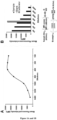
- An example of the method used for calculating anti-S 1 level is illustrated in Figure 1 .
- a calibration curve was obtained after serial dilutions of a highly anti-S 1-positive serum used as calibrator. A plateau of MFI was reached for dilution 1:400 ( Figure 1A ).
- Figure 1B At the screening dilution of 1:100, the sample used in this example showed a saturating signal.
- a higher 1:800 dilution was retained to compute Ab level by reference to the calibrator whose level was arbitrarily set to 100 AU/mL.
- the same method of calculation was used for computing the levels of anti-N IgG and anti-S 1 IgM Ab.
- ALBIA-IgG-S1/N was used to simultaneously investigate the presence of anti-S 1 and anti-N IgG Ab.
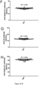
- a threshold of positivity was calculated as the mean titer + 3 SD of the 300 negative control sera, which yielded values of 7.29 and 20.98 AU/mL for anti-S1 and anti-N IgG Ab, respectively ( Figures 1C, 1D ).
- this threshold was 23.64 AU/mL ( Figure 1E ).
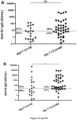
- the diagnostic performance of the assay was determined using a collection of 133 sera from SARS-CoV-2-specific PCR-positive patients that were collected at least 14 days after first COVID-19 symptoms.
- ROC curve analysis of ALBIA-IgG-S1/N confirmed the accuracy of the aforementioned threshold value, i.e., mean + 3 SD. Indeed, sensitivity was 97.7% and specificity was 98.0% at a 7.29 AU/mL threshold for anti-S1 IgG (2A, D). For anti- N IgG Ab, sensitivity was 100% and specificity was 98.7% at a threshold of 20.98 AU/mL ( Figures 2E ). For ALBIA-IgM-S1, sensitivity and specificity were 74.8 and 98.7% at a threshold of 23.64 AU/mL ( Figures 2C , F ).
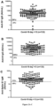
- 161 (84%) and 170 (89%) of the 192 patients of this series were positive for anti-S1 or anti-N IgG, respectively.
- Ab levels in seropositive patients were highly variable, ranging from 7.5 to 19,944 AU/mL, and from 24.74 to 491,992 AU/mL for anti-S1 or anti-N IgG, respectively.
- the sensitivity of the multiplexed anti-S1 plus anti-N IgG assay (90%) was greater than the sensitivity of anti-S1 and anti-N IgG taken separately (84 and 89%, respectively).
- the sensitivity increases to 91% if the results of the anti-S1 IgM assay are also taken into account.
- combining several antigens in the same well reduces the cost and handling time of the assay.
Landscapes
- Health & Medical Sciences (AREA)
- Life Sciences & Earth Sciences (AREA)
- Immunology (AREA)
- Engineering & Computer Science (AREA)
- Urology & Nephrology (AREA)
- Chemical & Material Sciences (AREA)
- Biomedical Technology (AREA)
- Virology (AREA)
- Molecular Biology (AREA)
- Hematology (AREA)
- Physics & Mathematics (AREA)
- General Physics & Mathematics (AREA)
- Biotechnology (AREA)
- Tropical Medicine & Parasitology (AREA)
- Food Science & Technology (AREA)
- Medicinal Chemistry (AREA)
- Biochemistry (AREA)
- Analytical Chemistry (AREA)
- Cell Biology (AREA)
- General Health & Medical Sciences (AREA)
- Microbiology (AREA)
- Pathology (AREA)
- Peptides Or Proteins (AREA)
- Measuring Or Testing Involving Enzymes Or Micro-Organisms (AREA)
Applications Claiming Priority (5)
| Application Number | Priority Date | Filing Date | Title |
|---|---|---|---|
| EP20315157 | 2020-04-14 | ||
| EP20305614 | 2020-06-09 | ||
| PCT/EP2021/059583 WO2021209463A1 (fr) | 2020-04-14 | 2021-04-13 | Procédés de détection de la présence d'anticorps spécifiques au coronavirus chez un sujet |
| EP21717462.2A EP4004547B1 (fr) | 2020-04-14 | 2021-04-13 | Procédés de détection de la présence d'anticorps spécifiques de coronavirus chez un sujet |
| EP23194718.5A EP4283299A3 (fr) | 2020-04-14 | 2021-04-13 | Procédés de détection de la présence d'anticorps spécifiques de coronavirus chez un sujet |
Related Parent Applications (2)
| Application Number | Title | Priority Date | Filing Date |
|---|---|---|---|
| EP23194718.5A Division EP4283299A3 (fr) | 2020-04-14 | 2021-04-13 | Procédés de détection de la présence d'anticorps spécifiques de coronavirus chez un sujet |
| EP21717462.2A Division EP4004547B1 (fr) | 2020-04-14 | 2021-04-13 | Procédés de détection de la présence d'anticorps spécifiques de coronavirus chez un sujet |
Publications (3)
| Publication Number | Publication Date |
|---|---|
| EP4459285A2 true EP4459285A2 (fr) | 2024-11-06 |
| EP4459285A3 EP4459285A3 (fr) | 2025-01-22 |
| EP4459285B1 EP4459285B1 (fr) | 2026-02-18 |
Family
ID=75438806
Family Applications (3)
| Application Number | Title | Priority Date | Filing Date |
|---|---|---|---|
| EP23194718.5A Pending EP4283299A3 (fr) | 2020-04-14 | 2021-04-13 | Procédés de détection de la présence d'anticorps spécifiques de coronavirus chez un sujet |
| EP24197643.0A Active EP4459285B1 (fr) | 2020-04-14 | 2021-04-13 | Procédés de détection de la présence d'anticorps spécifiques du coronavirus chez un sujet |
| EP21717462.2A Active EP4004547B1 (fr) | 2020-04-14 | 2021-04-13 | Procédés de détection de la présence d'anticorps spécifiques de coronavirus chez un sujet |
Family Applications Before (1)
| Application Number | Title | Priority Date | Filing Date |
|---|---|---|---|
| EP23194718.5A Pending EP4283299A3 (fr) | 2020-04-14 | 2021-04-13 | Procédés de détection de la présence d'anticorps spécifiques de coronavirus chez un sujet |
Family Applications After (1)
| Application Number | Title | Priority Date | Filing Date |
|---|---|---|---|
| EP21717462.2A Active EP4004547B1 (fr) | 2020-04-14 | 2021-04-13 | Procédés de détection de la présence d'anticorps spécifiques de coronavirus chez un sujet |
Country Status (4)
| Country | Link |
|---|---|
| US (1) | US20230194528A1 (fr) |
| EP (3) | EP4283299A3 (fr) |
| ES (1) | ES2966766T3 (fr) |
| WO (1) | WO2021209463A1 (fr) |
Families Citing this family (1)
| Publication number | Priority date | Publication date | Assignee | Title |
|---|---|---|---|---|
| WO2022008973A2 (fr) * | 2020-07-10 | 2022-01-13 | Covid Diagnostics Ltd. | Compositions, procédés et systèmes de détection de réponse immunitaire |
Citations (12)
| Publication number | Priority date | Publication date | Assignee | Title |
|---|---|---|---|---|
| GB1561042A (en) | 1975-07-23 | 1980-02-13 | Coulter Electronics | Method for detecting and separating antigens and antibodies in blood or other samples |
| US4499052A (en) | 1982-08-30 | 1985-02-12 | Becton, Dickinson And Company | Apparatus for distinguishing multiple subpopulations of cells |
| US5028545A (en) | 1987-06-16 | 1991-07-02 | Wallac Oy | Biospecific multianalyte assay method |
| US5981180A (en) | 1995-10-11 | 1999-11-09 | Luminex Corporation | Multiplexed analysis of clinical specimens apparatus and methods |
| US20040075907A1 (en) | 2002-08-20 | 2004-04-22 | John Moon | Diffraction grating-based encoded micro-particles for multiplexed experiments |
| US20040125424A1 (en) | 2002-09-12 | 2004-07-01 | Moon John A. | Diffraction grating-based encoded micro-particles for multiplexed experiments |
| US20040126875A1 (en) | 2002-09-12 | 2004-07-01 | Putnam Martin A. | Assay stick |
| US20040130761A1 (en) | 2002-09-12 | 2004-07-08 | John Moon | Chemical synthesis using diffraction grating-based encoded optical elements |
| US20040130786A1 (en) | 2002-09-12 | 2004-07-08 | Putnam Martin A. | Method of manufacturing of diffraction grating-based optical identification element |
| US20040132205A1 (en) | 2002-09-12 | 2004-07-08 | John Moon | Method and apparatus for aligning microbeads in order to interrogate the same |
| US20040179267A1 (en) | 2002-09-12 | 2004-09-16 | Moon John A. | Method and apparatus for labeling using diffraction grating-based encoded optical identification elements |
| US6916661B2 (en) | 1999-08-17 | 2005-07-12 | Luminex Corporation | Microparticles with multiple fluorescent signals and methods of using same |
Family Cites Families (4)
| Publication number | Priority date | Publication date | Assignee | Title |
|---|---|---|---|---|
| US7220852B1 (en) * | 2003-04-25 | 2007-05-22 | The United States Of America As Represented By The Secretary Of The Department Of Health And Human Services, Centers For Disease Control And Prevention | Coronavirus isolated from humans |
| WO2005012337A2 (fr) * | 2003-07-15 | 2005-02-10 | Crucell Holland B.V. | Peptides antigeniques du coronavirus du syndrome respiratoire aigu severe et applications de ceux-ci |
| US20050112559A1 (en) * | 2003-09-29 | 2005-05-26 | The Chinese University Of Hong Kong | Compositions and methods for diagnosing and preventing severe acute respiratory syndrome (SARS) |
| US20060188519A1 (en) * | 2004-06-14 | 2006-08-24 | To Cheung | Peptides, antibodies, and methods for the diagnosis of SARS |
-
2021
- 2021-04-13 WO PCT/EP2021/059583 patent/WO2021209463A1/fr not_active Ceased
- 2021-04-13 US US17/996,294 patent/US20230194528A1/en active Pending
- 2021-04-13 EP EP23194718.5A patent/EP4283299A3/fr active Pending
- 2021-04-13 EP EP24197643.0A patent/EP4459285B1/fr active Active
- 2021-04-13 EP EP21717462.2A patent/EP4004547B1/fr active Active
- 2021-04-13 ES ES21717462T patent/ES2966766T3/es active Active
Patent Citations (12)
| Publication number | Priority date | Publication date | Assignee | Title |
|---|---|---|---|---|
| GB1561042A (en) | 1975-07-23 | 1980-02-13 | Coulter Electronics | Method for detecting and separating antigens and antibodies in blood or other samples |
| US4499052A (en) | 1982-08-30 | 1985-02-12 | Becton, Dickinson And Company | Apparatus for distinguishing multiple subpopulations of cells |
| US5028545A (en) | 1987-06-16 | 1991-07-02 | Wallac Oy | Biospecific multianalyte assay method |
| US5981180A (en) | 1995-10-11 | 1999-11-09 | Luminex Corporation | Multiplexed analysis of clinical specimens apparatus and methods |
| US6916661B2 (en) | 1999-08-17 | 2005-07-12 | Luminex Corporation | Microparticles with multiple fluorescent signals and methods of using same |
| US20040075907A1 (en) | 2002-08-20 | 2004-04-22 | John Moon | Diffraction grating-based encoded micro-particles for multiplexed experiments |
| US20040125424A1 (en) | 2002-09-12 | 2004-07-01 | Moon John A. | Diffraction grating-based encoded micro-particles for multiplexed experiments |
| US20040126875A1 (en) | 2002-09-12 | 2004-07-01 | Putnam Martin A. | Assay stick |
| US20040130761A1 (en) | 2002-09-12 | 2004-07-08 | John Moon | Chemical synthesis using diffraction grating-based encoded optical elements |
| US20040130786A1 (en) | 2002-09-12 | 2004-07-08 | Putnam Martin A. | Method of manufacturing of diffraction grating-based optical identification element |
| US20040132205A1 (en) | 2002-09-12 | 2004-07-08 | John Moon | Method and apparatus for aligning microbeads in order to interrogate the same |
| US20040179267A1 (en) | 2002-09-12 | 2004-09-16 | Moon John A. | Method and apparatus for labeling using diffraction grating-based encoded optical identification elements |
Non-Patent Citations (57)
| Title |
|---|
| ALTSCHUL ET AL., J. MOL. BIOL., vol. 215, 1990, pages 403 - 410 |
| ALTSCHUL ET AL., NAT. GENET., vol. 6, 1994, pages 119 - 129 |
| ALTSCHUL ET AL., NUCLEIC ACIDS RES., vol. 25, 1997, pages 3389 - 3402 |
| ARNETT, F. C, THE AMERICAN RHEUMATISM ASSOCIATION, no. 1988, 1987 |
| AYOUBAA, A., CLIN. VIROL, vol. 129, 2020, pages 104521 |
| BENVENISTE O ET AL.: "Correlation of anti-signal recognition particle autoantibody levels with creatine kinase activity in patients with necrotizing myopathy", ARTHRITIS RHEUM, vol. 63, no. 7, July 2011 (2011-07-01), pages 1961 - 71, XP055004291, DOI: 10.1002/art.30344 |
| BISCHOF, E. ET AL.: "Understanding COVID-19 new diagnostic guidelines - a message of reassurance from an internal medicine doctor in Shanghai", SWISS MED. WKLY, vol. 150, 2020, pages w20216 |
| BRYAN, A ET AL.: "Performance Characteristics of the Abbott Architect SARS-CoV-2 IgG Assay and Seroprevalence in Boise, Idaho", J. CLIN. MICROBIOL, vol. 58, 2020, pages e00941 - 20 |
| BURBELO, P. D. ET AL.: "Sensitivity in detection of antibodies to nucleocapsid and spike proteins of severe acute respiratory syndrome coronavirus 2 in patients with Coronavirus Disease 2019", J. INFECT. DIS, vol. 222, 2020, pages 206 - 213, XP055818914, DOI: 10.1093/infdis/jiaa273 |
| CORPET ET AL., NUC. ACIDS RES., vol. 16, 1988, pages 10881 - 10890 |
| DROUOT L ET AL.: "Exploring necrotizing autoimmune myopathies with a novel immunoassay for anti-3-hydroxy-3-methyl-glutaryl-CoA reductase autoantibodies", ARTHRITIS RES THER, vol. 16, no. 1, 3 February 2014 (2014-02-03), pages R39, XP021177315, DOI: 10.1186/ar4468 |
| DUONG, Y. T.WRIGHT, G.JUSTMAN, J: "Antibody testing for coronavirus disease 2019: not ready for prime time", BMJ, vol. 370, 2020, pages m2655 |
| ECDC, 19 March 2020 (2020-03-19) |
| FARNSWORTH, C. W.ANDERSON, N. W: "SARS-CoV-2 serology: much hype, little data", CLIN. CHEM., vol. 66, 2020, pages 875 - 877 |
| FULWYLER ET AL.: "Meth. Cell Biol", vol. 33, 1990, COULTER ELECTRONICS INC, article "Flow Microsphere Immunoassay for the Quantitative and Simultaneous Detection of Multiple Soluble Analytes", pages: 613 - 629 |
| G. T. HERMANSON: "Practical Flow Cytometry", 1995, ACADEMIC PRESS |
| GEURTSVANKESSEL, C. H. ET AL.: "An evaluation of COVID-19 serological assays informs future diagnostics and exposure assessment", NAT. COMMUN, vol. 11, 2020, pages 3436 |
| GISHSTATES, NATURE GENET., vol. 3, 1993, pages 266 - 272 |
| GUO LET: "Profiling Early Humoral Response to Diagnose Novel Coronavirus Disease (COVID-19", CLIN INFECT DIS, 21 March 2020 (2020-03-21), pages ciaa310 |
| HEBERT, V. ET AL.: "Lack of association between chilblains outbreak and SARS-CoV-2: histological and serological findings from a new immunoassay", J. AM. ACAD. DERMATOL, vol. 83, 2020, pages 1434 - 1436 |
| HERMANSON, GREG T: "Bioconjugate techniques", 2013, ACADEMIC PRESS |
| HIGGINSSHARP, CABIOS, vol. 4, 1989, pages 151 - 153 |
| HIGGINSSHARP, GENE, vol. 73, 1988, pages 237 - 244 |
| HORAN ET AL.: "Immunoassays in the Clinical Laboratory", vol. 185, 1979, LISS, article "Fluid Phase Particle Fluorescence Analysis: Rheumatoid Factor Specificity Evaluated by Laser Flow Cytophotometry", pages: 189 |
| HUANG ET AL., COMP. APPLSBIOSCI., vol. 8, 1992, pages 155 - 165 |
| HUANG, A. T: "Systematic review of antibody mediated immunity to coronaviruses: antibody kinetics, correlates of protection, and association of antibody responses with severity of disease", MEDRXIV, 2020 |
| KRUTTGEN, A. ET AL.: "Comparison of four new commercial serologic assays for determination of SARS-CoV-2 IgG", J. CLIN. VIROL, vol. 128, 2020, pages 104394, XP086181761, DOI: 10.1016/j.jcv.2020.104394 |
| LAI, C. C.WANG, C. Y.KO, W. C.HSUEH, P. R: "In vitro diagnostics of coronavirus disease 2019: technologies and application", JMII, 2020 |
| LIMOGES UNIVERSITY HOSPITAL |
| LINDMO ET AL.: "Immunometric Assay Using Mixtures of Two Particle Types of Different Affinity", J. IMMUNOL. METH, vol. 126, 1990, pages 183 - 189, XP023973551, DOI: 10.1016/0022-1759(90)90149-P |
| LISBOA BASTOS ET AL.: "Diagnostic accuracy of serological tests for covid-19: systematic review and meta-analysis", BMJ, vol. 370, 2020, pages m2516, XP055759583, DOI: 10.1136/bmj.m2516 |
| LIU W ET AL., EVALUATION OF NUCLEOCAPSID AND SPIKE PR 1 OTEIN-BASED ELISAS FOR DETECTING ANTIBODIES AGAINST SARS-COV-2, Retrieved from the Internet <URL:https://doi.org/10.1101/2020.03.16.20035014> |
| LONG, Q. X. ET AL.: "Antibody responses to SARS-CoV-2 in patients with COVID-19", NAT. MED, vol. 26, 2020, pages 845 - 848, XP037173414, DOI: 10.1038/s41591-020-0897-1 |
| MADDEN ET AL., METH. ENZYMOL., vol. 266, 1996, pages 131 - 141 |
| MAHASE, E: "Covid-19: two antibody tests are ''highly specific'' but vary in sensitivity, evaluations find", BMJ, vol. 369, 2020, pages m2066 |
| MCHUGH ET AL.: "Clinical Flow Cytometry", 1993, WILLIAMS AND WILLIAMS, article "Microsphere-Based Fluorescence Immunoassays Using Flow Cytometry Instrumentation", pages: 535 - 544 |
| MCHUGH: "Flow Cytometry and the Application of Microsphere-Based Fluorescence Immunoassays", IMMUNOCHEMICA, vol. 5, 1991, pages 116 |
| MCHUGH: "Methods in Cell Biology", vol. 42, 1994, ACADEMIC PRESS, article "Flow Microsphere Immunoassay for the Quantitative and Simultaneous Detection of Multiple Soluble Analytes" |
| MEYER, B.DROSTEN, C.MULLER, M. A: "Serological assays for emerging coronaviruses: challenges and pitfalls", VIRUS RES, vol. 194, 2014, pages 175 - 183, XP029105622, DOI: 10.1016/j.virusres.2014.03.018 |
| MONTESINOS, I. ET AL.: "valuation of two automated and three rapid lateral.flow immunoassays for the detection of anti-SARS-CoV-2 antibodies", J. CLIN. VIROL, vol. 128, 2020, pages 104413, XP086181768, DOI: 10.1016/j.jcv.2020.104413 |
| NEEDLEMANWUNSCH, J. MOL. BIOL., vol. 48, 1970, pages 443 |
| OKBA N ET AL., SARS-COV-2 SPECIFIC ANTIBODY RESPONSES IN COVID-19 PATIENTS, Retrieved from the Internet <URL:https://doi.org/10.1101/2020.03.18.20038059> |
| PEARSON ET AL., METH. MOL. BIOL., vol. 24, 1994, pages 307 - 31 |
| PEARSONLIPMAN, PROC. NATL. ACAD. SCI. U.S.A., vol. 85, 1988, pages 2444 |
| SMITHGALL, M. C. ET AL.: "Types of assays for SARS-CoV-2 testing: a review", LAB. MED, vol. 51, 2020, pages 59 - 65 |
| SMITHWATERMAN, ADV. APPL. MATH., vol. 2, 1981, pages 482 |
| STEINKAMP ET AL., REVIEW OF SCIENTIFIC INSTRUMENTS, vol. 44, no. 9, 1973, pages 1301 - 1310 |
| STRASBOURG UNIVERSITY HOSPITAL |
| TAN, E. M. ET AL.: "The 1982 revised criteria for the classification of systemic lupus erythematosus", ARTHR. RHEUM, vol. 25, 1982, pages 1271 - 1277, XP055478543, DOI: THE 1982 REVISED CRITERIA FOR THE CLASSIFICATION OF SYSTEMIC LUPUS ERYTHEMATOSUS |
| TRAUGOTT, M. ET AL.: "Performance of SARS-CoV-2 antibody assays in different stages of the infection: comparison of commercial ELISA and rapid tests", J. INFECT. DIS, vol. 222, 2020, pages 352 - 366 |
| TROYANOV, Y. ET AL.: "Novel classification of idiopathic inflammatory myopathies based on overlap syndrome features and autoantibodies: analysis of 100 French Canadian patients", MEDICINE, vol. 84, 2005, pages 231 - 249 |
| VITALI, C. ET AL.: "Classification criteria for Sjogren's syndrome: a revised version of the European criteria proposed by the American-European Consensus Group", ANN. RHEUM, vol. 61, 2002, pages 554 - 558 |
| WILSON ET AL.: "A New Microsphere-Based Immunofluorescence Assay Using Flow Cytometry", J. IMMUNOL. METH, vol. 107, 1988, pages 225 - 230, XP023975301, DOI: 10.1016/0022-1759(88)90222-0 |
| XIA N ET AL., SEROLOGICAL TEST IS AN EFFICIENT SUPPLEMENT OF RNA DETECTION FOR CONFIRMATION OF SARS-COV-2 INFECTION, Retrieved from the Internet <URL:https://doi:10.20944/preprints202003.0184.v1> |
| XIANG J ET AL.: "Evaluation of Enzyme-Linked Immunoassay and Colloidal Gold-Immunochromatographic Assay Kit for Detection of Novel Coronavirus (SARS-Cov-2) Causing an Outbreak of Pneumonia (COVID-19", BMJ |
| XUE-FEI C ET AL.: "A Peptide-based Magnetic Chemiluminescence Enzyme Immunoassay for Serological Diagnosis", CORONA VIRUS DISEASE, 2019, pages 2020, Retrieved from the Internet <URL:https://doi.org/10.1101/2020.02.22.20026617> |
| ZHANGMADDEN, GENOME RES., vol. 7, 1997, pages 649 - 656 |
Also Published As
| Publication number | Publication date |
|---|---|
| EP4459285B1 (fr) | 2026-02-18 |
| US20230194528A1 (en) | 2023-06-22 |
| EP4283299A3 (fr) | 2024-02-28 |
| EP4459285A3 (fr) | 2025-01-22 |
| EP4283299A2 (fr) | 2023-11-29 |
| EP4004547A1 (fr) | 2022-06-01 |
| EP4004547B1 (fr) | 2023-10-11 |
| WO2021209463A1 (fr) | 2021-10-21 |
| ES2966766T3 (es) | 2024-04-24 |
Similar Documents
| Publication | Publication Date | Title |
|---|---|---|
| WO2018152124A1 (fr) | Essai par écoulement latéral immunochromatographique rapide pour la détection précoce d'une maladie zika | |
| US20230072761A1 (en) | Immunoassay for Detecting Zika Virus Infection | |
| Martinaud et al. | Evaluation of the Quotient® MosaiQ™ COVID-19 antibody microarray for the detection of IgG and IgM antibodies to SARS-CoV-2 virus in humans | |
| EP4459285B1 (fr) | Procédés de détection de la présence d'anticorps spécifiques du coronavirus chez un sujet | |
| Martínez-Subiela et al. | Measurement of anti SARS-CoV-2 RBD IgG in saliva: validation of a highly sensitive assay and effects of the sampling collection method and correction by protein | |
| WO2022109751A1 (fr) | Test hors laboratoire pour anticorps sars-cov | |
| Cassaniti et al. | Seroprevalence of SARS-CoV-2 in blood donors from the Lodi Red Zone and adjacent Lodi metropolitan and suburban area | |
| JP5168538B2 (ja) | 抗体−抗体コンジュゲートと抗体測定系診断薬への応用 | |
| WO2021202414A1 (fr) | Test multianalytique de la réponse immunitaire au virus sars-cov-2 conduisant au covid-19 | |
| US20210318311A1 (en) | Simultaneous detection of humoral and inflammatory biomarkers | |
| Drouot et al. | Evaluation of humoral immunity to SARS-CoV-2: diagnostic value of a new multiplex addressable laser bead immunoassay | |
| Wu et al. | Evaluation of a novel array-based toxoplasma, rubella, cytomegalovirus, and herpes simplex virus IgG enzyme linked immunosorbent assay and its comparison with Virion/Serion enzyme linked immunosorbent assays | |
| Feng et al. | Serological diagnosis of infectious mononucleosis by chemiluminescent immunoassay using capsid antigen p18 of Epstein-Barr virus | |
| WO2004048615A1 (fr) | Procede pour detecter une infection par vih recemment contractee | |
| Kaku et al. | Establishment of an immunofluorescence assay to detect IgM antibodies to Nipah virus using HeLa cells expressing recombinant nucleoprotein | |
| Filchtinski et al. | Application of the SARS-CoV-2-S1 ACE-2 receptor interaction as the basis of the fully automated assay to detect neutralizing SARS-CoV-2-S1 antibodies in blood samples | |
| WO2021221082A1 (fr) | FRAGMENT DE NUCLÉOCAPSIDE DÉRIVÉ DU SARS-CoV-2, ET MÉTHODE ET KIT DE DÉTECTION D'ANTICORPS ANTI-SARS-CoV-2 L'UTILISANT | |
| CA3181179A1 (fr) | Dosage a double multiplex pour de multiples isotypes d'immunoglobulines | |
| US20240353409A1 (en) | Novel serology assay for the detection of porcine viruses | |
| US20230358746A1 (en) | METHODS AND KITS FOR DETERMINING SEROSTATUS OF SARS-CoV-2 | |
| Lei et al. | HCV Antibody Rapid Test is Expected to Accelerate the Detection Accuracy of the Hepatitis C Virus | |
| JP2026514067A (ja) | ブタウイルスの検出のための血清学的アッセイ | |
| Chia et al. | Detection and Diagnosis Technologies | |
| Saha et al. | Limitations of serological screening for measles immunity in young health care workers in New Zealand | |
| Ursu et al. | The clinical utility of TORCH testing of pregnant women from northeastern Romanian population: application of electrochemiluminiscence assay |
Legal Events
| Date | Code | Title | Description |
|---|---|---|---|
| PUAI | Public reference made under article 153(3) epc to a published international application that has entered the european phase |
Free format text: ORIGINAL CODE: 0009012 |
|
| STAA | Information on the status of an ep patent application or granted ep patent |
Free format text: STATUS: THE APPLICATION HAS BEEN PUBLISHED |
|
| AC | Divisional application: reference to earlier application |
Ref document number: 4004547 Country of ref document: EP Kind code of ref document: P Ref document number: 4283299 Country of ref document: EP Kind code of ref document: P |
|
| AK | Designated contracting states |
Kind code of ref document: A2 Designated state(s): AL AT BE BG CH CY CZ DE DK EE ES FI FR GB GR HR HU IE IS IT LI LT LU LV MC MK MT NL NO PL PT RO RS SE SI SK SM TR |
|
| PUAL | Search report despatched |
Free format text: ORIGINAL CODE: 0009013 |
|
| AK | Designated contracting states |
Kind code of ref document: A3 Designated state(s): AL AT BE BG CH CY CZ DE DK EE ES FI FR GB GR HR HU IE IS IT LI LT LU LV MC MK MT NL NO PL PT RO RS SE SI SK SM TR |
|
| RIC1 | Information provided on ipc code assigned before grant |
Ipc: G01N 33/569 20060101AFI20241219BHEP |
|
| STAA | Information on the status of an ep patent application or granted ep patent |
Free format text: STATUS: REQUEST FOR EXAMINATION WAS MADE |
|
| 17P | Request for examination filed |
Effective date: 20250415 |
|
| STAA | Information on the status of an ep patent application or granted ep patent |
Free format text: STATUS: EXAMINATION IS IN PROGRESS |
|
| 17Q | First examination report despatched |
Effective date: 20250618 |
|
| GRAP | Despatch of communication of intention to grant a patent |
Free format text: ORIGINAL CODE: EPIDOSNIGR1 |
|
| STAA | Information on the status of an ep patent application or granted ep patent |
Free format text: STATUS: GRANT OF PATENT IS INTENDED |
|
| INTG | Intention to grant announced |
Effective date: 20251126 |
|
| GRAS | Grant fee paid |
Free format text: ORIGINAL CODE: EPIDOSNIGR3 |
|
| GRAA | (expected) grant |
Free format text: ORIGINAL CODE: 0009210 |
|
| STAA | Information on the status of an ep patent application or granted ep patent |
Free format text: STATUS: THE PATENT HAS BEEN GRANTED |
|
| AC | Divisional application: reference to earlier application |
Ref document number: 4283299 Country of ref document: EP Kind code of ref document: P Ref document number: 4004547 Country of ref document: EP Kind code of ref document: P |
|
| AK | Designated contracting states |
Kind code of ref document: B1 Designated state(s): AL AT BE BG CH CY CZ DE DK EE ES FI FR GB GR HR HU IE IS IT LI LT LU LV MC MK MT NL NO PL PT RO RS SE SI SK SM TR |
|
| REG | Reference to a national code |
Ref country code: CH Ref legal event code: F10 Free format text: ST27 STATUS EVENT CODE: U-0-0-F10-F00 (AS PROVIDED BY THE NATIONAL OFFICE) Effective date: 20260218 Ref country code: GB Ref legal event code: FG4D |
|
| REG | Reference to a national code |
Ref country code: IE Ref legal event code: FG4D |
|
| REG | Reference to a national code |
Ref country code: DE Ref legal event code: R096 Ref document number: 602021048509 Country of ref document: DE |
|
| PGFP | Annual fee paid to national office [announced via postgrant information from national office to epo] |
Ref country code: GB Payment date: 20260319 Year of fee payment: 6 |
|
| PGFP | Annual fee paid to national office [announced via postgrant information from national office to epo] |
Ref country code: FR Payment date: 20260319 Year of fee payment: 6 |