EP4470648A2 - Geladene tiefenfiltration von antigenbindenden proteinen - Google Patents
Geladene tiefenfiltration von antigenbindenden proteinen Download PDFInfo
- Publication number
- EP4470648A2 EP4470648A2 EP24193226.8A EP24193226A EP4470648A2 EP 4470648 A2 EP4470648 A2 EP 4470648A2 EP 24193226 A EP24193226 A EP 24193226A EP 4470648 A2 EP4470648 A2 EP 4470648A2
- Authority
- EP
- European Patent Office
- Prior art keywords
- antigen
- binding protein
- charged depth
- antibody
- molecules
- Prior art date
- Legal status (The legal status is an assumption and is not a legal conclusion. Google has not performed a legal analysis and makes no representation as to the accuracy of the status listed.)
- Pending
Links
Images


Classifications
-
- C—CHEMISTRY; METALLURGY
- C07—ORGANIC CHEMISTRY
- C07K—PEPTIDES
- C07K1/00—General methods for the preparation of peptides, i.e. processes for the organic chemical preparation of peptides or proteins of any length
- C07K1/14—Extraction; Separation; Purification
- C07K1/36—Extraction; Separation; Purification by a combination of two or more processes of different types
-
- B—PERFORMING OPERATIONS; TRANSPORTING
- B01—PHYSICAL OR CHEMICAL PROCESSES OR APPARATUS IN GENERAL
- B01D—SEPARATION
- B01D15/00—Separating processes involving the treatment of liquids with solid sorbents; Apparatus therefor
- B01D15/08—Selective adsorption, e.g. chromatography
- B01D15/26—Selective adsorption, e.g. chromatography characterised by the separation mechanism
- B01D15/36—Selective adsorption, e.g. chromatography characterised by the separation mechanism involving ionic interaction, e.g. ion-exchange, ion-pair, ion-suppression or ion-exclusion
- B01D15/361—Ion-exchange
- B01D15/362—Cation-exchange
-
- B—PERFORMING OPERATIONS; TRANSPORTING
- B01—PHYSICAL OR CHEMICAL PROCESSES OR APPARATUS IN GENERAL
- B01D—SEPARATION
- B01D15/00—Separating processes involving the treatment of liquids with solid sorbents; Apparatus therefor
- B01D15/08—Selective adsorption, e.g. chromatography
- B01D15/26—Selective adsorption, e.g. chromatography characterised by the separation mechanism
- B01D15/38—Selective adsorption, e.g. chromatography characterised by the separation mechanism involving specific interaction not covered by one or more of groups B01D15/265 and B01D15/30 - B01D15/36, e.g. affinity, ligand exchange or chiral chromatography
- B01D15/3804—Affinity chromatography
- B01D15/3809—Affinity chromatography of the antigen-antibody type, e.g. protein A, G or L chromatography
-
- C—CHEMISTRY; METALLURGY
- C07—ORGANIC CHEMISTRY
- C07K—PEPTIDES
- C07K1/00—General methods for the preparation of peptides, i.e. processes for the organic chemical preparation of peptides or proteins of any length
- C07K1/107—General methods for the preparation of peptides, i.e. processes for the organic chemical preparation of peptides or proteins of any length by chemical modification of precursor peptides
- C07K1/113—General methods for the preparation of peptides, i.e. processes for the organic chemical preparation of peptides or proteins of any length by chemical modification of precursor peptides without change of the primary structure
- C07K1/1133—General methods for the preparation of peptides, i.e. processes for the organic chemical preparation of peptides or proteins of any length by chemical modification of precursor peptides without change of the primary structure by redox-reactions involving cystein/cystin side chains
-
- C—CHEMISTRY; METALLURGY
- C07—ORGANIC CHEMISTRY
- C07K—PEPTIDES
- C07K1/00—General methods for the preparation of peptides, i.e. processes for the organic chemical preparation of peptides or proteins of any length
- C07K1/14—Extraction; Separation; Purification
- C07K1/16—Extraction; Separation; Purification by chromatography
- C07K1/18—Ion-exchange chromatography
-
- C—CHEMISTRY; METALLURGY
- C07—ORGANIC CHEMISTRY
- C07K—PEPTIDES
- C07K1/00—General methods for the preparation of peptides, i.e. processes for the organic chemical preparation of peptides or proteins of any length
- C07K1/14—Extraction; Separation; Purification
- C07K1/16—Extraction; Separation; Purification by chromatography
- C07K1/22—Affinity chromatography or related techniques based upon selective absorption processes
-
- C—CHEMISTRY; METALLURGY
- C07—ORGANIC CHEMISTRY
- C07K—PEPTIDES
- C07K1/00—General methods for the preparation of peptides, i.e. processes for the organic chemical preparation of peptides or proteins of any length
- C07K1/14—Extraction; Separation; Purification
- C07K1/34—Extraction; Separation; Purification by filtration, ultrafiltration or reverse osmosis
-
- C—CHEMISTRY; METALLURGY
- C07—ORGANIC CHEMISTRY
- C07K—PEPTIDES
- C07K16/00—Immunoglobulins [IG], e.g. monoclonal or polyclonal antibodies
- C07K16/06—Immunoglobulins [IG], e.g. monoclonal or polyclonal antibodies from serum
- C07K16/065—Purification, fragmentation
-
- C—CHEMISTRY; METALLURGY
- C07—ORGANIC CHEMISTRY
- C07K—PEPTIDES
- C07K16/00—Immunoglobulins [IG], e.g. monoclonal or polyclonal antibodies
- C07K16/18—Immunoglobulins [IG], e.g. monoclonal or polyclonal antibodies against material from animals or humans
- C07K16/28—Immunoglobulins [IG], e.g. monoclonal or polyclonal antibodies against material from animals or humans against receptors, cell surface antigens or cell surface determinants
- C07K16/2887—Immunoglobulins [IG], e.g. monoclonal or polyclonal antibodies against material from animals or humans against receptors, cell surface antigens or cell surface determinants against CD20
-
- C—CHEMISTRY; METALLURGY
- C07—ORGANIC CHEMISTRY
- C07K—PEPTIDES
- C07K2317/00—Immunoglobulins specific features
- C07K2317/10—Immunoglobulins specific features characterized by their source of isolation or production
- C07K2317/14—Specific host cells or culture conditions, e.g. components, pH or temperature
-
- C—CHEMISTRY; METALLURGY
- C07—ORGANIC CHEMISTRY
- C07K—PEPTIDES
- C07K2317/00—Immunoglobulins specific features
- C07K2317/40—Immunoglobulins specific features characterized by post-translational modification
-
- G—PHYSICS
- G01—MEASURING; TESTING
- G01N—INVESTIGATING OR ANALYSING MATERIALS BY DETERMINING THEIR CHEMICAL OR PHYSICAL PROPERTIES
- G01N27/00—Investigating or analysing materials by the use of electric, electrochemical, or magnetic means
- G01N27/26—Investigating or analysing materials by the use of electric, electrochemical, or magnetic means by investigating electrochemical variables; by using electrolysis or electrophoresis
- G01N27/416—Systems
- G01N27/447—Systems using electrophoresis
- G01N27/44756—Apparatus specially adapted therefor
- G01N27/44791—Microapparatus
Definitions
- the present disclosure relates to methods of producing an aqueous formulation comprising a re-oxidized antigen-binding protein.
- Antigen-binding proteins such as antibodies are currently used to treat millions of patients world-wide.
- Antigen-binding protein molecules are typically produced in mammalian cell culture systems and recovered using a standard series of filtration and chromatography steps (see, e.g., Liu et al., mAbs. 2(5): 480-499(2010 )).
- the structure and stability of antigen-binding protein molecules depend heavily on the disulfide bonds that link the two heavy chains and the heavy and light chains in each antigen-binding protein molecule, however, during the production and purification process, one or more disulfide bonds can be reduced to free thiol groups.
- the present disclosure is directed to methods of producing an aqueous formulation of an antigen-binding protein (such as an antigen-binding protein comprising an Fc region, an antibody, or a peptibody) or enhancing re-oxidation of such an antigen-binding protein and to formulations comprising a re-oxidized antigen-binding protein prepared according to these methods.
- an antigen-binding protein such as an antigen-binding protein comprising an Fc region, an antibody, or a peptibody
- the disclosure provides a method of producing an aqueous formulation of an antigen-binding protein (such as an antigen-binding protein comprising an Fc region, an antibody, or a peptibody) comprising (a) contacting an aqueous solution comprising antigen-binding protein molecules with a charged depth filter under conditions sufficient to achieve at least a 20% decrease, optionally a 30% or 40% decrease, in the percentage of reduced antigen-binding protein molecules, compared to the percentage of reduced antigen-binding protein molecules observed prior to step (a); and (b) optionally, measuring the amount (such as the total amount) or relative amount of reduced antigen-binding protein molecules.
- an antigen-binding protein such as an antigen-binding protein comprising an Fc region, an antibody, or a peptibody
- the disclosure provides a method of enhancing re-oxidization of an antigen-binding protein (such as an antigen-binding protein comprising an Fc region, an antibody, or a peptibody) comprising (a) contacting an aqueous solution comprising antigen-binding protein molecules with a charged depth filter under conditions sufficient to enhance re-oxidation of the antigen-binding protein molecules; and (b) optionally, measuring the amount (such as the total amount) or relative amount of reduced antigen-binding protein molecules.
- an antigen-binding protein such as an antigen-binding protein comprising an Fc region, an antibody, or a peptibody
- the amount (such as the total amount) or relative amount of reduced antigen-binding protein molecules is measured using non-reduced capillary electrophoresis with sodium dodecyl sulfate (nrCE-SDS).
- nrCE-SDS sodium dodecyl sulfate
- the total amount of reduced antigen-binding protein molecules after contact with the charged depth filter in step (a) is 10% or less of the total amount of antigen-binding protein molecules and/or is decreased by at least three-fold compared to before step (a).
- step (a) of the methods described herein is followed by and/or preceded by subjecting the aqueous solution of antigen-binding protein molecules to Protein A chromatography.
- a method according to the disclosure further comprises a step of inactivating one or more viruses in the aqueous solution of antigen-binding protein molecules and/or subjecting the aqueous solution of antigen-binding protein molecules to cation exchange chromatography and/or sparging air or oxygen into the aqueous solution of antigen-binding protein molecules.
- a method further comprises adding an inhibitor of thioredoxin or thioredoxin-like protein to the aqueous solution of antigen-binding protein molecules (see, e.g., U.S. Patent Publication No. 20090053786 ).
- a method of producing an aqueous formulation of an antigen-binding protein comprises contacting an aqueous solution comprising antigen-binding protein molecules with a charged depth filter under conditions sufficient to achieve at least a 20% decrease in the percentage of reduced antigen-binding protein molecules, compared to the percentage of reduced antigen-binding protein molecules observed prior to the contacting step, wherein the at least 20% decrease is determined using non-reduced capillary electrophoresis with sodium dodecyl sulfate (nrCE-SDS).
- nrCE-SDS non-reduced capillary electrophoresis with sodium dodecyl sulfate
- a method of enhancing re-oxidization of an antigen-binding protein comprising contacting an aqueous solution comprising antigen-binding protein molecules with a charged depth filter under conditions sufficient to achieve at least a two-fold increase in re-oxidation of the antigen-binding protein molecules following the contacting step, wherein the at least two-fold increase is determined using non-reduced capillary electrophoresis with sodium dodecyl sulfate (nrCE-SDS).
- a method described herein comprises (1) a Protein A chromatography step, optionally followed by charged depth filtration; (2) a viral inactivation step, optionally followed by charged depth filtration; and (3) a cation exchange chromatography step, optionally followed by charged depth filtration; further optionally comprising one or more of (4) a chromatography step optionally selected from salt-intolerant interaction chromatography, hydrophobic interaction chromatography, and mixed mode chromatography, optionally followed by charged depth filtration; (5) a virus filtration step, optionally followed by charged depth filtration; and (5) ultrafiltration and/or diafiltration, optionally followed by charged depth filtration.
- the filtrate is incubated, for example, at least about 1, 2, 3, 4, 5, 6, 7, 8, 9, 10, 12, 24 or more hours.
- the charged depth filter comprises a diatomaceous earth layer.
- the charged depth filter further comprises a cellulose layer and/or an inorganic layer, such as an inorganic layer comprises a polyamine resin.
- the charged depth filter comprises a positive ion, such as any one of sodium, calcium, magnesium, mercury, chromium, aluminum, potassium, lead, arsenic, cadmium, cobalt, iron, manganese, titanium, zinc, nickel, copper, or combinations thereof.
- the charged depth filter comprises one of the following combinations of positive ions: 1) copper and cobalt, 2) copper and cadmium, 3) cobalt and cadmium, or 4) copper, cobalt, and cadmium.
- the positive ion is a metal with a + 2 or higher oxidation state (such as + 3 or + 4).
- the method comprises contacting an aqueous solution comprising antigen-binding protein molecules with one, two, three, four, five, or more charged depth filter(s).
- the aqueous solution comprises an antigen-binding protein molecule which is an IgG antibody, such as an IgG1 or IgG2 antibody.
- an IgG antibody such as an IgG1 or IgG2 antibody.
- the antibody is an IgG1 antibody with a kappa light chain or an IgG1 antibody with a lambda light chain.
- the antigen-binding protein binds an antigen selected from the group consisting of CD3, CD4, CD8, CD19, CD20, CD34, HER2, HER3, HER4, the EGF receptor, LFA-1, Mol, p150, p95, VLA-4, ICAM-1, VCAM, alpha v/beta 3 integrin, vascular endothelial growth factor, growth hormone, thyroid stimulating hormone, follicle stimulating hormone, luteinizing hormone, growth hormone releasing factor, parathyroid hormone, mullerian-inhibiting substance, human macrophage inflammatory protein, erythropoietin, NGF-beta, platelet-derived growth factor, aFGF, bFGF, epidermal growth factor, TGF-alpha, TGF-beta1, TGF-beta2, TGF-beta3, TGF-beta4, TGF-beta5, IGF-I, IGF-II, des(1-3)-IGF-I, insulin, insulin, insulin
- the aqueous formulation comprises an antibody selected from the group consisting of abciximab, adalimumab, alemtuzumab, basiliximab, belimumab, bevacizumab, brentuximab vedotin, canakinumab, cetuximab, certolizumab pegol, daclizumab, denosumab, eculizumab, efalizumab, gemtuzumab, golimumab, ibritumomab tiuxetan, infliximab, ipilimumab, muromonab-CD3, natalizumab, nivolumab, ofatumumab, omalizumab, palivizumab, panitumumab, ranibizumab, rituximab, tocilizumab, tositumomab, trastuzumab, tras
- the aqueous formulation comprises rituximab or an antibody comprising 1, 2, 3, 4, 5, or 6 of the complementarity determining regions (CDRs) of rituximab
- the antibody can comprise (a) a light chain containing all three light chain CDRs of rituximab, (b) a heavy chain containing all three heavy chain CDRs of rituximab, or (c) both.
- the aqueous formulation comprises infliximab or an antibody comprising 1, 2, 3, 4, 5, or 6 of the complementarity determining regions (CDRs) of infliximab
- the antibody can comprise (a) a light chain containing all three light chain CDRs of infliximab, (b) a heavy chain containing all three heavy chain CDRs of infliximab, or (c) both.
- the aqueous formulation comprises ofatumumab or an antibody comprising 1, 2, 3, 4, 5, or 6 of the complementarity determining regions (CDRs) of ofatumumab
- the antibody can comprise (a) a light chain containing all three light chain CDRs of ofatumumab, (b) a heavy chain containing all three heavy chain CDRs of ofatumumab, or (c) both.
- the disclosure provides a formulation comprising a re-oxidized antigen-binding protein molecule (such as an antigen-binding protein comprising an Fc region, a fusion protein, an antibody, an antibody fragment or a peptibody) prepared using any of the methods described herein.
- a re-oxidized antigen-binding protein molecule such as an antigen-binding protein comprising an Fc region, a fusion protein, an antibody, an antibody fragment or a peptibody
- the present invention is based, at least in part, on the surprising discovery that material from a charged depth filter promotes the re-oxidation of antigen-binding molecules at least three-fold more than an uncharged depth filter control.
- Use of charged depth filtration to promote re-oxidation is particular desirable for antigen-binding molecules prone to reduction, such as IgG1 antibodies.
- the present disclosure provides methods of producing an aqueous formulation of an antigen-binding protein (such as an antigen-binding protein comprising an Fc region, an antibody, or a peptibody) or enhancing re-oxidation of an antigen-binding protein comprising contacting an aqueous solution comprising antigen-binding protein molecules with a charged depth filter under conditions sufficient to decrease the percentage of reduced antigen-binding protein molecules.
- an antigen-binding protein such as an antigen-binding protein comprising an Fc region, an antibody, or a peptibody
- enhancing re-oxidation of an antigen-binding protein comprising contacting an aqueous solution comprising antigen-binding protein molecules with a charged depth filter under conditions sufficient to decrease the percentage of reduced antigen-binding protein molecules.
- formulations comprising a re-oxidized antigen-binding protein (such as an antigen-binding protein comprising an Fc region, an antibody, or a peptibody) prepared using the
- the methods comprising a charged depth filter according to the present disclosure are more effective than other methods such as sparging with air, chilling, and sterile membrane filtration, for decreasing the amount of partially reduced antigen-binding protein molecules in the aqueous solution and thus remedy the fragmentation and aggregation issues that mar antigen-binding protein production processes and the resulting pharmaceutical formulations.
- any and all of the embodiments described for antibodies may also be used for an antigen binding protein, such as an antigen-binding protein comprising an Fc region (e.g., a peptibody).
- an antigen binding protein such as an antigen-binding protein comprising an Fc region (e.g., a peptibody).
- an antigen binding protein comprising an Fc region (e.g., a peptibody).
- an antigen binding protein such as an antigen-binding protein comprising an Fc region (e.g., a peptibody).
- an antigen binding protein such as an antigen-binding protein comprising an Fc region (e.g., a peptibody).
- any and all of the embodiments described for antigen-binding proteins also specifically apply, in each and every instance, to antibodies as defined herein.
- antigen-binding protein refers to a protein or polypeptide that comprises an antigen-binding region or antigen-binding portion that has a strong affinity for another molecule to which it binds (antigen).
- Antigen-binding proteins encompass antibodies, peptibodies, antibody fragments, antibody derivatives, antibody analogs, fusion proteins (including single-chain variable fragments (scFvs) and double-chain (divalent) scFvs), and antigen receptors including chimeric antigen receptors (CARs).
- antibody is used herein in accordance with its ordinary meaning in the biochemical and biotechnological arts.
- antibodies within the meaning of the term as it is used herein are those isolated from biological sources, including monoclonal and polyclonal antibodies, antibodies made by recombinant DNA techniques (also referred to at times herein as recombinant antibodies), including those made by processes that involve activating an endogenous gene and those that involve expression of an exogenous expression construct, including antibodies made in cell culture and those made in transgenic plants and animals, and antibodies made by methods involving chemical synthesis, including peptide synthesis and semi-synthesis.
- the prototypical IgG antibody is a tetrameric glycoprotein comprised of two identical light chain-heavy chain dimers joined together by disulfide bonds.
- Each heavy chain is comprised of a variable region and a constant region, which usually comprise three domains.
- the five heavy chain types define five classes of vertebrate antibodies (isotypes): IgA, IgD, IgE, IgG, and IgM.
- IgG subclasses There are four human IgG subclasses, IgG1, IgG2, IgG3, and IgG4, and two IgA subclasses, IgA1 and IgA2, for example.
- the antibody is a full-length antibody. All of these and others not specifically described above are included in the meaning of the term "antibody” or "antibodies" as used herein.
- charged depth filter or “depth filter” refers to a filter comprising a) porous matrix (e.g., 2 mm to 5 mm thick matrix) that filters a solution based on physical capture within the matrix channels and/or electrokinetic adsorption, e.g., due to a charge on the matrix.
- porous matrix e.g., 2 mm to 5 mm thick matrix
- electrokinetic adsorption e.g., due to a charge on the matrix.
- a variety of positively charged ions, preferably metal ions, are suitable for use in such a filter.
- Charged depth filters are available commercially from, for example, Cuno, Inc.
- EMD Millipore e.g., D0HC, C0HC, F0HC, A1HC, B1HC, X0HC
- Sartorius AG e.g., STA PLUS S series, ZETA PLUS SP series, ZETA PLUS LP series, ZETA PLUS CP series, ZETA PLUS LP BC series
- EMD Millipore e.g., D0HC, C0HC, F0HC, A1HC, B1HC, X0HC
- Sartorius AG e.g., SEITZ P series, SEITZ K series, SUPRADUR series, STAX series, SUPRACAP Series, SUPRAPAK series, SUPRADISC series.
- CDR complementarity determining region
- the disclosure provides a method of producing an aqueous formulation of an antigen-binding protein (such as an antigen-binding protein comprising an Fc region, a fusion protein, an antibody, an antibody fragment, or a peptibody) comprising (a) contacting an aqueous solution comprising antigen-binding protein molecules with a charged depth filter under conditions sufficient to achieve at least a 20% decrease in the percentage of reduced antigen-binding protein molecules, compared to the percentage of reduced antigen-binding protein molecules observed prior to step (a); and (b) optionally, measuring the amount (such as the total amount) or relative amount of reduced antigen-binding protein molecules.
- an antigen-binding protein such as an antigen-binding protein comprising an Fc region, a fusion protein, an antibody, an antibody fragment, or a peptibody
- the disclosure provides a method of enhancing re-oxidation of an antigen-binding protein (such as an antigen-binding protein comprising an Fc region, a fusion protein, an antibody, an antibody fragment or a peptibody) comprising (a) contacting an aqueous solution comprising antigen-binding protein molecules with a charged depth filter under conditions sufficient to enhance re-oxidation of the antigen-binding protein molecules; and (b) optionally, measuring the amount of relative amount of reduced antigen-binding protein molecules.
- an antigen-binding protein such as an antigen-binding protein comprising an Fc region, a fusion protein, an antibody, an antibody fragment or a peptibody
- Re-oxidation of antigen-binding protein molecules can be evidenced by a decrease in the amount (such as the total amount) or relative amount (e.g., percentage) of reduced antigen-binding protein molecules, compared to the amount (such as the total amount) or relative amount (e.g., percentage) of reduced antigen-binding protein molecules observed prior to step (a).
- the decrease in reduced antigen-binding protein molecules can be measured, for example, by quantifying the amount of antigen-binding protein fragments in the aqueous solution before and after contact with the charged depth filter to assess the degree of inter-chain disulfide bond breakage.
- One method of identifying size variants and quantifying the amount of partially reduced antigen-binding protein molecules in a sample comprises using nrCE-SDS to determine the percentage of pre-peak species corresponding to antigen-binding protein fragments (see, e.g., Guo et al., Electrophoresis. 29(12):2550-6 (2008 )).
- non-reducing buffer is added to a sample.
- the samples are injected into a silica capillary.
- the separation is performed using a capillary electrophoresis sodium dodecyl sulfate (CE-SDS) gel, and effective voltage and detection is performed, for example, at 220 nm by UV absorbance.
- CE-SDS capillary electrophoresis sodium dodecyl sulfate
- Other methods for measuring the purity of an aqueous formulation of an antigen-binding protein e.g., size exclusion chromatography (SEC), differentiate between protein aggregates and monomers, but do not distinguish between partially reduced and re-oxidized antigen-binding protein molecules in a sample and thus are not sufficient for use in the methods of the present disclosure.
- SEC size exclusion chromatography
- a method of producing an aqueous formulation of an antigen-binding protein comprises contacting an aqueous solution comprising antigen-binding protein molecules with a charged depth filter under conditions sufficient to achieve at least a 20% decrease in the percentage of reduced antigen-binding protein molecules, compared to the percentage of reduced antigen-binding protein molecules observed prior to the contacting step, wherein the at least 20% decrease is determined using nrCE-SDS.
- a method of enhancing re-oxidization of an antigen-binding protein comprising contacting an aqueous solution comprising antigen-binding protein molecules with a charged depth filter under conditions sufficient to achieve at least a two-fold increase in re-oxidation of the antigen-binding protein molecules following the contacting step, wherein the at least two-fold increase is determined using nrCE-SDS.
- the percentage of reduced antigen-binding protein (such as an antigen-binding protein comprising an Fc region, an antibody, or a peptibody) molecules or reduced disulfide bonds in the aqueous solution comprising antigen-binding protein molecules is decreased by at least 15%, at least 16%, at least 17%, at least 18%, at least 19%, at least 20%, at least 21%, at least 22%, at least 23%, at least 24%, at least 25%, at least 26%, at least 27%, at least 28%, at least 29%, at least 30%, at least 31%, at least 32%, at least 33%, at least 34%, at least 35%, at least 36%, at least 37%, at least 38%, at least 39%, at least 40%, at least 45%, at least 50%, at least 55%, at least 60%, at least 65%, at least 70%, at least 75%, at least 80%, at least 85%, at least 90%, or more, following contacting the aqueous solution with
- the total amount of reduced antigen-binding protein molecules after contacting an aqueous solution comprising antigen-binding protein molecules with a charged depth filter is less than 10%, for example, less than 9%, less than 8%, less than 7%, less than 6%, less than 5%, less than 4%, less than 3%, less than 2%, or less than 1%, of the total amount of antigen-binding protein molecules in the solution.
- the percentage of reduced antigen-binding protein molecules or reduced disulfide bonds in the aqueous solution comprising disulfide bonds is decreased by at least about 1.5 fold, for example, at least about 1.5-fold, at least about 2-fold, at least about 2.5-fold, at least about 3-fold, at least about 4-fold, at least about 5-fold, or more, after the aqueous solution is contacted with a charged depth filter as disclosed, compared to before the contacting step.
- a charged depth filter comprises at least one diatomaceous earth layer and/or a positively charged ion, preferably a metal ion.
- the diatomaceous earth layer comprises a high percentage (e.g., about 90%) of silica and/or is calcinated to remove organic matter.
- the charged depth filter further comprises a cellulose layer and/or an inorganic layer.
- the inorganic layer optionally comprises a resin binder that provides wet strength, for example, a polyamine resin such as polyamidoamine-epichlorohydrin (PAAE).
- PAAE polyamidoamine-epichlorohydrin
- the charge depth filter comprises at least one metal ion selected from the group consisting of sodium, calcium, magnesium, mercury, chromium, cadmium, aluminum, potassium, lead, arsenic, cobalt, iron, manganese, titanium, zinc, nickel, copper, and combinations thereof.
- the charged depth filter comprises one of the following combinations of metals : 1) copper and cobalt, 2) copper and cadmium, 3) cobalt and cadmium, or 4) copper, cobalt, and cadmium.
- the metal (or one or more or all metals in a combination of metals) has a + 2 or higher oxidation state (such as + 3 or + 4).
- Charged depth filters suitable for use in the methods of the disclosure include, but are not limited to, the MILLISTAK+ A1HC and X0HC filters (EMD Millipore, Billerica, MA), and the ZETA PLUS (e.g., ZETA PLUS 30SP) filter (Cuno, Inc., Meriden, CT).
- a charged depth filter according to the disclosure comprises one or more of the following media: HC, CE, DE, IM, CR, ZA, SP, HP, ZC, ELIS, LA, LP, EKS-P, EKM-P, SUPRA EK 1 P, KS 50 P, SUPRA 80 P, K 100 P. K 250 P, K 700 P, and K 900 P.
- Protocols for charged depth filtration are known in the art and are also available from the manufacturers of commercial charged depth filters.
- the charged depth filter is flushed with de-ionized water and equilibration buffer prior to loading the aqueous solution comprising antigen-binding protein molecules.
- the aqueous solution comprising antigen-binding protein molecules is loaded into the charged depth filter system to achieve a throughput between about 10 L/m 2 and about 1000 L/m 2 , for example, between about 350 L/m 2 and about 850 L/m 2 , between about 250 L/m 2 and about 450 L/m 2 , between about 150 L/m 2 and about 450 L/m 2 between about 50 L/m 2 and about 800 L/m 2 , or about 150 L/m 2 , about 200 L/m 2 , about 250 L/m 2 , about 300 L/m 2 , about 350 L/m 2 , about 400 L/m 2 , about 450 L/m 2 , about 500 L/m 2 , about 550 L/m 2 , about 600 L/m 2 , about 650 L/m 2 , about 700 L/m 2 , about 750 L/m 2 , about 800 L/m 2 , about 850 L/m 2 , or about 900 L/m 2 .
- the flow rate of the aqueous solution through the charged depth filter system is less than about 500 L/m 2 /h, for example, less than about 400 L/m 2 /h, less than about 300 L/m 2 /h, less than about 200 L/m 2 /h, less than about 100 L/m 2 /h, or less than about 50 L/m 2 , optionally at a pressure less than or equal to about 50 psi, for example, about 50 psi, less than about 50 psi, less than about 40 psi, less than about 30 psi, less than about 20 psi, or less than about 10 psi.
- the total amount of the aqueous solution comprising antigen-binding protein molecules is filtered through the charged depth filter system over about 5 hours or less, for example, about 5 hours, about 4.5 hours, about 4 hours, about 3.5 hours, about 3 hours, about 2.5 hours, about 2 hours, about 1.5 hours, about 1 hour, about 30 minutes, or less. In some embodiments, the total amount of the aqueous solution comprising antigen-binding protein molecules is filtered through the charged depth filter system over a time period of between about 1 and about 3 hours, such as about 1 to 2 hours, or about 1.5 to 2 hours.
- the aqueous solution comprising antigen-binding protein molecules is contacted with a charged depth filter at room temperature, i.e., about 20 °C to about 26 °C, for example, at about 20 °C, about 21 °C, about 22 °C, about 23 °C, about 24 °C, about 25 °C, or about 26 °C.
- the contacting step occurs at a temperature between about 1 °C or 2 °C and about 8 °C, for example, at about 2 °C, about 3 °C, about 4 °C, about 5 °C, about 6 °C, about 7 °C, or about 8 °C.
- a charged depth filter or material from a charged depth filter (such as the diatomaceous earth layer) is tested for the ability to re-oxidize an antigen-binding protein using any of the methods described herein.
- the antigen-binding protein is incubated with a charged depth filter or material from a charged depth filter (such as the diatomaceous earth layer) and then samples of the antigen-binding protein are taken at various time points (such as every 30 minutes for 1 or 2 hours) to measure the amount of reduced antigen-binding protein.
- material from a charged depth filter (such as the diatomaceous earth layer) is placed into a column and the antigen-binding protein is loaded onto the column and pushed through the column. The amount of reduced antigen-binding protein is measured for samples collected from the column.
- a method according to the disclosure further comprises subjecting the aqueous solution comprising antigen-binding protein molecules to Protein A chromatography.
- Techniques for Protein A chromatography are known in the art, and the process is routinely used to remove contaminants such as host cell protein, DNA, and viruses from a solution comprising antigen-binding protein molecules with an Fc region based on the affinity of Protein A for the Fc and/or Fab region of immunoglobulins.
- a neutral or basic loading buffer (such as pH 7 to 8) is used to bind the antigen-binding protein onto the Protein A resin.
- low pH is used to elute the antigen-binding protein from the Protein A resin, such as a pH between 3 and 5, such as 3 to 4, or 4 to 5.
- the aqueous solution comprising antigen-binding protein molecules is subjected to Protein A chromatography before being contacted with a charged depth filter.
- the aqueous solution comprising antigen-binding protein molecules is incubated for at least 1, 2, 3, 4, 5, 6, 7, 8, 9, 10, 12, 15, 20, 24, or more hours before being contacted with a charged depth filter.
- the aqueous solution comprising antigen-binding protein molecules is incubated for between 2 and 10 hours (such as between 2 to 24 hours, 4 to 20 hours, or 4 to 10 hours) before being contacted with a charged depth filter.
- the aqueous solution comprising antigen-binding protein molecules is first contacted with a charged depth filter and then afterwards subjected to Protein A chromatography.
- the aqueous solution comprising antigen-binding protein molecules is incubated for at least 1, 2, 3, 4, 5, 6, 7, 8, 9, 10, 12, 15, 20, 24, 32, 48 or more hours to promote re-oxidation before being subjected to protein A chromatography.
- the aqueous solution comprising antigen-binding protein molecules is incubated for between 2 and 32 hours (such as between 12 to 24 hours, 24 to 48 hours, or 24 to 32 hours) before being subjected to protein A chromatography.
- a method of the disclosure further comprises a step of inactivating one or more viruses present in the aqueous solution comprising antigen-binding protein molecules.
- the method comprises inactivating one or more viruses in an aqueous solution comprising antigen-binding protein molecules before contacting the solution with a charged depth filter.
- a method comprises inactivating one or more viruses in an aqueous solution comprising antigen-binding protein molecules after contacting the solution with a charged depth filter.
- a virus-inactivated aqueous solution comprising antigen-binding protein molecules is incubated for at least 1, 2, 3, 4, 5, 6, 7, 8, 9, 10, 12, 15, 20, 24, 32, 48 or more hours following charged depth filtration to promote re-oxidation.
- Methods for inactivating viruses are known in the art and generally comprise lowering the pH of an aqueous solution comprising antigen-binding protein molecules, e.g., to a pH between 3.0 and 4.0, for an extended period of time, such as about one hour.
- a method according to the disclosure comprises subjecting an aqueous solution comprising antigen-binding protein molecules to Protein A chromatography and a viral inactivation step, e.g., Protein A chromatography followed by viral inactivation, before being contacted with a charged depth filter.
- the aqueous solution comprising antigen-binding protein molecules is subjected to Protein A chromatography and a viral activation step, e.g., Protein A chromatography followed by viral inactivation, after being contacted with a charged depth filter.
- a viral activation step e.g., Protein A chromatography followed by viral inactivation
- the aqueous solution comprising antigen-binding protein molecules is subjected to Protein A chromatography, followed by being contacted with a charged depth filter, followed by viral inactivation.
- the aqueous solution comprising antigen-binding protein molecules is subjected to Protein A chromatography, followed by viral inactivation, and then contacted with a charged depth filter, followed by an incubation hold time of at least about 1, 2, 3, 4, 5, 6, 7, 8, 9, 10, 12, 14, 16, 18, 20, 24 or more hours.
- a method of the disclosure further comprises subjecting an aqueous solution comprising antigen-binding protein molecules to cation exchange (CEX) chromatography.
- CEX cation exchange
- Techniques for CEX chromatography are known in the art, and the process is routinely used to separate antibodies such as a human or humanized IgG1 and IgG2 antibodies based on the affinity of the antibodies for the negatively charged CEX resin.
- the aqueous solution comprising antigen-binding protein molecules is subjected to CEX chromatography before being contacted with a charged depth filter.
- the aqueous solution comprising antigen-binding protein molecules is first contacted with a charged depth filter and then subjected to CEX chromatography.
- an aqueous solution comprising antigen-binding protein molecules is subjected to Protein A chromatography and a viral inactivation step and then contacted with a charged depth filter, optionally with an incubation hold time of at least about 1, 2, 3, 4, 5, 6, 7, 8, 9, 10, 12, 14, 16, 18, 20, 24 or more hours followed charged depth filtration, before being subjected to CEX chromatography.
- an aqueous solution comprising antigen-binding protein molecules is subjected to Protein A chromatography followed by CEX chromatography before being contacted with a charged depth filter.
- a method according to the disclosure comprises contacting an aqueous solution comprising antigen-binding protein molecules with one charged depth filter.
- an aqueous solution comprising antigen-binding protein molecules is contacted with more than one charged depth filter, for example, two, three, four, or more charged depth filters, e.g., in series or in parallel or separated by other process steps such as centrifugation, microfiltration, ultrafiltration, diafiltration, Protein A chromatography, cation exchange chromatography, anion exchange chromatography, and/or viral inactivation/filtration.
- an aqueous solution comprising antigen-binding protein molecules is optionally contacted with a charged depth filter and then subjected to a Protein A chromatography step, optionally followed by a charged depth filtration step, then subjected to a viral inactivation step, optionally followed by a charged depth filtration step, then subjected to a cation exchange chromatography step, optionally followed by a charged depth filtration step, then subjected to another chromatography step, optionally selected from salt-intolerant interaction chromatography with primary amine ligand (STIC PA), hydrophobic interaction chromatography (HIC), and mixed mode chromatography (MMC), optionally followed by a charged depth filtration step, then subjected to a virus filtration step, optionally followed by a charged depth filtration step, then subjected to an ultra/diafiltration step, optionally followed by a charged depth filtration step. Further optionally, there is an incubation hold time of at least about 1,
- the effect of enhancing re-oxidation of the antigen-binding protein molecules continues for an extended period of time after the aqueous solution of antigen-binding protein molecules is contacted with the charged depth filter.
- the percentage of reduced antigen-binding protein molecules continues to decrease for at least one hour, for example, at least two hours, at least three hours, at least four hours, at least five hours, or more, following charged depth filtration, eventually reaching a steady state amount of reduced antigen-binding protein molecules that is minimal, e.g., after about 4 to about 24 hours.
- the percentage of reduced antigen-binding protein molecules in the aqueous solution continues to reduce at a temperature between about 2 °C and room temperature.
- the amount of partially reduced antigen-binding protein molecules in an aqueous solution of antigen-binding protein molecules continues to increase if the solution is not contacted with a charged depth filter (see Examples).
- the aqueous solution of antigen-binding protein molecules is further contacted with a composition comprising diatomaceous earth.
- the composition comprises diatomaceous earth that is acid washed and/or contains about 90% silicon dioxide.
- compositions comprising diatomaceous earth include, but are not limited to, Celite 545 Filter Aid (Fisher Scientific, Pittsburgh, PA) and HYFLO SUPERCEL (Sigma-Aldrich, St. Louis, MO).
- the aqueous solution of antigen-binding protein molecules is further contacted with a positive ion in solution, for example, a metal ion such as sodium, calcium, magnesium, mercury, molybdenum, chromium, cadmium, aluminum, potassium, cobalt, iron, manganese, titanium, zinc, nickel, copper, or combinations thereof.
- a metal ion such as sodium, calcium, magnesium, mercury, molybdenum, chromium, cadmium, aluminum, potassium, cobalt, iron, manganese, titanium, zinc, nickel, copper, or combinations thereof.
- the aqueous solution of antigen-binding protein molecules is further contacted with a one of the following combinations of metals: 1) copper and cobalt, 2) copper and cadmium, 3) cobalt and cadmium, or 4) copper, cobalt, and cadmium.
- the metal (or one or more or all of the metals in a combination of metals) has a + 2 or higher oxidation state (such as + 3 or + 4).
- the positive ion e.g., a dissolved metal ion
- the aqueous solution comprising antigen-binding protein molecules is sparged with air or oxygen, i.e., before, during, and/or after contacting the aqueous solution with a charged depth filter.
- the dissolved oxygen level in the bioreactor is increased, the HCCF vessel is prefilled with oxygen saturated buffer before collection of HCCF, and/or aeration in the bioreactor and/or HCCF vessel is increased.
- a method of producing an aqueous formulation of an antigen-binding protein (such as an antigen-binding protein comprising an Fc region, a fusion protein, an antibody, or a peptibody) or of enhancing re-oxidation of such an antigen-binding protein comprises (a) contacting an aqueous solution comprising antigen-binding protein molecules with at least one extractable from a charged depth filter under conditions sufficient to achieve at least a 20% decrease, optionally a 30% or 40% decrease, in the percentage of reduced antigen-binding protein molecules, compared to the percentage of reduced antigen-binding protein molecules observed prior to step (a); and (b) optionally, measuring the amount (such as the total amount) or relative amount of reduced antigen-binding protein molecules.
- an antigen-binding protein such as an antigen-binding protein comprising an Fc region, a fusion protein, an antibody, or a peptibody
- An extractable from a charged depth filter can be, for example, a positive ion described herein or other component present in the charged depth filter.
- the extractable is removed from a charged depth filter by contacting the charged depth filter with an acidic solution, e.g., H 2 SO 4 .
- the aqueous solution of antigen-binding protein molecules is contacted with at least one extractable from a charged depth filter in lieu of being contacted with a charged depth filter.
- the aqueous solution of antigen-binding protein molecules is contacted with at least one extractable from a charged depth filter in addition to being contacted with a charged depth filter.
- antigen-binding proteins according to the present disclosure comprise heavy and light chain polypeptides that have the same amino acid sequence as those that occur in and constitute naturally-occurring antibodies, and/or those that are made by hybridoma technologies, by activation of an endogenous gene (by homologous or non-homologous recombination, for instance), by expression of an exogenous gene under the control of an endogenous transcription control region, by expression of an exogenous expression construct, by semi-synthesis and by de novo synthesis, to name some techniques commonly employed for making antigen-binding proteins in accordance with the disclosure.
- antigen-binding proteins include those in whole or part having a de novo amino acid sequence, those having an amino acid sequence that matches in some way that of a naturally occurring antibody, but differs from it in other ways, those that have the same but different amino acid sequences as a naturally occurring counterpart or sequence relating thereto, but differ from the counterpart in one or more post-translational modifications, and those comprised in part of any of the foregoing (in part or in whole) fused to one or more polypeptide regions that can be of or derived from or related to a second, different antigen-binding protein polypeptide, and can be of or derived from any other polypeptide or protein, whether naturally occurring, resembling but differing therefrom, having a semi-de novo amino acid sequence and/or a de novo sequence, among others, as long as the antigen-binding protein structure comprises a disulfide bond that is capable of being reduced.
- antigen-binding proteins are proteins comprising one or more CDRs and/or CDR-derived and/or CDR-related regions of a naturally occurring or commercially available antigen-binding protein.
- antigen-binding proteins as used herein includes "peptibodies” comprising one or more antigen-specific peptides (e.g., two or three peptides in series) fused to an Fc region of an antibody. See, e.g., Shimamoto, MAbs. 4(5):586-91 (2012 ); U.S. Patent Publication 2014/0024111, published January 23, 2014 .
- modified proteins in accordance with all of the foregoing. Included among such modified proteins are proteins modified chemically by a non-covalent bond, covalent bond, or both a covalent and non-covalent bond. Also included are all of the foregoing further comprising one or more post-translational modifications which may be made by cellular modification systems or modifications introduced ex vivo by enzymatic and/or chemical methods, or introduced in other ways.
- antigen-binding proteins in accordance with the foregoing and with other aspects of the disclosure, see, for example, Protein Engineering: Principles and Practice, Jeffrey L. Cleland and Charles S. Craik, eds. Wiley-Liss, Inc., New York (1996 ), particularly therein Kelley, Robert F., "Engineering Therapeutic Antibodies," Chapter 15, pp. 399-434 and Hollinger, P. & Hudson, P., “Engineered antibody fragments and the rise of single domains,” Nature Biotechnology, September 2005, 1126-1136 , each of which is herein incorporated by reference in its entirety, particularly in parts pertinent to the structure and engineering of antigen-binding proteins, particularly biopharmaceutical antibodies and antibody-related pharmaceutical proteins in accordance with the disclosure.
- the antigen-binding protein belongs to a class particularly sensitive to reduction.
- the antigen-binding protein is an IgG1 or IgG2 antibody.
- the antibody has a lambda light chain.
- the antibody is selected from the group consisting of IgG1 ⁇ , IgG1 ⁇ , IgG2 ⁇ , and IgG2 ⁇ .
- human, humanized, and other antigen-binding proteins such as human and humanized antibodies, that do not engender significantly deleterious immune responses when administered to a human.
- antigen-binding proteins in accordance with all the foregoing that similarly do not cause a significantly deleterious immune responses when administered to non-humans, e.g., domesticated mammals.
- the antibody is selected from the group consisting of proteins that bind specifically to one or more CD proteins, HER receptor family proteins, cell adhesion molecules, growth factors, nerve growth factors, fibroblast growth factors, transforming growth factors (TGF), insulin-like growth factors, osteoinductive factors, insulin and insulin-related proteins, coagulation and coagulation-related proteins, colony stimulating factors (CSFs), other blood and serum proteins blood group antigens; receptors, receptor-associated proteins, growth hormone receptors, T-cell receptors; neurotrophic factors, neurotrophins, relaxins, interferons, interleukins, viral antigens, lipoproteins, integrins, rheumatoid factors, immunotoxins, surface membrane proteins, transport proteins, homing receptors, addressins, regulatory proteins, and immunoadhesins.
- CD proteins that bind specifically to one or more CD proteins, HER receptor family proteins, cell adhesion molecules, growth factors, nerve growth factors, fibroblast growth factors, transforming growth factors (TGF), insulin-
- an antigen-binding protein binds to one of more of the following, alone or in any combination: (i) CD proteins including but not limited to CD3, CD4, CD8, CD19, CD20, and CD34; (ii) HER receptor family proteins, including, for instance, HER2, HER3, HER4, and the EGF receptor; (iii) cell adhesion molecules, for example, LFA-1, Mol, p150,95, VLA-4, ICAM-1, VCAM, and alpha v/beta 3 integrin; (iv) growth factors, including but not limited to, for example, vascular endothelial growth factor ("VEGF"); growth hormone, thyroid stimulating hormone, follicle stimulating hormone, luteinizing hormone, growth hormone releasing factor, parathyroid hormone, mullerian-inhibiting substance, human macrophage inflammatory protein (MIP-1-alpha), erythropoietin (EPO), nerve growth factor, such as NGF-beta, plate
- the antigen-binding protein is selected from the group consisting of: antigen-binding proteins that bind any of: OPGL, myostatin, IL-4 receptor, IL1-R1, Ang2, NGF, CD22, IGF-1 receptor, B7RP-1, IFN gamma, TALL-1, stem cell factors, Flt-3, IL-17 or IL-17 receptor.
- an antibody or peptibody according to the disclosure can be characterized as follows: (i) OPGL specific antibodies and peptibodies (also referred to as RANKL specific antibodies, peptibodies), including fully humanized and human OPGL specific antibodies, particularly fully humanized monoclonal antibodies, including, but not limited to, the antibodies described in International Publication No.
- OPGL specific antibodies and peptibodies also referred to as RANKL specific antibodies, peptibodies
- fully humanized and human OPGL specific antibodies particularly fully humanized monoclonal antibodies, including, but not limited to, the antibodies described in International Publication No.
- WO 03/002713 which is incorporated herein in its entirety as to OPGL specific antibodies, particularly those having the sequences set forth therein, particularly, but not limited to, those denoted therein: 9H7; 18B2; 2D8; 2E11; 16E1; and 22B3, including the OPGL specific antibodies having either the light chain of SEQ ID NO: 2 as set forth therein in Figure 2 and/or the heavy chain of SEQ ID NO:4, as set forth therein in Figure 4 of WO 03/002713 , each of which is individually and specifically incorporated by reference herein in its entirety fully as disclosed in the foregoing publication; (ii) Myostatin binding agents or peptibodies, including myostatin specific peptibodies, particularly those described in US Application Publication No.
- WO 2003/030833 which is incorporated herein by reference in its entirety as to the same, particularly Ab526; Ab528; Ab531; Ab533; Ab535; Ab536; Ab537; Ab540; Ab543; Ab544; Ab545; Ab546; A551; Ab553; Ab555; Ab558; Ab559; Ab565; AbF1AbFD; AbFE; AbFJ; AbFK; AbG1D4; AbGC1E8; AbH1C12; Ab1A1; AblF; AblKAblP; and Ab1P, in their various permutations as described therein, each of which is individually and specifically incorporated by reference herein in its entirety fully as disclosed in the foregoing publication; (vi) NGF specific antibodies, including, in particular, but not limited to those described in US Application Publication No.
- NGF-specific antibodies including in particular, but not limited to, the NGF-specific antibodies therein designated 4D4, 4G6, 6H9, 7H2, 14D10 and 14D11, each of which is individually and specifically incorporated by reference herein in its entirety fully as disclosed in the foregoing publication; (vii) CD22 specific antibodies, such as those described in U.S.
- Patent 5,789,554 which is incorporated herein by reference in its entirety as to CD22 specific antibodies, particularly human CD22 specific antibodies, such as, but not limited to, humanized and fully human antibodies, including but not limited to humanized and fully human monoclonal antibodies, particularly including but not limited to human CD22 specific IgG antibodies, such as, for instance, a dimer of a human-mouse monoclonal hLL2 gamma-chain disulfide linked to a human-mouse monoclonal hLL2 kappa-chain, including, but limited to, for example, the human CD22 specific fully humanized antibody in Epratuzumab, CAS registry number 501423-23-0 ; (viii) IGF-1 receptor specific antibodies such as those described in International Patent Application No.
- human CD22 specific antibodies such as, but not limited to, humanized and fully human antibodies, including but not limited to humanized and fully human monoclonal antibodies, particularly including but not limited to human CD22 specific IgG antibodies, such as, for instance,
- IGF-1 receptor specific antibodies including but not limited to the IGF-1 specific antibodies therein designated L1H1, L2H2, L3H3, L4H4, L5H5, L6H6, L7H7, L8H8, L9H9, L10H10, L11H11, L12H12, L13H13, L14H14, L15H15, L16H16, L17H17, L18H18, L19H19, L20H20, L21H21, L22H22, L23H23, L24H24, L25H25, L26H26, L27H27, L28H28, L29H29, L30H30, L31H31, L32H32, L33H33, L34H34, L35H35, L36H36, L37H37, L38H38, L39H39, L40H40, L41H41, L42H42, L43H43, L44H
- IL-15 specific antibodies and peptibodies including particularly, for instance, but not limited to, HuMax IL-15 antibodies, such as, for instance, 146B7;
- IFN gamma specific antibodies especially human IFN gamma specific antibodies, particularly fully human anti-IFN gamma antibodies, such as, for instance, those described in US Patent Publication No.
- CD20 specific antibodies which are incorporated herein by reference in their entirety as to CD20 specific antibodies, particularly human CD20 specific antibodies, such as, but not limited to humanized and fully human antibodies, including but not limited to humanized and fully human monoclonal antibodies, particularly including but not limited to human CD20 specific IgG antibodies, including, but limited to, for example, the chimeric mouse/human CD20 specific antibody rituximab, CAS registry number 174722-31-7 , and ofatumumab, CAS registry number 679818-59-8 ; (xiv) calcitonin gene-related peptide (CGRP) specific antibodies; (xv) platelet specific (e.g., platelet glycoprotein IIb/IIIa (PAC-1) specific) antibodies; (xvi) sclerostin specific antibodies; and (xvii) bispecific antibodies, for example, bispecific T cell engagers (BiTEs), including bispecific antibodies having affinity for any of the foregoing protein targets.
- CGRP calcitonin gene-
- the antibody or peptibody is selected from the group consisting of proteins that bind specifically to one or more of: CD3, CD4, CD8, CD19, CD20, CD34; HER2, HER3, HER4, the EGF receptor; LFA-1, Mol, p150,95, VLA-4, ICAM-1, VCAM, alpha v/beta 3 integrin; vascular endothelial growth factor (VEGF); growth hormone, thyroid stimulating hormone, follicle stimulating hormone, luteinizing hormone, growth hormone releasing factor, parathyroid hormone, mullerian-inhibiting substance, human macrophage inflammatory protein (MIP-1-alpha), erythropoietin (EPO), NGF-beta, platelet-derived growth factor (PDGF), aFGF, bFGF, epidermal growth factor (EGF), TGF-alpha, TGF-beta1, TGF-beta2, TGF-beta3, TGF-beta4, TGF-beta5,
- the antibody is selected from the group consisting of: abciximab, adalimumab, alemtuzumab, basiliximab, belimumab, bevacizumab, brentuximab vedotin, canakinumab, cetuximab, certolizumab pegol, daclizumab, denosumab, eculizumab, efalizumab, gemtuzumab, golimumab, ibritumomab tiuxetan, infliximab, ipilimumab, muromonab-CD3, natalizumab, nivolumab, ofatumumab, omalizumab, palivizumab, panitumumab, ranibizumab, rituximab, tocilizumab, tositumomab, trastuzumab, ustekin
- the antibody is rituximab (see, e.g., U.S. Patent 5,843,439 ); or comprises a heavy chain variable region that is at least 90%, for example, at least 92%, at least 95%, at least 97%, or at least 99%, identical to SEQ ID NO: 1, and a light chain variable region that is at least 90%, for example, at least 92%, at least 95%, at least 97%, or at least 99%, identical to SEQ ID NO: 2; or comprises 1, 2, 3, 4, 5, or 6 of the CDRs of rituximab (SEQ ID NOs: 3-8).
- the antibody is infliximab (see, e.g., U.S.
- Patent 6,284,471 comprises a heavy chain variable region that is at least 90%, for example, at least 92%, at least 95%, at least 97%, or at least 99%, identical to SEQ ID NO: 9, and a light chain variable region that is at least 90%, for example, at least 92%, at least 95%, at least 97%, or at least 99%, identical to SEQ ID NO: 10.
- the antibody is ofatumumab (see, e.g., U.S.
- Patent 8,529,902 comprises a heavy chain variable region that is at least 90%, for example, at least 92%, at least 95%, at least 97%, or at least 99%, identical to SEQ ID NO: 11, and a light chain variable region that is at least 90%, for example, at least 92%, at least 95%, at least 97%, or at least 99%, identical to SEQ ID NO: 12.
- Antigen-binding proteins according to the invention encompass all of the foregoing and further include variants that retain all of the heavy chain CDRs thereof, and/or all of the light chain CDRs thereof, and comprise a region that is 70% or more, especially 80% or more, more especially 90% or more, yet more especially 95% or more, particularly 97% or more, more particularly 98% or more, yet more particularly 99% or more identical in amino acid sequence to a reference amino acid sequence of an antigen-binding protein, as illustrated above, particularly a pharmaceutical binding protein, such as a GenBank or other reference sequence of a reference protein. Identity in this regard can be determined using a variety of well-known and readily available amino acid sequence analysis software.
- Preferred software includes those that implement the Smith-Waterman algorithms, considered a satisfactory solution to the problem of searching and aligning sequences. Other algorithms also may be employed, particularly where speed is an important consideration.
- Commonly employed programs for alignment and homology matching of DNAs, RNAs, and polypeptides that can be used in this regard include FASTA, TFASTA, BLASTN, BLASTP, BLASTX, TBLASTN, PROSRCH, BLAZE, and MPSRCH, the latter being an implementation of the Smith-Waterman algorithm for execution on massively parallel processors made by MasPar.
- Particularly preferred variants in this regard have 50% to 150% of the activity of the aforementioned reference antigen-binding protein, particularly highly preferred embodiments in this regard have 60% to 125% of the activity of the reference antigen-binding protein, yet more highly preferred embodiments have 75% to 110% of the activity of the reference antigen-binding protein, still more highly preferred embodiments have 85% to 125% the activity of the reference, still more highly preferred embodiments have 90% to 110% of the activity of the reference.
- the disclosure provides formulations comprising a re-oxidized antigen-binding protein molecule prepared using any of the methods described herein.
- Many reagents and methods conventionally employed for the formulation of pharmaceutical antigen-binding protein formulations can be used for the formulations in accordance with various aspects and preferred embodiments of the disclosure.
- many methods and ingredients for formulating and using pharmaceuticals that are well-known and routine in the pertinent arts can be used in designing, making, and using formulations in accordance with various aspects and preferred embodiments of the disclosure relating thereto.
- Such methods and ingredients are described in, for example, REMINGTON: THE SCIENCE AND PRACTICE OF PHARMACY, 21st Ed.; Beringer et al.
- formulations comprising a re-oxidized antigen-binding protein in accordance with numerous aspects and embodiments of the disclosure will contain additional ingredients including, but not limited in any way to, excipients and other pharmaceutical agents.
- Formulations in accordance with various aspects and embodiments of the disclosure may contain, among others, excipients, as described below, including, but not limited to, ingredients for modifying, maintaining, or preserving, for example, osmolality, osmolarity, viscosity, clarity, color, tonicity, odor, sterility, stability, rate of dissolution or release, adsorption or penetration of the formulations and/or antigen-binding protein.
- Formulations will, of course, depend upon, for example, the particular antigen-binding protein being formulated, the other active agents, such as other pharmaceuticals, that will be comprised in the formulation, the intended route of administration, the method of administration to be employed, the dosage, the dosing frequency, and the delivery format, among others.
- Formulations in accordance with certain of the preferred embodiments in various aspects of the invention provide compositions comprising a re-oxidized antigen-binding protein and a carrier.
- concentration of the antigen-binding protein is between approximately, in mg/mL: 10 and 400, or 10 and 300, or 10 and 250, or 10 and 200, or 10 and 150, or 10 and 100, or 10 and 70, or 10 and 50.
- Formulations in accordance with certain of the preferred embodiments in various aspects of the disclosure provide compositions comprising a re-oxidized antigen-binding protein and a carrier, and further comprising one or more pharmaceutically acceptable salts; osmotic balancing agents (tonicity agents); anti-oxidants; antibiotics; antimycotics; bulking agents; lyoprotectants; anti-foaming agents; chelating agents; preservatives; colorants; analgesics; or additional pharmaceutical agents.
- osmotic balancing agents tonicity agents
- anti-oxidants antibiotics
- antimycotics antibiotics
- bulking agents lyoprotectants
- anti-foaming agents chelating agents
- preservatives colorants
- analgesics or additional pharmaceutical agents.
- the carrier is a solid, such as a powder in which a protein may be dispersed.
- the carrier is a liquid, particularly a liquid in which the self-buffering protein is highly soluble, particularly at concentrations that provide the desired buffer capacity.
- Liquid carriers may be organic or non-organic. Preferably they are aqueous, most preferably they are largely or entirely comprised of pure water.
- the carrier comprises a pharmaceutically acceptable buffer, e.g., acetate, succinate, citrate, histidine (imidazole), phosphate, Tris, or combinations thereof.
- the carrier may also comprises a biological buffer are biological buffers, such as those described in, among other texts: TEITZ TEXTBOOK OF CLINICAL CHEMISTRY, 3rd Ed., Burtis and Ashwood, eds., W.B. Saunders Company, Philadelphia, Pa. (1999 ), in particular in Tables 50-13 to 50-16, which are herein incorporated by reference in their entireties as to buffering agents and buffers; THE TOOLS OF BIOCHEMISTRY, Terrance G. Cooper, John Wiley & Sons, New York, N.Y.
- compositions comprising a re-oxidized antigen-binding protein and a carrier, in a solution that is hypotonic, isotonic, or hypertonic, preferably approximately isotonic, and may comprise one or more polyols including sugars, especially preferably any one or more of sorbitol, mannitol, sucrose, glucose, lactose, trehalose, propylene glycol, or glycerol.
- formulations according to the disclosure further comprise one or more pharmaceutically acceptable surfactants, preferably one or more of polysorbate 20, polysorbate 80, other fatty acid esters of sorbitan, polyethoxylates, or poloxamer 188.
- Additional excipients can be used in the formulations according to the disclosure for a wide variety of purposes, such as adjusting physical, chemical, or biological properties of formulations, such as adjustment of viscosity, and or processes of the invention to improve effectiveness and or to stabilize such formulations and processes against degradation and spoilage due to, for instance, stresses that occur during manufacturing, shipping, storage, pre-use preparation, administration, and thereafter.
- a formulation according to the disclosure comprises one or more salts, for example, to adjust the ionic strength and/or the isotonicity and/or the viscosity of the formulation and/or to improve the solubility and/or physical stability of the re-oxidized antigen-binding protein.
- the salt concentration is less than 150 mM, for example, less than 125 mM, less than 100 mM, less than 75 mM, less than 50 mM, or less than 25 mM
- a formulation according to the disclosure comprise one or more amino acids, for example, lysine, proline, serine, alanine, glycine, arginine, methionine, or combinations thereof, as, for example, a bulking agent, stabilizer, and/or antioxidant.
- a formulation according to the disclosure comprises one or more antioxidants, for example, one or more of a reducing agent, an oxygen/free-radical scavenger, or chelating agent, including, but not limited to, EDTA and glutathione.
- the amount of EDTA is between 0.5 and 15 mM, such as 1 to 10 mM, 2 to 8 mM, 3 to 7, mM, 3 to 4 mM, 4 to 5 mM, or 5 to 6 mM.
- the concentration of EDTA is 5 mM.
- the EDTA inhibits thioredoxin or thioredoxin-like protein.
- a formulation according to the disclosure comprises one or metal ions, including, but not limited to metals with a + 2 or higher oxidation state (such as + 3 or + 4), such as Mg +2 , Mn +2 , Ca +2 , Zn +2 , Cu +2 , Cd +2 , Co +2 , Sr +2 , or Al +3 .
- the concentration of metal is between 1 to 900 ppm, such as 10 to 800 ppm, 100 to 700 ppm, 10 to 100 ppm, 100 to 200 ppm, 200 to 300 ppm, 300 to 400 ppm, 400 to 500 ppm, 500 to 600 ppm, or 600 to 700 ppm.
- a formulation according to the disclosure comprises a preservative, to inhibit microbial growth and maintain sterility.
- suitable preservatives include, but are not limited to, benzyl alcohol, phenol, m-cresol, butyl alcohol, parabens, resorcinol, catechol, cyclohexanol, 3-pentanol, quaternary ammonium salts, and combinations thereof.
- Formulations in accordance with the disclosure may be administered by a variety of suitable routes, well-known to those skilled in the art of administering therapeutics to a subject.
- routes in a variety of embodiments include but are not limited to administration of the compositions orally, ocularly, mucosally, topically, rectally, pulmonarily, such as by inhalation spray, and epicutaneously.
- the following parenteral routes of administration also are useful in various embodiments of the invention: administration by intravenous, intraarterial, intracardiac, intraspinal, intrathecal, intraosseous, intraarticular, intrasynovial, intracutaneous, intradermal, subcutaneous, peritoneal, and/or intramuscular injection.
- intravenous, intraarterial, intracutaneous, intradermal, subcutaneous and/or intramuscular injection are used.
- subcutaneous, and/or intramuscular injection are used.
- Production and purification of recombinant antibody molecules from host cells grown in a bioreactor can result in the partial reduction of some of the antibody molecules, thereby reducing the therapeutic efficacy of the molecules.
- the partial reduction of the antibody molecules can begin in the bioreactor and continue throughout the production and purification process, even if aerating steps are added in an attempt to re-oxidize the reduced antibody molecules.
- the following Examples investigate the reduction of antibody molecules over the course of the production and purification process and the effects of one or more depth filtration steps on enhancing re-oxidation of the antibody molecules and reducing the percentage of reduced antibody molecules in an aqueous solution.
- Cell culture fluid comprising recombinant antibody molecules was harvested and clarified using cross-flow microfiltration.
- the harvested cell culture fluid (HCCF) was sparged with air and/or chilled and optionally subjected to one or more freeze/thaw cycles.
- Protein A chromatography was carried out in accordance with the resin manufacturer's instructions to form a Protein A pool.
- the amount of partially reduced antibody molecules in the HCCF and Protein A pool were analyzed using nrCE-SDS to determine the percentage of pre-peak species corresponding to partially reduced antibody molecules. Partially reduced antibodies were present in the HCCF, but the percentage of reduced antibodies was significantly higher in the Protein A pool compared to the HCCF.
- host cells producing an anti-CD20 IgG1 antibody with a kappa light chain having a heavy chain variable region of SEQ ID NO: 1 and a light chain variable region of SEQ ID NO: 2 were cultured in a bioreactor for up to 15 days.
- the cell culture fluid comprising the antibody was harvested and clarified using cross-flow microfiltration.
- the harvested cell culture fluid (HCCF) was then sparged with air and/or chilled to maintain 80 ⁇ 15% dissolved oxygen and/or a temperature of 2 °C to 8 °C.
- the sparged/chilled HCCF was brought to room temperature prior to being subjected to Protein A chromatography using an AKTA Explorer and MabSelect SuRE resin (GE Healthcare Life Sciences, Pittsburgh, PA) in accordance with the manufacturer's instructions to form a Protein A pool.
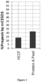
- the amount of partially reduced antibody molecules in the HCCF and Protein A pool were analyzed using nrCE-SDS to determine the percentage of pre-peak species corresponding to partially reduced antibody molecules ( Figure 1 ).
- the reduction of antibody molecules began in the bioreactor. Despite a decrease in the percentage of partially reduced antibodies after air sparging and chilling the cell culture fluid, reduction of the disulfide bonds continued post-harvest, and the percentage of pre-peak species was significantly higher in the Protein A pool compared to the harvested cell culture fluid. Therefore, the controlled oxygen and temperature conditions, i.e., air sparging and chilling of the harvested cell culture fluid, were not sufficient to prevent the partial reduction of the antibody molecules and did not sufficiently enhance re-oxidation.
- aqueous antibody solution comprising Antibody A was subjected to Protein A chromatography as described above and then a viral inactivation step comprising reducing the pH of the Protein A pool to about pH 3.6 for 1 hour and then adjusting to pH 5 was performed to form a neutralized viral inactivation pool (nVIP).
- the nVIP was subjected to charged depth filtration using a MILLISTAK+ A1HC charged depth filter system (EMD Millipore) followed by sterile filtration using a Millipore EXPRESS SHC hydrophilic filter in accordance with the manufacturer's instructions to form a filtered viral inactivation pool (FVIP).
- EMD Millipore MILLISTAK+ A1HC charged depth filter system
- FVIP Millipore EXPRESS SHC hydrophilic filter
- Filtration was performed using a normal flow filtration system (PendoTECH, Princeton, NJ) with the filter connected to a pressure sensor to monitor and control the pressure limit. The flowrate and pressure were maintained at ⁇ about 200 LMH and ⁇ about 50 psi, respectively.
- the filter was first flushed with deionized water for about 100 L/m 2 , followed by a equilibration phase with 30 mM acetate buffer, pH 5.0, for ⁇ about 50L/m 2 prior to loading the solution to be filtered in the range of about 50 L/m 2 to about 800 L/m 2 . At the end, the filter was flushed with about 20 L/m 2 of the equilibration buffer.
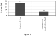
- the level of partial antibody reduction as the percentage of pre-peak species measured by nrCE-SDS in the Protein A pool and the FVIP was determined ( Figure 2 ). Following contact with the charged depth filter, the amount of partially reduced antibody in the Protein A pool was significantly decreased, with a greater than 3-fold decrease in the % pre-peaks observed. All of the Protein A pool product mass was recovered following filtration using the charged depth filter, demonstrating that the decrease in % pre-peak species was due to re-oxidation of partially reduced antibody following contact with the charged depth filter according to the disclosure and not loss of antibody molecules.
- the presence of thioredoxin and thioredoxin reductase was determined using mass spectrophotometry. Thioredoxin like proteins were present in the Protein A Pool, but the charged depth filtrate was free of both thioredoxin and thioredoxin reductase.
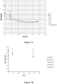
- a Protein A pool was prepared and contacted with a charged depth filter to form a FVIP as described above or not filtered further and then held at room temperature or a temperature between 2 °C and 8 °C, for up to 8 days.
- the level of partial antibody reduction as the percentage of pre-peak species measured by nrCE-SDS in the FVIP ( Figure 3A ) or non-charged depth filtered Protein A pool ( Figure 3B ) was determined.
- the percentage of pre-peak species in the FVIP continued to decrease following contact with the charged depth filter, and the re-oxidation was enhanced at both room temperature and between 2 °C and 8 °C.
- the amount of partially reduced antibody molecules in the FVIP decreased almost three-fold and reached a steady state level in three days or less.
- the percentage of pre-peak species in the non-filtered Protein A pool continued to increase over time at both temperatures, indicating further reduction of the antibody molecules, with faster reduction kinetics at room temperature compared to when chilled.
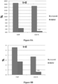
- the charged depth filtration was also effective in enhancing re-oxidation of antibody molecules when performed prior to Protein A chromatography.
- HCCF was subjected to either charged depth filtration using a MILLISTAK+ X0HC depth filter system (EMD Millipore) in accordance with the manufacturer's instructions and at a throughput of 350 L/m 2 or no filtration and then subjected to Protein A chromatography to form a Protein A pool.
- EMD Millipore MILLISTAK+ X0HC depth filter system
- the level of partial antibody reduction as the percentage of pre-peak species measured by nrCE-SDS in the charged depth filtered Protein A pool or non-charged depth filtered Protein A pool was determined ( Figure 4A and Figure 4B ).
- nVIP was produced as described in Example 2 and subjected to either charged depth filtration using a MILLISTAK+ A1HC depth filter system or sterile membrane filtration using a Millipore EXPRESS SHC hydrophilic filter at a throughput of 350 L/m 2 .
- the level of partial antibody reduction as the percentage of pre-peak species measured by nrCE-SDS in the nVIP loaded into the filter system and following filtration was determined ( Figure 5A and Figure 5B ).
- Charged depth filtration was shown to facilitate re-oxidation of even highly reduced material achieving a decrease in the percentage of pre-peak species from 45% in the load to less than 10% in the depth filtered filtrate ( Figure 5A ).
- An aqueous solution comprising Antibody A was subject to Protein A chromatography followed by (a) a viral inactivation step and then charged depth filtration using a MILLISTAK+ A1HC charged depth filter system to form a FVIP; (b) a viral inactivation step to form a nVIP followed by air sparging to 100% dissolved oxygen; or (c) viral inactivation only to form a nVIP followed up a hold step for up to 50 hours, or (d) the hold step only as a control.
- the level of partial antibody reduction as the percentage of pre-peak species measured by nrCE-SDS in the Protein A Pool, FVIP, nVIP oxygenated with air and nVIP without air was compared ( Figure 6 ).
- the starting percentage of pre-peak species was comparable among the Protein A pool and the nVIPs, regardless of whether the nVIP was oxygenated with air. In contrast, the amount of reduced antibody molecules following charged depth filtration in the FVIP was approximately two-fold lower. The percentage of pre-peak species in the FVIP continued to decrease during the hold, reaching a steady state level more than three-fold lower than the Protein A pool or non-oxygenated nVIP and more than two-fold lower than the oxygenated nVIP. The presence of 100% saturated oxygen had minimal impact, therefore, on re-oxidization of the antibody molecules and was much less effective than the charged depth filtration on decreasing the amount of partially reduced antibody molecules.
- throughput i.e., the total amount of aqueous solution contacted with a charged depth filter per square meter of filter
- throughput i.e., the total amount of aqueous solution contacted with a charged depth filter per square meter of filter
- Aqueous solutions comprising Antibody A were subjected to charged depth filtration using a MILLISTAK+ A1HC filter as described above at a throughput of 350 L/m 2 to 850 L/m 2 , and the level of partial antibody reduction as the percentage of pre-peak species was measured by nrCE-SDS for up to 24 hours post-filtration ( Figure 7A ).
- aqueous solution comprising Antibody A was subjected to Protein A chromatography and then a viral inactivation step to form a nVIP.
- the nVIP was subjected to (a) CEX chromatography only at 50 g/L using a FRACTOGEL SO3 resin (EMD Millipore) to form a CEX pool; (b) charged depth filtration only using a MILLISTAK+ A1HC depth filter system to form a FVIP, or (c) charged depth filtration to form a FVIP, followed by CEX chromatography, as described in (a) and (b), to form a CEX pool.
- the CEX step was designed to resolve high molecular weight (HMW) species in a bind and elute mode.
- Partial antibody molecules can dimerize on the resin via free thiol and elute out as HMW species.
- the level of partial antibody reduction as the percentage of pre-peak species was measured by nrCE-SDS before and after CEX chromatography of the nVIP ( Figure 8A ) and immediately following CEX chromatography of the charged depth filtered FVIP ( Figure 8B ) as well as 4 hours post-CEX chromatography of the charged depth filtered FVIP ( Figure 8C ).
- the percentage of HMW species was also determined as a measure of partial antibody molecules.
- the percentage of reduced pre-peak species in the non-charged depth filtered nVIP before and after CEX chromatography were comparable ( Figure 8A ).
- the level of reduced antibody in the FVIP was more than three-fold lower than in the nVIP following charged depth filtration.
- the percentage of pre-peak species in the charged depth filtered FVIP was further decreased by more than 1.5-fold immediately following CEX chromatography ( Figure 8B ) and was comparable at 0 and 4 hours post-CEX chromatography ( Figures 8B and 8C ).
- the percentage of HMW species in the charged depth filtered FVIP decreased following CEX chromatography, confirming a decrease in partial antibody molecules. The results indicated that re-oxidation of the antibody species following charged depth filtration continued on the CEX column and reached steady state.
- a charged depth filter was flushed with deionized water for 100 L/m 2 , followed by a 2-hour recirculation phase with a 0.1 M H 2 SO 4 solution to strip the filter and remove bound metals, or with a 100 mM acetate solution, pH 5.0, as a control.
- An aqueous solution comprising Antibody A was subjected to Protein A chromatography and then a viral inactivation step followed by a neutralization step to form a nVIP.
- the acetate and sulfuric solutions were spiked into nVIP samples at different volume ratios of 0.5, 1, 2 and 4 parts of buffer to 1 part of nVIP.
- the sulfuric solution contained bound metals and other material stripped from the charged depth filter.
- the sulfuric solution contained about 1500 parts per billion (ppb) Cu 2+ , while the acetate solution compared less than 5 ppb Cu 2+ .
- the level of partial antibody reduction as the percentage of pre-peak species in the spiked samples was measured by nrCE-SDS at 0, 2, 4 and 24 hours post-spike. As expected, unspiked nVIP exhibited no re-oxidation of the antibody molecules. In contrast, the percentage of pre-peak species in the nVIP spiked with the sulfuric solution were significantly lower ( Figure 9A ).
- the percentage of pre-peak species in the nVIP spiked with the sulfuric acid solution at a concentration of 1x or above reached steady state at t 0, indicated that contacting the nVIP with the components stripped from the charged depth filter efficiently enhanced re-oxidation.
- Spiking the nVIP with the acetate solution was not as efficient in facilitating re-oxidization of the antibody molecules as the sulfuric solution or direct contact of antibody with the depth filter ( Figure 9B ).
- Table 1 describes Processes A through N for preparing aqueous solutions comprising re-oxidized antibody molecules. TABLE 1 Process Process Steps A B C D E F G H I J K L M N Depth Filtration X X X X X X X Protein A Chromatography X X X X X X X X X X X X X X X X X X X X X Viral Inactivation X X X X X X X X X X X X X X X Cation Exchange Chromatography X X X X X X X X X X X X X X X X X X Cation Exchange Chromatography X X X X X X X X X X X X X X X X X X X Additional Chromatography (e.g., STIC PA, HIC, MMC
- aqueous solutions comprising reduced antigen-binding protein molecules are obtained without sparging the cell culture fluid with air.
- the aqueous solution is subjected to a charged depth filtration step to form a first filtrate, and the first filtrate is subjected to a Protein A chromatography step to form a first eluate.
- the first eluate is subjected to a charged depth filtration step to form a second filtrate, and the second filtrate is subjected to a low pH viral inactivation step to form a virally inactivated second filtrate.
- the virally inactivated second filtrate is subjected to a charged depth filtration step to form a third filtrate.
- the third filtrate is subjected to a cation exchange chromatography step to form a second eluate, followed by a charged depth filtration step to form a fourth filtrate.
- the fourth filtrate is subjected to one or more additional chromatography steps, for example, STIC PA, HIC, and/or MMC, to form a third (or fourth or fifth) eluate, followed by a charged depth filtration step to form a fifth filtrate.
- the aqueous solution is subjected to a Protein A chromatography step to form a first eluate.
- the first eluate is subjected to a charged depth filtration step to form a first filtrate, and the first filtrate is subjected to a low pH viral inactivation step to form a virally inactivated first filtrate.
- the virally inactivated first filtrate is subjected to a charged depth filtration step to form a second filtrate.
- the second filtrate is subjected to a cation exchange chromatography step to form a second eluate, followed by a charged depth filtration step to form a third filtrate.
- the third filtrate is subjected to one or more additional chromatography steps, for example, STIC PA, HIC, and/or MMC, to form a third (or fourth or fifth) eluate, followed by a charged depth filtration step to form a fourth filtrate.
- additional chromatography steps for example, STIC PA, HIC, and/or MMC
- the Process optionally further includes additional, subsequent purification steps, for example, one or more of ultrafiltration, diafiltration, and viral filtration steps, to achieve an aqueous solution comprising re-oxidized antibody molecules.
- the aqueous solution comprising re-oxidized antibody molecules achieved using the Process has a decreased percentage of reduced antigen-binding protein molecules, compared to the percentage of reduced antigen-binding protein molecules observed prior to the start of the Process.
Landscapes
- Chemical & Material Sciences (AREA)
- Organic Chemistry (AREA)
- Health & Medical Sciences (AREA)
- Molecular Biology (AREA)
- Analytical Chemistry (AREA)
- Life Sciences & Earth Sciences (AREA)
- Proteomics, Peptides & Aminoacids (AREA)
- Genetics & Genomics (AREA)
- Medicinal Chemistry (AREA)
- General Health & Medical Sciences (AREA)
- Biophysics (AREA)
- Biochemistry (AREA)
- Chemical Kinetics & Catalysis (AREA)
- Immunology (AREA)
- General Chemical & Material Sciences (AREA)
- Engineering & Computer Science (AREA)
- Water Supply & Treatment (AREA)
- Peptides Or Proteins (AREA)
- Medicines Containing Antibodies Or Antigens For Use As Internal Diagnostic Agents (AREA)
- Medicinal Preparation (AREA)
Applications Claiming Priority (4)
| Application Number | Priority Date | Filing Date | Title |
|---|---|---|---|
| US201562204831P | 2015-08-13 | 2015-08-13 | |
| PCT/US2016/046929 WO2017027861A1 (en) | 2015-08-13 | 2016-08-12 | Charged depth filtration of antigen-binding proteins |
| EP22211542.0A EP4209499B1 (de) | 2015-08-13 | 2016-08-12 | Geladene tiefenfiltration von antigenbindenden proteinen |
| EP16757780.8A EP3334747B1 (de) | 2015-08-13 | 2016-08-12 | Geladene tiefenfiltration von antigenbindenden proteinen |
Related Parent Applications (2)
| Application Number | Title | Priority Date | Filing Date |
|---|---|---|---|
| EP16757780.8A Division EP3334747B1 (de) | 2015-08-13 | 2016-08-12 | Geladene tiefenfiltration von antigenbindenden proteinen |
| EP22211542.0A Division EP4209499B1 (de) | 2015-08-13 | 2016-08-12 | Geladene tiefenfiltration von antigenbindenden proteinen |
Publications (2)
| Publication Number | Publication Date |
|---|---|
| EP4470648A2 true EP4470648A2 (de) | 2024-12-04 |
| EP4470648A3 EP4470648A3 (de) | 2025-03-05 |
Family
ID=56802689
Family Applications (3)
| Application Number | Title | Priority Date | Filing Date |
|---|---|---|---|
| EP16757780.8A Active EP3334747B1 (de) | 2015-08-13 | 2016-08-12 | Geladene tiefenfiltration von antigenbindenden proteinen |
| EP22211542.0A Active EP4209499B1 (de) | 2015-08-13 | 2016-08-12 | Geladene tiefenfiltration von antigenbindenden proteinen |
| EP24193226.8A Pending EP4470648A3 (de) | 2015-08-13 | 2016-08-12 | Geladene tiefenfiltration von antigenbindenden proteinen |
Family Applications Before (2)
| Application Number | Title | Priority Date | Filing Date |
|---|---|---|---|
| EP16757780.8A Active EP3334747B1 (de) | 2015-08-13 | 2016-08-12 | Geladene tiefenfiltration von antigenbindenden proteinen |
| EP22211542.0A Active EP4209499B1 (de) | 2015-08-13 | 2016-08-12 | Geladene tiefenfiltration von antigenbindenden proteinen |
Country Status (15)
| Country | Link |
|---|---|
| US (3) | US11098079B2 (de) |
| EP (3) | EP3334747B1 (de) |
| JP (1) | JP6889149B2 (de) |
| DK (2) | DK4209499T3 (de) |
| ES (2) | ES2995985T3 (de) |
| FI (2) | FI3334747T3 (de) |
| HR (2) | HRP20231544T1 (de) |
| HU (2) | HUE069195T2 (de) |
| LT (2) | LT4209499T (de) |
| MA (1) | MA42613A (de) |
| PL (1) | PL3334747T3 (de) |
| PT (2) | PT3334747T (de) |
| SG (1) | SG10202101105XA (de) |
| SI (2) | SI3334747T1 (de) |
| WO (1) | WO2017027861A1 (de) |
Families Citing this family (14)
| Publication number | Priority date | Publication date | Assignee | Title |
|---|---|---|---|---|
| US20150210753A1 (en) | 2012-07-26 | 2015-07-30 | Momenta Pharmaceuticals, Inc. | Glycoproteins with anti-inflammatory properties |
| WO2014186310A1 (en) | 2013-05-13 | 2014-11-20 | Momenta Pharmaceuticals, Inc. | Methods for the treatment of neurodegeneration |
| EP3058084A4 (de) | 2013-10-16 | 2017-07-05 | Momenta Pharmaceuticals, Inc. | Sialylierte glycoproteine |
| LT4209499T (lt) | 2015-08-13 | 2024-11-25 | Amgen Inc. | Antigeną surišančių baltymų įkrautas giluminis filtravimas |
| WO2018115879A1 (en) | 2016-12-21 | 2018-06-28 | Mereo Biopharma 3 Limited | Use of anti-sclerostin antibodies in the treatment of osteogenesis imperfecta |
| CN107973487B (zh) * | 2017-11-14 | 2021-03-02 | 东华大学 | 一种染整废水层级处理工艺及装置 |
| CN114981286B (zh) | 2019-05-03 | 2026-02-13 | 豪夫迈·罗氏有限公司 | 降低从纯化平台获得的组合物中酶水解活性速率的方法 |
| IL322315A (en) | 2019-05-14 | 2025-09-01 | Provention Bio Inc | Methods and preparations for preventing type 1 diabetes |
| AU2021208515A1 (en) | 2020-01-15 | 2022-08-04 | F. Hoffmann-La Roche Ag | Methods to decrease impurities from recombinant protein manufacturing processes |
| US20230071627A1 (en) * | 2020-02-03 | 2023-03-09 | Amgen Inc. | Multivariate Bracketing Approach for Sterile Filter Validation |
| CN111410688A (zh) * | 2020-03-13 | 2020-07-14 | 苏州智享众创孵化管理有限公司 | 一种提高双特异性抗体纯度的分离纯化方法 |
| AU2021287998B2 (en) | 2020-06-11 | 2026-03-12 | Benaroya Research Institute At Virginia Mason | Methods and compositions for preventing type 1 diabetes |
| US12565529B2 (en) | 2021-05-24 | 2026-03-03 | Provention Bio, Inc. | Methods for treating type 1 diabetes |
| WO2025252480A1 (en) | 2024-06-07 | 2025-12-11 | F. Hoffmann-La Roche Ag | Method for purifying plasmid dna |
Citations (39)
| Publication number | Priority date | Publication date | Assignee | Title |
|---|---|---|---|---|
| US4597966A (en) | 1985-01-09 | 1986-07-01 | Ortho Diagnostic Systems, Inc. | Histidine stabilized immunoglobulin and method of preparation |
| US5237054A (en) | 1987-02-20 | 1993-08-17 | Akzo Pharma | Stabilized aqueous composition containing antibodies |
| US5608038A (en) | 1993-12-28 | 1997-03-04 | Immuno Aktiengesellschaft | Highly concentrated immunoglobulin preparation and method for its production |
| US5654403A (en) | 1991-10-28 | 1997-08-05 | Burroughs Wellcome Co. | Immunoglobulins stabilized with a chelator of copper ions |
| EP0804163A1 (de) | 1994-08-19 | 1997-11-05 | Quadrant Holdings Cambridge Limited | Verbessertes verfahren zur stabilisierung biologischer substanzen der trocknung und anschliessendes lagerung sowie die entsprechenden zusammensetzungen |
| US5736137A (en) | 1992-11-13 | 1998-04-07 | Idec Pharmaceuticals Corporation | Therapeutic application of chimeric and radiolabeled antibodies to human B lymphocyte restricted differentiation antigen for treatment of B cell lymphoma |
| US5789554A (en) | 1994-08-12 | 1998-08-04 | Immunomedics, Inc. | Immunoconjugates and humanized antibodies specific for B-cell lymphoma and leukemia cells |
| US5843439A (en) | 1992-11-13 | 1998-12-01 | Anderson; Darrell R. | Therapeutic application of chimeric and radiolabeled antibodies to human B lymphocyte restricted differentiation antigen for treatment of B cell lymphoma |
| US5908826A (en) | 1991-03-08 | 1999-06-01 | Mitsui Toatsu Chemicals Inc. | Freeze-dried preparation containing monoclonal antibody |
| US5945098A (en) | 1990-02-01 | 1999-08-31 | Baxter International Inc. | Stable intravenously-administrable immune globulin preparation |
| US6171586B1 (en) | 1997-06-13 | 2001-01-09 | Genentech, Inc. | Antibody formulation |
| US6252055B1 (en) | 1996-05-24 | 2001-06-26 | Glaxo Wellcome Inc. | Concentrated antibody preparation |
| US6267958B1 (en) | 1995-07-27 | 2001-07-31 | Genentech, Inc. | Protein formulation |
| US6284471B1 (en) | 1991-03-18 | 2001-09-04 | New York University Medical Center | Anti-TNFa antibodies and assays employing anti-TNFa antibodies |
| US6288030B1 (en) | 1993-12-22 | 2001-09-11 | Amgen Inc. | Stem cell factor formulations and methods |
| US6485932B1 (en) | 1997-03-20 | 2002-11-26 | Common Services Agency | Composition comprising immunoglobulin |
| WO2002096457A2 (en) | 2001-05-31 | 2002-12-05 | Novartis Ag | Stable liquid formulations of antibodies |
| WO2003002713A2 (en) | 2001-06-26 | 2003-01-09 | Abgenix, Inc. | Antibodies to opgl |
| US20030023586A1 (en) | 2000-03-03 | 2003-01-30 | Super Internet Site System Pty Ltd. | On-line geographical directory |
| WO2003030833A2 (en) | 2001-10-11 | 2003-04-17 | Amgen Inc. | Angiopoietin-2 specific binding agents |
| WO2003057134A2 (en) | 2001-10-11 | 2003-07-17 | Amgen, Inc. | Specific binding agents of human angiopoietin-2 |
| US20030138421A1 (en) | 2001-08-23 | 2003-07-24 | Van De Winkel Jan G.J. | Human Antibodies specific for interleukin 15 (IL-15) |
| US20030138417A1 (en) | 2001-11-08 | 2003-07-24 | Kaisheva Elizabet A. | Stable liquid pharmaceutical formulation of IgG antibodies |
| US20030195156A1 (en) | 2001-05-11 | 2003-10-16 | Amgen Inc. | Peptides and related molecules that bind to TALL-1 |
| US20040071702A1 (en) | 2001-08-23 | 2004-04-15 | Genmab, Inc. | Human antibodies specific for interleukin 15 (IL-15) |
| US20040097712A1 (en) | 2002-09-06 | 2004-05-20 | Amgen, Inc. A Corporation Of The State Of Delaware | Therapeutic human anti-IL1-R1 monoclonal antibody |
| WO2004055164A2 (en) | 2002-12-13 | 2004-07-01 | Abgenix, Inc. | System and method for stabilizing antibodies with histidine |
| US20040181033A1 (en) | 2002-12-20 | 2004-09-16 | Hq Han | Binding agents which inhibit myostatin |
| US20040197324A1 (en) | 2003-04-04 | 2004-10-07 | Genentech, Inc. | High concentration antibody and protein formulations |
| US20050004353A1 (en) | 2002-10-16 | 2005-01-06 | Amgen, Inc., A Corporation Of The State Of Delaware | Human anti-IFN-gamma neutralizing antibodies as selective IFN-gamma pathway inhibitors |
| US6875432B2 (en) | 2000-10-12 | 2005-04-05 | Genentech, Inc. | Reduced-viscosity concentrated protein formulations |
| US20050074821A1 (en) | 2003-07-15 | 2005-04-07 | Wild Kenneth D. | Human anti-NGF neutralizing antibodies as selective NGF pathway inhibitors |
| WO2005044854A2 (en) | 2003-11-04 | 2005-05-19 | Chiron Corporation | Antagonist anti-cd40 monoclonal antibodies and methods for their use |
| WO2005047331A2 (en) | 2003-11-07 | 2005-05-26 | Immunex Corporation | Antibodies that bind interleukin-4 receptor |
| WO2005063291A1 (ja) | 2003-12-25 | 2005-07-14 | Kirin Beer Kabushiki Kaisha | 抗体を含有する安定な水性医薬製剤 |
| US20080311078A1 (en) | 2005-06-14 | 2008-12-18 | Gokarn Yatin R | Self-Buffering Protein Formulations |
| US20090053786A1 (en) | 2007-07-09 | 2009-02-26 | Yung-Hsiang Kao | Prevention of disulfide bond reduction during recombinant production of polypeptides |
| US8529902B2 (en) | 2002-10-17 | 2013-09-10 | Genmab A/S | Human monoclonal antibodies against CD20 |
| US20140024111A1 (en) | 2008-01-07 | 2014-01-23 | Amgen Inc. | Host cell for making antibody fc-heterodimeric molecules using electrostatic steering effects |
Family Cites Families (47)
| Publication number | Priority date | Publication date | Assignee | Title |
|---|---|---|---|---|
| US6214221B1 (en) | 1999-02-22 | 2001-04-10 | Henry B. Kopf | Method and apparatus for purification of biological substances |
| WO2003066669A2 (en) | 2002-02-04 | 2003-08-14 | Millipore Corporation | Process for removing protein aggregates and virus from a protein solution |
| US8188231B2 (en) * | 2002-09-27 | 2012-05-29 | Xencor, Inc. | Optimized FC variants |
| US7157276B2 (en) | 2003-06-20 | 2007-01-02 | Biogen Idec Inc. | Use of depth filtration in series with continuous centrifugation to clarify mammalian cell cultures |
| CN1922207B (zh) | 2004-02-27 | 2012-06-06 | 奥克特珐玛股份有限公司 | 提供纯化的、病毒安全的抗体制剂的方法 |
| KR101270829B1 (ko) | 2004-09-23 | 2013-06-07 | 제넨테크, 인크. | 시스테인 유전자조작 항체 및 접합체 |
| JP5553963B2 (ja) | 2004-10-22 | 2014-07-23 | アムジエン・インコーポレーテツド | 組換え抗体をリフォールディングする方法 |
| WO2007035283A1 (en) | 2005-09-15 | 2007-03-29 | Wyeth | Protein floculation using salts |
| US20100234577A1 (en) | 2006-06-14 | 2010-09-16 | Smithkline Beecham Corporation | Methods for purifying antibodies using ceramic hydroxyapatite |
| WO2008036899A2 (en) | 2006-09-22 | 2008-03-27 | Amgen Inc. | Methods for removing viral contaminants during protein purification |
| US20100056759A1 (en) | 2006-10-19 | 2010-03-04 | Tolerx, Inc. | Methods and Compositions for Efficient Removal of Protein A from Binding Molecule Preparations |
| DE102008037678A1 (de) | 2008-08-14 | 2010-02-25 | Sartorius Stedim Biotech Gmbh | Tiefenfilterschicht mit anorganischem Schichtdoppelhydroxid |
| AU2009347206C1 (en) | 2008-10-20 | 2016-12-08 | Abbvie Inc. | Isolation and purification of antibodies using Protein A affinity chromatography |
| ES2551854T3 (es) | 2009-04-30 | 2015-11-24 | Ablynx N.V. | Procedimiento para la producción de anticuerpos de dominio |
| US20120208986A1 (en) | 2009-10-20 | 2012-08-16 | Wenger Marc D | Use of mixed mode chromatography for the capture and purification of basic antibody products |
| ES2813398T3 (es) | 2009-10-20 | 2021-03-23 | Abbvie Inc | Aislamiento y purificación de anticuerpos anti-IL-13 usando cromatografía de afinidad a Proteína A |
| GB201006753D0 (en) | 2010-04-22 | 2010-06-09 | Biotest Ag | Process for preparing an immunolobulin composition |
| WO2011140405A1 (en) * | 2010-05-07 | 2011-11-10 | Millipore Corporation | Enhanced clarification media |
| WO2011146179A2 (en) | 2010-05-18 | 2011-11-24 | Abbott Laboratories | Apparatus and process of purification of proteins |
| WO2012051147A1 (en) | 2010-10-11 | 2012-04-19 | Abbott Laboratories | Processes for purification of proteins |
| AU2011317021B2 (en) | 2010-10-20 | 2015-07-09 | Medimmune, Llc | Methods for processing inclusion bodies |
| US20120238730A1 (en) | 2011-03-15 | 2012-09-20 | Abbott Laboratories | Integrated approach to the isolation and purification of antibodies |
| CN103649297B (zh) | 2011-07-08 | 2017-03-01 | Emd密理博公司 | 用于一次性生物技术方法的改良深层滤器 |
| FR2977893B1 (fr) | 2011-07-11 | 2015-02-20 | Lab Francais Du Fractionnement | Procede de preparation d'un concentre d'immunoglobulines polyvalentes |
| AU2012290004B2 (en) | 2011-08-02 | 2014-11-27 | Takeda Pharmaceutical Company Limited | Systems and methods to increase protein yield from recombinant manufacturing processes |
| US9096648B2 (en) | 2011-08-19 | 2015-08-04 | Emd Millipore Corporation | Methods of reducing level of one or more impurities in a sample during protein purification |
| US10344050B2 (en) * | 2011-10-27 | 2019-07-09 | Genmab A/S | Production of heterodimeric proteins |
| RU2648999C2 (ru) | 2011-12-22 | 2018-03-29 | Дженентек, Инк. | Способы повышения эффективности этапов очистки белка, находящихся ниже по потоку, с использованием мембранной ионообменной хроматографии |
| US20140154270A1 (en) | 2012-05-21 | 2014-06-05 | Chen Wang | Purification of non-human antibodies using kosmotropic salt enhanced protein a affinity chromatography |
| TW201348246A (zh) * | 2012-05-21 | 2013-12-01 | Abbvie Inc | 利用蛋白質a親和性層析之人類、人類化或嵌合抗體之新穎純化 |
| US9249182B2 (en) | 2012-05-24 | 2016-02-02 | Abbvie, Inc. | Purification of antibodies using hydrophobic interaction chromatography |
| EP2682168A1 (de) | 2012-07-02 | 2014-01-08 | Millipore Corporation | Reinigung biologischer Moleküle |
| US9920124B2 (en) | 2012-12-20 | 2018-03-20 | Medimmune, Llc | Methods of producing immunoconjugates |
| WO2014159554A1 (en) | 2013-03-14 | 2014-10-02 | Abbvie Inc. | Low acidic species compositions and methods for producing the same using displacement chromatography |
| CA2926301A1 (en) | 2013-03-14 | 2014-10-02 | Abbvie Inc. | Low acidic species compositions and methods for producing and using the same |
| WO2014145744A1 (en) | 2013-03-15 | 2014-09-18 | Alder Biopharmaceuticals, Inc. | Antibody purification and purity monitoring |
| CN105263946A (zh) | 2013-06-04 | 2016-01-20 | 新加坡科技研究局 | 蛋白质纯化方法 |
| US10513555B2 (en) | 2013-07-04 | 2019-12-24 | Prothena Biosciences Limited | Antibody formulations and methods |
| EP3815766A1 (de) | 2013-08-30 | 2021-05-05 | EMD Millipore Corporation | Zusammengesetzte tiefenfiltermedien mit hoher kapazität und geringem entnahmevolumen |
| US8946395B1 (en) | 2013-10-18 | 2015-02-03 | Abbvie Inc. | Purification of proteins using hydrophobic interaction chromatography |
| GB201318840D0 (en) | 2013-10-24 | 2013-12-11 | Univ Leeds | Method and device for protein preparation |
| US20160272674A1 (en) | 2013-11-07 | 2016-09-22 | Abbvie Inc. | Isolation and purification of antibodies |
| KR20160118369A (ko) | 2014-02-27 | 2016-10-11 | 에이전시 포 사이언스, 테크놀로지 앤드 리서치 | 항체 정제 프로세스 |
| EP3119871A4 (de) | 2014-03-17 | 2018-01-03 | Regeneron Pharmaceuticals, Inc. | Klärung einer säugerzellenkultur |
| WO2015198320A1 (en) | 2014-06-24 | 2015-12-30 | Insight Biopharmaceuticals Ltd. | Methods of purifying antibodies |
| EP3236772A4 (de) | 2014-12-22 | 2018-10-17 | Alexion Pharmaceuticals, Inc. | Verfahren zur aufreinigung von rekombinanten proteinen |
| LT4209499T (lt) | 2015-08-13 | 2024-11-25 | Amgen Inc. | Antigeną surišančių baltymų įkrautas giluminis filtravimas |
-
2016
- 2016-08-12 LT LTEP22211542.0T patent/LT4209499T/lt unknown
- 2016-08-12 LT LTEPPCT/US2016/046929T patent/LT3334747T/lt unknown
- 2016-08-12 DK DK22211542.0T patent/DK4209499T3/da active
- 2016-08-12 ES ES22211542T patent/ES2995985T3/es active Active
- 2016-08-12 MA MA042613A patent/MA42613A/fr unknown
- 2016-08-12 HU HUE22211542A patent/HUE069195T2/hu unknown
- 2016-08-12 HR HRP20231544TT patent/HRP20231544T1/hr unknown
- 2016-08-12 FI FIEP16757780.8T patent/FI3334747T3/fi active
- 2016-08-12 DK DK16757780.8T patent/DK3334747T5/da active
- 2016-08-12 ES ES16757780T patent/ES2964640T3/es active Active
- 2016-08-12 SG SG10202101105XA patent/SG10202101105XA/en unknown
- 2016-08-12 US US15/751,231 patent/US11098079B2/en active Active
- 2016-08-12 SI SI201631774T patent/SI3334747T1/sl unknown
- 2016-08-12 HR HRP20241510TT patent/HRP20241510T1/hr unknown
- 2016-08-12 WO PCT/US2016/046929 patent/WO2017027861A1/en not_active Ceased
- 2016-08-12 SI SI201631860T patent/SI4209499T1/sl unknown
- 2016-08-12 PT PT167577808T patent/PT3334747T/pt unknown
- 2016-08-12 PT PT222115420T patent/PT4209499T/pt unknown
- 2016-08-12 HU HUE16757780A patent/HUE064252T2/hu unknown
- 2016-08-12 JP JP2018506988A patent/JP6889149B2/ja active Active
- 2016-08-12 PL PL16757780.8T patent/PL3334747T3/pl unknown
- 2016-08-12 EP EP16757780.8A patent/EP3334747B1/de active Active
- 2016-08-12 EP EP22211542.0A patent/EP4209499B1/de active Active
- 2016-08-12 EP EP24193226.8A patent/EP4470648A3/de active Pending
- 2016-08-12 FI FIEP22211542.0T patent/FI4209499T3/fi active
-
2021
- 2021-07-28 US US17/386,692 patent/US20210363180A1/en active Pending
-
2025
- 2025-01-15 US US19/022,213 patent/US20250230187A1/en active Pending
Patent Citations (42)
| Publication number | Priority date | Publication date | Assignee | Title |
|---|---|---|---|---|
| US4597966A (en) | 1985-01-09 | 1986-07-01 | Ortho Diagnostic Systems, Inc. | Histidine stabilized immunoglobulin and method of preparation |
| US5237054A (en) | 1987-02-20 | 1993-08-17 | Akzo Pharma | Stabilized aqueous composition containing antibodies |
| US5945098A (en) | 1990-02-01 | 1999-08-31 | Baxter International Inc. | Stable intravenously-administrable immune globulin preparation |
| US5908826A (en) | 1991-03-08 | 1999-06-01 | Mitsui Toatsu Chemicals Inc. | Freeze-dried preparation containing monoclonal antibody |
| US6284471B1 (en) | 1991-03-18 | 2001-09-04 | New York University Medical Center | Anti-TNFa antibodies and assays employing anti-TNFa antibodies |
| US5654403A (en) | 1991-10-28 | 1997-08-05 | Burroughs Wellcome Co. | Immunoglobulins stabilized with a chelator of copper ions |
| US5792838A (en) | 1991-10-28 | 1998-08-11 | Glaxo Wellcome Inc. | Method for stabilizing immunoglobulin compositions |
| US5843439A (en) | 1992-11-13 | 1998-12-01 | Anderson; Darrell R. | Therapeutic application of chimeric and radiolabeled antibodies to human B lymphocyte restricted differentiation antigen for treatment of B cell lymphoma |
| US5736137A (en) | 1992-11-13 | 1998-04-07 | Idec Pharmaceuticals Corporation | Therapeutic application of chimeric and radiolabeled antibodies to human B lymphocyte restricted differentiation antigen for treatment of B cell lymphoma |
| US6288030B1 (en) | 1993-12-22 | 2001-09-11 | Amgen Inc. | Stem cell factor formulations and methods |
| US5608038A (en) | 1993-12-28 | 1997-03-04 | Immuno Aktiengesellschaft | Highly concentrated immunoglobulin preparation and method for its production |
| US5789554A (en) | 1994-08-12 | 1998-08-04 | Immunomedics, Inc. | Immunoconjugates and humanized antibodies specific for B-cell lymphoma and leukemia cells |
| EP0804163A1 (de) | 1994-08-19 | 1997-11-05 | Quadrant Holdings Cambridge Limited | Verbessertes verfahren zur stabilisierung biologischer substanzen der trocknung und anschliessendes lagerung sowie die entsprechenden zusammensetzungen |
| US6821515B1 (en) | 1995-07-27 | 2004-11-23 | Genentech, Inc. | Protein formulation |
| US6267958B1 (en) | 1995-07-27 | 2001-07-31 | Genentech, Inc. | Protein formulation |
| US6252055B1 (en) | 1996-05-24 | 2001-06-26 | Glaxo Wellcome Inc. | Concentrated antibody preparation |
| US6485932B1 (en) | 1997-03-20 | 2002-11-26 | Common Services Agency | Composition comprising immunoglobulin |
| US6171586B1 (en) | 1997-06-13 | 2001-01-09 | Genentech, Inc. | Antibody formulation |
| US20030023586A1 (en) | 2000-03-03 | 2003-01-30 | Super Internet Site System Pty Ltd. | On-line geographical directory |
| US6875432B2 (en) | 2000-10-12 | 2005-04-05 | Genentech, Inc. | Reduced-viscosity concentrated protein formulations |
| US20030195156A1 (en) | 2001-05-11 | 2003-10-16 | Amgen Inc. | Peptides and related molecules that bind to TALL-1 |
| WO2002096457A2 (en) | 2001-05-31 | 2002-12-05 | Novartis Ag | Stable liquid formulations of antibodies |
| WO2003002713A2 (en) | 2001-06-26 | 2003-01-09 | Abgenix, Inc. | Antibodies to opgl |
| US20040071702A1 (en) | 2001-08-23 | 2004-04-15 | Genmab, Inc. | Human antibodies specific for interleukin 15 (IL-15) |
| US20030138421A1 (en) | 2001-08-23 | 2003-07-24 | Van De Winkel Jan G.J. | Human Antibodies specific for interleukin 15 (IL-15) |
| WO2003030833A2 (en) | 2001-10-11 | 2003-04-17 | Amgen Inc. | Angiopoietin-2 specific binding agents |
| WO2003057134A2 (en) | 2001-10-11 | 2003-07-17 | Amgen, Inc. | Specific binding agents of human angiopoietin-2 |
| US20030229023A1 (en) | 2001-10-11 | 2003-12-11 | Oliner Jonathan Daniel | Specific binding agents of human angiopoietin-2 |
| US20030138417A1 (en) | 2001-11-08 | 2003-07-24 | Kaisheva Elizabet A. | Stable liquid pharmaceutical formulation of IgG antibodies |
| US20040097712A1 (en) | 2002-09-06 | 2004-05-20 | Amgen, Inc. A Corporation Of The State Of Delaware | Therapeutic human anti-IL1-R1 monoclonal antibody |
| US20050004353A1 (en) | 2002-10-16 | 2005-01-06 | Amgen, Inc., A Corporation Of The State Of Delaware | Human anti-IFN-gamma neutralizing antibodies as selective IFN-gamma pathway inhibitors |
| US8529902B2 (en) | 2002-10-17 | 2013-09-10 | Genmab A/S | Human monoclonal antibodies against CD20 |
| WO2004055164A2 (en) | 2002-12-13 | 2004-07-01 | Abgenix, Inc. | System and method for stabilizing antibodies with histidine |
| US20040181033A1 (en) | 2002-12-20 | 2004-09-16 | Hq Han | Binding agents which inhibit myostatin |
| US20040197324A1 (en) | 2003-04-04 | 2004-10-07 | Genentech, Inc. | High concentration antibody and protein formulations |
| US20050074821A1 (en) | 2003-07-15 | 2005-04-07 | Wild Kenneth D. | Human anti-NGF neutralizing antibodies as selective NGF pathway inhibitors |
| WO2005044854A2 (en) | 2003-11-04 | 2005-05-19 | Chiron Corporation | Antagonist anti-cd40 monoclonal antibodies and methods for their use |
| WO2005047331A2 (en) | 2003-11-07 | 2005-05-26 | Immunex Corporation | Antibodies that bind interleukin-4 receptor |
| WO2005063291A1 (ja) | 2003-12-25 | 2005-07-14 | Kirin Beer Kabushiki Kaisha | 抗体を含有する安定な水性医薬製剤 |
| US20080311078A1 (en) | 2005-06-14 | 2008-12-18 | Gokarn Yatin R | Self-Buffering Protein Formulations |
| US20090053786A1 (en) | 2007-07-09 | 2009-02-26 | Yung-Hsiang Kao | Prevention of disulfide bond reduction during recombinant production of polypeptides |
| US20140024111A1 (en) | 2008-01-07 | 2014-01-23 | Amgen Inc. | Host cell for making antibody fc-heterodimeric molecules using electrostatic steering effects |
Non-Patent Citations (11)
| Title |
|---|
| "Protein Engineering: Principles and Practice,", 1996, WILEY-LISS, INC. |
| ARAKAWA ET AL.: "Solvent interactions in pharmaceutical formulations", PHARM RES., vol. 8, no. 3, 1991, pages 285 - 91, XP009052919, DOI: 10.1023/A:1015825027737 |
| BERINGER ET AL.: "REMINGTON: THE SCIENCE AND PRACTICE OF PHARMACY", 1999, LIPPINCOTT, WILLIAMS & WILKINS |
| GUO ET AL., ELECTROPHORESIS, vol. 29, no. 12, 2008, pages 2550 - 6 |
| HOLLINGER, P.HUDSON, P: "Engineered antibody fragments and the rise of single domains", NATURE BIOTECHNOLOGY, September 2005 (2005-09-01), pages 1126 - 1136, XP008076746, DOI: 10.1038/nbt1142 |
| KELLEY, ROBERT F., ENGINEERING THERAPEUTIC ANTIBODIES, vol. 15, pages 399 - 434 |
| KENDRICK ET AL.: "Physical stabilization of proteins in aqueous solution", RATIONAL DESIGN OF STABLE PROTEIN FORMULATIONS: THEORY AND PRACTICE |
| LIU ET AL., MABS., vol. 2, no. 5, 2010, pages 480 - 499 |
| RANDOLPH ET AL.: "Surfactant-protein interactions", PHARM BIOTECHNOL., vol. 13, 2002, pages 159 - 75, XP055647590, DOI: 10.1007/978-1-4615-0557-0_7 |
| SHIMAMOTO, MABS, vol. 4, no. 5, 2012, pages 586 - 91 |
| TERRANCE G. COOPER: "PROTEIN PURIFICATION PRINCIPLES AND PRACTICE", 1994, JOHN WILEY & SONS |
Also Published As
| Publication number | Publication date |
|---|---|
| HRP20231544T1 (hr) | 2024-03-15 |
| DK3334747T5 (da) | 2024-10-07 |
| HRP20241510T1 (hr) | 2025-01-03 |
| ES2964640T3 (es) | 2024-04-08 |
| US11098079B2 (en) | 2021-08-24 |
| DK3334747T3 (da) | 2023-12-11 |
| SI3334747T1 (sl) | 2024-02-29 |
| PT3334747T (pt) | 2023-12-12 |
| PL3334747T3 (pl) | 2024-04-02 |
| MA42613A (fr) | 2018-06-20 |
| LT4209499T (lt) | 2024-11-25 |
| JP2018526357A (ja) | 2018-09-13 |
| SG10202101105XA (en) | 2021-03-30 |
| FI4209499T3 (fi) | 2024-11-05 |
| HUE069195T2 (hu) | 2025-02-28 |
| EP3334747A1 (de) | 2018-06-20 |
| EP4470648A3 (de) | 2025-03-05 |
| US20250230187A1 (en) | 2025-07-17 |
| ES2995985T3 (en) | 2025-02-11 |
| WO2017027861A1 (en) | 2017-02-16 |
| US20180230180A1 (en) | 2018-08-16 |
| PT4209499T (pt) | 2024-11-08 |
| EP4209499A1 (de) | 2023-07-12 |
| US20210363180A1 (en) | 2021-11-25 |
| HUE064252T2 (hu) | 2024-02-28 |
| SI4209499T1 (sl) | 2025-01-31 |
| LT3334747T (lt) | 2023-12-27 |
| EP3334747B1 (de) | 2023-09-27 |
| EP4209499B1 (de) | 2024-08-07 |
| JP6889149B2 (ja) | 2021-06-18 |
| DK4209499T3 (da) | 2024-11-11 |
| FI3334747T3 (fi) | 2023-11-30 |
Similar Documents
| Publication | Publication Date | Title |
|---|---|---|
| US20250230187A1 (en) | Charged depth filtration of antigen-binding proteins | |
| US12545703B2 (en) | Method to produce an immunoglobulin preparation with improved yield | |
| JP2025072467A (ja) | 自己緩衝タンパク質製剤 | |
| CN1671741A (zh) | 浓缩抗体的缓冲剂制剂及其使用方法 | |
| JP2021515893A (ja) | 質量分光測定法による分析のためのポリペプチドの連続消化 | |
| KR20210145152A (ko) | 단백질-함유 제제들의 안정화를 위한 조성물들 및 방법들 | |
| EP4214226A1 (de) | Verfahren zur verarbeitung einer probe zur peptidkartierungsanalyse | |
| US8796430B2 (en) | Method to produce an immunoglobulin preparation with improved yield | |
| HK40120510A (en) | Charged depth filtration of antigen-binding proteins | |
| HK40096795B (en) | Charged depth filtration of antigen-binding proteins | |
| HK40096795A (en) | Charged depth filtration of antigen-binding proteins | |
| WO2025068923A2 (en) | IMMUNOGLOBULIN FORMULATIONS DEPLETED IN IgA | |
| HK1119939A (en) | Self-buffering protein formulations | |
| HK1181682B (en) | Method for preparing an enriched igg composition from plasma | |
| HK1203838B (en) | Method for preparing an enriched igg composition from plasma | |
| HK1170168B (en) | Method for preparing an enriched igg composition from plasma |
Legal Events
| Date | Code | Title | Description |
|---|---|---|---|
| PUAI | Public reference made under article 153(3) epc to a published international application that has entered the european phase |
Free format text: ORIGINAL CODE: 0009012 |
|
| STAA | Information on the status of an ep patent application or granted ep patent |
Free format text: STATUS: THE APPLICATION HAS BEEN PUBLISHED |
|
| AC | Divisional application: reference to earlier application |
Ref document number: 3334747 Country of ref document: EP Kind code of ref document: P Ref document number: 4209499 Country of ref document: EP Kind code of ref document: P |
|
| AK | Designated contracting states |
Kind code of ref document: A2 Designated state(s): AL AT BE BG CH CY CZ DE DK EE ES FI FR GB GR HR HU IE IS IT LI LT LU LV MC MK MT NL NO PL PT RO RS SE SI SK SM TR |
|
| REG | Reference to a national code |
Ref country code: DE Ref legal event code: R079 Free format text: PREVIOUS MAIN CLASS: B01D0015380000 Ipc: C07K0001113000 |
|
| PUAL | Search report despatched |
Free format text: ORIGINAL CODE: 0009013 |
|
| AK | Designated contracting states |
Kind code of ref document: A3 Designated state(s): AL AT BE BG CH CY CZ DE DK EE ES FI FR GB GR HR HU IE IS IT LI LT LU LV MC MK MT NL NO PL PT RO RS SE SI SK SM TR |
|
| RIC1 | Information provided on ipc code assigned before grant |
Ipc: B01D 15/36 20060101ALI20250129BHEP Ipc: B01D 15/38 20060101ALI20250129BHEP Ipc: C07K 16/00 20060101ALI20250129BHEP Ipc: C07K 1/34 20060101ALI20250129BHEP Ipc: C07K 1/113 20060101AFI20250129BHEP |
|
| REG | Reference to a national code |
Ref country code: HK Ref legal event code: DE Ref document number: 40120510 Country of ref document: HK |
|
| STAA | Information on the status of an ep patent application or granted ep patent |
Free format text: STATUS: REQUEST FOR EXAMINATION WAS MADE |
|
| 17P | Request for examination filed |
Effective date: 20250905 |
|
| RAV | Requested validation state of the european patent: fee paid |
Extension state: MA Effective date: 20250905 |
|
| RAX | Requested extension states of the european patent have changed |
Extension state: BA Payment date: 20250905 Extension state: ME Payment date: 20250905 |