EP4480966A1 - Anticorps anti-cd28 humanisé et anticorps bispécifique l'associant avec un anticorps anti-cd40 - Google Patents
Anticorps anti-cd28 humanisé et anticorps bispécifique l'associant avec un anticorps anti-cd40 Download PDFInfo
- Publication number
- EP4480966A1 EP4480966A1 EP23755846.5A EP23755846A EP4480966A1 EP 4480966 A1 EP4480966 A1 EP 4480966A1 EP 23755846 A EP23755846 A EP 23755846A EP 4480966 A1 EP4480966 A1 EP 4480966A1
- Authority
- EP
- European Patent Office
- Prior art keywords
- seq
- set forth
- chain variable
- variable region
- lcdr2
- Prior art date
- Legal status (The legal status is an assumption and is not a legal conclusion. Google has not performed a legal analysis and makes no representation as to the accuracy of the status listed.)
- Pending
Links
Images













































Classifications
-
- A—HUMAN NECESSITIES
- A61—MEDICAL OR VETERINARY SCIENCE; HYGIENE
- A61K—PREPARATIONS FOR MEDICAL, DENTAL OR TOILETRY PURPOSES
- A61K35/00—Medicinal preparations containing materials or reaction products thereof with undetermined constitution
- A61K35/12—Materials from mammals; Compositions comprising non-specified tissues or cells; Compositions comprising non-embryonic stem cells; Genetically modified cells
- A61K35/14—Blood; Artificial blood
- A61K35/17—Lymphocytes; B-cells; T-cells; Natural killer cells; Interferon-activated or cytokine-activated lymphocytes
-
- A—HUMAN NECESSITIES
- A61—MEDICAL OR VETERINARY SCIENCE; HYGIENE
- A61K—PREPARATIONS FOR MEDICAL, DENTAL OR TOILETRY PURPOSES
- A61K39/00—Medicinal preparations containing antigens or antibodies
- A61K39/395—Antibodies; Immunoglobulins; Immune serum, e.g. antilymphocytic serum
-
- A—HUMAN NECESSITIES
- A61—MEDICAL OR VETERINARY SCIENCE; HYGIENE
- A61K—PREPARATIONS FOR MEDICAL, DENTAL OR TOILETRY PURPOSES
- A61K40/00—Cellular immunotherapy
- A61K40/10—Cellular immunotherapy characterised by the cell type used
- A61K40/11—T-cells, e.g. tumour infiltrating lymphocytes [TIL] or regulatory T [Treg] cells; Lymphokine-activated killer [LAK] cells
-
- A—HUMAN NECESSITIES
- A61—MEDICAL OR VETERINARY SCIENCE; HYGIENE
- A61K—PREPARATIONS FOR MEDICAL, DENTAL OR TOILETRY PURPOSES
- A61K40/00—Cellular immunotherapy
- A61K40/30—Cellular immunotherapy characterised by the recombinant expression of specific molecules in the cells of the immune system
- A61K40/31—Chimeric antigen receptors [CAR]
-
- A—HUMAN NECESSITIES
- A61—MEDICAL OR VETERINARY SCIENCE; HYGIENE
- A61K—PREPARATIONS FOR MEDICAL, DENTAL OR TOILETRY PURPOSES
- A61K40/00—Cellular immunotherapy
- A61K40/30—Cellular immunotherapy characterised by the recombinant expression of specific molecules in the cells of the immune system
- A61K40/33—Antibodies; T-cell engagers
-
- A—HUMAN NECESSITIES
- A61—MEDICAL OR VETERINARY SCIENCE; HYGIENE
- A61K—PREPARATIONS FOR MEDICAL, DENTAL OR TOILETRY PURPOSES
- A61K40/00—Cellular immunotherapy
- A61K40/40—Cellular immunotherapy characterised by antigens that are targeted or presented by cells of the immune system
- A61K40/41—Vertebrate antigens
- A61K40/42—Cancer antigens
- A61K40/4202—Receptors, cell surface antigens or cell surface determinants
- A61K40/4203—Receptors for growth factors
- A61K40/4205—Her-2/neu/ErbB2, Her-3/ErbB3 or Her 4/ ErbB4
-
- A—HUMAN NECESSITIES
- A61—MEDICAL OR VETERINARY SCIENCE; HYGIENE
- A61K—PREPARATIONS FOR MEDICAL, DENTAL OR TOILETRY PURPOSES
- A61K40/00—Cellular immunotherapy
- A61K40/40—Cellular immunotherapy characterised by antigens that are targeted or presented by cells of the immune system
- A61K40/41—Vertebrate antigens
- A61K40/42—Cancer antigens
- A61K40/4202—Receptors, cell surface antigens or cell surface determinants
- A61K40/421—Immunoglobulin superfamily
- A61K40/4211—CD19 or B4
-
- A—HUMAN NECESSITIES
- A61—MEDICAL OR VETERINARY SCIENCE; HYGIENE
- A61K—PREPARATIONS FOR MEDICAL, DENTAL OR TOILETRY PURPOSES
- A61K40/00—Cellular immunotherapy
- A61K40/40—Cellular immunotherapy characterised by antigens that are targeted or presented by cells of the immune system
- A61K40/41—Vertebrate antigens
- A61K40/42—Cancer antigens
- A61K40/4202—Receptors, cell surface antigens or cell surface determinants
- A61K40/4214—Receptors for cytokines
- A61K40/4215—Receptors for tumor necrosis factors [TNF], e.g. lymphotoxin receptor [LTR], CD30
-
- A—HUMAN NECESSITIES
- A61—MEDICAL OR VETERINARY SCIENCE; HYGIENE
- A61K—PREPARATIONS FOR MEDICAL, DENTAL OR TOILETRY PURPOSES
- A61K40/00—Cellular immunotherapy
- A61K40/40—Cellular immunotherapy characterised by antigens that are targeted or presented by cells of the immune system
- A61K40/41—Vertebrate antigens
- A61K40/42—Cancer antigens
- A61K40/4202—Receptors, cell surface antigens or cell surface determinants
- A61K40/4214—Receptors for cytokines
- A61K40/4217—Receptors for interleukins [IL]
-
- A—HUMAN NECESSITIES
- A61—MEDICAL OR VETERINARY SCIENCE; HYGIENE
- A61K—PREPARATIONS FOR MEDICAL, DENTAL OR TOILETRY PURPOSES
- A61K40/00—Cellular immunotherapy
- A61K40/40—Cellular immunotherapy characterised by antigens that are targeted or presented by cells of the immune system
- A61K40/41—Vertebrate antigens
- A61K40/42—Cancer antigens
- A61K40/4254—Adhesion molecules, e.g. NRCAM, EpCAM or cadherins
- A61K40/4255—Mesothelin [MSLN]
-
- A—HUMAN NECESSITIES
- A61—MEDICAL OR VETERINARY SCIENCE; HYGIENE
- A61P—SPECIFIC THERAPEUTIC ACTIVITY OF CHEMICAL COMPOUNDS OR MEDICINAL PREPARATIONS
- A61P35/00—Antineoplastic agents
-
- C—CHEMISTRY; METALLURGY
- C07—ORGANIC CHEMISTRY
- C07K—PEPTIDES
- C07K16/00—Immunoglobulins [IG], e.g. monoclonal or polyclonal antibodies
- C07K16/005—Immunoglobulins [IG], e.g. monoclonal or polyclonal antibodies constructed by phage libraries
-
- C—CHEMISTRY; METALLURGY
- C07—ORGANIC CHEMISTRY
- C07K—PEPTIDES
- C07K16/00—Immunoglobulins [IG], e.g. monoclonal or polyclonal antibodies
- C07K16/18—Immunoglobulins [IG], e.g. monoclonal or polyclonal antibodies against material from animals or humans
- C07K16/28—Immunoglobulins [IG], e.g. monoclonal or polyclonal antibodies against material from animals or humans against receptors, cell surface antigens or cell surface determinants
- C07K16/2803—Immunoglobulins [IG], e.g. monoclonal or polyclonal antibodies against material from animals or humans against receptors, cell surface antigens or cell surface determinants against the immunoglobulin superfamily
-
- C—CHEMISTRY; METALLURGY
- C07—ORGANIC CHEMISTRY
- C07K—PEPTIDES
- C07K16/00—Immunoglobulins [IG], e.g. monoclonal or polyclonal antibodies
- C07K16/18—Immunoglobulins [IG], e.g. monoclonal or polyclonal antibodies against material from animals or humans
- C07K16/28—Immunoglobulins [IG], e.g. monoclonal or polyclonal antibodies against material from animals or humans against receptors, cell surface antigens or cell surface determinants
- C07K16/2803—Immunoglobulins [IG], e.g. monoclonal or polyclonal antibodies against material from animals or humans against receptors, cell surface antigens or cell surface determinants against the immunoglobulin superfamily
- C07K16/2818—Immunoglobulins [IG], e.g. monoclonal or polyclonal antibodies against material from animals or humans against receptors, cell surface antigens or cell surface determinants against the immunoglobulin superfamily against CD28 or CD152
-
- C—CHEMISTRY; METALLURGY
- C07—ORGANIC CHEMISTRY
- C07K—PEPTIDES
- C07K16/00—Immunoglobulins [IG], e.g. monoclonal or polyclonal antibodies
- C07K16/18—Immunoglobulins [IG], e.g. monoclonal or polyclonal antibodies against material from animals or humans
- C07K16/28—Immunoglobulins [IG], e.g. monoclonal or polyclonal antibodies against material from animals or humans against receptors, cell surface antigens or cell surface determinants
- C07K16/2803—Immunoglobulins [IG], e.g. monoclonal or polyclonal antibodies against material from animals or humans against receptors, cell surface antigens or cell surface determinants against the immunoglobulin superfamily
- C07K16/2827—Immunoglobulins [IG], e.g. monoclonal or polyclonal antibodies against material from animals or humans against receptors, cell surface antigens or cell surface determinants against the immunoglobulin superfamily against B7 molecules, e.g. CD80, CD86
-
- C—CHEMISTRY; METALLURGY
- C07—ORGANIC CHEMISTRY
- C07K—PEPTIDES
- C07K16/00—Immunoglobulins [IG], e.g. monoclonal or polyclonal antibodies
- C07K16/18—Immunoglobulins [IG], e.g. monoclonal or polyclonal antibodies against material from animals or humans
- C07K16/28—Immunoglobulins [IG], e.g. monoclonal or polyclonal antibodies against material from animals or humans against receptors, cell surface antigens or cell surface determinants
- C07K16/2866—Immunoglobulins [IG], e.g. monoclonal or polyclonal antibodies against material from animals or humans against receptors, cell surface antigens or cell surface determinants against receptors for cytokines, lymphokines, interferons
-
- C—CHEMISTRY; METALLURGY
- C07—ORGANIC CHEMISTRY
- C07K—PEPTIDES
- C07K16/00—Immunoglobulins [IG], e.g. monoclonal or polyclonal antibodies
- C07K16/18—Immunoglobulins [IG], e.g. monoclonal or polyclonal antibodies against material from animals or humans
- C07K16/28—Immunoglobulins [IG], e.g. monoclonal or polyclonal antibodies against material from animals or humans against receptors, cell surface antigens or cell surface determinants
- C07K16/2878—Immunoglobulins [IG], e.g. monoclonal or polyclonal antibodies against material from animals or humans against receptors, cell surface antigens or cell surface determinants against the NGF-receptor/TNF-receptor superfamily, e.g. CD27, CD30, CD40, CD95
-
- C—CHEMISTRY; METALLURGY
- C07—ORGANIC CHEMISTRY
- C07K—PEPTIDES
- C07K16/00—Immunoglobulins [IG], e.g. monoclonal or polyclonal antibodies
- C07K16/18—Immunoglobulins [IG], e.g. monoclonal or polyclonal antibodies against material from animals or humans
- C07K16/28—Immunoglobulins [IG], e.g. monoclonal or polyclonal antibodies against material from animals or humans against receptors, cell surface antigens or cell surface determinants
- C07K16/30—Immunoglobulins [IG], e.g. monoclonal or polyclonal antibodies against material from animals or humans against receptors, cell surface antigens or cell surface determinants from tumour cells
-
- C—CHEMISTRY; METALLURGY
- C07—ORGANIC CHEMISTRY
- C07K—PEPTIDES
- C07K16/00—Immunoglobulins [IG], e.g. monoclonal or polyclonal antibodies
- C07K16/18—Immunoglobulins [IG], e.g. monoclonal or polyclonal antibodies against material from animals or humans
- C07K16/32—Immunoglobulins [IG], e.g. monoclonal or polyclonal antibodies against material from animals or humans against translation products of oncogenes
-
- A—HUMAN NECESSITIES
- A61—MEDICAL OR VETERINARY SCIENCE; HYGIENE
- A61K—PREPARATIONS FOR MEDICAL, DENTAL OR TOILETRY PURPOSES
- A61K2239/00—Indexing codes associated with cellular immunotherapy of group A61K40/00
- A61K2239/10—Indexing codes associated with cellular immunotherapy of group A61K40/00 characterized by the structure of the chimeric antigen receptor [CAR]
- A61K2239/11—Antigen recognition domain
- A61K2239/13—Antibody-based
-
- A—HUMAN NECESSITIES
- A61—MEDICAL OR VETERINARY SCIENCE; HYGIENE
- A61K—PREPARATIONS FOR MEDICAL, DENTAL OR TOILETRY PURPOSES
- A61K2239/00—Indexing codes associated with cellular immunotherapy of group A61K40/00
- A61K2239/27—Indexing codes associated with cellular immunotherapy of group A61K40/00 characterized by targeting or presenting multiple antigens
- A61K2239/29—Multispecific CARs
-
- A—HUMAN NECESSITIES
- A61—MEDICAL OR VETERINARY SCIENCE; HYGIENE
- A61K—PREPARATIONS FOR MEDICAL, DENTAL OR TOILETRY PURPOSES
- A61K2239/00—Indexing codes associated with cellular immunotherapy of group A61K40/00
- A61K2239/46—Indexing codes associated with cellular immunotherapy of group A61K40/00 characterised by the cancer treated
- A61K2239/48—Blood cells, e.g. leukemia or lymphoma
-
- A—HUMAN NECESSITIES
- A61—MEDICAL OR VETERINARY SCIENCE; HYGIENE
- A61K—PREPARATIONS FOR MEDICAL, DENTAL OR TOILETRY PURPOSES
- A61K2239/00—Indexing codes associated with cellular immunotherapy of group A61K40/00
- A61K2239/46—Indexing codes associated with cellular immunotherapy of group A61K40/00 characterised by the cancer treated
- A61K2239/59—Reproductive system, e.g. uterus, ovaries, cervix or testes
-
- C—CHEMISTRY; METALLURGY
- C07—ORGANIC CHEMISTRY
- C07K—PEPTIDES
- C07K2317/00—Immunoglobulins specific features
- C07K2317/20—Immunoglobulins specific features characterized by taxonomic origin
- C07K2317/21—Immunoglobulins specific features characterized by taxonomic origin from primates, e.g. man
-
- C—CHEMISTRY; METALLURGY
- C07—ORGANIC CHEMISTRY
- C07K—PEPTIDES
- C07K2317/00—Immunoglobulins specific features
- C07K2317/20—Immunoglobulins specific features characterized by taxonomic origin
- C07K2317/24—Immunoglobulins specific features characterized by taxonomic origin containing regions, domains or residues from different species, e.g. chimeric, humanized or veneered
-
- C—CHEMISTRY; METALLURGY
- C07—ORGANIC CHEMISTRY
- C07K—PEPTIDES
- C07K2317/00—Immunoglobulins specific features
- C07K2317/30—Immunoglobulins specific features characterized by aspects of specificity or valency
- C07K2317/31—Immunoglobulins specific features characterized by aspects of specificity or valency multispecific
-
- C—CHEMISTRY; METALLURGY
- C07—ORGANIC CHEMISTRY
- C07K—PEPTIDES
- C07K2317/00—Immunoglobulins specific features
- C07K2317/30—Immunoglobulins specific features characterized by aspects of specificity or valency
- C07K2317/33—Crossreactivity, e.g. for species or epitope, or lack of said crossreactivity
-
- C—CHEMISTRY; METALLURGY
- C07—ORGANIC CHEMISTRY
- C07K—PEPTIDES
- C07K2317/00—Immunoglobulins specific features
- C07K2317/50—Immunoglobulins specific features characterized by immunoglobulin fragments
- C07K2317/52—Constant or Fc region; Isotype
-
- C—CHEMISTRY; METALLURGY
- C07—ORGANIC CHEMISTRY
- C07K—PEPTIDES
- C07K2317/00—Immunoglobulins specific features
- C07K2317/50—Immunoglobulins specific features characterized by immunoglobulin fragments
- C07K2317/55—Fab or Fab'
-
- C—CHEMISTRY; METALLURGY
- C07—ORGANIC CHEMISTRY
- C07K—PEPTIDES
- C07K2317/00—Immunoglobulins specific features
- C07K2317/50—Immunoglobulins specific features characterized by immunoglobulin fragments
- C07K2317/56—Immunoglobulins specific features characterized by immunoglobulin fragments variable (Fv) region, i.e. VH and/or VL
- C07K2317/565—Complementarity determining region [CDR]
-
- C—CHEMISTRY; METALLURGY
- C07—ORGANIC CHEMISTRY
- C07K—PEPTIDES
- C07K2317/00—Immunoglobulins specific features
- C07K2317/50—Immunoglobulins specific features characterized by immunoglobulin fragments
- C07K2317/56—Immunoglobulins specific features characterized by immunoglobulin fragments variable (Fv) region, i.e. VH and/or VL
- C07K2317/567—Framework region [FR]
-
- C—CHEMISTRY; METALLURGY
- C07—ORGANIC CHEMISTRY
- C07K—PEPTIDES
- C07K2317/00—Immunoglobulins specific features
- C07K2317/60—Immunoglobulins specific features characterized by non-natural combinations of immunoglobulin fragments
- C07K2317/62—Immunoglobulins specific features characterized by non-natural combinations of immunoglobulin fragments comprising only variable region components
- C07K2317/622—Single chain antibody (scFv)
-
- C—CHEMISTRY; METALLURGY
- C07—ORGANIC CHEMISTRY
- C07K—PEPTIDES
- C07K2317/00—Immunoglobulins specific features
- C07K2317/70—Immunoglobulins specific features characterized by effect upon binding to a cell or to an antigen
- C07K2317/73—Inducing cell death, e.g. apoptosis, necrosis or inhibition of cell proliferation
-
- C—CHEMISTRY; METALLURGY
- C07—ORGANIC CHEMISTRY
- C07K—PEPTIDES
- C07K2317/00—Immunoglobulins specific features
- C07K2317/70—Immunoglobulins specific features characterized by effect upon binding to a cell or to an antigen
- C07K2317/76—Antagonist effect on antigen, e.g. neutralization or inhibition of binding
-
- C—CHEMISTRY; METALLURGY
- C07—ORGANIC CHEMISTRY
- C07K—PEPTIDES
- C07K2317/00—Immunoglobulins specific features
- C07K2317/90—Immunoglobulins specific features characterized by (pharmaco)kinetic aspects or by stability of the immunoglobulin
- C07K2317/92—Affinity (KD), association rate (Ka), dissociation rate (Kd) or EC50 value
Definitions
- the present invention relates to antibodies, and in particular, the present invention relates to a humanized anti-CD28 antibody and a bispecific antibody thereof with an anti-CD40 antibody.
- T lymphocytes When tissues are damaged or infected, the immune system is activated, and B and T lymphocytes, macrophages, eosinophils, neutrophils, etc., are recruited and activated.
- Antigen presenting cells APCs
- APCs Antigen presenting cells
- T-helper cells T H cells
- T H cells express immunoglobulins on their surface and are activated upon recognition of the antigen displayed by APCs.
- Activated T H cells in turn activate certain B cell clones to proliferate and differentiate into plasma cells, which then produce and secrete an antibody against the exogenous protein.
- the antibody binds to cells expressing the exogenous protein and thus targets these cells to allow the killing of these cells by immune effector cells[1].
- CD28 Cluster of differentiation 28
- CD80 B7.1
- CD86 B7.2
- CD28 binding lowers the T cell activation threshold and results in the enhancement of TCR signaling events necessary for efficient cytokine production (by enhancing transcriptional activity and messenger RNA stability), cell cycle progression, survival, metabolic regulation, and T cell response.
- CD28 is a key factor in the immune synapse (IS) tissue, in which CD28 enhances intimate contact between T cells and APCs.
- the CD28-specific monoclonal antibody (mAb) 9.3 can induce T cell proliferation without co-stimulation, i.e., the CD28-specific mAb, which is independent of antigen receptor binding, activates resting T lymphocytes[3, 4].
- the 9.3 antibody is derived from mouse hybridoma cells[4] and has high immunogenicity in humans. Philip Tan et al. used CDR grafting and pairing of a hypervariable loop region with a human V gene to humanize the 9.3 antibody[5] and grafted the CDR of the murine antibody onto a human framework, thereby greatly reducing the immunogenicity of the chimeric antibody.
- the humanized 9.3 chimeric antibody has a reduced ability to bind to an antigen.
- T lymphocytes When tissues are damaged or infected, the immune system is activated, and B and T lymphocytes, macrophages, eosinophils, neutrophils, etc., are recruited and activated.
- Antigen presenting cells APCs
- APCs Antigen presenting cells
- T-helper cells T H cells
- T H cells express immunoglobulins on their surface and are activated upon recognition of the antigen displayed by APCs.
- Activated T H cells in turn activate certain B cell clones to proliferate and differentiate into plasma cells, which then produce and secrete an antibody against the exogenous protein.
- the antibody binds to cells expressing the exogenous protein and thus targets these cells to allow the killing of these cells by immune effector cells[1].
- Contact of T H cells with B cells stimulates B cell proliferation and the conversion of immunoglobulin (Ig) from IgM to IgG, IgA or IgE.
- Ig immunoglobulin
- the CD40/CD40L receptor-ligand interaction plays an important role in mediating the contact of T H cells with B cells.
- CD40 is a costimulatory member of the tumor necrosis factor receptor (TNFR) superfamily.
- Human CD40 is a type I transmembrane glycoprotein, consists of 277 amino acids, has a molecular weight of 30,600, and is structurally divided into a signal peptide, an extracellular domain, a transmembrane domain, and an intracellular domain.
- TNFR tumor necrosis factor receptor
- CD40 forms trimers and has higher-order protein clustering properties required for optimal signaling.
- Human CD40L is a member of the tumor necrosis factor (TNF) superfamily, belongs to a type II transmembrane glycoprotein, and has an extracellular domain, a transmembrane domain, and an intracellular domain from the C-terminus to the N-terminus.
- CD40L was originally found on the surface of activated CD4+ T cells, and the CD40-CD40L interaction between B and T cells is also critical for the germinal center (GC) response. Functions of CD40 on B cells.
- CD40 as a co-stimulator, together with cytokines or other stimuli, promotes B cell activation and proliferation.
- CD40 stimulation causes up-regulation of CD80 and CD86 on human peripheral blood B cells and naive, memory, and germinal center B cells.
- CD40 signaling also induces up-regulation of CD95/Fas and major histocompatibility complex class II (MHCII) on human B cells. In addition to early activation, CD40 signaling also promotes further progression in activation through cell cycle and B cell expansion. CD40, when binding to an anti-CD20 antibody, an anti-IL-4 antibody, or an anti-IL-21 antibody, can also stimulate rapid proliferation of circulating and tissue-resident B cells.
- MHCII major histocompatibility complex class II
- CD40 is also expressed on other hematopoietic cells, such as monocytes and dendritic cells, and can promote growth of these cells and cytokine release therefrom, as well as release signals to induce activation, proliferation and cytokine production of CD4+ T cells expressing CD40L.
- the CD40 signaling pathway also affects the function of non-hematopoietic cells, including endothelial cells, epithelial cells, fibroblasts, and neural cells.
- CD40L is also widely expressed on a variety of cells, including mast cells, basophils, B cells, NK cells, macrophages, megakaryocytes, and platelets[6].
- the CD40 signaling pathway has a wide range of effects in cell biology. Although CD40 is considered to be the most important binding protein for CD40L, CD40L also binds to other proteins of the integrin family.
- CD40 plays a broad role in immune regulation by mediating T cell interactions with B cells as well as other cells.
- An agonistic CD40 antibody can be used as an immunostimulant to enhance the immune response in an individual.
- Several anti-CD40 antibodies currently being developed have low activity.
- the tumor microenvironment is composed of blood vessels, myeloid-derived suppressor cells (MDSCs), antigen presenting cells (APCs), lymphocytes, neutrophils, tumor-associated macrophages (TAMs), fibroblasts, extracellular matrix, cytokines, growth factors, etc., and is closely related to the generation and metastasis of tumors.
- the tumor microenvironment is involved in expelling T cells outside the tumor edges, inhibiting the accumulation of T cells in the tumor and inhibiting the proliferation of T cells[7].
- a bispecific antibody (diabody or BsAb for short) can simultaneously bind to two different antigen epitopes, bridge two kinds of cells carrying different antigens, redirect effector T cells to tumor cells, and promote the function of the effector T cells.
- the diabody herein is formed by an scFv targeting CD28 and an scFv targeting CD40 in tandem, and such a diabody is also referred to as BiTE (bispecific T-cell engager or bispecific T-cell redirecting antibody).
- the CD28-scFv acts as an agonist for the CD28 molecule
- the CD40-scFv is an agonist for the CD40 molecule. Therefore, the CD28-CD40 diabody can simultaneously activate CD28 and CD40 signaling pathways and play an important role in immune response.
- CD40 signaling pathway affects the differentiation of CD4 + T cells into effector cells.
- CD4 + T cells transiently express CD40L (CD40 ligand), CD40L interacts with CD40 on other cells to cause degradation of CD40L, and soluble CD40L is released outside the cell.
- CD40L is dependent on the costimulatory molecule CD28.
- CD28 binds to its ligands CD80 and CD86 before it can induce complete activation and differentiation of T cells.
- CD40 expressed by APCs binds to CD40L of T cells, promoting the expression of CD80 and CD86 on APCs.
- CD80/CD86 up-regulates CD40L, thereby producing a positive feedback effect.
- the CD40 signaling pathway affects dendritic cells and macrophages.
- Dendritic cells as antigen presenting cells, present an antigen to T cells via the interaction between the major histocompatibility complex (MHC) and the T cell receptor (TCR), thus allowing effector T cells to function.
- MHC major histocompatibility complex
- TCR T cell receptor
- the CD28-CD40 diabody activates CD40 on dendritic cells and thereby up-regulates expression of MHC and costimulatory molecule, thus resulting in maturation and activation of the dendritic cells and pre-activation of CD8 T cells.
- the downstream signaling pathway of CD40 in turn activates the MAPK and NFKb signaling pathways via TRAF6, thereby promoting survival and maturation of dendritic cells and cytokine release therefrom.
- macrophages may enhance the immune response by secreting IL-10, IDO, and TGF-beta.
- the CD40 signaling pathway affects NK cells.
- NK cells can be directly activated through the CD40L/CD40 interaction of itself, thereby promoting proliferation, activation of NK cells and killing of target cells. In the tumor microenvironment, the activation of the CD40 pathway can increase the killing effect of NK cells.
- CD40 signaling pathway granulocytes. Granulocytes also express CD40 under different circumstances.
- the CD40 pathway promotes the survival of human peripheral blood eosinophils and the release of macrophage colony-stimulating factor (GMCSF). Eosinophils can also act as antigen presenting cells, activating numerous T cell helper responses (pro-inflammation and inhibition of inflammation).
- the CD28-CD40 diabody not only provides a costimulatory signal for T cell activation, but also can stimulate various immune cells (such as dendritic cells, macrophages, NK cells, granulocytes, etc.) to activate and function, and thus the immune response is remarkably enhanced.
- various immune cells such as dendritic cells, macrophages, NK cells, granulocytes, etc.
- the present invention provides a humanized anti-CD28 antibody or an antigen-binding fragment thereof, wherein the humanized anti-CD28 antibody comprises a heavy chain variable region comprising an HCDR1, an HCDR2 and an HCDR3 and a light chain variable region comprising an LCDR1, an LCDR2 and an LCDR3, wherein,
- the heavy chain variable region of the humanized anti-CD28 antibody comprises an HFR1, an HFR2, an HFR3, and an HFR4, and the light chain variable region of the humanized anti-CD28 antibody comprises an LFR1, an LFR2, an LFR3, and an LFR4, wherein,
- the HFR1, the HFR2, the HFR3, and the HFR4 comprised in the heavy chain variable region of the humanized anti-CD28 antibody are selected from the group consisting of:
- the LFR1, the LFR2, the LFR3, and the LFR4 comprised in the light chain variable region of the humanized anti-CD28 antibody are selected from the group consisting of:
- the HFR1, the HFR2, the HFR3, and the HFR4 comprised in the heavy chain variable region and the LFR1, the LFR2, the LFR3, and the LFR4 comprised in the light chain variable region of the humanized anti-CD28 antibody are selected from the group consisting of:
- the heavy chain variable region of the humanized anti-CD28 antibody comprises a sequence selected from SEQ ID NOs: 31-35.
- the light chain variable region of the humanized anti-CD28 antibody comprises a sequence selected from SEQ ID NOs: 43-45.
- the heavy chain variable region and the light chain variable region of the humanized anti-CD28 antibody are selected from the following combinations:
- the heavy chain variable region of the humanized anti-CD28 antibody is fused to an immunoglobulin heavy chain constant region and/or the light chain variable region of the humanized anti-CD28 antibody is fused to an immunoglobulin light chain constant region.
- the immunoglobulin heavy chain constant region comprises SEQ ID NO: 81 and the immunoglobulin light chain constant region comprises SEQ ID NO: 82.
- the humanized anti-CD28 antibody is an scFv.
- the humanized anti-CD28 scFv comprises a heavy chain variable region and a light chain variable region linked via a peptide linker.
- sequence of the peptide linker in the humanized anti-CD28 scFv comprises SEQ ID NO: 80.
- the heavy chain variable region in the humanized anti-CD28 scFv is located at the N-terminus or C-terminus of the light chain variable region.
- Another aspect of the present invention provides a multispecific antibody, which comprises at least a first antigen-binding moiety and a second antigen-binding moiety, wherein the first antigen-binding moiety comprises the aforementioned humanized anti-CD28 antibody or antigen-binding fragment thereof, and the second antigen-binding moiety and the first antigen-binding moiety bind to different antigens.
- the second antigen-binding moiety specifically binds to a tumor antigen or is capable of stimulating an immune cell.
- the second antigen-binding moiety is an anti-CD40 antibody.
- the first antigen-binding moiety is an scFv and the second antigen-binding moiety is an scFv.
- the first antigen-binding moiety and the second antigen-binding moiety are linked via a peptide linker.
- the peptide linker comprises the sequence of SEQ ID NO: 79.
- the first antigen-binding moiety is located at the N-terminus or C-terminus of the second antigen-binding moiety.
- the multispecific antibody is a bispecific antibody.
- the bispecific antibody comprises the first antigen-binding moiety and the second antigen-binding moiety, wherein the second antigen-binding moiety is an anti-CD40 antibody or an antigen-binding fragment thereof, wherein the anti-CD40 antibody has an EC50 of less than 0.04919 ⁇ g/mL for binding to human CD40 as determined by ELISA, or the anti-CD40 antibody has an EC50 of less than 3.44 ⁇ 10 -7 M for binding to human CD40 as determined by ELISA.
- the anti-CD40 antibody is obtained by affinity maturation of a parent antibody and exhibits greater binding affinity for CD40 than the parent antibody;
- the parent antibody has a heavy chain variable region and a light chain variable region, wherein the heavy chain variable region comprises an HCDR1 comprising SEQ ID NO: 46, an HCDR2 comprising SEQ ID NO: 47, and an HCDR3 comprising SEQ ID NO: 48, and the light chain variable region comprises an LCDR1 comprising SEQ ID NO: 54, an LCDR2 comprising SEQ ID NO: 55, and an LCDR3 comprising SEQ ID NO: 56.
- a heavy chain variable region of the anti-CD40 antibody comprises an HCDR1 comprising a sequence selected from SEQ ID NO: 46 and SEQ ID NO: 62, an HCDR2 comprising SEQ ID NO: 47, and an HCDR3 comprising SEQ ID NO: 48.
- the heavy chain variable region of the anti-CD40 antibody comprises:
- a light chain variable region of the anti-CD40 antibody comprises: an LCDR1 comprising SEQ ID NO: 54, an LCDR2 comprising a sequence selected from SEQ ID NOs: 55 and 64-66, and an LCDR3 comprising a sequence selected from SEQ ID NOs: 56 and 67-68.
- the light chain variable region of the anti-CD40 antibody comprises:
- the heavy chain variable region and the light chain variable region of the anti-CD40 antibody comprise:
- the heavy chain variable region of the anti-CD40 antibody comprises:
- the light chain variable region of the anti-CD40 antibody comprises an LFR1 comprising SEQ ID NO: 57, an LFR2 comprising SEQ ID NO: 58, an LFR3 comprising SEQ ID NO: 59, and an LFR4 comprising SEQ ID NO: 60.
- the heavy chain variable region of the anti-CD40 antibody has at least 70% sequence identity to a sequence selected from SEQ ID NOs: 69-73.
- the heavy chain variable region of the anti-CD40 antibody comprises a sequence selected from SEQ ID NOs: 69-73.
- the light chain variable region of the anti-CD40 antibody has at least 70% sequence identity to a sequence selected from SEQ ID NOs: 74-78.
- the light chain variable region of the anti-CD40 antibody comprises a sequence selected from SEQ ID NOs: 74-78.
- the anti-CD40 antibody is an scFv.
- the anti-CD40 scFv comprises a heavy chain variable region and a light chain variable region linked via a peptide linker.
- sequence of the peptide linker in the anti-CD40 scFv comprises SEQ ID NO: 80.
- the heavy chain variable region in the anti-CD40 scFv is located at the N-terminus or C-terminus of the light chain variable region.
- Another aspect of the present invention provides a polynucleotide encoding the aforementioned humanized anti-CD28 antibody or antigen-binding fragment thereof or the aforementioned multispecific antibody or bispecific antibody.
- Another aspect of the present invention provides a vector comprising the aforementioned polynucleotide.
- the vector is a plasmid vector, a viral vector, a circular RNA comprising the polynucleotide, a precursor RNA from which the circular RNA can be obtained by circularization, or a vector comprising a DNA template for the precursor RNA, wherein the circular RNA comprises, in sequence, an internal ribosome entry site (IRES) element, the polynucleotide, and a polyA.
- IRS internal ribosome entry site
- the polyA is at least 45 nucleotides in length; preferably, the polyA is at least 70 nucleotides in length.
- Another aspect of the present invention provides a host cell comprising the aforementioned polynucleotide or the aforementioned vector.
- Another aspect of the present invention provides a pharmaceutical composition
- a pharmaceutical composition comprising the aforementioned humanized anti-CD28 antibody or antigen-binding fragment thereof, the aforementioned multispecific antibody, the aforementioned bispecific antibody, the aforementioned polynucleotide or the aforementioned vector and a pharmaceutically acceptable carrier.
- Another aspect of the present invention provides a pharmaceutical combination, which comprises a combination of: a) the aforementioned humanized anti-CD28 antibody or antigen-binding fragment thereof, the aforementioned multispecific antibody, the aforementioned bispecific antibody, or the aforementioned polynucleotide, or the aforementioned vector; and b) an immune cell; or provides an immune cell expressing the aforementioned humanized anti-CD28 antibody or antigen-binding fragment thereof, the aforementioned multispecific antibody, or the aforementioned bispecific antibody.
- the immune cell is a T cell, an NK cell, an NKT cell, a macrophage, a neutrophil, or a granulocyte.
- the immune cell recombinantly expresses a chimeric antigen receptor (CAR), a T cell receptor (TCR), or a bispecific T cell engager (BiTE), wherein the CAR, the TCR, or the BiTE binds to a tumor antigen or a viral antigen.
- the immune cell is a CAR-T cell.
- the CAR targets mesothelin, CD123, BCMA, HER2, II,13Ra2, CD19, or B7H3.
- the CAR targeting mesothelin, CD123, BCMA, HER2, IL13Ra2, CD19, or B7H3 is as defined herein.
- Another aspect of the present invention provides a method for enhancing the killing effect of an immune cell on a target cell in a subject, which comprises administering to the subject receiving the immune cell the aforementioned humanized anti-CD28 antibody or antigen-binding fragment thereof, the aforementioned multispecific antibody, the aforementioned bispecific antibody, the aforementioned polynucleotide, the aforementioned vector, or the aforementioned pharmaceutical composition.
- the immune cell is as defined above.
- the present invention also provides use of the aforementioned humanized anti-CD28 antibody or antigen-binding fragment thereof, the aforementioned multispecific antibody, the aforementioned bispecific antibody, the aforementioned polynucleotide, the aforementioned vector, or the aforementioned pharmaceutical composition in enhancing the killing effect of an immune cell on a target cell in a subject or in preparing a medicament for enhancing the killing effect of an immune cell on a target cell in a subject.
- the immune cell is as defined above.
- Another aspect of the present invention provides a method for killing a target cell in a subject, which comprises administering to the subject the aforementioned pharmaceutical combination or the aforementioned immune cell.
- the target cell is a tumor cell.
- the tumor cell expresses mesothelin, CD123, BCMA, HER2, IL13Ra2, CD19, or B7H3.
- the immune cell is a CAR-T cell.
- the present invention also provides use of the aforementioned pharmaceutical combination or the aforementioned immune cell in killing a target cell in a subject or in preparing a medicament for killing a target cell in a subject.
- Another aspect of the present invention provides a method for treating a disease in a subject in need thereof, which comprises administering to the subject a therapeutically effective amount of the aforementioned pharmaceutical combination or the aforementioned immune cell.
- the disease is a tumor, a cancer, a viral infection, or an autoimmune disease.
- the cancer expresses mesothelin, CD123, BCMA, HER2, IL13Ra2, or B7H3.
- the cancer is a solid tumor or a hematologic cancer.
- the cancer is acute myeloid leukemia (AML), B-type acute lymphocytic leukemia (B-ALL), T-type acute lymphocytic leukemia (T-ALL), B-cell precursor acute lymphocytic leukemia (BCP-ALL), or blastic placytoid dendritic cell neoplasm (BPDCN), non-Hodgkin's lymphoma, chronic lymphocytic leukemia, acute lymphocytic leukemia, human B-cell precursor leukemia, multiple myeloma, or malignant lymphoma.
- AML acute myeloid leukemia
- B-ALL B-type acute lymphocytic leukemia
- T-ALL T-type acute lymphocytic leukemia
- BCP-ALL B-cell precursor acute lymphocytic leukemia
- BPDCN blastic placytoid dendritic cell neoplasm
- non-Hodgkin's lymphoma non-Hodgkin's
- the cancer is mesothelioma, pancreatic cancer, ovarian cancer, lung cancer, breast cancer, gastric cancer, cervical cancer, urothelial cancer, esophageal cancer, bladder cancer, colorectal cancer, endometrial cancer, renal cancer, head and neck cancer, sarcoma, glioblastoma, prostate cancer, thyroid cancer, or glioma.
- Another aspect of the present invention provides use of the aforementioned pharmaceutical combination or the aforementioned immune cell in treating the aforementioned diseases or in preparing a medicament for treating the aforementioned diseases.
- Another aspect of the present invention provides a method for inducing T cell proliferation, which comprises contacting the aforementioned humanized anti-CD28 antibody or antigen-binding fragment thereof, the aforementioned multispecific antibody, the aforementioned bispecific antibody, the aforementioned polynucleotide, or the aforementioned vector with a T cell.
- the method further comprises introducing the aforementioned polynucleotide or the aforementioned vector into the T cell and allowing the T cell to express the aforementioned humanized anti-CD28 antibody or antigen-binding fragment thereof or the aforementioned multispecific antibody.
- the T cell expresses a chimeric antigen receptor (CAR).
- CAR chimeric antigen receptor
- Another aspect of the present invention provides use of the aforementioned humanized anti-CD28 antibody or antigen-binding fragment thereof, the aforementioned multispecific antibody, the aforementioned bispecific antibody, the aforementioned polynucleotide, or the aforementioned vector in inducing T cell proliferation or in preparing a medicament for inducing T cell proliferation.
- the numerical value ranges described herein include the end values within the ranges, all intermediate ranges and subranges (e.g., a range between certain intermediate value and certain end value), and all individual numerical values, particularly intermediate ranges and subranges with integer values as end values, and individual integer values.
- any intermediate ranges and subranges and all individual numerical values described in the numerical value ranges can be excluded from the numerical value ranges.
- nucleic acid sequence and “polynucleotide” are used interchangeably and refer to a sequence formed by linking bases, sugars, and a phosphate backbone.
- the nucleic acid sequence of the present invention may be a deoxyribonucleic acid sequence (DNA) or a ribonucleic acid sequence (RNA) and may comprise natural bases or unnatural bases. It may be single-stranded or double-stranded and may be a coding sequence or a non-coding sequence.
- polypeptide and protein are used interchangeably and refer to a polymer of amino acid residues. Such polymers may contain natural or unnatural amino acid residues and include, but are not limited to, peptides, oligopeptides, dimers, trimers, and multimers composed of amino acid residues. Full-length proteins and fragments thereof are encompassed by this definition.
- the term also includes polypeptides with post-expression modifications, such as glycosylation, sialylation, acetylation, phosphorylation, and similar modifications.
- isolated protein or “isolated polypeptide” refers to a protein or polypeptide that: (1) is not associated with naturally associated components that accompany it in its native state; (2) is free of other proteins from the same species; (3) is expressed by a cell from a different species; or (4) does not occur in nature.
- a polypeptide that is chemically synthesized or synthesized in a cellular system different from the cell from which it naturally originates will be “isolated” from the components to which it naturally binds.
- a protein may also be rendered substantially free of the components to which it naturally binds by isolation, using protein purification techniques well known in the art. Proteins or polypeptides, such as antibodies or antigen-binding fragments thereof, referred to in the present invention are preferably isolated.
- isolated nucleic acid refers to a nucleic acid molecule of genomic, cDNA, or synthetic origin, or a combination thereof, which is separated from other nucleic acid molecules present in the natural source of the nucleic acid.
- isolated includes nucleic acid molecules which are separated from the chromosome with which the genomic DNA is naturally associated.
- an "isolated" nucleic acid is free of sequences which naturally flank the nucleic acid (i.e., sequences located at the 5' and 3' ends of the nucleic acid of interest).
- the polynucleotides or nucleic acid sequences mentioned in the present invention are preferably isolated.
- the description of the nucleic acid sequence is usually from the 5' end to the 3' end, and the description of the amino acid sequence is usually from the N-terminus to the C-terminus.
- antigenic epitope which may also be called an “antigenic determinant”, refers to a chemical functional group on the surface of an antigen that determines the specificity of the antigen. An antigenic epitope can be recognized and bound by an antigen binding site in an antibody.
- antibody used herein is used in its broadest sense and includes immunoglobulins or other types of molecules that comprise one or more antigen-binding domains that specifically bind to an antigen, and it is a protein or polypeptide that exhibits binding specificity for a particular antigen.
- Specific examples of antibodies may include (e.g., classical four-chain antibody molecules), single-chain antibodies, single-domain antibodies, multispecific antibodies, and the like.
- a complete antibody which may also be referred to herein as a full-length antibody or a classical antibody molecule, is an immunoglobulin molecule composed of four polypeptide chains (e.g., IgG) or a multimer thereof (e.g., IgA or IgM).
- the four polypeptide chains comprise two identical heavy (H) chains and two identical light (L) chains, which are interconnected by disulfide bonds to form a tetramer.
- Each heavy chain consists of a heavy chain variable region ("HCVR” or "VH") and a heavy chain constant region (CH, including domains CH1, CH2, and CH3).
- Each light chain consists of a light chain variable region ("LCVR” or "VL”) and a light chain constant region (CL).
- the constant regions of the heavy and light chains are not directly involved in binding of the antibody to the antigen, but exhibit a variety of effector functions, such as being able to mediate binding of the antibody to host tissues or factors, including various cells of the immune system (e.g., effector cells) and the first component of the classical complement system (C1q).
- the variable regions of the heavy and light chains form the antigen binding sites, and the VH and VL each have hypervariable regions called complementarity determining regions (CDRs), which have a relatively high degree of variation in amino acid composition and arrangement order and are key positions for the binding of an antibody to an antigen, with more conserved sequences called framework regions (FRs) interposed therebetween.
- CDRs complementarity determining regions
- Each of VH and VL consists of three CDRs and four FRs, arranged from the amino terminus to the carboxyl terminus in the following order: FR1, CDR1, FR2, CDR2, FR3, CDR3, FR4.
- the three heavy chain complementarity determining regions are referred to as HCDR1 (CDR-H1), HCDR2 (CDR-H2), and HCDR3 (CDR-H3)
- the four heavy chain framework regions are referred to as HFR1 (FR-H1), HFR2 (FR-H2), HFR3 (FR-H3), and HFR4 (FR-H4)
- the three light chain complementarity determining regions are referred to as LCDR1 (CDR-L1), LCDR2 (CDR-L2), and LCDR3 (CDR-L3)
- the four light chain framework regions are referred to as LFR1 (FR-L1), LFR2 (FR-L2), LFR3 (FR-L3), and LFR4 (FR-L
- CDR complementarity determining region
- the precise boundaries of CDRs may be defined according to various numbering systems known in the art, e.g., according to the Kabat numbering system ( Kabat et al., Sequences of Proteins of Immunological Interest, 5th Ed. Public Health Service, National Institutes of Health, Bethesda, Md., 1991 ), Chothia numbering system ( Chothia & Lesk (1987) J. Mol. Biol.
- the CDRs are defined using the Kabat, Chothia, AbM, Contact, or IMGT numbering system, but it should be understood that CDRs of antibodies in the present invention may also be defined using other numbering systems.
- framework region or "FR” used herein refers to those amino acid sequences in the variable regions of an antibody except the CDR sequences defined above.
- Antibodies may be divided into five major distinct classes, namely IgA, IgD, IgE, IgG, and IgM, based on the amino acid sequence of the heavy chain constant region of the antibody. These antibody types may be further divided into subclasses according to the size of the hinge region, the position of interchain disulfide bonds, and the difference in molecular weight, e.g., IgGl, IgG2a, IgG2b, IgG3, and IgG4. Light chains may be divided into two classes, namely ⁇ and ⁇ , according to the amino acid composition and arrangement of the light chain constant region of the antibody. Antibodies of the present invention include antibodies of any of the aforementioned classes or subclasses.
- the term "monoclonal antibody” used herein refers to a homogeneous antibody directed against only a particular epitope of an antigen. In contrast to common polyclonal antibody formulations that typically comprise different antibodies directed against different epitopes, each monoclonal antibody is directed against a single epitope on the antigen. Although monoclonal antibodies may be obtained by hybridoma culturing, in the present invention, the modifier "monoclonal" indicates the homogeneous property of the antibody and should not be construed as requiring production of the antibody by any particular method. The monoclonal antibodies of the present invention may be produced by recombinant DNA methods or obtained by other screening methods.
- antigen-binding fragment refers to one or more portions of a complete antibody that retain the ability to specifically bind to an epitope against which the antibody is directed, see, e.g., Fundamental Immunology, Ch.7 (Paul,W., ed., 2th Edition, Raven Press, N.Y. (1989 )).
- antigen-binding fragment examples include: (1) a Fab fragment: an antigen-binding fragment produced by digesting a complete antibody with papain and consisting of a complete light chain (comprising a VL domain and a CL domain) of a full-length antibody and a heavy chain variable region VH and a heavy chain constant region domain CH1 of the full-length antibody, without a hinge region; (2) a F(ab') 2 fragment: an antigen-binding fragment produced by digesting a complete antibody with pepsin and comprising complete light chains (comprising a VL domain and a CL domain) of two full-length antibodies and heavy chain variable regions VH, heavy chain constant region domains CH1 and hinge regions of the two full-length antibodies, which may be regarded as a fragment formed by pairing two Fab' fragments having a disulfide bond therebetween; (3) a Fab' fragment that may be formed by cleavage of the disulfide bond between the F(ab') 2 hinge regions by treatment with a
- antigen-binding fragment generally comprises at least one or both of a heavy chain variable region and a light chain variable region of a complete antibody and may also comprise a portion or the entirety of a light chain constant region and/or a heavy chain constant region of the complete antibody, e.g., may comprise one or two or more of a CL domain, a CH1 domain, a CH2 domain, and a CH3 domain.
- Fc region refers to the C-terminal region of an immunoglobulin heavy chain, i.e., the fragment crystallizable (Fc) that constitutes the handle region of a Y-shaped classical antibody molecule, including the second and third constant domains of the heavy chain (CH2 and CH3 domains).
- the Fc region can be obtained by hydrolysis of a complete antibody molecule using papain.
- the Fc region may comprise a hinge, a CH2, and a CH3. Dimerization between two Fc-containing polypeptides may be mediated when the Fc region comprises a hinge.
- the Fc fragment may be derived from IgG, IgM, IgD, IgE, or IgA.
- the Fc region is derived from IgG1, IgG2, IgG3, or IgG4.
- the "Fc fragment” also includes variant Fc fragments derived from a native Fc fragment, modified but retaining its effector functions.
- a "variant Fc fragment” comprises an amino acid sequence having at least one amino acid substitution relative to the amino acid sequence of the native Fc fragment.
- the variant Fc fragment has at least one amino acid substitution as compared to the parent Fc fragment (native Fc fragment), e.g., about 1 to about 10 amino acids are substituted, and preferably about 1 to about 5 amino acid substitutions are present.
- the variant Fc fragment Fc region has at least about 80% sequence identity, at least about 90% sequence identity, at least about 95% sequence identity, at least about 96% sequence identity, at least about 97% sequence identity, at least about 98% sequence identity, or at least about 99% sequence identity to the parent Fc fragment.
- Effector functions of the "Fc fragment” may include binding to Fc receptors, Clq binding and complement dependent cytotoxicity (CDC), antibody-dependent cell-mediated cytotoxicity (ADCC), mediating phagocytosis, and the like.
- single-domain antibody refers to an antigen-binding polypeptide having a single variable domain and comprising three complementarity determining regions (CDRs).
- CDRs complementarity determining regions
- Common single-domain antibodies comprise a heavy chain but lack the light chain normally found in antibodies, such as camelidae sdAbs (see e.g., Hamers-Casterman et al., Nature 363:446-8 (1993 ); Greenberg et al., Nature 374:168-73 (1995 ); Hassanzadeh-Ghassabeh et al., Nanomedicine (Lond), 8:1013-26 (2013 )).
- a single-domain antibody may be artificially engineered from a camelidae heavy chain antibody, which is called V H H and has the following structure from the N-terminus to the C-terminus: FR1-CDR1-FR2-CDR2-FR3-CDR3-FR4.
- a single-domain antibody may also be considered as a particular "antigen-binding fragment" of a full-length antibody.
- single-chain antibody used herein refers to a single polypeptide chain comprising VL and VH domains, wherein the VL and VH are connected by a linker (see, e.g., Bird et al., Science 242:423-426 (1988 ); Huston et al., Proc. Natl. Acad. Sci. USA 85:5879-5883 (1988 ); and Pluckthun, The Pharmacology of Monoclonal Antibodies, 113, Roseburg & Moore, Springer-Verlag, New York, 269-315 (1994 )).
- Such scFv molecules may have a general structure of NH 2 -VL-linker-VH-COOH or NH 2 -VH-linker-VL-COOH.
- An appropriate linker may consist of repeated GGGGS amino acid sequence or a variant thereof.
- a linker having the amino acid sequence (GGGGS) 4 or variants thereof may be used ( Holliger et al., (1993), Proc. Natl. Acad. Sci. USA 90:6444-6448 ).
- a disulfide bond may also be present between the VH and VL of the scFv.
- the term "di-scFv" refers to an antibody fragment formed by connecting two scFvs.
- antibodies may be prepared by a variety of techniques, such as the hybridoma technique (see, e.g., Kohler et al., Nature, 256:495, 1975 ), the recombinant DNA technique (see, e.g., U.S. Patent Application No. 4,816,567 ), or the phage antibody library technique (see, e.g., Clackson et al., Nature, 352:624-628, 1991 , or Marks et al., J. Mol. Biol. 222:581-597, 1991 ).
- antibodies may be purified by well-known techniques, such as affinity chromatography using protein A or protein G. Subsequently or alternatively, the specific antigen (the target molecule recognized by the antibody) or an epitope thereof may be immobilized on the column and the immunospecific antibody is purified by the immunoaffinity chromatography.
- affinity chromatography using protein A or protein G.
- the specific antigen the target molecule recognized by the antibody
- an epitope thereof may be immobilized on the column and the immunospecific antibody is purified by the immunoaffinity chromatography.
- purification of antibodies reference may be made to e.g., D.Wilkinson The Engineer, published by The Engineer, Inc., Philadelphia PA (Vol.14, No.8 (Apr. 17, 2000), pp.25-28 ).
- chimeric antibody refers to an antibody in which a portion of the light chain or/and heavy chain is derived from one antibody (which may be derived from a particular species or belong to a particular antibody class or subclass) and another portion of the light chain or/and heavy chain is derived from another antibody (which may be derived from the same or a different species or belong to the same or a different antibody class or subclass), but which still retains binding activity for a target antigen ( Morrison et al., Proc. Natl. Acad. Sci. USA, 81:6851-6855(1984 )).
- the term “chimeric antibody” may refer to an antibody comprising heavy and light chain variable region sequences from one species and constant region sequences from another species, e.g., an antibody having human constant regions and murine heavy and light chain variable regions.
- human antibody used herein refers to an antibody in which the entire sequence (e.g., variable regions and constant regions) of the antibody is derived from a human immunoglobulin.
- Non-human antibody refers to an antibody in which the entire sequence (e.g., variable regions and constant regions) of the antibody is derived from an immunoglobulin of a non-human species; for example, an antibody derived from a murine immunoglobulin may be referred to as a murine antibody.
- humanized antibody refers to a chimeric antibody obtained by engineering a non-human antibody, e.g., an antibody having variable regions and constant regions (if any) that are not derived from a human immunoglobulin, to allow it to have increased homology to a human antibody sequence.
- a non-human antibody donor antibody
- all or part of the non-CDR regions are derived from a human immunoglobulin (receptor antibody).
- the humanized antibody generally retains the desired properties of the donor antibody, including but not limited to, antigen specificity, affinity, reactivity, ability to increase the activity of immune cells, ability to enhance the immune response, and the like.
- the donor antibody may be a mouse, rat, rabbit, or non-human primate (e.g., cynomolgus monkey) antibody having a desired property (e.g., antigen specificity, affinity, reactivity, ability to increase the activity of immune cells, and/or ability to enhance the immune response).
- Humanized antibodies are particularly advantageous in that they can retain the desired properties of non-human donor antibodies (e.g., a murine antibody) and effectively reduce the immunogenicity of the non-human donor antibodies (e.g., a murine antibody) in human subjects as well.
- humanized antibodies due to matching issues between the CDRs of a donor antibody and the FRs of an acceptor antibody, humanized antibodies are generally lower than non-human donor antibodies (e.g., a murine antibody) regarding the desired properties (e.g., antigen specificity, affinity, reactivity, ability to increase the activity of immune cells, and/or ability to enhance the immune response).
- the framework regions (FRs) in the humanized antibody may comprise amino acid residues of both the human acceptor antibody and the corresponding non-human donor antibody; for example, the humanized antibody may comprise a back mutation.
- the term "back mutation" is a mutation introduced to an amino acid of a humanized antibody, and an amino acid resulting from the mutation corresponds to an amino acid in a parent antibody (e.g., a donor antibody, e.g., a murine antibody).
- framework residues from the parent antibody may be retained during humanization of the antibody to substantially retain the binding properties of the parent antibody while minimizing potential immunogenicity of the resulting antibody.
- the parent antibody is a murine antibody.
- back mutations change human framework residues to parent murine residues.
- framework residues that can be back-mutated include, but are not limited to, canonical residues, interface packing residues, unusual parent residues which are close to the binding site, residues in the "Vernier Zone" (which forms a platform on which the CDRs rest) ( Foote&Winter, 1992, J. Mol. Biol. 224, 487-499 ), and residues close to CDRH3.
- affinity-matured antibody used herein refers to an antibody that binds to the same antigen as a reference antibody but has a higher binding affinity for that antigen than the reference antibody.
- An affinity-matured antibody may be obtained by mutation of a reference antibody, in which case the reference antibody may also be referred to as the parent antibody.
- An affinity-matured antibody typically has one or more amino acid changes in one or more CDRs relative to a parent antibody, and these changes result in an increase in the affinity of the antibody for the target antigen as compared to the parent antibody without these changes.
- Exemplary affinity-matured antibodies will have nanomolar or even picomolar affinity for the target antigen. Methods for generating affinity-matured antibodies are known in the art.
- Affinity maturation may also be performed by random mutagenesis of CDR and/or framework residues, see, e.g.: Barbas et al., Proc. Nat. Acad. Sci. USA, 91:3809-3813 (1994 ); Schier et al., Gene, 169:147-155 (1995 ); Yelton et al., J. Immunol., 155: 1994-2004 (1995 ); Jackson et al., J. Immunol., 154(7):3310-3319 (1995 ); Hawkins et al., J. Mol. Biol., 226:889-896 (1992 ). Affinity-matured variants may then be screened by phage display libraries and phage ELISA.
- multispecific antibody refers to an antibody capable of specifically binding to multiple antigenic epitopes, including bispecific antibodies, trispecific antibodies, or antibodies of greater multi specificity.
- the multiple epitopes may be different epitopes on the same antigen or different epitopes on different antigens.
- a multispecific antibody comprises a plurality of antigen-binding moieties. Each antigen-binding moiety targets one epitope and the epitopes targeted by the plurality of antigen-binding moieties are different from each other.
- Each antigen-binding moiety may be independently selected from a monoclonal antibody (e.g., IgG immunoglobulin), a Fab fragment, a Fab' fragment, a F(ab') 2 fragment, an Fv fragment, an Fd fragment, an scFv, a dsFv, a single-domain antibody, a chimeric antibody, a humanized antibody, an affinity-matured antibody, and the like.
- One antigen-binding moiety may be functionally linked (e.g., by chemical coupling, genetic fusion, noncovalent binding, or other means) to one or more additional antigen-binding moieties to form a multispecific antibody.
- target means that an antibody or an antigen-binding fragment thereof forms, together with an antigen, a complex relatively stable under physiological conditions and preferably does not exhibit significant binding to other undesired antigens.
- One molecule may target, be directed to, or specifically bind to no less than one molecule.
- a bispecific antibody may have a higher binding affinity for two different antigens relative to other molecules.
- Specific binding may be characterized by the equilibrium dissociation constant KD for antigen-antibody binding (smaller KD indicates tighter binding) and also by the EC50 value for antibody-antigen binding.
- Methods for determining whether two molecules bind specifically are well known in the art and include, e.g., equilibrium dialysis, surface plasmon resonance, and the like.
- EC50 concentration for 50% of maximum effect
- concentration concentration that causes 50% of the maximum effect.
- ELISA enzyme-linked immunosorbent assay
- concentration of the antibody molecule at which half of the maximum detectable signal e.g., colorimetric or fluorescent intensity
- ka refers to the association rate constant for a particular antibody-antigen interaction, which indicates the association rate of the antibody with its target antigen or the rate of complex formation between the antibody and antigen and is expressed in M -1 s -1 ;
- kd refers to the dissociation rate constant for a particular antibody-antigen interaction, which indicates the dissociation rate of the antibody from its target antigen or the dissociation rate of the separation of the antigen-antibody complex into a free antibody and an antigen and is expressed in s -1 .
- KD refers to the equilibrium dissociation constant for a particular antibody-antigen interaction, which is derived from the ratio of kd to ka (i.e., kd/ka) and is expressed in molar concentration (M). KD may be used to measure the affinity of the binding of an antibody to its binding partner (e.g., an antigen). A smaller KD indicates either tighter binding between the antibody and the antigen or higher affinity between the antibody and the antigen. For example, an antibody with an equilibrium dissociation constant in the nanomolar (nM) range binds more tightly to a particular antigen than an antibody with a dissociation constant in the micromolar ( ⁇ M) range.
- the KD value of an antibody may be determined using methods well known in the art.
- One method for determining the ka, kd, and KD values of an antibody is to use surface plasmon resonance, typically using a biosensor system such as the Biacore TM system.
- Another method for determining the KD value of an antibody is biolayer interferometry (BLI).
- chimeric antigen receptor refers to a fusion protein comprising an extracellular domain (i.e., binding domain) capable of binding to an antigen, a transmembrane domain, and an intracellular domain comprising one or more intracellular signaling domains derived from a signal transduction protein. These intracellular signaling domains are generally distinct from the polypeptide from which the extracellular domain is derived.
- the extracellular domain may be any protein molecule or a portion thereof and can specifically bind to a predetermined antigen. In some embodiments, the extracellular domain comprises an antibody or an antigen-binding fragment thereof.
- the intracellular domain may be any known oligopeptide or polypeptide domain that functions to transmit a signal that causes activation or inhibition of an intracellular biological process, e.g., activation of an immune cell, such as a T cell or an NK cell.
- the intracellular domain typically comprises an immunoreceptor tyrosine activation motif (ITAM), such as the signaling domain from the CD3 ⁇ molecule, which is responsible for activating immune effector cells and producing the killing effect.
- the chimeric antigen receptor may also comprise a signal peptide at the amino terminus responsible for the intracellular localization of the fusion protein, as well as a hinge region between the extracellular domain and the transmembrane domain.
- the intracellular signaling domain may also comprise a costimulatory domain from, e.g., 4-1BB or CD28 molecule.
- CART refers to a T cell capable of expressing or producing a CAR.
- a CART cell is typically obtained by engineering a T cell to allow it to comprise a nucleic acid molecule encoding a CAR.
- CART cells may be obtained by the engineering of autologous T cells or allogeneic T cells.
- recombinant refers to the expression of an antibody or an antigen-binding fragment thereof of the present invention by recombinant DNA techniques (which include, e.g., DNA splicing and transgenic expression), e.g., the expression of an antibody or an antigen-binding fragment thereof using a recombinant expression vector expression system transfected into a non-human mammal (e.g., a mouse) or a cell (e.g., a CHO cell), wherein the antibody may be or is isolated from an antibody of a recombinant or combinatorial human antibody library.
- DNA techniques which include, e.g., DNA splicing and transgenic expression
- a recombinant expression vector expression system transfected into a non-human mammal (e.g., a mouse) or a cell (e.g., a CHO cell), wherein the antibody may be or is isolated from an antibody of a recombinant or combinatorial human antibody library.
- sequence identity refers to the percentage of nucleotides or amino acid residues that are identical at corresponding positions when two or more sequences are aligned in such a way that they match maximally, i.e., taking into account gaps and insertions. Alignment of sequences and calculation of percent sequence identity may be performed using suitable computer programs known in the art. Such programs include local alignment programs and global alignment programs, including but not limited to BLAST, ALIGN, ClustalW, EMBOSS Needle, and the like.
- BLAST Basic Local Alignment Search Tool
- vector refers to any molecule, such as a nucleic acid, a plasmid, or a virus, capable of transporting a heterologous nucleic acid contained therein into a host cell (either in free form or integrated into the genome of the host cell).
- a vector may exist independently and autonomously replicate after being introduced into a host cell (e.g., a bacterial vector having a bacterial origin of replication and an episomal mammalian vector) or may be integrated into the genome of the host cell and replicated together with the host genome (e.g., a non-episomal mammalian vector).
- a vector used to express a target gene in a host cell is referred to as an "expression vector” or a "recombinant expression vector".
- the vector may comprise regulatory elements such as an origin of replication, expression regulation sequences (e.g., a promoter and/or an enhancer), and/or selectable marker genes (e.g., an antibiotic resistance gene and genes useful in colorimetric assays, such as ⁇ -galactose) operably linked to the target gene.
- expression vectors encoding the heavy and light chains may be transfected into host cells by standard techniques.
- the term "transfect" is intended to encompass a variety of techniques usually used for introducing an exogenous DNA into a prokaryotic or eukaryotic host cell, such as electroporation, calcium phosphate precipitation, DEAE-dextran transfection, or the like.
- the antibody of the present invention in prokaryotic or eukaryotic host cells, the antibody is expressed in eukaryotic cells, most preferably in mammalian host cells. This is because such eukaryotic cells, particularly mammalian cells, are more likely than prokaryotic cells to assemble and secrete a correctly folded and immunologically active antibody.
- host cell refers to a cell that may be or has been a recipient of a vector or an isolated polynucleotide.
- Host cells may be prokaryotic cells or eukaryotic cells.
- Exemplary eukaryotic cells include mammalian cells, such as primate or non-primate animal cells; fungal cells, such as yeast; plant cells; and insect cells.
- Nonlimiting exemplary mammalian cells include, but are not limited to, CHO cells, HEK-293 cells, BHK cells, or PER-C6 cells, as well as derivative cells thereof, such as 293-6E, CHO-DG44, CHO-K1, CHO-S, and CHO-DS cells, and may also be immune effector cells, such as T cells.
- Host cells include progeny of a single host cell, and the progeny may not necessarily be completely identical, e.g., in morphology or in genomic DNA complement, to the original parent cell due to natural, accidental, or deliberate mutation.
- a host cell may be an isolated cell or cell line and also includes cells transfected in vivo with the nucleic acid molecule or expression vector provided herein.
- the mouse 9.3 antibody is humanized by means of homology modeling and back mutation, so that the defect of reduced affinity is overcome, and the humanized 9.3 antibody has stronger functionality.
- the present invention solves the weak point of low affinity of the CD40 antibody.
- CD28 as used herein, which may also be referred to as cluster of differentiation 28 or Tp44, is an antigen expressed on the surface of T cells as a costimulatory receptor and may provide costimulatory signals required for T cell activation and survival.
- CD28 herein refers to any CD28 protein from any vertebrate source, including mammals such as primates (e.g., humans), non-human primates (e.g., rhesus monkeys), and rodents (e.g., mice and rats).
- the present invention provides a humanized anti-CD28 antibody, wherein the humanized anti-CD28 antibody is obtained by performing humanization on the basis of a murine anti-CD28 antibody, and the murine anti-CD28 antibody may be an antibody 9.3.
- the humanized anti-CD28 antibody of the present invention may specifically recognize CD28, particularly human CD28, including soluble CD28 or CD28 expressed on the cell surface.
- Soluble CD28 includes native CD28 proteins as well as recombinant CD28 proteins or variants thereof, monomeric or dimeric CD28 constructs, CD28 lacking a transmembrane domain, and the like.
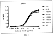
- the humanized anti-CD28 antibody of the present invention has an EC 50 for binding to human CD28 of about 0.008 ⁇ g/mL to about 0.016 ⁇ g/mL, such as about 0.008 ⁇ g/mL to about 0.013 ⁇ g/mL, about 0.008 ⁇ g/mL to about 0.011 ⁇ g/mL, about 0.008 ⁇ g/mL to about 0.010 ⁇ g/mL, or about 0.008 ⁇ g/mL to about 0.009 ⁇ g/mL; further, about 8.00 ng/mL to about 15.50 ng/mL, such as about 8.27 ng/mL to about 15.09 ng/mL, about 8.27ng/mL to about 15.07 ng/mL, about 8.27 ng/mL to about 12.67 ng/mL, about 8.27 ng/mL to about 12.65 ng/mL, about 8.27 ng/mL to about 12.59 ng/mL, about 8.27 ng/
- the humanized anti-CD28 antibody of the present invention has an EC 50 for binding to human CD28 of about 8.27 ng/mL, about 9.14 ng/mL, about 10.40 ng/mL, about 10.90 ng/mL, about 12.11 ng/mL, about 12.54 ng/mL, about 12.59 ng/mL, about 12.65 ng/mL, about 12.67 ng/mL, about 15.07 ng/mL, or about 15.09 ng/mL.
- the EC 50 is determined by ELISA, which may be determining the binding activity of the humanized antibody for human CD28 (e.g., human CD28 containing 6 histidines at the N-terminus), and the concentration of human CD28 used for the measurement of the binding activity may be, e.g., 2 ⁇ g/mL.
- human CD28 e.g., human CD28 containing 6 histidines at the N-terminus
- concentration of human CD28 used for the measurement of the binding activity may be, e.g., 2 ⁇ g/mL.
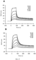
- the humanized anti-CD28 antibody of the present invention has an equilibrium dissociation constant KD for binding to human CD28 of about 6.1 ⁇ 10 -9 M to about 1.2 ⁇ 10 -8 M, such as about 6.1 ⁇ 10 -9 M to about 1.1 ⁇ 10 -8 M, about 6.1 ⁇ 10 -9 M to about 9.6 ⁇ 10 -9 M, about 6.1 ⁇ 10 -9 M to about 9.2 ⁇ 10 -9 M, about 6.1 ⁇ 10 -9 M to about 8.4 ⁇ 10 -9 M, about 6.1 ⁇ 10 -9 M to about 7.5 ⁇ 10 -9 M, about 6.1 ⁇ 10 -9 M to about 6.6 ⁇ 10 -9 M, or about 6.1 ⁇ 10 -9 M to about 6.2 ⁇ 10 -9 M; further, about 6.13 ⁇ 10 -9 M to about 1.15 ⁇ 10 -8 M, about 6.13 ⁇ 10 -9 M to about 1.12 ⁇ 10 -8 M, about 6.13 ⁇ 10 -9 M to about 1.09 ⁇ 10 -8 M, about
- the humanized anti-CD28 antibody of the present invention has an equilibrium dissociation constant KD for binding to human CD28 of about 6.13 ⁇ 10 -9 M, about 6.54 ⁇ 10 -9 M, about 6.59 ⁇ 10 -9 M, about 7.44 ⁇ 10 -9 M, about 8.36 ⁇ 10 -9 M, about 9.13 ⁇ 10 -9 M, about 9.59 ⁇ 10 -9 M, about 1.08 ⁇ 10 -8 M, about 1.09 ⁇ 10 -8 M, about 1.12 ⁇ 10 -8 M, or about 1.15 ⁇ 10 -8 M.
- the equilibrium dissociation constant KD for binding is determined by biolayer interferometry (BLI).
- the three heavy chain CDRs of the murine anti-CD28 antibody may be linked to the four heavy chain FRs of the human antibody (acceptor antibody) for humanization of the antibody heavy chain variable region, and/or the three light chain CDRs of the murine anti-CD28 antibody may be linked to the four light chain FRs of the human antibody for humanization of the antibody light chain variable region.
- the humanized anti-CD28 antibody may comprise a humanized heavy chain variable region and/or a humanized light chain variable region.
- the heavy chain variable region of the murine anti-CD28 antibody comprises an HCDR1 comprising SEQ ID NO: 1, an HCDR2 comprising SEQ ID NO: 2, and an HCDR3 comprising SEQ ID NO: 3.
- the light chain variable region of the murine anti-CD28 antibody comprises an LCDR1 comprising SEQ ID NO: 9, an LCDR2 comprising SEQ ID NO: 10, and an LCDR3 comprising SEQ ID NO: 10.
- the acceptor antibody used in the humanization may be any human germline antibody sequence; a human germline antibody sequence having the highest sequence identity to the heavy chain variable region of the donor antibody may be selected as the acceptor antibody for humanization of the antibody heavy chain variable region, and a human germline antibody sequence having the highest sequence identity to the light chain variable region of the donor antibody may be selected as the acceptor antibody for humanization of the antibody light chain variable region.
- the human germline IGHV4 sequence is used as the receptor antibody for humanization of the heavy chain variable region of the murine anti-CD28 antibody.
- the human germline IGKV1 sequence is used as the receptor antibody for humanization of the light chain variable region of the murine anti-CD28 antibody.
- the humanized anti-CD28 antibody of the present invention may further comprise amino acid substitutions in the CDRs of the murine anti-CD28 antibody.
- the humanized anti-CD28 antibody may comprise amino acid substitutions in the HCDR1, the HCDR2, and/or the HCDR3, and may also comprise amino acid substitutions in the LCDR1, the LCDR2, and/or the LCDR3.
- the HCDR2 comprised in the heavy chain variable region of the humanized anti-CD28 antibody has amino acid substitutions at one or more of positions S12, A12, and M15 relative to SEQ ID NO: 2 (amino acid residue numbering is based on SEQ ID NO:2, with the first amino acid residue of SEQ ID NO: 2 as position 1).
- the HCDR2 comprised in the heavy chain variable region of the humanized anti-CD28 antibody has an amino acid substitution at position M15 relative to SEQ ID NO: 2, and the amino acid substitution is preferably M15K.
- the HCDR2 comprised in the heavy chain variable region of the humanized anti-CD28 antibody has amino acid substitutions at positions S12, A12, and M15 relative to SEQ ID NO: 2, and the amino acid substitutions are preferably S12P, A13S, and M15K.
- the heavy chain variable region of the humanized anti-CD28 antibody comprises an HCDR1 comprising SEQ ID NO: 1, an HCDR2 comprising SEQ ID NO: 17 or 18, and an HCDR3 comprising SEQ ID NO: 3.
- the heavy chain variable region and the light chain variable region of the humanized anti-CD28 antibody comprise: (1) an HCDR1 comprising SEQ ID NO: 1, an HCDR2 comprising SEQ ID NO: 2, an HCDR3 comprising SEQ ID NO: 3, an LCDR1 comprising SEQ ID NO: 9, an LCDR2 comprising SEQ ID NO: 10, and an LCDR3 comprising SEQ ID NO: 10; (2) an HCDR1 comprising SEQ ID NO: 1, an HCDR2 comprising SEQ ID NO: 17, an HCDR3 comprising SEQ ID NO: 3, an LCDR1 comprising SEQ ID NO: 9, an LCDR2 comprising SEQ ID NO: 10, and an LCDR3 comprising SEQ ID NO: 10; or (3) an HCDR1 comprising SEQ ID NO: 1, an HCDR2 comprising SEQ ID NO: 18, an HCDR3 comprising SEQ ID NO: 3, an LCDR1 comprising SEQ ID NO: 9, an LCDR2 comprising SEQ ID NO: 10, and an LCDR3 comprising SEQ ID NO
- the humanized anti-CD28 antibody of the present invention may also comprise a back mutation in the FR regions derived from human germline antibody sequences, that is, some of the amino acid residues in the FR regions are substituted with corresponding residues from the murine antibody to restore or improve the specificity and affinity of the antibody.
- the heavy chain variable region of the humanized anti-CD28 antibody of the present invention comprises an HFR1 comprising a sequence selected from SEQ ID NOs: 19-22, an HFR2 comprising a sequence selected from SEQ ID NOs: 23-26, an HFR3 comprising a sequence selected from SEQ ID NOs: 27-30, and an HFR4 comprising SEQ ID NO: 7.
- the light chain variable region of the humanized anti-CD28 antibody of the present invention comprises an LFR1 comprising a sequence selected from SEQ ID NOs: 36-37, an LFR2 comprising a sequence selected from SEQ ID NOs: 38-39, an LFR3 comprising a sequence selected from SEQ ID NOs: 40-42, and an LFR4 comprising SEQ ID NO: 15.
- the heavy chain variable region of the humanized anti-CD28 antibody of the present invention comprises: (1) an HFR1 comprising SEQ ID NO: 19, an HFR2 comprising SEQ ID NO: 23, an HFR3 comprising SEQ ID NO: 27, and an HFR4 comprising SEQ ID NO: 7; (2) an HFR1 comprising SEQ ID NO: 20, an HFR2 comprising SEQ ID NO: 24, an HFR3 comprising SEQ ID NO: 28, and an HFR4 comprising SEQ ID NO: 7; (3) an HFR1 comprising SEQ ID NO: 21, an HFR2 comprising SEQ ID NO: 25, an HFR3 comprising SEQ ID NO: 29, and an HFR4 comprising SEQ ID NO: 7; (4) an HFR1 comprising SEQ ID NO: 21, an HFR2 comprising SEQ ID NO: 26, an HFR3 comprising SEQ ID NO: 30, and an HFR4 comprising SEQ ID NO: 7; or (5) an HFR1 comprising SEQ ID NO: 22, an HFR2 comprising SEQ ID NO: 22, an H
- the light chain variable region of the humanized anti-CD28 antibody of the present invention comprises: (1) an LFR1 comprising SEQ ID NO: 36, an LFR2 comprising SEQ ID NO: 38, an LFR3 comprising SEQ ID NO: 40, and an LFR4 comprising SEQ ID NO: 15; (2) an LFR1 comprising SEQ ID NO: 37, an LFR2 comprising SEQ ID NO: 39, an LFR3 comprising SEQ ID NO: 41, and an LFR4 comprising SEQ ID NO: 15; or (3) an LFR1 comprising SEQ ID NO: 37, an LFR2 comprising SEQ ID NO: 39, an LFR3 comprising SEQ ID NO: 42, and an LFR4 comprising SEQ ID NO: 15.
- the heavy chain variable region and the light chain variable region of the humanized anti-CD28 antibody of the present invention comprise: (1) an HFR1 comprising SEQ ID NO: 19, an HFR2 comprising SEQ ID NO: 23, an HFR3 comprising SEQ ID NO: 27, an HFR4 comprising SEQ ID NO: 7, an LFR1 comprising SEQ ID NO: 36, an LFR2 comprising SEQ ID NO: 38, an LFR3 comprising SEQ ID NO: 40, and an LFR4 comprising SEQ ID NO: 15; (2) an HFR1 comprising SEQ ID NO: 19, an HFR2 comprising SEQ ID NO: 23, an HFR3 comprising SEQ ID NO: 27, an HFR4 comprising SEQ ID NO: 7, an LFR1 comprising SEQ ID NO: 37, an LFR2 comprising SEQ ID NO: 39, an LFR3 comprising SEQ ID NO: 41, and an LFR4 comprising SEQ ID NO: 15; (3) an HFR1 comprising SEQ ID NO: 20, an HFR2 comprising SEQ ID NO:
- the heavy chain variable region of the humanized anti-CD28 antibody of the present invention comprises a sequence selected from SEQ ID NOs: 31-35. In some embodiments, the light chain variable region of the humanized anti-CD28 antibody of the present invention comprises a sequence selected from SEQ ID NOs: 43-45.
- the heavy chain variable region and the light chain variable region of the humanized anti-CD28 antibody of the present invention are selected from the following combinations: (1) a heavy chain variable region comprising SEQ ID NO: 31 and a light chain variable region comprising SEQ ID NO: 43; (2) a heavy chain variable region comprising SEQ ID NO: 31 and a light chain variable region comprising SEQ ID NO: 44; (3) a heavy chain variable region comprising SEQ ID NO: 32 and a light chain variable region comprising SEQ ID NO: 44; (4) a heavy chain variable region comprising SEQ ID NO: 32 and a light chain variable region comprising SEQ ID NO: 45; (5) a heavy chain variable region comprising SEQ ID NO: 33 and a light chain variable region comprising SEQ ID NO: 43; (6) a heavy chain variable region comprising SEQ ID NO: 33 and a light chain variable region comprising SEQ ID NO: 44; (7) a heavy chain variable region comprising SEQ ID NO: 33 and a light chain variable region comprising SEQ ID NO:
- the humanized anti-CD28 antibody of the present invention may be any antibody form comprising the humanized heavy chain variable region and/or the humanized light chain variable region described above, e.g., a full-length antibody (e.g., IgG), a single-chain antibody, or a single-domain antibody.
- the humanized anti-CD28 antibody of the present invention comprises a constant region of an immunoglobulin (such as IgG, e.g., IgG1, IgG2, IgG3, or IgG4), e.g., a constant region of IgG1, IgG2, IgG3, or IgG4.
- the constant region is a constant region of a human IgG, e.g., a constant region of human IgG1, human IgG2, human IgG3, or human IgG4.
- the humanized heavy chain variable region described above is fused to an immunoglobulin heavy chain constant region (including CH1, CH2, and/or CH3).
- the humanized light chain variable region described above is fused to an immunoglobulin light chain constant region (CL).
- the immunoglobulin heavy chain constant region comprises SEQ ID NO: 81.
- the immunoglobulin light chain constant region comprises SEQ ID NO: 82.
- the humanized anti-CD28 antibody of the present invention is a single-chain antibody (scFv), in which the humanized heavy chain variable region is fused to the humanized light chain variable region via a peptide linker.
- the humanized heavy chain variable region and the humanized light chain variable region may be linked in a different order; for example, the C-terminus of the humanized heavy chain variable region is fused to the N-terminus of the humanized light chain variable region via a peptide linker, or the N-terminus of the humanized heavy chain variable region is fused to the N-terminus of the humanized light chain variable region via a peptide linker.
- Peptide linkers for linking the humanized heavy chain variable region and the humanized light chain variable region are well known to those skilled in the art and may be, e.g., a flexible linker such as a glycine-serine polymer.
- the peptide linker for linking the humanized heavy chain variable region and the humanized light chain variable region is GGGGSGGGGSGGGGS (SEQ ID NO: 80).
- the humanized anti-CD28 antibody of the present invention may also be a single-domain antibody, e.g., a single-domain antibody that comprises a heavy chain variable region but does not comprise a light chain variable region. Methods for preparing single-domain antibodies are well known to those skilled in the art.
- the present invention also relates to an antigen-binding fragment of the humanized anti-CD28 antibody, such as a Fab fragment, a F(ab') 2 fragment, or a Fab' fragment of a full-length antibody.
- an antigen-binding fragment of the humanized anti-CD28 antibody such as a Fab fragment, a F(ab') 2 fragment, or a Fab' fragment of a full-length antibody.
- the humanized anti-CD28 antibody or the binding fragment thereof of the present invention may also be fused, as part of a fusion protein, to other functional polypeptides.
- CD40 is a glycoprotein expressed on the cell surface, belongs to the tumor necrosis factor receptor (TNFR) superfamily, and plays a central role in the immune system. It is expressed on a variety of immune cells, such as B cells, dendritic cells, monocytes, and macrophages.
- TNFR tumor necrosis factor receptor
- CD40 herein refers to any CD40 protein from any vertebrate source, including mammals such as primates (e.g., humans), non-human primates (e.g., rhesus monkeys), and rodents (e.g., mice and rats).
- the present invention provides an affinity-matured anti-CD40 antibody.
- the affinity-matured CD40 antibody has increased binding affinity for CD40 as identified by phage display library.
- Affinity maturation using phage display libraries has been described, e.g., in Lowman et al., Biochemistry 30(45):10832-10838 (1991 ); reference may also be made to Hawkins et al., J. Mol Biol. 254: 889-896 (1992 ) and the examples hereinafter. Without being strictly limited to the following description, this process may be described simply as follows: mutating one or more sites within a predetermined region of a parent antibody to generate possible amino acid substitutions at each site.
- the antibody mutants thus generated are displayed in a monovalent fashion from filamentous phage particles as fusions to the gene III product of M13 packaged within each particle.
- the phage expressing the various mutants may be cycled through rounds of binding selection (e.g., determination of the binding affinity of the mutants for an antigen by ELISA), followed by isolation and sequencing of those mutants which display high affinity.
- binding selection e.g., determination of the binding affinity of the mutants for an antigen by ELISA
- isolation and sequencing of those mutants which display high affinity e.g., determination of the binding affinity of the mutants for an antigen by ELISA
- the anti-CD40 antibody variants of the present invention are identified using the phage display technique, other techniques may also be used to identify anti-CD40 antibody variants with improved binding affinity, including affinity-matured anti-CD40 antibody variants.
- the parent antibody may be, e.g., an anti-CD40 antibody having the following heavy chain variable region and light chain variable region: a heavy chain variable region that comprises an HCDR1 comprising SEQ ID NO: 46, an HCDR2 comprising SEQ ID NO: 47 and an HCDR3 comprising SEQ ID NO: 48 and a light chain variable region that comprises an LCDR1 comprising SEQ ID NO: 54, an LCDR2 comprising SEQ ID NO: 55 and an LCDR3 comprising SEQ ID NO: 56.
- the VH of the parent antibody comprises SEQ ID NO: 53, and/or the VL comprises SEQ ID NO: 61.
- the parent antibody comprises a VH having at least 70%, at least 75%, at least 80%, at least 85%, at least 90%, at least 91%, at least 92%, at least 93%, at least 94%, at least 95%, at least 96%, at least 97%, at least 98%, or at least 99% sequence identity to SEQ ID NO: 53 and/or a VL having at least 70%, at least 75%, at least 80%, at least 85%, at least 90%, at least 91%, at least 92%, at least 93%, at least 94%, at least 95%, at least 96%, at least 97%, at least 98%, or at least 99% sequence identity to SEQ ID NO: 61.
- the parent antibody may be a full-length antibody, e.g., an immunoglobulin (such as IgG, e.g., IgG1, IgG2, IgG3, or IgG4), an scFv or a single-domain antibody, or an antigen-binding fragment thereof, such as a Fab fragment, a F(ab') 2 fragment, or a Fab' fragment.
- an immunoglobulin such as IgG, e.g., Ig., IgG1, IgG2, IgG3, or IgG4
- an scFv or a single-domain antibody or an antigen-binding fragment thereof, such as a Fab fragment, a F(ab') 2 fragment, or a Fab' fragment.
- the affinity-matured anti-CD40 antibody of the present invention has a higher binding affinity for CD40 (e.g., human CD40) compared with the parent antibody. In some embodiments, the affinity-matured anti-CD40 antibody of the present invention has a lower EC 50 value for binding to CD40 (e.g., human CD40) compared with the parent antibody. In some embodiments, the affinity-matured anti-CD40 antibody of the present invention binds to human CD40 with an EC 50 of less than about 0.04919 ⁇ g/mL. In some embodiments, the affinity-matured anti-CD40 antibody of the present invention binds to human CD40 with an EC 50 of less than about 3.44 ⁇ 10 -7 M.
- the EC 50 is determined by ELISA, which may be determining the binding activity of the antibody for human CD40 (e.g., human CD40-His), and the concentration of human CD28 used for the determination of the binding activity may be, e.g., 2 ⁇ g/mL.
- human CD40 e.g., human CD40-His
- concentration of human CD28 used for the determination of the binding activity may be, e.g., 2 ⁇ g/mL.
- the affinity-matured anti-CD40 antibody of the present invention comprises a VH and a VL and has one, two, or more amino acid substitutions in at least one CDR relative to the parent antibody.
- the affinity-matured anti-CD40 antibody of the present invention comprises a VH and a VL and has one, two, or more amino acid substitutions in at least one of the HCDR1, HCDR2, HCDR3, LCDR1, LCDR2, and LCDR3 relative to the parent antibody.
- the VH of the affinity-matured anti-CD40 antibody of the present invention has one, two, or more amino acid substitutions in at least one of the HCDR1, HCDR2, and HCDR3 relative to the parent antibody.
- the VL of the affinity-matured anti-CD40 antibody of the present invention has one, two, or more amino acid substitutions in at least one of the LCDR1, LCDR2, and LCDR3 relative to the parent antibody.
- the VH of the affinity-matured anti-CD40 antibody of the present invention comprises an HCDR1 comprising a sequence selected from SEQ ID NO: 46 and SEQ ID NO: 62, an HCDR2 comprising SEQ ID NO: 47, and an HCDR3 comprising SEQ ID NO: 48.
- the VL of the affinity-matured anti-CD40 antibody of the present invention comprises an LCDR1 comprising SEQ ID NO: 54, an LCDR2 comprising a sequence selected from SEQ ID NOs: 55 and 64-66, and an LCDR3 comprising a sequence selected from SEQ ID NOs: 56 and 67-68.
- the VH of the affinity-matured anti-CD40 antibody of the present invention comprises: (1) an HCDR1 comprising SEQ ID NO: 46, an HCDR2 comprising SEQ ID NO: 47, and an HCDR3 comprising SEQ ID NO: 48; or (2) an HCDR1 comprising SEQ ID NO: 62, an HCDR2 comprising SEQ ID NO: 47, and an HCDR3 comprising SEQ ID NO: 48.
- the VL of the affinity-matured anti-CD40 antibody of the present invention comprises: (1) an LCDR1 comprising SEQ ID NO: 54, an LCDR2 comprising SEQ ID NO: 64, and an LCDR3 comprising SEQ ID NO: 56; (2) an LCDR1 comprising SEQ ID NO: 54, an LCDR2 comprising SEQ ID NO: 55, and an LCDR3 comprising SEQ ID NO: 67; (3) an LCDR1 comprising SEQ ID NO: 54, an LCDR2 comprising SEQ ID NO: 55, and an LCDR3 comprising SEQ ID NO: 68; (4) an LCDR1 comprising SEQ ID NO: 54, an LCDR2 comprising SEQ ID NO: 65, and an LCDR3 comprising SEQ ID NO: 56; (5) an LCDR1 comprising SEQ ID NO: 54, an LCDR2 comprising SEQ ID NO: 66, and an LCDR3 comprising SEQ ID NO: 56.
- the VH and VL of the affinity-matured anti-CD40 antibody of the present invention comprises: (1) an HCDR1 comprising SEQ ID NO: 62, an HCDR2 comprising SEQ ID NO: 47, an HCDR3 comprising SEQ ID NO: 48, an LCDR1 comprising SEQ ID NO: 54, an LCDR2 comprising SEQ ID NO: 64, and an LCDR3 comprising SEQ ID NO: 56; (2) an HCDR1 comprising SEQ ID NO: 62, an HCDR2 comprising SEQ ID NO: 47, an HCDR3 comprising SEQ ID NO: 48, an LCDR1 comprising SEQ ID NO: 54, an LCDR2 comprising SEQ ID NO: 55, and an LCDR3 comprising SEQ ID NO: 67; (3) an HCDR1 comprising SEQ ID NO: 62, an HCDR2 comprising SEQ ID NO: 47, an HCDR3 comprising SEQ ID NO: 48, an LCDR1 comprising SEQ ID NO: 54,
- the affinity-matured anti-CD40 antibody of the present invention does not comprise a VH that comprises an HCDR1 comprising SEQ ID NO: 46, an HCDR2 comprising SEQ ID NO: 47, and an HCDR3 comprising SEQ ID NO: 48.
- the affinity-matured anti-CD40 antibody of the present invention does not comprise a VL that comprises an LCDR1 comprising SEQ ID NO: 54, an LCDR2 comprising SEQ ID NO: 55, and an LCDR3 comprising SEQ ID NO: 56.
- the HFR region and/or LFR region of the affinity-matured anti-CD40 antibody of the present invention are the same as those of the parent antibody.
- the VH of the affinity-matured anti-CD40 antibody of the present invention may comprise an HFR1 comprising SEQ ID NO: 49, an HFR2 comprising SEQ ID NO: 50, an HFR3 comprising SEQ ID NO: 51, and an HFR4 comprising SEQ ID NO: 52.
- the VL of the affinity-matured anti-CD40 antibody of the present invention may comprise an LFR1 comprising SEQ ID NO: 57, an LFR2 comprising SEQ ID NO: 58, an LFR3 comprising SEQ ID NO: 59, and an LFR4 comprising SEQ ID NO: 60.
- the affinity-matured anti-CD40 antibody of the present invention may have one or more amino acid substitutions in the FR region relative to the parent antibody.
- the HFR1 of the affinity-matured anti-CD40 antibody of the present invention has an amino acid substitution at position E10 relative to SEQ ID NO: 49 (amino acid residue numbering is based on SEQ ID NO: 49, with the first amino acid residue of SEQ ID NO: 49 as position 1).
- the HFR1 of the affinity-matured anti-CD40 antibody of the present invention has amino acid substitution E10Q relative to SEQ ID NO: 49.
- the VH of the affinity-matured anti-CD40 antibody of the present invention comprises an HFR1 comprising SEQ ID NO: 63, an HFR2 comprising SEQ ID NO: 50, an HFR3 comprising SEQ ID NO: 51, and an HFR4 comprising SEQ ID NO: 52.
- the VH of the affinity-matured anti-CD40 antibody of the present invention comprises a sequence selected from SEQ ID NOs: 69-73 and/or the VL comprises a sequence selected from SEQ ID NOs: 74-78.
- the affinity-matured anti-CD40 antibody of the present invention comprises a VH having at least 70%, at least 75%, at least 80%, at least 85%, at least 90%, at least 91%, at least 92%, at least 93%, at least 94%, at least 95%, at least 96%, at least 97%, at least 98%, or at least 99% sequence identity to a sequence selected from SEQ ID NOs: 69-73 and/or a VL having at least 70%, at least 75%, at least 80%, at least 85%, at least 90%, at least 91%, at least 92%, at least 93%, at least 94%, at least 95%, at least 96%, at least 97%, at least 98%, or at least 99% sequence identity to
- the affinity-matured anti-CD40 antibody of the present invention is any antibody form comprising the heavy chain variable region and/or the light chain variable region described above, e.g., a full-length antibody (such as IgG, e.g., IgG1, IgG2, IgG3, or IgG4), a single-chain antibody, or a single-domain antibody.
- the affinity-matured anti-CD40 antibody of the present invention comprises a constant region of an immunoglobulin (e.g., an IgG), such as a constant region of IgG1, IgG2, IgG3, or IgG4.
- the constant region is a constant region of a human IgG, e.g., a constant region of human IgG1, human IgG2, human IgG3, or human IgG4.
- the heavy chain variable region described above is fused to an immunoglobulin heavy chain constant region (including CH1, CH2, and/or CH3).
- the light chain variable region described above is fused to an immunoglobulin light chain constant region (CL).
- the affinity-matured anti-CD40 antibody of the present invention is a single-chain antibody (scFv), in which the heavy chain variable region is fused to the light chain variable region via a peptide linker.
- the heavy chain variable region and the light chain variable region may be linked in a different order; for example, the C-terminus of the heavy chain variable region is fused to the N-terminus of the light chain variable region via a peptide linker, or the N-terminus of the heavy chain variable region is fused to the N-terminus of the light chain variable region via a peptide linker.
- Peptide linkers for linking the heavy chain variable region and the light chain variable region are well known to those skilled in the art and may be, e.g., a flexible linker such as a glycine-serine polymer.
- the peptide linker for linking the heavy chain variable region and the light chain variable region is GGGGSGGGGSGGGGS (SEQ ID NO: 80).
- the affinity-matured anti-CD40 antibody of the present invention may also be a single-domain antibody, e.g., a single-domain antibody that comprises a heavy chain variable region but does not comprise a light chain variable region.
- a single-domain antibody that comprises a heavy chain variable region but does not comprise a light chain variable region.
- the present invention also relates to an antigen-binding fragment of the affinity-matured anti-CD40 antibody, such as a Fab fragment, a F(ab') 2 fragment, or a Fab' fragment of a full-length antibody.
- an antigen-binding fragment of the affinity-matured anti-CD40 antibody such as a Fab fragment, a F(ab') 2 fragment, or a Fab' fragment of a full-length antibody.
- the affinity-matured anti-CD40 antibody or the binding fragment thereof of the present invention may also be fused, as part of a fusion protein, to other functional polypeptides.
- the present invention also provides a multispecific antibody, including but not limited to a bispecific antibody, a trispecific antibody, or a tetraspecific antibody.
- the multispecific antibody comprises at least two antigen-binding moieties: a first antigen-binding moiety and a second antigen-binding moiety.
- the first antigen-binding moiety and the second antigen-binding moiety specifically bind to different antigens or different epitopes of the same antigen.
- the first antigen-binding moiety and the second antigen-binding moiety may each independently be a full-length antibody, a single-chain antibody, a single-domain antibody, or an antigen-binding fragment thereof, such as a Fab fragment, a F(ab') 2 fragment, a Fab' fragment, or an Fv fragment of a full-length antibody.
- an antigen-binding fragment thereof such as a Fab fragment, a F(ab') 2 fragment, a Fab' fragment, or an Fv fragment of a full-length antibody.
- the "first” and “second” are for the purpose of distinction only and do not imply a particular order.
- the first antigen-binding moiety is an anti-CD28 antibody, e.g., the murine anti-CD28 antibody or the humanized anti-CD28 antibody described herein, and the second antigen-binding moiety is capable of specifically binding to a second antigen that is different from CD28.
- the second antigen-binding moiety may specifically bind to a tumor antigen or is capable of stimulating an immune cell.
- the second antigen-binding moiety is an anti-CD40 antibody, e.g., the parent anti-CD40 antibody or the affinity-matured anti-CD40 antibody described herein.
- the first antigen-binding moiety is an anti-CD40 antibody, e.g., the parent anti-CD40 antibody or the affinity-matured anti-CD40 antibody described herein, and the second antigen-binding moiety is capable of specifically binding to a second antigen that is different from CD40.
- the second antigen-binding moiety may specifically bind to a tumor antigen or is capable of stimulating an immune cell.
- the second antigen-binding moiety is an anti-CD28 antibody, e.g., the murine anti-CD28 antibody or the humanized anti-CD28 antibody described herein.
- the first antigen-binding moiety may be any anti-CD28 antibody, e.g., the murine anti-CD28 antibody or the humanized anti-CD28 antibody described herein.
- the second antigen-binding moiety may be any anti-CD40 antibody, e.g., the parent anti-CD40 antibody or the affinity-matured anti-CD40 antibody described herein.
- the first antigen-binding moiety and the second antigen-binding moiety of the bispecific antibody are both an scFv and are linked via a peptide linker.
- the first antigen-binding moiety is an anti-CD28 scFv and the second antigen-binding moiety is an anti-CD40 scFv.
- the C-terminus of the first antigen-binding moiety is linked to the N-terminus of the second antigen-binding moiety via a peptide linker.
- the N-terminus of the first antigen-binding moiety is linked to the C-terminus of the second antigen-binding moiety via a peptide linker.
- peptide linkers for linking the first antigen-binding moiety and the second antigen-binding moiety of the bispecific antibody are well known to those skilled in the art and may be, e.g., a flexible linker, such as a glycine-serine polymer.
- the peptide linker for linking the first antigen-binding moiety and the second antigen-binding moiety of the bispecific antibody is GGGGS (SEQ ID NO: 79).
- the present invention also provides a polynucleotide encoding the humanized anti-CD28 antibody or the antigen-binding fragment thereof, the affinity-matured anti-CD40 antibody or the antigen-binding fragment thereof, or the multispecific antibody comprising the anti-CD28 antibody or the antigen-binding fragment thereof and/or the anti-CD40 antibody or the antigen-binding fragment thereof (e.g., a bispecific antibody, which may be called CD28-CD40 diabody for short hereinafter) of the present invention.
- the polynucleotide may be a DNA or an RNA (e.g., an mRNA).
- the present invention also provides a vector comprising the polynucleotide described above, which may be used to express the antibody or the antigen-binding fragment thereof described above in a host cell.
- the vector may be an expression vector, such as a plasmid vector or a viral vector.
- the viral vector may be a retroviral vector, a lentiviral vector, an adenoviral vector or an adeno-associated viral vector.
- the vector may be a circular RNA of the polynucleotide described above, and the circular RNA comprises, in sequence, an internal ribosome entry site (IRES) element, the polynucleotide described above, and a polyA.
- the circular RNA may be used for expressing the antibody described above.
- the IRES may be derived from a virus.
- the IRES sequence is an IRES derived from Coxsackie virus B3 (CVB3) or Coxsackie virus A (CVB 1/2), encephalomyocarditis virus (EMCV), Taura syndrome virus, tripartite virus, Theiler's encephalomyelitis virus, simian virus 40, Solenopsis invicta virus 1, Rhopalosiphum padi virus, reticuloendotheliosis virus, human poliovirus 1, Plautia stali intestine virus, Kashmir bee virus, human rhinovirus 2, Homalodisca coagulata virus-1, human immunodeficiency virus type 1, Himetobi P virus , hepatitis C virus, hepatitis A virus, hepatitis GB virus, foot and mouth disease virus, human enterovirus 71, equine rhinitis virus, Ectropis obliqua picorna-like virus, Drosophila C virus
- the circular RNA of the present invention may be prepared by the general strategies of RNA circularization, such as enzymatic methods using RNA or DNA ligases or ribozymatic methods using self-splicing introns. See Petkovic, S. & Muller, S., "RNA circularization strategies in vivo and in vitro", Nucleic Acids Research, 43(4): 2454-2465 (2015 ); Beadudry, D.
- RNA molecules Nucleic Acids Research, 23(15): 3064-3066 (1995 ); Micura, R., "Cyclic Oligoribonucleotides (RNA) by Solid-Phase Synthesis", Chemistry A European Journal, 5(7): 2077-2082 (1999 ).
- the present invention also provides a precursor RNA for preparing the circular RNA, wherein the circular RNA may be obtained by circularizing the precursor RNA, and the precursor RNA may also be used as a vector comprising the polynucleotide described above.
- the precursor RNA comprises a circularization element, an internal ribosome entry site (IRES) element, the polynucleotide described above, and a polyA.
- the circularization element comprises a first intron sequence located at 5' position of the internal ribosome entry site (IRES) element and a second intron sequence located at 3' position of the polyA.
- the first intron sequence and the second intron sequence are derived from Group I or Group II intron self-splicing sequences.
- the first intron sequence comprises a 3' Group I intron segment containing 3' splice site dinucleotides and the second intron element comprises a 5' Group I intron segment containing 5' splice site dinucleotides.
- the precursor RNA further comprises a 5' spacer sequence between the first intron sequence and the internal ribosome entry site (IRES) element and a 3' spacer sequence between the polyA and the second intron element.
- the precursor RNA further comprises a 5' homology arm outside the first intron element and a 3' homology arm outside the second intron element.
- the vector comprising the polynucleotide encoding the antibody or the antigen-binding fragment described above may also be a vector for producing the precursor RNA described above, and the vector comprises a DNA template for the precursor RNA, which may be transcribed to obtain the precursor RNA.
- the transcription may be in vitro or in a cell.
- polyA used herein is an abbreviation for polyadenylation and refers to a sequence that is consisting of consecutive adenine nucleotides and is at least 30 in length.
- the polyA sequence may be a ribonucleic acid sequence or a deoxyribonucleic acid sequence.
- the length of consecutive adenine nucleotides in a polyA sequence may be at least 30 nucleotides, e.g., at least 45 nucleotides, at least 50 nucleotides, at least 55 nucleotides, at least 60 nucleotides, at least 65 nucleotides, at least 70 nucleotides, at least 75 nucleotides, at least 80 nucleotides, at least 85 nucleotides, at least 90 nucleotides, at least 95 nucleotides, at least 100 nucleotides, at least 105 nucleotides, at least 110 nucleotides, at least 115 nucleotides, at least 120 nucleotides, at least 125 nucleotides, at least 130 nucleotides, at least 135 nucleotides, at least 140 nucleotides, at least 145 nucleotides, at least 150 nucleotides, at least 155 nucleotides, at least 160 nucle
- the length of consecutive adenine nucleotides in the polyA sequence may be 30-240 nucleotides, e.g., 40-230 nucleotides, 45-220 nucleotides, 50-210 nucleotides, 60-200 nucleotides, 70-190 nucleotides, 80-180 nucleotides, 90-170 nucleotides, 100-160 nucleotides, 110-150 nucleotides, or 120-140 nucleotides.
- the polyA sequence may consist of only consecutive adenine nucleotides.
- the polyA is at least 45 nucleotides in length. In some embodiments, the polyA is at least 70 nucleotides in length.
- the present invention also provides a method for producing the circular RNA described above, which comprises circularizing the precursor RNA described above to obtain the circular RNA.
- the method further comprises transcribing the DNA template for the precursor RNA contained in the vector to obtain the precursor RNA.
- the method further comprises purifying the circular RNA, e.g., by interaction between oligo dT and polyA using an oligo dT-based capture method.
- the humanized anti-CD28 antibody or the antigen-binding fragment thereof of the present invention can be used to induce T cell proliferation, activate T cells, and promote the killing effect of immune effector cells (e.g., T cells, such as CAR-T cells).
- T cells such as CAR-T cells.
- the humanized anti-CD28 antibody, the affinity-matured anti-CD40 antibody or the antigen-binding fragment thereof of the present invention, together with an antibody that specifically binds to an antigen on the surface of a tumor cell can form a bispecific antibody, e.g., BiTE (bispecific T-cell engager), or a multispecific antibody to enable targeted activation of T cells of oneself to kill tumor cells.
- BiTE bispecific T-cell engager
- the present invention provides a method for inducing T cell proliferation using the humanized anti-CD28 antibody or the antigen-binding fragment thereof of the present invention or use of the humanized anti-CD28 antibody or the antigen-binding fragment thereof of the present invention in preparing a medicament for inducing T cell proliferation.
- the method comprises contacting the humanized anti-CD28 antibody or the antigen-binding fragment thereof of the present invention, the multispecific antibody comprising the humanized anti-CD28 antibody or the antigen-binding fragment thereof of the present invention, the polynucleotide encoding the humanized anti-CD28 antibody or the antigen-binding fragment thereof of the present invention, the vector comprising the polynucleotide, or the pharmaceutical composition comprising the antibody or the antigen-binding fragment thereof, the multispecific antibody, the polynucleotide, or the vector with a T cell, such that the humanized anti-CD28 antibody or the antigen-binding fragment thereof stimulates T cell activation and induces the proliferation thereof.
- the polynucleotide or vector may be introduced into the T cell and the humanized anti-CD28 antibody or the antigen-binding fragment thereof is thereby expressed in the T cell to stimulate T cell activation and induce the proliferation thereof.
- the polynucleotide may be a DNA or an RNA, e.g., an mRNA.
- the T cell may be a CAR-T cell.
- the humanized anti-CD28 antibody or the antigen-binding fragment thereof of the present invention or the multispecific antibody comprising the humanized anti-CD28 antibody or the antigen-binding fragment thereof of the present invention can be administered to a subject in combination with a CAR-T cell to enhance the killing effect of the CAR-T cell on the target cell.
- the affinity-matured anti-CD40 antibody of the present invention can be used to stimulate immune cells (e.g., dendritic cells, macrophages, NK cells, granulocytes, etc.) and thus can be used to stimulate the immune response, e.g., activating antigen presenting cells (APCs) such as dendritic cells (DCs), stimulating activation and/or proliferation of B cells, or inducing cytotoxicity of effector T cells, and can also be used to treat tumors.
- APCs antigen presenting cells
- DCs dendritic cells
- B cells e.g., cytotoxicity of effector T cells
- the humanized anti-CD28 antibody, the affinity-matured anti-CD40 antibody or the antigen-binding fragment thereof of the present invention together with an antibody that specifically binds to an antigen on the surface of a tumor cell, can form a bispecific antibody or a multispecific antibody to activate cytotoxic T cells to kill tumor cells.
- the present invention provides a method for enhancing an immune response in a subject using the affinity-matured anti-CD40 antibody or the antigen-binding fragment thereof of the present invention or use of the affinity-matured anti-CD40 antibody or the antigen-binding fragment thereof of the present invention in preparing a medicament for enhancing an immune response in a subject.
- the method comprises administering to the subject the affinity-matured anti-CD40 antibody or the antigen-binding fragment thereof of the present invention, the multispecific antibody comprising the affinity-matured anti-CD40 antibody or the antigen-binding fragment thereof of the present invention, the polynucleotide encoding the affinity-matured anti-CD40 antibody or the antigen-binding fragment thereof of the present invention or the vector comprising the polynucleotide, or the pharmaceutical composition comprising the antibody or the antigen-binding fragment thereof, the multispecific antibody, the polynucleotide, or the vector.
- the polynucleotide may be a DNA or an RNA, e.g., an mRNA.
- the term "enhance an immune response” used herein refers to stimulating, eliciting, increasing, modifying, or enhancing any response of the immune system of a subject.
- the immune response may be a cellular response (i.e., a cell-mediated, e.g., cytotoxic T lymphocyte-mediated response) or a humoral response (i.e., an antibody-mediated response) and may be a primary or secondary immune response.
- Exemplary immune response enhancement includes inducing proliferation of T cells (e.g., CD4+ T cells or CD8+ T cells), stimulating activation of T cells (e.g., CD4+ T cells or CD8+ T cells), activating resting T lymphocytes, promoting activation and proliferation of B cells, activating dendritic cells, promoting proliferation and activation of NK cells, increasing killing effect of NK cells, promoting survival of human peripheral blood eosinophils, promoting release of macrophage colony-stimulating factor (GMCSF), increasing survival rate of T cells and/or B cells, improving antigen presentation by antigen presenting cells (e.g., dendritic cells), improving antigen clearance, increasing production of cytokines (e.g., interleukin-2 or IFN-gamma), and the like.
- T cells e.g., CD4+ T cells or CD8+ T cells
- stimulating activation of T cells e.g., CD4+ T cells or CD8+ T cells
- An anti-CD28 antibody e.g., a humanized anti-CD28 antibody of the present invention or antigen-binding fragment thereof, together with an anti-CD40 antibody (e.g., the affinity-matured anti-CD40 antibody of the present invention) or an antigen-binding fragment thereof, can form a multispecific antibody, e.g., a bispecific antibody (which may be called CD28-CD40 diabody for short hereinafter).
- a bispecific antibody which may be called CD28-CD40 diabody for short hereinafter.
- the anti-CD28 antibody or the antigen-binding fragment thereof can be used as the agonist of a CD28 molecule and can provide a costimulatory signal for T cell activation
- the anti-CD40 antibody or the antigen-binding fragment thereof is the agonist of a CD40 molecule and can stimulate various immune cells (such as dendritic cells, macrophages, NK cells, granulocytes, etc.) to activate and play a role
- the CD28-CD40 diabody can play an important role in immune response, e.g., enhancing the immune response of a subject, improving the effect of cellular immunotherapy, and particularly promoting the killing effect of CAR-T cells.
- cellular immunotherapy refers to the treatment of a disease, such as a tumor, an autoimmune disease, or an infectious disease, using cells contained in the immune system that are capable of resisting diseases or capable of helping the immune system resist diseases.
- Cells that can be used for cellular immunotherapy include, but are not limited to, T cells, NK cells, dendritic cells, regulatory T cells, cytotoxic T Cells (CTLs), macrophages, and the like.
- CTLs cytotoxic T Cells
- autoimmune cells can be separated from a subject's blood or tumor, and then these cells are cultured in vitro before being transfused into the subject to kill the tumor cells.
- the immune cells can be genetically engineered to express a tumor-specific receptor and then cultured and transfused back to the patient, e.g., CAR-T cells.
- the present invention provides a pharmaceutical combination of the CD28-CD40 diabody of the present invention and an immune cell, which can be used to kill a target cell in a subject.
- the CD28-CD40 diabody of the present invention can enhance the killing effect of the immune cells on a target cell.
- the present invention also provides a method for enhancing the killing effect of an immune cell on a target cell in a subject, which comprises administering to a subject receiving the immune cell the CD28-CD40 diabody of the present invention, the polynucleotide encoding the diabody, the vector comprising the polynucleotide, or the pharmaceutical composition comprising the diabody, the polynucleotide, or the vector.
- the polynucleotide encoding the CD28-CD40 diabody of the present invention or the vector comprising the polynucleotide may be introduced into an immune cell, so that the immune cell can express the diabody.
- the polynucleotide may be a DNA or an RNA, e.g., an mRNA.
- the vector may be a plasmid vector or a viral vector, such as a lentiviral vector, an adenoviral vector, an adeno-associated viral vector, a retroviral vector, and the like.
- the vector may also be a circular RNA described above, a precursor RNA, or a vector comprising a DNA template for the precursor RNA.
- the "introducing” may be understood as "transfecting".
- the polynucleotide or vector may be transfected into an immune cell by, e.g., electroporation, calcium phosphate precipitation, DEAE-dextran transfection, virus-like particle infection, and other known manners.
- the immune cell capable of expressing the CD28-CD40 diabody of the present invention can be administered to a subject to kill target cells in the subject.
- the present invention also provides an immune cell capable of expressing the CD28-CD40 diabody of the present invention.
- immune cell refers to immune cells used in cellular immunotherapy and having a therapeutic effect, e.g., immune cells that can kill target cells in cellular immunotherapy, such as T cells, NK cells, dendritic cells, regulatory T cells, cytotoxic T Cells (CTLs), macrophages, and the like.
- target cell refers to a target cell to which the cellular immunotherapy is directed.
- the immune cell comprises a CAR or can express a CAR.
- the immune cell is a CAR-T cell.
- the CAR-T cell comprises a binding domain which can specifically bind to the antigen on the surface of a target cell, and thus can specifically kill the target cell.
- the method of the present invention can enhance the killing effect of the immune cell on the target cell compared with administration of the immune cell alone.
- the killing effect can be determined, e.g., by a reduction in the number of target cells.
- the target cell is a tumor cell.
- the present invention also provides use of the CD28-CD40 diabody of the present invention, the polynucleotide encoding the diabody, and the vector comprising the polynucleotide in preparing a medicament for enhancing the killing effect of an immune cell on a target cell in a subject.
- the present invention also provides a genetically engineered immune effector cell capable of expressing the CD28-CD40 diabody of the present invention, and the immune effector cell may be a T cell, an NK cell, an NKT cell, a macrophage, a neutrophil, or a granulocyte.
- the immune effector cell may comprise a polynucleotide encoding the CD28-CD40 diabody of the present invention or a vector comprising the polynucleotide.
- the genetically engineered immune effector cell further recombinantly expresses a chimeric antigen receptor (CAR), a T cell receptor (TCR), or a bispecific T cell engager (BiTE), wherein the CAR, TCR, or BiTE may be a CAR, a TCR, or a BiTE that binds to a tumor antigen or a viral antigen.
- CAR chimeric antigen receptor
- TCR T cell receptor
- BiTE bispecific T cell engager
- the viral antigen may be selected from HPV, EBV, and HIV
- the tumor antigen may be selected from Her2, NY-ESO-1, CD19, CD20, CD22, PSMA, c-Met, GPC3, IL13ra2, EGFR, CD123, CD7, GD2, PSCA, EBV16-E7, H3.3, EGFRvIII, BCMA, and mesothelin.
- the CAR, the TCR, or the BiTE binds to mesothelin (MESO), CD123, BCMA, HER2, IL13Ra2, B7H3, or CD19.
- the CAR, TCR, or BiTE may bind to TSHR, CD19; CD123; CD22; CD30; CD171; CS-1; C-type lectin-like molecule-1, CD33; epidermal growth factor receptor variant III (EGFRvIII); ganglioside G2 (GD2); ganglioside GD3; TNF receptor family members; B cell maturation antigen (BCMA); Tn antigen ((Tn Ag) or (GalNAca-Ser/Thr)); prostate-specific membrane antigen (PSMA); receptor tyrosine kinase-like orphan receptor 1 (ROR1); Fms-like tyrosine kinase 3 (FLT3); tumor-associated glycoprotein 72 (TAG72); CD38; CD44v6; carcinoembryonic antigen (CEA); epithelial cell adhesion molecule (EPCAM); B7H3 (CD276); KIT (CD117); interleukin-13 receptor subunit ⁇ -2
- the genetically engineered immune effector cell comprises a first polynucleotide encoding the CD40-CD28 diabody of the present invention and a second polynucleotide encoding the CAR, TCR, or BiTE (CAR/TCR/BiTE).
- the first polynucleotide and the second polynucleotide may be separately expressed in separate expression cassettes or may be co-expressed in the same expression cassette. When co-expressed in the same expression cassette, the first polynucleotide and the second polynucleotide may have a linker between them.
- the linker may be a self-cleaving linker, e.g., 2A linkers, such as T2A, P2A, or F2A.
- the polynucleotide encoding the CD28-CD40 diabody of the present invention or a vector comprising the polynucleotide may be introduced into an immune cell, so that the immune cell can express the diabody.
- the first polynucleotide and the second polynucleotide may be DNA or RNA, e.g., mRNA.
- the first polynucleotide and the second polynucleotide may be introduced into the immune effector cell by any means known in the art, for example, they may be introduced into the immune effector cell by transfecting the immune effector cell with a vector or mRNA.
- the vector may be a plasmid vector or a viral vector, such as a lentiviral vector, an adenoviral vector, an adeno-associated viral vector, a retroviral vector, and the like.
- the vector may also be a circular RNA described above, a precursor RNA, or a vector comprising a DNA template for the precursor RNA.
- the CAR-T cell comprises a CAR with a HER2-binding molecule (such as an anti-HER2 antibody) as a binding domain and targets a cell expressing HER2.
- the CAR-T cell comprises a CAR with a mesothelin-binding molecule (e.g., an anti-mesothelin antibody) as a binding domain and targets a cell expressing mesothelin.
- the target cell of the CAR-T cell is a tumor cell expressing HER2 or mesothelin.
- the CAR may comprise a signal peptide, a hinge region, a transmembrane domain, and an intracellular signaling domain.
- the intracellular signaling domain may further comprise a costimulatory domain.
- the signal peptide may include a CD8 signal peptide or a GM-CSF signal peptide.
- the hinge region of the CAR may include a hinge region of CD28, CD8, IgG1, IgG4, IgD, 4-1BB, CD4, CD27, CD7, CD8A, PD-1, ICOS, OX40, NKG2D, NKG2C, Fc ⁇ RI ⁇ , BTLA, GITR, DAP10, TIM1, SLAM, CD30, or LIGHT, preferably a CD8 hinge region.
- the transmembrane domain of the CAR may include a transmembrane domain of CD8, CD28, CD3 ⁇ (CD3e), 4-1BB, CD4, CD27, CD7, PD-1, TRAC, TRBC, CD3 ⁇ , CTLA-4, LAG-3, CD5, ICOS, OX40, NKG2D, 2B4, CD244, Fc ⁇ RI ⁇ , BTLA, CD30, GITR, HVEM, DAP10, CD2, NKG2C, LIGHT, DAP12, CD40L (CD154), TIM1, CD226, DR3, CD45, CD80, CD86, CD9, CD16, CD22, CD33, CD37, CD64, or SLAM, preferably a CD8 transmembrane (TM) domain.
- CD8 transmembrane (TM) domain preferably a CD8 transmembrane (TM) domain.
- the intracellular signaling domain of the CAR may include an intracellular signaling domain of CD3 ⁇ , CD3 ⁇ , CD3 ⁇ , CD3 ⁇ , CD79a, CD79b, FceRI ⁇ , FceRI ⁇ , Fc ⁇ RIIa, DAP10, or DAP-12, preferably a CD3 ⁇ intracellular signaling domain.
- the intracellular signaling domain of the CAR may further comprise a costimulatory domain, such as a costimulatory domain of CD28, 4-1BB (CD137), CD27, CD2, CD7, CD8A, CD8B, OX40, CD226, DR3, SLAM, CDS, ICAM-1, NKG2D, NKG2C, B7-H3, 2B4, Fc ⁇ RI ⁇ , BTLA, GITR, HVEM, DAP10, DAP12, CD30, CD40, CD40L, TIM1, PD-1, LFA-1, LIGHT, JAML, CD244, CD100, ICOS, CD40, or MyD88, preferably a costimulatory domain of 4-1BB.
- the CAR having a "BBZ" structure refers to a CAR having a 4-1BB costimulatory molecule, generally comprising a CD8 hinge domain, a CD8 transmembrane (TM) domain, a 4-1BB costimulatory domain, and a CD3 ⁇ domain.
- the CAR used in the present invention has a BBZ structure.
- the CAR comprises an antibody or an antigen-binding fragment thereof as a binding domain.
- the antibody or the antigen-binding fragment specifically binds to mesothelin (MESO), CD123, BCMA, HER2, IL13Ra2, or B7H3.
- the antibody is an scFv.
- the scFv comprises a heavy chain variable region (VH) fused to the N-terminus or C-terminus of a light chain variable region (VL).
- VH heavy chain variable region
- VL light chain variable region
- the antibody that specifically binds to mesothelin also referred to as an anti-mesothelin (or anti-MESO or anti-MSLN) antibody, as described in PCT/CN2021/112767 (incorporated herein by reference in its entirety), comprises a light chain variable region comprising an LCDR1, an LCDR2 and an LCDR3 and a heavy chain variable region comprising an HCDR1, an HCDR2 and an HCDR3, wherein the LCDR1, the LCDR2, the LCDR3, the HCDR1, the HCDR2, and the HCDR3 are selected from the group consisting of:
- the anti-MESO antibody comprises a light chain variable region and a heavy chain variable region selected from the group consisting of:
- the anti-MESO antibody is an anti-MESO scFv and may comprise an amino acid sequence selected from SEQ ID NOs: 202-216.
- the CAR comprises a binding domain that may specifically bind to mesothelin, which may be referred to as a CAR targeting mesothelin.
- the CAR targeting mesothelin may be a CAR described in PCT/CN2021/112767 (incorporated herein by reference in its entirety).
- the CAR targeting mesothelin may comprise any one of the anti-mesothelin antibodies described above.
- the CAR targeting mesothelin may comprise an amino acid sequence selected from SEQ ID NOs: 217-231.
- the antibody that specifically binds to CD123 is an antibody described in PCT/CN2021/112748 (incorporated herein by reference in its entirety), and comprises a light chain variable region comprising an LCDR1, an LCDR2 and an LCDR3 and a heavy chain variable region comprising an HCDR1, an HCDR2 and an HCDR3, wherein the LCDR1, the LCDR2, the LCDR3, the HCDR1, the HCDR2, and the HCDR3 are selected from the group consisting of:
- the anti-CD123 antibody comprises a light chain variable region and a heavy chain variable region selected from the group consisting of:
- the anti-CD123 antibody is an anti-CD123 scFv and may comprise any amino acid sequence selected from SEQ ID NOs: 583-617.
- the CAR comprises a binding domain that may specifically bind to CD123, which may be referred to as a CAR targeting CD123.
- the CAR targeting CD123 may be a CAR described in PCT/CN2021/112748 (incorporated herein by reference in its entirety).
- the CAR targeting CD123 may comprise any one of the anti-CD123 antibodies described above.
- the CAR targeting CD123 may comprise an amino acid sequence selected from SEQ ID NOs: 580-582.
- the antibody that specifically binds to BCMA also referred to as an anti-BCMA antibody, as described in PCT/CN2021/112798 (incorporated herein by reference in its entirety), comprises a light chain variable region comprising an LCDR1, an LCDR2 and an LCDR3 and a heavy chain variable region comprising an HCDR1, an HCDR2 and an HCDR3, wherein the LCDR1, the LCDR2, the LCDR3, the HCDR1, the HCDR2, and the HCDR3 are selected from the group consisting of:
- the anti-BCMA antibody comprises a light chain variable region and a heavy chain variable region selected from the group consisting of:
- the anti-BCMA antibody is an anti-BCMA scFv and may comprise an amino acid sequence selected from SEQ ID NOs: 323-334.
- the CAR comprises a binding domain that may specifically bind to BCMA, which may be referred to as a CAR targeting BCMA.
- the CAR targeting BCMA may be a CAR described in PCT/CN2021/112798 (incorporated herein by reference in its entirety).
- the CAR targeting BCMA may comprise any one of the anti-BCMA antibodies described above.
- the CAR targeting BCMA may comprise an amino acid sequence selected from SEQ ID NOs: 335-346.
- the antibody that specifically binds to CD19 also referred to as an anti-CD19 antibody, as described in the Chinese patent application CN202210274255.1 (publication No. CN114349863A , which is incorporated herein by reference in its entirety), comprises a light chain variable region comprising an LCDR1, an LCDR2 and an LCDR3 and a heavy chain variable region comprising an HCDR1, an HCDR2 and an HCDR3, wherein the LCDR1, the LCDR2, the LCDR3, the HCDR1, the HCDR2, and the HCDR3 may be an LCDR1 set forth in SEQ ID NO: 628, an LCDR2 set forth in SEQ ID NO: 629, an LCDR3 set forth in SEQ ID NO: 630, an HCDR1 set forth in SEQ ID NO: 631, an HCDR2 set forth in SEQ ID NO: 632, and an HCDR3 set forth in SEQ ID NO: 633.
- the anti-CD19 antibody comprises a light chain variable region set forth in SEQ ID NO: 635 and a heavy chain variable region set forth in SEQ ID NO: 634.
- the anti-CD19 antibody is an anti-CD19 scFv and may comprise an amino acid sequence set forth in SEQ ID NO: 636.
- the CAR comprises a binding domain that may specifically bind to CD19, which may be referred to as a CAR targeting CD19.
- the CAR targeting CD19 may be a CAR described in the Chinese patent application CN202210274255.1 (publication No. CN114349863A , which is incorporated herein by reference in its entirety).
- the CAR targeting CD19 may comprise any one of the anti-CD19 antibodies described above.
- the CAR targeting CD19 may comprise an amino acid sequence set forth in SEQ ID NO: 637.
- the antibody that specifically binds to HER2 also referred to as an anti-HER2 antibody, as described in the Chinese patent application CN202210750853.1 (publication No. CN114805584A , which is incorporated herein by reference in its entirety), comprises a light chain variable region comprising an LCDR1, an LCDR2 and an LCDR3 and a heavy chain variable region comprising an HCDR1, an HCDR2 and an HCDR3, wherein the LCDR1, the LCDR2, the LCDR3, the HCDR1, the HCDR2, and the HCDR3 may be an LCDR1 set forth in SEQ ID NO: 618, an LCDR2 set forth in SEQ ID NO: 619, an LCDR3 set forth in SEQ ID NO: 620, an HCDR1 set forth in SEQ ID NO: 621, an HCDR2 set forth in SEQ ID NO: 622, and an HCDR3 set forth in SEQ ID NO: 623.
- the anti-HER2 antibody comprises a light chain variable region set forth in SEQ ID NO: 624 and a heavy chain variable region set forth in SEQ ID NO: 625.
- the anti-HER2 antibody is an anti-HER2 scFv and may comprise an amino acid sequence set forth in SEQ ID NO: 626.
- the CAR comprises a binding domain that may specifically bind to HER2, which may be referred to as a CAR targeting HER2.
- the CAR targeting HER2 may be a CAR described in the Chinese patent application CN202210750853.1 (publication No. CN114805584A , which is incorporated herein by reference in its entirety).
- the CAR targeting HER2 may comprise any one of the anti-HER2 antibodies described above.
- the CAR targeting HER2 may comprise an amino acid sequence set forth in SEQ ID NO: 627.
- the antibody that specifically binds to IL13Ra2 also referred to as an anti-IL13Ra2 antibody, as described in the Chinese patent application CN202210743595.4 (publication No. CN114805581A , which is incorporated herein by reference in its entirety), comprises a light chain variable region comprising an LCDR1, an LCDR2 and an LCDR3 and a heavy chain variable region comprising an HCDR1, an HCDR2 and an HCDR3, wherein the LCDR1, the LCDR2, the LCDR3, the HCDR1, the HCDR2, and the HCDR3 may be an LCDR1 set forth in SEQ ID NO: 638, an LCDR2 set forth in SEQ ID NO: 639, an LCDR3 set forth in SEQ ID NO: 640, an HCDR1 set forth in SEQ ID NO: 641, an HCDR2 set forth in SEQ ID NO: 642, and an HCDR3 set forth in SEQ ID NO: 642.
- the anti-IL13Ra2 antibody comprises a light chain variable region set forth in SEQ ID NO: 644 and a heavy chain variable region set forth in SEQ ID NO: 645.
- the anti-IL13Ra2 antibody is an anti-IL13Ra2 scFv and may comprise an amino acid sequence set forth in SEQ ID NO: 646.
- the CAR comprises a binding domain that may specifically bind to IL13Ra2, which may be referred to as a CAR targeting IL13Ra2.
- the CAR targeting IL13Ra2 may be a CAR described in the Chinese patent application CN202210743595.4 (publication No. CN114805581A , which is incorporated herein by reference in its entirety).
- the CAR targeting IL13Ra2 may comprise any one of the anti-IL13Ra2 antibodies described above.
- the CAR targeting IL13Ra2 may comprise an amino acid sequence set forth in SEQ ID NO: 647.
- the antibody that specifically binds to B7H3, also referred to as an anti-B7H3 antibody comprises a light chain variable region comprising an LCDR1, an LCDR2 and an LCDR3 and a heavy chain variable region comprising an HCDR1, an HCDR2 and an HCDR3, wherein the LCDR1, the LCDR2, the LCDR3, the HCDR1, the HCDR2, and the HCDR3 may be an LCDR1 set forth in SEQ ID NO: 648, an LCDR2 set forth in SEQ ID NO: 649, an LCDR3 set forth in SEQ ID NO: 650, an HCDR1 set forth in SEQ ID NO: 651, an HCDR2 set forth in SEQ ID NO: 652, and an HCDR3 set forth in SEQ ID NO: 653.
- the anti-B7H3 antibody comprises a light chain variable region set forth in SEQ ID NO: 654 and a heavy chain variable region set forth in SEQ ID NO: 655.
- the anti-B7H3 antibody is an anti-B7H3 scFv and may comprise an amino acid sequence set forth in SEQ ID NO: 656.
- the CAR comprises a binding domain that may specifically bind to B7H3, which may be referred to as a CAR targeting B7H3.
- the CAR targeting B7H3 may be a CAR described in the Chinese patent application CN202210714289.8 (publication No. CN114773477A , which is incorporated herein by reference in its entirety).
- the CAR targeting B7H3 may comprise any one of the anti-B7H3 antibodies described above.
- the CAR targeting B7H3 may comprise an amino acid sequence set forth in SEQ ID NO: 657.
- the humanized anti-CD28 antibody and the affinity-matured anti-CD40 antibody or the antigen-binding fragment thereof, the multispecific antibody comprising them, the CD28-CD40 diabody, the polynucleotide encoding them, the vector comprising the polynucleotide, or the immune effector cell expressing the CD28-CD40 diabody of the present invention may be used as a medicament for treating a tumor, a cancer, a viral infection, an autoimmune disease, or the like, which may be formulated into a pharmaceutical composition.
- the humanized anti-CD28 antibody and the affinity-matured anti-CD40 antibody or the antigen-binding fragment thereof, the multispecific antibody comprising them, the CD28-CD40 diabody, the polynucleotide encoding them, or the vector comprising the polynucleotide may be used in combination with an immune effector cell.
- the immune effector cell may be selected from a CAR T cell, a TCR T cell, TIL, CIK, LAK, and MIL.
- the present invention provides a method for treating a disease, comprising administering to a subject in need of treatment the humanized anti-CD28 antibody and the affinity-matured anti-CD40 antibody or the antigen-binding fragment thereof, the multispecific antibody comprising them, the CD28-CD40 diabody, the polynucleotide encoding them, the vector comprising the polynucleotide, or the immune effector cell expressing the CD28-CD40 diabody.
- the disease may be a tumor, a cancer, a viral infection, or an autoimmune disease.
- the treatment method may comprise: (a) administering to a subject in need of treatment the humanized anti-CD28 antibody and the affinity-matured anti-CD40 antibody or the antigen-binding fragment thereof, the multispecific antibody comprising them, the CD28-CD40 diabody, the polynucleotide encoding them, or the vector comprising the polynucleotide, and (b) administering to the subject immune cell therapy.
- Steps (a) and (b) may be performed simultaneously or sequentially, wherein step (a) may be performed before or after step (b).
- the immune cell therapy in step (b) may include CAR T therapy, TCR T therapy, TIL therapy, CIK therapy, LAK therapy, and MIL therapy.
- the cancer is a solid tumor or a hematologic cancer (e.g., leukemia).
- the cancer is acute myeloid leukemia (AML), B-type acute lymphocytic leukemia (B-ALL), T-type acute lymphocytic leukemia (T-ALL), B-cell precursor acute lymphocytic leukemia (BCP-ALL), or blastic placytoid dendritic cell neoplasm (BPDCN).
- the disease e.g., cancer
- the disease is characterized by disease cells expressing mesothelin, CD123, BCMA, HER2, CD19, IL13Ra2, and/or B7H3.
- the cancer is a cancer expressing CD123. In some embodiments, the cancer is AML expressing CD123. In some embodiments, the cancer is mesothelioma. In some embodiments, the mesothelioma is pleural mesothelioma, peritoneal mesothelioma, or pericardial mesothelioma. In some embodiments, the cancer is pancreatic cancer. In some embodiments, the pancreatic cancer is pancreatic ductal carcinoma. In some embodiments, the cancer is ovarian cancer. In some embodiments, the cancer is epithelial ovarian cancer. In some embodiments, the cancer is lung cancer.
- the cancer is non-Hodgkin's lymphoma, chronic lymphocytic leukemia, acute lymphocytic leukemia, human B-cell precursor leukemia, multiple myeloma, or malignant lymphoma.
- the cancer is breast cancer, gastric cancer, ovarian cancer, cervical cancer, urothelial cancer, esophageal cancer, bladder cancer, colorectal cancer, endometrial cancer, renal cancer, lung cancer, pancreatic cancer, head and neck cancer, sarcoma, glioblastoma, prostate cancer, or thyroid cancer.
- the cancer is glioma or head and neck cancer.
- subject refers to any organism to which the antibody or the antigen-binding fragment thereof of the present invention may be administered, e.g., for experimental, diagnostic, prophylactic, and/or therapeutic purposes.
- Typical subjects include animals (e.g., mammals such as mice, rats, rabbits, non-human primates (e.g., chimpanzees and other apes and monkey species), and humans).
- the subject may be a mammal, particularly a human, including a female or a male, and including a neonate, an infant, a juvenile, a youth, an adult, or an elderly adult, and further including various races and ethnicities.
- a subject refers to an individual in need of diagnosis, treatment, or prevention of a disease or disorder, and the subject may have the disease or disorder, or be at risk of having the disease or disorder.
- treat refers to providing a beneficial or desired clinical outcome to a disease, such as eliminating the disease, alleviating symptoms, reducing the extent of the disease, stabilizing, ameliorating or relieving the state of the disease, or slowing the progression of the disease.
- the measurement of the treatment outcome may be based, for example, on the results of a physical examination, a pathological test, and/or a diagnostic test known in the art.
- the treatment may also refer to prolonging survival as compared to the expected survival of a subject if not receiving treatment.
- the treatment may also refer to reducing the incidence or morbidity of the disease, or the recurrence thereof, as compared to a disease that would occur in the absence of this measure.
- Clinically, such treatment may also be referred to as prophylaxis.
- the terms “pharmaceutical composition” and “pharmaceutical formulation” of the present invention are used interchangeably.
- the pharmaceutical composition may comprise a pharmaceutically acceptable carrier.
- pharmaceutically acceptable carrier used herein refers to any carrier comprised in a pharmaceutical composition as an inactive ingredient, which enables the pharmaceutical composition to have a suitable appearance and properties for administration.
- the pharmaceutically acceptable carrier such as a stabilizer, a diluent, an additive, an auxiliary agent, an excipient, and the like, has substantially no long-term or permanent adverse effects when administered to a subject.
- “Pharmaceutically acceptable carrier” should be a pharmaceutically inert material, which is substantially free of biological activity and constitutes an integral part of the formulation.
- the pharmaceutical composition, the pharmaceutical combination, and the immune cell (including CAR-T cell) of the present invention may be formulated for various modes of administration according to known techniques. See, for example, Remington, The Science and Practice of Pharmacy (9th Ed. 1995 ).
- an active agent is generally mixed with a pharmaceutically acceptable carrier and the like.
- the pharmaceutically acceptable carrier must be acceptable, i.e., it is compatible with any other ingredients in the formula and must not be deleterious to a subject.
- Pharmaceutically acceptable carriers may include, but are not limited to, buffers, excipients, stabilizers, preservatives, wetting agents, surfactants, emulsifiers, or combinations thereof.
- buffers include, but are not limited to, acetic acid, citric acid, histidine, boric acid, formic acid, succinic acid, phosphoric acid, carbonic acid, malic acid, aspartic acid, Tris buffer, HEPPSO, HEPES, neutral buffered saline, phosphate-buffered saline, and the like.
- the pharmaceutical composition, the pharmaceutical combination, and the immune cell (including CAR-T cell) of the present invention may be administered in any mode suitable for diseases to be treated (or prevented) and subjects.
- the mode of administration may include, but is not limited to, parenteral or non-parenteral routes, including oral, sublingual, buccal, transdermal, rectal, vaginal, intradermal, intranasal routes, or parenteral routes, such as intravenous injection (i.v.), intraperitoneal, intradermal, subcutaneous, intramuscular, intracranial, intrathecal, intratumoral, transdermal, transmucosal, intra-articular, intrathecal, intrathecal, intrahepatic, intraneural, or intracranial injection or infusion.
- the pharmaceutical composition may be directly injected into tumors, lymph nodes, tissues, organs, or sites of infection.
- Dosage forms suitable for oral administration include, but are not limited to, tablets, capsules, powders, pills, granules, suspensions, solutions or pre-concentrates of solutions, and emulsions or pre-concentrates of emulsions.
- Pharmaceutically acceptable carriers that may be used in oral dosage forms include water, ethylene glycol, oils, alcohols, flavoring agents, preservatives, colorants, and the like. Carriers such as starches, sugars, microcrystalline cellulose, diluents, fillers, lubricants, granulating agents, lubricants, binders, stabilizers, disintegrants, and the like may be used to prepare oral solid formulations such as powders, capsules, or tablets.
- Dosage forms suitable for parenteral administration include, but are not limited to, sterile liquid formulations, such as isotonic aqueous solutions, emulsions, suspensions, dispersions, or viscous compositions, which may be buffered to reach a desired pH value.
- the parenteral dosage form may be ready-to-use, or may be a dry product ready to be dissolved or suspended in a pharmaceutically acceptable carrier.
- the parenteral dosage form may be a sterile formula, or may be sterilized before administration to a subject.
- Pharmaceutically acceptable carriers that may be used to provide parenteral dosage forms include, but are not limited to, water for injection; aqueous carriers, such as, but not limited to, sodium chloride injection, Ringer's injection, and glucose injection; water-soluble carriers, such as, but not limited to, ethanol, polyethylene glycol, and polypropylene glycol; non-aqueous carriers, such as, but not limited to, corn oil, cottonseed oil, peanut oil, sesame oil, ethyl oleate, isopropyl myristate, and benzyl benzoate; and solubilizers, such as cyclodextrin.
- aqueous carriers such as, but not limited to, sodium chloride injection, Ringer's injection, and glucose injection
- water-soluble carriers such as, but not limited to, ethanol, polyethylene glycol, and polypropylene glycol
- non-aqueous carriers such as, but not limited to, corn oil, cottonseed oil, peanut oil, sesame oil, e
- the pharmaceutical composition, the pharmaceutical combination, and the immune cell (including CAR-T cell) of the present invention are administered to a subject in a therapeutically effective amount.
- therapeutically effective amount and “effective amount” used herein are used interchangeably, and refer to an amount that is effective, at dosages and for periods of time necessary, to achieve the desired therapeutic effect.
- the therapeutically effective amount may vary depending on various factors, such as the disease state, age, sex, and weight of the individual, and the ability of a treatment method or a combination of treatment methods to elicit a desired response in the individual.
- the effective amount may refer to an amount that causes a detectable change in a biological or chemical activity. The detectable change may be detected and/or further quantified by those skilled in the relevant art.
- the "effective amount” may specify an amount that maintains a desired physiological state, i.e., reduces or prevents a significant decline and/or promotes improvement in the disease condition.
- the amount and frequency of administration will be determined by factors such as the condition of a subject (e.g., age, weight, sex, and response of the subject to the drug), as well as the type and severity of the subject's disease, although appropriate dosages may be determined by clinical trials.
- the pharmaceutical composition, the pharmaceutical combination, and the immune cell (including CAR-T cell) of the present invention may be administered to a subject in a therapeutically effective amount of about 0.5 to about 250 mg/kg, e.g., about 1 to about 250 mg/kg, about 2 to about 200 mg/kg, about 3 to about 120 mg/kg, about 5 to about 250 mg/kg, about 10 to about 200 mg/kg, or about 20 to about 120 mg/kg.
- an effective amount of immune cells may be, for example, 5 ⁇ 10 6 cells, 1 ⁇ 10 7 cells, 2 ⁇ 10 7 cells, 5 ⁇ 10 7 cells, 1 ⁇ 10 8 cells, 2 ⁇ 10 8 cells, 5 ⁇ 10 8 cells, 1 ⁇ 10 9 cells, 2 ⁇ 10 9 cells, or 5 ⁇ 10 9 cells administered in a single dose.
- the pharmaceutical composition, the pharmaceutical combination, and the immune cell (including CAR-T cell) of the present invention may be administered once or twice a day; or once every 2, 3, 4, 5, 6, 7, 8, 9, or 10 days, once every 1, 2, 3, 4, 5, or 6 weeks, or once every 1, 2, 3, 4, 5, or 6 months or longer.
- the pharmaceutical composition may also be administered on a regimen of several times (e.g., 1, 2, 3, 4, or 5 times) a week or several times (e.g., 1, 2, 3, 4, 5, 6, 7, 8, 9, or 10 times) a month.
- the pharmaceutical composition may be administered once a day for 5 consecutive days, followed by a rest for two consecutive days.
- composition refers to a combination of two or more drugs administered to a subject over a period of time (e.g., simultaneously, or several minutes to several hours apart, or even several days or several weeks apart).
- the two or more drugs may be formulated into the same pharmaceutical formulation, or may be formulated into different pharmaceutical formulations, respectively.
- the pharmaceutical composition, the pharmaceutical combination, and the immune cell (including CAR-T cell) of the present invention may be used in combination with other drugs and treatment methods, such as other anti-tumor drugs, chemotherapy, or radiation therapy.
- used in combination refers to the administration of two (or more) different drugs and/or therapies to a subject during treatment.
- the two or more drugs and/or therapies in the combination may be administered by different routes and regimens.
- the two or more drugs and/or treatment methods may be administered to the subject simultaneously or sequentially.
- administration of a second drug or therapy begins, administration of one drug or therapy is still ongoing, thus there is an overlap in the administration.
- Such a regimen may be referred to herein as "simultaneously”.
- the two or more drugs and/or therapies may be formulated together in a single dosage form, or separately in two or more separate dosage forms.
- CDRs of a heavy chain variable region and a light chain variable region of an antibody are defined by the Kabat nomenclature. Unless otherwise stated, the methods and materials of the examples described below are all conventional products commercially available.
- Homology modeling also referred to as “comparative modeling” predicts the structure of an unknown protein according to the sequence homology of known structures based on the principle that "if two proteins have sufficiently high sequence similarity, they are likely to have very similar three-dimensional structures".
- the accuracy of the prediction model can reach 80%; if the homology reaches 50%, the accuracy of the model can reach 95%; if the homology reaches more than 70%, the prediction model is considered to completely represent the true structure of the protein. However, there are special cases where the structures are different although the sequence homology is high.
- the optimal structure model is shown in FIG. 1 .
- the homology of the sequences of light and heavy chains of the 9.3 antibody to the template sequence in the PDB structure database exceeded 90%, and the antibody structure was common, so that the reliability of the prediction model exceeded 95%.
- the charge distribution on the antibody surface was uniform, so that the possibility of forming an aggregate was low.
- Example 3 Identification of Molecular Weight and Purity of Humanized Antibodies by SDS-PAGE (SDS-Polyacrylamide Gel Electrophoresis)
- the molecular weight and purity of the full-length humanized antibodies (IgG1) under non-reducing and reducing conditions were detected by SDS-PAGE electrophoresis, thereby facilitating the development of the later production process.
- the ratio of monomers and polymers of the humanized antibody was measured under non-reducing conditions by the SEC method to identify the purity of the antibody.
- the binding activity of the humanized antibody to the antigen CD28 was detected by the ELISA method.
- the experimental procedures were as follows:
- the humanized antibody huVH1huVL1 (9.3h11 for short) with the highest affinity was selected, the VH and VL of this humanized antibody were combined with a CD40 antibody 40.52 in the form of scFv in tandem to form a bispecific antibody (diabody or BsAb for short), and the two different scFvs were linked via GGGGS (G 4 S for short).
- the VH and VL in each scFv were linked via three G 4 S (GGGGSGGGGSGGGGS).
- the experimental procedures for the preparation of Her2-CAR-T cells and the killing-promoting effects of the diabodies were as follows: (1) Preparation of target cells: 10 ⁇ g of CD40 mRNA (A549-CD40 for short) was electroporated into A549-CBG cells by using a BTX ECM 830 electroporator, or no mRNA was electroporated (A549-no EP for short, no EP in the present invention means that no mRNA was electroporated). The electroporation conditions were 300 V and 0.5 ms. After 24 hours, the expression level of CD40 was detected by flow cytometry and PE-anti-CD40 antibody. FIG. 6 shows that CD40 was expressed on the surface of A549 cells at a relatively high level.
- CAR-T cells 0.5 ⁇ g of Her2-CAR (4D5.BBZ) mRNA and 15 ⁇ g of BsAb mRNA or 5 ⁇ g of A40C28 (see Table 8) were co-electroporated into CD3 + T cells as Her2-CAR-T cells.
- the electroporation conditions were 500 V and 0.7 ms.
- Her2-CAR (4D5.BBZ) was a chimeric antigen receptor (CAR) and comprised an anti-HER2 scFV, a CD8 hinge domain, a CD8 transmembrane (TM) domain, a 4-1BB costimulatory domain, and CD3 ⁇ , wherein the VH and VL in the anti-HER2 scFV were identical to the well-known anti-HER2 murine antibody 4D5.
- the expression levels of the diabodies were detected by flow cytometry, CD40-Fc, and PE-anti-Fc antibody ( FIG. 7 ); the expression level of Her2-CAR was detected by Her2-Fc and PE-anti-Fc antibody ( FIG. 8 ).
- BsAbs (15 ⁇ g) CAR (0.5 ⁇ g) E1 / / E2 / 4D5.BBZ E3 1412-4D11 4D5.BBZ E4 4.1 4D5.BBZ E5 4.2 4D5.BBZ E6 4.3 4D5.BBZ E7 4.4 4D5.BBZ E8 4.5 4D5.BBZ E9 4.6 4D5.BBZ E10 4.7 4D5.BBZ E11 4.8 4D5.BBZ E12 8.6 4D5.BBZ E13 A40C28 (5 ⁇ g) 4D5.BBZ 2.
- the killing-promoting effects of the diabodies 4.1-4.8 and the differences between these diabodies and the parent 8.6, the 1412-4D11 diabody, and A40C28 (LACO) were detected by mesothelin (Meso for short)-CAR-T cells.
- the experimental procedures for the preparation of Meso-CAR-T cells and the killing-promoting effects of the diabodies were as follows:
- a bispecific antibody refers to a genetically engineered antibody that can specifically target 2 different epitopes on the same molecule or on different molecules simultaneously.
- the diabody herein consisted of scFv antibodies targeting CD28 and CD40, respectively, in tandem, that is, an scFv antibody targeting CD28 was formed by a VH and a VL of an antibody targeting CD28 (IgG1 or other forms), an scFv antibody targeting CD40 was formed by a VH and a VL of an antibody targeting CD40 (Fab or other forms), and the two scFv antibodies were linked in tandem.
- the two different scFvs were linked via GGGGS (G 4 S for short).
- VH and VL in each scFv were linked via three G 4 S (GGGGSGGGGSGGGGS) (see Table 10).
- 1412 and 9.3 were CD28 antibodies, and 4D11, F2.103, F5.77, 40.37, 40.38, 40.45, 40.47, and 40.52 were all CD40 antibodies.
- 9.3h11 was the humanized antibody huVH1VL1 of 9.3, and 4052.12, 4052.17, 4052.25, 4052.28, and 4052.36 were affinity-matured antibodies of 40.52.
- A40C28 comprised an anti-CD40 scFv antibody, a transmembrane region, and an intracellular region of CD28 protein.
- the CD40 antigen was specifically and highly expressed on the surface of some tumor cells, and the CD40 antibody at one terminus of the diabody could recognize and bind to CD40 on the surface of tumors; the CD28 antibody at the other terminus could bind to and activate the CD28 costimulatory signaling pathway on T cells, enhancing T cell functions. Therefore, the diabody could bind to CD40 on the tumor cells and the CD28 molecule on the T cells simultaneously, and the distance between the tumor cells and the T cells was shortened by bridging, thereby making it easier for the T cells to kill tumors.
- the plasmid construction method of the bispecific antibody was as follows: VH and VL fragments of the antibody were amplified by PCR, and subcloned into a pDA vector by a connection mode of Gibbson assembly homology arm recombination. The correctness of the plasmid was identified by Sanger sequencing. Table 10. Composition information of diabodies No.
- mRNA was transcribed from a DNA template in vitro for subsequent detection of the functions of the diabodies at a cellular level.
- the killing-promoting effects of the diabodies were detected by co-culturing the diabodies, CAR-T cells, and target cells.
- the CD28 antibody 9.3 and CD40 antibodies (4D11, F2.103, F5.77, 40.37, 40.38, 40.45, 40.47, and 40.52) constituted diabodies (see Table 10), and mRNA and CAR mRNA of these diabodies were co-electroporated into T cells.
- the diabody could be expressed by T cells and secreted out of the cells to form a diabody in a free state.
- the diabodies 9.3-F2.103 and 9.3-40.52 could promote the release of more IL-2 and IFN-gamma cytokines as compared to Her2-CAR-T cells alone ( FIG. 18 ), could reduce the fluorescence value of the target cells to be lower, and had stronger ability to improve the killing of the target cells ( FIG. 19 ).
- Meso-CAR-T cells secreting diabodies were co-cultured with target cells, the release of cytokines IL-2 and IFN-gamma and the killing of the target cells were similar to Her2-CAR-T cell co-culture ( FIGs. 20 , 21 , and 22 ).
- the diabody 9.3-40.52 was selected for subsequent optimization experiments.
- the relative positions of the VH and VL as well as 9.3 and 40.52 were recombined to obtain 8 different kinds of diabodies, which were named "8.1, 8.2 ... and 8.8" (see Table 10), to construct different cells, and the level of gene overexpression was detected ( FIG. 23 ).
- the diabodies 8.5 and 8.6 could promote the release of more IFN-gamma ( FIG. 24 ), and could enable the CAR-T cells to kill more target cells.
- the killing-promoting ability of these diabodies was still weaker than that of the diabody 1412-4D11 ( FIG. 25 ).
- Similar experimental results were obtained after Meso-CAR-T cells secreting the diabodies 8.1-8.8 were co-cultured with target cells ( FIGs. 26 , 27 , and 28 ).
- the CD40 antibody (A40C) in A40C28 was combined with the 9.3 antibody to form diabodies, which were named "3.1, 3.2 ... and 3.8" (see Table 10).
- the diabodies 3.4 and 3.7 could enable the CAR-T cells to kill more target cells, the killing-promoting ability of which was stronger than that of the diabody 8.5 and 8.6 ( FIGs. 29 and 30 ).
- Similar experimental results were obtained after Meso-CAR-T cells secreting the diabodies 3.1-3.8 were co-cultured with target cells ( FIGs. 31 , 32 , and 33 ).
- the killing-promoting ability of the diabodies 3.4 and 3.7 was stronger than that of the diabodies 8.5 and 8.6, which may be caused by the different affinities of CD40 antibodies. Therefore, the affinity of the CD40 antibody (in the form of a human classical monoclonal antibody IgG1) was detected by in vitro binding ELISA.
- the brief experimental procedures were as follows:
- this example constructed a mutant Fab antibody library by random point mutation of CDR regions of an Fab antibody of 40.52, and antibodies with high affinities were screened from the mutant library.
- the Fab antibody with the high affinity was panned by phage screening.
- H1 wells in P1 and P8 plates were both parent controls, and a single clone with an OD450 value greater than that of the parent (grey wells in FIG. 36 ) was picked, colony PCR was performed, and DNA sequences were identified by Sanger sequencing to obtain a plurality of affinity-matured anti-CD40 Fab antibodies, wherein the sequences of antibodies 4052.12, 4052.17, 4052.25, 4052.28, and 4052.36 are shown in FIG. 35 and in the "Sequence" section below.
- the DNA sequence of the correctly sequenced positive single clone was subcloned into a pDA vector for in vitro preparation of mRNA.
- the mRNA prepared in vitro was used for a subsequent secondary killing effect experiment to detect the effects of the CD40 affinity-matured antibodies.
- a plurality of candidate CD40 affinity-matured antibodies were obtained by phage screening.
- VHs and VLs of the CD40 affinity-matured antibody and the CD28 antibody constituted a diabody (mature diabody for short), and then the diabody composed of the affinity-matured molecules was detected for the killing-promoting effect by co-culturing the diabody with Her2-CAR-T cells (or Meso-CAR-T cells) and target cells.
- the killing curve of target cells was observed by co-culturing Meso-CAR-T cells with different target cells, and the release of cytokines in the co-culture supernatant was detected.
- Recombinant antibodies were expressed in vitro by 293F suspension cells and purified by protein A medium.
- the experimental procedures were as follows: The V-region of the antibody was inserted into an expression vector containing the IgG1 framework.
- the expression plasmid was transiently transfected into the 293F suspension cells by PEI, and the supernatant was collected after 4 days of culturing. Protein A was added to the supernatant to capture the antibody, and the antibody was eluted with an eluent, thus obtaining a purified antibody.
- the yield of antibody was calculated by measuring A280, and the purity of the antibody was detected by SDS-PAGE electrophoresis. The results are shown in FIG. 45 .
- the antibody had a single band and good purity under nonreduced conditions ( FIG. 45-A ); under reduced conditions ( FIG.
- Example 19 Detection of Binding Activities of Affinity-Matured Antibodies by ELISA (Enzyme Linked Immunosorbent Assay)
- the affinity kinetics of the affinity-matured antibodies were detected using surface plasmon resonance (SPR) technique.
- the experimental procedures were as follows: The binding affinities between the antibodies and the CD40 antigen were detected by SPR technique using Biacore 2000 (Cytiva, Sweden). All experiments were performed at 25 °C. The machine was first equilibrated with 10 mM Glycine-HCl (pH 1.5) for 30 s, and then the antibody 40.52 or 4052.17 at a concentration of 2 ⁇ g/mL was captured using a protein A chip with PBST (pH 7.4, containing 0.05% tween-20) flowing at a rate of 10 ⁇ L/min. CD40 diluted in a 2-fold gradient (3.125 - 100 nM) was injected into the mobile phase.
- the association time was set at 90 s and the dissociation time at 300 s.
- the chip surface was regenerated by incubation with 10 mM Glycine-HCl (pH 1.5) for 30 s to remove the bound CD40 and antibody.
- the affinity of the antibody was simulated using a biphasic ligand model (heterogenerous ligand mode) in the build-in Biacore Evaluation software of the machine.
- the functional CAR-T cells were prepared by infecting T cells with a lentivirus containing a CAR structure.
- the diabody 5.17 consisted of the affinity-matured CD40 antibody 4052.17 and the humanized CD28 antibody 9.3h11.
- the 5.17 and mesothelin CAR (M12) were combined to form M12-5.17 and 5.17-M12.
- the experimental procedures were as follows: A lentiviral plasmid containing a CAR structure (M12), a CAR and a diabody (M12-5.17 or 5.17-M12) or a CAR and LACO molecule (A40C2828-M12) and a helper plasmid were transiently transfected into 293T cells for virus packaging, the supernatant was collected, and the virus was concentrated after high speed centrifugation. The viral titer was determined, and human T cells that had been activated by CD3/CD28 Dynabeads (Thermo, #11161D) were infected at an MOI of 3. On day 5 after infection, beads were removed and CAR-T cells were continued to be cultured until day 13. The transduction efficiency of the CAR-T cells was detected, and the CAR-T cells were cryopreserved.
- CD3/CD28 Dynabeads Thermo, #11161D
- the transduction efficiency of the CAR-T cells was detected as follows: 5E6 CAR-T cells were plated into each well of a 96-well plate, and the plate was washed once with PBS. 50 ⁇ L of Meso-Fc or CD40-Fc protein at a final concentration of 2 ⁇ g/mL was added to each well, and the mixture was incubated for 30 min at 4 °C. The cells were washed 3 times with PBS, and 50 ⁇ L of PE-anti-Fc antibody at a final concentration of 1 ⁇ g/mL was added to each well. The mixture was incubated at 4 °C for 30 min. After being washed twice with PBS, the cells were detected by a flow cytometer.
- the lentivirus-transduced CAR-T cells obtained in Example 21 were co-cultured with target cells, the killing curves of the target cells were observed, and the release of cytokines in the co-culture supernatant was detected. At the same time, the functional difference comparison with M12 and A40C2828-M12 was performed.
- Example 23 Specificity Detection of Lentivirus-Transduced CAR-T cells
- the lentivirus-transduced CAR-T cells obtained in Example 21 were co-cultured with different target cells, the killing curves of the target cells were observed, and the release of cytokines in the co-culture supernatant was detected.
- the experimental results ( FIG. 51 ) show that the specificity of M12-5.17 was better than that of 5.17-M12.
- the lentivirus-transduced CAR-T cells obtained in Example 21 were reinfused to tumor-bearing NSG mice, tumor changes were observed, and the in vivo efficacy of the CAR-T cells was detected.
- FIGs. 53 and 54 show that after the reinfusion of low-dose CAR-T, compared with the control group (UTD), the M12-only CAR group, and the A40C2828-M12 group, M12-5.17 CAR-T cells were able to reduce the mean fluorescence value of the tumor more rapidly ( FIGs. 53-A and B ), the tumor size decreased more rapidly ( FIG. 53-C ), and there was no significant change in mouse body weight ( FIG. 53-D ) and no mouse deaths ( FIG. 53-E ).
- both the M12-5.17 and A40C2828-M12 groups showed rapid decrease in tumor size and mean fluorescence value compared with the control group (UTD) and the M12-only CAR group ( FIG. 53-A, B and C ); mice in the A40C2828-M12 group showed significant weight loss, and mice in the other groups didn't show significant change in body weight ( FIG. 53-D ); the A40C2828-M12 group showed the worst survival curve of mice, and the survival curve of the M12-5.17 group was better than that of the M12-only CAR group but was not as well as that of the control group ( FIG. 53-E ).
- the following examples 25-29 relate to antibodies, CARs and CAR-Ts targeting mesothelin (Meso)
- Anti-mesothelin antibodies were prepared using a fully human antibody phage display library according to the following procedures:
- FIG. 56 provides a schematic diagram of a pDA-CAR vector for producing CAR mRNA.
- the pDA-CAR plasmid was then linearized by digestion with Spe1.
- the linearized vector was purified using the PCR Cleanup kit and eluted with RNase-free water. The concentration of DNA was determined by nanodrop and checked by agarose DNAgel.
- In vitro transcription (IVT) was then carried out according to the experimental scheme of the manufacturer (Thermofisher, Cat. No.: AM13455). Briefly, a 20- ⁇ L system containing 1 ⁇ g of template DNA, NTP/ARCA buffer, T7 buffer, GTP, T7 enzyme, and RNase-free H 2 O was added to a 0.2-mL PCR tube and incubated at 37 °C for 3 h.
- Tumor cell lines including A549-CBG (human lung cancer (carcinoma) cells), H226-CBG (human lung cancer (malignant epithelial tumor) cells), MOLM14-CBG (human leukemia cells), ASPC1-CBG (human pancreatic tumor cells), HCC70-CBG (human breast cancer cells), and OVCAR3-CBG (human ovarian cancer cells), were cultured in the RPMI-1640 medium containing 10% FCS.
- T cells Primary lymphocytes from normal donors were stimulated with anti-CD3/CD28 immunomagnetic beads (Life Technologies) and cultured in the R10 medium (RPMI-1640 containing 10% FCS, penicillin-streptomycin (100 ⁇ ), HEPES (100 ⁇ ), sodium pyruvate (100 ⁇ ), Glutamax (100 ⁇ ), and NEAA (100 ⁇ )). On day 10 after stimulation, T cells were cryopreserved at 1 ⁇ 10 8 cells/vial in a solution consisting of 90% FCS and 10% DMSO.
- A549 tumor cells and T cells were collected and washed 3 times with the Opti-MEM medium.
- the cell pellet was resuspended in the Opti-MEM medium and the cell concentration was adjusted to 1 ⁇ 10e7 cells/mL.
- 10 ⁇ g of RNA was aliquoted into 1.5-mL EP tubes, 100 ⁇ L of T cells or A549 cells were added, followed by mixing well. 100 ⁇ L of cells mixed with RNA were added to the BTX electroporation cuvette and slight tapping was performed to avoid air bubbles.
- Electroporation was performed using a BTX instrument under the following parameters: for T cells: 500 V voltage, 0.7 ms; for A549 tumor cells: 300 V voltage, 0.5 ms. The cells were then transferred to a pre-warmed medium and cultured at 37 °C.
- the binding of mesothelin CART cells to the mesothelin-Fc recombinant protein was determined by FACS staining.
- T cells without CAR molecules served as the control ("Mock").
- the ectopic expression level of mesothelin was correlated with the amount of mesothelin mRNA introduced into A549 cells by electroporation.
- the cytotoxicity of mesothelin CART cells against tumor cells was determined in an in vitro cytotoxicity assay.
- EGFP-expressing tumor cell lines or EGFP-A549 cells electroporated with different amounts of tumor antigen were seeded at 3000 cells/100 ⁇ L/well into a flat-bottom 96-well plate.
- CART cells were diluted to a proper concentration, seeded at 100 ⁇ L/well, and co-cultured with tumor cells according to different E/T ratios such as 10:1, 3:1, and 1:1.
- the co-culture plate was placed in an IncuCyte S3 instrument, and scanning parameters were set. After scanning for 3 days, the total green object integrated intensity (GCU ⁇ ⁇ m 2 /well) was analyzed, and the killing efficiency was calculated.
- FIG. 59 shows that CART cells expressing anti-mesothelin scFv-M4, -M22, -M28, and-M31 effectively blocked the growth of A549 cells, indicating that these scFv-based CART cells are quite cytotoxic to tumor cells.
- CART cells expressing anti-mesothelin scFv-M4, -M6, -M13, -M20, -M27, -M31, and -M37 CAR maintained a strong killing effect on A549 tumor cells with less ectopic expression of mesothelin (electroporated with 2 ⁇ g of mesothelin mRNA).
- CART cells expressing anti-mesothelin scFv-M7, - M8, -M9, -M10, -M11, -M12, -M15, -M23, -M24, -M32, -M35, and -M38 selectively showed high cytotoxicity only for tumor cells with high expression of mesothelin, not for tumor cells with low expression of mesothelin, and thus exhibited excellent safety because mesothelin is also expressed in some normal tissues.
- the anti-mesothelin scFv-M12, -M24 and -M32 CART cells had a mild killing effect on A549 tumor cells with low expression of mesothelin (2 ⁇ g group), but had a strong killing effect on A549 tumor cells with high expression of mesothelin (10 ⁇ g group).
- the results show that these CART cells will specifically target tumor cells with high expression of mesothelin and do not kill normal tissues with low expression of mesothelin.
- FIG. 63 shows FACS staining of OVCAR3 (human ovarian cancer cells), H226 (human lung cancer (malignant epithelial tumor) cells), ASPC1 (human pancreatic tumor cells), A549 (human lung cancer (carcinoma) cells), and HCC70 (human breast cancer cells) with the isotype control and anti-mesothelin mAb.
- OVCAR3 human ovarian cancer cells
- H226 human lung cancer (malignant epithelial tumor) cells
- ASPC1 human pancreatic tumor cells
- A549 human lung cancer (carcinoma) cells
- HCC70 human breast cancer cells
- CD107a is an early activation marker for T cells.
- the activation of mesothelin CARTs by tumor cells expressing mesothelin was determined by CD107a staining, and the procedures are as follows: The PE-CD107a mAb was added to a 96-well plate at 20 ⁇ L/ well; the tumor cells were diluted to 2 ⁇ 10e6/mL and seeded to the 96-well round plate (100 ⁇ L/well); the CAR-T cells obtained in Example 28 above were diluted to 1 ⁇ 10e6/mL and seeded to the 96-well round plate (100 ⁇ L/well); the plate was centrifuged at 500 rpm for 5 min to allow the cells to adhere, and the cells were then cultured at 37 °C for 1 h; GolgiStop was subjected to 1500-fold dilution with a medium and added to each well (20 ⁇ L/well); the cells were cultured at 37 °C for another 2.5 h, stained with the
- FIG. 64 shows the CD107a staining of anti-mesothelin M12 and M32 CAR-T cells in the assay of coculturing with OVCAR3, H226, ASPC1, A549, and HCC70 and killing.
- the following examples 30-33 relate to antibodies, CARs and CAR-Ts targeting BCMA
- Anti-BCMA antibodies were prepared using a fully human antibody phage display library according to the following procedures:
- the absorbance at 450 nm was read in a microplate reader.
- Table 17 below provides ELISA 96-well plate readings for three representative anti-human BCMA-Fc monoclonal phages. Clones with a gray highlight were positive clones. In the phage ELISA assay, a total of 36 positive clones were selected, and scFv fragments were amplified by PCR and sequenced.
- the PCR products were then sequenced by sanger sequencing (the sequence of the forward primer is set forth in SEQ ID NO: 661: aacaattgaattcaggagga, and the sequence of the reverse primer is set forth in SEQ ID NO: 662: cctcctaagaagcgtagtc).
- CDR regions of the scFvs were analyzed by the abysis website (http://abysis.org/), and for the numbering rules, the Kabat, Chothia, AbM, Contact, or IMGT system was used, see Table 18. Table 18.
- the anti-BCMA scFv was constructed into a bicistronic lentiviral CAR expression vector containing an IRES-truncated EGFR (tEGFR) expression cassette.
- the lentivirus was produced by transient transfection of 293T cells, followed by purification by ultracentrifugation and concentration. T cells were transduced with the CAR lentivirus to generate CAR-T cells, which were then cultured for 10 days.
- CAR-T cells were collected and stained with 5 ⁇ g/mL CD19-Fc protein (Ctrl Fc protein) or BCMA-Fc recombinant protein for 30 min at 4 °C. After washing, the CAR-T cells were stained with the anti-human IgG Fc and anti-EGFR mAb. The samples were analyzed using a flow cytometer. As shown in FIGs. 65A and 65B , T cells expressing CARs (containing the following anti-BCMA scFvs) showed binding to BCMA-Fc ( FIG. 65B ) and were selected for further study: BCMA21, BCMA22, BCMA23, BCMA24, BCMA27, BCMA28, BCMA30, BCMA31, BCMA32, BCMA33, BCMA34, and BCMA35.
- BCMA CARTs, CAR%, and expression levels CART scFv CAR % MFI 1 NBC10 22% 1.2E+05 2 FHVH33 84% 3.2E+05 3 BCMA21 31% 1.6E+05 4 BCMA22 12% 7.5E+04 5 BCMA23 18% 2.2E+05 6 BCMA24 23% 9.4E+04 7 BCMA27 27% 1.7E+05 8 BCMA28 29% 6.5E+04 9 BCMA30 25% 4.6E+04 10 BCMA31 37% 2.5E+05 11 BCMA32 19% 5.6E+04 12 BCMA33 27% 2.6E+05 13 BCMA34 16% 9.5E+04 14 BCMA35 18% 1.1E+05 15 B38M 51% 5.8E+05 16 NTD
- T cells were transduced by lentiviral vectors to express different BCMA CARs.
- Table 19 above shows CART cells used in the studies disclosed herein, percentages of cells expressing CARs, and their respective expression levels.
- FIGs. 66A and 66B show the frequencies and expression levels ("MFI" is mean fluorescence intensity) of CAR+T cells, respectively.
- MFI is mean fluorescence intensity
- FIG. 67 shows the comparable frequencies of CAR+CD8 cells in the CARTs tested.
- FIG. 68 shows the phenotype of CART cells.
- the frequency of naive T cell populations was higher in BCMA27 (#7), BCMA31 (#10), and BCMA33 (#12) T cells than in other samples, indicating that these T cells were less differentiated.
- BCMA expression in different tumor cell lines was detected by FACS staining ( FIG. 69A ) and RT-PCR ( FIG. 69B ).
- BCMA expression in Jeko-1 (low level), Raji (medium level) and RPMI-8226 cells (high level) was detected by FACS staining.
- RT-PCR analysis showed that BCMA was expressed in Nalm6, although at a low level.
- the CART cells obtained in Example 31 above were co-cultured with Jeko-1 cells and RPMI-8226 tumor cells.
- the production of INF- ⁇ and IL-2 was detected.
- FIGs. 70A (INF- ⁇ ) and 70B (IL-2) of the 12 CARTs produced by the present invention, BCMA23 (#5; SEQ ID NO: 337), BCMA24 (#6; SEQ ID NO: 338), BCMA27 (#7; SEQ ID NO: 339), BCMA31 (#10; SEQ ID NO: 342), and BCMA33 (#12; SEQ ID NO: 344) were able to produce more cytokines than other CART cells (including NBC10 and B38M CAR T cells).
- BCMA23 (#5), BCMA24 (#6), BCMA31 (#10), and BCMA33 (#12) CART cells showed different degrees of cytotoxicity for Jeko-1 cells, where BCMA31 (#10) showed the highest cytotoxicity and was able to effectively eliminate Jeko-1.
- BCMA21 (#3; SEQ ID NO: 335), BCMA22 (#4; SEQ ID NO: 336), BCMA23 (#5; SEQ ID NO: 337), BCMA24 (#6; SEQ ID NO: 338), BCMA27 (#7; SEQ ID NO: 339), BCMA31 (#10; SEQ ID NO: 342), BCMA33 (#12; SEQ ID NO: 344), BCMA34 (#13; SEQ ID NO: 345), and BCMA35 (#14; SEQ ID NO: 346) showed different levels of cytotoxicity for RPMI-8226 cells, where BCMA21 (#3), BCMA23 (#5), BCMA24 (#6), BCMA27 (#7), BCMA31 (#10), and BCMA33 (#12) effectively eliminated RPMI-8226 cells.
- the following examples 34-37 relate to antibodies, CARs and CAR-Ts targeting CD123
- FIG. 74 provides a schematic diagram of a pDA-CAR vector for producing CAR mRNA.
- the CD 123 CAR mRNA was prepared by in vitro transcription (IVT).
- the pDA-CAR plasmid was linearized by digestion with Spe1 and purified by the PCR Cleanup kit.
- IVT concentration of the RNA product was determined by nanodrop and checked by performing the PAGE gel method.
- the CD123 CAR mRNAs obtained in Example 35 above were each introduced into T cells by electroporation according to the following procedures: T cells were collected, washed with the Opti-MEM medium, and resuspended at 1 ⁇ 10e 7 /mL with the Opti-MEM medium; 10 ⁇ g of RNA and 100 ⁇ L of T cells were aliquoted, mixed well, and electroporated under the following parameters (BTX machine): 500 V, 0.7 ms; the cells were then transferred to a pre-warmed medium and cultured at 37 °C.
- T cells were collected, washed with the Opti-MEM medium, and resuspended at 1 ⁇ 10e 7 /mL with the Opti-MEM medium
- 10 ⁇ g of RNA and 100 ⁇ L of T cells were aliquoted, mixed well, and electroporated under the following parameters (BTX machine): 500 V, 0.7 ms; the cells were then transferred to a pre-warmed medium and
- CD123 CART cells The binding of CD123 CART cells to the CD123-Fc recombinant protein was detected by FACS staining.
- Empty vector (Mock) was a control T cell without CAR molecule.
- A549 tumor cells were electroporated with different amounts of CD123 mRNA.
- the electroporation procedure was the same as described above, and only the parameters were changed, with the parameters 300 V, 0.5 ms used.
- CD123 expression in A549 tumor cells was detected by FACS staining of isotype or anti-CD123 antibodies and A549 cells electroporated with different amounts of CD123 mRNA.
- FIG. 76 A549 cells weakly expressed endogenous CD123, and the ectopic expression level of CD123 correlated with the amount of CD123 mRNA electroporated into A549 cells.
- the cytotoxicity of CD123 CART cells against tumor cells was detected in an in vitro cytotoxicity assay.
- Tumor cells expressing EGFP or EGFP-A549 cells electroporated with different amounts of tumor antigen were seeded at 3000 cells/100 ⁇ L/well into a flat-bottom 96-well plate; CART cells were diluted to a proper concentration and seeded to the 100 ⁇ L/well tumor cells according to different E/T ratios (e.g., 10:1, 3:1, or 1:1), and the co-culture plate was placed into the IncuCyte S3 machine and scanning parameters were set. After scanning for 3 days, the total green object integrated intensity (GCU ⁇ ⁇ m 2 /well) was analyzed to calculate the killing efficiency.
- GCU ⁇ ⁇ m 2 /well the total green object integrated intensity
- FIGs. 77 and 78 show killing curves of different mRNA-based anti-CD123 CART cells for A549-GFP tumor cells at an E/T ratio of 10:1 ( FIG. 77 ) or 3:1 ( FIG. 78 ).
- CART cells expressing anti-CD123 scFv-C2, -C3, -C4, -C6, -C9, -C11, -C13, -C14, - C15, -C16, -C17, -C19, -C21, -C23, -C24, and -C32 effectively blocked the growth of A549 cells and even eliminated A549 cells, although A549 cells expressed endogenous CD123 at a low level, indicating that these scFv-based CART cells have high cytotoxicity for tumor cells.
- FIGs. 79 and 80 show killing curves of different mRNA-based anti-CD123 CART cells for A549-GFP tumor cells expressing exogenous CD123 (electroporated with 10 ⁇ g of CD123 mRNA) at an E/T ratio of 10:1 ( FIG. 79 ) or 3:1 ( FIG. 80 ). As shown in FIGs.
- FIG. 81 shows the results of FACS staining of A549, SK-OV3, Jeko-1, Molm-14, SupT-1, 293T, Nalm-6, and PC-3 cells with the PE-isotype control and PE-anti-CD123 mAb. As shown in FIG. 81 , most of the tested tumor cell lines did not express CD123, and only the Molm-14 expressed a relatively high level of CD123.
- CD107a is an early activation marker for T cells.
- the activation of CD123 CARTs by tumor cells expressing CD123 was detected by CD107a staining according to the following procedures: The PE-CD107a mAb was added to a 96-well plate at 20 ⁇ L/ well; the tumor cells were diluted to 2 ⁇ 10e 6 /mL and seeded to the 96-well round plate (100 ⁇ L/well); the CART cells obtained in Example 36 above were diluted to 1 ⁇ 10e 6 /mL and seeded to the 96-well round plate (100 ⁇ L/well); the plate was centrifuged at 500 rpm for 5 min to allow the cells to adhere well, and the cells were then cultured at 37 °C for 1 h; Golgi stop was subjected to 1500-fold dilution with a medium and added to each well (20 ⁇ L/well); the cells were cultured at 37 °C for another 2.5 h, stained with the anti-CD3-APC and anti-
- CD123-C5 the activation of CARTs (expressing anti-CD123-C5, anti-CD123-C7, and anti-CD123-C11) by tumor cells expressing CD123 was detected by CD107a staining.
- the cells tested included A549 electroporated with 10 ⁇ g, 0.1 ⁇ g, and 0 ⁇ g of CD123 mRNA, SK-OV3, PC-3, cord blood-derived CD34 hematopoietic stem cells (CD34+ cord), bone marrow-derived hematopoietic stem cells (CD34+ M), Molm-14, Nalm6, Jeko-1 tumor cell line, and freshly isolated patient AML tumor cells (CD123+).
- CD34+ cord cord blood-derived CD34 hematopoietic stem cells
- CD34+ M bone marrow-derived hematopoietic stem cells
- Molm-14 Molm-14
- Nalm6, Jeko-1 tumor cell line the activation of CARTs (expressing anti-CD123-C5, anti-CD123-
- CART cells expressing anti-CD123-C5, -C7, and - C11 were specifically activated by tumor cells with relatively high CD123 expression, in particular CD 123+AMI, tumor cells, but not by tumor cell lines with low CD123 expression. These results show that tumor cells expressing CD123 can activate CD123 CARTs.
- sequence numbers corresponding to the sequences of antibody VHs and VLs, scFvs and CARs involved in Examples 25-37 above are shown in Table 21 below. Table 21. Sequence NO Sequence Sequence NO Sequence NO Sequence 172 M6 VL 192 M13 VH 212 M27 scFv 173 M7 VL 193 M15 VH 213 M28 scFv 174 M8 VL 194 M20 VH 214 M31 scFv 175 M10 VL 195 M22 VH 215 M32 scFv 176 M12 VL 196 M24 VH 216 M37 scFv 177 M13 VL 197 M27 VH 217 M6 CAR 178 M15 VL 198 M28 VH 218 M7 CAR 179 M20 VL 199 M31 VH 219 M8 CAR 180 M22 VL 200 M32 VH 220 M10 CAR 181 M24 VL 201 M37 VH
Landscapes
- Health & Medical Sciences (AREA)
- Chemical & Material Sciences (AREA)
- Life Sciences & Earth Sciences (AREA)
- Immunology (AREA)
- General Health & Medical Sciences (AREA)
- Organic Chemistry (AREA)
- Veterinary Medicine (AREA)
- Public Health (AREA)
- Animal Behavior & Ethology (AREA)
- Medicinal Chemistry (AREA)
- Epidemiology (AREA)
- Genetics & Genomics (AREA)
- Molecular Biology (AREA)
- Biophysics (AREA)
- Biochemistry (AREA)
- Proteomics, Peptides & Aminoacids (AREA)
- Pharmacology & Pharmacy (AREA)
- Cell Biology (AREA)
- Virology (AREA)
- Nuclear Medicine, Radiotherapy & Molecular Imaging (AREA)
- General Chemical & Material Sciences (AREA)
- Chemical Kinetics & Catalysis (AREA)
- Oncology (AREA)
- Engineering & Computer Science (AREA)
- Biotechnology (AREA)
- Biomedical Technology (AREA)
- Developmental Biology & Embryology (AREA)
- Hematology (AREA)
- Zoology (AREA)
- Bioinformatics & Cheminformatics (AREA)
- Microbiology (AREA)
- Mycology (AREA)
- Peptides Or Proteins (AREA)
Applications Claiming Priority (4)
| Application Number | Priority Date | Filing Date | Title |
|---|---|---|---|
| CN2022076504 | 2022-02-16 | ||
| CN2022076506 | 2022-02-16 | ||
| CN2022076503 | 2022-02-16 | ||
| PCT/CN2023/076535 WO2023155845A1 (fr) | 2022-02-16 | 2023-02-16 | Anticorps anti-cd28 humanisé et anticorps bispécifique l'associant avec un anticorps anti-cd40 |
Publications (2)
| Publication Number | Publication Date |
|---|---|
| EP4480966A1 true EP4480966A1 (fr) | 2024-12-25 |
| EP4480966A4 EP4480966A4 (fr) | 2026-01-28 |
Family
ID=87577580
Family Applications (1)
| Application Number | Title | Priority Date | Filing Date |
|---|---|---|---|
| EP23755846.5A Pending EP4480966A4 (fr) | 2022-02-16 | 2023-02-16 | Anticorps anti-cd28 humanisé et anticorps bispécifique l'associant avec un anticorps anti-cd40 |
Country Status (4)
| Country | Link |
|---|---|
| US (1) | US20250381223A1 (fr) |
| EP (1) | EP4480966A4 (fr) |
| CN (1) | CN118696062A (fr) |
| WO (2) | WO2023155844A1 (fr) |
Families Citing this family (2)
| Publication number | Priority date | Publication date | Assignee | Title |
|---|---|---|---|---|
| AU2023356326A1 (en) | 2022-10-06 | 2025-03-27 | Bicara Therapeutics Inc. | Multispecific proteins and related methods |
| WO2025093033A1 (fr) * | 2023-11-03 | 2025-05-08 | 普米斯生物技术(珠海)有限公司 | Anticorps bispécifique anti-pd-1/cd40 et son utilisation |
Family Cites Families (14)
| Publication number | Priority date | Publication date | Assignee | Title |
|---|---|---|---|---|
| US4816567A (en) | 1983-04-08 | 1989-03-28 | Genentech, Inc. | Recombinant immunoglobin preparations |
| AU2015371265B2 (en) * | 2014-12-24 | 2021-06-03 | Neximmune, Inc | Nanoparticle compositions and methods for immunotherapy |
| EP3443006B1 (fr) * | 2016-04-13 | 2023-08-02 | Sanofi | Protéines de liaison trispécifiques et/ou trivalentes |
| US20200223925A1 (en) * | 2018-12-21 | 2020-07-16 | Hoffmann-La Roche Inc. | Tumor-targeted superagonistic cd28 antigen binding molecules |
| MX2021007320A (es) * | 2018-12-21 | 2021-07-07 | Hoffmann La Roche | Moleculas agonistas de union al antigeno cd28 que actuan sobre el tumor. |
| WO2020253722A1 (fr) * | 2019-06-17 | 2020-12-24 | Eucure (Beijing) Biopharma Co., Ltd | Anticorps anti-cd40 et leurs utilisations |
| JP2022553108A (ja) * | 2019-10-23 | 2022-12-21 | リビジェン バイオファーマ ホールディングス リミテッド | 抗cd40結合分子およびそのようなものを含む二重特異性抗体 |
| US20210214454A1 (en) * | 2020-01-10 | 2021-07-15 | Symphogen A/S | Anti-cd40 antibodies and compositions |
| EP4118118A1 (fr) * | 2020-03-09 | 2023-01-18 | Bristol-Myers Squibb Company | Anticorps dirigés contre cd40 à activité agoniste améliorée |
| TWI851885B (zh) * | 2020-03-30 | 2024-08-11 | 大陸商正大天晴藥業集團股份有限公司 | 結合cd40的抗體及其用途 |
| CN114349863B (zh) | 2022-03-21 | 2022-07-12 | 上海优替济生生物医药有限公司 | 一种抗cd19抗体及其制备方法和用途 |
| CN114773477B (zh) | 2022-06-23 | 2022-09-09 | 上海优替济生生物医药有限公司 | 抗b7-h3抗体及其用途 |
| CN114805584B (zh) | 2022-06-30 | 2022-09-09 | 上海优替济生生物医药有限公司 | 抗原结合蛋白及其用途 |
| CN114805581B (zh) | 2022-06-29 | 2022-10-14 | 上海优替济生生物医药有限公司 | 靶向il13ra2的抗体、嵌合抗原受体及其用途 |
-
2023
- 2023-02-16 WO PCT/CN2023/076533 patent/WO2023155844A1/fr not_active Ceased
- 2023-02-16 EP EP23755846.5A patent/EP4480966A4/fr active Pending
- 2023-02-16 CN CN202380022165.6A patent/CN118696062A/zh active Pending
- 2023-02-16 WO PCT/CN2023/076535 patent/WO2023155845A1/fr not_active Ceased
- 2023-02-16 US US18/838,574 patent/US20250381223A1/en active Pending
Also Published As
| Publication number | Publication date |
|---|---|
| WO2023155845A1 (fr) | 2023-08-24 |
| WO2023155844A1 (fr) | 2023-08-24 |
| US20250381223A1 (en) | 2025-12-18 |
| EP4480966A4 (fr) | 2026-01-28 |
| CN118696062A (zh) | 2024-09-24 |
Similar Documents
| Publication | Publication Date | Title |
|---|---|---|
| JP7843827B2 (ja) | Bcmaを標的とするキメラ抗原受容体及びその使用方法 | |
| JP7504347B2 (ja) | Claudin18.2結合部分及びそれらの使用 | |
| CN112673022B (zh) | 针对cd33的单结构域抗体及其构建体 | |
| JP7701911B2 (ja) | Ceacam5およびcd3に対する二特異性抗体 | |
| JP2024032975A (ja) | 単一ドメイン抗体に基づくキメラ抗原受容体及びその使用方法 | |
| CN111094350B (zh) | 调节由细胞表达的生物活性的抗体 | |
| CA3032581A1 (fr) | Traitement du cancer au moyen d'un recepteur d'antigene chimerique en combinaison avec un inhibiteur d'une molecule de macrophage pro-m2 | |
| JP2020504627A (ja) | 抗pd−1抗体及びその使用 | |
| CN111094348B (zh) | 双特异性抗pd1-抗tim3抗体 | |
| EP3902838A1 (fr) | Fractions de liaison à cd30, récepteurs d'antigènes chimériques et leurs utilisations | |
| IL322492A (en) | New anti-PD-1 antibodies | |
| JP2018522564A (ja) | 抗グリピカン3抗体およびその使用 | |
| TWI806087B (zh) | 免疫細胞銜接多特異性結合蛋白及其製備和應用 | |
| JP2019536432A (ja) | プログラム死1(pd−1)に対する新規モノクローナル抗体 | |
| IL304898A (en) | New anti-CD24 antibodies | |
| US20230257475A1 (en) | Multispecific chimeric antigen receptors and uses thereof | |
| KR20250046264A (ko) | 클라우딘6 겹합 모이어티 및 이의 용도 | |
| US20250381223A1 (en) | Humanized anti-cd28 antibody and bispecific antibody thereof with anti-cd40 antibody | |
| CA3238571A1 (fr) | Anticorps bispecifique contre tigit et pd-l1, composition pharmaceutique de celui-ci et son utilisation | |
| KR20210141544A (ko) | 소형 배출 차단제 | |
| CN115010810A (zh) | 抗ctla-4抗体及其应用 | |
| AU2022230011A1 (en) | Antibody against nkp46 and application of antibody | |
| AU2021218069B2 (en) | Anti-HER2 affibody and switchable chimeric antigen receptor using same as switch molecule | |
| CN113698492A (zh) | 人间皮素嵌合抗原受体及其用途 | |
| WO2022253240A1 (fr) | Anticorps ciblant des complexes peptide afp/cmh et leurs utilisations |
Legal Events
| Date | Code | Title | Description |
|---|---|---|---|
| STAA | Information on the status of an ep patent application or granted ep patent |
Free format text: STATUS: THE INTERNATIONAL PUBLICATION HAS BEEN MADE |
|
| PUAI | Public reference made under article 153(3) epc to a published international application that has entered the european phase |
Free format text: ORIGINAL CODE: 0009012 |
|
| STAA | Information on the status of an ep patent application or granted ep patent |
Free format text: STATUS: REQUEST FOR EXAMINATION WAS MADE |
|
| 17P | Request for examination filed |
Effective date: 20240912 |
|
| AK | Designated contracting states |
Kind code of ref document: A1 Designated state(s): AL AT BE BG CH CY CZ DE DK EE ES FI FR GB GR HR HU IE IS IT LI LT LU LV MC ME MK MT NL NO PL PT RO RS SE SI SK SM TR |
|
| DAV | Request for validation of the european patent (deleted) | ||
| DAX | Request for extension of the european patent (deleted) | ||
| A4 | Supplementary search report drawn up and despatched |
Effective date: 20260102 |
|
| RIC1 | Information provided on ipc code assigned before grant |
Ipc: C07K 16/28 20060101AFI20251219BHEP Ipc: C07K 16/30 20060101ALI20251219BHEP Ipc: C07K 16/32 20060101ALI20251219BHEP Ipc: C07K 16/46 20060101ALI20251219BHEP Ipc: C12N 15/13 20060101ALI20251219BHEP Ipc: C12N 15/63 20060101ALI20251219BHEP Ipc: C12N 5/10 20060101ALI20251219BHEP Ipc: A61K 39/395 20060101ALI20251219BHEP Ipc: A61P 35/00 20060101ALI20251219BHEP Ipc: A61K 40/11 20250101ALI20251219BHEP Ipc: A61K 40/31 20250101ALI20251219BHEP Ipc: A61K 40/33 20250101ALI20251219BHEP Ipc: A61K 40/42 20250101ALI20251219BHEP |