EP4481383A1 - Procédé d'analyse immunologique, complexe, procédé de production de complexe et réactif pour analyse immunologique - Google Patents
Procédé d'analyse immunologique, complexe, procédé de production de complexe et réactif pour analyse immunologique Download PDFInfo
- Publication number
- EP4481383A1 EP4481383A1 EP23775113.6A EP23775113A EP4481383A1 EP 4481383 A1 EP4481383 A1 EP 4481383A1 EP 23775113 A EP23775113 A EP 23775113A EP 4481383 A1 EP4481383 A1 EP 4481383A1
- Authority
- EP
- European Patent Office
- Prior art keywords
- antibody
- complex
- bound
- antigen
- binding
- Prior art date
- Legal status (The legal status is an assumption and is not a legal conclusion. Google has not performed a legal analysis and makes no representation as to the accuracy of the status listed.)
- Pending
Links
Images



Classifications
-
- G—PHYSICS
- G01—MEASURING; TESTING
- G01N—INVESTIGATING OR ANALYSING MATERIALS BY DETERMINING THEIR CHEMICAL OR PHYSICAL PROPERTIES
- G01N33/00—Investigating or analysing materials by specific methods not covered by groups G01N1/00 - G01N31/00
- G01N33/48—Biological material, e.g. blood, urine; Haemocytometers
- G01N33/50—Chemical analysis of biological material, e.g. blood, urine; Testing involving biospecific ligand binding methods; Immunological testing
- G01N33/53—Immunoassay; Biospecific binding assay; Materials therefor
- G01N33/5306—Improving reaction conditions, e.g. reduction of non-specific binding, promotion of specific binding
-
- G—PHYSICS
- G01—MEASURING; TESTING
- G01N—INVESTIGATING OR ANALYSING MATERIALS BY DETERMINING THEIR CHEMICAL OR PHYSICAL PROPERTIES
- G01N33/00—Investigating or analysing materials by specific methods not covered by groups G01N1/00 - G01N31/00
- G01N33/48—Biological material, e.g. blood, urine; Haemocytometers
- G01N33/50—Chemical analysis of biological material, e.g. blood, urine; Testing involving biospecific ligand binding methods; Immunological testing
- G01N33/53—Immunoassay; Biospecific binding assay; Materials therefor
- G01N33/531—Production of immunochemical test materials
- G01N33/532—Production of labelled immunochemicals
-
- G—PHYSICS
- G01—MEASURING; TESTING
- G01N—INVESTIGATING OR ANALYSING MATERIALS BY DETERMINING THEIR CHEMICAL OR PHYSICAL PROPERTIES
- G01N33/00—Investigating or analysing materials by specific methods not covered by groups G01N1/00 - G01N31/00
- G01N33/48—Biological material, e.g. blood, urine; Haemocytometers
- G01N33/50—Chemical analysis of biological material, e.g. blood, urine; Testing involving biospecific ligand binding methods; Immunological testing
- G01N33/53—Immunoassay; Biospecific binding assay; Materials therefor
- G01N33/543—Immunoassay; Biospecific binding assay; Materials therefor with an insoluble carrier for immobilising immunochemicals
- G01N33/54393—Improving reaction conditions or stability, e.g. by coating or irradiation of surface, by reduction of non-specific binding, by promotion of specific binding
-
- G—PHYSICS
- G01—MEASURING; TESTING
- G01N—INVESTIGATING OR ANALYSING MATERIALS BY DETERMINING THEIR CHEMICAL OR PHYSICAL PROPERTIES
- G01N2458/00—Labels used in chemical analysis of biological material
- G01N2458/30—Electrochemically active labels
Definitions
- the present disclosure relates to an immunological analysis method, a complex, a method for producing a complex, and a reagent for immunological analysis.
- an antibody which is bound to a labeling substance such as a luminescent substance, is allowed to bind to an antigen that is included in a biological specimen, and the concentration of the antigen is measured based on the detection result of a signal emitted from the labeling substance.
- Patent Document 1 describes an immunological analysis method that detects a signal emitted from a labeling substance that is bound to an antibody by means of a biotin-avidin reaction.
- Patent Document 1 Japanese Patent Application Laid-Open ( JP-A) No. 2010-107207
- affinity of the antibody with respect to the antigen is one of the factors that affect the detection sensitivity.
- affinity of the antibody with respect to the antigen is one of the factors that affect the detection sensitivity.
- the location of the antigen-recognition site may affect whether the measurement is possible or the detection sensitivity.
- the present disclosure aims to provide an immunological analysis method, a complex, a method for producing a complex, and a reagent for immunological analysis, by which a favorable detection sensitivity is achieved without using an antibody that has a sufficient degree of affinity with respect to an antigen.
- the means for solving the foregoing problem includes the following embodiments.
- an immunological analysis method a complex, a method for producing a complex, and a reagent for immunological analysis, by which a favorable detection sensitivity is achieved without using an antibody that has a sufficient degree of affinity with respect to an antigen, are provided.
- any numerical range described using the expression “from ... to " represents a range in which numerical values described before and after the "to” are included in the range as a minimum value and a maximum value, respectively.
- an upper limit value or a lower limit value described in one numerical range may be replaced with an upper limit value or a lower limit value in another numerical range described in a stepwise manner.
- the upper limit value or the lower limit value in the numerical range may be replaced with a value shown in the Examples.
- the content of the respective component refers to the total content of the plural kinds of substances present in the composition, unless otherwise specified.
- the immunological analysis method includes a process of binding an antibody to an antigen, and the antibody is included in a complex that includes a labeling substance and has a configuration in which plural antibodies are bound to each other via a scaffold compound.
- an antibody which is bound to an antigen
- a further antibody exists in the vicinity of the antibody in the same complex. Therefore, if the antibody dissociates from an antigen, the likelihood that the antibody or a different antibody in the vicinity of the antibody will rebind to the antigen is higher than a case in which a conventional antibody is bound individually to an antigen. It is thought that the high likelihood of rebinding compensates for the insufficient affinity of the antibody with respect to an antigen, and contributes to an improvement in detection sensitivity.
- the method of the present disclosure in addition to achieving a favorable detection sensitivity without using an antibody with a high degree of affinity, it is possible to try a greater number of combinations of antibodies when studying an immunological analysis method. Therefore, for example, it is possible to efficiently promote the development of reagents for immunological analysis.
- the method for conducting the immunological analysis method according to the present disclosure is not particularly limited, as long as the method detects a signal emitted from a labeling substance by way of antigen-antibody reaction.
- the method include electrochemiluminescence immunoassay in which an electrochemiluminescent material is used as a labeling substance (ECLIA), enzyme immunoassay in which an enzyme is used as a labeling substance (EIA), radioimmunoassay in which a radioisotope is used as a labeling substance (RIA), chemiluminescence immunoassay in which a chemiluminescent material is used as a labeling substance (CIA), fluorescence immunoassay in which a fluorescent material is used as a labeling substance (FIA), and bioluminescence immunoassay in which a bioluminescent material is used as a labeling substance (BLIA).
- ELIA electrochemiluminescence immunoassay in which an electrochemiluminescent material is used as a labeling substance
- EIA enzyme immunoassay in which an enzyme is used as a labeling substance
- RIA radioimmunoassay in which a radioisotope is used as
- the immunological analysis method according to the present disclosure is preferably conducted by electrochemiluminescence immunoassay (ECLIA).
- ELIA electrochemiluminescence immunoassay
- the composite used in the immunological analysis method according to the present disclosure includes a configuration in which plural antibodies are bound to each other via a scaffold compound.
- the configuration can be prepared by, for example, using a compound having plural sites capable of binding to an antibody directly or indirectly, as a scaffold compound, and allowing plural antibodies to bind to the plural sites of the scaffold compound.
- the scaffold compound may be a biological compound or a synthetic compound.
- the scaffold compound may be a biotin-binding protein.
- the biotin-binding protein has four sites at which biotin is bound in a molecule. Therefore, by using a biotin-binding protein as the scaffold compound, and using an antibody to which biotin is bound as the antibody, it is possible to form a complex in which plural antibodies are crosslinked via a biotin-binding protein.
- biotin-binding protein examples include streptavidin and avidin, and streptavidin with less non-specific binding is preferred.
- the method of binding an antibody to a scaffold compound is not particularly limited, and examples thereof include a biotin-avidin method, physical adsorption, a method using hydrogen bonding, a method using van der Waals binding, and a method using a crosslinker. Among these, a method that does not require cumbersome processes for the binding is preferred.
- the complex includes a labeling substance.
- the manner of including a labeling substance in the complex is not particularly limited.
- a labeling substance may be bound to either one of a scaffold compound or an antibody, or may be bound to each of a scaffold compound and an antibody.
- the labeling substance may be directly bound to one or both the scaffold compound and the antibody, or may be bound to one or both the scaffold compound and the antibody via an organic molecular chain or the like.
- the labeling substance refers to a substance that engages in the emission of a signal for detection.
- the labeling substance may be a substance that emits a signal spontaneously, or may be a substance that emits a signal in an indirect manner.
- the type of the labeling substance is not particularly limited, and examples thereof include known labeling substances such as electrochemiluminescent materials, enzymes, radioisotopes, chemiluminescent materials, fluorescent materials and bioluminescent materials.
- an electrochemiluminescent material when used as the labeling substance, specific examples thereof include metal complexes such as ruthenium complexes and osmium complexes.
- the molecular weight of the labeling substance is preferably less than the molecular weight of an antibody to which the labeling substance is bound. Specifically, the molecular weight of the labeling substance is preferably 150,000 or less, more preferably 10,000 or less, further preferably 5,000 or less, particularly preferably 2,000 or less.
- the labeling material preferably has a size that does not sterically hinder the formation of a complex together with the plural antibodies.
- the process of binding an antibody to an antigen may include a process of binding a first antibody to an antigen, and a process of binding a second antibody to the antigen to which the fist antibody has been bound, wherein the second antibody is included in the complex.
- the method of the present disclosure may be a sandwich method.
- the first antibody is preferably bound to an insoluble carrier.
- an insoluble carrier it is possible to immobilize the complex to the insoluble carrier via the first antibody and the antigen, and detect a signal emitted from a labeling substance included in the complex in an efficient manner.
- the method for detecting a signal emitted from a labeling substance may be selected depending on the type of the labeling substance.
- the labeling substance when an electrochemiluminescent material is used as the labeling substance, the labeling substance is caused to emit light via electrochemical reaction upon application of a voltage thereto, and the intensity of emission is detected.
- the insoluble carrier to which the first antibody is bound may be selected depending on the method of performing the immunological analysis, and examples thereof include particles, an immunoplate and a membrane.
- the insoluble carrier is preferably particles, more preferably particles having a magnetic property (hereinafter, also referred to as magnetic particles).
- the average particle diameter thereof may be selected from a range of from 0.5 ⁇ m to 10 ⁇ m, preferably from 1 ⁇ m to 5 ⁇ m, more preferably from 2.5 ⁇ m to 4 ⁇ m.
- the foregoing average particle diameter refers to an arithmetic average value of the measured maximum diameters of 50 particles that are randomly selected from those observed with a scanning electron microscope or the like.
- the particles may be totally composed of a magnetic body, or the particles may be partly composed of a magnetic body.
- the particles that are partly composed of a magnetic body include those having core particles that are covered with a magnetic layer including a magnetic body.
- the magnetic particles preferably have core particles including a resin.
- the type of the resin is not particularly limited, and may be selected from known resins.
- the magnetic body included in the magnetic layer may be selected from metals and metal oxides having magnetic properties, such as metals in periods 4-6 and groups 8-10 in the periodic table, lanthanoids and magnetic iron oxides, without particular restriction.
- the method for binding the first antibody to the magnetic particles is not particularly limited.
- the first antibody may be bound to the magnetic particles via a functional group disposed at a surface of the magnetic particles, such as an epoxy group, an amino group, a mercapto group, a carboxy group, or an acid anhydride group.
- the functional group may be disposed at a surface of the magnetic particles using a silane compound or the like.
- the method according to the present disclosure may include a process other than the process of binding the antibody to the antigen in a biological sample.
- the method may include a process of removing the antigen or the antibody that is free in a biological sample.
- the method according to the present disclosure may be applied to the test of a sample obtained from a living body. Specifically, the method may be applied to the detection of a target substance (antigen), or the measurement of the concentration thereof, included in a sample.
- a target substance antigen
- the type of the sample obtained from a living body is not particularly limited, and may be selected from blood, blood serum, blood plasma, lymph fluid, urine, stercus, ascites fluid, pleural effusion, and cerebral spinal fluid.
- the target substance included in a sample is not particularly limited, as long as it serves as the antigen.
- the method according to the present disclosure can achieve a favorable detection sensitivity without using an antibody having a high affinity with respect to an antigen.
- the method according to the present disclosure can achieve a favorable detection sensitivity even in a case in which the dissociation constant of the antibody with respect to the antigen is 1.00 ⁇ 10 -8 or more.
- the dissociation constant of an antibody with respect to an antigen refers to a value obtained by dividing a dissociation rate constant (Kd) by an association rate constant (Ka). The smaller the dissociation constant is, the higher the affinity of the antibody with respect to the antigen is.
- the dissociation constant of an antibody with respect to an antigen is measured by the method described in the Examples.
- the time of the formation of a complex is not particularly limited, as long as it is prior to the process of binding an antibody to an antigen.
- the time of the formation of a complex may be temporally connected to the process of binding an antibody to an antigen, or may not be temporally connected to the process of binding an antibody to an antigen.
- Examples of the cases in which the time of the formation of a complex is not temporally connected to the process of binding an antibody to an antigen includes a case in which the antibody is included in the complex at the stage of producing the reagent.
- the complex according to the present disclosure is a complex, including a labeling substance and having a configuration in which plural antibodies are bound to each other via a scaffold compound.
- the complex according to the present disclosure is used as the antibody for the immunological analysis method, it is possible to achieve a favorable detection sensitivity with respect to the antigen, without using an antibody having a high affinity with respect to the antigen.
- the method for producing a complex according to the present disclosure includes:
- the compound capable of binding to a scaffold compound may be biotin, and the scaffold compound may be a biotin-binding protein.
- the method may include:
- the complex in which the labeling substance is bound to the scaffold compound may be obtained by, for example, mixing the antibody to which biotin is bound with a biotin-binding protein, as the scaffold compound, to which a labeling substance is bound, and causing biotin to bind to the biotin-binding protein.
- the complex in which the labeling substance is bound to the antibody may be obtained by, for example, mixing the antibody to which a labeling substance and biotin are bound with a biotin-binding protein, as the scaffold compound, and causing biotin to bind to the biotin-binding protein.
- the binding of biotin or a labeling substance to the antibody or the biotin-binding protein may be performed by, for example, causing a functional group that is bound to biotin or the labeling substance, such as an NHS ester group or a sulfo-NHS ester group, to react with a primary amino group of the antibody or the biotin-binding protein.
- a functional group that is bound to biotin or the labeling substance such as an NHS ester group or a sulfo-NHS ester group
- the reagent for immunological analysis according to the present disclosure includes the foregoing complex according to the present disclosure.
- the reagent according to the present disclosure When the reagent according to the present disclosure is used as a reagent for immunological analysis, a favorable detection sensitivity can be achieved without using an antibody having high affinity with respect to an antigen.
- the reagent according to the present disclosure may be a combination of a reagent including the complex and a reagent including other components (i.e., a reagent kit).
- a reagent including other components include a reagent including a calibrator, a standardized antigen, antibody-binding beads or the like, a sample diluent, a solution for reaction, a BF washing solution, and a luminescent electrolytic solution.
- the reagent according to the present disclosure may be used in the foregoing immunological analysis method according to the present disclosure.
- a mouse-derived anti-AFP monoclonal antibody, reactive to alpha-fetoprotein (AFP) as an antigen with or without a sugar chain (hereinafter, referred to as the first antibody) was prepared as the antibody to be bound to magnetic particles.
- rat-derived anti-AFP monoclonal antibodies Three types of rat-derived anti-AFP monoclonal antibodies (hereinafter, referred to as the second antibodies A, B and C), specifically reactive to AFP that had been subjected to glycan cleavage using Endoglycosidase F3 (Endo F3, EC3.2.1.96) as an enzyme, were prepared as the antibodies to bind to the AFP bound to the first antibody.
- Endoglycosidase F3 Endoglycosidase F3
- the dissociation constant (KD) of the antibodies were measured using BIACORE T200 and BIACORE T200 Evaluation software (Cytiva) as an index of the affinity of the antibody with respect to the antigen. The measurement was conducted using HBS-EP buffer solution (Cytiva) at a rate of 30 ⁇ L/min. The dissociation rate constant and the association rate constant were calculated by kinetics assay of 1: 1 binding model, and the KD (M) was obtained by dividing the dissociation rate constant by the association rate constant. The results are shown in Table 1. Table 1 Antibody Type Second Antibody A Second Antibody B Second Antibody C First Antibody KD (M) 1.63 ⁇ 10 -8 3.66 ⁇ 10 -8 2.57 ⁇ 10 -9 1.69 ⁇ 10 -10
- Magnetic particles were obtained from a dispersion (30 mg) of magnetic particles (Sekisui Medical, average particle diameter: 3 ⁇ m, having core particles made of resin covered with a magnetic layer, and having epoxy groups at a surface) by magnetically gathering the magnetic particles with a magnet and removing a solvent. Subsequently, the magnetic particles were washed by adding 1 mL of BPS-1 (140 mM K 2 HPO 4 , 10 mM KH 2 PO 4 , 150 mM NaCl, pH 7.8) to the magnetic particles and stirring. The magnetic particles were magnetically gathered using a magnet and a solvent was removed.
- BPS-1 140 mM K 2 HPO 4 , 10 mM KH 2 PO 4 , 150 mM NaCl, pH 7.8
- the magnetic particles were mixed with 1 mL of the first antibody, at a concentration adjusted to 1 mg/mL with PBS-1, and the mixture was stirred with a rotor at 25°C for 72 hours. Thereafter, the magnetic particles were washed three times with PBS-1.
- the magnetic particles were mixed with 1 mL of diluent A (50 mM HEPES, 150 mM NaCl, 10 mM EDTA3Na, 20% N102 (NOF CORPORATION), 0.1% BSA, 200 ⁇ g/mL mouse-derived IgG, 0.01% Tween 20, 0.05% ProClin 300, pH 7.2), and the mixture was stirred with a rotor at 25°C for 24 hours.
- the magnetic particles were washed three times with the same diluent, and 1 mL of the same diluent was added, thereby obtaining an undiluted solution containing the magnetic particles to which the first antibody was bound.
- the undiluted solution was diluted with diluent A to the final concentration of 1.5 mg/mL, thereby obtaining a suspension containing the magnetic particles to which the first antibody was bound.
- the concentration of the second antibody was adjusted to 2 mg/mL (13.3 ⁇ M, diluted with PBS-1), and the second antibody was allowed to react with EZ-Link Sulfo-NHS-LC-LC-Biotin (Thermo Fisher Scientific) by an amount 20 times as much as the second antibody (267 ⁇ M). The reaction was conducted at 25°C for 30 minutes.
- the reactant was dialyzed with PBS (pH 7.2) to remove unreacted EZ-Link Sulfo-NHS-LC-Biotin, thereby obtaining a solution containing the second antibody to which biotin was bound.
- the dialysis was conducted twice at 4°C by an amount of 100 times as much as the reactant.
- a streptavidin recombinant (Fujifilm Wako Pure Chemical) was subjected to solution displacement with PBS-1.
- the ruthenium complex NHS was added to the streptavidin recombinant that had been subjected to the solution displacement, at a molar ratio of 1:5, and the mixture was allowed to stand to react at 25°C for 30 minutes.
- the reaction was terminated by adding a 2M glycine solution by an amount of 6% (w/w) and allowing to stand at 25°C for 30 minutes.
- the reactant was dialyzed with PBS (pH 7.2) to remove the ruthenium complex-bound glycine, thereby obtaining a solution containing streptavidin to which the ruthenium complex was bound.
- the dialysis was conducted twice at 4°C by an amount of 100 times as much as the reactant.
- the ruthenium complex-bound streptavidin was added to diluent A at a final concentration of 25 ⁇ g/mL, and the mixture was stirred uniformly at room temperature for 5 minutes. Subsequently, the biotin-bound second antibody was added to the mixture at a final concentration of 5 ⁇ g/mL, and the mixture was stirred at room temperature for 5 minutes, thereby preparing a complex.
- the analysis was directed to the biotin-bound second antibody A, the ruthenium complex-bound streptavidin, and a mixture thereof.
- the mixture was prepared such that the final concentration of the ruthenium complex-bound streptavidin was 1000 ⁇ g/mL and the final concentration of the biotin-bound second antibody A was 200 ⁇ g/mL, and the mixture was diluted appropriately with an eluent.
- the molecular weight was determined using a gel filtration markers kit (Oriental Yeast Co., Ltd., molecular weight: 12,400-290,000). The results are shown in FIG. 1A and FIG. 1B .
- the biotin-bound second antibody A is indicated by a
- the ruthenium complex-bound streptavidin is indicated by b
- the mixture thereof is indicated by c.
- the dotted line in FIG. 1B refers to the peaks of the molecular weight markers.
- the molecular weights of the markers are 12,400, 32,000, 67,000, 142,000 and 290,000 in the order from the right.
- a sample was prepared by diluting an AFP antigen derived from human hepatoma cell line HepG2 (Meridian) with an antigen diluent (50 mM HEPES, 150 mM NaCl, 10 mM EDTA3Na, 1.0% BSA, 0.01% Tween 20, 0.05% ProClin 300, pH 7.2).
- an antigen diluent 50 mM HEPES, 150 mM NaCl, 10 mM EDTA3Na, 1.0% BSA, 0.01% Tween 20, 0.05% ProClin 300, pH 7.2.
- the concentration of the AFP was determined using ⁇ TASWako AFP-L3 (Fujifilm Wako Chemicals). Specifically, the concentration of the AFP was adjusted at 2000 ng/mL based on the measurement values obtained by ⁇ TASWako AFP-L3, and the concentrations of the antigen in the samples used for the measurements were adjusted by diluting with the antigen diluent in a stepwise manner. The antigen diluent was used as a blank sample.
- the electrochemiluminescence immunoassay was conducted using a full-automatic electrochemiluminescence immunoassay machine Picolumi III (Sekisui Medical).
- a measurement specimen (10 ⁇ L) was dispensed into a prescribed reaction tube, and a specimen treatment solution (10mM MES, 0.1% BSA, 20 ⁇ g/mL Endo F3 (GenScript), 0.05% ProClin 300, pH 5.5) was added thereto, and the reaction tube was allowed to stand at 37°C for 15 minutes (glycan-cleavage treatment).
- a specimen treatment solution (10mM MES, 0.1% BSA, 20 ⁇ g/mL Endo F3 (GenScript), 0.05% ProClin 300, pH 5.5) was added thereto, and the reaction tube was allowed to stand at 37°C for 15 minutes (glycan-cleavage treatment).
- a container containing the suspension of the magnetic particles to which the first antibody was bound, and a container containing a solution of the complex, were set at the predetermined positon of the machine, respectively.
- the measurement specimen after the completion of the glycan-cleavage treatment was set at the predetermined position of the machine, and the measurement was conducted.
- the suspension of magnetic particles 25 ⁇ L is dispensed into the reaction tube and allowed to cause antigen-antibody reaction at 28°C for approximately 4.8 minutes (first reaction).
- the reactant is subjected to BF separation with a prescribed BF washing solution to remove the unreacted specimen.
- the solution of the complex 100 ⁇ L is added and allowed to cause antigen-antibody reaction at 28°C for approximately 3 minutes (second reaction: formation of sandwich).
- second reaction formation of sandwich
- the magnetic particles after the reaction were suspended in a solution of a prescribed substrate (tripropylamine), and allowed to cause electrochemical luminescence at a prescribed position of the machine.
- a prescribed substrate tripropylamine
- the electrochemiluminescence immunoassay was conducted in the same manner as Example 1, except that the second antibody to which the ruthenium complex was directly bound was used instead of the complex.
- the concentration of the second antibody was adjusted to 2 ⁇ g/mL.
- the second antibody was subjected to solution displacement with PBS-1 and the concentration thereof was adjusted to 2 mg/mL.
- the ruthenium complex NHS was added to the second antibody at a molar ratio of 1: 10, and the mixture was allowed to stand to react at 25°C for 30 minutes.
- the reaction was terminated by adding a 2M glycine solution by an amount of 6% (w/w) and allowing to stand at 25°C for 30 minutes.
- the reactant was dialyzed with PBS (pH 7.2) to remove the ruthenium complex-bound glycine, thereby obtaining a solution containing the ruthenium complex-bound second antibody.
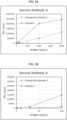
- FIG. 2 to FIG. 4 are the graphs showing the results of electrochemiluminescence immunoassay conducted in Example 1 and Comparative Example 1 for comparison.
- FIG. 2A and FIG. 2B are the graphs showing the results in which the second antibody A was used
- FIG. 3A and FIG. 3B are the graphs showing the results in which the second antibody B was used
- FIG. 4A and FIG. 4B are the graphs showing the results in which the second antibody C was used.
- the solid line indicates the results of Example 1 (with formation of the complex) and the dotted line indicates the results of Comparative Example 1 (without formation of the complex).
- Example 1 in which the analysis was conducted with the second antibody included in the complex, exhibited a higher detection sensitivity than Comparative Example 1, in which the analysis was conducted with the second antibody not included in the complex.
- Comparative Example 1 did not exhibit an antigen concentration-dependent increase in count up to 30 ng/mL, failing to conduct a precise measurement.
- Example 1 exhibited an antigen concentration-dependent increase in count from 1 ng/mL, successfully conducting a precise measurement.
- the electrochemiluminescence immunoassay was conducted in the same manner as Example 1, except that the second antibody was allowed to react with the antigen (formation of sandwich) without the formation of the complex, and subsequently the ruthenium complex was bound to the second antibody via streptavidin.
- a measurement specimen (30 ⁇ L) was dispensed into a prescribed reaction tube, and a specimen treatment solution (10mM MES, 0.1% BSA, 20 ⁇ g/mL Endo F3 (GenScript), 0.05% ProClin 300, pH 5.5) was added thereto, and the reaction tube was allowed to stand at 37°C for 15 minutes (glycan-cleavage treatment).
- a specimen treatment solution (10mM MES, 0.1% BSA, 20 ⁇ g/mL Endo F3 (GenScript), 0.05% ProClin 300, pH 5.5) was added thereto, and the reaction tube was allowed to stand at 37°C for 15 minutes (glycan-cleavage treatment).
- the antigen is allowed to react with the second antibody for 1.2 minutes from the start of the measurement (first reaction).
- the suspension of the magnetic particles 25 ⁇ L is dispensed into the reaction tube, and the antigen is allowed to react with the first antibody at 28°C for approximately 4.8 minutes (second reaction, formation of sandwich).
- the reactant is subjected to BF separation with a prescribed BF washing solution to remove the unreacted antigen and the unreacted second antibody.
- a solution of the ruthenium-labeled streptavidin 100 ⁇ L
- streptavidin is allowed to bind to biotin that is bound to the second antibody (third reaction).
- the reactant was subjected to BF separation with a prescribed BF washing solution to remove the unreacted ruthenium-labeled streptavidin.
- the complex generated was suspended in a solution of a prescribed substrate (tripropylamine), and allowed to cause electrochemical luminescence.
- the results in which the second antibody A was used as the second antibody are shown in Table 4.
- the count at respective antigen concentrations in Table 4 refers to a value calculated by subtracting a blank count from the count obtained by the measurement (specific count).
- the blank count shown in Table 4 refers to a value measured at an antigen concentration of 0 ng/mL.
- Comparative Example 2 in which a labeling substance was bound to the second antibody via streptavidin, after binding the second antibody to the antigen, exhibited a lower detection sensitivity than Comparative Example 1, in which the ruthenium complex was directly bound to the second antibody.
- Samples were prepared at different concentrations of the second antibody (5 ⁇ g/mL to 40 ⁇ g/mL) by the method described in Comparative Example 1.
- the second antibody the second antibody B was used.
- the "blank deduction” in Table 5 refers to a specific count value, which is a value obtained from subtracting a blank count from the count, and the "S/N ratio” refers to a value obtained by dividing the count by the blank count.
- Table 5 Antigen (ng/mL) Second Antibody ( ⁇ g/mL) 5 10 20 40 Count 0 261.0 279.8 393.8 500.1 1 396.9 363.0 469.1 604.4 10 2852.3 2787.4 3287.9 3358.8 100 132638.7 131376.0 141508.5 135465.3 1000 1976947.7 2043923.3 2079213.5 2115883.2 Specific Count (Blank Deduction) 1 135.9 83.2 75.3 104.3 10 2591.3 2507.6 2894.1 2858.7 100 132377.7 131096.2 141114.7 134965.2 1000 1976686.7 2043643.5 2078819.7 2115383.1 S/N Ratio 1 1.52 1.3 1.19 1.21 10 10.93 9.96 8.35
- reaction molar ratio of the second antibody and the ruthenium complex NHS was set at 1:10 or 1:20.
- the number of the ruthenium complex with respect to a single molecule of the antibody was 6.4 or 10.2.
- the second antibody the second antibody B was used.
Landscapes
- Health & Medical Sciences (AREA)
- Life Sciences & Earth Sciences (AREA)
- Immunology (AREA)
- Engineering & Computer Science (AREA)
- Chemical & Material Sciences (AREA)
- Molecular Biology (AREA)
- Biomedical Technology (AREA)
- Hematology (AREA)
- Urology & Nephrology (AREA)
- Biotechnology (AREA)
- Biochemistry (AREA)
- Cell Biology (AREA)
- Food Science & Technology (AREA)
- Medicinal Chemistry (AREA)
- Physics & Mathematics (AREA)
- Analytical Chemistry (AREA)
- Microbiology (AREA)
- General Health & Medical Sciences (AREA)
- General Physics & Mathematics (AREA)
- Pathology (AREA)
- Chemical Kinetics & Catalysis (AREA)
- Investigating Or Analysing Materials By The Use Of Chemical Reactions (AREA)
- Peptides Or Proteins (AREA)
Applications Claiming Priority (2)
| Application Number | Priority Date | Filing Date | Title |
|---|---|---|---|
| JP2022050800 | 2022-03-25 | ||
| PCT/JP2023/012019 WO2023182520A1 (fr) | 2022-03-25 | 2023-03-24 | Procédé d'analyse immunologique, complexe, procédé de production de complexe et réactif pour analyse immunologique |
Publications (2)
| Publication Number | Publication Date |
|---|---|
| EP4481383A1 true EP4481383A1 (fr) | 2024-12-25 |
| EP4481383A4 EP4481383A4 (fr) | 2025-06-04 |
Family
ID=88101747
Family Applications (1)
| Application Number | Title | Priority Date | Filing Date |
|---|---|---|---|
| EP23775113.6A Pending EP4481383A4 (fr) | 2022-03-25 | 2023-03-24 | Procédé d'analyse immunologique, complexe, procédé de production de complexe et réactif pour analyse immunologique |
Country Status (5)
| Country | Link |
|---|---|
| US (1) | US20250216384A1 (fr) |
| EP (1) | EP4481383A4 (fr) |
| JP (1) | JPWO2023182520A1 (fr) |
| CN (1) | CN118891523A (fr) |
| WO (1) | WO2023182520A1 (fr) |
Families Citing this family (1)
| Publication number | Priority date | Publication date | Assignee | Title |
|---|---|---|---|---|
| CN119438573A (zh) * | 2024-10-23 | 2025-02-14 | 广东普门生物医疗科技有限公司 | 电化学发光免疫检测试剂盒及其制备方法和应用 |
Family Cites Families (6)
| Publication number | Priority date | Publication date | Assignee | Title |
|---|---|---|---|---|
| MX2009004460A (es) * | 2006-10-26 | 2009-07-09 | Abbott Lab | Inmunoensayo de analitos en muestras que contienen anticuerpos anti-analito endogenos. |
| JP2010107207A (ja) | 2008-10-28 | 2010-05-13 | Panasonic Corp | 免疫分析試薬及びそれを用いた免疫分析方法 |
| WO2010054185A1 (fr) * | 2008-11-06 | 2010-05-14 | Ambit Biosciences Corporation | Dosage de biomarqueur de la tyrosine kinase 3 apparentée à fms phosphorylée |
| US9005910B2 (en) * | 2010-04-14 | 2015-04-14 | Eiken Kagaku Kabushiki Kaisha | Complex of labeled probes and water-soluble carrier |
| WO2019189870A1 (fr) * | 2018-03-30 | 2019-10-03 | 積水メディカル株式会社 | Procédé de traitement d'antigènes |
| JP2022050800A (ja) | 2020-09-18 | 2022-03-31 | ヤマハ発動機株式会社 | 鞍乗型車両 |
-
2023
- 2023-03-24 EP EP23775113.6A patent/EP4481383A4/fr active Pending
- 2023-03-24 CN CN202380028510.7A patent/CN118891523A/zh active Pending
- 2023-03-24 US US18/849,260 patent/US20250216384A1/en active Pending
- 2023-03-24 WO PCT/JP2023/012019 patent/WO2023182520A1/fr not_active Ceased
- 2023-03-24 JP JP2024509279A patent/JPWO2023182520A1/ja active Pending
Also Published As
| Publication number | Publication date |
|---|---|
| CN118891523A (zh) | 2024-11-01 |
| JPWO2023182520A1 (fr) | 2023-09-28 |
| WO2023182520A1 (fr) | 2023-09-28 |
| EP4481383A4 (fr) | 2025-06-04 |
| US20250216384A1 (en) | 2025-07-03 |
Similar Documents
| Publication | Publication Date | Title |
|---|---|---|
| EP3460474B1 (fr) | Procédé et kit de détection de molécule cible | |
| CN108318680B (zh) | 一种抗药抗体的检测方法及检测试剂盒 | |
| KR101814385B1 (ko) | 표식화 프로브-수용성 담체 복합체 | |
| CN112505322A (zh) | 阿尔茨海默病标志物p-Tau217检测试剂盒及其制造方法 | |
| CN112649616A (zh) | 用于心肌肌钙蛋白i检测的组合物及应用和磁微球电化学发光免疫检测试剂盒与检测方法 | |
| EP4019973B1 (fr) | Méthode de détection de plusieurs analytes | |
| CN1216112A (zh) | 发光特异性结合检测法 | |
| CN113176404A (zh) | 一种全血样本多指标联检的试剂盒及其使用方法 | |
| EP4481383A1 (fr) | Procédé d'analyse immunologique, complexe, procédé de production de complexe et réactif pour analyse immunologique | |
| CN113049835A (zh) | 联合检测试剂盒及检测方法、免疫分析系统 | |
| CN110954693A (zh) | 一种肿瘤标志物Cyfra21-1的Simoa试剂盒及其应用 | |
| CN113933521A (zh) | 磁微粒化学发光检测试剂盒及其制备方法和应用 | |
| JP3899029B2 (ja) | 免疫学的分析方法 | |
| CN112051404A (zh) | 一种肌红蛋白检测试剂盒及其应用 | |
| CN114563578A (zh) | 人膜性肾病诊断试剂盒 | |
| CN112485452A (zh) | 金属团簇作为人工抗体定量蛋白丰度的方法 | |
| JP7605555B2 (ja) | 免疫学的分析方法及び免疫学的分析試薬キット | |
| CN116125067A (zh) | 抗链霉亲和素抗体检测试剂盒、检测方法及应用 | |
| CN1888904B (zh) | 一种指示免疫反应中异嗜性抗体干扰的方法和试剂盒 | |
| EP4481385A1 (fr) | Réactif de dosage immunologique et procédé de dosage immunologique | |
| CN116593720B (zh) | 一种抗原单体检测的校准质控品及其应用 | |
| US20230221309A1 (en) | Method, use of the method and kit for detecting bioindicators in a sample | |
| CN119804843A (zh) | 全血样本中待测物的免疫学检测方法 | |
| CN121703409A (zh) | 一种基于吖啶酯化学发光的信号放大体系和试剂盒 | |
| CN121522171A (zh) | 一种多重级联放大的超灵敏化学发光试剂盒及其应用 |
Legal Events
| Date | Code | Title | Description |
|---|---|---|---|
| STAA | Information on the status of an ep patent application or granted ep patent |
Free format text: STATUS: THE INTERNATIONAL PUBLICATION HAS BEEN MADE |
|
| PUAI | Public reference made under article 153(3) epc to a published international application that has entered the european phase |
Free format text: ORIGINAL CODE: 0009012 |
|
| STAA | Information on the status of an ep patent application or granted ep patent |
Free format text: STATUS: REQUEST FOR EXAMINATION WAS MADE |
|
| 17P | Request for examination filed |
Effective date: 20240919 |
|
| AK | Designated contracting states |
Kind code of ref document: A1 Designated state(s): AL AT BE BG CH CY CZ DE DK EE ES FI FR GB GR HR HU IE IS IT LI LT LU LV MC ME MK MT NL NO PL PT RO RS SE SI SK SM TR |
|
| A4 | Supplementary search report drawn up and despatched |
Effective date: 20250507 |
|
| RIC1 | Information provided on ipc code assigned before grant |
Ipc: G01N 33/543 20060101ALI20250429BHEP Ipc: G01N 33/483 20060101ALI20250429BHEP Ipc: G01N 33/53 20060101AFI20250429BHEP |
|
| DAV | Request for validation of the european patent (deleted) | ||
| DAX | Request for extension of the european patent (deleted) |