EP4483887A1 - Zellpräparation - Google Patents
Zellpräparation Download PDFInfo
- Publication number
- EP4483887A1 EP4483887A1 EP23774429.7A EP23774429A EP4483887A1 EP 4483887 A1 EP4483887 A1 EP 4483887A1 EP 23774429 A EP23774429 A EP 23774429A EP 4483887 A1 EP4483887 A1 EP 4483887A1
- Authority
- EP
- European Patent Office
- Prior art keywords
- cells
- cell
- positive
- agent
- cell preparation
- Prior art date
- Legal status (The legal status is an assumption and is not a legal conclusion. Google has not performed a legal analysis and makes no representation as to the accuracy of the status listed.)
- Pending
Links
Images



Classifications
-
- A—HUMAN NECESSITIES
- A61—MEDICAL OR VETERINARY SCIENCE; HYGIENE
- A61P—SPECIFIC THERAPEUTIC ACTIVITY OF CHEMICAL COMPOUNDS OR MEDICINAL PREPARATIONS
- A61P25/00—Drugs for disorders of the nervous system
-
- A—HUMAN NECESSITIES
- A61—MEDICAL OR VETERINARY SCIENCE; HYGIENE
- A61K—PREPARATIONS FOR MEDICAL, DENTAL OR TOILETRY PURPOSES
- A61K35/00—Medicinal preparations containing materials or reaction products thereof with undetermined constitution
- A61K35/12—Materials from mammals; Compositions comprising non-specified tissues or cells; Compositions comprising non-embryonic stem cells; Genetically modified cells
- A61K35/28—Bone marrow; Haematopoietic stem cells; Mesenchymal stem cells of any origin, e.g. adipose-derived stem cells
-
- A—HUMAN NECESSITIES
- A61—MEDICAL OR VETERINARY SCIENCE; HYGIENE
- A61P—SPECIFIC THERAPEUTIC ACTIVITY OF CHEMICAL COMPOUNDS OR MEDICINAL PREPARATIONS
- A61P25/00—Drugs for disorders of the nervous system
- A61P25/14—Drugs for disorders of the nervous system for treating abnormal movements, e.g. chorea, dyskinesia
- A61P25/16—Anti-Parkinson drugs
-
- A—HUMAN NECESSITIES
- A61—MEDICAL OR VETERINARY SCIENCE; HYGIENE
- A61P—SPECIFIC THERAPEUTIC ACTIVITY OF CHEMICAL COMPOUNDS OR MEDICINAL PREPARATIONS
- A61P25/00—Drugs for disorders of the nervous system
- A61P25/18—Antipsychotics, i.e. neuroleptics; Drugs for mania or schizophrenia
-
- A—HUMAN NECESSITIES
- A61—MEDICAL OR VETERINARY SCIENCE; HYGIENE
- A61P—SPECIFIC THERAPEUTIC ACTIVITY OF CHEMICAL COMPOUNDS OR MEDICINAL PREPARATIONS
- A61P25/00—Drugs for disorders of the nervous system
- A61P25/24—Antidepressants
-
- A—HUMAN NECESSITIES
- A61—MEDICAL OR VETERINARY SCIENCE; HYGIENE
- A61P—SPECIFIC THERAPEUTIC ACTIVITY OF CHEMICAL COMPOUNDS OR MEDICINAL PREPARATIONS
- A61P25/00—Drugs for disorders of the nervous system
- A61P25/28—Drugs for disorders of the nervous system for treating neurodegenerative disorders of the central nervous system, e.g. nootropic agents, cognition enhancers, drugs for treating Alzheimer's disease or other forms of dementia
-
- A—HUMAN NECESSITIES
- A61—MEDICAL OR VETERINARY SCIENCE; HYGIENE
- A61P—SPECIFIC THERAPEUTIC ACTIVITY OF CHEMICAL COMPOUNDS OR MEDICINAL PREPARATIONS
- A61P43/00—Drugs for specific purposes, not provided for in groups A61P1/00-A61P41/00
-
- C—CHEMISTRY; METALLURGY
- C12—BIOCHEMISTRY; BEER; SPIRITS; WINE; VINEGAR; MICROBIOLOGY; ENZYMOLOGY; MUTATION OR GENETIC ENGINEERING
- C12N—MICROORGANISMS OR ENZYMES; COMPOSITIONS THEREOF; PROPAGATING, PRESERVING, OR MAINTAINING MICROORGANISMS; MUTATION OR GENETIC ENGINEERING; CULTURE MEDIA
- C12N5/00—Undifferentiated human, animal or plant cells, e.g. cell lines; Tissues; Cultivation or maintenance thereof; Culture media therefor
- C12N5/06—Animal cells or tissues; Human cells or tissues
- C12N5/0602—Vertebrate cells
- C12N5/0634—Cells from the blood or the immune system
- C12N5/0647—Haematopoietic stem cells; Uncommitted or multipotent progenitors
-
- C—CHEMISTRY; METALLURGY
- C12—BIOCHEMISTRY; BEER; SPIRITS; WINE; VINEGAR; MICROBIOLOGY; ENZYMOLOGY; MUTATION OR GENETIC ENGINEERING
- C12N—MICROORGANISMS OR ENZYMES; COMPOSITIONS THEREOF; PROPAGATING, PRESERVING, OR MAINTAINING MICROORGANISMS; MUTATION OR GENETIC ENGINEERING; CULTURE MEDIA
- C12N2501/00—Active agents used in cell culture processes, e.g. differentation
- C12N2501/10—Growth factors
- C12N2501/165—Vascular endothelial growth factor [VEGF]
-
- C—CHEMISTRY; METALLURGY
- C12—BIOCHEMISTRY; BEER; SPIRITS; WINE; VINEGAR; MICROBIOLOGY; ENZYMOLOGY; MUTATION OR GENETIC ENGINEERING
- C12N—MICROORGANISMS OR ENZYMES; COMPOSITIONS THEREOF; PROPAGATING, PRESERVING, OR MAINTAINING MICROORGANISMS; MUTATION OR GENETIC ENGINEERING; CULTURE MEDIA
- C12N2502/00—Coculture with; Conditioned medium produced by
- C12N2502/28—Vascular endothelial cells
Definitions
- the present invention relates to cell preparations and the like for treatment of diseases that can be expected to be cured by promoting new tissue formation.
- NON-PATENT DOCUMENT 1 indicates that for myocardial infarction, administration of bone marrow mononuclear cells obtained by specific gravity centrifugation allows formation of a new heart microvascular network, leading to an improvement in cardiac function.
- NON-PATENT DOCUMENT 2 indicates that for limb ischemia, administration of peripheral blood-derived CD34-positive cells purified using a specific cell-surface antigen allows formation of a new limb microvascular network, leading to an improvement in ischemia syndrome.
- NON-PATENT DOCUMENT 3 indicates that for limb ischemia, administration of bone marrow mononuclear cells obtained by specific gravity centrifugation allows formation of a new limb microvascular network, leading to an improvement in ischemia syndrome.
- NON-PATENT DOCUMENT 4 indicates that for cerebral infarction, administration of umbilical cord blood-derived CD34-positive cells purified using a specific cell-surface antigen allows formation of a new cerebral microvascular network, leading to an improvement in neurological function.
- NON-PATENT DOCUMENT 5 indicates that for cerebral infarction, administration of bone marrow mononuclear cells obtained by specific gravity centrifugation allows formation of a new cerebral microvascular network, leading to an improvement in neurological function.
- NON-PATENT DOCUMENT 6 indicates that allogeneic hematopoietic stem cell transplantation may cause damage to a microvascular network, leading to symptomatic deterioration after myocardial infarction.
- NON-PATENT DOCUMENT 7 indicates that allogeneic hematopoietic stem cell transplantation may cause damage to a microvascular network, leading to symptomatic deterioration after cerebral infarction.
- Newborn neurons are incorporated into a nervous network and function therein.
- the frequency of the birth of neurons (neurogenesis) in the hippocampus increases during learning or in an enriched environment.
- hippocampal neurogenesis would suggest the possibility of a novel method for treatment of an age-related decrease in neurological function, psychiatric disorders, and dementia.
- the present invention has been made. It is an object of the present invention to provide a cell preparation having an effective therapeutic effect for diseases that can be expected to be cured by promoting new tissue formation.
- a cell preparation according to the present invention has CD18-positive cells.
- a cell preparation having an effective therapeutic effect on ischemic disease is obtained.
- neurogenesis can be effectively promoted in the hippocampus.
- a cell preparation according to the present invention is characterized by having CD18-positive cells.
- the CD18-positive cells are preferably a stem cell.
- Stem cells have the ability to differentiate into various cells, and the self-renewal ability. Examples of stem cells include adult stem cells and pluripotent stem cells.
- adult stem cells exit in adult tissues, and are not fully differentiated and have pluripotency to some extent.
- adult stem cells include hematopoietic stem cells, mesenchymal stem cells, neural stem cells, intestinal epithelial stem cells, hair follicle stem cells, and pigment stem cells.
- Pluripotent stem cells have pluripotency, which is the ability to differentiate into all types of cells constituting an organism, and can continue to proliferate endlessly while maintaining pluripotency when cultured in vitro under appropriate conditions.
- pluripotent stem cells include embryonic stem cells (ES cells), embryonic germ stem cells (EG cells), germline stem cells (GS cells), and artificial pluripotent stem cells (iPS cells).
- the stem cell is preferably a hematopoietic stem cell.
- Hematopoietic stem cells have the pluripotency to differentiate into all blood cell lineages of blood cells, and the self-renewal ability while maintaining the pluripotency.
- Hematopoietic stem cells are isolated from the above-described tissue, typically using expression of a surface marker specific to the cells (hematopoietic stem cell specific marker). Concerning expression of such a marker, human-derived hematopoietic cells are typically characterized by being CD34-positive. More specifically, hematopoietic stem cells are a CD34-positive (+), CD38-negative (-), CD90-positive (+), and CD45RA-negative (-) cell, preferably a CD34 + CD38 - CD90 + CD45RA - CD49f + cell.
- Tissues from which hematopoietic stem cells are derived are not particularly limited as long as hematopoietic stem cells exist therein.
- the tissue is preferably hematopoietic tissue. Examples of the tissue include bone marrow, umbilical cord blood, peripheral blood, and liver.
- CD34-positive cells mean CD34-positive enriched cells, which can, for example, be obtained by concentrating CD34-positive cells from a sample of bone marrow, peripheral blood, placenta, umbilical cord blood, or the like using a technique well known in the art.
- CD34-positive cells can, for example, be concentrated using CD34-immobilized beads or a cell sorter.
- a preferable example of CD34-positive cells is a CD34-positive mononuclear cell.
- the hematopoietic stem cells in the present invention are allogeneic cells in light of medical convenience, but do not exclude autologous cells.
- the hematopoietic stem cells are prepared from an allogeneic umbilical cord blood sample that is a pharmaceutical composition according to the present invention.
- the hematopoietic stem cells are prepared from a banked cell group.
- CD18 is also called ⁇ 2 integrin.
- Integrins are molecules that have a key role in cell adhesion and are expressed in various cells. Integrins are transmembrane proteins that are a dimer composed of ⁇ and ⁇ chains. Twenty-four integrins are each composed of one of 18 ⁇ chains and one of 8 ⁇ chains.
- CD18 is also called leukocyte integrin, and the expression thereof is specific to leukocytes.
- the present inventors paid attention to the fact that CD18 is also expressed in hematopoietic stem cells and the like in addition to leukocytes to find that CD18 has a key role in cell adhesion of hematopoietic stem cells.
- CD18 is normally incapable of binding to a ligand, and when activated, undergoes a reversible three-dimensional structural change to be able to bind to a ligand.
- CD18-positive cells are a cell group in which the percentage of cells having a CD18-positive reaction in, for example, analysis by flow cytometry is at least 60%, preferably at least 70%, more preferably at least 80%, and even more preferably at least 90%.
- CD18-positive cells may, for example, also be a cell group in which, in a flow cytometry assay, the median intensity of fluorescence (MIF) of a CD18 antigen can be maintained at least two times, preferably at least three times, and more preferably at least four times as high as an MIF obtained when reacted with an isotype control antibody.
- MIF median intensity of fluorescence
- Glucose transporters are membrane proteins on the plasma membrane that are required for transport of glucose across the biological membrane, and are called a glucose transporting carrier.
- 13 GLUTs have been reported (GLUT-1, -2, -3, -4, -5, -6, - 7, -8, -9, -10, -11, and -12, and HMIT).
- GLUT1-GLUT4 which belong to class I, the expression sites and roles thereof have been relatively well clarified.
- GLUT1-GLUT4 all have a molecular weight of about 50 kDa and share the same structure in which the protein spans the cell membrane 12 times.
- a class-I glucose transporter (GLUT1) has a key role in transport of glucose from cerebral blood vessels to the brain. The present inventors found that GLUT1 is deeply involved with exhibition of the effect of hematopoietic stem cells.
- Connexin is a generic term for a family of junction membrane proteins forming gap junctions. More than 12 subtypes of connexins having different molecular weights have been found. A connexin having a molecular weight of 26 kDa is called connexin 26, and a connexin having a molecular weight of 43 kDa is called connexin 43. Connexin 43 is more abundant in the central nervous system and cardiomyocytes than are the other members of the connexin family, and is presumed to have adverse effects such as arrhythmias when the amount thereof is reduced. The present inventors found that connexin 43 is deeply involved with exhibition of the effect of hematopoietic stem cells.
- Jagged1 is a ligand for a Notch receptor.
- the Notch signaling pathway is very important to cell-cell communication.
- the present inventors found that Jagged1 is deeply involved with exhibition of the effect of hematopoietic stem cells.
- the blood vessel regeneration promotion mechanism of hematopoietic stem cells provides a simple cell-cell interaction (interaction between hematopoietic stem cells and damaged blood vessel endothelial cells) immediately after administration of the cells, and does not require engraftment, proliferation, or differentiation of the transplanted stem cells. If CD18 is expressed, the administered hematopoietic stem cells effectively adhere to blood vessel endothelial cells. If a connexin is expressed, the administered hematopoietic stem cells are effectively linked to blood vessel endothelial cells through gap junctions.
- a glucose transporter GLUT
- blood vessel endothelial cells are effectively supplied with energy from hematopoietic stem cells, which have abundant glucose, through gap junctions.
- Jagged1 Jagged1 is expressed, cell-cell communication is effectively established between the administered hematopoietic stem cells and blood vessel endothelial cells.
- a cell preparation that can be expected to have an excellent therapeutic effect is characterized by having CD18-positive cells.
- a glucose transporter GLUT (particularly preferably GLUT1) is preferably expressed.
- a connexin (particularly preferably connexin 43) is preferably expressed.
- Jagged1 is preferably expressed.
- GLUT glucose transporter
- a cell preparation that can be expected to have an excellent therapeutic effect is characterized by having a connexin-positive cell (preferably connexin 43-positive cell).
- Jagged1 in stem cells is important to regeneration promotion effects. Therefore, a cell preparation that can be expected to have an excellent therapeutic effect is characterized by having Jagged1-positive cells.
- the cell preparation according to the present invention is for treatment and/or prevention of diseases that can be expected to be cured by new tissue formation.
- treatment includes treatment of syndromes, improvement of syndromes, and inhibition of progression of syndromes.
- prevention includes inhibition and slowing of the onset of diseases, and includes not only prevention of diseases before the onset thereof but also prevention of the recurrence of diseases after the treatment thereof.
- the cell preparation according to the present invention is also for ischemic diseases such as cerebral infarction, myocardial infarction, limb ischemia, renal infarction, pulmonary infarction, splenic infarction, and intestinal infarction.
- ischemic diseases such as cerebral infarction, myocardial infarction, limb ischemia, renal infarction, pulmonary infarction, splenic infarction, and intestinal infarction.
- the cell preparation according to the present invention is intended to cause formation of new microvessels around the ischemic area by administering stem cells in which CD18 is highly expressed, thereby overcoming ischemic diseases.
- Diseases targeted by the cell preparation according to the present invention are not limited to those described in the examples.
- the cell preparation according to the present invention can be used in any of the hyperacute, acute, subacute, and chronic phases.
- the cell preparation according to the present invention is suitably used in the acute and subacute phases for acute-onset ischemic diseases such as acute myocardial infarction and cerebral infarction.
- the hyperacute phase is within 12 hours from the onset, in which tissue cell death can be highly likely to be prevented by stenting, thrombolysis, thrombectomy, or the like.
- the acute phase is within 12 hours to 7 days from the onset.
- the subacute phase is within 1 to 4 weeks from the onset.
- the chronic period is at least one month from the onset.
- the cell preparation according to the present invention can be used suitably for ischemic diseases that follow a chronic course, such as limb ischemia caused by chronic circulatory disorders, regardless of timing.
- the cell preparation according to the present invention also targets diseases to whose treatment hippocampal neurogenesis contributes.
- the cell preparation according to the present invention is a neurogenesis promotion agent that is characterized by having CD18-positive cells and allows hippocampal neurogenesis.
- neural stem and progenitor cells exist in the hippocampus, which is a specific brain region, even during the period of maturity, and that these cells grow and differentiate to produce new neurons. Newborn neurons play a key role in formation of neural networks, memory generation, and the like.
- hippocampal neural stem and progenitor cells are reduced not only when the animal is physically and mentally stressed by, for example, restraint stress, but also when the animal is only mentally stressed by, for example, resident-intruder stress. This suggests that inhibition of hippocampal neurogenesis may be involved with the causes of psychiatric disorders such as depression.
- promotion of neurogenesis in the hippocampus allows prevention and/or treatment of an age-related decrease in neurological function, and prevention and/or treatment of diseases associated with an age-related impairment of neurological function. Furthermore, promotion of neurogenesis in the hippocampus allows prevention and/or treatment of dementia associated with neurodegenerative diseases, and prevention and/or treatment of psychiatric disorders.
- the hippocampus is located in the medial temporal lobe of the cerebrum at the base of the inferior horn of the lateral ventricle, and is a cortical site essential for the formation of explicit memory such as episodic memory.
- the dentate gyrus is an appendage of the hippocampus, but is herein considered to be part of the hippocampus.
- the dentate gyrus consists of three layers: a granule cell layer as a middle layer; a molecular layer located immediately above the middle layer and having a considerably low cell density; and a polymorphic cell layer located below the middle layer and appearing to have sparsely scattered cells.
- the CD18-positive cells are preferably connexin 43-positive. All tissues and organs in the adult body have various forms of junctions between adjacent cells to maintain tissue homeostasis. Connexin 43, which is a molecule having a key role in cell-cell gap junctions, is expressed in, for example, osteoblasts, osteocytes, endovascular cells, and neurons. The present inventors found that connexin 43-positive cells have a key role in neurogenesis in the hippocampus (especially, neurogenesis in the dentate gyrus). Connexin 43-positive cells are a group of cells in which connexin 43 is highly expressed. The high expression of connexin 43 is not particularly limited.
- the percentage of cells in a cell group exhibiting a connexin 43-positive reaction as analyzed by flow cytometry analysis is at least 60%, preferably at least 70%, more preferably at least 80%, and even more preferably at least 90%, it is considered that connexin 43 is highly expressed in the cell group.
- the administration route of the cell preparation according to the present invention is preferably intravenous, intraarterial, intraportal, or local tissue.
- the number of cells to be administered is not particularly limited and is determined, as appropriate, according to the age, weight, disease, and symptom of the target patient, and the technique of administration.
- the pharmaceutical composition according to the present invention is prepared such that hematopoietic stem cells are administered at a rate of about 10 4 to 10 6 cells/kg, preferably at least 10 4 cells/kg, more preferably at least 5 ⁇ 10 4 cells/kg, more preferably at least 5 ⁇ 10 5 cells/kg, and optionally 2 ⁇ 10 6 cells.
- the pharmaceutical composition according to the present invention is prepared such that hematopoietic stem cells are administered at a rate of about 10 4 to 10 6 cells/kg, preferably at least 10 4 cells/kg, and more preferably about 5 ⁇ 10 4 cells/kg.
- the total number of cells (mononuclear cells) is about 100 times as large as the numerical value of the hematopoietic stem cells.
- an angiogenesis promotion factor can also be included.
- the angiogenesis promotion factor is not particularly limited.
- As the angiogenesis promotion factor for example, VEGF, angiopoietins, PDGF, TGF- ⁇ , FGF, PlGF, matrix metalloproteinases, plasminogen activators, and the like, preferably angiopoietins, can be used.
- Angiopoietins, which are glycoproteins, are growth factors that promote vasculogenesis or angiogenesis.
- Angiopoietins include angiopoietin 1 (Ang1), angiopoietin 2 (Ang2), angiopoietin 3 (Ang3), and angiopoietin 4 (Ang4).
- Ang1 angiopoietin 1
- Ang2 angiopoietin 2
- Ang3 angiopoietin 3
- Ang4 angiopoietin 4
- ANGPTLs angiopoietin-related proteins or angiopoietin- like proteins.
- additives that are commonly used in the art can be used if necessary.
- the additives include tonicity agents, stabilizing agents, buffering agents, preserving agents, chelating agents, antioxidants, and solubilizing agents.
- the tonicity agents include sugars, such as glucose, sorbitol, and mannitol, sodium chloride, glycerol, propylene glycol, and polyethylene glycol.
- the stabilizing agents include sodium sulfite.
- the buffering agents include borate buffers, phosphate buffers, citrate buffers, tartrate buffers, and acetate buffers.
- Examples of the preserving agents include paraoxybenzoic acid esters, benzyl alcohol, chlorocresol, phenethyl alcohol, and benzethonium chloride.
- Examples of the chelating agents include disodium EDTA and sodium citrate.
- Examples of the antioxidants include sodium sulfite, sodium hydrogen sulfite, sodium ascorbate, and sodium thiosulfate.
- Examples of the solubilizing agents include dextran, polyvinyl pyrrolidone, sodium benzoate, ethylenediamine, salicylamide, nicotinamide, and polyoxyethylene hydrogenated castor oil derivatives.
- the cell preparation is produced by a method described below, although the present invention is not limited to the method.
- the method has, for example: obtaining a buffy coat layer from a sample containing hematopoietic stem cells; obtaining mononuclear cells by removing, from the buffy coat layer, inhibitory cells that inhibit a therapeutic effect; and obtaining, from the mononuclear cells, CD18 high mononuclear cells in which CD18 is highly expressed.
- the method also has, for example: obtaining a buffy coat layer from a sample containing hematopoietic stem cells; obtaining mononuclear cell cells by removing, from the buffy coat layer, inhibitory cells that inhibit a therapeutic effect; obtaining, from the mononuclear cells, CD34 + mononuclear cells that are CD34-positive; and obtaining, from the CD34-positive mononuclear cells, CD34 + CD18 high mononuclear cells in which CD18 is highly expressed.
- the method also has, for example: selecting a sample that contains a large number of CD18 high expression cells from a plurality of blood samples collected from a plurality of patients; and removing, from the blood samples, inhibitory cells that inhibit a therapeutic effect.
- the method also has, for example: increasing CD18 high expression cells by causing a cytokine such as TNF ⁇ to act with the collected blood samples; and removing, from the blood samples in which CD18 high expression cells have been increased, inhibitory cells that inhibit a therapeutic effect.
- a cytokine such as TNF ⁇
- heparin is often used as an anticoagulant.
- heparin is not used, and coagulation is inhibited by dilution with physiological saline and use of a divalent ion chelating agent.
- an umbilical cord blood, bone marrow fluid, or peripheral blood sample preferably an umbilical cord blood sample
- density gradient centrifugation For an umbilical cord blood sample, mononuclear cells that are accumulated in a buffy coat are obtained by density gradient centrifugation.
- density gradient centrifugation for example, commercially available media for density gradient centrifugation, such as Ficoll (registered trademark) and Percoll (registered trademark), are preferably used.
- the buffy coat may contain inhibitory cells that inhibit the efficacy of the therapeutic effect of the cellular preparation. Therefore, it is desirable to remove such inhibitory cells from the buffy coat.
- inhibitory cells in the buffy coat are neutrophils and dead cells.
- the upper limit of centrifugal force for separating the inhibitory cells is, for example, 1000 g, 900 g, 800 g, 700 g, or 600 g, and the lower limit thereof is 500 g or 400 g.
- the centrifugal force is, for example, in the range of 400-1000 g, 400-900 g, 400-800 g, 400-700 g, or 400-600 g.
- the centrifugal force is preferably 400-800 g, more preferably 500-800 g.
- the precipitate in the buffy coat layer after centrifugation has an upper layer that is a fraction in which neutrophils and dead cells are concentrated, and a lower layer that is a fraction in which mononuclear cells are concentrated.
- neutrophils and dead cells in the upper layer of the precipitate are selectively removed by gradually tilting the container toward the horizontal.
- centrifugation under the same conditions and then passing through a 40- ⁇ m filter is desirably performed in order to remove neutrophils and dead cells from the mononuclear cell fraction.
- hematopoietic stem cells are obtained from the mononuclear cells in the buffy coat.
- Hematopoietic stem cells are obtained by concentrating CD34-positive cells according to a known technique.
- the concentration of CD34-positive cells can be performed using CD34-immobilized beads or a cell sorter.
- CD18-positive cells are obtained from the CD34-positive mononuclear cells in the buffy coat by concentration.
- concentration of CD18-positive cells can be performed using CD18-immobilized beads or a cell sorter to obtain cells in which CD18 is highly expressed.
- CD34 + CD18 high mononuclear cells are obtained. It should be noted that the order of CD34 + CD18 high is not limited to this embodiment. Cells in which CD18 is highly expressed may be obtained from mononuclear cells in the buffy coat before CD34-positive cells may be concentrated from the CD18 high expression cells.
- Mononuclear cells were purified from each of umbilical cord blood in which aggregates did not exist and umbilical cord blood in which large aggregates existed, by Ficoll-Paque specific gravity centrifugation, were washed with PBS, and thereafter, were co-cultured with HUVECs ( FIG. 1 ). It should be noted that in FIG. 1 , CBMNC indicates human umbilical cord blood-derived mononuclear cells.
- a co-culture test was conducted as follows. Specifically, umbilical cord blood was gently placed on an equal amount of Ficoll-Paque, followed by centrifugation at 400 g for 30 minutes. At this time, the acceleration and deceleration of the centrifuge were set to the minimum values. A buffy coat layer was removed from the umbilical cord blood specimen after centrifugation. An equal amount of a PBS containing a 10% anticoagulant-citrate-dextrose solution (ACD solution) was added to the buffy coat layer, followed by centrifugation again at 400 g for 5 minutes. At this time, it is not necessary to set the acceleration and deceleration of the centrifuge to the minimum values.
- ACD solution 10% anticoagulant-citrate-dextrose solution
- the precipitate in the buffy coat layer obtained by centrifugation had an upper layer that was a fraction in which neutrophils and dead cells were concentrated, and a lower layer that was a fraction in which mononuclear cells were concentrated. Therefore, during discarding of the supernatant, neutrophils and dead cells in the upper layer of the precipitate were selectively removed by gradually tilting the container toward the horizontal. Furthermore, the precipitate was centrifuged at 400 g for 5 minutes and was passed through a 40- ⁇ m filter to remove neutrophils and dead cells contained in the mononuclear cell fraction. Next, HUVECs were cultured in a HuMedia-EB2 culture medium with serum and growth additives according to the manufacturer's protocol, and thereafter, were dissociated with trypsin and recovered.
- APC allophycocyanin
- VEGF vascular endothelial growth factor
- the cell mixture was washed twice with phosphate buffered saline (PBS), and was stained with a phycoerythrin (PE)-conjugated anti-human CD31 antibody (BD Bioscience), a fluorescein isothiocyanate (FITC)-conjugated anti-human CD45 antibody, and 7-aminoactinomycin D (7AAD).
- PE phycoerythrin
- FITC fluorescein isothiocyanate
- 7AAD 7-aminoactinomycin D
- vascular endothelial cells undergoing angiogenesis take up VEGF from outside the cell, it is considered that umbilical cord blood having a higher APC-labeled VEGF level more strongly promotes angiogenesis, or in other words, has a more effective therapeutic effect on cerebral infarction.
- a PBS prepared to contain heparin at a final concentration of 2 units/ml was erroneously added, instead of a PBS containing a 10% anticoagulant-citrate-dextrose solution (ACD solution).
- ACD solution 10% anticoagulant-citrate-dextrose solution
- umbilical cord blood was gently placed on an equal amount of Ficoll Paque, followed by centrifugation at 400 g for 30 minutes. At this time, the acceleration and deceleration of the centrifuge were set to the minimum values.
- a buffy coat layer was removed from the umbilical cord blood specimen after centrifugation.
- a PBS prepared to contain heparin at a final concentration of 2 units/mL was erroneously added to the buffy coat layer, followed by centrifugation again at 400 g for 5 minutes, so that the buffy coat layer underwent precipitation.
- centrifugation was performed at 400 g for 5 minutes, followed by filtration through a 40- ⁇ m filter, in order to remove neutrophils and dead cells from the mononuclear cell fraction.
- Umbilical cord blood mononuclear cells thus obtained were subjected to the co-culture test described above.
- umbilical cord blood was placed on an equal amount of Ficoll-Paque, followed by centrifugation at 400 g for 30 minutes.
- a buffy coat layer was removed from the umbilical cord blood specimen after centrifugation.
- An equal amount of a PBS containing a 10% anticoagulant-citrate-dextrose solution (ACD solution) was added, followed by centrifugation again at 400 g for 5 minutes.
- the precipitate in the buffy coat layer was separated by gradually tilting the container toward the horizontal and thereby discarding the supernatant.
- the precipitate was resuspended in a PBS, followed by centrifugation at 400 g for 5 minutes, and then filtration through a 40- ⁇ m filter.
- the mononuclear cell suspension was equally divided, followed by centrifugation at 400 g for 5 minutes. After discarding of the supernatant, one of the two precipitates was suspended in a PBS, and the other was suspended in a PBS prepared to contain heparin at a final concentration of 2 units/mL, followed by incubation at room temperature for 5 minutes.
- the cell suspensions were washed with a PBS, and were stained with a phycoerythrin (PE)-conjugated anti-human CD34 antibody (BD Bioscience), 7-aminoactinomycin D (7AAD) (BD Bioscience), fluorescein isothiocyanate (FITC)-conjugated anti-human CD45 antibody (BD Bioscience), or various adhesion factor antibodies (an integrin ⁇ 4 antibody (Beckman Coulter), an integrin ⁇ 5 antibody (Beckman Coulter), a CD18 antibody (Beckman Counter), or a CXCR4 antibody (MBL)) conjugated with allophycocyanin (APC).
- PE phycoerythrin
- 7AAD 7-aminoactinomycin D
- FITC fluorescein isothiocyanate
- various adhesion factor antibodies an integrin ⁇ 4 antibody (Beckman Coulter), an integrin ⁇ 5 antibody (Beckman Coulter), a
- integrin ⁇ 4 is a representative adhesion factor that functions in hematopoietic stem cell homing and cell adhesion.
- the adhesion factors whose expression was significantly changed due to the heparin treatment were integrin ⁇ 5, C-X-C chemokine receptor type 4 (CXCR4), and CD18. Some of these molecules underwent a large reduction in expression, and others underwent almost no reduction in expression. In particular, it was CD18 that underwent a large change in expression due to the heparin treatment.
- umbilical cord blood was placed on an equal amount of Ficoll-Paque, followed by centrifugation at 400 g for 30 minutes.
- a buffy coat layer was removed from the umbilical cord blood specimen after centrifugation.
- An equal amount of a PBS containing a 10% anticoagulant-citrate-dextrose solution (ACD solution) was added, followed by centrifugation again at 400 g for 5 minutes.
- the precipitate in the buffy coat layer was separated by gradually tilting the container toward the horizontal and thereby discarding the supernatant.
- the precipitate was resuspended in a PBS, followed by centrifugation at 400 g for 5 minutes, and then filtration through a 40- ⁇ m filter.
- the resultant precipitate was suspended again in a PBS. Next, aliquots of the suspension were removed and stained with a PE-conjugated anti-human CD34 antibody, 7AAD, an FITC-conjugated anti-human CD45 antibody, and an APC-conjugated CD18 antibody.
- the remaining suspension was used to conduct a co-culture test with HUVECs (see (1) above).
- the co-culture experiment was conducted under a condition that an allophycocyanin (APC)-labeled vascular endothelial growth factor (VEGF, final concentration: 3 ⁇ g/mL) and umbilical cord blood mononuclear cells (cell count including 1 ⁇ 10 4 CD34-positive cells) were added to the HUVECs (1 ⁇ 10 5 cells).
- APC allophycocyanin
- VEGF vascular endothelial growth factor
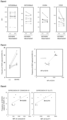
- the left portion of FIG. 4 shows differences in the VEGF uptake capability of HUVECs between each co-cultured umbilical cord blood.
- the right portion of FIG. 4 shows a correlation between expression of CD18 in hematopoietic stem cells (CD34-positive and 7AAD-negative) and the VEGF uptake capability of HUVECs in the co-culture experiment. It was found that the expression intensity of CD18 as an indicator of the VEGF uptake promotion capability, which was found by an accidental heparin treatment, i.e., an erroneous operation, is useful as a selectable marker for mononuclear cells having a high therapeutic effect.
- the precipitate in the buffy coat layer was resuspended in a PBS, followed by centrifugation at 400 g for 5 minutes and then filtration through a 40- ⁇ m filter, and then resuspension in a PBS.
- aliquots of the suspension were removed and stained with a PE-conjugated anti-human CD34 antibody, 7AAD, an FITC-conjugated anti-human CD45 antibody, and an APC-conjugated glucose transporter (GLUT1) antibody.
- the remaining suspension was used to conduct a co-culture test with HUVECs (see (1) above).
- the co-culture experiment was conducted under a condition that an allophycocyanin (APC)-labeled vascular endothelial growth factor (VEGF, final concentration: 3 ⁇ g/mL) and umbilical cord blood mononuclear cells (cell count including 1 ⁇ 10 4 CD34-positive cells) were added to HUVECs (1 ⁇ 10 5 cells).
- APC allophycocyanin
- VEGF vascular endothelial growth factor
- the right portion of FIG. 5 shows a correlation between expression of a glucose transporter (GLUT1) in hematopoietic stem cells (CD34-positive, CD45-positive, and 7AAD-negative) and the VEGF uptake capability of HUVECs in the co-culture experiment. It was found that the expression intensity of a glucose transporter (GLUT1) as an indicator of VEGF uptake promotion capability is useful as a selectable marker for mononuclear cells having a high therapeutic effect.
- APC allophycocyanin
- VEGF
- the precipitate in the buffy coat layer was resuspended in a PBS, followed by centrifugation at 400 g for 5 minutes and then filtration through a 40- ⁇ m filter, and then resuspension in a PBS.
- aliquots of the suspension were removed and stained with a PE-conjugated anti-human CD34 antibody, 7AAD, an FITC-conjugated anti-human CD45 antibody, and an APC-conjugated connexin 43 antibody.
- the remaining suspension was used to conduct a co-culture test with HUVECs (see (1) above).
- the co-culture experiment was conducted under a condition that an allophycocyanin (APC)-labeled vascular endothelial growth factor (VEGF, final concentration: 3 ⁇ g/mL) and umbilical cord blood mononuclear cells (cell count including 1 ⁇ 10 4 CD34-positive cells) were added to HUVECs (1 ⁇ 10 5 cells).
- APC allophycocyanin
- VEGF vascular endothelial growth factor
- the left portion of FIG. 5 shows a correlation between expression of connexin 43 in hematopoietic stem cells (CD34-positive, CD45-positive, and 7AAD-negative) and the VEGF uptake capability of HUVECs in the co-culture experiment. It was found that the expression intensity of connexin 43 as an indicator of VEGF uptake promotion capability is useful as a selectable marker for mononuclear cells having a high therapeutic effect.
- a mononuclear cell fraction was removed from umbilical cord blood by Ficoll-Paque specific gravity centrifugation described above, and was equally divided.
- CD34-positive, CD45-positive, and 7AAD-negative The expression level of a CD18 antigen in hematopoietic stem cells (CD34-positive, CD45-positive, and 7AAD-negative) was assessed using a FACS Canto II cell analyzer (BD Bioscience).
- cryoprotection solution cell banker
- hematopoietic stem cells were assessed using a technique similar to that described above.
- umbilical cord blood mononuclear cells having high expression of CD18 underwent a significant decrease in CD18 expression when those before freezing and after thawing were compared, while umbilical cord blood mononuclear cells having low expression of CD18 remained low before freezing and after thawing.
- the method for collecting cells having a high therapeutic effect had the following steps. Specifically, the method for collecting cells has: a collection step of collecting human bone marrow fluid with a diluent that does not contain heparin; and a separation step of adding a specific gravity centrifugal liquid that does not contain heparin to the collected human bone marrow fluid, and performing specific gravity centrifugation.
- the method was specifically carried out as follows. A 23-G needle was attached to a 2.5-ml syringe, and the syringe was filled with 2 ml of physiological saline. Both ends of a mouse femur were cut with surgical scissors. The needle was inserted into one end of the bone marrow cavity.
- the physiological saline was quickly injected to obtain bone marrow fluid that flowed out from the other end.
- An anticoagulant such as heparin is typically often used because bone marrow fluid coagulates easily. In the present invention, however, heparin was not used, and only dilution with physiological saline was used to inhibit coagulation of the bone marrow fluid.
- the bone marrow fluid that was diluted with physiological saline to inhibit coagulation was placed on a specific gravity centrifugal liquid (Ficoll-premium, manufactured by GE), followed by centrifugation at 600g for 20 minutes. Thereafter, a cell group in the buffy coat layer was collected.
- a specific gravity centrifugal liquid (Ficoll-premium, manufactured by GE)
- Dead cells were excluded from the analysis using a 7AAD (7-amino-actinomycin D, manufactured by BD Bioscience, product number: 559925, dilution factor: 50x) reagent.
- the respective isotype controls were used as negative controls.
- the positive reaction by flow cytometry is defined as a reaction in which the intensity of fluorescence at a particular wavelength emitted by a fluorophore used for labeling is within a range of fluorescent intensity higher than the intensity of the background measured without use of the primary antibody.
- Jagged1 is expressed in CD34-positive cells ( FIG. 8 ). Therefore, in order to study the regeneration promotion effect of the Jagged1 molecule, a Jagged1 peptide having Jagged1 activity (H-Cys-Asp-Asp-Tyr-Tyr-Tyr-Gly-Phe-Gly-Cys-Asn-Lys-Phe-Cys-Arg-Pro-Arg-OH) was added to a HUVEC culture medium. The VEGF uptake promotion effect of the Jagged1 peptide was studied.
- the brain was removed and fixed in 2% paraformaldehyde. Thereafter, the brain was sectioned into 20- ⁇ m sections using a vibratome (manufactured by Leica Camera AG). The sections were stained with an anti-nestin antibody (manufactured by Merck Millipore, product number: MAB5326, dilution factor: 1:200), an anti-doublecortin antibody (DCX; manufactured by Merck Millipore, product no. AB2253, dilution factor: 1:350), and DAPI for nuclear staining (manufactured by Thermo Fisher Scientific Inc., product no. D1306, dilution factor: 1: 1,000) (CD18 cell-administered mice: 7 mice, a total of 22 sections, PBS-administered mice: 6 mice, a total of 20 sections).
- an anti-nestin antibody manufactured by Merck Millipore, product number: MAB5326, dilution factor: 1:200
- DCX anti-double
- a confocal image was captured using a fluorescence microscope (BZ-X810, manufactured by Keyence Corporation) to assess newborn neuron marker (nestin)-positive cells in the granule cell layer of the hippocampal dentate gyrus and newborn neuron (DCX)-positive cells in the granule cell layer.
- BZ-X810 manufactured by Keyence Corporation
- DCX newborn neuron
- neurogenesis is impaired in the hippocampus.
- neurogenesis is promoted in the hippocampus, which allows prevention and/or treatment of such diseases.
- neurogenesis is promoted in the hippocampus according to the present invention, which allows improvement or slowing of aging.
- the present invention is useful in treatment of cerebral infarction and age-related neurological disorders.
Landscapes
- Health & Medical Sciences (AREA)
- Life Sciences & Earth Sciences (AREA)
- Engineering & Computer Science (AREA)
- Biomedical Technology (AREA)
- Bioinformatics & Cheminformatics (AREA)
- Chemical & Material Sciences (AREA)
- General Health & Medical Sciences (AREA)
- Veterinary Medicine (AREA)
- Animal Behavior & Ethology (AREA)
- Public Health (AREA)
- Medicinal Chemistry (AREA)
- Pharmacology & Pharmacy (AREA)
- Organic Chemistry (AREA)
- Neurology (AREA)
- Neurosurgery (AREA)
- General Chemical & Material Sciences (AREA)
- Nuclear Medicine, Radiotherapy & Molecular Imaging (AREA)
- Chemical Kinetics & Catalysis (AREA)
- Cell Biology (AREA)
- Developmental Biology & Embryology (AREA)
- Immunology (AREA)
- Biotechnology (AREA)
- Hematology (AREA)
- Zoology (AREA)
- Psychiatry (AREA)
- Epidemiology (AREA)
- Virology (AREA)
- Wood Science & Technology (AREA)
- Genetics & Genomics (AREA)
- Microbiology (AREA)
- General Engineering & Computer Science (AREA)
- Psychology (AREA)
- Hospice & Palliative Care (AREA)
- Biochemistry (AREA)
- Pain & Pain Management (AREA)
- Medicines Containing Material From Animals Or Micro-Organisms (AREA)
Applications Claiming Priority (2)
| Application Number | Priority Date | Filing Date | Title |
|---|---|---|---|
| JP2022046721 | 2022-03-23 | ||
| PCT/JP2023/007766 WO2023181830A1 (ja) | 2022-03-23 | 2023-03-02 | 細胞製剤 |
Publications (2)
| Publication Number | Publication Date |
|---|---|
| EP4483887A1 true EP4483887A1 (de) | 2025-01-01 |
| EP4483887A4 EP4483887A4 (de) | 2025-06-25 |
Family
ID=88100694
Family Applications (1)
| Application Number | Title | Priority Date | Filing Date |
|---|---|---|---|
| EP23774429.7A Pending EP4483887A4 (de) | 2022-03-23 | 2023-03-02 | Zellpräparation |
Country Status (5)
| Country | Link |
|---|---|
| US (1) | US20250195581A1 (de) |
| EP (1) | EP4483887A4 (de) |
| JP (1) | JPWO2023181830A1 (de) |
| CN (1) | CN118900693A (de) |
| WO (1) | WO2023181830A1 (de) |
Family Cites Families (2)
| Publication number | Priority date | Publication date | Assignee | Title |
|---|---|---|---|---|
| US7732126B2 (en) * | 2004-09-03 | 2010-06-08 | University Of Maryland, Baltimore | Integrin CD18 is a novel stromal stem cell marker and functions to promote osteogenesis |
| US20070031387A1 (en) * | 2005-08-08 | 2007-02-08 | Kyoto University | Method for treating and/or preventing spinal cord injury |
-
2023
- 2023-03-02 EP EP23774429.7A patent/EP4483887A4/de active Pending
- 2023-03-02 CN CN202380029209.8A patent/CN118900693A/zh active Pending
- 2023-03-02 WO PCT/JP2023/007766 patent/WO2023181830A1/ja not_active Ceased
- 2023-03-02 US US18/848,627 patent/US20250195581A1/en active Pending
- 2023-03-02 JP JP2024509904A patent/JPWO2023181830A1/ja active Pending
Also Published As
| Publication number | Publication date |
|---|---|
| US20250195581A1 (en) | 2025-06-19 |
| EP4483887A4 (de) | 2025-06-25 |
| CN118900693A (zh) | 2024-11-05 |
| WO2023181830A1 (ja) | 2023-09-28 |
| JPWO2023181830A1 (de) | 2023-09-28 |
Similar Documents
| Publication | Publication Date | Title |
|---|---|---|
| KR101730052B1 (ko) | 심근경색의 수복 재생을 유도하는 다능성 간세포 | |
| KR102689261B1 (ko) | 정제 혈소판의 제조 방법 | |
| AU2018243554B2 (en) | Generation of therapeutic cells using extracellular components of target organs | |
| JP2011529706A (ja) | 間充織間質細胞集団、ならびにそれを単離および使用する方法 | |
| KR20080056302A (ko) | 태반 유래 줄기세포로부터 생산된 희소돌기 아교세포 | |
| Friedlander et al. | Progenitor cells and retinal angiogenesis | |
| Chou et al. | CD44 fucosylation on mesenchymal stem cell enhances homing and macrophage polarization in ischemic kidney injury | |
| WO2005007176A1 (ja) | 間葉系細胞を有効成分とする体内投与用脳神経疾患治療薬 | |
| US8574899B2 (en) | Methods for augmentation collection of placental hematopoietic stem cells and uses thereof | |
| EP2488187A2 (de) | Assay zur vorhersage der therapeutischen wirkung mesenchymaler stromazellen sowie verwendungsverfahren dafür | |
| Gortner et al. | Regenerative therapies in neonatology: clinical perspectives | |
| CN114901806B (zh) | 细胞群以及其取得方法 | |
| JP7176766B2 (ja) | Cd31陽性cd45陰性cd200陽性の哺乳動物細胞からなる細胞集団、およびその利用 | |
| EP4483887A1 (de) | Zellpräparation | |
| Li et al. | Analysis of the origin and population dynamics of cardiac progenitor cells in a donor heart model | |
| JP2007528229A (ja) | 臍帯血から分離された多能性幹細胞及びこれを含有する虚血性壊死疾患治療用細胞治療剤 | |
| US20220072050A1 (en) | Dual Stem Cell Therapy for Neurological Conditions | |
| EP4364747A1 (de) | Pharmazeutische zusammensetzung mit bestrahlten hämatopoietischen stammzellen | |
| US20210236557A1 (en) | Immature dental pulp stem cells and methods of use to treat bone marrow failure | |
| Pellegrini | Myoblast Transplantation: Murine Studies | |
| Pischiutta | Mesenchymal stromal cells for traumatic brain injury | |
| HK1128933A (en) | Co-culture of placental stem cells and stem cells from a second source |
Legal Events
| Date | Code | Title | Description |
|---|---|---|---|
| STAA | Information on the status of an ep patent application or granted ep patent |
Free format text: STATUS: THE INTERNATIONAL PUBLICATION HAS BEEN MADE |
|
| PUAI | Public reference made under article 153(3) epc to a published international application that has entered the european phase |
Free format text: ORIGINAL CODE: 0009012 |
|
| STAA | Information on the status of an ep patent application or granted ep patent |
Free format text: STATUS: REQUEST FOR EXAMINATION WAS MADE |
|
| 17P | Request for examination filed |
Effective date: 20240924 |
|
| AK | Designated contracting states |
Kind code of ref document: A1 Designated state(s): AL AT BE BG CH CY CZ DE DK EE ES FI FR GB GR HR HU IE IS IT LI LT LU LV MC ME MK MT NL NO PL PT RO RS SE SI SK SM TR |
|
| A4 | Supplementary search report drawn up and despatched |
Effective date: 20250522 |
|
| RIC1 | Information provided on ipc code assigned before grant |
Ipc: C12N 5/10 20060101ALI20250516BHEP Ipc: C12N 5/0797 20100101ALI20250516BHEP Ipc: C12N 5/0789 20100101ALI20250516BHEP Ipc: C12N 5/0775 20100101ALI20250516BHEP Ipc: C12N 5/074 20100101ALI20250516BHEP Ipc: C12N 5/0735 20100101ALI20250516BHEP Ipc: A61P 43/00 20060101ALI20250516BHEP Ipc: A61P 25/28 20060101ALI20250516BHEP Ipc: A61P 25/24 20060101ALI20250516BHEP Ipc: A61P 25/18 20060101ALI20250516BHEP Ipc: A61P 25/16 20060101ALI20250516BHEP Ipc: A61P 25/00 20060101ALI20250516BHEP Ipc: A61K 35/51 20150101ALI20250516BHEP Ipc: A61K 35/28 20150101ALI20250516BHEP Ipc: A61K 35/12 20150101AFI20250516BHEP |
|
| DAV | Request for validation of the european patent (deleted) | ||
| DAX | Request for extension of the european patent (deleted) |