EP4485351A1 - Procédé d'affinage d'un masque de segmentation - Google Patents
Procédé d'affinage d'un masque de segmentation Download PDFInfo
- Publication number
- EP4485351A1 EP4485351A1 EP23315261.0A EP23315261A EP4485351A1 EP 4485351 A1 EP4485351 A1 EP 4485351A1 EP 23315261 A EP23315261 A EP 23315261A EP 4485351 A1 EP4485351 A1 EP 4485351A1
- Authority
- EP
- European Patent Office
- Prior art keywords
- user
- segmentation mask
- segmentation
- image
- algorithm
- Prior art date
- Legal status (The legal status is an assumption and is not a legal conclusion. Google has not performed a legal analysis and makes no representation as to the accuracy of the status listed.)
- Pending
Links
Images


Classifications
-
- G—PHYSICS
- G06—COMPUTING OR CALCULATING; COUNTING
- G06T—IMAGE DATA PROCESSING OR GENERATION, IN GENERAL
- G06T7/00—Image analysis
- G06T7/10—Segmentation; Edge detection
- G06T7/11—Region-based segmentation
-
- G—PHYSICS
- G06—COMPUTING OR CALCULATING; COUNTING
- G06T—IMAGE DATA PROCESSING OR GENERATION, IN GENERAL
- G06T5/00—Image enhancement or restoration
- G06T5/20—Image enhancement or restoration using local operators
-
- G—PHYSICS
- G06—COMPUTING OR CALCULATING; COUNTING
- G06N—COMPUTING ARRANGEMENTS BASED ON SPECIFIC COMPUTATIONAL MODELS
- G06N20/00—Machine learning
- G06N20/10—Machine learning using kernel methods, e.g. support vector machines [SVM]
-
- G—PHYSICS
- G06—COMPUTING OR CALCULATING; COUNTING
- G06N—COMPUTING ARRANGEMENTS BASED ON SPECIFIC COMPUTATIONAL MODELS
- G06N20/00—Machine learning
- G06N20/20—Ensemble learning
-
- G—PHYSICS
- G06—COMPUTING OR CALCULATING; COUNTING
- G06N—COMPUTING ARRANGEMENTS BASED ON SPECIFIC COMPUTATIONAL MODELS
- G06N3/00—Computing arrangements based on biological models
- G06N3/02—Neural networks
- G06N3/04—Architecture, e.g. interconnection topology
- G06N3/0464—Convolutional networks [CNN, ConvNet]
-
- G—PHYSICS
- G06—COMPUTING OR CALCULATING; COUNTING
- G06N—COMPUTING ARRANGEMENTS BASED ON SPECIFIC COMPUTATIONAL MODELS
- G06N3/00—Computing arrangements based on biological models
- G06N3/02—Neural networks
- G06N3/08—Learning methods
-
- G—PHYSICS
- G06—COMPUTING OR CALCULATING; COUNTING
- G06T—IMAGE DATA PROCESSING OR GENERATION, IN GENERAL
- G06T12/00—Tomographic reconstruction from projections
-
- G—PHYSICS
- G06—COMPUTING OR CALCULATING; COUNTING
- G06T—IMAGE DATA PROCESSING OR GENERATION, IN GENERAL
- G06T7/00—Image analysis
- G06T7/0002—Inspection of images, e.g. flaw detection
- G06T7/0012—Biomedical image inspection
-
- G—PHYSICS
- G06—COMPUTING OR CALCULATING; COUNTING
- G06T—IMAGE DATA PROCESSING OR GENERATION, IN GENERAL
- G06T7/00—Image analysis
- G06T7/10—Segmentation; Edge detection
-
- G—PHYSICS
- G06—COMPUTING OR CALCULATING; COUNTING
- G06T—IMAGE DATA PROCESSING OR GENERATION, IN GENERAL
- G06T7/00—Image analysis
- G06T7/10—Segmentation; Edge detection
- G06T7/194—Segmentation; Edge detection involving foreground-background segmentation
-
- G—PHYSICS
- G06—COMPUTING OR CALCULATING; COUNTING
- G06V—IMAGE OR VIDEO RECOGNITION OR UNDERSTANDING
- G06V10/00—Arrangements for image or video recognition or understanding
- G06V10/20—Image preprocessing
- G06V10/25—Determination of region of interest [ROI] or a volume of interest [VOI]
-
- G—PHYSICS
- G06—COMPUTING OR CALCULATING; COUNTING
- G06V—IMAGE OR VIDEO RECOGNITION OR UNDERSTANDING
- G06V10/00—Arrangements for image or video recognition or understanding
- G06V10/40—Extraction of image or video features
- G06V10/54—Extraction of image or video features relating to texture
-
- G—PHYSICS
- G06—COMPUTING OR CALCULATING; COUNTING
- G06V—IMAGE OR VIDEO RECOGNITION OR UNDERSTANDING
- G06V10/00—Arrangements for image or video recognition or understanding
- G06V10/40—Extraction of image or video features
- G06V10/56—Extraction of image or video features relating to colour
-
- G—PHYSICS
- G06—COMPUTING OR CALCULATING; COUNTING
- G06V—IMAGE OR VIDEO RECOGNITION OR UNDERSTANDING
- G06V10/00—Arrangements for image or video recognition or understanding
- G06V10/70—Arrangements for image or video recognition or understanding using pattern recognition or machine learning
- G06V10/74—Image or video pattern matching; Proximity measures in feature spaces
- G06V10/75—Organisation of the matching processes, e.g. simultaneous or sequential comparisons of image or video features; Coarse-fine approaches, e.g. multi-scale approaches; using context analysis; Selection of dictionaries
- G06V10/758—Involving statistics of pixels or of feature values, e.g. histogram matching
-
- G—PHYSICS
- G06—COMPUTING OR CALCULATING; COUNTING
- G06T—IMAGE DATA PROCESSING OR GENERATION, IN GENERAL
- G06T2207/00—Indexing scheme for image analysis or image enhancement
- G06T2207/10—Image acquisition modality
- G06T2207/10072—Tomographic images
- G06T2207/10088—Magnetic resonance imaging [MRI]
-
- G—PHYSICS
- G06—COMPUTING OR CALCULATING; COUNTING
- G06T—IMAGE DATA PROCESSING OR GENERATION, IN GENERAL
- G06T2207/00—Indexing scheme for image analysis or image enhancement
- G06T2207/10—Image acquisition modality
- G06T2207/10072—Tomographic images
- G06T2207/10101—Optical tomography; Optical coherence tomography [OCT]
-
- G—PHYSICS
- G06—COMPUTING OR CALCULATING; COUNTING
- G06T—IMAGE DATA PROCESSING OR GENERATION, IN GENERAL
- G06T2207/00—Indexing scheme for image analysis or image enhancement
- G06T2207/10—Image acquisition modality
- G06T2207/10072—Tomographic images
- G06T2207/10104—Positron emission tomography [PET]
-
- G—PHYSICS
- G06—COMPUTING OR CALCULATING; COUNTING
- G06T—IMAGE DATA PROCESSING OR GENERATION, IN GENERAL
- G06T2207/00—Indexing scheme for image analysis or image enhancement
- G06T2207/10—Image acquisition modality
- G06T2207/10072—Tomographic images
- G06T2207/10108—Single photon emission computed tomography [SPECT]
-
- G—PHYSICS
- G06—COMPUTING OR CALCULATING; COUNTING
- G06T—IMAGE DATA PROCESSING OR GENERATION, IN GENERAL
- G06T2207/00—Indexing scheme for image analysis or image enhancement
- G06T2207/10—Image acquisition modality
- G06T2207/10072—Tomographic images
- G06T2207/10112—Digital tomosynthesis [DTS]
-
- G—PHYSICS
- G06—COMPUTING OR CALCULATING; COUNTING
- G06T—IMAGE DATA PROCESSING OR GENERATION, IN GENERAL
- G06T2207/00—Indexing scheme for image analysis or image enhancement
- G06T2207/10—Image acquisition modality
- G06T2207/10116—X-ray image
-
- G—PHYSICS
- G06—COMPUTING OR CALCULATING; COUNTING
- G06T—IMAGE DATA PROCESSING OR GENERATION, IN GENERAL
- G06T2207/00—Indexing scheme for image analysis or image enhancement
- G06T2207/10—Image acquisition modality
- G06T2207/10132—Ultrasound image
-
- G—PHYSICS
- G06—COMPUTING OR CALCULATING; COUNTING
- G06T—IMAGE DATA PROCESSING OR GENERATION, IN GENERAL
- G06T2207/00—Indexing scheme for image analysis or image enhancement
- G06T2207/20—Special algorithmic details
- G06T2207/20081—Training; Learning
-
- G—PHYSICS
- G06—COMPUTING OR CALCULATING; COUNTING
- G06T—IMAGE DATA PROCESSING OR GENERATION, IN GENERAL
- G06T2207/00—Indexing scheme for image analysis or image enhancement
- G06T2207/20—Special algorithmic details
- G06T2207/20084—Artificial neural networks [ANN]
-
- G—PHYSICS
- G06—COMPUTING OR CALCULATING; COUNTING
- G06T—IMAGE DATA PROCESSING OR GENERATION, IN GENERAL
- G06T2207/00—Indexing scheme for image analysis or image enhancement
- G06T2207/20—Special algorithmic details
- G06T2207/20092—Interactive image processing based on input by user
- G06T2207/20104—Interactive definition of region of interest [ROI]
-
- G—PHYSICS
- G06—COMPUTING OR CALCULATING; COUNTING
- G06T—IMAGE DATA PROCESSING OR GENERATION, IN GENERAL
- G06T2207/00—Indexing scheme for image analysis or image enhancement
- G06T2207/30—Subject of image; Context of image processing
- G06T2207/30004—Biomedical image processing
-
- G—PHYSICS
- G06—COMPUTING OR CALCULATING; COUNTING
- G06T—IMAGE DATA PROCESSING OR GENERATION, IN GENERAL
- G06T2207/00—Indexing scheme for image analysis or image enhancement
- G06T2207/30—Subject of image; Context of image processing
- G06T2207/30004—Biomedical image processing
- G06T2207/30096—Tumor; Lesion
-
- G—PHYSICS
- G06—COMPUTING OR CALCULATING; COUNTING
- G06T—IMAGE DATA PROCESSING OR GENERATION, IN GENERAL
- G06T2210/00—Indexing scheme for image generation or computer graphics
- G06T2210/41—Medical
Definitions
- Image segmentation has various functions for processing image data, for example in the context of image detection, image content detection or image classification.
- image segmentation may for instance serve for an automated or semi-automated, lesion or ROI (region of interest) detection.
- Examples of medical imaging methods comprise ultrasound imaging, mammography and tomosynthesis, and other.
- CNNs Convolutional Neural Networks
- a computer-implemented method of refining a segmentation mask comprising:
- a user may input hints on regions to include or exclude the region(s) from the first segmentation mask in a fuzzy manner.
- Obtaining the second segmentation mask may be automatically guided by the user's hints.
- the first segmentation mask may become user-adaptive.
- the adaptation or refinement of the first segmentation mask may thereby be considered as at least semi-automatically, as the user only needs to provide fuzzy user hints instead of a precise modification of the mask, leading to advantageous saving of his/her time and efforts.
- the user input may thus also be referred to as user constraints.
- the first segmentation mask may for example be automatically generated by a computer-implemented algorithm, for instance based on an AI-based algorithm.
- the first segmentation mask may be associated with a region of interest in the image.
- the region of interest may be associated with only a part of the image. Accordingly, the first segmentation mask may cover for example only a part of the image, i.e. the region of interest (ROI).
- ROI region of interest
- the user-defined area of the image may be adapted by the user to modify the first segmentation mask.
- the user-defined area of the image may be determined and/or adjusted by the user in order to modify the first segmentation mask.
- Modifying the segmentation mask may thus imply extending the first segmentation mask and/or reducing the first segmentation mask.
- the user-defined area may comprise a first user-defined area adapted by the user to extend the first segmentation mask.
- the user-defined area may comprise a second user-defined area adapted by the user to restrict the first segmentation mask.
- the algorithm may be configured to modify the first segmentation mask based on the user-defined area in a fuzzy manner by applying a penalization rule.
- the penalization rule may be predefined or may be defined or selected by the user or the system during the method of the present disclosure is performed.
- the penalization rule may also be preset and/or predefined, and adapted or adaptable during the method of the present disclosure is performed.
- the user-defined area may be used as a soft criterion based on which the first segmentation mask may be modified.
- the algorithm may be configured to include the first user-defined area into the first segmentation mask in a fuzzy manner by applying a penalization rule.
- the algorithm may be configured to exclude the second user-defined area from the first segmentation mask in a fuzzy manner, by applying a penalization rule.
- the algorithm may comprise at least a penalization rule.
- the penalization rule may be configured to penalize a violation of the user-defined area.
- the penalization rule may be configured to penalize a violation of extending the first segmentation mask by the first user-defined area.
- this case concerns the condition that the basically refined first segmentation mask, as basically refined based on the image data, does not match with the first user-defined area. Accordingly, the basic refinement based on the image data may be considered as a violation of the user input. This violation may be penalized.
- the penalization rule may be configured to penalize a violation of excluding the second user-defined area from the first segmentation mask.
- this case concerns the condition that the basically refined first segmentation mask, as basically refined based on the image data, does not fully exclude the second user-defined area. Accordingly, the basic refinement based on the image data may be considered as a violation of the user input. This violation may be penalized.
- the user input may further comprise a user-defined confidence level which may be associated with the user-defined area.
- the algorithm may be configured to penalize a violation of the user input as a function of the user-defined confidence level.
- parameters may be given in the method such that confidence on the user input can be tuned, for example manually by the user.
- the user input may be addressed in a fuzzy framework, such that the confidence can be adjusted in the automatic segmentation, as also described below. This advantageously provides more flexibility on user error tolerance.
- the image data may comprise statistical criteria on image features, for example low-level image features.
- the image features may comprise for example at least one of contours, edges, angles, intensity, color, motion and/or texture.
- the image data may be used by the algorithm to discriminate between image regions to refine the first segmentation mask.
- the algorithm may be configured to exploit statistics of local likelihood, and/or contextual distributions based on the image data to adjust a segmentation boundary of the first segmentation mask.
- the function F ( ⁇ ) may be configured to penalize a violation of the user input. Accordingly, the function F ( ⁇ ) may for example represent the fuzzy penalization of the violation of the user input by the segmentation.
- the function R ( ⁇ ) may represent a regularization penalization term of the segmentation mask.
- the algorithm for example the function D ( ⁇ ), may be configured to compare the local distribution around each of a plurality of image points to the distribution of an image area inside the first segmentation mask and that of outside the first segmentation mask, such that points of similar contextual information may be grouped together in the second segmentation mask.
- the algorithm may use an optimal transport metric (for example a Weisserstein metric) to compare the distributions.
- an optimal transport metric for example a Weisserstein metric
- the optimal transport metric may be defined by the function D ( ⁇ ).
- An image point may comprise an image pixel, an image voxel, or an image point of any dimension.
- the algorithm for example the function D ( ⁇ ), may be configured to take into account local likelihood information, such that points likelihoods from an image area inside the first segmentation mask from an image area outside the first segmentation mask may be in competition, which separates the points of the image inside area from the image outside area,
- the algorithm for example the function R ( ⁇ ), may be configured to smooth the segmentation boundary of the second segmentation mask.
- the first segmentation mask may be automatically generated by a computer-implemented algorithm.
- the image may be a medical image and/or an image obtained by a medical imaging system, optionally an ultrasound imaging system.
- the first and/or second segmentation mask may be displayed on a display device.
- the first segmentation mask may be displayed on a display device.
- the user input may be received via an input device.
- the user input and the first segmentation mask may be provided to a computing device.
- the computer-implemented algorithm may be performed by the computing device to obtain the second segmentation mask.
- the second segmentation mask may be displayed on the display device, optionally adjacent to or instead of the first segmentation mask.
- the method may be iterated.
- the second segmentation mask of an iteration N may be used as a first segmentation mask in an iteration N+1.
- the method may be performed at least partially in real-time or pseudo real time.
- obtaining a second segmentation mask by refining the first segmentation mask may be performed in real-time or in quasi real-time, such that a user providing the user input does not perceive any significant delay between inputting the user input and obtaining the second segmentation mask.
- the present disclosure may further relate to a method of generating a training dataset for an artificial intelligence (AI) algorithm, comprising:
- the present disclosure may further relate to a method of training an artificial intelligence (AI) algorithm, comprising:
- AI artificial intelligence
- machine learning models such as decision trees, random forests, logistic regression, gradient boosting, neural networks, such as Convolutional Neural Networks (CNNs), and/or support vector machines.
- Predefined algorithms may be used to perform classification and prediction tasks, such as image recognition.
- a decision tree algorithm may be used to classify images of objects based on their features.
- Other examples of machine leaning models may comprise deep learning models, such as neural networks, k-nearest neighbors, and/or clustering algorithms, which may be used for tasks such as anomaly detection and data segmentation.
- AI may refer to the development of computer systems and algorithms that may perform tasks that typically require human intelligence, such as visual perception. AI systems and algorithms may be trained using large sets of data and sophisticated algorithms that may allow them to identify patterns and trends, make predictions, and optimize outcomes.
- the present disclosure may further refer to a computing device, comprising:
- the computing device may be configured to calculate or receive from an external system a first segmentation mask associated with an image, receive a user input comprising a user-defined area of the image, obtain or calculate a second segmentation mask by refining the first segmentation mask based on image data of the image using a computer-implemented algorithm, wherein the algorithm is configured to penalize a violation of the user input.
- the computer-executable instructions may for instance comprise the computer-implemented algorithm.
- the computing device may be configured to be associated with a display device, such that the first and/or second segmentation masks may be displayed on the display device.
- the computing device may be configured to be associated with an input device configured to receive the user input and to provide it to the computing device.
- the processor may be a component of electronic devices that may be responsible for carrying out computational tasks.
- a processing unit may be or may comprise a Central Processing Unit (CPU), Graphics Processing Unit (GPU), Digital Signal Processor (DSP), Field-Programmable Gate Array (FPGA), and/or Application-Specific Integrated Circuit (ASIC).
- CPU Central Processing Unit
- GPU Graphics Processing Unit
- DSP Digital Signal Processor
- FPGA Field-Programmable Gate Array
- ASIC Application-Specific Integrated Circuit
- the method according to the present disclosure may also run on a virtual server.
- the present disclosure may also relate to a system for refining a segmentation mask, the system comprising means for carrying out the method according to any examples of the present disclosure.
- the system may comprise or may be a computing device, as described above.
- the present disclosure may also relate to an imaging system (e.g., ultrasound imaging) comprising means for carrying out the method according to any examples of the present disclosure.
- an imaging system e.g., ultrasound imaging
- the present disclosure may also relate to a computer program comprising instructions which, when the program is executed by a computer, cause the computer to carry out the method according to any examples of the present disclosure.
- the present disclosure may also relate to a computer-readable medium comprising instructions which, when executed by a computer, cause the computer to carry out the method according to any examples of the present disclosure.
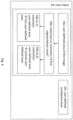
- Fig. 1 schematically shows a method of refining a segmentation mask according to examples of the present disclosure.
- the method may comprise a plurality of operations.
- One or several operations may for example be implemented by a respective software module.
- an image may be provided.
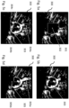
- Fig. 5a shows an example of an image 300a.
- the image may for example be provided by an imaging system, for instance a medical imaging system.
- the image may be associated with a medium scanned by the imaging system.
- the imaging system may comprise at least one of: mammography, tomosynthesis, magnetic resonance imaging (MRI), single-photon emission computerized tomography (SPECT) scan, positron emission tomography (PET) scan, optical coherence tomography (OCCT), optical tomography (OCT), X-ray exam, and ultrasound imaging.
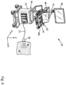
- An exemplary ultrasound system is shown in fig. 6 .
- the image may be obtained by a first system which performs the method of fig. 1 or the image may be prepared or obtained by a second system and provided to the first system.
- a first segmentation mask is provided.
- the first segmentation mask may be obtained in operation S2, based on image data of the image of operation S1.
- the first segmentation mask may be associated with a region of interest, ROI, in the image.
- the ROI may be or may comprise a lesion in the medium, for example a human or animal tissue.
- the first segmentation mask may be obtained by detecting and segmenting a region of interest (ROI) in the medium.
- ROI region of interest
- a predefined computer-implemented segmentation algorithm may be used, for example an AI based algorithm, as for example a neural network, such as a convolutional neural network (CNN).
- the region of interest may be associated with only a part of the image.
- the first segmentation mask may cover for example only a part of the image, i.e. the region of interest (ROI).
- the image and/or the first segmentation mask is merely obtained from a communication interface, a data storage or there like.
- the image and/or the first segmentation mask may be predetermined and/or pre-stored and provided to a system performing the method according to the present disclosure.
- the first segmentation mask may be displayed.
- the first segmentation mask may be displayed on top of the image, as shown in the example illustrated in fig. 5b .
- a user input is provided.
- the user input may comprise a user-defined area, as for example shown in fig. 5c .
- the user-defined area of the image may be provided by the user to modify the first segmentation mask.
- a second segmentation mask is obtained by refining the first segmentation mask based on image data of the image using a computer-implemented algorithm.
- the algorithm thereby penalizes a violation of the user input.
- the second segmentation mask may be displayed.
- the second segmentation mask may be displayed on the display device, optionally adjacent to or instead of the first segmentation mask. The latter case is shown in the example of fig. 5d .
- the method may be iterated in an operation S7.
- the second segmentation mask of an iteration N may be used as a first segmentation mask in an iteration N+1. Accordingly, a user may successively refine the segmentation mask by modifying the (first) segmentation mask in each iteration.
- the method may be performed at least partially in real-time.
- obtaining the second segmentation mask in operation S5 may be performed in real-time or in quasi real-time, such that a user providing the user input does not perceive any significant delay, when concentrating on the image displayed on a display device.
- Fig. 2 shows a method of obtaining second segmentation mask according to examples of the present disclosure.
- the method of fig. 2 may correspond to or be a particular example of the operation S5 of the method of fig. 1 .
- the method of fig. 2 in particular the operation S5, may be performed by the computer-implemented algorithm according to the present disclosure.
- the operation S5 may comprise a basic refinement operation S5a1, wherein the first segmentation mask is basically refined, for instance based on image data.
- the image data may comprise statistical criteria on image features, for example low-level image features.
- the image features may comprise for example at least one of contours, edges, angles, intensity, color, motion and/or texture.
- the image data may be used by the algorithm to discriminate between image regions to refine the first segmentation mask.
- the operation S5a1 may be performed using the algorithm, or some rules defined by the algorithm, as described in the following.
- the algorithm may be configured to exploit statistics of local likelihood, and/or contextual distributions based on the image data to adjust a segmentation boundary of the first segmentation mask.
- the algorithm may be configured to be adaptable based on a characteristic of the medium scanned by the imaging system, for example the type of medium (for instance a type of organ, such as a liver or a breast).
- x may be an image point, for example a pixel, a voxel, or a point of any dimension.
- the function D ( ⁇ ) may represent a penalization term configured to penalize a reduced level of fitting of the first segmentation mask compared to the image data. Accordingly, the function D ( ⁇ ) may represent the penalization of bad fitness of the first segmentation mask compared to image data. Accordingly, in case of a reduced level of fitting (i.e. a relatively bad fitness) the penalization according to function D ( ⁇ ) may be increased.
- the function F ( ⁇ ) may be configured to penalize a violation of the user input. Accordingly, the function F ( ⁇ ) may for example represent the fuzzy penalization of the violation of the user input by the segmentation.
- the function R ( ⁇ ) may represent a regularization penalization term of the segmentation mask.
- the algorithm for example the function D ( ⁇ ), may be configured to compare the local distribution around each of a plurality of image points to the distribution of an image area inside the first segmentation mask and that of outside the first segmentation mask, such that points of similar contextual information may be grouped together in the second segmentation mask.
- the algorithm may use an optimal transport metric (for example a Weisserstein metric) to compare the distributions.
- an optimal transport metric for example a Weisserstein metric
- the optimal transport metric may be defined by the function D ( ⁇ ).
- An image point may comprise an image pixel, an image voxel, or an image point of any dimension.
- the algorithm for example the function D ( ⁇ ), may be further configured to take into account local likelihood information, such that points likelihoods from an image area inside the first segmentation mask from an image area outside the first segmentation mask may be in competition, which separates the points of the image inside area from the image outside area.
- the algorithm for example the function R ( ⁇ ), may be configured to smooth the segmentation boundary of the second segmentation mask.
- the operation S5 and/or S5a1 comprises an operation S5a2 for penalizing a violation of the user input through the basic refinement operation.
- the algorithm may comprise a penalization rule.
- the penalization rule may be configured to penalize a violation of the user-defined area.
- the user-defined area may be used for example as a soft criterion based on which the first segmentation mask may be modified.
- the algorithm may be configured to include the first user-defined area into the first segmentation mask in a fuzzy manner by applying a predefined penalization rule.
- the algorithm may be configured to exclude the second user-defined area from the first segmentation mask in a fuzzy manner by applying a penalization rule.
- the computer-implemented algorithm may basically be configured to refine the first segmentation mask based on image data of the image, cf. operation S5a1.
- the algorithm penalizes a violation of the user input, i.e. constrains the basic refinement based on image data in view of the user input, cf. S5a2. Accordingly, in case the user-defined area does not match the basic refinement, this basic refinement is constrained to better respect the user-defined area.
- the operation S5, S5a1 and/or S5a2 may comprise an optional operation S5a3 of considering a user-defined confidence level.
- the user input may further comprise a user-defined confidence level which may be associated with the user-defined area.
- the algorithm may be configured to penalize a violation of the user input as a function of the user-defined confidence level.
- parameters may be given in the method such that confidence on the user input can be tuned, for example manually by the user.
- Fig. 3 shows an exemplary method of training an artificial intelligence (AI) algorithm according to examples of the present disclosure.
- a set i.e. a plurality
- the operation may include providing images which are respectively associated with the first segmentation masks.
- the images are desirably different to each other and/or the segmentation masks are desirably different to each other.
- Said first segmentation masks may correspond to the segmentation mask of operation S2 of fig. 1 . Accordingly, the operation S11 may correspond to a plurality of operations S2 providing different first segmentation masks.
- a plurality of second segmentation masks may be obtained.
- Said second segmentation masks may correspond to the segmentation mask of operation S5 of fig. 1 .
- the operation S12 may correspond to a plurality of operations S5 providing different second segmentation masks.
- the second segmentation masks may be obtained using the method according to the present disclosure.
- a training dataset may be generated.
- the training dataset may comprise a set of samples.
- Each sample may comprise a first segmentation mask and/or the associated second segmentation mask, optionally together with the associated image.
- an artificial intelligence (AI) algorithm may be trained using the training dataset.
- AI artificial intelligence
- AI algorithms may comprise machine learning models, such as decision trees, random forests, logistic regression, gradient boosting, neural networks, such as Convolutional Neural Networks (CNNs), and/or support vector machines.
- the AI algorithm may be trained in a supervised manner using the first segmentation masks and/or the respective images as input and the respective second segmentation masks as target output.
- the AI algorithm may learn to predict a second refined segmentation mask based on an image and/or a first segmentation mask.
- the trained AI algorithm may hence automate or at least partially automate the manual work of providing a user input.
- Fig. 4 shows method of providing and processing a user input according to examples of the present disclosure.
- the method of fig. 4 may correspond to or be a particular example of the operation S4 of the method of fig. 1 .
- the method of fig. 2 in particular the operation S4, may be performed by a software module configured to receive and optionally process the user input.
- the operation S4 comprises an operation S4a of providing a user-defined area of the image.
- the user defined area may be provided by a user by manually drawing the area using a user interface device associated with a computing device on which the method is running.
- the user interface device may be for example a touch screen device (for instance the control panel display screen 49 illustrated on fig. 6 ).
- operation S4b it may be determined whether the user-defined area is intended to be an extension or restriction of first segmentation mask.
- the method or the respective software module may be associated with additional input means, which can be used by the user to select the type of user-defined area.
- the additional input means may be for instance one or several physical or software-implemented buttons of a user interface device.
- the type of user-defined area is detected automatically by the method. For example, in case more than 50% of the user-defined area is outside the first segmentation mask (cf. for example the user-defined area 304a illustrated in fig. 5c ), it may be determined that the user-defined area is intended to extend the first segmentation mask. Moreover, in case more than 50% of the user-defined area is inside the first segmentation mask (cf. for example the user-defined area 304b as shown in fig. 5c ), it may be determined that the user-defined area is intended to be removed from the first segmentation mask.
- the user-defined area may be labelled as first user-defined area, which is intended to be added to the first segmentation mask.
- the user-defined area may be labelled as second user-defined area, which is intended to be removed from the first segmentation mask.
- the operation S4 may comprise providing a user-defined confidence level for each provided user-defined area.
- the user-defined confidence level may be associated with the user-defined area.
- the user-defined confidence level may be a number between 0 and 100 which may be manually inputted by the user using the user interface device.
- the user-defined area in particular the first and/or second user-defined area, optionally together with one or several confidence levels, may then be handed over to the operation S5 or its above-mentioned respective algorithm, in which the second segmentation mask is obtained.
- Fig. 5a shows a schematic example of an image according to operation S1 of the method of Fig. 1 .
- the image 300 may be for example an ultrasound image, or any other type of image, as described above in context of operation S1 in fig. 1 .
- the image may display an ultrasound scanned medium.
- the medium can be for example a part of a human body or animal or material.
- the image may be displayed on a display device, as described above and in the context of fig. 6 below.
- the image may comprise a region of interest 302, for example a suspected lesion in the medium.
- the region of interest may be manually selected by a user by drawing a bounding box 301 comprising the region of interest.
- the region of interest is automatically determined, detected.
- Fig. 5b shows a schematic example of a first segmentation mask according to operations S2 and S3 of the method of Fig. 1 .
- the first segmentation mask 303 may automatically be determined, as described above in context of operation S2 of fig. 1 . The determination may also be based on the manually drawn bounding box 301.
- the first segmentation mask 303 may be displayed together with the image 300, in particular by overlaying the image 300.
- the first segmentation mask 303 may thus indicate the borders of the region of interest, e.g. of a supposed lesion in a scanned medium, a muscle, an organ or part of it.
- a user may wish to review and in some cases adapt the first segmentation mask 303 in view of his/her skills and experience.
- the user may wish to modify the border of the first segmentation mask by local extension and/or limitation of the mask.
- FIGS 5c and 5d An example of addressing this problem is shown in figures 5c and 5d .
- Fig. 5c shows a schematic example of a first segmentation mask and user input according to operation S4 of the method of Fig. 1 .
- the system and method may provide means allowing the user to interact in order to provide a user-defined area likely to be included in the ROI, and an area likely to be excluded.
- the user has provided two user-defined areas 304a and 304b, e.g. by drawing lines on the display device, and/or by setting one or several marker points 304c which define the border of the user-defined area.
- the user-defined area 304a may be labelled as first user-defined area, which is intended to be added to the first segmentation mask.
- the user-defined area 304b may be labelled as second user-defined area, which is intended to be removed from the first segmentation mask.
- the user may additionally input a user-defined confidence level (or levels) for one or each or some of the user-defined areas 304a and 304b.
- Fig. 5d shows a schematic example of a second segmentation mask according to operations S5 and S6 of the method of Fig. 1 .
- the second refined segmentation mask 305 is obtained based on image data of the image 300, using a computer-implemented algorithm, wherein the algorithm is configured to penalize a violation of the user input.
- an image area 305a has been included in the second segmentation mask.
- Said added image area 305a is determined by the algorithm based on image data, wherein the user input (i.e. the first user-defined area 304a) may be used as a fuzzy user input.
- an image area 305b has been removed from the second segmentation mask.
- Said added image area 305a is determined by the algorithm based on image data, wherein the user input (i.e. the second user-defined area 304b) is used as a fuzzy user input.
- the user input does not need to be very accurate.
- the statistical terms of the algorithm according to the present disclosure can provide a more accurate segmentation boundary, while the user constraints can be taken into account in a fuzzy manner.
- Fig. 6 shows a schematic drawing of an ultrasound system 10 according to examples of the present disclosure.
- the ultrasound system 10 may be for example a system for refining a segmentation mask according to the present disclosure.
- the system may for example be configured to perform the method of the present disclosure.
- the method according to the present disclosure may also be (at least partially) performed by an external system.
- the ultrasound imaging system 10 may comprise:
- the probe 20 may be associated to the processing unit 30 via a cable 21 and/or via wireless connection(s), and it is able to emit ultrasound waves W into a medium M and to receive ultrasound waves W from the medium M, said received ultrasound waves being consequent or resulting from reflections of said emitted ultrasound waves on diffusing particles inside medium M.
- the display screen 50 may be a screen for visualizing the image processed by the processing unit 30.
- the display 50 may also visualize other information such as scales used in the image, and/or configuration information for the processing or any information such as help information or contextual gesture help for the touch pad 42.
- the display screen may by articulated on a support arm 51 for better positioning for the user.
- the display screen is usually a high-definition screen of a great size (at least 20 inches) for better image visualization to the user.
- the control panel 40a is for example a portion of the system casing 31, said portion comprising a panel casing having a substantially flat surface inclined towards the user for manipulation by one hand of said user.
- the control panel 40a may be moved by a hanger upwards and downward to adapt to the user size, and may be optionally moved frontward and rearward for being adapted to the user position.
- the control panel 40a may include a control panel display screen 49 for visualizing several configuration information or any information dedicated to the user.
- the processing unit 30 may be configured to refine a segmentation mask according to the present disclosure.
- the processing unit may also send data to an external device for at least partially performing the method of the present disclosure on the external device.
- Examples of an external device include a server, a computer, a dedicated workstation , a device for displaying images obtained from the electronic control device or any other external device. Accordingly, the method according to the present disclosure may be carried out by at least one of the processing unit or any of the external devices.
- the process for building the image data based on acquired data i.e. compiling the ultrasound image data, may be carried out by the same processing device as that one for optimizing the optimizing a process, or (at least in part) by another one.
- the system 10 may include at least one processing unit (or processor) and memory.
- the processor and memory unit may be incorporated into the system or may be a computer or computer communicatively linked thereto.
- memory storing, instructions to evaluate ultrasound data or otherwise perform the methods described herein
- RAM volatile
- nonvolatile such as RAM, flash memory, etc.
- the system 10 may also include storage devices (removable and/or non-removable) including, but not limited to, magnetic or optical disks or tape.
- the system 10 may also have input device(s) such as keyboard, mouse, pen, voice input, etc.
- connections such as 4G/5G, WIFI, LAN, WAN, point to point, etc.
- the connections may be operable to facility point-to-point communications, connection-oriented communications, connectionless communications, etc.
- the system 10 may typically include some form of computer readable media.
- Computer readable media can be any available media that can be accessed by processing unit (or processor) or other devices comprising the operating environment.
- Computer readable media may comprise computer storage media and communication media.
- Computer storage media includes volatile and nonvolatile, removable and non-removable media implemented in any method or technology for storage of information such as computer readable instructions, data structures, program modules or other data.
- Computer storage media does not include communication media.
- Communication media embodies computer readable instructions, data structures, program modules, or other data in a modulated data signal such as a carrier wave or other transport mechanism and includes any information delivery media.
- modulated data signal means a signal that has one or more of its characteristics set or changed in such a manner as to encode information in the signal.
- communication media includes wired media such as a wired network or direct-wired connection, and wireless media such as acoustic, RF, infrared, microwave, and other wireless media. Combinations of the any of the above should also be included within the scope of computer readable media.
- the system 10 may be a single computer operating in a networked environment using logical connections to one or more remote computers.
- the remote computer may be a personal computer, a server, a router, a network PC, a peer device or other common network node, and typically includes many or all of the elements described above as well as others not so mentioned.
- the logical connections may include any method supported by available communications media.
Landscapes
- Engineering & Computer Science (AREA)
- Theoretical Computer Science (AREA)
- Physics & Mathematics (AREA)
- General Physics & Mathematics (AREA)
- Computer Vision & Pattern Recognition (AREA)
- Software Systems (AREA)
- Evolutionary Computation (AREA)
- Computing Systems (AREA)
- Artificial Intelligence (AREA)
- Multimedia (AREA)
- General Health & Medical Sciences (AREA)
- Health & Medical Sciences (AREA)
- Medical Informatics (AREA)
- General Engineering & Computer Science (AREA)
- Mathematical Physics (AREA)
- Data Mining & Analysis (AREA)
- Life Sciences & Earth Sciences (AREA)
- Computational Linguistics (AREA)
- Biophysics (AREA)
- Molecular Biology (AREA)
- Biomedical Technology (AREA)
- Nuclear Medicine, Radiotherapy & Molecular Imaging (AREA)
- Radiology & Medical Imaging (AREA)
- Quality & Reliability (AREA)
- Databases & Information Systems (AREA)
- Ultra Sonic Daignosis Equipment (AREA)
- Image Analysis (AREA)
- Image Processing (AREA)
Priority Applications (5)
| Application Number | Priority Date | Filing Date | Title |
|---|---|---|---|
| EP23315261.0A EP4485351A1 (fr) | 2023-06-29 | 2023-06-29 | Procédé d'affinage d'un masque de segmentation |
| US18/658,697 US20250005762A1 (en) | 2023-06-29 | 2024-05-08 | Method of refining segmentation mask |
| JP2024076917A JP2025013742A (ja) | 2023-06-29 | 2024-05-10 | セグメンテーションマスクを改善する方法 |
| KR1020240066243A KR20250001892A (ko) | 2023-06-29 | 2024-05-22 | 세그멘테이션 마스크를 정제하는 방법 |
| CN202410682503.5A CN119228818A (zh) | 2023-06-29 | 2024-05-29 | 精细化分割掩模的方法 |
Applications Claiming Priority (1)
| Application Number | Priority Date | Filing Date | Title |
|---|---|---|---|
| EP23315261.0A EP4485351A1 (fr) | 2023-06-29 | 2023-06-29 | Procédé d'affinage d'un masque de segmentation |
Publications (1)
| Publication Number | Publication Date |
|---|---|
| EP4485351A1 true EP4485351A1 (fr) | 2025-01-01 |
Family
ID=87473936
Family Applications (1)
| Application Number | Title | Priority Date | Filing Date |
|---|---|---|---|
| EP23315261.0A Pending EP4485351A1 (fr) | 2023-06-29 | 2023-06-29 | Procédé d'affinage d'un masque de segmentation |
Country Status (5)
| Country | Link |
|---|---|
| US (1) | US20250005762A1 (fr) |
| EP (1) | EP4485351A1 (fr) |
| JP (1) | JP2025013742A (fr) |
| KR (1) | KR20250001892A (fr) |
| CN (1) | CN119228818A (fr) |
Citations (1)
| Publication number | Priority date | Publication date | Assignee | Title |
|---|---|---|---|---|
| EP3382642A1 (fr) * | 2017-03-27 | 2018-10-03 | Siemens Healthcare GmbH | Système d'annotation et de segmentation hautement intégré pour imagerie médicale |
Family Cites Families (6)
| Publication number | Priority date | Publication date | Assignee | Title |
|---|---|---|---|---|
| WO2016191870A1 (fr) * | 2015-06-01 | 2016-12-08 | The Governors Of The University Of Alberta | Modélisation de surface d'une structure échogène segmentée pour la détection et la mesure d'anomalies anatomiques |
| US12456209B2 (en) * | 2019-08-28 | 2025-10-28 | Hover Inc. | Systems and methods for estimating feature locations based on image analysis and homography |
| EP4042372A1 (fr) * | 2019-10-10 | 2022-08-17 | Koninklijke Philips N.V. | Segmentation d'une image médicale |
| US12094190B2 (en) * | 2021-02-19 | 2024-09-17 | Arizona Board Of Regents On Behalf Of Arizona State University | Systems, methods, and apparatuses for implementing medical image segmentation using interactive refinement |
| WO2023069445A1 (fr) * | 2021-10-18 | 2023-04-27 | Google Llc | Élimination de distraction basée sur une entrée d'utilisateur dans des éléments multimédias |
| US12198349B2 (en) * | 2022-01-25 | 2025-01-14 | GE Precision Healthcare LLC | Methods and systems for real-time image 3D segmentation regularization |
-
2023
- 2023-06-29 EP EP23315261.0A patent/EP4485351A1/fr active Pending
-
2024
- 2024-05-08 US US18/658,697 patent/US20250005762A1/en active Pending
- 2024-05-10 JP JP2024076917A patent/JP2025013742A/ja active Pending
- 2024-05-22 KR KR1020240066243A patent/KR20250001892A/ko not_active Ceased
- 2024-05-29 CN CN202410682503.5A patent/CN119228818A/zh active Pending
Patent Citations (1)
| Publication number | Priority date | Publication date | Assignee | Title |
|---|---|---|---|---|
| EP3382642A1 (fr) * | 2017-03-27 | 2018-10-03 | Siemens Healthcare GmbH | Système d'annotation et de segmentation hautement intégré pour imagerie médicale |
Non-Patent Citations (1)
| Title |
|---|
| WENXIAN YANG ET AL: "User-Friendly Interactive Image Segmentation Through Unified Combinatorial User Inputs", IEEE TRANSACTIONS ON IMAGE PROCESSING, IEEE, USA, vol. 18, no. 9, 1 September 2010 (2010-09-01), pages 2470 - 2479, XP011316788, ISSN: 1057-7149 * |
Also Published As
| Publication number | Publication date |
|---|---|
| CN119228818A (zh) | 2024-12-31 |
| KR20250001892A (ko) | 2025-01-07 |
| US20250005762A1 (en) | 2025-01-02 |
| JP2025013742A (ja) | 2025-01-28 |
Similar Documents
| Publication | Publication Date | Title |
|---|---|---|
| US12067724B2 (en) | Method and system for image segmentation and identification | |
| Golan et al. | Lung nodule detection in CT images using deep convolutional neural networks | |
| JP4949264B2 (ja) | 医療画像データ内の腫瘍境界を自動的に検出及び区分するシステム及び方法 | |
| US10991093B2 (en) | Systems, methods and media for automatically generating a bone age assessment from a radiograph | |
| US11810291B2 (en) | Medical image synthesis of abnormality patterns associated with COVID-19 | |
| US10111632B2 (en) | System and method for breast cancer detection in X-ray images | |
| US8270688B2 (en) | Method for intelligent qualitative and quantitative analysis assisting digital or digitized radiography softcopy reading | |
| JP7350582B2 (ja) | モデル学習用のシステム、装置、及び学習方法 | |
| US20180260957A1 (en) | Automatic Liver Segmentation Using Adversarial Image-to-Image Network | |
| US20200074634A1 (en) | Recist assessment of tumour progression | |
| US20050207630A1 (en) | Lung nodule detection and classification | |
| CN111899244B (zh) | 图像分割、网络模型的训练方法及装置,及电子设备 | |
| CN101002228A (zh) | 结节边界检测 | |
| CN110276741B (zh) | 结节检测及其模型训练的方法和装置以及电子设备 | |
| EP4498327A1 (fr) | Appareil et procédé pour aider à trouver une mise en correspondance de régions dans des images médicales | |
| EP3716157A1 (fr) | Appareil et procédé d'apprentissage de modèles | |
| AU2019204365C1 (en) | Method and System for Image Segmentation and Identification | |
| de Almeida et al. | A deep unsupervised saliency model for lung segmentation in chest X-ray images | |
| JP2022553076A (ja) | 画像のセグメンテーションのためのシステム及び方法 | |
| EP4485351A1 (fr) | Procédé d'affinage d'un masque de segmentation | |
| JPWO2007013300A1 (ja) | 異常陰影候補検出方法及び異常陰影候補検出装置 | |
| Saidani | Efficient Tumor Detection in Medical Imaging Using Advanced Object Detection Model: A Deep Learning Approach. | |
| Rajput et al. | U-Net: A Versatile Deep Learning Architecture for Multi-Disease Detection | |
| TWI767439B (zh) | 醫療影像分析系統及其方法 | |
| Nomura et al. | Can the spherical gold standards be used as an alternative to painted gold standards for the computerized detection of lesions using voxel-based classification? |
Legal Events
| Date | Code | Title | Description |
|---|---|---|---|
| PUAI | Public reference made under article 153(3) epc to a published international application that has entered the european phase |
Free format text: ORIGINAL CODE: 0009012 |
|
| STAA | Information on the status of an ep patent application or granted ep patent |
Free format text: STATUS: THE APPLICATION HAS BEEN PUBLISHED |
|
| AK | Designated contracting states |
Kind code of ref document: A1 Designated state(s): AL AT BE BG CH CY CZ DE DK EE ES FI FR GB GR HR HU IE IS IT LI LT LU LV MC ME MK MT NL NO PL PT RO RS SE SI SK SM TR |
|
| STAA | Information on the status of an ep patent application or granted ep patent |
Free format text: STATUS: REQUEST FOR EXAMINATION WAS MADE |
|
| 17P | Request for examination filed |
Effective date: 20250701 |
|
| RAP3 | Party data changed (applicant data changed or rights of an application transferred) |
Owner name: SUPERSONIC IMAGINE |