EP4491630A1 - Récepteur de lymphocytes t pour la leucémie myéloïde aiguë (lma) - Google Patents
Récepteur de lymphocytes t pour la leucémie myéloïde aiguë (lma) Download PDFInfo
- Publication number
- EP4491630A1 EP4491630A1 EP23386059.2A EP23386059A EP4491630A1 EP 4491630 A1 EP4491630 A1 EP 4491630A1 EP 23386059 A EP23386059 A EP 23386059A EP 4491630 A1 EP4491630 A1 EP 4491630A1
- Authority
- EP
- European Patent Office
- Prior art keywords
- cells
- chain
- tcr
- seq
- cell
- Prior art date
- Legal status (The legal status is an assumption and is not a legal conclusion. Google has not performed a legal analysis and makes no representation as to the accuracy of the status listed.)
- Withdrawn
Links
Images




Classifications
-
- C—CHEMISTRY; METALLURGY
- C07—ORGANIC CHEMISTRY
- C07K—PEPTIDES
- C07K14/00—Peptides having more than 20 amino acids; Gastrins; Somatostatins; Melanotropins; Derivatives thereof
- C07K14/435—Peptides having more than 20 amino acids; Gastrins; Somatostatins; Melanotropins; Derivatives thereof from animals; from humans
- C07K14/705—Receptors; Cell surface antigens; Cell surface determinants
- C07K14/70503—Immunoglobulin superfamily
- C07K14/7051—T-cell receptor (TcR)-CD3 complex
-
- A—HUMAN NECESSITIES
- A61—MEDICAL OR VETERINARY SCIENCE; HYGIENE
- A61K—PREPARATIONS FOR MEDICAL, DENTAL OR TOILETRY PURPOSES
- A61K40/00—Cellular immunotherapy
- A61K40/10—Cellular immunotherapy characterised by the cell type used
- A61K40/11—T-cells, e.g. tumour infiltrating lymphocytes [TIL] or regulatory T [Treg] cells; Lymphokine-activated killer [LAK] cells
-
- A—HUMAN NECESSITIES
- A61—MEDICAL OR VETERINARY SCIENCE; HYGIENE
- A61K—PREPARATIONS FOR MEDICAL, DENTAL OR TOILETRY PURPOSES
- A61K40/00—Cellular immunotherapy
- A61K40/30—Cellular immunotherapy characterised by the recombinant expression of specific molecules in the cells of the immune system
- A61K40/32—T-cell receptors [TCR]
-
- A—HUMAN NECESSITIES
- A61—MEDICAL OR VETERINARY SCIENCE; HYGIENE
- A61K—PREPARATIONS FOR MEDICAL, DENTAL OR TOILETRY PURPOSES
- A61K40/00—Cellular immunotherapy
- A61K40/40—Cellular immunotherapy characterised by antigens that are targeted or presented by cells of the immune system
- A61K40/41—Vertebrate antigens
- A61K40/42—Cancer antigens
- A61K40/4201—Neoantigens
-
- A—HUMAN NECESSITIES
- A61—MEDICAL OR VETERINARY SCIENCE; HYGIENE
- A61P—SPECIFIC THERAPEUTIC ACTIVITY OF CHEMICAL COMPOUNDS OR MEDICINAL PREPARATIONS
- A61P35/00—Antineoplastic agents
-
- A—HUMAN NECESSITIES
- A61—MEDICAL OR VETERINARY SCIENCE; HYGIENE
- A61P—SPECIFIC THERAPEUTIC ACTIVITY OF CHEMICAL COMPOUNDS OR MEDICINAL PREPARATIONS
- A61P35/00—Antineoplastic agents
- A61P35/02—Antineoplastic agents specific for leukemia
-
- C—CHEMISTRY; METALLURGY
- C07—ORGANIC CHEMISTRY
- C07K—PEPTIDES
- C07K14/00—Peptides having more than 20 amino acids; Gastrins; Somatostatins; Melanotropins; Derivatives thereof
- C07K14/435—Peptides having more than 20 amino acids; Gastrins; Somatostatins; Melanotropins; Derivatives thereof from animals; from humans
- C07K14/705—Receptors; Cell surface antigens; Cell surface determinants
- C07K14/70503—Immunoglobulin superfamily
-
- C—CHEMISTRY; METALLURGY
- C12—BIOCHEMISTRY; BEER; SPIRITS; WINE; VINEGAR; MICROBIOLOGY; ENZYMOLOGY; MUTATION OR GENETIC ENGINEERING
- C12N—MICROORGANISMS OR ENZYMES; COMPOSITIONS THEREOF; PROPAGATING, PRESERVING, OR MAINTAINING MICROORGANISMS; MUTATION OR GENETIC ENGINEERING; CULTURE MEDIA
- C12N9/00—Enzymes; Proenzymes; Compositions thereof; Processes for preparing, activating, inhibiting, separating or purifying enzymes
- C12N9/10—Transferases (2.)
- C12N9/12—Transferases (2.) transferring phosphorus containing groups, e.g. kinases (2.7)
-
- C—CHEMISTRY; METALLURGY
- C12—BIOCHEMISTRY; BEER; SPIRITS; WINE; VINEGAR; MICROBIOLOGY; ENZYMOLOGY; MUTATION OR GENETIC ENGINEERING
- C12Y—ENZYMES
- C12Y207/00—Transferases transferring phosphorus-containing groups (2.7)
- C12Y207/10—Protein-tyrosine kinases (2.7.10)
-
- A—HUMAN NECESSITIES
- A61—MEDICAL OR VETERINARY SCIENCE; HYGIENE
- A61K—PREPARATIONS FOR MEDICAL, DENTAL OR TOILETRY PURPOSES
- A61K2239/00—Indexing codes associated with cellular immunotherapy of group A61K40/00
- A61K2239/31—Indexing codes associated with cellular immunotherapy of group A61K40/00 characterized by the route of administration
-
- A—HUMAN NECESSITIES
- A61—MEDICAL OR VETERINARY SCIENCE; HYGIENE
- A61K—PREPARATIONS FOR MEDICAL, DENTAL OR TOILETRY PURPOSES
- A61K2239/00—Indexing codes associated with cellular immunotherapy of group A61K40/00
- A61K2239/38—Indexing codes associated with cellular immunotherapy of group A61K40/00 characterised by the dose, timing or administration schedule
-
- A—HUMAN NECESSITIES
- A61—MEDICAL OR VETERINARY SCIENCE; HYGIENE
- A61K—PREPARATIONS FOR MEDICAL, DENTAL OR TOILETRY PURPOSES
- A61K2239/00—Indexing codes associated with cellular immunotherapy of group A61K40/00
- A61K2239/46—Indexing codes associated with cellular immunotherapy of group A61K40/00 characterised by the cancer treated
- A61K2239/48—Blood cells, e.g. leukemia or lymphoma
-
- C—CHEMISTRY; METALLURGY
- C07—ORGANIC CHEMISTRY
- C07K—PEPTIDES
- C07K2319/00—Fusion polypeptide
- C07K2319/01—Fusion polypeptide containing a localisation/targetting motif
- C07K2319/03—Fusion polypeptide containing a localisation/targetting motif containing a transmembrane segment
-
- C—CHEMISTRY; METALLURGY
- C07—ORGANIC CHEMISTRY
- C07K—PEPTIDES
- C07K2319/00—Fusion polypeptide
- C07K2319/33—Fusion polypeptide fusions for targeting to specific cell types, e.g. tissue specific targeting, targeting of a bacterial subspecies
Definitions
- the present disclosure concerns antigen binding proteins based on sequences from T-cell receptors (TCRs).
- TCRs T-cell receptors
- the proteins disclosed herein, and cells expressing them, may be used in cancer therapy.
- Allo-HSCT allogeneic hematopoietic stem cell transplantation
- Allo-HSCT remains, however, associated with high relapse rates as well as transplant-related morbidity and mortality resulting from donor T cells attacking healthy recipient cells, causing severe graft-versus-host disease (GVHD), and often a suitable donor cannot be identified. This has prompted development of alternative cellular therapies.
- Successful targeting of the lymphoid-specific molecule CD19 in chimeric antigen receptor (CAR) T-cell therapy of acute lymphocytic leukemia has led to attempts at also directing CARs to myeloid cell surface antigens, including CD33, CD123 and FLT3, overexpressed on AML cells.
- CAR chimeric antigen receptor
- CAR T therapies targeting these molecules are associated with toxicities and a need for transplantation to rescue normal hematopoiesis after a short period of treatment. It is also unclear whether all AML propagating cells express these antigens, and therefore whether the targeting has curative potential.
- FLT3 FMS-like tyrosine kinase 3
- ITD internal tandem duplications
- TKD tyrosine kinase domain
- FLT3 mutations are secondary events in leukemogenesis, they are associated with accelerated clonal expansion and disease progression, and treatment with the tyrosine kinase inhibitor (TKI) midostaurin in patients with FLT3 mutations receiving standard induction chemotherapy does significantly increase long-term survival.
- TKI tyrosine kinase inhibitor
- the present disclosure provides proteins comprising a targeting unit able to recognize epitopes comprising the D835Y when presented by HLA on the surface of cells.
- Cells expressing a TCR comprising the targeting unit mediated highly specific and efficient killing of primary AML cells harboring the FLT3 D835Y mutation in vitro, and in vivo.
- the TCR (TCR FLT3D/Y ) is restricted by HLA-A*02:01 (hereafter HLA-A2), expressed in approximately 50% of Caucasians.
- T-cells redirected with TCR FLT3D/Y specifically and efficiently eliminate primary human AML cells harboring FLT3 D835Y in vitro and in vivo, while sparing cells expressing wild type (WT) FLT3.
- the present disclosure provides a protein for specific binding to a human leukocyte antigen (HLA) type A2 presenting an HLA-class 1 peptide comprising the sequence YIMSDSNYV (SEQ ID NO: 1),
- the extracellular ⁇ -chain constant domain is represented by SEQ ID NO: 10 and the extracellular ⁇ -chain constant domain represented by SEQ ID NO: 11.
- the extracellular ⁇ -chain constant domain is fused to a transmembrane domain.
- the extracellular ⁇ -chain constant domain is fused to a transmembrane domain.
- each of the extracellular ⁇ -chain constant domain and the extracellular ⁇ -chain constant domain are fused to a transmembrane domain.
- the transmembrane domain is fused to an intracellular signaling domain.
- the protein comprises a full-length ⁇ -chain and/or a full-length ⁇ -chain.
- the protein comprises a full-length ⁇ -chain represented by SEQ ID NO: 12 and/or a full-length ⁇ -chain represented by SEQ ID NO: 13, or alternatively, a protein according to claim 1 comprising a full-length ⁇ -chain represented by SEQ ID NO: 14 and/or a full-length ⁇ -chain represented by SEQ ID NO: 15.
- the present disclosure provides one or more recombinant nucleic acid molecules which alone or together encode a protein according to the first aspect.
- the present disclosure provides one or more cDNA molecules encoding a protein according to the first aspect.
- the present disclosure provides an immune effector cell transduced with a nucleic acid according to the second aspect.
- the present disclosure provides an immune effector cell expressing a protein according to the first aspect.
- the present disclosure provides an immune effector cell according to the fourth or fifth aspect, wherein said cell is a T-helper cell, a cytotoxic T-cell or a natural killer cell.
- the present disclosure provides a pharmaceutical composition comprising the proteins according to the first aspect, the nucleic acid according to second aspect, or a cell according to the fourth or fifth aspect.
- the present disclosure provides a pharmaceutical composition for intravenous administration comprising T-cells expressing a protein according to any one of the first aspect in their cell membrane.
- the present disclosure provides a method for treatment of acute myeloid leukemia (AML) comprising the steps of
- steps b) and c) in the nineth aspect can be performed in any order, and step c) may be performed at any time.
- the present disclosure provides a method for treatment of acute myeloid leukemia (AML) comprising the steps of
- Neoantigens represent an attractive group of targets in cancer immunotherapy, as they are tumor-specific and can be recognized by T cells as foreign in context of MHC. Neoantigenic burden is an important determinant of clinical success upon checkpoint inhibition, and case reports have demonstrated that neoantigen-reactive T cells can mediate clinical responses. This has brought hope that T cells genetically modified to express TCRs derived from neoantigen-reactive T cells could provide efficient adoptive cell therapy. Patient T cells do, however, spontaneously recognize only 1-2% of candidate neoantigens predicted to be expressed and presented on HLA in solid cancer. As the large majority is unique to the individual patient, therapeutic targeting of neoantigens generally becomes a highly personalized effort.
- the present disclosure provides a TCR recognizing an epitope encoded by the recurrent driver mutation D835Y in the tyrosine-kinase domain of FLT3 in AML presented on the prevalent HLA-A2 allele.
- This TCR selected from healthy donor T-cell repertoires, mediated highly specific and efficient killing of primary AML cells harboring the FLT3 D835Y mutation in vitro, and in vivo.
- Different clinically relevant aspects of therapeutic effects were demonstrated in different in vivo mouse PDX models. This included an almost complete removal of mutated CD34- AML in Model 1 with high engraftment, and complete removal of CD34+ AML in Model 2 with lower levels of reconstitution.
- TCR FLT3D/Y cells rejected patient AML cells in a setting resembling MRD. TCR FLT3D/Y cells thus efficiently eliminated leukemia with both high and low disease burden.
- AML cells that are CD34 + have previously been shown to propagate leukemia in vivo.
- TCR FLT3D/Y cells in vitro can specifically and efficiently target and kill CD34 + FLT3 D/Y mutated AML cells.
- Studies in humans would, however, be required to confirm that the TCR FLT3D/Y cells could achieve this also in vivo. Taken together, this suggests that a therapeutic TCR targeting a single, shared neoantigen has the potential of eliminating the reservoir of leukemia stem cells in AML.
- T cells are, on the other hand, the most sensitive tools available for detection of peptide-MHC complexes.
- Our technology utilizing mRNA-transduced dendritic cells to prime naive T cells from healthy donors provides means to identify neoepitopes and reactive TCRs in a single assay. Presentation of the FLT3 D/Y neoepitope on leukemic cells was confirmed by efficient killing of primary HLA-A2 pos AML cells expressing D835Y, but not the less frequent D835E or D835H mutations or WT FLT3, by TCR FLT3D/Y cells, further demonstrating TCR specificity.
- TCRs memory T cell responses in patient material are interrogated. Identification of TCRs with sufficient affinity is essential for clinical impact.
- a hurdle might be induction of T-cell tolerance due to long-standing co-evolution between cancer and the immune system in absence of inflammation, and lack of sufficient priming. This might contribute to the low spontaneous reactivity that tumor-infiltrating T cells show to the large repertoire of predicted neoantigens in melanoma.
- immune responses to neoantigen vaccines seem to be dominated by de novo responses induced in naive T cells.
- TILs tumor-infiltrating lymphocytes
- TCR FLT3D/Y cells did not, however, react to a panel of 26 HLA-A2 pos cell lines of different tissue origins, and spared normal blood cells and AML cells expressing the FLT3 WT sequence or alternative D835 mutations.
- Treatment with TCR FLT3D/Y cells would be limited to the 3-4% of patients that in addition to expressing the mutation also express HLA-A*02:01 in the Caucasian population. While recurrent FLT3 mutations are frequently known to represent secondary and accelerating AML mutations, our re-analysis of 58 published FLT3 D835Y-mutated AML cases demonstrated that this FLT3 mutation frequently is clonal, and in some cases also might be the initiating mutation or secondary only to a CH mutation. These data are in agreement with the high VAF for FLT3 D835Y in all included AML patients harboring this mutation, where the only selection criterion up front was presence of the mutation.
- TCRs can access recurrent mutations currently inaccessible to CARs. Moreover, TCRs might have inherent advantages relative to CARs that improve T-cell persistence and antigen sensitivity.
- TCR-based therapies are mostly applied in the form of genetically modified T cells, although soluble bispecific TCR engagers have emerged as new opportunities for off-the-shelf therapies at lower cost. The results presented here show that a TCR targeting a single shared neoantigen generated from a healthy donor can provide highly efficacious and specific cancer treatment in vivo in multiple disease-relevant models, paving the way for future off-the-shelf, tumor-specific immunotherapies.
- HLA human leukocyte antigen
- telomere binding By “specific binding” is meant that the protein binds specifically to its target under physiological conditions. Accordingly, a protein for specific binding to a human leukocyte antigen type A2 (HLA-A2) presenting an HLA-class 1 peptide will bind to the HLA-A2 if the cognate HLA-class 1 peptide is presented, but is unlikely to bind when a random HLA-class 1 peptide is presented.
- HLA-A2 human leukocyte antigen type A2
- a protein for specific binding to a HLA-A2 presenting YIMSDSNYV will bind to the HLA-A2 if YIMSDSNYV (SEQ ID NO: 1) is presented, but display little or no binding to HLA type A2 when for instance DIMSDSNYV (SEQ ID NO: 18) is presented.
- HLA-class 1 peptides herein comprise 9 or 10 amino acid residues.
- the target antigen in the present disclosure is HLA-A2 presenting an HLA-class 1 peptide comprising or consisting of YIMSDSNYV (SEQ ID NO: 1).
- Specific binding to a target may be distinguished from off-target or non-specific binding.
- the proteins herein bind their targets with a higher affinity than they bind other molecules (or at least most other molecules).
- the binding of a protein to a target may be measured by any suitable method known in the art, e.g. surface plasmon resonance (SPR) or by binding of soluble, multimerized peptide-HLA complexes to T cells expressing the TCR.
- SPR surface plasmon resonance
- a full-length ⁇ -chain comprises, from N-terminal to C-terminal, a variable domain with three CDRs, an extracellular constant domain, a transmembrane domain and a cytoplasmic domain.
- a full-length ⁇ -chain is represented by SEQ ID NO: 12.
- Another example of a full-length ⁇ -chain is represented by SEQ ID NO: 14.
- a full-length ⁇ -chain comprises, from N-terminal to C-terminal, a variable domain with 3 CDRs, an extracellular constant domain, a transmembrane domain and a cytoplasmic domain.
- a full-length ⁇ -chain is represented by SEQ ID NO 13.
- Another example of a full-length ⁇ -chain is represented by SEQ ID NO: 15.
- TCRs herein comprise a full-length ⁇ -chain and a full-length ⁇ -chain.
- An ⁇ -chain variable domain comprises three complementarity-determining regions (CDRs); CDR1, CDR2 and CDR3 numbered from the N-terminal. Said CDRs are flanked by framework regions.
- An ⁇ -chain variable domain can thus be represented: N-FRAMEWORK1 CDR1 FRAMEWORK2 CDR2 FRAMEWORK3 CDR3 FRAMEWORK4
- ⁇ -chain CDR1 is represented by TTSDR (SEQ ID NO: 2)
- the ⁇ -chain CDR2 is represented by LLSNGAV (SEQ ID NO: 3)
- ⁇ -chain CDR3 is represented by CAVVGGSQGNLIF (SEQ ID NO: 4)
- ⁇ -chain variable domain is set forth in SEQ ID NO: 8.
- Other ⁇ -chain variable domains may have more than 90% sequence identity when compared to SEQ ID NO: 8, provided they comprise the same three CDRs.
- Other ⁇ -chain variable domains may have more than 95% sequence identity when compared to SEQ ID NO: 8, provided they comprise the same three CDRs.
- a ⁇ -chain variable domain comprises three complementarity-determining regions (CDRs); CDR1, CDR2 and CDR3 numbered from the N-terminal. Said CDRs are flanked by framework regions.
- a ⁇ -chain variable domain can thus be represented: N-FRAMEWORK1 CDR1 FRAMEWORK2 CDR2 FRAMEWORK3 CDR3 FRAMEWORK4
- ⁇ -chain CDR1 is represented by MDHEN (SEQ ID NO: 5)
- ⁇ -chain CDR2 is represented by SYDVKM (SEQ ID NO: 6)
- the ⁇ -chain CDR3 is represented by CASSLSPGGGYGYTF (SEQ ID NO: 7)
- ⁇ -chain variable domain is set forth in SEQ ID NO: 9.
- Other ⁇ -chain variable domains may have more than 90% sequence identity when compared to SEQ ID NO: 9, provided they comprise the same three CDRs.
- Other ⁇ -chain variable domains may have more than 95% sequence identity when compared to SEQ ID NO: 9, provided they comprise the same three CDRs.
- An ⁇ -chain variable domain can together with a ⁇ -chain variable domain form an antigen binding unit.
- Such antigen binding units may position the six CDRs to specifically bind to a target under physiological conditions.
- the minimal antigen binding unit comprises an ⁇ -chain variable domain and a ⁇ -chain variable domain.
- the ⁇ -chain variable domain and the ⁇ -chain variable domain may be connected by a peptide linker from the C-terminal of the first variable domain to the N-terminal of the second variable domain.
- the C-terminal of an ⁇ -chain variable domain may be fused to the N-terminal of an extracellular ⁇ -chain constant domain like in the full-length ⁇ -chain.
- an extracellular ⁇ -chain constant domain is set forth by SEQ ID NO: 10.
- the C-terminal of a ⁇ -chain variable domain may be fused to the N-terminal of an extracellular ⁇ -chain constant domain like in the full-length ⁇ -chain.
- an extracellular ⁇ -chain constant domain is set forth by SEQ ID NO: 11.
- the first chain comprises an extracellular ⁇ -chain constant domain (located C-terminal to the variable domain, for example directly C-terminal such that the N-terminal amino acid of the constant domain is joined by a peptide bond to the C-terminal amino acid of the variable domain).
- the second chain comprises an extracellular ⁇ -chain constant domain (located C-terminal to the variable domain, for example directly C-terminal to the variable domain).
- both the first chain and the second chain comprise an extracellular constant domain.
- the constant domains may be human TCR constant domains, or TCR constant domains from another animal, for instance the constant domains may be murine constant domains.
- the constant domains may be synthetic constant domains, or constant domains derived from a synthetic protein.
- both constant domains may be from the same species or different species.
- the ⁇ -chain variable domain and the ⁇ -chain variable domain may be located in two separate chains like in naturally occurring TCRs.
- proteins comprising an ⁇ -chain variable domain and a ⁇ -chain variable domain, which together form an antigen binding unit may have many different formats.
- single chain TCRs are known and soluble fragments of TCRs are also known.
- polypeptide chain comprising the ⁇ -chain variable domain and polypeptide chain comprising the ⁇ -chain variable domain may be joined covalently or non-covalently as desired.
- the separate polypeptide chains of the proteins disclosed herein may be non-covalently joined by dimerization domains like coiled coils with opposite charges.
- leucine zippers are used to non-covalently join the chains.
- Leucine zippers, and their sequences, are well-known in the art, and are reviewed in e.g. Busch & Sassone-Corsi (Trends in Genetics 6: 36-40, 1990 ). Combinations of covalent and non-covalent interactions may also be used to join truncated or full-length ⁇ - and ⁇ -chains.
- the separate polypeptide chains of the proteins disclosed herein may be covalently joined (e.g. by one or more disulphide bonds formed between the side chains of cysteine residues).
- disulphide bonds formed between the side chains of cysteine residues.
- an additional cysteine bridge may be introduced by insertion or by substituting a suitable amino acid residue on each chain with a cysteine residue.
- the additional cysteine residues should be introduced at locations such that formation of a disulphide bond between the introduced residues leads to the formation of a functional complex in which the variable regions are correctly located and orientated with respect to each other, e.g. as disclosed by Boulter, J.M. et al. (Protein Eng. Des. Sei. 16(9): 707-711, 2003 ).
- the protein disclosed herein comprises an extracellular ⁇ -chain constant domain comprising the amino acid sequence set forth in SEQ ID NO: 10, or an amino acid sequence having at least 90 % or 95 % sequence identity thereto; and the second chain comprises an extracellular ⁇ -chain constant domain comprising the amino acid sequence set forth in SEQ ID NO: 11, or an amino acid sequence having at least 90 % or 95 % sequence identity thereto.
- These constant domains may comprise cysteine residues able to form one, two or three interchain cysteine bridges.
- receptors comprising an ⁇ -chain variable domain and a ⁇ -chain variable domain are known. Such receptors usually comprise one or two transmembrane domains. An example of such receptor comprising a single transmembrane domain is provided in WO2019069125 . An example of such receptors comprising two transmembrane domains are conventional ⁇ TCRs.
- the C-terminal of an extracellular ⁇ -chain constant domain may be fused to the N-terminal of a transmembrane domain like in the full-length ⁇ -chain.
- a suitable ⁇ -chain transmembrane domain is LSVMGLRILLLKVAGFNLLMTLRL, set forth by SEQ ID NO: 21.
- the C-terminal of an extracellular ⁇ -chain constant domain may be fused to the N-terminal of a transmembrane domain like in the full-length ⁇ -chain.
- a suitable ⁇ -chain transmembrane domain is SATILYEILLGKATLYAVLVSGLVLM, set forth by SEQ ID NO: 22.
- the C-terminal of a transmembrane domain may be fused to the N-terminal of an intracellular signaling domain like in the full-length ⁇ -chain or in the full-length ⁇ -chain.
- An ⁇ -chain intracellular domain, WSS is represented by SEQ ID NO: 23.
- a ⁇ -chain intracellular domain, AMVKKKNS is represented by SEQ ID NO: 24.
- intracellular signaling domains may also be used.
- a variety of intracellular signaling domains are known, and they can be combined and tailored to fit the endogenous signaling machinery in the immune cells.
- the intracellular signaling domain comprises a "signal 1" domain like the signaling domains obtainable from the human proteins CD3 ⁇ , FcR- ⁇ , CD3 ⁇ etc.
- signal 1 e.g. CD3 ⁇ signaling domain
- the intracellular signaling domain comprises a costimulatory domain.
- Such domains are well known and often referred to as “signal 2" domains, and they are believed to, subsequently of “signal 1" domains, convey a signal via costimulatory molecules.
- Examples of such commonly used human “signal 2" domains include 4- 1BB signaling domain, CD28 signaling domain and ICOS signaling domain.
- a conventional leader sequence may be introduced N-terminally for facilitating location in the cell membrane.
- the leader sequence is believed to be trimmed off and will likely not be present in the functional receptors in the cell membrane.
- soluble proteins may be isolated and purified, using techniques known in the art.
- the protein may be produced using a host eukaryotic cell.
- the protein may be expressed such that it comprises a leader sequence which directs it for export.
- the protein is exported from the production cell into the culture medium.
- the culture medium is then separated from the production cell, e.g. by centrifugation.
- Soluble proteins provided herein may be linked or conjugated to a therapeutic or diagnostic agent, or a carrier which comprises or contains a therapeutic or diagnostic agent.
- a therapeutic agent is an agent used in therapy. By therapy is meant the treatment or prevention of a disease.
- the therapeutic agent may be an agent useful in the treatment of a neoplastic condition, particularly cancer.
- nucleic acid molecules encoding the proteins.
- the nucleic acid molecules provided herein may be an isolated nucleic acid molecule and may include DNA or RNA or chemical derivatives of DNA or RNA.
- the term "nucleic acid molecule” specifically includes single- and double-stranded forms of DNA and RNA.
- the nucleic acid e.g. DNA or RNA
- the nucleic acid may be circular or linear.
- a "recombinant" nucleic acid molecule is a nucleic acid molecule synthesized using recombinant techniques, e.g. molecular cloning.
- the recombinant nucleic acid molecule provided herein encodes a protein as provided herein.
- such recombinant nucleic acids may have promoters allowing expression of the proteins in cellular systems.
- the recombinant nucleic acid molecule or construct provided herein may be provided within a vector.
- the term "vector” as used herein refers to a vehicle into which the nucleic acid molecule or construct provided herein may be introduced (e.g. be covalently inserted) from which the specific binding molecule encoded by the nucleic acid molecule may be expressed and/or the nucleic acid molecule/construct cloned.
- the vector may accordingly be a cloning vector or an expression vector.
- the recombinant nucleic acid molecule provided herein may be provided within a recombinant construct comprising the recombinant nucleic acid molecule linked to a heterologous nucleic acid sequence.
- heterologous as used herein is meant a nucleic acid sequence which is not natively linked to the nucleic acid molecule described herein, i.e. which is not linked to the nucleic acid molecule described herein in nature.
- the term “linked” as used herein with respect to the construct may simply mean that the nucleic acid molecule is directly joined to a heterologous nucleic acid sequence.
- the nucleic acid molecule provided herein is operatively linked to a heterologous expression control sequence.
- expression control sequence refers to nucleotide sequences located upstream of, within, or downstream of a coding sequence, and which influence transcription, RNA processing or stability, or translation of the associated coding sequence (i.e. which influence any aspect of expression of the encoded specific binding molecule).
- Expression control sequences include promoters, promoter elements such as a TATA box or a B recognition element, operators, enhancers, translation leader sequences, terminator sequences and suchlike.
- the recombinant nucleic acid molecule comprises a cDNA molecule.
- cDNA as used herein is meant cDNA in its true and original sense (i.e. DNA synthesized by reverse transcription of mRNA), DNA amplified from original cDNA, and also DNA that is equivalent to cDNA.
- DNA that is equivalent to cDNA is DNA that encodes a protein as provided herein and that lacks introns, such that it resembles a protein-coding sequence obtained by reverse transcription of mRNA.
- the nucleic acid molecule encoding a protein as provided herein is codon-optimized, in particular the binding protein may be encoded by a codon-optimized cDNA sequence.
- a nucleic acid molecule provided herein may thus encode a protein as provided herein in the form of a single polypeptide comprising a first chain (e.g. an ⁇ -chain) linked to a second chain (e.g. ⁇ -chain) by a selfsplicing 2A peptide.
- the 2A peptide may have any suitable sequence, such that it has selfsplicing activity.
- 2A peptide sequences are known, any of which may be used according to the present disclosure, e.g. 2A sequences disclosed in Wang et al., 2015 (Scientific Reports 5: Article No. 16273 ) and WO 2019/166463 .
- the proteins as provided herein may be encoded with an N-terminal leader sequence.
- leader sequences are believed to direct the proteins to the cell membrane, either for export (in the case of a soluble protein) or insertion into the membrane (in the case of a binding protein comprising a transmembrane domain), and upon export of the protein or its insertion into the membrane, are trimmed off to yield the mature protein.
- An exemplary ⁇ -chain N-terminal leader sequence may have the amino acid sequence MKSLRVLLVILWLQLSWVWSQ, set forth in SEQ ID NO: 19 and exemplary ⁇ -chain N-terminal leader sequences may have the amino acid sequence MGIRLLCRVAFCFLAVGLV, set forth in SEQ ID NO: 20.
- SEQ ID NO: 19 and SEQ ID NO: 20 are used as leader sequences.
- leader sequences may be used according to the present disclosure, but in one embodiment the first chain (e.g. ⁇ -chain) is encoded with a leader sequence comprising the amino acid sequence set forth in SEQ ID NO: 19, or an amino acid sequence having at least 80 % or 90 % sequence identity thereto; and the second chain (e.g. ⁇ -chain) is encoded with a leader sequence comprising the amino acid sequence set forth in SEQ ID NO: 20, or an amino acid sequence having at least 80 % or 90 % sequence identity thereto.
- the first chain e.g. ⁇ -chain
- the second chain e.g. ⁇ -chain
- kits comprising a first nucleic acid molecule encoding a first chain of a binding protein as provided herein and a second nucleic acid molecule encoding a second chain as provided herein.
- the kit comprises a pair of nucleic acid molecules, one encoding a first chain and the other a second chain as described herein.
- the two nucleic acid molecules of the kit may be provided in a single container (i.e. in a mixture of the two nucleic acid molecules) or in separate containers.
- the nucleic acid molecules may be provided in an aqueous solution (e.g. in water or a suitable buffer such as TE buffer) or may be provided in a lyophilized form.
- sequence identity when referring to "sequence identity" of proteins, an amino acid sequence having at least x% identity to a second amino acid sequence means that x% represents the number of amino acid residues in the first sequence which are identical to their matched amino acid residues of the second sequence when both sequences are optimally aligned via a global alignment, relative to the total length of the second amino acid sequence. Both sequences are optimally aligned when x is maximum by using the comparison matrix BLOSUM62 with gap costs: existence 12, extension 4.
- Framework regions may tolerate substitution, addition or deletion of amino acid residues while keeping the ability to position the CDRs for specific binding to a target epitope.
- sequences of the constant domains are quite conserved. However, some conservative amino acid substitutions may be tolerated within the constant domains.
- conservative amino acid substitution refers to an amino acid substitution in which one amino acid residue is replaced with another amino acid residue having a similar side chain.
- Amino acids with similar side chains tend to have similar properties, and thus a conservative substitution of an amino acid important for the structure or function of a polypeptide may be expected to affect polypeptide structure/function less than a non-conservative amino acid substitution at the same position.
- Families of amino acid residues having similar side chains have been defined in the art, including basic side chains (e.g. lysine, arginine, histidine), acidic side chains (e.g. aspartic acid, glutamic acid), uncharged polar side chains (e.g.
- a conservative amino acid substitution may be considered to be a substitution in which a particular amino acid residue is substituted for a different amino acid residue in the same family.
- the immune cells expressing the disclosed proteins herein may be isolated from a patient or a compatible donor by leukapheresis or other suitable methods.
- Such primary cells may for example be T cells or NK cells.
- autologous T cells both cytotoxic T cells, T helper cells or mixtures of these
- the immune cells expressing the proteins disclosed herein may also be cell lines suitable for clinical use like NK-92 cells.
- the preferred cells are human when the intended patient is human.
- the pharmaceutical compositions herein can be a composition suitable for administration of therapeutic cells to a patient.
- the most common administration route for T-cells is intravenous administration.
- said pharmaceutical compositions may for example be sterile aqueous solutions with a neutral pH.
- said pharmaceutical compositions may for example be sterile aqueous solutions with a physiological pH.
- a patient's peripheral blood mononuclear cells may be obtained via a standard leukapheresis procedure.
- the mononuclear cells may be enriched for T-cells, before transducing them with a lentiviral vector or mRNA encoding the proteins disclosed herein, in particular TCRs. Said cells may then be activated with anti-CD3/CD28 antibody coated beads.
- the transduced T-cells may be expanded in cell culture, washed, and formulated into a sterile suspension, which can be cryopreserved. If so, the product is thawed prior to administration.
- the human ⁇ -chain can be represented: TRBV28-1-CASSLSPGGGYGYTF-TRBJ1 -2
- the human ⁇ -chain can be represented: TRAV39-1-CAVVGGSQGNLIF-TRAJ42
- T cells reactive to FLT3 D835Y HLA-A*02:01 can be induced by culture of naive healthy donor T cells have not been identified in samples from AML patients at diagnosis or post allo-HSCT.
- Naive T cells from a large number of HLA-A2 positive healthy blood donors were co-cultured with autologous monocyte-derived dendritic cells (moDCs) electroporated with mRNA encoding a 9-mer and 10-mer peptide with the FLT3 D835Y mutation in position 1, predicted to be strong binders to HLA-A*02:01, respectively.
- the sequence was flanked by 9 - 11 amino acids at the N-terminal and C-terminal end (target mRNA, encoding KICDFGLARYIMSDSNYVVRGNVRLARLP), or control mRNA.
- PBMCs peripheral blood mononuclear cells
- TCR FLT3D/Y is efficiently expressed in T cells and recognizes a mutant peptide with higher pHLA stability as compared with the corresponding wt peptide
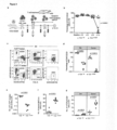
- TCR FLT3D/Y sequence was efficiently expressed in third party peripheral blood (PB) T cells by retroviral transduction, and are called TCR FLT3D/Y cells ( Fig. 1c ). Binding to HLA-A2, as well as endogenous processing and presentation of the peptide, was demonstrated by immunopeptidomics and targeted mass spectrometry analysis of primary AML cells from two patients, using a monoallelic cell line overexpressing the D835Y mutation as a positive control ( Fig. 1d ).
- TCR FLT3D/Y re-directed CD8 + T cells stained positively with FLT3 D/Y pMHC multimers and anti-mouse TCR- ⁇ -PE (reactive to the mouse constant region introduced into the TCR and negatively with anti-human TCR ⁇ , indicating preferential pairing of the introduced TCR- ⁇ and ⁇ -chains and suppression of the endogenous TCR.
- Both CD8 + and CD4 + T cells exhibited a predominantly naive profile and expanded similarly to T cells transduced with a control TCR in vitro, the clinically applied NY-ESO1-specific 1G4 (TCR 1G4 ), indicating lack of fratricide.
- TCR FLT3D/Y cells show high peptide sensitivity and specifically recognize the FLT3 D/Y epitope in an HLA-A2-restricted manner
- TCR 1G4 cells recognized the cognate NY-ESO1 peptide with an EC 50 of 5.5nM, in agreement with previous data showing EC 50 of 7.7nM (41).
- TCR FLT3D/Y cells showed no or negligible reactivity to a panel of 26 HLA-A2 pos cell lines of different tissue origins, unless pre-loaded with mutant peptide, suggesting a high degree of peptide and HLA specificity ( Fig. 1g ).
- TCR FLT3D/Y cells were screened for IFN- ⁇ production in response to target cells loaded with each peptide in a library of 161 peptides representing single amino acid-substituted variants of the cognate peptide ( Fig. 1h ).
- Peptide motifs harboring any "permitted" alternative amino acids in each position were queried in the human proteome databases UniProtKB/Swiss Prot by employing the ScanProsite tool (https://prosite.expasy.org/scanprosite/), identifying 28 additional 9mers in the human proteome potentially recognized by the TCR FLT3D/Y cells.
- TCR FLT3D/Y cells specifically recognize and kill primary FLT3 D835Y AML cells in vitro
- the FLT3 D835Y mutation had the highest variant allele frequency (VAF) among identified recurrent driver single nucleotide variants (SNVs)/indels in 22 cases (45%). In two additional cases, it was only preceded by a recurrent SRSF2 and TET2 mutation, known to occur in normal individuals with clonal hematopoiesis (CH) and requiring additional driver mutations to transform to AML.
- VAF variant allele frequency
- TCR FLT3D/Y cells killed AML cells from 11 patients.
- DNA sequencing of mononuclear samples dominated by myeloid cells ( FIG. 2a ), showed high FLT3 D835Y clonal involvement in the eight FLT3 D835Y pos patients ( Fig. 2b ).
- TCR FLT3D/Y cells killed myeloid cells from AML patients 1-7 effectively at effector:target (E:T) ratios as low as 1:2 (mean 87%, range 53.3% - 98.7%, Fig. 2c and 2d ).
- CD3 + T cells and CD19 + CD20 + B cells were not significantly affected ( Fig.
- TCR FLT3D/Y cells were very low, and we therefore also investigated the effect of TCR FLT3D/Y cells on isolated B cells from healthy donors, which demonstrated lack of killing.
- the HLA-restriction of TCR FLT3D/Y was confirmed by lack of killing of myeloid HLA-A2 neg FLT3 D835Y patient cells (pt. 8, Fig. 2c and 2d ).
- Specificity for the D835Y substitution was demonstrated by lack of recognition of AML samples with alternative amino acid substitutions in D835 position (pt. 9-10) or expressing FLT3 WT (pt. 11) ( Fig. 2d ).
- TCR FLT3D/Y cells derived from AML patients killed autologous leukemia cells with similar efficacy and selectivity as third party T cells, mimicking a clinical setting ( Fig. 2e ). In most cases, 2-4 experiments were performed per patient sample, with only 1 experiment performed due to limited amount of material in a few instances, as described in figure legends.
- TCR FLT3D/Y cells efficiently target human leukemia cells in vivo
- TCR FLT3D/Y cells recognized endogenously presented antigen in a small experiment using an in vivo xenograft mouse model engrafted with a leukemia cell line. Since FLT3 D835Y leukemic cell lines are not commercially available, we introduced the mutation into different leukemia cell lines and demonstrated that TCR FLT3D/Y cells killed >95% of the leukemic cells in 24h at low E:T ratios (1:2). BV173 D835Y cells (cell line origin: B cell precursor leukemia) were next transplanted into NOD-scid IL2Rg null (NSG) mice.
- NSG NOD-scid IL2Rg null
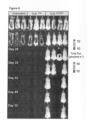
- mice receiving the therapeutic TCR two survived for the duration of the experiment (52 days), one remained leukemia free but was sacrificed on day 41 due to GVHD, whereas one relapsed day 28 day and was found dead on day 32 (see Figure 6 ).
- no leukemic cells were detected in the bone marrow (BM) of the two surviving TCR FLT3D/Y cell-treated mice, while transduced T cells in PB and BM persisted.
- BM bone marrow
- TCR FLT3D/Y cells efficiently eliminate primary AML in vivo in patient-derived xenograft models, including AML-propagating cells
- mice engrafted with primary AML cells in different patient-derived xenograft models.
- mice were closely monitored for potential allo-reactivity mediated by endogenous TCRs of the TCR-transduced T cells derived from a third-party donor, which could otherwise confound the anti-tumor reactivity mediated by the TCR FLT3D/Y .
- terminal analysis of all TCR 1G4 -treated and TCR FLT3D/Y -treated mice was performed on day 15 after T-cell infusion when tumor burden started to decline slightly also in control mice ( Fig. 3b ).
- BM analysis demonstrated high CD33 + AML engraftment in TCR 1G4 -treated mice (mean 86.9 ⁇ 5.3%), which was reduced to mean 0.5 ⁇ 0.2% in TCR FLT3D/Y -treated mice, with a similar reduction in the spleen ( Fig. 3c-d ).
- An almost complete elimination of FLT3 D/Y AML cells in BM was confirmed by ddPCR analysis, quantifying clonally involved cells ( Fig. 3e ).
- efficient targeting of human AML cells in TCR FLT3D/Y- treated mice resulted in recovery of mouse hematopoiesis, which was severely suppressed by high leukemic burden in TCR 1G4 -treated mice ( Fig. 3f ).
- TCR-transduced T cells were detected in BM, spleen and PB of all mice throughout the experiment ( Fig. 3g ).
- mice had relatively low levels of PB human CD33 + engraftment 34 days after T-cell injection (mean 6.0 ⁇ 3.1% hCD45 + CD33 + cells), whereas BM engraftment was much higher (mean 23.7 ⁇ 5.8%) ( Fig. 4b ).
- TCR FLT3D/Y treated mice this was reduced to 1.9 ⁇ 0.7% in PB and 1.7 ⁇ 0.3 % in BM.
- Terminal BM analysis day 34 after T-cell injection revealed distinct populations of CD33 + CD34 - (mean 19.4 ⁇ 4.8%) and CD33 + CD34 + cells (mean 4.4 ⁇ 1.0 %) in control TCR 1G4 -treated mice ( Fig. 4b and 4c ).
- DNA sequencing confirmed that cells engrafted in NSG-SGM3 mice were represented by the same exonic mutations as those detected in AML blasts from BM of patient 1, with FLT3 D835Y and WT1 H507P representing the only detected recurrent driver mutations. Further quantification by ddPCR demonstrated that FLT3 D835Y and WT1 H507P mutations represented the dominating clone, with a VAF >45% in patient 1 as well as in engrafted NSG-SGM3 mice ( Fig. 4d ).
- mice engrafted with patient 1 AML cells were performed into NSG mice, a mouse strain that in contrast to NSG-SGM3 mice does not specifically enhance human myeloid lineages.
- NSG-SGM3 model we observed efficient elimination of the FLT3 D/Y primary AML cells also in this MRD setting ( Fig. 5a-b ).
- TCR FLT3D/Y treatment resulted in undetectable levels of CD34 + FLT3 D/Y AML cells in PB, spleen and BM of all mice engrafted with AML cells from patient 1, compatible with complete elimination of FLT3 D/Y AML propagating cells in vivo through specific TCR FLT3D/Y targeting. More definitive support for this would, however, require longer follow up of mice to allow for potential outgrowth of rare and therapy-resistant AML stem cells that might have escaped T-cell recognition. A long follow-up time was not possible in models 1-3 due to continued presence of TCR T cells having endogenous TCR repertoires that cause allo-reactivity and xenoreactivity over time.
- mice to establish whether all AML-propagating cells have been eliminated by the TCR FLT3D/Y , requires assessment in the absence of TCR FLT3D/Y T cells.
- patient 1 AML cells were cultured with TCR FLT3D/Y or TCR 1G4 cells or without T cells in vitro for 48 hours prior to transplantation into NSG mice ( Fig. 5c ).
- the very few human T cells to which the AML cells were exposed in vitro did not persist in vivo.
- the mice did not get infusions of IL-2. Potential AML development could therefore be followed for 28 weeks.
- mice injected with AML cells from control cultures without T cells or with control TCR 1G4 cells showed progressive and high leukemic engraftment.
- PBMCs from healthy donor buffy coats were obtained from the blood bank of Oslo University Hospital, and PB or BM mononuclear cells from leukemia patients were from biobanked, cryopreserved material (ethical approval # 2018/879 and 2018/1246).
- Mononuclear cells were isolated by density-gradient centrifugation (Axis-Shield) and were typed for HLA-A2 expression by flow cytometry.
- genomic DNA was extracted (QIAGEN DNeasy purification kit) from patient primary cells, and samples were sequenced using the TruSight TM Myeloid Panel (Illumina, San Diego, CA, USA).
- Epstein Barr-virus transformed lymphoblastoid cell lines were generated from HLA-A2 pos and HLA-A2 neg PBMCs as described previously (68).
- the following cell lines were obtained from American Type Culture Collection (ATCC) or German Collection of Microorganisms and Cell Cultures (DSMZ): NALM-6, BV173, RS4;11, T2, RD, U-2 OS, FM-6, UT-7, HeLa, HaCaT, MCF-7, K562, ML-2, COLO 668, EA.hy926, U-87 MG, Daoy, HCT-116, CHP-212, OCI-M2, HEK 293, Hep G2, MV-4-11, EoL-1, MOLM-13, Caco-2, HEL 92.1.7, B721.221, Phoenix-AMPHO.
- a minigene was designed to encode predicted epitopes (https://services.healthtech.dtu.dk/service.php?NetMHC-4.0 ) containing the FLT3 D835Y mutation, codon-optimized and synthesized by Genscript. Subsequently, it was cloned into the pCIpA102 vector for in vitro mRNA transcription using the RiboMAX LargeScale RNA production system (Promega), as previously described (69, 70). The minigene encoded the FLT3 amino acid sequence KICDFGLAR YIMSDSNYVV RGNVRLARLP.
- Monocytes from PBMCs of HLA-A2 pos healthy donors were isolated on day -4 using CD14-reactive microbeads and AutoMACS Pro Separator (Miltenyi Biotec), while the remaining CD14 neg PBMC fraction was cryopreserved for later use.
- the monocytes were then cultured for three days in CellGro GMP DC medium (CellGenix) supplemented with 1% (vol/vol) human serum (HS, Trina biotech), 1% (vol/vol) penicillin/streptomycin (P/S, Sigma-Aldrich), 50 IU/ml Interleukin (IL)-4 (PeproTech) and 800 IU/ml GM-CSF (Genzyme).
- the monocyte-derived-dendritic cells were matured for 14-16 h by adding lipopolysaccharide (LPS; Sigma-Aldrich) and IFN- ⁇ (PeproTech) to final concentrations of 10 ng/ml and 100 IU/ml respectively.
- LPS lipopolysaccharide
- IFN- ⁇ IFN- ⁇
- naive CD8 + T cells were isolated from the autologous CD14 neg cryopreserved PBMCs by use of AutoMACS Pro Separator and CD8 + T-cell isolation kit mixed with CD45RO- and CD57-reactive beads (Miltenyi Biotec).
- cryopreserved PB samples were thawed and resuspended in Iscove's Modified Dulbecco's (IMDM) supplemented with 20% (vol/vol) FCS (Trina biotech) and 0.1mg/ml DNase. Between 2-4 mill viable cells were recovered and were resuspended at a concentration of 1M/ml and pulsed with the FLT3 D/Y peptide at 100ng/ml for 2 hours at 37°C.
- IMDM Iscove's Modified Dulbecco's
- T cells were then washed and resuspended at a concentration of 3.75M/ml in IMDM 5% HS, 1x P/S and 20 U/mL IL-2 before culture for 5 days.
- Identification of T cells reactive to the peptide was performed by staining with pMHC multimers conjugated to PE and APC as described above.
- PBMCs from three healthy donors were isolated, mixed in equal ratio (1:1:1) and were then irradiated with 35 Gy, washed and resuspended in X-vivo 20 medium (Lonza, BioNordika) supplemented with 5% HS and 1% P/S (T-cell medium).
- 0.2 ⁇ 10 6 irradiated cells were placed into tissue culture treated 96-well plates and were supplemented with 100 ⁇ L of T-cell medium containing 2 ⁇ g/ml phytohaemagglutinin (PHA; Remel Thermo Scientific), 80 ng/ml IL-2 (R&D Systems) and 4 ng/ml IL-15 (PeproTech).
- PHA phytohaemagglutinin
- R&D Systems R&D Systems
- 4 ng/ml IL-15 PeproTech
- the FLT3 D/Y co-cultures were then harvested and stained with LiveDead Fixable Near-IR, anti-CD3, -CD8, PE- and APC-conjugated pMHC multimers and using the FACS Aria II (BD Biosciences) cell sorter, CD8 + , pMHC double positive multimer populations were sorted as single cells into 96-well plates containing feeders. After 7 days, cultures were supplied with fresh T-cell medium containing 1750 U/ml IL-2 and 4ng/ml IL-15, and expanding clones were identified by microscopy. At day 14 growing clones were restimulated with feeder cells prepared as described above and stained with FLT3 D/Y pMHC multimers. To assess functionality after further expansion, the clones were stimulated with K562 cells pulsed with FLT3 D/Y or FLT3 WT peptides and assessed for upregulation of CD137.
- FACS Aria II BD Biosciences
- Paired TCR ⁇ and ⁇ chains from two clones and 55 single cells reactive to FLT3 D/Y were amplified using a protocol described previously that was modified and adapted for the targeted amplification of TCR ⁇ and ⁇ transcripts (37, 54, 68).
- MiXCR script was used to analyze sequencing data, and an in-house python script TCR primer was used to reconstruct full-length TCR chains as described previously (54, 73).
- Output was manually verified for each sample in IMGT/V-Quest (74).
- Variable TCR- ⁇ and TCR- ⁇ fragments of identified TCRs were codon-optimized, synthesized and cloned by Genscript.
- the FLT3 D/Y and NY-ESO-1 (1G4) specific TCRs were transduced into HLA-A2 pos healthy donor-derived and patient-derived PBMCs for both in vitro and in vivo experiments.
- 12-well tissue culture-treated plates were coated with anti-CD3 (clone OKT3, eBioscience) and anti-CD28 (clone CD28.6, eBioscience) antibodies for at least 2 hours at 37°C.
- PBMCs were harvested, resuspended in CellGro GMP DC medium supplemented with 5% human serum, IL-7 and IL-15, mixed with retroviral supernatant, placed in non-tissue culture treated 6-well plates pre-coated with Retronectin (20 ⁇ g/ml, Takara) and spinoculated at 900 g for 60 min.
- a second spinoculation was performed the following day with fresh retroviral supernatant and transduction efficiency was determined after 3 days by staining with anti-mouse TCR- ⁇ chain antibody and/or pMHC multimer followed by flow cytometry.
- cells Prior to functional experiments, cells were cultured for 48-72 h in CellGro GMP DC medium containing low concentrations of cytokines (0.5 ng/ml IL-7 and IL-15). Alternatively, cells were frozen for later experiments.
- BV173, ML-2, RS4;11 and NALM-6 cell lines were also transduced as described above using retroviral supernatant containing the FLT3 D/Y minigene.
- the BV173 cell line was stably transduced to express the FLT3 D/Y minigene, firefly luciferase and green fluorescent protein (GFP) and was designated as BV173 D835Y . All transduced cell lines were subsequently purified by FACS sorting, expanded and frozen for use in later experiments.
- IP-MS Immunoprecipitation-Targeted Mass spectrometry
- Mono-allelic B721.221 cells expressing HLA-A*02:01 were transduced using retroviral supernatant encoding the mutant FLT3 amino acid sequence VLVTHGKVVKICDFGLAR YIMSDSNYV VRGNARLPVK.
- One hundred million cells from the B721.221 and four hundred million cells from the patient samples (patient 1 and 3) were each lysed in PBS containing 1% lauryl maltoside, 0.5mM EDTA, 1mM PMSF and Sigma protease inhibitors (1:200) for 1h at 4°C (1 mL lysis buffer per 100 million cells).
- the clarified cell lysates (12.000 RPM, 15 min), were then added to 200uL of AminoLink plus bead slurry (ThermoFisher Scientific) coated with pan HLA class I specific antibody (W6/32, BioXcell) and incubated for 3h at 4°C.
- the HLA class I antibody was coupled to the AminoLink plus bead slurry, following the manufacturers suggestions (10 mg of W6/32 antibody was coupled to 2 mL of amino link plus bead slurry).
- the beads were washed with 3 mL of 0.1M Tris-HCl/150mM NaCl, 3 mL of 0.1M Tris-HCl/400mM NaCl, and again with 3 mL of 0.1M Tris-HCl/150mM NaCl and finally with 3 mL of 0.1 M Tris-HCl.
- the HLA bound peptides were then eluted with 1 mL of 1% TFA, followed by another two elutions, each with 1mL of 1% TFA.
- the peptide elutions were pooled and loaded onto the Discovery DSC-C18 SPE column for desalting before MS analysis.
- the peptides were vacuum concentrated and dissolved in 25 ⁇ L of 3% acetonitrile containing 0.1% TFA, following spike-in with 200 picograms of heavy isotope labelled peptide (YI( 13 C 6 , 15 N)MSDSNYV) prior to targeted MS analysis.
- the peptide solution (5 ⁇ L) was analyzed using an Easy nLC 1000 system (Thermo Fisher Scientific, Sunnyvale, CA, USA) connected to a Q Exactive HF mass spectrometer (ThermoElectron, Bremen, Germany) equipped with a nano electrospray ion source.
- an EASY-Spray ES902 column (C18, 2 ⁇ m beads, 100 ⁇ , 75 ⁇ m inner diameter) capillary of 25 cm bed length was used. A flow rate of 300nL/min was employed with a solvent gradient of 7-35% B in 55 min, to 90% B in 3 min. Solvent A was 0.1% formic acid and solvent B was 0.1% formic acid/90% acetonitrile.
- PRM Parallel Reaction Monitoring
- the maximum allowed ion accumulation for the MS/MS spectrum was 120ms.
- Raw data were analyzed using the XcaliburTM software and Skyline (MacCoss Lab Software).
- HLA-A*02:01 molecules were prepared in-house, according to previously described approaches (71, 75, 76).
- the pMHC stability assay was performed as previously described (37), with minor modifications. Briefly, UV-mediated peptide-exchange reactions were performed for 1h, followed by incubation of the resulting product at 4°C. The next day, streptavidin coated beads were washed twice with PBS-Tween 1%. The peptide-HLA monomers were coupled with the washed beads for 10 minutes at room temperature. After coupling, the beads were washed twice with PBS-Tween 1% and resuspended in 200ul. An aliquot of 20ul beads was taken aside for the 0-hour time point, while the remaining beads were incubated at 37°C.
- PB and BM cells For surface antibody staining of human PB and BM cells, antibodies were added to cells for 15-20 min at 4°C followed by washing steps. For intracellular staining, cells were suspended in Cytofix/Cytoperm (BD Bioscience) solution for 20 minutes, washed with Perm/Wash buffer (BD Bioscience) and then stained with antibodies. For mouse PB, BM and spleen, cells were processed into single cell suspension as previously described (77). All samples were Fc-Receptor blocked (human; Miltenyi Biotec, mouse; produced by mouse hybridoma cell line clone 2.4 G2, ATCC, HB-197) for minimum of 10 minutes at 4°C prior to staining with prediluted conjugated antibodies for 15-20 minutes at 4°C.
- Fc-Receptor blocked human; Miltenyi Biotec, mouse; produced by mouse hybridoma cell line clone 2.4 G2, ATCC, HB-197
- Anti-human CD3 (SK7, UCH-T1, HIT3 ⁇ ), - CD4 (OKT4, RPA-T4, SK3), -CD8a (HIT8a, RPA-T8), -CD11b (ICRF44), -CD13 (WM15), -CD14 (HCD14, M5E2, M ⁇ P9), -CD16 (3G8, NKP15), -CD19 (HIB19, SJ25c1, 4G7), -CD20 (2H7), -CD33 (P67.6, VM-53), -CD34 (561, 581, 8G12), - CD38 (HB-7), -CD40 (LOB7/6 Serotec) -CD45 (30-F11, H130), -CD45RA (HI100), -CD45RO (UCHL1), -CD56 (NCAM, N901 Beckman Co
- Anti-mouse TCR- ⁇ chain (H57-597) was used to determine transduction efficiency of the TCR 1G4 or TCR FLT3D/Y in human cells and monitor transduced T cells used for in vivo treatment in mice.
- Live/Dead Fixable Near-IR and Aqua Dead Cell Stain kit (Life Technologies), DAPI (Invitrogen) or 7-AAD (BioLegend) was used to exclude dead cells during flow cytometry.
- Cell trace violet (CTV) (Life Technologies) was used to label the TCR-transduced PBMCs in T cell activation and killing assays.
- Human mature blood lineages were gated as single, viable, mTer119 (erythrocyte) negative and further defined as followed: myeloid; hCD45 + CD33 + , B cells; hCD45 + CD19 + , T cells; hCD45 + CD19-CD33-CD3 + CD4 + /CD8 + , therapeutic T cells; hCD45 + CD19-CD33-CD3 + CD8 + mTCR- ⁇ + .
- the percentage of myeloid was determined as a fraction of combined mouse and human leukocytes (mCD45 + hCD45 + ), subtracting hCD3 + events accounting for infused T cells.
- FACS analysis was performed on BD LSR II flow cytometer (BD Biosciences) or BD LSRFortessa (BD Biosciences), while cell sorting was performed on FACSAria Fusion (BD Biosciences). Data were analyzed using FlowJo (TreeStar) or FACS DIVA (BD Biosciences) software.
- FlowJo T-Distributed Stochastic Neighbor Embedding
- t -SNE T-Distributed Stochastic Neighbor Embedding
- TCR-transduced T cells were co-cultured with cell lines or primary patient tumor cells at an effector:target cell ratio of 1:2 (100,000:200,000 cells/well) and reactivity was investigated by measuring CD137 upregulation or release of IFN- ⁇ .
- target cells were pulsed with FLT3 D/Y or FLT3 WT peptide (purities >90%), or 161 single amino acid substituted variants of the FLT3 D/Y peptide (purities >70%) (GenScript Biotech) for 1-2 h or electroporated with mRNA encoding either WT FLT3, or the FLT3 D/Y minigene.
- Reagents for IFN- ⁇ ELISA were acquired from BD Pharmingen or R&D Systems: mouse anti-human IFN- ⁇ capture antibody (NIB42), Biotin Mouse Anti-Human IFN- ⁇ detection antibody (4S.B3), streptavidin-HRP, stabilized tetramethylbenzidine and hydrogen peroxide as substrate solutions, sulfuric acid as stop solution, and recombinant human IFN- ⁇ protein as standard.
- the assay was performed according to manufacturer's instructions.
- Transduced cell lines stably expressing the FLT3 D/Y minigene were co-cultured with CTV-labeled TCR-transduced T cells at an effector:target cell ratio of 1:2 (75,000:150,000 cells/well) for 48 h in round bottom 96-well plates in triplicates. Following co-culture, cells were harvested, washed, and stained with human anti-CD3, -CD8, -CD4 and Live/Dead NIR for 15-20 minutes. Cells were then washed and resuspended in 200 ⁇ L of FACS buffer containing 10,000 CountBright Absolute Counting Beads (Thermo Fisher).
- IMDM Iscove's Modified Dulbecco's Medium
- FCS Trina biotech
- Normal CD19 + B cells were isolated from healthy donor buffy coat (BC) mononuclear cells using CD19-reactive microbeads and AutoMACS Pro Separator (Miltenyi Biotec) and were transferred to round bottom 96-well plates for assays measuring cytotoxicity on target cells.
- Individualized antibody panels and gating strategies to identify malignant blasts and normal leukocyte populations were designed after reviewing diagnostic phenotyping available in the hospital records. Allogeneic or, for patients 1, 2 and 3, also autologous patient-derived T cells transduced with TCRs, were used in the experiments. TCR-transduced cells were pre-labeled with CTV dye to distinguish them from target cells.
- cytotoxicity assays 75,000 T cells per well were co-incubated with 150,000 target cells in 3 parallels per condition for 72 h and then stained with individualized antibody panels for flow cytometry.
- CountBright Absolute Counting Beads were used as described above.For patients 1-6 and 8, myeloid cells were identified as CD3 - CD19 - CD20 - , normal T cells as CD3 + and normal B cells as CD19 + CD20 + .
- Cells from patient 7 were obtained from Jackson Laboratory (Stock ID J000106565) and patient-specific phenotypic markers CD33 and CD19 were used to identify the leukemia cells based on characterization profile from the provider.
- mice 4 ⁇ 10 6 or 1 ⁇ 10 6 cells of the human B-ALL cell line BV173 D835Y retrovirally transduced to express the FLT3 D835Y mutation and GFP and firefly luciferase were injected on day -14 through the tail vein. After leukemia was established and confirmed by BLI on day -1, mice were treated with 10 7 T cells, transduced with either TCR 1G4 or TCR FLT3D/Y . A separate group of control mice did not receive any T cell injections. All mice were injected daily with 2500 IU IL-2 (R&D Systems) by intra-peritoneal (IP) injections.
- IP intra-peritoneal
- mice were observed for clinical signs of GVHD, and were sacrificed if they developed more than 20% weight loss, hunched posture, ruffled fur or limb paralysis. The experiment was terminated 7.5 weeks after T-cell injection and surviving mice in treated groups were euthanized by cervical dislocation. In all three experimental cohorts, bone marrows were collected at time of sacrifice and processed for flow cytometry to analyze the presence of T cells and tumor cells.
- mice were housed 2-5 mice per cage in IVC-Mouse GM 500 cages with a light cycle of 4 a.m - 4 p.m (patient 7) or 6 a.m - 6 p.m (patient 1), 21 °C and 50% humidity.
- BM was collected from femur, tibia and crista from both hind legs of all mice.
- Model 1 NOD.Cg- Prkdc scid Il2rg tm1Wjl Tg(CMV-IL3,CSF2,KITLG)1Eav/MloySzJ (NSG-SGM3) mice stably engrafted with primary FLT3 D/Y HLA-A2 pos AML cells from patient 7 at 5 weeks of age were obtained from the Jackson Laboratory (Stock ID J000106565).
- mice Upon arrival (5.5 weeks after transplantation), PB myeloid engraftment was confirmed by flow cytometry and mice were allocated to treatment groups (TCR FLT3D/Y or TCR 1G4 cells) based on their engraftment levels to ensure that the mean human myeloid engraftment was similar between the two groups before infusion of T cells.
- Cryopreserved TCR-transduced T cells were thawed and cultured in X-VIVO TM 20 medium (Lonza) supplemented with 5 % human serum, 1% Penicillin/Streptavidin and 5ng/ml IL-7 and IL-15 for 3-4 days prior to treatment.
- T cells containing 5 ⁇ 10 6 CD8 + mTCR- ⁇ + T cells were injected through the lateral tail vein, and all mice received daily IP injections of 2500 IU human IL-2 (R&D Systems).
- half of the mice received a second dose of 5 ⁇ 10 6 CD4-depleted (Miltenyi Biotec) TCR FLT3D/Y or TCR 1G4 T cells.
- CD4-depleted Miltenyi Biotec
- TCR FLT3D/Y or TCR 1G4 T cells As no difference was observed between the mice receiving one, or two, doses of T cells in either leukemic engraftment or T cell expansion, the data from these mice were pooled.
- the effect of the T cells was monitored in serially collected PB, and at termination 15 days after T cell treatment in the BM and spleen through detailed flow cytometry analysis.
- Model 2 CD34 + cells from patient 1 (FLT3 D/Y and HLA-A2 pos ) were obtained from cryopreserved BM MNCs by CD34 magnetic bead enrichment (Miltenyi Biotec) according to manufacturer's instructions and as previously described (78). 3 ⁇ 10 5 CD34 + cells/mouse were intrafemorally injected into sub-lethally irradiated (3.3 Gy, X-ray source) female and male NSG-SGM3 mice (The Jackson Laboratory, stock 013062) 9 weeks of age.
- Engraftment was monitored through flow cytometry analysis of PB and upon confirmation of stable myeloid engraftment 7 weeks after transplantation, mice were allocated to treatment groups (TCR FLT3D/Y or TCR 1G4 cells) based on their engraftment levels as described for patient 7. Males and females were distributed equally into the treatment groups. Cryopreserved T cells were thawed and cultured as described above and T cells containing 5 ⁇ 10 6 CD8 + mTCR- ⁇ + T cells were infused into each mouse by lateral tail vain injections and all mice received daily IP injections of 2500 IU human IL-2 for 2 weeks followed by less frequent injections. The effect of the T cells was monitored in serially collected PB and at termination 34 days after T cell treatment in the BM and spleen.
- Model 3 In order to establish mice with low leukemic engraftment levels and mimic an MRD setting, secondary transplantation of BM cells from patient 1-engrafted NSG mice (The Jackson Laboratory, stock 005557) was performed into sub-lethally irradiated (2.5 Gy, X-ray source) female NSG mice 12-13 weeks of age through intrafemoral injections. Following confirmation of stable engraftment 20 weeks after secondary transplantation, mice were treated with TCR FLT3D/Y or TCR 1G4 CD4- depleted (Miltenyi Biotec) T cells (10 ⁇ 10 6 T cells, containing 90 % CD8 and 10 % CD4 T cells). All mice were injected daily with IP injections of 2500 IU human IL-2. The effect of the T cells was evaluated in BM 11 days after T-cell treatment.
- Model 4 Following secondary transplantation of BM from mice engrafted with patient 1 cells, BM from three NOD.Cg- Prkdc scid Il2rg tm1Sug Tg(CMV-IL2)4-2Jic/JicTac (NOG-hIL2, Taconic) mice was isolated and cultured for 48 h in CellGro GMP DC medium supplemented with 5% human serum, IL-7 and IL-15 either with no T cells or with TCR 1G4 or TCR FLT3D/Y cells (effector:target cell ratio, 1:2). After 48h, the contents of each well were harvested and intrafemorally injected into sub-lethally irradiated (2.25-2.5 Gy) female NSG mice 8-13 weeks of age.
- Engraftment levels were monitored in serially collected PB samples by flow cytometry (FACS). As no difference in engraftment levels were observed between donor mice, data were pooled from two individual experiments. Experimental mice were housed 2-5 mice per cage in IVC-Mouse GM 500 cages with a light cycle of 6 a.m - 6 p.m, 21 °C and 45% humidity. Differences in engraftment dynamics between mice engrafted with AML cells pre-cultured with TCR FLT3D/Y , TCR 1G4 or without T cells was assessed by multilevel linear regression using the R package "lmerTest". Sampling time points and groups are treated as an interaction term with random effects for individual mice.
- DNA was isolated by Maxwell RSC Cultured Cell DNA Kit (Promega) and whole-exome sequencing libraries were prepared with Lotus DNA Library Prep Kit (IDT). Exon regions were captured using xGen Exome Hyb Panel v2 and xGen Hybridization and Wash Kit (IDT) on 2022/07/04-2022/07/05. After mixing with 1% of PhiX, prepared libraries were sequenced using NextSeq high output kit (300 cycles) with the setting of Read1: 151, Index 1: 8, Index2: 8, Read2: 151. Sequencing reads were aligned to the human (GRCh37) genome reference using Burrows-Wheeler Aligner version 0.7.17 with default parameter settings. PCR duplicates were marked with biobambam version 2.0.87.
- GenomonFisher https://github.com/Genomon-Project/GenomonFisher
- ddPCR droplet digital PCR
- Emulsified PCR reactions were run on a thermal cycler (Bio-Rad) incubating the plates at 95°C for 10 min followed by 40 cycles at 94°C for 30 sec and 55°C for 60 sec, followed by 10 min incubation at 98°C. Plates were read on a QX200 droplet reader (Bio-Rad) and results analyzed using QuantaSoft v1.5.38.1118 software (Bio-Rad). QuantaSoft software was used to calculate Variant Allele Frequencies (VAFs) and their 95% confidential intervals of the mutations based on Poisson distribution as described in Bio-Rad's "Droplet Digital PCR applications guide”.
- VAFs Variant Allele Frequencies
- the absolute numbers of FLT3 D/Y cells in the AML PDX mice were determined by the fraction of mutated cells identified by ddPCR, the frequency of phenotypically defined subsets (hCD45 + CD3- or hCD45 + CD33 + CD34- and hCD45 + CD33 + CD34 + , respectively, for patient 7 and 1) in the BM determined by flow cytometry, and the total number of MNCs in the BM counted with Sysmex.
- VAF Variant allele frequency
- the order of FLT3 mutations in AML using VAF and mutated cell fraction was determined from the publicly available mutation list identified by targeted DNA sequencing in Morita et al (44).
Landscapes
- Health & Medical Sciences (AREA)
- Life Sciences & Earth Sciences (AREA)
- Chemical & Material Sciences (AREA)
- Organic Chemistry (AREA)
- General Health & Medical Sciences (AREA)
- Animal Behavior & Ethology (AREA)
- Veterinary Medicine (AREA)
- Genetics & Genomics (AREA)
- Public Health (AREA)
- Zoology (AREA)
- Immunology (AREA)
- Medicinal Chemistry (AREA)
- Biochemistry (AREA)
- Molecular Biology (AREA)
- Epidemiology (AREA)
- Wood Science & Technology (AREA)
- Bioinformatics & Cheminformatics (AREA)
- Engineering & Computer Science (AREA)
- Chemical Kinetics & Catalysis (AREA)
- Proteomics, Peptides & Aminoacids (AREA)
- Gastroenterology & Hepatology (AREA)
- General Engineering & Computer Science (AREA)
- Biophysics (AREA)
- Pharmacology & Pharmacy (AREA)
- Toxicology (AREA)
- Nuclear Medicine, Radiotherapy & Molecular Imaging (AREA)
- Cell Biology (AREA)
- General Chemical & Material Sciences (AREA)
- Biomedical Technology (AREA)
- Biotechnology (AREA)
- Microbiology (AREA)
- Hematology (AREA)
- Oncology (AREA)
- Peptides Or Proteins (AREA)
Priority Applications (2)
| Application Number | Priority Date | Filing Date | Title |
|---|---|---|---|
| EP23386059.2A EP4491630A1 (fr) | 2023-07-12 | 2023-07-12 | Récepteur de lymphocytes t pour la leucémie myéloïde aiguë (lma) |
| PCT/EP2024/069490 WO2025012317A1 (fr) | 2023-07-12 | 2024-07-10 | Récepteur de lymphocytes t pour la leucémie myéloïde aiguë (lma) |
Applications Claiming Priority (1)
| Application Number | Priority Date | Filing Date | Title |
|---|---|---|---|
| EP23386059.2A EP4491630A1 (fr) | 2023-07-12 | 2023-07-12 | Récepteur de lymphocytes t pour la leucémie myéloïde aiguë (lma) |
Publications (1)
| Publication Number | Publication Date |
|---|---|
| EP4491630A1 true EP4491630A1 (fr) | 2025-01-15 |
Family
ID=87473898
Family Applications (1)
| Application Number | Title | Priority Date | Filing Date |
|---|---|---|---|
| EP23386059.2A Withdrawn EP4491630A1 (fr) | 2023-07-12 | 2023-07-12 | Récepteur de lymphocytes t pour la leucémie myéloïde aiguë (lma) |
Country Status (2)
| Country | Link |
|---|---|
| EP (1) | EP4491630A1 (fr) |
| WO (1) | WO2025012317A1 (fr) |
Citations (6)
| Publication number | Priority date | Publication date | Assignee | Title |
|---|---|---|---|---|
| WO2009059804A2 (fr) * | 2007-11-05 | 2009-05-14 | Medinnova As | Polypeptides |
| WO2019069125A1 (fr) | 2017-10-06 | 2019-04-11 | Oslo Universitetssykehus Hf | Récepteurs d'antigènes chimériques |
| WO2019166463A1 (fr) | 2018-02-27 | 2019-09-06 | Oslo Universitetssykehus Hf | Molécules de liaison spécifiques pour htert |
| US20200291355A1 (en) * | 2019-02-20 | 2020-09-17 | Rubius Therapeutics, Inc. | Engineered erythroid cells including loadable antigen-presenting polypeptides and methods of use |
| WO2023060217A1 (fr) * | 2021-10-08 | 2023-04-13 | Baylor College Of Medicine | Récepteurs de lymphocytes t transgéniques ciblant des néo-antigènes pour le diagnostic, la prévention et/ou le traitement de cancers hématologiques |
| WO2023077038A2 (fr) * | 2021-10-28 | 2023-05-04 | Genentech, Inc. | Systèmes et méthodes pour identifier des antigènes associés au cmh pour une intervention thérapeutique |
-
2023
- 2023-07-12 EP EP23386059.2A patent/EP4491630A1/fr not_active Withdrawn
-
2024
- 2024-07-10 WO PCT/EP2024/069490 patent/WO2025012317A1/fr not_active Ceased
Patent Citations (6)
| Publication number | Priority date | Publication date | Assignee | Title |
|---|---|---|---|---|
| WO2009059804A2 (fr) * | 2007-11-05 | 2009-05-14 | Medinnova As | Polypeptides |
| WO2019069125A1 (fr) | 2017-10-06 | 2019-04-11 | Oslo Universitetssykehus Hf | Récepteurs d'antigènes chimériques |
| WO2019166463A1 (fr) | 2018-02-27 | 2019-09-06 | Oslo Universitetssykehus Hf | Molécules de liaison spécifiques pour htert |
| US20200291355A1 (en) * | 2019-02-20 | 2020-09-17 | Rubius Therapeutics, Inc. | Engineered erythroid cells including loadable antigen-presenting polypeptides and methods of use |
| WO2023060217A1 (fr) * | 2021-10-08 | 2023-04-13 | Baylor College Of Medicine | Récepteurs de lymphocytes t transgéniques ciblant des néo-antigènes pour le diagnostic, la prévention et/ou le traitement de cancers hématologiques |
| WO2023077038A2 (fr) * | 2021-10-28 | 2023-05-04 | Genentech, Inc. | Systèmes et méthodes pour identifier des antigènes associés au cmh pour une intervention thérapeutique |
Non-Patent Citations (7)
| Title |
|---|
| BOULTER, J.M. ET AL., PROTEIN ENG. DES. SEI., vol. 16, no. 9, 2003, pages 707 - 711 |
| BUSCHSASSONE-CORSI, TRENDS IN GENETICS, vol. 6, 1990, pages 36 - 40 |
| ÇAKMAK GÖRÜR NESE ANTONIO: "Targeting the hematopoietic stem cell antigen FLT3 by high-affinity T cell receptor for the treatment of high-risk acute myeloid leukemia", 27 January 2021 (2021-01-27), XP093142955, Retrieved from the Internet <URL:https://refubium.fu-berlin.de/bitstream/handle/fub188/29059/Dissertation_Cakmak_Gorur.pdf?sequence=4&isAllowed=y> [retrieved on 20240319] * |
| ÇAKMAK-GÖRÜR NESE ET AL: "Intracellular expression of FLT3 in Purkinje cells: implications for adoptive T-cell therapies", BLOOD CANCER JOURNAL, NATURE PUBLISHING GROUP UK, LONDON, vol. 33, no. 4, 3 January 2019 (2019-01-03), pages 1039 - 1043, XP036858887, ISSN: 0887-6924, [retrieved on 20190103], DOI: 10.1038/S41375-018-0330-7 * |
| COHEN ET AL., CANCER RESEARCH, vol. 67, no. 8, 2007, pages 3898 - 903 |
| GIANNAKOPOULOU EIRINI ET AL: "A T cell receptor targeting a recurrent driver mutation in FLT3 mediates elimination of primary human acute myeloid leukemia in vivo", NATURE CANCER, vol. 4, no. 10, 2 October 2023 (2023-10-02), pages 1474 - 1490, XP093142865, ISSN: 2662-1347, Retrieved from the Internet <URL:https://www.nature.com/articles/s43018-023-00642-8> DOI: 10.1038/s43018-023-00642-8 * |
| WANG ET AL., SCIENTIFIC REPORTS, vol. 5, 2015 |
Also Published As
| Publication number | Publication date |
|---|---|
| WO2025012317A1 (fr) | 2025-01-16 |
Similar Documents
| Publication | Publication Date | Title |
|---|---|---|
| US20230041030A1 (en) | Antigen-binding proteins targeting shared neoantigens | |
| CN109476726B (zh) | 在治疗HvG病中使用的融合蛋白 | |
| TWI835730B (zh) | Tcr及肽 | |
| JP7491532B2 (ja) | HvG疾患の処置における使用のためのCAR | |
| US20220119477A1 (en) | Tcr and peptides | |
| WO2016184592A1 (fr) | Récepteur de lymphocytes t présentant une spécificité pour un peptide de la myéloperoxydase et utilisations associées | |
| US12365717B2 (en) | Specific t cell receptors against epitopes of mutant MYD88L265P protein for adoptive T cell therapy | |
| KR20260042612A (ko) | 야생형 항원 및 강력한 펩타이드 미모토프를 인식하는 환자 종양으로부터 단리된 t 세포 수용체에 대한 항원 발견 | |
| EP4651887A1 (fr) | Récepteurs de lymphocytes t | |
| WO2025012317A1 (fr) | Récepteur de lymphocytes t pour la leucémie myéloïde aiguë (lma) | |
| Wang et al. | Peptide-Specific CARs Recognize WT1 Promiscuously Presented by Diverse HLA Class II Alleles | |
| US12209137B2 (en) | Antigen binding proteins specifically binding CT45 | |
| US20260132205A1 (en) | Car for use in the treatment of hvg disease | |
| US20260000710A1 (en) | Novel Treatments for Cardiovascular Related Disease | |
| JP2024537353A (ja) | 改変された結合タンパク質及びその治療上の使用 | |
| HK40063050B (en) | Car for use in the treatment of hvg disease | |
| HK40063050A (en) | Car for use in the treatment of hvg disease | |
| EA045443B1 (ru) | Tcr и пептиды |
Legal Events
| Date | Code | Title | Description |
|---|---|---|---|
| PUAI | Public reference made under article 153(3) epc to a published international application that has entered the european phase |
Free format text: ORIGINAL CODE: 0009012 |
|
| STAA | Information on the status of an ep patent application or granted ep patent |
Free format text: STATUS: THE APPLICATION HAS BEEN PUBLISHED |
|
| AK | Designated contracting states |
Kind code of ref document: A1 Designated state(s): AL AT BE BG CH CY CZ DE DK EE ES FI FR GB GR HR HU IE IS IT LI LT LU LV MC ME MK MT NL NO PL PT RO RS SE SI SK SM TR |
|
| STAA | Information on the status of an ep patent application or granted ep patent |
Free format text: STATUS: THE APPLICATION IS DEEMED TO BE WITHDRAWN |
|
| 18D | Application deemed to be withdrawn |
Effective date: 20250716 |