EP4491712A1 - Procédé de détection de cellules contaminantes dans une population de cellules dérivées de cellules souches - Google Patents
Procédé de détection de cellules contaminantes dans une population de cellules dérivées de cellules souches Download PDFInfo
- Publication number
- EP4491712A1 EP4491712A1 EP24164881.5A EP24164881A EP4491712A1 EP 4491712 A1 EP4491712 A1 EP 4491712A1 EP 24164881 A EP24164881 A EP 24164881A EP 4491712 A1 EP4491712 A1 EP 4491712A1
- Authority
- EP
- European Patent Office
- Prior art keywords
- cells
- stromal
- cell
- culture medium
- population
- Prior art date
- Legal status (The legal status is an assumption and is not a legal conclusion. Google has not performed a legal analysis and makes no representation as to the accuracy of the status listed.)
- Withdrawn
Links
- 210000004027 cell Anatomy 0.000 title abstract description 434
- 238000000034 method Methods 0.000 title abstract description 81
- 239000000356 contaminant Substances 0.000 title abstract description 56
- 210000000130 stem cell Anatomy 0.000 title abstract description 42
- 239000001963 growth medium Substances 0.000 abstract description 73
- 210000001778 pluripotent stem cell Anatomy 0.000 abstract description 41
- 210000002536 stromal cell Anatomy 0.000 abstract description 37
- 230000001965 increasing effect Effects 0.000 abstract description 6
- 230000004069 differentiation Effects 0.000 description 43
- 229940126534 drug product Drugs 0.000 description 35
- 239000000825 pharmaceutical preparation Substances 0.000 description 35
- 230000014509 gene expression Effects 0.000 description 31
- 239000003550 marker Substances 0.000 description 30
- 108091003079 Bovine Serum Albumin Proteins 0.000 description 28
- 238000003556 assay Methods 0.000 description 28
- 239000012091 fetal bovine serum Substances 0.000 description 28
- 210000003890 endocrine cell Anatomy 0.000 description 27
- 108010065472 Vimentin Proteins 0.000 description 25
- 102000013127 Vimentin Human genes 0.000 description 24
- 239000003102 growth factor Substances 0.000 description 24
- 210000005048 vimentin Anatomy 0.000 description 24
- 230000009996 pancreatic endocrine effect Effects 0.000 description 21
- 239000002609 medium Substances 0.000 description 20
- 108090000623 proteins and genes Proteins 0.000 description 20
- 239000006144 Dulbecco’s modified Eagle's medium Substances 0.000 description 19
- 102000008730 Nestin Human genes 0.000 description 19
- 108010088225 Nestin Proteins 0.000 description 19
- 210000005055 nestin Anatomy 0.000 description 19
- 239000000047 product Substances 0.000 description 18
- 239000000523 sample Substances 0.000 description 18
- 239000013589 supplement Substances 0.000 description 18
- 102000003693 Hedgehog Proteins Human genes 0.000 description 15
- 108090000031 Hedgehog Proteins Proteins 0.000 description 15
- VBEQCZHXXJYVRD-GACYYNSASA-N uroanthelone Chemical compound C([C@@H](C(=O)N[C@H](C(=O)N[C@@H](CS)C(=O)N[C@@H](CC(N)=O)C(=O)N[C@@H](CS)C(=O)N[C@H](C(=O)N[C@@H]([C@@H](C)CC)C(=O)NCC(=O)N[C@@H](CC=1C=CC(O)=CC=1)C(=O)N[C@@H](CO)C(=O)NCC(=O)N[C@@H](CC(O)=O)C(=O)N[C@@H](CCCNC(N)=N)C(=O)N[C@@H](CS)C(=O)N[C@@H](CCC(N)=O)C(=O)N[C@@H]([C@@H](C)O)C(=O)N[C@@H](CCCNC(N)=N)C(=O)N[C@@H](CC(O)=O)C(=O)N[C@@H](CC(C)C)C(=O)N[C@@H](CCCNC(N)=N)C(=O)N[C@@H](CC=1C2=CC=CC=C2NC=1)C(=O)N[C@@H](CC=1C2=CC=CC=C2NC=1)C(=O)N[C@@H](CCC(O)=O)C(=O)N[C@@H](CC(C)C)C(=O)N[C@@H](CCCNC(N)=N)C(O)=O)C(C)C)[C@@H](C)O)NC(=O)[C@H](CO)NC(=O)[C@H](CC(O)=O)NC(=O)[C@H](CC(C)C)NC(=O)[C@H](CO)NC(=O)[C@H](CCC(O)=O)NC(=O)[C@@H](NC(=O)[C@H](CC=1NC=NC=1)NC(=O)[C@H](CCSC)NC(=O)[C@H](CS)NC(=O)[C@@H](NC(=O)CNC(=O)CNC(=O)[C@H](CC(N)=O)NC(=O)[C@H](CC(C)C)NC(=O)[C@H](CS)NC(=O)[C@H](CC=1C=CC(O)=CC=1)NC(=O)CNC(=O)[C@H](CC(O)=O)NC(=O)[C@H](CC=1C=CC(O)=CC=1)NC(=O)[C@H](CO)NC(=O)[C@H](CO)NC(=O)[C@H]1N(CCC1)C(=O)[C@H](CS)NC(=O)CNC(=O)[C@H]1N(CCC1)C(=O)[C@H](CC=1C=CC(O)=CC=1)NC(=O)[C@H](CO)NC(=O)[C@@H](N)CC(N)=O)C(C)C)[C@@H](C)CC)C1=CC=C(O)C=C1 VBEQCZHXXJYVRD-GACYYNSASA-N 0.000 description 15
- 101800003838 Epidermal growth factor Proteins 0.000 description 14
- 102400001368 Epidermal growth factor Human genes 0.000 description 14
- 229940088679 drug related substance Drugs 0.000 description 14
- 229940116977 epidermal growth factor Drugs 0.000 description 14
- 238000000684 flow cytometry Methods 0.000 description 14
- 239000008186 active pharmaceutical agent Substances 0.000 description 13
- 210000004413 cardiac myocyte Anatomy 0.000 description 13
- 239000006143 cell culture medium Substances 0.000 description 13
- 238000001514 detection method Methods 0.000 description 13
- 210000001519 tissue Anatomy 0.000 description 12
- 102000008186 Collagen Human genes 0.000 description 11
- 108010035532 Collagen Proteins 0.000 description 11
- 108010038512 Platelet-Derived Growth Factor Proteins 0.000 description 11
- 102000010780 Platelet-Derived Growth Factor Human genes 0.000 description 11
- 229920001436 collagen Polymers 0.000 description 11
- 150000001875 compounds Chemical class 0.000 description 11
- 102000004169 proteins and genes Human genes 0.000 description 11
- DUYSYHSSBDVJSM-KRWOKUGFSA-N sphingosine 1-phosphate Chemical compound CCCCCCCCCCCCC\C=C\[C@@H](O)[C@@H](N)COP(O)(O)=O DUYSYHSSBDVJSM-KRWOKUGFSA-N 0.000 description 11
- 102000010792 Chromogranin A Human genes 0.000 description 10
- 108010038447 Chromogranin A Proteins 0.000 description 10
- 102100033601 Collagen alpha-1(I) chain Human genes 0.000 description 10
- 102100037362 Fibronectin Human genes 0.000 description 10
- 108010029483 alpha 1 Chain Collagen Type I Proteins 0.000 description 10
- 239000007640 basal medium Substances 0.000 description 10
- 238000000338 in vitro Methods 0.000 description 10
- 238000001727 in vivo Methods 0.000 description 10
- NOESYZHRGYRDHS-UHFFFAOYSA-N insulin Chemical compound N1C(=O)C(NC(=O)C(CCC(N)=O)NC(=O)C(CCC(O)=O)NC(=O)C(C(C)C)NC(=O)C(NC(=O)CN)C(C)CC)CSSCC(C(NC(CO)C(=O)NC(CC(C)C)C(=O)NC(CC=2C=CC(O)=CC=2)C(=O)NC(CCC(N)=O)C(=O)NC(CC(C)C)C(=O)NC(CCC(O)=O)C(=O)NC(CC(N)=O)C(=O)NC(CC=2C=CC(O)=CC=2)C(=O)NC(CSSCC(NC(=O)C(C(C)C)NC(=O)C(CC(C)C)NC(=O)C(CC=2C=CC(O)=CC=2)NC(=O)C(CC(C)C)NC(=O)C(C)NC(=O)C(CCC(O)=O)NC(=O)C(C(C)C)NC(=O)C(CC(C)C)NC(=O)C(CC=2NC=NC=2)NC(=O)C(CO)NC(=O)CNC2=O)C(=O)NCC(=O)NC(CCC(O)=O)C(=O)NC(CCCNC(N)=N)C(=O)NCC(=O)NC(CC=3C=CC=CC=3)C(=O)NC(CC=3C=CC=CC=3)C(=O)NC(CC=3C=CC(O)=CC=3)C(=O)NC(C(C)O)C(=O)N3C(CCC3)C(=O)NC(CCCCN)C(=O)NC(C)C(O)=O)C(=O)NC(CC(N)=O)C(O)=O)=O)NC(=O)C(C(C)CC)NC(=O)C(CO)NC(=O)C(C(C)O)NC(=O)C1CSSCC2NC(=O)C(CC(C)C)NC(=O)C(NC(=O)C(CCC(N)=O)NC(=O)C(CC(N)=O)NC(=O)C(NC(=O)C(N)CC=1C=CC=CC=1)C(C)C)CC1=CN=CN1 NOESYZHRGYRDHS-UHFFFAOYSA-N 0.000 description 10
- 210000003734 kidney Anatomy 0.000 description 10
- 238000010899 nucleation Methods 0.000 description 10
- 102100031611 Collagen alpha-1(III) chain Human genes 0.000 description 9
- 101000993285 Homo sapiens Collagen alpha-1(III) chain Proteins 0.000 description 9
- 101001126417 Homo sapiens Platelet-derived growth factor receptor alpha Proteins 0.000 description 9
- 108010051742 Platelet-Derived Growth Factor beta Receptor Proteins 0.000 description 9
- 102100030485 Platelet-derived growth factor receptor alpha Human genes 0.000 description 9
- 102100026547 Platelet-derived growth factor receptor beta Human genes 0.000 description 9
- 206010012601 diabetes mellitus Diseases 0.000 description 9
- 230000002124 endocrine Effects 0.000 description 9
- 210000001900 endoderm Anatomy 0.000 description 9
- 102100024155 Cadherin-11 Human genes 0.000 description 8
- 102100036364 Cadherin-2 Human genes 0.000 description 8
- 102100031519 Collagen alpha-1(VI) chain Human genes 0.000 description 8
- 102100036213 Collagen alpha-2(I) chain Human genes 0.000 description 8
- 102100031518 Collagen alpha-2(VI) chain Human genes 0.000 description 8
- 102100036725 Epithelial discoidin domain-containing receptor 1 Human genes 0.000 description 8
- 101710131668 Epithelial discoidin domain-containing receptor 1 Proteins 0.000 description 8
- 102100029279 Homeobox protein SIX1 Human genes 0.000 description 8
- 101000762236 Homo sapiens Cadherin-11 Proteins 0.000 description 8
- 101000714537 Homo sapiens Cadherin-2 Proteins 0.000 description 8
- 101000941581 Homo sapiens Collagen alpha-1(VI) chain Proteins 0.000 description 8
- 101000875067 Homo sapiens Collagen alpha-2(I) chain Proteins 0.000 description 8
- 101000941585 Homo sapiens Collagen alpha-2(VI) chain Proteins 0.000 description 8
- 101001027128 Homo sapiens Fibronectin Proteins 0.000 description 8
- 101000634171 Homo sapiens Homeobox protein SIX1 Proteins 0.000 description 8
- 101000613577 Homo sapiens Paired box protein Pax-2 Proteins 0.000 description 8
- 101000601664 Homo sapiens Paired box protein Pax-8 Proteins 0.000 description 8
- 101001069727 Homo sapiens Paired mesoderm homeobox protein 1 Proteins 0.000 description 8
- 101000612134 Homo sapiens Procollagen C-endopeptidase enhancer 1 Proteins 0.000 description 8
- 101000800116 Homo sapiens Thy-1 membrane glycoprotein Proteins 0.000 description 8
- 101000860430 Homo sapiens Versican core protein Proteins 0.000 description 8
- 101150029107 MEIS1 gene Proteins 0.000 description 8
- 108700041619 Myeloid Ecotropic Viral Integration Site 1 Proteins 0.000 description 8
- 102000047831 Myeloid Ecotropic Viral Integration Site 1 Human genes 0.000 description 8
- 102100040852 Paired box protein Pax-2 Human genes 0.000 description 8
- 102100037502 Paired box protein Pax-8 Human genes 0.000 description 8
- 102100033786 Paired mesoderm homeobox protein 1 Human genes 0.000 description 8
- 102100041026 Procollagen C-endopeptidase enhancer 1 Human genes 0.000 description 8
- 102100033523 Thy-1 membrane glycoprotein Human genes 0.000 description 8
- 102100028437 Versican core protein Human genes 0.000 description 8
- 230000010261 cell growth Effects 0.000 description 8
- 238000012258 culturing Methods 0.000 description 8
- 230000003291 dopaminomimetic effect Effects 0.000 description 8
- 210000001671 embryonic stem cell Anatomy 0.000 description 8
- 102100029284 Hepatocyte nuclear factor 3-beta Human genes 0.000 description 7
- 101001062347 Homo sapiens Hepatocyte nuclear factor 3-beta Proteins 0.000 description 7
- 210000004263 induced pluripotent stem cell Anatomy 0.000 description 7
- 102000004877 Insulin Human genes 0.000 description 6
- 108090001061 Insulin Proteins 0.000 description 6
- 241000699670 Mus sp. Species 0.000 description 6
- 239000002775 capsule Substances 0.000 description 6
- 238000004113 cell culture Methods 0.000 description 6
- 238000011161 development Methods 0.000 description 6
- 230000018109 developmental process Effects 0.000 description 6
- 210000001654 germ layer Anatomy 0.000 description 6
- 230000035945 sensitivity Effects 0.000 description 6
- 230000004083 survival effect Effects 0.000 description 6
- 102000018233 Fibroblast Growth Factor Human genes 0.000 description 5
- 108050007372 Fibroblast Growth Factor Proteins 0.000 description 5
- 101000603702 Homo sapiens Neurogenin-3 Proteins 0.000 description 5
- 238000007796 conventional method Methods 0.000 description 5
- 230000006862 enzymatic digestion Effects 0.000 description 5
- 210000002950 fibroblast Anatomy 0.000 description 5
- 229940126864 fibroblast growth factor Drugs 0.000 description 5
- 230000012010 growth Effects 0.000 description 5
- 229940125396 insulin Drugs 0.000 description 5
- 239000011159 matrix material Substances 0.000 description 5
- 210000001127 pigmented epithelial cell Anatomy 0.000 description 5
- 230000002207 retinal effect Effects 0.000 description 5
- 241000766026 Coregonus nasus Species 0.000 description 4
- 102100033420 Keratin, type I cytoskeletal 19 Human genes 0.000 description 4
- 102100038553 Neurogenin-3 Human genes 0.000 description 4
- 102000004887 Transforming Growth Factor beta Human genes 0.000 description 4
- 108090001012 Transforming Growth Factor beta Proteins 0.000 description 4
- 102000013814 Wnt Human genes 0.000 description 4
- 108050003627 Wnt Proteins 0.000 description 4
- 239000003795 chemical substances by application Substances 0.000 description 4
- 210000004039 endoderm cell Anatomy 0.000 description 4
- 238000010820 immunofluorescence microscopy Methods 0.000 description 4
- 238000004020 luminiscence type Methods 0.000 description 4
- 239000000203 mixture Substances 0.000 description 4
- 108010017843 platelet-derived growth factor A Proteins 0.000 description 4
- 230000019491 signal transduction Effects 0.000 description 4
- 108091032973 (ribonucleotides)n+m Proteins 0.000 description 3
- FWBHETKCLVMNFS-UHFFFAOYSA-N 4',6-Diamino-2-phenylindol Chemical compound C1=CC(C(=N)N)=CC=C1C1=CC2=CC=C(C(N)=N)C=C2N1 FWBHETKCLVMNFS-UHFFFAOYSA-N 0.000 description 3
- HJCMDXDYPOUFDY-WHFBIAKZSA-N Ala-Gln Chemical compound C[C@H](N)C(=O)N[C@H](C(O)=O)CCC(N)=O HJCMDXDYPOUFDY-WHFBIAKZSA-N 0.000 description 3
- 102000003974 Fibroblast growth factor 2 Human genes 0.000 description 3
- 108090000379 Fibroblast growth factor 2 Proteins 0.000 description 3
- WSFSSNUMVMOOMR-UHFFFAOYSA-N Formaldehyde Chemical compound O=C WSFSSNUMVMOOMR-UHFFFAOYSA-N 0.000 description 3
- 102000051325 Glucagon Human genes 0.000 description 3
- 108060003199 Glucagon Proteins 0.000 description 3
- 206010061598 Immunodeficiency Diseases 0.000 description 3
- 108010066302 Keratin-19 Proteins 0.000 description 3
- 102100026450 POU domain, class 3, transcription factor 4 Human genes 0.000 description 3
- 101710133389 POU domain, class 3, transcription factor 4 Proteins 0.000 description 3
- 208000018737 Parkinson disease Diseases 0.000 description 3
- 210000001789 adipocyte Anatomy 0.000 description 3
- 238000004458 analytical method Methods 0.000 description 3
- 238000000339 bright-field microscopy Methods 0.000 description 3
- 239000006285 cell suspension Substances 0.000 description 3
- 238000002659 cell therapy Methods 0.000 description 3
- 230000002596 correlated effect Effects 0.000 description 3
- 239000003814 drug Substances 0.000 description 3
- 210000002889 endothelial cell Anatomy 0.000 description 3
- 238000010195 expression analysis Methods 0.000 description 3
- 210000002744 extracellular matrix Anatomy 0.000 description 3
- MASNOZXLGMXCHN-ZLPAWPGGSA-N glucagon Chemical compound C([C@@H](C(=O)N[C@H](C(=O)N[C@@H](CCC(N)=O)C(=O)N[C@@H](CC=1C2=CC=CC=C2NC=1)C(=O)N[C@@H](CC(C)C)C(=O)N[C@@H](CCSC)C(=O)N[C@@H](CC(N)=O)C(=O)N[C@@H]([C@@H](C)O)C(O)=O)C(C)C)NC(=O)[C@H](CC(O)=O)NC(=O)[C@H](CCC(N)=O)NC(=O)[C@H](C)NC(=O)[C@H](CCCNC(N)=N)NC(=O)[C@H](CCCNC(N)=N)NC(=O)[C@H](CO)NC(=O)[C@H](CC(O)=O)NC(=O)[C@H](CC(C)C)NC(=O)[C@H](CC=1C=CC(O)=CC=1)NC(=O)[C@H](CCCCN)NC(=O)[C@H](CO)NC(=O)[C@H](CC=1C=CC(O)=CC=1)NC(=O)[C@H](CC(O)=O)NC(=O)[C@H](CO)NC(=O)[C@@H](NC(=O)[C@H](CC=1C=CC=CC=1)NC(=O)[C@@H](NC(=O)CNC(=O)[C@H](CCC(N)=O)NC(=O)[C@H](CO)NC(=O)[C@@H](N)CC=1NC=NC=1)[C@@H](C)O)[C@@H](C)O)C1=CC=CC=C1 MASNOZXLGMXCHN-ZLPAWPGGSA-N 0.000 description 3
- 229960004666 glucagon Drugs 0.000 description 3
- 238000003384 imaging method Methods 0.000 description 3
- 230000006698 induction Effects 0.000 description 3
- 230000005764 inhibitory process Effects 0.000 description 3
- 230000000977 initiatory effect Effects 0.000 description 3
- 210000003716 mesoderm Anatomy 0.000 description 3
- 230000037361 pathway Effects 0.000 description 3
- 210000003668 pericyte Anatomy 0.000 description 3
- 239000004033 plastic Substances 0.000 description 3
- 230000008569 process Effects 0.000 description 3
- 238000011002 quantification Methods 0.000 description 3
- 230000011664 signaling Effects 0.000 description 3
- 210000000329 smooth muscle myocyte Anatomy 0.000 description 3
- 238000012546 transfer Methods 0.000 description 3
- 230000035899 viability Effects 0.000 description 3
- 210000002237 B-cell of pancreatic islet Anatomy 0.000 description 2
- 238000003734 CellTiter-Glo Luminescent Cell Viability Assay Methods 0.000 description 2
- 102000010834 Extracellular Matrix Proteins Human genes 0.000 description 2
- 108010037362 Extracellular Matrix Proteins Proteins 0.000 description 2
- 108010067306 Fibronectins Proteins 0.000 description 2
- WQZGKKKJIJFFOK-GASJEMHNSA-N Glucose Natural products OC[C@H]1OC(O)[C@H](O)[C@@H](O)[C@@H]1O WQZGKKKJIJFFOK-GASJEMHNSA-N 0.000 description 2
- 102000018697 Membrane Proteins Human genes 0.000 description 2
- 108010052285 Membrane Proteins Proteins 0.000 description 2
- 241001465754 Metazoa Species 0.000 description 2
- 238000003559 RNA-seq method Methods 0.000 description 2
- 102000040945 Transcription factor Human genes 0.000 description 2
- 108091023040 Transcription factor Proteins 0.000 description 2
- 102100026893 Troponin T, cardiac muscle Human genes 0.000 description 2
- 101710165323 Troponin T, cardiac muscle Proteins 0.000 description 2
- 239000000427 antigen Substances 0.000 description 2
- 108091007433 antigens Proteins 0.000 description 2
- 102000036639 antigens Human genes 0.000 description 2
- 210000000227 basophil cell of anterior lobe of hypophysis Anatomy 0.000 description 2
- 238000012054 celltiter-glo Methods 0.000 description 2
- 238000005119 centrifugation Methods 0.000 description 2
- 230000000875 corresponding effect Effects 0.000 description 2
- 229960000265 cromoglicic acid Drugs 0.000 description 2
- IMZMKUWMOSJXDT-UHFFFAOYSA-N cromoglycic acid Chemical compound O1C(C(O)=O)=CC(=O)C2=C1C=CC=C2OCC(O)COC1=CC=CC2=C1C(=O)C=C(C(O)=O)O2 IMZMKUWMOSJXDT-UHFFFAOYSA-N 0.000 description 2
- 238000005138 cryopreservation Methods 0.000 description 2
- 201000010099 disease Diseases 0.000 description 2
- 208000037265 diseases, disorders, signs and symptoms Diseases 0.000 description 2
- VYFYYTLLBUKUHU-UHFFFAOYSA-N dopamine Chemical compound NCCC1=CC=C(O)C(O)=C1 VYFYYTLLBUKUHU-UHFFFAOYSA-N 0.000 description 2
- 210000003981 ectoderm Anatomy 0.000 description 2
- 230000000694 effects Effects 0.000 description 2
- 210000002919 epithelial cell Anatomy 0.000 description 2
- 238000011156 evaluation Methods 0.000 description 2
- 230000003176 fibrotic effect Effects 0.000 description 2
- 239000008103 glucose Substances 0.000 description 2
- 238000007490 hematoxylin and eosin (H&E) staining Methods 0.000 description 2
- 210000005260 human cell Anatomy 0.000 description 2
- 238000003365 immunocytochemistry Methods 0.000 description 2
- 239000003112 inhibitor Substances 0.000 description 2
- 239000007788 liquid Substances 0.000 description 2
- 210000001161 mammalian embryo Anatomy 0.000 description 2
- 239000012913 medium supplement Substances 0.000 description 2
- 108020004999 messenger RNA Proteins 0.000 description 2
- 239000002207 metabolite Substances 0.000 description 2
- 238000012986 modification Methods 0.000 description 2
- 230000004048 modification Effects 0.000 description 2
- 210000002894 multi-fate stem cell Anatomy 0.000 description 2
- 210000003577 pancreatic endocrine progenitor Anatomy 0.000 description 2
- 238000007747 plating Methods 0.000 description 2
- 230000002062 proliferating effect Effects 0.000 description 2
- 238000003753 real-time PCR Methods 0.000 description 2
- 239000013074 reference sample Substances 0.000 description 2
- 238000012174 single-cell RNA sequencing Methods 0.000 description 2
- 238000009168 stem cell therapy Methods 0.000 description 2
- 238000009580 stem-cell therapy Methods 0.000 description 2
- 239000000758 substrate Substances 0.000 description 2
- 238000002054 transplantation Methods 0.000 description 2
- 101150084750 1 gene Proteins 0.000 description 1
- PRDFBSVERLRRMY-UHFFFAOYSA-N 2'-(4-ethoxyphenyl)-5-(4-methylpiperazin-1-yl)-2,5'-bibenzimidazole Chemical compound C1=CC(OCC)=CC=C1C1=NC2=CC=C(C=3NC4=CC(=CC=C4N=3)N3CCN(C)CC3)C=C2N1 PRDFBSVERLRRMY-UHFFFAOYSA-N 0.000 description 1
- VUVUVNZRUGEAHB-CYBMUJFWSA-N 7-(3,5-dimethyl-4-isoxazolyl)-8-methoxy-1-[(1R)-1-(2-pyridinyl)ethyl]-3H-imidazo[4,5-c]quinolin-2-one Chemical compound C1([C@@H](C)N2C3=C4C=C(C(=CC4=NC=C3NC2=O)C2=C(ON=C2C)C)OC)=CC=CC=N1 VUVUVNZRUGEAHB-CYBMUJFWSA-N 0.000 description 1
- 102000010825 Actinin Human genes 0.000 description 1
- 108010063503 Actinin Proteins 0.000 description 1
- 102000007469 Actins Human genes 0.000 description 1
- 108010085238 Actins Proteins 0.000 description 1
- 102000015735 Beta-catenin Human genes 0.000 description 1
- 108060000903 Beta-catenin Proteins 0.000 description 1
- 108010049955 Bone Morphogenetic Protein 4 Proteins 0.000 description 1
- 102000007350 Bone Morphogenetic Proteins Human genes 0.000 description 1
- 108010007726 Bone Morphogenetic Proteins Proteins 0.000 description 1
- 102100024505 Bone morphogenetic protein 4 Human genes 0.000 description 1
- 241000283690 Bos taurus Species 0.000 description 1
- 102000001805 Bromodomains Human genes 0.000 description 1
- 108050009021 Bromodomains Proteins 0.000 description 1
- OYPRJOBELJOOCE-UHFFFAOYSA-N Calcium Chemical compound [Ca] OYPRJOBELJOOCE-UHFFFAOYSA-N 0.000 description 1
- 108091007854 Cdh1/Fizzy-related Proteins 0.000 description 1
- 102000038594 Cdh1/Fizzy-related Human genes 0.000 description 1
- 206010011732 Cyst Diseases 0.000 description 1
- 102000012804 EPCAM Human genes 0.000 description 1
- 101150084967 EPCAM gene Proteins 0.000 description 1
- 108010011459 Exenatide Proteins 0.000 description 1
- 102000003972 Fibroblast growth factor 7 Human genes 0.000 description 1
- 108090000385 Fibroblast growth factor 7 Proteins 0.000 description 1
- 101800000224 Glucagon-like peptide 1 Proteins 0.000 description 1
- DTHNMHAUYICORS-KTKZVXAJSA-N Glucagon-like peptide 1 Chemical compound C([C@@H](C(=O)N[C@@H]([C@@H](C)CC)C(=O)N[C@@H](C)C(=O)N[C@@H](CC=1C2=CC=CC=C2NC=1)C(=O)N[C@@H](CC(C)C)C(=O)N[C@@H](C(C)C)C(=O)N[C@@H](CCCCN)C(=O)NCC(=O)N[C@@H](CCCNC(N)=N)C(N)=O)NC(=O)[C@H](CCC(O)=O)NC(=O)[C@H](CCCCN)NC(=O)[C@H](C)NC(=O)[C@H](C)NC(=O)[C@H](CCC(N)=O)NC(=O)CNC(=O)[C@H](CCC(O)=O)NC(=O)[C@H](CC(C)C)NC(=O)[C@H](CC=1C=CC(O)=CC=1)NC(=O)[C@H](CO)NC(=O)[C@H](CO)NC(=O)[C@@H](NC(=O)[C@H](CC(O)=O)NC(=O)[C@H](CO)NC(=O)[C@@H](NC(=O)[C@H](CC=1C=CC=CC=1)NC(=O)[C@@H](NC(=O)CNC(=O)[C@H](CCC(O)=O)NC(=O)[C@H](C)NC(=O)[C@@H](N)CC=1N=CNC=1)[C@@H](C)O)[C@@H](C)O)C(C)C)C1=CC=CC=C1 DTHNMHAUYICORS-KTKZVXAJSA-N 0.000 description 1
- 108090000100 Hepatocyte Growth Factor Proteins 0.000 description 1
- 102100021866 Hepatocyte growth factor Human genes 0.000 description 1
- 102100029283 Hepatocyte nuclear factor 3-alpha Human genes 0.000 description 1
- 102100028096 Homeobox protein Nkx-6.2 Human genes 0.000 description 1
- 101001062353 Homo sapiens Hepatocyte nuclear factor 3-alpha Proteins 0.000 description 1
- 101000632186 Homo sapiens Homeobox protein Nkx-2.2 Proteins 0.000 description 1
- 101000578254 Homo sapiens Homeobox protein Nkx-6.1 Proteins 0.000 description 1
- 101000578258 Homo sapiens Homeobox protein Nkx-6.2 Proteins 0.000 description 1
- 101000599951 Homo sapiens Insulin-like growth factor I Proteins 0.000 description 1
- 101000998020 Homo sapiens Keratin, type I cytoskeletal 18 Proteins 0.000 description 1
- 101000998011 Homo sapiens Keratin, type I cytoskeletal 19 Proteins 0.000 description 1
- 101001065609 Homo sapiens Lumican Proteins 0.000 description 1
- ZGSXEXBYLJIOGF-ALFLXDJESA-N IWR-1-endo Chemical compound C=1C=CC2=CC=CN=C2C=1NC(=O)C(C=C1)=CC=C1N1C(=O)[C@@H]2[C@H](C=C3)C[C@H]3[C@@H]2C1=O ZGSXEXBYLJIOGF-ALFLXDJESA-N 0.000 description 1
- 102100037852 Insulin-like growth factor I Human genes 0.000 description 1
- JVTAAEKCZFNVCJ-UHFFFAOYSA-M Lactate Chemical compound CC(O)C([O-])=O JVTAAEKCZFNVCJ-UHFFFAOYSA-M 0.000 description 1
- 102100032114 Lumican Human genes 0.000 description 1
- FYYHWMGAXLPEAU-UHFFFAOYSA-N Magnesium Chemical compound [Mg] FYYHWMGAXLPEAU-UHFFFAOYSA-N 0.000 description 1
- 102000005650 Notch Receptors Human genes 0.000 description 1
- 108010070047 Notch Receptors Proteins 0.000 description 1
- 102100026456 POU domain, class 3, transcription factor 3 Human genes 0.000 description 1
- 101710133393 POU domain, class 3, transcription factor 3 Proteins 0.000 description 1
- 102100041030 Pancreas/duodenum homeobox protein 1 Human genes 0.000 description 1
- 102100040918 Pro-glucagon Human genes 0.000 description 1
- 102000003923 Protein Kinase C Human genes 0.000 description 1
- 108090000315 Protein Kinase C Proteins 0.000 description 1
- 101710183548 Pyridoxal 5'-phosphate synthase subunit PdxS Proteins 0.000 description 1
- 101150057140 TACSTD1 gene Proteins 0.000 description 1
- 229940124149 Tankyrase inhibitor Drugs 0.000 description 1
- 108700019146 Transgenes Proteins 0.000 description 1
- DFPAKSUCGFBDDF-ZQBYOMGUSA-N [14c]-nicotinamide Chemical compound N[14C](=O)C1=CC=CN=C1 DFPAKSUCGFBDDF-ZQBYOMGUSA-N 0.000 description 1
- 238000009825 accumulation Methods 0.000 description 1
- 108010076089 accutase Proteins 0.000 description 1
- 230000009471 action Effects 0.000 description 1
- 239000004480 active ingredient Substances 0.000 description 1
- 239000000654 additive Substances 0.000 description 1
- 230000000996 additive effect Effects 0.000 description 1
- 239000000556 agonist Substances 0.000 description 1
- SHGAZHPCJJPHSC-YCNIQYBTSA-N all-trans-retinoic acid Chemical compound OC(=O)\C=C(/C)\C=C\C=C(/C)\C=C\C1=C(C)CCCC1(C)C SHGAZHPCJJPHSC-YCNIQYBTSA-N 0.000 description 1
- 235000001014 amino acid Nutrition 0.000 description 1
- 150000001413 amino acids Chemical class 0.000 description 1
- 238000013459 approach Methods 0.000 description 1
- 239000011324 bead Substances 0.000 description 1
- 230000004071 biological effect Effects 0.000 description 1
- 230000015572 biosynthetic process Effects 0.000 description 1
- 210000001109 blastomere Anatomy 0.000 description 1
- 210000000988 bone and bone Anatomy 0.000 description 1
- 229940112869 bone morphogenetic protein Drugs 0.000 description 1
- 239000011575 calcium Substances 0.000 description 1
- 229910052791 calcium Inorganic materials 0.000 description 1
- 150000001720 carbohydrates Chemical class 0.000 description 1
- 235000014633 carbohydrates Nutrition 0.000 description 1
- 230000000747 cardiac effect Effects 0.000 description 1
- 230000022131 cell cycle Effects 0.000 description 1
- 239000002771 cell marker Substances 0.000 description 1
- 230000011748 cell maturation Effects 0.000 description 1
- 230000003833 cell viability Effects 0.000 description 1
- 230000001413 cellular effect Effects 0.000 description 1
- 230000008614 cellular interaction Effects 0.000 description 1
- 230000008859 change Effects 0.000 description 1
- 238000012512 characterization method Methods 0.000 description 1
- JUFFVKRROAPVBI-PVOYSMBESA-N chembl1210015 Chemical compound C([C@@H](C(=O)N[C@@H]([C@@H](C)CC)C(=O)N[C@@H](CCC(O)=O)C(=O)N[C@@H](CC=1C2=CC=CC=C2NC=1)C(=O)N[C@@H](CC(C)C)C(=O)N[C@@H](CCCCN)C(=O)N[C@@H](CC(=O)N[C@H]1[C@@H]([C@@H](O)[C@H](O[C@H]2[C@@H]([C@@H](O)[C@@H](O)[C@@H](CO[C@]3(O[C@@H](C[C@H](O)[C@H](O)CO)[C@H](NC(C)=O)[C@@H](O)C3)C(O)=O)O2)O)[C@@H](CO)O1)NC(C)=O)C(=O)NCC(=O)NCC(=O)N1[C@@H](CCC1)C(=O)N[C@@H](CO)C(=O)N[C@@H](CO)C(=O)NCC(=O)N[C@@H](C)C(=O)N1[C@@H](CCC1)C(=O)N1[C@@H](CCC1)C(=O)N1[C@@H](CCC1)C(=O)N[C@@H](CO)C(N)=O)NC(=O)[C@H](CC(C)C)NC(=O)[C@H](CCCNC(N)=N)NC(=O)[C@@H](NC(=O)[C@H](C)NC(=O)[C@H](CCC(O)=O)NC(=O)[C@H](CCC(O)=O)NC(=O)[C@H](CCC(O)=O)NC(=O)[C@H](CCSC)NC(=O)[C@H](CCC(N)=O)NC(=O)[C@H](CCCCN)NC(=O)[C@H](CO)NC(=O)[C@H](CC(C)C)NC(=O)[C@H](CC(O)=O)NC(=O)[C@H](CO)NC(=O)[C@@H](NC(=O)[C@H](CC=1C=CC=CC=1)NC(=O)[C@@H](NC(=O)CNC(=O)[C@H](CCC(O)=O)NC(=O)CNC(=O)[C@@H](N)CC=1NC=NC=1)[C@@H](C)O)[C@@H](C)O)C(C)C)C1=CC=CC=C1 JUFFVKRROAPVBI-PVOYSMBESA-N 0.000 description 1
- 239000003153 chemical reaction reagent Substances 0.000 description 1
- 238000007621 cluster analysis Methods 0.000 description 1
- 210000002808 connective tissue Anatomy 0.000 description 1
- 238000011109 contamination Methods 0.000 description 1
- 208000031513 cyst Diseases 0.000 description 1
- 210000004292 cytoskeleton Anatomy 0.000 description 1
- 230000006378 damage Effects 0.000 description 1
- 230000001419 dependent effect Effects 0.000 description 1
- 238000003745 diagnosis Methods 0.000 description 1
- 230000029087 digestion Effects 0.000 description 1
- LOKCTEFSRHRXRJ-UHFFFAOYSA-I dipotassium trisodium dihydrogen phosphate hydrogen phosphate dichloride Chemical compound P(=O)(O)(O)[O-].[K+].P(=O)(O)([O-])[O-].[Na+].[Na+].[Cl-].[K+].[Cl-].[Na+] LOKCTEFSRHRXRJ-UHFFFAOYSA-I 0.000 description 1
- 230000006806 disease prevention Effects 0.000 description 1
- 239000006185 dispersion Substances 0.000 description 1
- 238000010494 dissociation reaction Methods 0.000 description 1
- 230000005593 dissociations Effects 0.000 description 1
- 229960003638 dopamine Drugs 0.000 description 1
- 239000002552 dosage form Substances 0.000 description 1
- 210000003317 double-positive, alpha-beta immature T lymphocyte Anatomy 0.000 description 1
- 239000000975 dye Substances 0.000 description 1
- 230000003511 endothelial effect Effects 0.000 description 1
- 238000005516 engineering process Methods 0.000 description 1
- 210000002322 enterochromaffin cell Anatomy 0.000 description 1
- 230000002255 enzymatic effect Effects 0.000 description 1
- 208000017338 epidermoid cysts Diseases 0.000 description 1
- 235000020774 essential nutrients Nutrition 0.000 description 1
- 229960001519 exenatide Drugs 0.000 description 1
- 230000001605 fetal effect Effects 0.000 description 1
- 239000000834 fixative Substances 0.000 description 1
- 238000000799 fluorescence microscopy Methods 0.000 description 1
- 239000012595 freezing medium Substances 0.000 description 1
- 239000012737 fresh medium Substances 0.000 description 1
- 230000006870 function Effects 0.000 description 1
- 238000002825 functional assay Methods 0.000 description 1
- 239000007789 gas Substances 0.000 description 1
- 230000002068 genetic effect Effects 0.000 description 1
- 210000004602 germ cell Anatomy 0.000 description 1
- 239000000122 growth hormone Substances 0.000 description 1
- 239000012510 hollow fiber Substances 0.000 description 1
- 229940088597 hormone Drugs 0.000 description 1
- 238000010166 immunofluorescence Methods 0.000 description 1
- 238000012744 immunostaining Methods 0.000 description 1
- 238000000099 in vitro assay Methods 0.000 description 1
- 230000001939 inductive effect Effects 0.000 description 1
- 239000004615 ingredient Substances 0.000 description 1
- 229910052500 inorganic mineral Inorganic materials 0.000 description 1
- 210000004153 islets of langerhan Anatomy 0.000 description 1
- 239000011777 magnesium Substances 0.000 description 1
- 229910052749 magnesium Inorganic materials 0.000 description 1
- 210000004962 mammalian cell Anatomy 0.000 description 1
- 239000000463 material Substances 0.000 description 1
- 230000035800 maturation Effects 0.000 description 1
- 210000001259 mesencephalon Anatomy 0.000 description 1
- 230000002503 metabolic effect Effects 0.000 description 1
- -1 microcarrier Substances 0.000 description 1
- 230000003278 mimic effect Effects 0.000 description 1
- 239000011707 mineral Substances 0.000 description 1
- 235000010755 mineral Nutrition 0.000 description 1
- 230000000116 mitigating effect Effects 0.000 description 1
- 230000000921 morphogenic effect Effects 0.000 description 1
- 210000000933 neural crest Anatomy 0.000 description 1
- 235000015097 nutrients Nutrition 0.000 description 1
- 230000003204 osmotic effect Effects 0.000 description 1
- 239000006174 pH buffer Substances 0.000 description 1
- 210000000496 pancreas Anatomy 0.000 description 1
- 230000015031 pancreas development Effects 0.000 description 1
- 210000002571 pancreatic alpha cell Anatomy 0.000 description 1
- 239000000546 pharmaceutical excipient Substances 0.000 description 1
- 239000002953 phosphate buffered saline Substances 0.000 description 1
- 238000012545 processing Methods 0.000 description 1
- 230000002250 progressing effect Effects 0.000 description 1
- 230000001737 promoting effect Effects 0.000 description 1
- 230000004044 response Effects 0.000 description 1
- 229930002330 retinoic acid Natural products 0.000 description 1
- 238000010839 reverse transcription Methods 0.000 description 1
- 102000000568 rho-Associated Kinases Human genes 0.000 description 1
- 108010041788 rho-Associated Kinases Proteins 0.000 description 1
- 230000003248 secreting effect Effects 0.000 description 1
- 150000003384 small molecules Chemical class 0.000 description 1
- 239000000243 solution Substances 0.000 description 1
- 229960005322 streptomycin Drugs 0.000 description 1
- 238000006467 substitution reaction Methods 0.000 description 1
- 238000004114 suspension culture Methods 0.000 description 1
- ZRKFYGHZFMAOKI-QMGMOQQFSA-N tgfbeta Chemical compound C([C@H](NC(=O)[C@H](C(C)C)NC(=O)CNC(=O)[C@H](CCC(O)=O)NC(=O)[C@H](CCCNC(N)=N)NC(=O)[C@H](CC(N)=O)NC(=O)[C@H](CC(C)C)NC(=O)[C@H]([C@@H](C)O)NC(=O)[C@H](CCC(O)=O)NC(=O)[C@H]([C@@H](C)O)NC(=O)[C@H](CC(C)C)NC(=O)CNC(=O)[C@H](C)NC(=O)[C@H](CO)NC(=O)[C@H](CCC(N)=O)NC(=O)[C@@H](NC(=O)[C@H](C)NC(=O)[C@H](C)NC(=O)[C@@H](NC(=O)[C@H](CC(C)C)NC(=O)[C@@H](N)CCSC)C(C)C)[C@@H](C)CC)C(=O)N[C@@H]([C@@H](C)O)C(=O)N[C@@H](C(C)C)C(=O)N[C@@H](CC=1C=CC=CC=1)C(=O)N[C@@H](C)C(=O)N1[C@@H](CCC1)C(=O)N[C@@H]([C@@H](C)O)C(=O)N[C@@H](CC(N)=O)C(=O)N[C@@H](CCC(O)=O)C(=O)N[C@@H](C)C(=O)N[C@@H](CC=1C=CC=CC=1)C(=O)N[C@@H](CCCNC(N)=N)C(=O)N[C@@H](C)C(=O)N[C@@H](CC(C)C)C(=O)N1[C@@H](CCC1)C(=O)N1[C@@H](CCC1)C(=O)N[C@@H](CCCNC(N)=N)C(=O)N[C@@H](CCC(O)=O)C(=O)N[C@@H](CCCNC(N)=N)C(=O)N[C@@H](CO)C(=O)N[C@@H](CCCNC(N)=N)C(=O)N[C@@H](CC(C)C)C(=O)N[C@@H](CC(C)C)C(O)=O)C1=CC=C(O)C=C1 ZRKFYGHZFMAOKI-QMGMOQQFSA-N 0.000 description 1
- 230000002103 transcriptional effect Effects 0.000 description 1
- 229960001727 tretinoin Drugs 0.000 description 1
- 210000002444 unipotent stem cell Anatomy 0.000 description 1
- 230000003827 upregulation Effects 0.000 description 1
- 238000012800 visualization Methods 0.000 description 1
- 239000011782 vitamin Substances 0.000 description 1
- 229930003231 vitamin Natural products 0.000 description 1
- 235000013343 vitamin Nutrition 0.000 description 1
- 229940088594 vitamin Drugs 0.000 description 1
Images


Classifications
-
- G—PHYSICS
- G01—MEASURING; TESTING
- G01N—INVESTIGATING OR ANALYSING MATERIALS BY DETERMINING THEIR CHEMICAL OR PHYSICAL PROPERTIES
- G01N33/00—Investigating or analysing materials by specific methods not covered by groups G01N1/00 - G01N31/00
- G01N33/48—Biological material, e.g. blood, urine; Haemocytometers
- G01N33/50—Chemical analysis of biological material, e.g. blood, urine; Testing involving biospecific ligand binding methods; Immunological testing
- G01N33/53—Immunoassay; Biospecific binding assay; Materials therefor
- G01N33/569—Immunoassay; Biospecific binding assay; Materials therefor for microorganisms, e.g. protozoa, bacteria, viruses
- G01N33/56966—Animal cells
-
- C—CHEMISTRY; METALLURGY
- C12—BIOCHEMISTRY; BEER; SPIRITS; WINE; VINEGAR; MICROBIOLOGY; ENZYMOLOGY; MUTATION OR GENETIC ENGINEERING
- C12N—MICROORGANISMS OR ENZYMES; COMPOSITIONS THEREOF; PROPAGATING, PRESERVING, OR MAINTAINING MICROORGANISMS; MUTATION OR GENETIC ENGINEERING; CULTURE MEDIA
- C12N5/00—Undifferentiated human, animal or plant cells, e.g. cell lines; Tissues; Cultivation or maintenance thereof; Culture media therefor
- C12N5/06—Animal cells or tissues; Human cells or tissues
- C12N5/0602—Vertebrate cells
- C12N5/0652—Cells of skeletal and connective tissues; Mesenchyme
-
- G—PHYSICS
- G01—MEASURING; TESTING
- G01N—INVESTIGATING OR ANALYSING MATERIALS BY DETERMINING THEIR CHEMICAL OR PHYSICAL PROPERTIES
- G01N33/00—Investigating or analysing materials by specific methods not covered by groups G01N1/00 - G01N31/00
- G01N33/48—Biological material, e.g. blood, urine; Haemocytometers
- G01N33/50—Chemical analysis of biological material, e.g. blood, urine; Testing involving biospecific ligand binding methods; Immunological testing
- G01N33/5005—Chemical analysis of biological material, e.g. blood, urine; Testing involving biospecific ligand binding methods; Immunological testing involving human or animal cells
- G01N33/5008—Chemical analysis of biological material, e.g. blood, urine; Testing involving biospecific ligand binding methods; Immunological testing involving human or animal cells for testing or evaluating the effect of chemical or biological compounds, e.g. drugs, cosmetics
- G01N33/5044—Chemical analysis of biological material, e.g. blood, urine; Testing involving biospecific ligand binding methods; Immunological testing involving human or animal cells for testing or evaluating the effect of chemical or biological compounds, e.g. drugs, cosmetics involving specific cell types
- G01N33/5073—Stem cells
Definitions
- the present invention relates to the field of stem cells. Particularly, it relates to a method for detecting specific type of contaminant cells in a population of cells differentiated from stem cells.
- hPSC Human Pluripotent Stem Cells
- PEC pancreatic endocrine cells
- cardiomyocytes etc.
- Step wise in vitro differentiation of hPSC is carried out to obtain an inexhaustible source of any desired cell type to be explored as transplantable stem cell therapy products for treatment of various diseases.
- the major impediment in using the desired cells as a cell therapy product is the potential safety risk due to contamination with unwanted cells.
- One type of unwanted cells or contaminant is an off-target cell population(s) of stromal like cells. Contaminant stromal like cells can arise during several stages of the differentiation of hPSC into desired cells. Accordingly, highly sensitive and specific methods to detect such contaminant cells is a critical safety requirement of hPSC-derived cell therapy products for treatment of various diseases.
- the present invention provides an in vitro method for detecting or identifying contaminant cells in a population of cells differentiated from stem cells.
- the present invention relates to a stromal like cell assay.
- the present invention provides a method for identifying contaminant stromal like cells in a population of non-stromal cells differentiated from human pluripotent stem cells comprising the steps of:
- detecting the contaminant stromal like cells is by way of identifying one or more markers of the stromal like cells selected from a group consisting of COL1A1, COL1A2, COL3A1, COL6A1, COL6A2, FN1, THY1, VIM, NES, CDH2, PDGFRA, PDGFRB, MEIS1, SIX1, PAX2, PAX8, PRRX1, PCOLCE, VCAN, DDR1, DDR2, SPARC and CDH11.
- the present invention provides a culture medium for selectively expanding the proportion of stromal like cells within a population of cells comprising non-stromal cells differentiated from human pluripotent stem cells.
- the present invention provides a cell population comprising non-stromal cells differentiated from human pluripotent stem cells and contaminant stromal like cells, wherein less than 1% of the cells are contaminant stromal like cells, for use as a medicament.
- Contaminant cells can arise during several stages of hPSC differentiation and even a small amount of contaminant cells in the final drug product can be a potential safety concern.
- Conventional methods such as flow cytometry, quantitative PCR, digital droplet PCR and fluorescence microscopy can be applied to measure a marker(s) specific to contaminant cells directly in the drug product.
- to detect contaminant cells such assays rely on the contaminant cells being present in the drug product at levels above the lower limit of detection and lower limit of quantification (LLOD and LLOQ) for the specific assays.
- the present invention relates to a cell culture medium that allows for the selective expansion of stromal-like cells present in hPSC-derived non-stromal drug products. Since the expansion is specific to the stromal-like cells which is an off-target population, selectively the stromal like cells will grow but not the non-stromal cells. Hence, the proportion between stromal-like cells and drug product will increase. Thus, the selective expansion will allow for an increased sensitivity for the stromal-like cells in hPSC-derived drug products. The expanded stromal-like cells can subsequently be detected, characterized, and quantified using various conventional detection methods.
- the combination of the cell culture system for selective expansion of stromal like cells and conventional methods for detecting stromal-like cells can reliably identify and quantify stromal-like cells with a significantly higher sensitivity and specificity than the conventional methods alone.
- stem cell refers to an undifferentiated cell having proliferative capacity (particularly self-renewal competence) but maintaining differentiation potency.
- stem cell includes categories such as pluripotent stem cell, multipotent stem cell, and the like according to their differentiation potentiality.
- pluripotent stem cell refers to a stem cell capable of being cultured in vitro and having a potency to differentiate into any cell lineage belonging to the three germ layers (ectoderm, mesoderm, endoderm) and/or extraembryonic tissue (pluripotency).
- Examples of the pluripotent stem cell (PSC) include embryonic stem cell (ESC), EG cell (embryonic germ cell), induced pluripotent stem cell (iPSC) and the like.
- the cells of the method of the present invention are pluripotent stem cells.
- human pluriopotent stem cell refers to human pluripotent stem cells that can be derived from any source and that are capable, under appropriate conditions, of producing human progeny of different cell types that are derivatives of all the three germ layers (endoderm, mesoderm, and ectoderm).
- the cells of the method of the present invention are human pluripotent stem cells.
- induced pluripotent stem cell also known as iPS cells or iPSCs
- iPS cells iPSCs
- iPSCs induced pluripotent stem cell
- the cells of the method of the present invention are induced pluripotent stem cells.
- embryonic stem cell means a pluripotent stem cell derived from parthenotes as described in e.g. WO 2003/046141 , the contents of which are incorporated by reference in their entirety. Additionally, embryonic stem cells can be produced from a single blastomere or by culturing an inner cell mass obtained without the destruction of the embryo. Embryonic stem cells are available from given organizations and are also commercially available. Preferably, the methods and products of the present invention are based on human PSCs, i.e. stem cells derived from either human induced pluripotent stem cells or human embryonic stem cells, including parthenotes.
- multipotent stem cell means a stem cell having a potency to differentiate into plural types of tissues or cells, though not all kinds and is typically restricted to one germ layer.
- unipotent stem cell means a stem cell having a potency to differentiate into only one specific tissue or cell.
- non-native means that the cells although derived from pluripotent stem cells, which may have human origin, is an artificial construct, that does not exist in nature. In general, it is an object within the field of stem cell therapy to provide cells, which resemble the cells of the human body as much as possible. However, it may never become possible to mimic the development which the pluripotent stem cells undergo during the embryonic and fetal stage to such an extent that the mature cells are indistinguishable from native cells of the human body. Inherently, in an embodiment of the present invention, the cells are artificial.
- the term "artificial” in reference to cells may comprise material naturally occurring in nature but modified to a construct not naturally occurring. This includes human stem cells, which are differentiated into non-naturally occurring cells mimicking the cells of the human body.
- the term "undifferentiated" pluripotent stem cell or human pluripotent stem cell or induced pluripotent stem cell means that such cell has not differentiated into another cell type.
- stromal like cells means cells that originate from the mesodermal germ layer. Neural crest and germ layers other than mesoderm can also give rise to certain stromal-like cell types.
- the stromal-like cells resemble cell types of human connective tissue. The most common stromal-like cell type is fibroblasts that produces extracellular matrix and collagens.
- Other stromal-like cells include but are not limited to pericytes, mesenchymal stromal cells, adipocyte stromal cells, endothelial cells, smooth muscle cells.
- stromal like cells are fibroblasts.
- the stromal-like cells are characterized by expression of one or more markers including but not limited to COL1A1, COL1A2, COL3A1, COL6A1, COL6A2, FN1, THY1, VIM, NES, CDH2, PDGFRA, PDGFRB, MEIS1, SIX1, PAX2, PAX8, PRRX1, PCOLCE, VCAN, DDR1, DDR2, SPARC and CDH11.
- markers including but not limited to COL1A1, COL1A2, COL3A1, COL6A1, COL6A2, FN1, THY1, VIM, NES, CDH2, PDGFRA, PDGFRB, MEIS1, SIX1, PAX2, PAX8, PRRX1, PCOLCE, VCAN, DDR1, DDR2, SPARC and CDH11.
- stromal-like cells are NESTIN + cells.
- stromal-like cells are CHGA - /NESTIN + cells.
- stromal-like cells are cTNT - /NESTIN + cells.
- stromal-like cells are CHGA - /cTNT - /NESTIN + cells.
- contaminant cells mean any cells or cell type that are undesirable or unwanted in a cell population.
- contaminant cells are non-endocrine cells.
- the contaminant cells are stromal-like cells.
- the contaminant cells are fibroblasts.
- off-target cell population refers to a cell population of undesired cells that may be of same type or a mixture of different type of cells or cell lineages.
- stromal cells is an off-target cell population.
- drug substance refers to an active ingredient that is intended to furnish biological activity or other direct effect in the diagnosis, cure, mitigation, treatment or prevention of disease or to affect the structure or any function of the human body.
- the drug substance is non-stromal cells.
- the drug substance is PEC.
- the drug substance is cardiomyocytes.
- the drug substance is dopaminergic progenitors.
- the drug substance is retinal pigmented epithelial cells.
- drug product refers to finished dosage form that contains drug Substance, generally in association with one or more other ingredients (e.g. excipients).
- the drug product is non-stromal cells.
- the drug substance is PEC.
- the drug product is cardiomyocytes.
- the drug product is dopaminergic progenitors.
- the drug product is retinal pigmented epithelial cells.
- non-stromal cells refers to cells that are not stromal-like cells.
- non-stromal cells are cells differentiated from human pluripotent stem cells.
- non-stromal cells are pancreatic endocrine cells.
- non-stromal cells are cardiomyocytes.
- non-stromal cells are dopaminergic progenitors.
- non-stromal cells are retinal pigmented epithelial cells.
- expressing or expression in relation to a gene or protein refers to the presence of an RNA molecule, which can be detected using assays such as reverse transcription quantitative polymerase chain reaction (RT-qPCR), RNA sequencing and the like, and/or a protein, which can be detected for example using antibody-based assays such as flow cytometry, immunocytochemistry/immunofluorescence, and the like.
- assays such as reverse transcription quantitative polymerase chain reaction (RT-qPCR), RNA sequencing and the like
- antibody-based assays such as flow cytometry, immunocytochemistry/immunofluorescence, and the like.
- a gene or protein may be considered expressed when a minimum of one molecule is detected such as in RNA sequencing, or the limit of detection above background/noise levels may be defined in relation to control samples such as in flow cytometry.
- a cell can be defined by the positive or negative expression of a marker, i e. the properties and state of a cell may equally be correlated based on the expression of a certain marker as well as the lack thereof. When referring to specific markers the presence or lack of expression may be denoted with + (plus) or - (minus) signs, respectively.
- expression level refers to the degree of gene expression and/or gene product activity in a cell. Expression level can be determined in arbitrary absolute units or normalized units (relative to known expression levels of a control reference).
- the term "marker” refers to a naturally occurring identifiable expression made by a cell which can be correlated with certain properties of the cell and serves to identify, predict or characterise a cell or cell population.
- a marker may be referred to by gene.
- a marker may be in the form of mRNA or protein for e.g., protein on the cell surface.
- the marker is a genetic or proteomic expression, which can be detected and correlated with the identity of the cell.
- the markers may be referred to by gene. This can readily be translated into the expression of the corresponding mRNA and proteins.
- the term "negative” or “-” when used in reference to any marker such as a surface protein or transcription factor disclosed herein refers to the marker not being expressed in a cell or a population of cells, while the term “weak” or “low” refers to the marker being expressed at a reduced level in a cell as compared to the mean expression of the marker in a population of cells or as compared to a reference sample.
- double negative refers to two markers not being expressed in a cell or a population of cells.
- the term “positive” or “+” when used in reference to any marker such as a surface protein or transcription factor disclosed herein refers to the marker being expressed in a cell or a population of cells, while the term “high” or “strong” refers to the marker being expressed at an increased level in a cell as compared to the mean expression of the marker in a population of cells or as compared to a reference sample.
- double positive refers to two markers being expressed in a cell or a population of cells.
- method refers to a process comprising or consisting of one or more or a series of steps performed to obtain the desired outcome or product.
- method and “protocol”, when referring to processes for differentiating cells, may be used interchangeably.
- step in relation to methods as described herein is to be understood as a stage, where something is undertaken and/or an action is performed. It will be understood by one of ordinary skill in the art when the step(s) to be performed is a first step or an intermediate step occurring between one or more steps or a final step and/or the steps undertaking are concurrent and/or successive and/or continuous.
- day and similarly day in vitro (DIV), in reference to the protocols, refers to a specific time for carrying out certain steps during the differentiation procedure. It will be understood by one of ordinary skill in the art when the day is expressed alternatively in hours.
- day 0 refers to the initiation of the protocol, this be by for example but not limited to plating the cells or transferring the cells to an incubator or contacting the cells in their current cell culture medium with a compound prior to transfer of the cells.
- the initiation of the protocol will be by transferring the cells, such as e.g.
- undifferentiated stem cells definitive endoderm cells, pancreatic endoderm cell, pancreatic endocrine progenitor (EP) cells or pancreatic endocrine (PEC) cells to a different cell culture medium and/or container such as, but not limited to, by plating or incubating, and/or with the first contacting of the cells with a compound or compounds that affects the undifferentiated stem cells in such a way that a differentiation process is initiated.
- day X When referring to "day X", such as day 1, day 2 etc., it is relative to the initiation of the protocol at day 0.
- day X is meant to encompass a time span such as of +/-10 hours, +/-8 hours, +/-6 hours, +/-4 hours, +/-2 hours, or +/-1 hours.
- DE02 means definitive endoderm at day 2
- DE04 means definitive endoderm at day 4 etc.
- the phrase "from at about day X to at about day Y" refers to a day at which an event starts from.
- the phrase provides an interval of days on which the event may start from. For example, if “cells are contacted with a differentiating factor from at about day 3 to at about day 5" then this is to be construed as encompassing all the options: “the cells are contacted with a differentiating factor from about day 3", “the cells are contacted with a differentiating factor from about day 4", and “the cells are contacted with a differentiating factor from about day 5". Accordingly, this phrase should not be construed as the event only occurring in the interval from day 3 to day 5. This applies mutatis mutandis to the phrase "to at about day X to at about day Y".
- identifying or “detecting” refers to establishing the presence of stromal-like cells using one or several conventional methods.
- detecting is done by immunofluorescence microscopy-based methods.
- detecting is done by way of identifying one or more markers.
- detecting is done by identifying one or more markers of the stromal like cells, the markers selected from a group consisting of COL1A1, COL1A2, COL3A1, COL6A1, COL6A2, FN1, THY1, VIM, NES, CDH2, PDGFRA, PDGFRB, MEIS1, SIX1, PAX2, PAX8, PRRX1, PCOLCE, VCAN, DDR1, DDR2, SPARC and CDH11.
- detecting is done by flow cytometry.
- detecting is done by gene expression analysis.
- detecting is done by measuring metabolites in cell culture medium.
- detecting is done by measuring cell numbers.
- method for identifying contaminant stromal like cells in a population of non-stromal cells differentiated from human pluripotent stem cells comprising the steps of:
- method for identifying contaminant stromal like cells in a population of PEC differentiated from human pluripotent stem cells comprising the steps of:
- method for identifying contaminant stromal like cells in a population of PEC differentiated from human pluripotent stem cells comprising the steps of:
- method for identifying contaminant stromal like cells in a population of PEC differentiated from human pluripotent stem cells comprising the steps of:
- method for identifying contaminant stromal like cells in a population of PEC differentiated from human pluripotent stem cells comprising the steps of:
- method for identifying contaminant stromal like cells in a population of cardiomyocytes differentiated from human pluripotent stem cells comprising the steps of:
- the term "culturing” refers to a continuous procedure, which is employed throughout the method to maintain the viability of the cells at their various stages. After the cells of interest have been isolated from, for example but not limited to, living tissue or embryo, they are subsequently maintained under carefully controlled conditions. These conditions vary for each cell type, but generally consist of a suitable vessel with a substrate and/or medium that supplies the essential nutrients (amino acids, carbohydrates, vitamins, minerals), growth factors, hormones, and gases (CO 2 , O 2 ), and regulates the physiochemical environment (pH buffer, osmotic pressure, temperature).
- the term “selectively expanding” or “selective expansion” or “selectively expanded” means expanding, growing or proliferating only specific cells in a cell population whereas the remaining cells do not expand, proliferate or grow.
- the cells that are selectively expanding are contaminant cells.
- the cells that are selectively expanding are stromal like cells.
- the cells that are selectively expanding is performed for at least 8 days.
- the step of selectively expanding is performed for 8 days.
- the step of selectively expanding is performed for at least 10 days.
- the step of selectively expanding is performed for 10 days.
- the step of selectively expanding is performed for at least 12 days.
- the step of selectively expanding is performed for 12 days.
- the step of selectively expanding is performed for at least 14 days.
- the step of selectively expanding is performed for 14 days.
- the step of selectively expanding is performed for 14 days.
- culture medium or “cell culture medium” refers to a liquid or gel designed to support the growth of cells.
- Cell culture media generally comprise an appropriate source of energy and compounds which regulate the cell cycle.
- the culture medium comprises a basal medium, selected from a group consisting of MCDB131, DMEM and F12.
- the culture medium comprises MCDB131 as basal medium.
- supply refers to a culture medium additive that can improve mammalian cell growth and viability.
- a supplement is selected from a group consisting of Fetal Bovine Serum (FBS), ITS-X, and B27.
- FBS Fetal Bovine Serum
- ITS-X ITS-X
- B27 B27
- culture medium comprises FBS as a supplement.
- growth factors refers to a compound added to a culture medium to stimulate and enhance survival or growth of cells in the culture medium.
- Exemplary growth factors include Fibroblast Growth Factor (FGF) family including bovine Fibroblast Growth Factor (bFGF), Epidermal Growth Factor (EGF), Platelet-derived Growth Factor (PDGF), Sonic Hedgehog (SHH) and Sphingosine-1-phosphate.
- FGF Fibroblast Growth Factor
- bFGF bovine Fibroblast Growth Factor
- EGF Epidermal Growth Factor
- PDGF Platelet-derived Growth Factor
- SHH Sonic Hedgehog
- Sphingosine-1-phosphate Sphingosine-1-phosphate.
- one or more growth factor(s) is added to a culture medium to stimulate and enhance the survival and growth of contaminant stromal like cells in the culture medium.
- the growth factor is a FGF family member.
- the growth factor is basic fibroblast growth factor (bFGF)
- bFGF is added to a culture medium to stimulate and enhance the survival and growth of contaminant stromal like cells in the culture medium.
- one or more growth factor(s) is added to a culture medium to stimulate and enhance the survival and growth of only contaminant stromal like cells, not the non-stromal cells in the culture medium.
- one or more supplements and growth factors are added to a culture medium to stimulate and enhance the survival and growth of contaminant stromal like cells in the culture medium.
- one or more supplements and growth factor(s) is added to a culture medium to stimulate and enhance the survival and growth of only contaminant stromal like cells, not the non-stromal cells in the culture medium.
- selectively expanding the stromal like cells in the population of cells in a culture medium comprising 2ng/ml to 200ng/ml bFGF and FBS.
- selectively expanding the stromal like cells in the population of cells in a culture medium comprising 20ng/ml bFGF and FBS.
- incubator refers to any suitable incubator that may support a cell culture.
- suitable incubator include culture dish, petri dish and plate (microtiter plate, microplate, deep well plate etc. of 6 well, 24 well, 48 well, 96 well, 384 well, 9600 well and the like), flask, chamber slide, tube, Cell Factory, roller bottle, spinner flask, hollow fiber, microcarrier, or bead.
- the term "providing or obtaining stem cells" when referred to in a protocol means obtaining a batch of cells by methods such as described above and optionally transferring the cells into a different environment such as by seeding onto a new substrate.
- stem cells are fragile to such transfer and the procedure requires diligence and that maintaining the stem cells in the origin cell culture medium may facilitate a more sustainable transfer of the cells before replacing a cell culture medium with another cell culture medium more suitable for a further differentiation process.
- in vitro means that the cells are provided and maintained outside of the human or animal body, such as in a vessel like a flask, multiwell or petri dish. It follows that the cells are cultured in a cell culturing medium.
- the term “differentiation” or “differentiate” or “differentiating” refers broadly to the process wherein cells progress from an undifferentiated state or a state different from the intended differentiated state to a specific differentiated state, e.g. from an immature state to a less immature state or from an immature state to a mature state, which may occur continuously as the method is performed. Changes in cell interaction and maturation occur as cells lose markers of undifferentiated cells or gain markers of differentiated cells. Loss or gain of a single marker can indicate that a cell has "fully differentiated". "Fully differentiated" cells are the final stage of a developmental lineage and cannot further differentiate.
- differentiation factor refers to a compound added to stem cells to enhance their differentiation into mature cells.
- differentiation factor is added to pluripotent stem cell to enhance their differentiation into definitive endoderm cell.
- differentiation factors include hepatocyte growth factor, keratinocyte growth factor, exendin-4, basic fibroblast growth factor, insulin-like growth factor-1, epidermal growth factor platelet-derived growth factor, and glucagon-like peptide 1.
- differentiation of the cells comprises culturing the cells in a medium comprising one or more differentiation factors.
- differentiation of the cells comprises culturing the cells in a medium comprising one or more differentiation factors.
- the term "for further differentiation” or “capable of further differentiation” refers to the ability of a differentiated cell to progress from a differentiated state to a more mature state either directly or in a stepwise manner e.g. definitive endoderm "for further differentiation" into insulin producing cells means that the definitive endoderm cells are capable of differentiating into insulin producing cells when subjected to a suitable differentiation protocol.
- contacting in reference to culturing or differentiating cells is meant exposing the cells to e.g. a specific compound by placing the specific compound in a location that will allow it to touch the cell in order to produce "contacted" cells.
- the contacting may be accomplished using any suitable means.
- a non-limiting example of contacting is by adding the compound to a cell culture medium of the cells. The contacting of the cells is assumed to occur as long as the cells and specific compound are in proximity, e.g. the compound is present in a suitable concentration in the cell culture medium.
- hPSC pancreatic endocrine cells
- PEC pancreatic endocrine
- PEC pancreatic endocrine
- PEC pancreatic endocrine
- DE definitive endoderm
- PE pancreatic endoderm
- EP endocrine progenitor
- PEC pancreatic islet cells
- DE Madsen et al. - Nat Biotechnol. - 2006 Dec, 24(12):1481-3
- DE is commonly derived by treating hPSC with transforming growth factor ⁇ and WNT/ ⁇ -Catenin agonists ( D'Amour et al.
- Fibroblast growth factor, retinoic acid, sonic hedgehog, epidermal growth factor and bone morphogenic protein signalling pathways have all been implicated in pancreas development and manipulation of these pathways at distinct stages of the differentiation promote highly enriched populations of PE ( D'Amour et al. - Nat Biotechnol. - 2006 Nov;24(11):1392-401 , Kroon et al. - Nat Biotechnol. - 2008 Apr;26(4):443- 52 , Nostro et al. - Development - 2011 Mar;138(5):861-71 , Rezania et al. - Diabetes - 2012 Aug;61(8):2016-29 , Mfopou et al.
- Modulation of the actin cytoskeleton as well as dispersion of PE to single cells followed by reaggregation cells to clusters can induce NEUROG3 10 expression and differentiation to EP and hPSC-endocrine cells ( Mamidi - Nature. - 2018 Dec;564(7734):114-118 , Hogrebe et al. - Nat Biotechnol. - 2020 Apr;38(4):460-470 ).
- Inhibition of TGF ⁇ signalling and Notch signalling progressed PE to a pancreatic endocrine phenotype ( Rezania et al. - Diabetes. - 2011 Jan;60(1):239-47 , Nostro et al. - Development.
- Bromodomain and extraterminal (BET) protein inhibition with I-BET151 or JQ1 enhanced the number of NEUROG3 endocrine progenitor (EP) cells ( Huijbregts et al. - Diabetes. - 2019 Apr;68(4):761-773 ).
- Sodium cromoglicate (SCG) was identified in a small molecule screen and induced pancreatic endocrine differentiation in PE through the inhibition of bone morphogenetic protein 4 signalling pathway ( Kondo et al. - Diabetologia. - 2017 25 Aug;60(8):1454-1466 .). Bone morphogenetic protein has been implicated in endocrine induction ( Nostro et al. - Development.
- pancreatic endocrine cell types resembling their in vivo counterparts have been derived and characterized in detail.
- a protocol for generating pancreatic endocrine progenitor cells is described in WO2015/028614 which is incorporated herein by reference in its entirety.
- Glucagon expressing alpha-like cells derived from hPSC display molecular and functional characteristics of bona fide pancreatic alpha cells ( Rezania et al. - 35 Diabetes - 2011 Jan;60(1):239-47 , Peterson et al. - Nat Common. - 2020 May 7;11(1):2241 ).
- Differentiation protocols for maturing hPSC-derived beta-like cells that are capable of secreting insulin in response to elevated glucose concentrations have recently been reported ( Rezania et al. - Nat Biotechnol. - 2014 Nov;32(11):1121-33 , Pagliuca et al. - Cell.
- the PEC may be further treated with a cryopreservation medium and lowering temperature to obtain cryopreserved single cells.
- a cryopreservation medium and lowering temperature to obtain cryopreserved single cells.
- differentiated cells refers to cells such as pluripotent stem cells which have progressed from an undifferentiated state to a less immature state. Differentiated cells may be e.g., less immature specialized cell such as progenitor cells or matured fully into a specialized/terminal cell type.
- cell lineage refers to the developmental origin of a cell type or cell types starting from pluripotent stem cells and progressing to less immature cells and further to specialized/terminally differentiated cell types.
- the term "cell population” refers to a plurality of cells in the same culture.
- the cell population may be e.g., a mixture of cells of different types, or cells at various developmental stages such as cells at various maturity stages towards the same or similar specialized feature or it may be a more homogeneous composition of cells with common markers.
- Human pluripotent stem cells can be differentiated to cell types of the three germ layers. Protocols for differentiation of hPSC to specific cell types have been published, including cardiomyocytes ( Continuous WNT Control Enables Advanced hPSC Cardiac Processing and Prognostic Surface Marker Identification in Chemically Defined Suspension Culture, Stem Cell Reports, 2019 Oct 8; 13(4):775 .), pancreatic endocrine cells ( Reversal of diabetes with insulin-producing cells derived in vitro from human pluripotent stem cells, Nature Biotechnology, 32, pages 1121-1133 (2014 ); Generation of functional human pancreatic ⁇ cells in vitro, Cell.
- cardiomyocytes Continuous WNT Control Enables Advanced hPSC Cardiac Processing and Prognostic Surface Marker Identification in Chemically Defined Suspension Culture, Stem Cell Reports, 2019 Oct 8; 13(4):775 .
- pancreatic endocrine cells Reversal of diabetes with insulin-producing cells derived in vitro from human pluripot
- hPSC-derived cell types are dissociated into a single cell suspension using enzymatic digestion. Briefly, cells are washed with phosphate-buffered saline without calcium and magnesium (PBS-/-, Gibco, product no. 14190-169). PBS-/- is removed and an enzymatic digestion agent is added to the cells and incubated for 37°C until cells are dispersed to single cells. Examples of enzymatic digestion agent are TrypLE Select (Gibco, product no. 12563-011) or Accutase Cell detachment solution (STEMCELL Technologies, product no. 07920). The enzymatic digestion is quenched by addition of excess culture medium and cells are pelleted by centrifugation. Cell number and viability is determined using an automated cell counter.
- hPSC-derived cell types (cells) in single cell suspension from example 1 are washed and subsequently resuspended in stromal like cell culture medium.
- stromal like cell culture medium contains MCDB131 medium (Gibco, product no. 10372-019) supplemented with 1% (v/v) Glutamax (Gibco, cat. no. 35050038), 0.1% (v/v) penicillin-Streptomycin (P/S Gibco, product no. 15140-122) and 2% (v/v) fetal bovine serum (Sigma, product no. F2442). 20ng/ml of human recombinant basic fibroblast growth factor (bFGF, Peprotech, product no. 100-18B) is added to the medium.
- bFGF basic fibroblast growth factor
- iMatrix-511MG (Nippi, product. no. 890-005) is added to the medium used for seeding cells, but not for subsequent medium change.
- Cells are seeded onto standard tissue culture plastic vessels. Seeding concentration can be varied across a wide range, but cells are often seeded at 10.000 cells/cm 2 . After seeding, cells are incubated at 37°C, 5% CO 2 . Medium is replenished every other day. Emergence and expansion of stromal like cells is monitored regularly using bright-field microscopy. In certain examples cells are cultured for a defined number of days to allow for sufficient expansion of stromal like cells for subsequent detection. An example of culture time is 10 days, after where the cell culture is harvested for detection of stromal like cells.
- the cell culture can be passaged, and stromal like cell can be further expanded as shown in fig. 3 and 4 .
- medium is aspirated from cell cultures and cells are subsequently washed with PBS-/-.
- PBS-/- is removed and an enzymatic digestion agent is added to the cells and incubated for 37°C until cells are dispersed to single cells.
- the digestion is terminated by adding excess stromal medium (MCDB131 + Glutamax + P/S + FBS), cells are transferred to a centrifugation tube and pelleted.
- Cells are resuspended in stromal medium and cell number and viability is determined using an automated cell counter. Cells are seeded according to description above.
- Stromal like cells can be expanded across multiple passages and cryopreserved.
- Stromal like cells can be detected using a variety of methods, including flow cytometry, immunocytochemistry, and gene expression analysis.
- cell cultures are fixed directly on the tissue culture plastic using formaldehyde or other fixative agents. Cells are subsequently permeabilized and stained with antibodies directed against stromal like cell specific antigens.
- One of the several markers for stromal like cells is Nestin (NES).
- One of the markers for endocrine cells is FOXA2 (FOXA2).
- Secondary antibodies coupled to fluorescence molecules allows for the detection of cells expressing the specific antigens.
- Nuclei dyes such as DAPI or Hoechst 33342 allows for detection of all cells in the culture.
- the number of stromal like cells can be quantified using cell imaging reader platforms such as BioTek Cutation C10.
- Expansion of stromal like cells can also be detected using various methods of cell growth. Such methods include determining the number of cells present in a culture after a defined number of culture days or measuring consumption of nutrients (e.g. glucose) and accumulation of metabolites (e.g. lactate) in culture medium. Alternatively, detection of stromal like cells can be carried out by any suitable detection method known in the art.
- nutrients e.g. glucose
- metabolites e.g. lactate
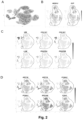
- stromal like cells To identify optimal culture conditions for stromal like cells, combinations of basal medium, medium supplements, extracellular matrix and growth factors were evaluated by assessing stromal like cell expansion over four days. Expanded stromal like cells were seeded at 500 cells/well in 96-well plates precoated with specific extracellular matrixes. Cells were seeded in MCDB131 medium containing 1% Glutamax, 2% FBS and 0.1% P/S and allowed to adhere to the tissue culture plastic for 2h at 37°C, 5% CO 2 . Medium was then aspirated and replaced with different combinations of basal medium, medium supplements and growth factors listed in table 2 below.
- DMEM/ F12 ITS-X 1% Fibronecti n 5ug/cm2 CHIR 2uM 12.
- DMEM/ F12 Empty - Fibronecti n 5ug/cm2 bFGF 20ng/ml 23.
- DMEM( HG) ITS-X 1% Collagen 5ug/cm2 bFGF 20ng/ml 24.
- MCDB1 31 Empty - iMatrix 5ug/cm2 CHIR 2uM
- Cells were cultured for 2 days at 37°C, 5% CO 2 and medium was replenished with fresh medium according to table 2. After four days of culture the total number of cells were quantified using the CellTiter-Glo Luminescent Cell Viability Assay (Promega, cat. no. G7570) as shown in Fig. 9 . Briefly, half of the medium was aspirated from the wells and a similar volume of CellTiter-Glo Luminescent reagent was added to the wells. Plates were mixed for two minutes on an orbital shaker and subsequently incubated for 10min at RT before recording the luminescence signal using a microplate reader. Baseline was established by recording the luminescence signal for stromal like cells after the initial 2h seeding step. Expansion of cells had occurred if the luminescence signal was higher at day four compared to the signal after 2h. The higher the luminescence signal, the more expansion had occurred.
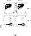
- stromal like cell assay To illustrate the higher sensitivity of the stromal like cell assay in detecting stromal like cells compared to conventional detection methods and to be able to predict the level of contaminating stromal like cells in vivo, a comparison between flow cytometry and stromal like cell assay was conducted.
- Three pancreatic endocrine cell samples (sample A, B and C) were evaluated by flow cytometry, using a marker for endocrine cells (CHGA) and a marker for stromal like cells (VIM).
- CHGA marker for endocrine cells
- VIM marker for stromal like cells
- stromal like cell assay Fig.6B
- Cells were cultured in 6-well tissue culture plates (6 wells pr. sample) for 10 days and subsequently stained for a marker for endocrine cells (FOXA2) and a marker for stromal like cells (NESTIN).
- FOXA2 a marker for endocrine cells
- NESTIN a marker for stromal like cells
- the number of stromal cells were quantified using an BioTek Cytation C10 cell imaging reader.
- the number of stromal like cells quantified in the stromal like cell assay demonstrated that sample A had the highest proportion of stromal like cells, followed by sample B, whereas very few stromal like cells were detected in sample C ( Fig. 6C ).
- an in vitro assay for stromal like cells should ideally predict the levels of contaminating stromal like cells expanding in vivo.
- the three samples were also transplanted under the kidney capsule of immunocompromised mice for 12 weeks. The transplanted kidneys were explanted, and the cell composition of the grafts were evaluated using histology.
- Fig. 6D shows the quantified amount of contaminating stromal cells (stromal VIM/total human cells) in mice transplanted with the three samples. Mice receiving sample A had the highest amount of contaminating stromal like cell, followed by sample B, whereas mice receiving sample C had the lowest amount of contaminating stromal like cells in the grafts.
- the in vivo outcome is in alignment with the prediction of the stromal like cell assay, but not flow cytometry, demonstrating the superiority of the stromal like cell assay to a conventional detection method such as flow cytometry.
Landscapes
- Health & Medical Sciences (AREA)
- Life Sciences & Earth Sciences (AREA)
- Engineering & Computer Science (AREA)
- Biomedical Technology (AREA)
- Immunology (AREA)
- Chemical & Material Sciences (AREA)
- Cell Biology (AREA)
- Biotechnology (AREA)
- Hematology (AREA)
- Molecular Biology (AREA)
- Urology & Nephrology (AREA)
- Bioinformatics & Cheminformatics (AREA)
- General Health & Medical Sciences (AREA)
- Zoology (AREA)
- Biochemistry (AREA)
- Microbiology (AREA)
- Food Science & Technology (AREA)
- Pathology (AREA)
- Physics & Mathematics (AREA)
- Analytical Chemistry (AREA)
- Tropical Medicine & Parasitology (AREA)
- Organic Chemistry (AREA)
- General Physics & Mathematics (AREA)
- Medicinal Chemistry (AREA)
- Wood Science & Technology (AREA)
- Genetics & Genomics (AREA)
- Rheumatology (AREA)
- Developmental Biology & Embryology (AREA)
- General Engineering & Computer Science (AREA)
- Toxicology (AREA)
- Virology (AREA)
- Micro-Organisms Or Cultivation Processes Thereof (AREA)
Priority Applications (1)
| Application Number | Priority Date | Filing Date | Title |
|---|---|---|---|
| EP24164881.5A EP4491712A1 (fr) | 2024-03-20 | 2024-03-20 | Procédé de détection de cellules contaminantes dans une population de cellules dérivées de cellules souches |
Applications Claiming Priority (1)
| Application Number | Priority Date | Filing Date | Title |
|---|---|---|---|
| EP24164881.5A EP4491712A1 (fr) | 2024-03-20 | 2024-03-20 | Procédé de détection de cellules contaminantes dans une population de cellules dérivées de cellules souches |
Publications (1)
| Publication Number | Publication Date |
|---|---|
| EP4491712A1 true EP4491712A1 (fr) | 2025-01-15 |
Family
ID=90368500
Family Applications (1)
| Application Number | Title | Priority Date | Filing Date |
|---|---|---|---|
| EP24164881.5A Withdrawn EP4491712A1 (fr) | 2024-03-20 | 2024-03-20 | Procédé de détection de cellules contaminantes dans une population de cellules dérivées de cellules souches |
Country Status (1)
| Country | Link |
|---|---|
| EP (1) | EP4491712A1 (fr) |
Citations (5)
| Publication number | Priority date | Publication date | Assignee | Title |
|---|---|---|---|---|
| WO2003046141A2 (fr) | 2001-11-26 | 2003-06-05 | Advanced Cell Technology, Inc. | Procedes de production et d'utilisation de noyaux de cellules somatiques humaines reprogrammees et de cellules souches humaines autologues et isogeniques |
| WO2012175633A1 (fr) | 2011-06-21 | 2012-12-27 | Novo Nordisk A/S | Induction efficace de l'endoderme definitif à partir de cellules souches pluripotentes |
| WO2014033322A1 (fr) | 2012-09-03 | 2014-03-06 | Novo Nordisk A/S | Génération d'endoderme pancréatique à partir de cellules souches pluripotentes à l'aide de petites molécules |
| WO2015028614A1 (fr) | 2013-08-30 | 2015-03-05 | Novo Nordisk A/S | Génération de cellules progénitrices endocrines à partir de cellules souches pluripotentes humaines à l'aide de petites molécules |
| WO2019048690A1 (fr) | 2017-09-11 | 2019-03-14 | Novo Nordisk A/S | Enrichissement de cellules coexprimant nkx6.1 et le peptide c, dérivées in vitro à partir de cellules souches |
-
2024
- 2024-03-20 EP EP24164881.5A patent/EP4491712A1/fr not_active Withdrawn
Patent Citations (5)
| Publication number | Priority date | Publication date | Assignee | Title |
|---|---|---|---|---|
| WO2003046141A2 (fr) | 2001-11-26 | 2003-06-05 | Advanced Cell Technology, Inc. | Procedes de production et d'utilisation de noyaux de cellules somatiques humaines reprogrammees et de cellules souches humaines autologues et isogeniques |
| WO2012175633A1 (fr) | 2011-06-21 | 2012-12-27 | Novo Nordisk A/S | Induction efficace de l'endoderme definitif à partir de cellules souches pluripotentes |
| WO2014033322A1 (fr) | 2012-09-03 | 2014-03-06 | Novo Nordisk A/S | Génération d'endoderme pancréatique à partir de cellules souches pluripotentes à l'aide de petites molécules |
| WO2015028614A1 (fr) | 2013-08-30 | 2015-03-05 | Novo Nordisk A/S | Génération de cellules progénitrices endocrines à partir de cellules souches pluripotentes humaines à l'aide de petites molécules |
| WO2019048690A1 (fr) | 2017-09-11 | 2019-03-14 | Novo Nordisk A/S | Enrichissement de cellules coexprimant nkx6.1 et le peptide c, dérivées in vitro à partir de cellules souches |
Non-Patent Citations (54)
| Title |
|---|
| "Continuous WNT Control Enables Advanced hPSC Cardiac Processing and Prognostic Surface Marker Identification in Chemically Defined Suspension Culture", STEM CELL REPORTS, vol. 13, no. 4, 8 October 2019 (2019-10-08), pages 775 |
| "Cytometry: Part B", vol. 86, 1 January 2008, SAN DIEGO [U.A.] : ACAD. PRESS, US, article HIROSHI KUBOTA ET AL: "Culture of Rodent Spermatogonial Stem Cells, Male Germline Stem Cells of the Postnatal Animal", pages: 59 - 84, XP055405432, DOI: 10.1016/S0091-679X(08)00004-6 * |
| "Preclinical and dose-ranging assessment of hESC-derived dopaminergic progenitors for a clinical trial on Parkinson's disease", CELL STEM CELL, vol. 31, 4 January 2024 (2024-01-04), pages 25 - 38 |
| "Preclinical Efficacy and Safety of a Human Embryonic Stem Cell-Derived Midbrain Dopamine Progenitor Product", MSK-DA01, CELL STEM CELL, vol. 28, 4 February 2021 (2021-02-04), pages 217 - 229 |
| "Preclinical quality, safety, and efficacy of a human embryonic stem cell-derived product for the treatment of Parkinson's disease", STEM-PD, CELL STEM CELL, vol. 30, 5 October 2023 (2023-10-05), pages 1299 - 1314 |
| "Preclinical study of induced pluripotent stem cell-derived dopaminergic progenitor cells for Parkinson's disease", NAT COMMON., vol. 11, 2020, pages 3369 |
| "Reversal of diabetes with insulin-producing cells derived in vitro from human pluripotent stem cells", NATURE BIOTECHNOLOGY, vol. 32, 2014, pages 1121 - 1133 |
| AMERI ET AL., STAM CELLS, vol. 28, no. 1, January 2010 (2010-01-01), pages 45 - 56 |
| AUGSORNWORAWAT ET AL., CELL REP., vol. 32, no. 8, 25 August 2020 (2020-08-25), pages 108067 |
| BALBOA ET AL., FUNCTIONAL, METABOLIC AND TRANSCRIPTIONAL MATURATION OF STEM CELL DERIVED BETA CELLS, 31 March 2021 (2021-03-31), pages 437748v1 |
| CHEN ET AL., NAT CHEM BIOL., vol. 5, no. 4, April 2009 (2009-04-01), pages 258 - 65 |
| D'AMOUR ET AL., NAT BIOTECHNOL., vol. 23, no. 12, December 2005 (2005-12-01), pages 1534 - 41 |
| D'AMOUR ET AL., NAT BIOTECHNOL., vol. 24, no. 11, November 2006 (2006-11-01), pages 1392 - 401 |
| DOCHERTY ET AL., DIABETES, vol. 70, no. 11, November 2021 (2021-11-01), pages 2554 - 2567 |
| FUNA ET AL., CELL STEM CELL, vol. 16, no. 6, 4 June 2015 (2015-06-04), pages 639 - 52 |
| GARY S. L. PEH ET AL: "Optimization of Human Corneal Endothelial Cells for Culture: The Removal of Corneal Stromal Fibroblast Contamination Using Magnetic Cell Separation", INTERNATIONAL JOURNAL OF BIOMATERIALS, vol. 2012, 1 January 2012 (2012-01-01), pages 1 - 8, XP055464920, ISSN: 1687-8787, DOI: 10.1155/2012/601302 * |
| HOGREBE ET AL., NAT BIOTECHNOL., vol. 38, no. 4, April 2020 (2020-04-01), pages 460 - 470 |
| HUIJBREGTS ET AL., DIABETES, vol. 68, no. 4, April 2019 (2019-04-01), pages 761 - 773 |
| KISSELBACH LYNN ET AL: "CD90 Expression on human primary cells and elimination of contaminating fibroblasts from cell cultures", CYTOTECHNOLOGY, vol. 59, no. 1, 1 January 2009 (2009-01-01), Dordrecht, pages 31 - 44, XP093199450, ISSN: 0920-9069, Retrieved from the Internet <URL:http://link.springer.com/article/10.1007/s10616-009-9190-3/fulltext.html> DOI: 10.1007/s10616-009-9190-3 * |
| KONDO ET AL., DIABETOLOGIA, vol. 60, no. 8, 25 August 2017 (2017-08-25), pages 1454 - 1466 |
| KROON ET AL., NAT BIOTECHNOL., vol. 26, no. 4, April 2008 (2008-04-01), pages 443 - 52 |
| KUBO ET AL., DEVELOPMENT, vol. 131, no. 7, April 2004 (2004-04-01), pages 1651 - 62 |
| LINGE C ET AL: "A method for removal of fibroblasts from human tissue culture systems", EXPERIMENTAL CELL RESEARCH, ELSEVIER, AMSTERDAM, NL, vol. 185, no. 2, 1 December 1989 (1989-12-01), pages 519 - 528, XP024854998, ISSN: 0014-4827, [retrieved on 19891201], DOI: 10.1016/0014-4827(89)90320-0 * |
| LIU ET AL., NAT COMMUN., vol. 12, no. 1, 7 June 2021 (2021-06-07), pages 3330 |
| MADSEN ET AL., NAT BIOTECHNOL., vol. 24, no. 12, December 2006 (2006-12-01), pages 1481 - 3 |
| MAMIDI, NATURE, vol. 564, no. 7734, December 2018 (2018-12-01), pages 114 - 118 |
| MCGRATH ET AL., DIABETES, vol. 64, no. 7, July 2015 (2015-07-01), pages 2497 - 505 |
| MFOPOU ET AL., GASTROENTEROLOGY, vol. 138, no. 7, June 2010 (2010-06-01), pages 2233 - 45 |
| NAIR ET AL., NAT CELL BIOL., vol. 21, no. 2, February 2019 (2019-02-01), pages 263 - 274 |
| NOSTRO ET AL., DEVELOPMENT, vol. 138, no. 5, March 2011 (2011-03-01), pages 861 - 71 |
| NOSTRO ET AL., STEM CELL REPORTS, vol. 4, no. 4, 14 April 2015 (2015-04-14), pages 591 - 604 |
| PAGLIUCA ET AL.: "Generation of functional human pancreatic f3 cells in vitro", CELL, vol. 159, no. 2, 9 October 2014 (2014-10-09), pages 428 - 439, XP029073423, DOI: 10.1016/j.cell.2014.09.040 |
| PELEKANOS REBECCA A. ET AL: "Isolation and Expansion of Mesenchymal Stem/Stromal Cells Derived from Human Placenta Tissue", JOURNAL OF VISUALIZED EXPERIMENTS, vol. 379154204, no. 112, 1 January 2016 (2016-01-01), pages 54204, XP055967287, Retrieved from the Internet <URL:https://www.jove.com/pdf/54204/jove-protocol-54204-isolation-expansion-mesenchymal-stemstromal-cells-derived-from-human> DOI: 10.3791/54204 * |
| PETERSEN ET AL., STEM CELL REPORTS., vol. 9, no. 4, 10 October 2017 (2017-10-10), pages 1246 - 1261 |
| PETERSON ET AL., NAT COMMON., vol. 11, no. 1, 7 May 2020 (2020-05-07), pages 2241 |
| RAMOND ET AL., DEVELOPMENT., vol. 145, no. 16, 15 August 2018 (2018-08-15), pages dev165480 |
| REZANIA ET AL., 35 DIABETES, vol. 60, no. 1, January 2011 (2011-01-01), pages 239 - 47 |
| REZANIA ET AL., DIABETES - 20, vol. 60, no. 1, January 2011 (2011-01-01), pages 239 - 47 |
| REZANIA ET AL., DIABETES, vol. 60, no. 1, January 2011 (2011-01-01), pages 239 - 47 |
| REZANIA ET AL., DIABETES, vol. 61, no. 8, August 2012 (2012-08-01), pages 2016 - 29 |
| REZANIA ET AL., DIFFERENTIATION OF HUMAN EMBRYONIC STEM CELLS, 23 June 2015 (2015-06-23) |
| REZANIA ET AL., NAT 15 BIOTECHNOL., vol. 32, no. 11, November 2014 (2014-11-01), pages 121 - 33 |
| REZANIA ET AL., NAT BIOTECHNOL., vol. 32, no. 11, November 2014 (2014-11-01), pages 1121 - 33 |
| REZANIA ET AL., STEM CELL, vol. 31, no. 11, November 2013 (2013-11-01), pages 2432 - 42 |
| RUSS ET AL., EMBO J., vol. 34, no. 13, 2 July 2015 (2015-07-02), pages 1759 - 72 |
| SAALBACH A ET AL: "The fibroblast-specific MAb AS02: a novel tool for detection and elimination of human fibroblasts", CELL AND TISSUE RESEARCH, SPRINGER, DE, vol. 290, no. 3, 1 December 1997 (1997-12-01), pages 593 - 599, XP002634899, ISSN: 0302-766X, DOI: 10.1007/S004410050964 * |
| SHARON ET AL., CELL REP, vol. 27, no. 8, 21 May 2019 (2019-05-21), pages 2281 - 2291 |
| SHARON ET AL., CELL REP., vol. 27, no. 8, 21 May 2019 (2019-05-21), pages 2281 - 2291 |
| TOYODA ET AL., STEM CELL REP., vol. 9, no. 2, 8 August 2017 (2017-08-08), pages 419 - 428 |
| VELAZCO-CRUZ ET AL., STEM CELL REPORTS., vol. 12, no. 2, 12 February 2019 (2019-02-12), pages 351 - 365 |
| VERES ET AL., NATURE, vol. 569, no. 7756, 10 May 2019 (2019-05-10), pages 368 - 373 |
| VERES ET AL.: "Charting cellular identity during human in vitro β-cell differentiation", NATURE, vol. 569, no. 7756, May 2019 (2019-05-01), pages 368 - 373, XP036791200, DOI: 10.1038/s41586-019-1168-5 |
| ZHANG ET AL., DEV. CELL, vol. 50, no. 3, 5 August 2019 (2019-08-05), pages 367 - 380 |
| ZHU ET AL., CELL STEM CELL, vol. 18, no. 6, 2 June 2016 (2016-06-02), pages 755 - 768 |
Similar Documents
| Publication | Publication Date | Title |
|---|---|---|
| US12503685B2 (en) | Cortical interneurons and other neuronal cells produced by the directed differentiation of pluripotent and multipotent cells | |
| Xu et al. | Activin, BMP and FGF pathways cooperate to promote endoderm and pancreatic lineage cell differentiation from human embryonic stem cells | |
| EP2604685B1 (fr) | Procédé de production de cellules produisant une hormone pancréatique | |
| EP3060652B1 (fr) | Production in vitro de cellules souches d'intestin antérieur | |
| EP2505639B1 (fr) | Procédé pour la fabrication de cellules de production d'hormones pancréatiques | |
| US9708582B2 (en) | Method of differentiating stem cells | |
| JP2019516384A (ja) | オルガノイド作製および疾患モデル化のための組成物および方法 | |
| CN116179470B (zh) | 肝脏类器官高速和大量制备方法、以及使用其筛选药物功效和毒性的方法 | |
| Kumar et al. | Generation of an expandable intermediate mesoderm restricted progenitor cell line from human pluripotent stem cells | |
| TW201803983A (zh) | 多能性幹細胞分化成腸的中腸內胚層細胞 | |
| KR102146274B1 (ko) | 장관 오가노이드의 제조 방법 및 이의 용도 | |
| US20240067931A1 (en) | Methods and compositions for small molecule based pancreatic beta cell induction | |
| EP2134836B1 (fr) | Système combiné de différenciation in vitro, pouvant être mis à l'échelle, pour des cellules souches embryonnaires humaines en vue d'une application de dosage direct dans des plaques à multiples puits | |
| EP4491712A1 (fr) | Procédé de détection de cellules contaminantes dans une population de cellules dérivées de cellules souches | |
| EP3950933A1 (fr) | Population de cellules comprenant des cellules souches pluripotentes et son procédé de production | |
| JP2025520898A (ja) | 幹細胞の膵臓内分泌細胞への分化 | |
| EP4198121A1 (fr) | Procédé permettant d'obtenir un organoïde neuromusculaire et son utilisation | |
| WO2025093467A1 (fr) | Procédés pour empêcher l'apparition de cellules indésirables pendant la différenciation de cellules souches pluripotentes humaines | |
| JP2024523738A (ja) | ニューロンおよびその前駆体が富化された細胞集団を提供するための方法 | |
| WO2024059208A2 (fr) | Compositions et procédés pour obtenir des organoïdes intestinaux humains en suspension ayant une couche mésothéliale séreuse | |
| Stachelscheid | Spontaneous and hepatic differentiation of human embryonic stem cells in 3D perfusion culture bioreactors | |
| Rancati et al. | Jamie Trott,* Ee Kim Tan, Sheena Ong, Drew M. Titmarsh, Simon LIJ Denil, Maybelline Giam, Cheng Kit Wong, Jiaxu Wang, 2 Mohammad Shboul, Michelle Eio, Justin Cooper-White, 3 Simon M. Cool |
Legal Events
| Date | Code | Title | Description |
|---|---|---|---|
| PUAI | Public reference made under article 153(3) epc to a published international application that has entered the european phase |
Free format text: ORIGINAL CODE: 0009012 |
|
| STAA | Information on the status of an ep patent application or granted ep patent |
Free format text: STATUS: THE APPLICATION HAS BEEN PUBLISHED |
|
| AK | Designated contracting states |
Kind code of ref document: A1 Designated state(s): AL AT BE BG CH CY CZ DE DK EE ES FI FR GB GR HR HU IE IS IT LI LT LU LV MC ME MK MT NL NO PL PT RO RS SE SI SK SM TR |
|
| STAA | Information on the status of an ep patent application or granted ep patent |
Free format text: STATUS: THE APPLICATION HAS BEEN WITHDRAWN |
|
| 18W | Application withdrawn |
Effective date: 20250120 |