EP4494558A1 - Procédé mis en oeuvre par ordinateur pour l'estimation d'un risque de trouble du rythme cardiaque dans le coeur d'un patient - Google Patents
Procédé mis en oeuvre par ordinateur pour l'estimation d'un risque de trouble du rythme cardiaque dans le coeur d'un patient Download PDFInfo
- Publication number
- EP4494558A1 EP4494558A1 EP23186229.3A EP23186229A EP4494558A1 EP 4494558 A1 EP4494558 A1 EP 4494558A1 EP 23186229 A EP23186229 A EP 23186229A EP 4494558 A1 EP4494558 A1 EP 4494558A1
- Authority
- EP
- European Patent Office
- Prior art keywords
- heart
- patient
- points
- mapping
- value
- Prior art date
- Legal status (The legal status is an assumption and is not a legal conclusion. Google has not performed a legal analysis and makes no representation as to the accuracy of the status listed.)
- Pending
Links
Images
Classifications
-
- A—HUMAN NECESSITIES
- A61—MEDICAL OR VETERINARY SCIENCE; HYGIENE
- A61B—DIAGNOSIS; SURGERY; IDENTIFICATION
- A61B5/00—Measuring for diagnostic purposes; Identification of persons
- A61B5/72—Signal processing specially adapted for physiological signals or for diagnostic purposes
- A61B5/7235—Details of waveform analysis
- A61B5/7264—Classification of physiological signals or data, e.g. using neural networks, statistical classifiers, expert systems or fuzzy systems
- A61B5/7267—Classification of physiological signals or data, e.g. using neural networks, statistical classifiers, expert systems or fuzzy systems involving training the classification device
-
- A—HUMAN NECESSITIES
- A61—MEDICAL OR VETERINARY SCIENCE; HYGIENE
- A61B—DIAGNOSIS; SURGERY; IDENTIFICATION
- A61B5/00—Measuring for diagnostic purposes; Identification of persons
- A61B5/24—Detecting, measuring or recording bioelectric or biomagnetic signals of the body or parts thereof
- A61B5/316—Modalities, i.e. specific diagnostic methods
- A61B5/318—Heart-related electrical modalities, e.g. electrocardiography [ECG]
- A61B5/346—Analysis of electrocardiograms
- A61B5/349—Detecting specific parameters of the electrocardiograph cycle
-
- A—HUMAN NECESSITIES
- A61—MEDICAL OR VETERINARY SCIENCE; HYGIENE
- A61B—DIAGNOSIS; SURGERY; IDENTIFICATION
- A61B5/00—Measuring for diagnostic purposes; Identification of persons
- A61B5/72—Signal processing specially adapted for physiological signals or for diagnostic purposes
- A61B5/7235—Details of waveform analysis
- A61B5/7264—Classification of physiological signals or data, e.g. using neural networks, statistical classifiers, expert systems or fuzzy systems
-
- G—PHYSICS
- G16—INFORMATION AND COMMUNICATION TECHNOLOGY [ICT] SPECIALLY ADAPTED FOR SPECIFIC APPLICATION FIELDS
- G16H—HEALTHCARE INFORMATICS, i.e. INFORMATION AND COMMUNICATION TECHNOLOGY [ICT] SPECIALLY ADAPTED FOR THE HANDLING OR PROCESSING OF MEDICAL OR HEALTHCARE DATA
- G16H50/00—ICT specially adapted for medical diagnosis, medical simulation or medical data mining; ICT specially adapted for detecting, monitoring or modelling epidemics or pandemics
- G16H50/20—ICT specially adapted for medical diagnosis, medical simulation or medical data mining; ICT specially adapted for detecting, monitoring or modelling epidemics or pandemics for computer-aided diagnosis, e.g. based on medical expert systems
-
- G—PHYSICS
- G16—INFORMATION AND COMMUNICATION TECHNOLOGY [ICT] SPECIALLY ADAPTED FOR SPECIFIC APPLICATION FIELDS
- G16H—HEALTHCARE INFORMATICS, i.e. INFORMATION AND COMMUNICATION TECHNOLOGY [ICT] SPECIALLY ADAPTED FOR THE HANDLING OR PROCESSING OF MEDICAL OR HEALTHCARE DATA
- G16H50/00—ICT specially adapted for medical diagnosis, medical simulation or medical data mining; ICT specially adapted for detecting, monitoring or modelling epidemics or pandemics
- G16H50/30—ICT specially adapted for medical diagnosis, medical simulation or medical data mining; ICT specially adapted for detecting, monitoring or modelling epidemics or pandemics for calculating health indices; for individual health risk assessment
Definitions
- the present invention relates to the field of computer assisted medical diagnosis. More particularly, the invention relates to a computer-implemented method for the quantification of a risk of heart rhythm disorder of a patient, possibly leading to a sudden cardiac death.
- Cardiomyopathies might cause a malfunction of the cardiac conduction system, leading to abnormal heart rhythms or cardiac arrhythmias, as ventricular tachycardia (VT), or atrial fibrillation (AF).
- VT ventricular tachycardia
- AF atrial fibrillation
- the scar from a cardiomyopathy, for instance from a previous heart attack, might form abnormal electrical circuits within the heart, which causes VT.
- Such arrythmias might cause sudden cardiac death, also named SCD.
- SCD sudden cardiac death
- arrhythmias For instance, it is known to treat arrhythmias with an invasive catheter-based ablation.
- This ablation consists of inserting a catheter into the heart through veins or arteries to burn an area of the heart tissue that is causing arrhythmias.
- This procedure requires an assessment of the cardiac region contributing to the arrhythmia and subsequently cauterizing a region of cardiac muscle to disrupt the arrhythmia. Even if this procedure is commonly used, it carries risks due to its invasive nature, its complexity, and its length.
- an implantable cardioverter-defibrillator also named ICD
- ICD implantable cardioverter-defibrillator
- Heart rhythm disorders are usually diagnosed by a cardiologist from an analysis of electrocardiogram signals (ECGs) acquired from electrodes positioned on the patient's chest or arranged on a catheter inserted into the patient's heart during an electrophysiological mapping. Since ECGs are provided from local electrodes, it might not provide a complete picture of the patient's heart health, even when they are provided from an electrophysiological mapping. Moreover, their interpretation depends on the expertise of the cardiologist and might lead to incorrect readings and false positives or negatives.
- ECGs electrocardiogram signals
- the object of the present invention is to answer to this need.
- the subject of the invention is a computer-implemented method for the estimation of a risk of heart rhythm disorder in a patient's heart, the method comprising:
- a mapping of points of a tissue of a patient's heart is supplied.
- This mapping of points might result from one or more imaging and/or mapping techniques of the tissue, from which a 3D representation of all or part of the tissue is obtained.
- the 3D representation is associated to local features which are representative, directly, or indirectly and partly, or fully, of the presence or the absence of arrythmias. These local features might be physiological, geometrical, electrical and/or structural characteristic, resulting directly from computing on the mapping of points and/or from another data acquisition technique.
- An example of local characteristic might be the thickness of the tissue, for instance determined with measuring the distance separating closest points of inner and outer surface of the tissue.
- the thickness of the wall may help to identify areas responsible of arrythmias, such as chamber's wall segment particularly thin or morphological isthmuses, i.e., areas having a thickness lowerthan the thickness of adjacent zones.
- isthmuses might create electrical pathway problems on the heart conduction system, such as re-entry circuits.
- An example of classification might be the cellular composition of the tissue.
- part of the muscular tissue is replaced by adipocytes or by calcified structures; such structural changes tend to weaken the thickness of the heart as well and might be responsible of arrhythmias.
- the mapping of points is automatically segmented, from its local features, to delineate at least a region of interest in the tissue which might be responsible, directly, or indirectly and partly, or fully, of the presence or the absence of an arrythmia.
- the seek of such region of interest is therefore not limited to a local part of the heart.
- this region of interest might be delineated as being an area of lower thickness located between a couple of adjacent areas with a same greater thickness.
- this region of interest might be delineated as being a continuous area located in a specific heart chamber and having a maximum thickness lower than a predetermined threshold.
- a trained machine learning algorithm might be then implemented directly on the segmented area, with or without considering the associated parameter values/classifications, and/or on features extracted from the segmented area to provide a value which might, directly or indirectly, lead to the estimation of a risk value, as a probability, indicating whether said patient's heart presents a heart rhythm disorder. Due to the trained machine learning algorithm, the possibility of a misleading interpretation of the cardiologist is thus reduced. Said risk value might be directly interpreted by the cardiologist to determine the probability of occurrence of a heart condition, as a sudden cardiac arrest, a sudden cardiac death, or a myocardial infection. As a variant, said risk value might be post-processed, considering further data related to the patient, to provide a further interpretable risk value of a heart condition.
- a "computer” may refer, but is not limited to, a desktop computer, a laptop computer, a tablet computer, a piece of testing equipment, a network appliance, a controller, a digital signal processor, a computational engine within an appliance, a consumer-electronic device, a portable computing device, and/or another electronic device, and/or any combination thereof appropriate to the relevant task at hand.
- the method steps might be all executed on a same device. As a variant, the method steps might be executed on several devices, connected by wires or by a wireless connection.
- a "mapping of points of a tissue” is a set of points digitally representing this tissue, said points can be encoded computationally by sequences or lists of numerical values, in particular sequences of at least one numerical value representing the coordinates of said points in a particular reference.
- the coordinates can be specified in any type of chart, for instance in cartesian, spherical cylindrical coordinates or any other type of geometrical chart in one, two or three spatial dimensions.
- a mapping of points might be 2D mapping of points or a 3D mapping of points.
- a 3D mapping of points might be a 3D model of the heart, namely one or more structured mesh, each comprising a plurality of vertices connected with each otherto define a plurality of faces and/or volumes which approximate internal and/or external surfaces and/or volumes of all or part of the heart.
- a 3D model might be a triangle mesh or a quadrilateral mesh, or more generally a polygonal mesh, which comprises a plurality of vertices, with edges connecting pairs of vertices and/or polygonal faces connecting a closed set of vertices.
- a 3D model might be a tetrahedral mesh or a hexahedral mesh or a prism mesh, or more generally a volume mesh, which comprises a plurality of vertices, with polyhedrons connecting a closed set of polygons defined by closed set of vertices.
- each vertex and/or edge and/or polygon and/or polyhedron might be labelled as being a part of a specific sub-part of the heart.
- a 3D model might be stored as a list of vertices each associated with spatial coordinates and with a set of connections to other vertices, or as a list of vertices associated with spatial coordinates and a list of polygons or faces each defined by a subset of vertices contained in said list of vertices, being understood that any other suitable method of storage might be used in the context of the present specification.
- a 3D mapping of points might be an unstructured set of points, namely a point cloud, where each point might be or not connected to another point of the point cloud.
- said "3D mapping of points” might be (or might be computed from) one of, or a combination of two or more of : an electrophysiological map, resulting from an electrocardiogram (ECG) and/or from an invasive mapping, an anatomical and/or a functional map, resulting from a computed tomography(CT)-scan, a spectral computed tomography(SCT)-scan, a positron emission tomography(PET)-scan, a single photon emission computed tomography(SPECT)-scan, a photon-counting computed tomography(PCCT)-scan or a magnetic resonance imaging(MRI)-scan, and an electro-anatomical map, resulting from an invasive mapping.
- ECG electrocardiogram
- SCT spectral computed tomography
- PET positron emission tomography
- SPECT single photon emission computed tomography
- PCCT photon-counting computed tomography
- MRI magnetic resonance imaging
- a parameter indicating a local characteristic of said tissue at a point position or around a point position might be an electrical, a physiological, a physical or a geometrical feature of an area of the tissue whose location corresponds to said position.
- an electrical feature might be an activation time, an electric potential, or a conduction velocity.
- a physiological feature might be a tissue density.
- a physical or a geometrical feature might be a tissue thickness, such as a myocardial thickness. It is understood that any other suitable local feature of the heart, which is representative, directly, or indirectly and partly, or fully, of the presence or the absence of arrythmias, might be used in the context of the present specification.
- said parameter value associated to a point of said mapping of points indicates a local thickness of said tissue at or around said point position.
- a "classification indicating a local characteristic of a tissue at a point position or around a point position” might be a class indicating the type of cells composing the tissue at or around a location corresponding said position and/or a class indicating the major type of cells composing the tissue at or around a location corresponding said position and/or a class indicating the proportion of the types of cells composing the tissue at or around a location corresponding said position.
- said type of tissue might be a muscle tissue, a fat tissue, or a calcified tissue.
- a "classification indicating a local characteristic of a tissue at a point position or around a point position” might be a label previously attributed to a point and indicated whether a part of the tissue at or around said point position belongs to a cardiomyopathy scar or a myocardial fibrosis.
- said classification associated to a point of said mapping of points indicates the presence of fat, calcification and/or muscle in said tissue at or around said point position.
- the mapping of points comprises at least a mesh of a wall of the heart, said mesh being generated from one or more images of the heart, each vertex of said mesh is associated with at least one value of a local characteristic of the wall at the heart's point corresponding to this vertex and the segmentation step comprises the selection of a plurality of vertices of said mesh from their associated values, the segmented region being determined from said selected plurality of vertices.
- the method comprises a preliminary step of acquiring a 3D image and/or recording a 3D image of a patient's heart or a region of his heart, the mesh being computed from said 3D image.
- the 3D image may be acquired directly from an image acquisition device such as a CT-scan or MRI.
- the 3D image may be obtained from a recording medium on which it is stored, such as a local memory or a distant database, the 3D image having been acquired beforehand the method.
- the step of acquiring the 3D image can be performed by tomography.
- These techniques are also identified as CT-scan, SCT-scan, PCCT-scan, PET-scan, SPECT-scan, or CAT-scan and are based on the measurement of X-ray absorption by the tissues of an organ.
- Tomography provides a plurality of 2D images each representing a slice of the organ, which are then combined to reconstruct the 3D image of the anatomical structure of the observed organ.
- the 3D image comprises a volumetric distribution of pixels, or voxels. Two or more 3D images might be acquired from distinct tomography techniques, or modalities, to built then a multimodal and/or a multidimensional 3D image.
- the method according to the invention can thus comprise a 3D modeling step of the 3D image of the heart to generate a 3D model forming then the mapping of points.
- the 3D modelling step comprises a modeling step of at least one layer, or one wall of the heart shown in the 3D image, such as an inner face and an outer face of the layer of the heart.
- Said 3D model might be a mesh, especially a polygonal or a polyhedron mesh, of said layer, especially of said inner and outer faces.
- a thickness value at a point position or around a point position might be determined with computing the distance between the inner face and the outer face at said position.
- a classification of the type of tissue at a point position or around a point position might be determined with considering the Hounsfield units encoding the radiodensity at said position.
- the method comprises, further to the step of receiving the mapping of points, a step of identifying a set of points, from said mapping of points, corresponding to a predetermined heart chamber of said patient's heart; and wherein said segmentation step is implemented only on the identified set of points corresponding to said heart chamber.
- Said heart chamber might be a left ventricle, a right ventricle, a left atrium or a right atrium and might be manually defined by the implementation of the method and according to the patient's medical history.
- Said prior identification step might help to improve the segmentation step, since the characteristics of the tissue from which the risk value is computed might be different from one chamber to the other, and even within the same chamber.
- said set of points might be morphologically and/or anatomically segmented from the mapping of points with identifying a set of points which define together a volume whose shape corresponds globally to the shape of the predetermined heart chamber and/or with identifying a set of points which belong to a same area labelled with an anatomical landmark.
- an area of a tissue matching a predefined electrical conductivity criterion might be any area of the tissue having an electrical conductivity lower than a given threshold and/or being defined by a border corresponding to a local gradient of electrical conductivity greater than a given threshold.
- said segmentation step comprises the segmentation of the mapping of points from said parameter values and/or from said classifications to identify at least a region of interest of a predetermined kind; and wherein the risk value computing step comprises:
- a « machine learning algorithm» is a computer-implemented algorithm which purpose is to estimate a risk value, from one or more morphological feature values provided as inputs, by means of decision rules (otherwise called knowledge base) which have been previously learned from a set of reference data, also called training dataset.
- said kind of region of interest might be:
- Said kind of region of interest might be manually defined by the cardiologist and/or automatically determined, prior to the implementation of the method and according to the patient's medical history.
- said segmentation step comprises a direct thresholding of the parameter values of the points of said mapping of points, for instance following a comparison of their thickness to a thickness level, for example of 1mm, 2mm, 3mm, 4mm or 5mm.
- said segmentation step comprises the segmentation of the mapping of points from said parameter values and/or from said classifications to identify at least a couple of areas of interest in said mapping of points, each area of interest corresponding to an area of said tissue matching a predefined electrical conductivity criterion, wherein the points of the mapping of points extending between said couple of areas of interest define together said region of interest whether their parameter values is lower than the parameter values of said couple of areas of interest.
- said segmentation step might comprise an iterative decrease of an electrical conductivity threshold; and, for each iterative value of said electrical conductivity threshold, an identification step of two or more groups of points of said mapping of points, wherein each group of points is selected from their parameter values and/or from their classifications so as to define an area of said tissue having an electrical conductivity lower than said iterative value of said electrical conductivity threshold, said iterative decrease of the electrical conductivity threshold being stopped when the border of each corresponding groups of points remains globally identical from the previous iteration to the current iteration, said corresponding groups of points thus defining said areas of interest.
- each area of interest is progressively identified with decreasing a thickness threshold until the area remains the same, meaning that this area has globally a same thickness, that the decreasing step required to further change the shape of this area is high and that the border of this area corresponds to a local gradient of electrical conductivity greater than a given threshold.
- said segmentation step might be implemented from any suitable region growing method, with considering points labelled with a parameter value lower than a given threshold as seed points and recursively considering neighbors of these points with estimating whether they should be considered as part of a region of interest upon their parameter values.
- a region of interest identified within the mapping of points might be modified, for instance with smoothing or eroding its border.
- said value extraction sub-step might comprise the computing of a value of one or more, or a combination, of the following features:
- said value extraction sub-step might comprise the computing of a value of one or more, or a combination, of the following features:
- said value extraction sub-step might comprise the computing of a value of one or more, or a combination, of the following features:
- said trained machine learning algorithm might be a densely connected neural network comprising at least one input layer comprising as many input nodes, or artificial neurons, as predetermined morphological features whose values are extracted in the value extraction sub-step, one or more dense hidden layers each comprising a plurality of nodes, and an output layer comprising a single output node from which said risk value is provided.
- the input layer might comprise 31 input nodes, to which is supplied an input vector comprising the extracted values, namely the mean, min and max values of the isthmus's absolute thickness; the isthmus's relative thickness; the thickness gradient at the boundary of the isthmus; the isthmus's width, the isthmus's length, the isthmus's width over length ratio; the curvature of the entrance and the exit of the isthmus; and the thickness gradient at the entrance and the exit of the isthmus, and the volume density of fat tissue and of calcification tissue in the isthmus.
- the input layer might comprise 31 input nodes, to which is supplied an input vector comprising the extracted values, namely the mean, min and max values of the isthmus's absolute thickness; the isthmus's relative thickness; the thickness gradient at the boundary of the isthmus; the isthmus's width, the isthmus's length, the isthmus's width over length ratio; the curvature of the entrance and the exit of the isthmus; and the thickness gradient at
- the densely connected neural network might comprise a first hidden layer comprising 25 nodes and a second hidden layer comprising 25 nodes.
- Each node of the first hidden layer is fully connected to each input node of the input layer
- each node of the second hidden layer is fully connected to each node of the first hidden layer
- the single output node of the output layer is fully connected to each node of the second hidden layer.
- Each node of the hidden layers might be configured with a rectified linear activation function ReLU, and the single node of the output layer might be configured with a logistic function.
- the number of considered morphological features, the number of input nodes of the input layer, the number of hidden layers, the number of nodes of each hidden layer and the activation function associated to each node might be adapted in any appropriate manner without departing from the scope of the disclosure.
- the method comprises a preliminary training step of the densely connected neural network, using a training dataset consisting of a plurality of training vectors, each training vector being associated to a patient and comprising a mapping of points of the patient's heart labelled with an indication whether said patient suffers from heart rhythm disorder.
- a training step at least one region of interest is segmented from the mapping of points of each training vector, and values of predetermined morphological feature are extracted from said region of interest and supplied to the densely connected neural network, which predicts a risk of heart rhythm disorder for the associated patient.
- the error between the prediction and the indication labelling the training vector is computed, and the synaptic weights and the biases of the nodes of the layers are adjusted using gradient backpropagation of the error to minimize a cost function.
- the training dataset might comprise cardiac data aggregated for 500 patients, from which a plurality has a heart which does not suffer from arrythmia, another plurality has a heart which suffers from arrythmia although the arrythmia has not causes a SCD and another plurality has a heart suffering from arrythmia which has caused a SCD.
- values of the 31 above-mentioned morphological features of at least an isthmus have been extracted, while the corresponding mapping of points labelled with an indication whether said patient's heart has suffered from SCD.
- a batch gradient descent is used for the training step and the weights of the layers are adjusted until a mean square error is less than 10-5.
- the amount of data of the training dataset, the type of data used in the training dataset, the method of gradient backpropagation and the function cost might be adapted in any appropriate manner without departing from the scope of the disclosure.
- said parameter values and/or said classifications associated to the points forming said segmented region of interest form together a parameter and/or classification map of said region of interest; and wherein the risk value computing step comprises a sub-step of implementing said trained machine learning algorithm on said parameter and/or classification map to estimate said risk value, wherein said trained machine learning algorithm have been trained to estimate, from a parameter and/or classification map of a region of interest of a patient's heart, a probability indicating whether said patient's heart presents a heart rhythm disorder.
- said trained machine learning algorithm might comprise at least a first stage configured to provide, from said parameter and/or classification map, a plurality of feature maps and a second stage configured to estimate, from said features maps, said risk value.
- Said first stage might be a graph convolution neural network (GCNN) or a convolution neural network (CNN) comprising a contracting pathway receiving said parameter and/or classification map as an input and being configured to extract features from said parameter and/or classification map.
- said contracting pathway might comprise one or more image or graph convolution layers, wherein each convolution layer processes its input one or more convolution filters or kernels, each convolution layer being associated to an activation layer.
- said contracting pathway might also comprise one or more pooling layers, and a final flattening layer from which is provided a one-dimensional vector containing said extracted features.
- said graph convolution neural network might comprise two consecutive graph convolution layers, followed by a pooling layer, followed again by three further graph convolution layers and a final flattening layer.
- the two first graph convolution layers might each comprise 5000 nodes, a 64x64 filter and a rectified linear activation function ReLU
- the two second graph convolution layers might each comprise 500 nodes, a 64x64 filter and a rectified linear activation function ReLU
- the last comprises 500 nodes and a 1x1 filter.
- the pooling layer might be a max pooling layer.
- the number of convolution layers and pooling layer, the number of nodes of each convolution layer, the size of each filter, the type of pooling function and the type of activation function might be adapted in any appropriate manner without departing from the scope of the disclosure.
- the second stage might be a fully connected neural network comprising a succession of fully connected layers, the first of which being an input layer receiving said one-dimensional vector provided from the graph convolution neural network, and the last of which being an output layer comprising a single output node from which said risk value is provided.
- said fully connected neural network might comprise a first layer of 128 nodes, a second layer of 64 nodes, a third layer of 32 nodes and a last layer of a single node.
- Each node of the three first layers might be configured with a rectified linear activation function ReLU, and the single node of the last layer might be configured with a logistic function.
- the number of nodes of each layer and the activation function associated to each node might be adapted in any appropriate manner without departing from the scope of the disclosure.
- the method comprises a preliminary training step of both the graph convolution neural network and the fully connected neural network, using a training dataset consisting of a plurality of training vectors, each training vector being associated to a patient and comprising a mapping of points of the patient's heart labelled with an indication whether said patient suffers from heart rhythm disorder.
- a training dataset consisting of a plurality of training vectors, each training vector being associated to a patient and comprising a mapping of points of the patient's heart labelled with an indication whether said patient suffers from heart rhythm disorder.
- at least one region of interest is segmented from the mapping of points of each training vector, and the parameter and/or classification map of said region of interest, either formatted as a picture or as a graph, is supplied to the machine learning algorithm, which predicts a risk of heart rhythm disorder for the associated patient.
- the error between the prediction and the indication labelling the training vector is computed, and the weights of the filters of the convolution layers and the weights of the fully connected layers are adjusted using gradient backpropagation
- the training dataset might comprise cardiac data aggregated for 600 patients, from which a plurality has suffers from ventricular tachycardia, ventricular fibrillation and cardiac arrest.
- a 3D model of his heart has been acquired and segmented to identify a region corresponding to a cardiomyopathy scar, said 3D model being labelled with an indication whether said patient's heart has suffered from SCD.
- a batch gradient descent is used for the training step and the weights of the layers are adjusted using a binary cross-entropy loss as cost function.
- the amount of data of the training dataset, the type of data used in the training dataset, the method of gradient backpropagation and the function cost might be adapted in any appropriate manner without departing from the scope of the disclosure.
- said trained machine learning algorithm might be implemented on a region of interest in said mapping of points corresponding to a hypertrophic segment of a wall of heart's chamber, as the left ventricle myocardium, or to a thin-walled segment of the most distal portion of the left ventricle myocardium, wherein said trained machine learning algorithm have been trained to estimate, from a region of interest of 3D model of patient's heart, a probability indicating whether said region of interest might be responsible of a heart rhythm disorder.
- said risk value estimated with implementing the at least one trained machine learning algorithm on the input determined from said segmented region of interest, is a first probability indicating whether said patient's heart presents a heart rhythm disorder
- the method comprises:
- the risk value estimated from the trained machine learning algorithm implemented from the mapping of points can be adjusted, confirmed or infirmed using another modality likely to indicate a probability of occurrence of an arrythmia.
- said second probability might be estimated from another modality, as a second imaging modality.
- said second probability might be estimated from the patient's medical history or from patient's family medical history.
- Said third probability might be computed with multiplying the first probability with the second probability, with computing any linear /or logarithmic combinations of the first and second probabilities, or with retaining the higher or the lower value among the first and second probabilities as the third probability.
- said step of second probability computing comprises:
- Said electrograms signals might be real-time electrocardiogram signals, each originating from at least one of a plurality of electrodes of a mapping catheter being guided into said organ during an electrophysiological mapping.
- said electrograms signals might be acquired by a device worn by the patient, as a 12 lead-ECG placed on the chest.
- the first machine learning algorithm can be trained to estimate a probability of occurrence of an arrythmia, while the second machine learning algorithm might be trained to estimate a risk of seriousness of the arrythmia or whether said arrythmia might cause a SCD.
- a timewise feature of an electrogram signal is a feature which is intrinsically related to the time domain or frequency domain analysis of the electrogram signals.
- said timewise feature extracting sub-step comprises the estimation of a value of a length cycle from said one or more of said electrograms signals.
- Said value of a length cycle might be a time interval between two successive R Peaks in the electrogram signal which has the highest amplitude among all the electrograms signals.
- said value of a length cycle might be estimated with firstly measuring a normalized autocorrelation function of said electrogram signal and with estimating a local extremum of said normalized autocorrelation.
- said normalized autocorrelation function can be estimated by computing the scalar product of a first vector comprising the T first data of the electrogram signal and a second vector comprising the T last data of the electrogram signal, divided by the product of the Euclidean norms of the first vector and the second vector.
- a maximum and a minimum value of the normalized autocorrelation function can be determined, and then the value selected for the length cycle is chosen as that for which the value of T provides the first local extremum for which the normalized autocorrelation function calculated with this value of T is greater than a set threshold, as for example the difference between the maximum value subtracted by 0.3 times the difference between the maximum value and the minimum value.
- said value of a length cycle might be estimated with firstly measuring a normalized mean square error function of said electrogram signal and with estimating a local extremum of said normalized mean square error function.
- said normalized mean square error function might be estimated by computing for each value of T the Euclidean norm squared by the difference between the first vector and the second vector, divided by the product of the largest absolute value in the first vector by the largest absolute value in the second vector.
- a maximum and a minimum value of the normalized mean square error function are determined, and then the value selected for the length cycle is chosen as the one for which the value of T provides the first local extremum for which the normalized mean square error function calculated with this value of T is smaller than a set threshold, as for example the difference between the minimum value added by 0.2 times the difference between the maximum value and the minimum value.
- said timewise feature extracting sub-step comprises the estimation of a value of the frequency of said one or more of said electrograms signals, for instance within the FFT of said one or more of said electrograms signals, and/or the amplitude of said one or more of said electrograms signals.
- a morphological feature of an electrogram signal is a feature which is intrinsically related to morphological analysis of the signal, i.e., analysis based on or related to mathematical morphology.
- said morphological feature extracting sub-step comprises the estimation of a norm of said one or more of said electrograms signals.
- said norm might be the Euclidean norm of the electrogram signal and/or the integrated absolute derivative of the electrogram signal, namely the H10 norm.
- said trained machine learning algorithm might be a boosting-based machine learning algorithm comprising as many input nodes, as timewise and/or morphological features whose values are extracted in the value extraction sub-step.
- said boosting-based machine learning algorithm might be a gradient boosting-based machine learning algorithm comprising six input nodes, to which is supplied an input vector comprising the values of the following timewise and/or morphological features, said values being extracted from the electrogram signal which has the highest amplitude among all the electrograms signals : a first cycle length estimation, a second cycle length estimation, the frequency, the amplitude, the Euclidean norm and the integrated absolute derivative of the electrogram signal.
- the number of considered timewise and/or morphological features, the number of input nodes, the number of terminal nodes and the tree size might be adapted in any appropriate manner without departing from the scope of the disclosure.
- the method comprises a preliminary training step of the boosting-based machine learning algorithm, using a training dataset consisting of a plurality of training vectors, each training vector being associated to a patient and comprising patient's electrograms signals which has been labelled with an indication whether said patient suffers from heart rhythm disorder.
- values of timewise and/or morphological features are extracted from electrograms signals of each training vector and supplied to the boosting-based machine learning algorithm, which predicts a risk of heart rhythm disorder for the associated patient.
- the error between the prediction and the indication labelling the training vector is computed, and the nodes weights are adjusted using gradient boosting backpropagation of the error to minimize a cost function.
- the training dataset might comprise 275000 electrograms signals labelled with a value indicating whether the corresponding patient's heart has suffered from a heart rhythm disorder. Values of the six timewise and/or morphological features have been extracted from each of these electrograms signals and supplied to the gradient boosting-based machine learning algorithm for its training, using a logistic loss as cost function.
- said step of second probability computing comprises a sub-step of implementing said trained machine learning algorithm on said set of electrophysiological data to estimate said second probability, wherein said trained machine learning algorithm have been trained to estimate, from a set of electrophysiological data , a probability indicating whether said patient's heart presents a heart rhythm disorder.
- said trained machine learning algorithm might comprise at least a first stage configured to provide, from said set of electrograms signals, a plurality of feature maps and a second stage configured to estimate, from said features maps, said risk value.
- Said first stage might be a convolutional neural network and said second stage might be a fully connected neural network.
- said convolution neural network might comprise a first convolution layer configured to extract, from a set of 16 electrograms signals provided as input to the convolution neural network, 200 features, followed by a second convolution layer which extracts 3000 features from the first layer, a third convolution layer which extracts 20000 features from the second layer, a fourth convolution layer which extracts 8000 features from a max pooling layer following the third layer, and a fifth convolution layer which extracts 1000 features from the second layer.
- the fifth convolution layer is linked to a fully connected neural network, forming said second stage and which comprises two fully connected layers comprising respectively 50 and 10 nodes, an array of probabilities being provided from the last fully connected layer.
- the number of convolution layers, the number of nodes of each convolution layer, the size of each filter, the type of pooling function and the type of activation function of the first stage, and the number of fully connected layers, the number of nodes of each fully connected layer and the type of activation function of the second stage might be adapted in any appropriate manner without departing from the scope of the disclosure.
- the training of this convolutional neural network might made using by using 275000 electrogram signals labelled with a value indicating whether the corresponding patient's heart has suffered from a heart rhythm disorder.
- the training is using a binary cross entropy loss.
- the subject of the invention is also a computing device for the implementation of the method according to one of the embodiments, said computing device comprising a memory arranged to receive at least one mapping of points and a computing unit arranged to implement said segmentation step and said risk value computing step from said mapping of points.
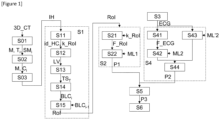
- Figure 1 is a logic flow diagram that depicts a computer-implemented method, according to an embodiment of the invention
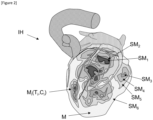
- Figure 2 shows a schematic representation of a 3D mapping of points representing a tissue of a patient's heart
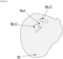
- Figure 3 shows a schematic representation of the result of a segmentation step of the method depicted in Figure 1 when implemented on the 3D mapping of points of Figure 2 and morphological features extracted from this result;
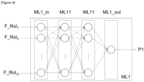
- Figure 4 shows a schematic representation of a first machine learning algorithm implemented in the method of Figure 1 ;
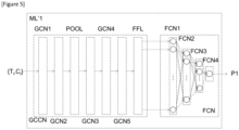
- Figure 5 shows a schematic representation of a variant of the first machine learning algorithm implemented in the method of Figure 1 .
- Figure 1 shows a computer-implemented method for estimation of a risk of heart rhythm disorder in a patient's heart.
- a 3D image 3D_CT of the patient's heart has been acquired from a CT-scan method, and a 3D model IH of the patient's heart has been generated from the 3D image.
- an inner surface or layer and an outer surface or layer of the patient's heart myocardium have been modelized from the 3D image 3D_CT with defining a plurality of vertices of meshes representing said surfaces.
- This modeling results in a mesh M of the inner surface or layer or of the outer surface or layer of the myocardium, wherein each vertex M i of the mesh M is therefore labelled with the value of the thickness T i of the myocardium, meaning the thickness computed from these inner and outer surfaces or layers, at the location of the heart corresponding to the position of this vertex.
- Said mesh M comprises then a plurality of superimposed sub-meshes SM i , with each sub-mesh corresponding to a thickness range of the myocardium.
- FIG. 2 shows an example of a 3D model IH.
- the model IH comprises a mesh M comprising six superimposed thickness sub-meshes SM 1 to SM 6 corresponding each to a range of myocardium thickness, namely 5mm to 6mm, 4mm to 5mm, 3mm to 4mm, 2mm to 3mm, 1mm to 2mm, and 0 to 1mm.
- SM 1 to SM 6 corresponding each to a range of myocardium thickness, namely 5mm to 6mm, 4mm to 5mm, 3mm to 4mm, 2mm to 3mm, 1mm to 2mm, and 0 to 1mm.
- regions of greater thickness have been depicted darker than regions of lower thickness.
- each point of the 3D image has been associated to a numerical value in terms of Hounsfield units encoding the radiodensity at said point. Since calcification type cells, fat type cells and muscle type cells do not present a same radiodensity, it is therefore possible to discriminate, for a slice of the myocardium at a defined location of the heart, the proportion of calcification, fat and muscle type cells composing said slice.
- Each vertex M i of the mesh M has been therefore labelled with a class C i indicating the major type of cells composing the myocardium at the location of the heart corresponding to the position of this vertex.
- said class C i is selected among classes C 1 "calcification tissue", C 2 "fat tissue” and C 3 "muscle tissue”.
- steps S01 to S02 might be executed by another computing unit than the one used for the rest of the method. Moreover, steps S01 to S02 might be executed fully automatically by a computing unit or with a human assistance.
- the resulting 3D model IH might be stored on a recording medium, such as a memory of the computer or an external memory support or a distant database through which it is accessible to the computer.
- the 3D model IH is provided to the computing unit of the computer.
- a step S1 at least one region of interest Rol, which matches a predefined electrical conductivity criterion, is segmented in said 3D model IH.
- a heart chamber identifier id_HC and a kind of region of interest k_Rol to be segmented are provided in a first sub-step S11, either manually by a cardiologist or automatically from the patient's medical history.
- said heart chamber identifier id_HC is a left ventricle and said kind of region of interest k_Rol is a myocardial isthmus having an electrical gradient which has the consequence of inducing a slow down of the electrical conduction of the heart.
- a set of points LV is morphologically segmented from the 3D model IH with identifying a set of points which define together a volume whose shape corresponds globally to the shape of the heart chamber corresponding to identifier id_HC, namely the left ventricle.
- the below-described sub-steps of the segmentation step S1 are implemented only on the identified set of points LV corresponding to said left ventricle.
- the segmentation step comprises then a segmentation, in the set of points LV, of a couple of areas of interest BLC, which each matches a predefined electrical conductivity criterion.
- said segmentation step S1 comprises an iterative decrease of an electrical conductivity threshold, being approximated here by a thickness threshold TS T .
- Said thickness threshold TS T is thus progressively decrease, in a sub-step S13, from a maximum value, namely 6 mm, to a minimum value, namely 1 mm.
- the newly defined areas BLC i are compared with the previous defined areas BLC i-1 .
- the border of a newly defined area BLC i remains globally the same than the corresponding previously defined area BLC i-1 , the iteration is stopped for this area and the set of connected vertices defining said area BLC i is classified as an area of interest BLC.
- Each area of interest BLC resulting from the step S15 has then globally a same thickness and its border corresponds to a quick change of thickness toward its adjacent regions, meaning that this border corresponds to a local gradient of electrical conductivity greater than a given threshold.
- sub-steps S13 to S15 might be implemented with considering classes C i rather than thickness values T i of the vertices, or with considering a combination of these classes and values.
- said sub-steps S13 to S15 might be replaced with a single thresholding of the thickness values T i of the vertices of the mesh M, for instance following a comparison to a given thickness level, for example of 1mm, 2mm, 3mm, 4mm or 5mm.
- said segmentation step S1 might be replaced with any suitable region growing method, with considering vertices of the mesh M labelled with thickness values T i lower than a given threshold as seed points and recursively considering neighbors of these vertices with estimating whether they should be considered as part of a region of interest upon their thickness values T i .
- Figure 3 shows an example of a couple of areas of interest BLC and the isthmus Rol extending between these areas of interest BLC, resulting from the segmentation of the mesh M according to the step S1.
- a first probability P1 indicating whether said patient's heart presents a heart rhythm disorder is estimated.
- values of several morphological features F_Rol are extracted from the region of interest Rol.
- the types of considered morphological features F_Rol depend on the kind of said region of interest k_Rol.
- the considered morphological features F_Rol are the isthmus's absolute thickness; the isthmus's relative thickness; the thickness gradient at the boundary of the isthmus; the isthmus's width, the isthmus's length, the isthmus's width over length ratio; the curvature of the entrance and the exit of the isthmus; and the thickness gradient at the entrance and the exit of the isthmus, and the volume density of fat tissue and of calcification tissue in the isthmus.
- a mean value, a minimum value and a maximum value are computed from the vertices M i of the isthmus Rol and the adjacent areas BLC, from their coordinates and from their thickness values T i .
- a value of the volume density is computed from the vertices M i of the isthmus Rol and from their classes C i .
- a sub-step S22 the extracted values of the features F_Rol are supplied, as an input vector, to a first trained machine learning algorithm ML1 for the prediction of the probability P1 that the patient's heart presents a heart rhythm disorder.
- Figure 4 shows an architecture of an appropriate machine learning algorithm ML1 for the implementation of the sub-step S22.
- Said machine learning algorithm ML1 is a densely connected neural network comprising an input layer comprising an input layer ML1_in with 31 input nodes for the reception of the extracted values of the features F_Rol and an output layer ML1_out with a single output node from which the probability P1 is provided.
- the densely connected neural network ML1 comprises a first hidden layer ML11, comprising 25 nodes, and a second hidden layer ML12, comprising 25 nodes.
- Each node of the first hidden layer ML11 is fully connected to each input node of the input layer ML1_in
- each node of the second hidden layer ML12 is fully connected to each node of the first hidden layer ML11
- the single output node of the output layer ML1_out is fully connected to each node of the second hidden layer ML12.
- Each node of the hidden layers is configured with a rectified linear activation function ReLU
- the single node of the output layer is configured with a logistic function.
- the densely connected neural network ML1 has been previously trained with a training dataset to estimate, from a set of values of the morphological features F_Rol, a probability that a patient's heart presents a heart rhythm disorder.
- the training dataset comprises heart's 3D models of 500 patients, from which a plurality has a heart which does not suffer from arrythmia, another plurality has a heart which suffers from arrythmia although the arrythmia has not causes a SCD and another plurality has a heart suffering from arrythmia which has caused a SCD.
- values of the 31 above-mentioned morphological features F_Rol have been extracted from its heart's 3D model, while the model has been labelled with an indication whether said patient's heart has suffered from SCD.
- a batch gradient descent has been used for the training step and the weights of the layers have been adjusted until a mean square error is less than 10-5.
- a graph formed by the thickness values T i and the classes C i of the vertices M i of the isthmus Rol might be directly supplied to a machine learning algorithm ML'1, comprising a graph convolution neural network GCNN as a first stage and a fully connected network FCN as a second stage, said machine algorithm ML'1 being trained to predict said first probability P1 from a graph of thickness values and classes.
- a machine learning algorithm ML'1 comprising a graph convolution neural network GCNN as a first stage and a fully connected network FCN as a second stage, said machine algorithm ML'1 being trained to predict said first probability P1 from a graph of thickness values and classes.
- the graph convolution neural network GCNN comprises two consecutive graph convolution layers GCNN1 and GCNN2, followed by a pooling layer POOL, followed again by three further graph convolution layers GCNN3, GCCN4 and GCNN5, and a final flattening layer FFL.
- the two first graph convolution layers GCNN1 and GCNN2 each comprises 5000 nodes, a 64x64 filter and a rectified linear activation function ReLU
- the two second graph convolution layers GCNN3 and GCNN4 each comprises 500 nodes, a 64x64 filter and a rectified linear activation function ReLU
- the last layer GCNN5 comprises 500 nodes and a 1 ⁇ 1 filter.
- the pooling layer POOL is a max pooling layer.
- the fully connected neural network FCN comprises a first layer FCN1 of 128 nodes, a second layer FCN2 of 64 nodes, a third layer FCN3 of 32 nodes and a last layer FCN4 of a single node.
- Each node of the three first layers FCN1, FCN2 and FCN3 is configured with a rectified linear activation function ReLU, and the single node of the last layer FCN4 is configured with a logistic function.
- the machine learning algorithm ML'1 has been previously trained with a training dataset to estimate, from a given graph, a probability that a patient's heart presents a heart rhythm disorder.
- the training dataset comprises heart's 3D models of 600 patients, from which a plurality has suffers from ventricular tachycardia, ventricular fibrillation and cardiac arrest.
- an isthmus has been segmented from the 3D model, which has been labelled with an indication whether said patient's heart has suffered from SCD.
- a batch gradient descent has been used for the training step and the weights of the layers of the first and second stages have been adjusted using a binary cross-entropy loss as cost function.
- Only one of the first machine learning algorithm ML1 and its variant ML'1 might be used to estimate the first probability, or both might be used simultaneously to estimate two probabilities, which are then combined to estimate the first probability.
- a set of electrocardiograms signals ECG of said patient's heart are received in a step S3.
- ECG might be real-time electrocardiogram signals, each originating from at least one of a plurality of electrodes of a mapping catheter being guided into said organ during an electrophysiological mapping or electrocardiograms signals acquired by a device worn by the patient, as a 12 lead-ECG placed on its chest.
- a second probability P2 indicating whether said patient's heart presents a heart rhythm disorder, is computed from said set of electrocardiogram signals ECG.
- a sub-step S41 values of several predetermined timewise and morphological features F_ECG are extracted from the electrocardiogram signal which has the highest amplitude among all the electrocardiogram signals ECG.
- timewise and morphological features are considered for the extraction sub-step S41: a first cycle length estimation based on the ECG signal autocorrelation, a second cycle length estimation based on the ECG signal mean square error, the frequency, the amplitude, the Euclidean norm and the integrated absolute derivative of the electrocardiogram signal.
- a sub-step S42 the extracted values of the features F_ECG are supplied, as an input vector, to a second trained machine learning algorithm ML2 for the prediction of the second probability P2 that the patient's heart presents a heart rhythm disorder.
- the second machine learning algorithm ML2 is a gradient boosting-based machine learning algorithm.
- the gradient boosting-based machine learning algorithm ML2 has been previously trained with a training dataset to estimate, from a set of values of timewise and morphological features F_ECG of an electrocardiogram signal, a probability that a patient's heart presents a heart rhythm disorder.
- the training dataset comprises 275000 electrocardiogram signals labelled with a value indicating whether the corresponding patient's heart has suffered from a heart rhythm disorder. Values of the six timewise and morphological features have been extracted from each of these electrograms signals and supplied to the gradient boosting-based machine learning algorithm for its training, using a logistic loss as cost function.
- the set of electrocardiogram signals ECG are directly supplied, in a sub-step S43 to an alternative machine learning algorithm ML'2.
- Said trained machine learning algorithm ML'2 comprise a convolutional neural network CNN, as a first stage, trained to provide, from said set of electrocardiogram signals ECG, a plurality of feature maps, and a fully connected neural network, as a second stage, trained to estimate, from said features maps, a second probability P2.
- the training of this machine learning algorithm ML'2 is made using by using the 275000 electrogram signals labelled with a value indicating whether the corresponding patient's heart has suffered from a heart rhythm disorder.
- the training is using a binary cross entropy loss.
- both machine learning algorithms ML2 and ML'2 are used simultaneously to estimate two probabilities, which are then combined in a step S44 to estimate the second probability P2.
- only one of these machine learning algorithm ML2 and ML'2 might be used to estimate the second probability P2.
- a step S5 the first and second probabilities P1 and P2 are combined to estimate a third probability P3, which might be interpretated by a cardiologist to derive a risk value indicating whether said patient's heart presents a heart rhythm disorder.
- Said third probability P3 is computed with multiplying the first probability P1 with the second probability P2, although linear /or logarithmic combinations of the first and second probabilities or highest/lowest value among the first and second probabilities might be considered to estimate the third probability P3.
- the third probability P3 might be displayed on a computer display for being interpretated by the cardiologist, helping him to accurately determine the most recommended treatment.
- the third probability P3 might be storer in a computer memory for a later interpretation.
- the segmentation step S1 might be a segmentation of an arrhythmogenic zone, as myocardium scar in which the electrical circulation is sustained according to a rhythm different from the cardiac sinus rhythm; a segmentation of an hypertrophic segment of a wall of heart's chamber, as a segment of the left ventricle myocardium; or a segmentation of an apical aneurysm or a thin-walled segment of the most distal portion of the left ventricle myocardium.
- the corresponding morphological features, and the appropriate machine learning algorithm might be adapted in any appropriate manner without departing from the scope of the disclosure.
- implementations may also be implemented by software programs executable by a computer system. Further, implementations may include distributed processing and parallel processing, especially for processing in parallel several or all data in the data sets.
Landscapes
- Health & Medical Sciences (AREA)
- Engineering & Computer Science (AREA)
- Life Sciences & Earth Sciences (AREA)
- Public Health (AREA)
- Medical Informatics (AREA)
- Biomedical Technology (AREA)
- Artificial Intelligence (AREA)
- Physics & Mathematics (AREA)
- General Health & Medical Sciences (AREA)
- Pathology (AREA)
- Biophysics (AREA)
- Heart & Thoracic Surgery (AREA)
- Molecular Biology (AREA)
- Surgery (AREA)
- Animal Behavior & Ethology (AREA)
- Cardiology (AREA)
- Veterinary Medicine (AREA)
- Data Mining & Analysis (AREA)
- Epidemiology (AREA)
- Primary Health Care (AREA)
- Databases & Information Systems (AREA)
- Evolutionary Computation (AREA)
- Fuzzy Systems (AREA)
- Mathematical Physics (AREA)
- Computer Vision & Pattern Recognition (AREA)
- Physiology (AREA)
- Psychiatry (AREA)
- Signal Processing (AREA)
- Measurement And Recording Of Electrical Phenomena And Electrical Characteristics Of The Living Body (AREA)
Priority Applications (2)
| Application Number | Priority Date | Filing Date | Title |
|---|---|---|---|
| EP23186229.3A EP4494558A1 (fr) | 2023-07-18 | 2023-07-18 | Procédé mis en oeuvre par ordinateur pour l'estimation d'un risque de trouble du rythme cardiaque dans le coeur d'un patient |
| PCT/EP2024/069584 WO2025016848A1 (fr) | 2023-07-18 | 2024-07-11 | Procédé mis en œuvre par ordinateur pour l'estimation d'un risque de trouble du rythme cardiaque dans le cœur d'un patient |
Applications Claiming Priority (1)
| Application Number | Priority Date | Filing Date | Title |
|---|---|---|---|
| EP23186229.3A EP4494558A1 (fr) | 2023-07-18 | 2023-07-18 | Procédé mis en oeuvre par ordinateur pour l'estimation d'un risque de trouble du rythme cardiaque dans le coeur d'un patient |
Publications (1)
| Publication Number | Publication Date |
|---|---|
| EP4494558A1 true EP4494558A1 (fr) | 2025-01-22 |
Family
ID=87419035
Family Applications (1)
| Application Number | Title | Priority Date | Filing Date |
|---|---|---|---|
| EP23186229.3A Pending EP4494558A1 (fr) | 2023-07-18 | 2023-07-18 | Procédé mis en oeuvre par ordinateur pour l'estimation d'un risque de trouble du rythme cardiaque dans le coeur d'un patient |
Country Status (2)
| Country | Link |
|---|---|
| EP (1) | EP4494558A1 (fr) |
| WO (1) | WO2025016848A1 (fr) |
Citations (1)
| Publication number | Priority date | Publication date | Assignee | Title |
|---|---|---|---|---|
| US20220344025A1 (en) * | 2021-04-16 | 2022-10-27 | Physcade, Inc. | Personalized heart rhythm therapy |
-
2023
- 2023-07-18 EP EP23186229.3A patent/EP4494558A1/fr active Pending
-
2024
- 2024-07-11 WO PCT/EP2024/069584 patent/WO2025016848A1/fr active Pending
Patent Citations (1)
| Publication number | Priority date | Publication date | Assignee | Title |
|---|---|---|---|---|
| US20220344025A1 (en) * | 2021-04-16 | 2022-10-27 | Physcade, Inc. | Personalized heart rhythm therapy |
Non-Patent Citations (1)
| Title |
|---|
| CIACCIO EDWARD J ET AL: "Structure and function of the ventricular tachycardia isthmus", HEART RHYTHM, ELSEVIER, US, vol. 19, no. 1, 6 August 2021 (2021-08-06), pages 137 - 153, XP086913045, ISSN: 1547-5271, [retrieved on 20210806], DOI: 10.1016/J.HRTHM.2021.08.001 * |
Also Published As
| Publication number | Publication date |
|---|---|
| WO2025016848A1 (fr) | 2025-01-23 |
Similar Documents
| Publication | Publication Date | Title |
|---|---|---|
| US11350888B2 (en) | Risk prediction for sudden cardiac death from image derived cardiac motion and structure features | |
| US20190090774A1 (en) | System and method for localization of origins of cardiac arrhythmia using electrocardiography and neural networks | |
| CN110555388A (zh) | 基于cnn和lstm的心内异常激动点定位模型构建方法 | |
| JP2021186686A (ja) | コヒーレントアルゴリズムを使用したローカルノイズ識別 | |
| JP7487965B2 (ja) | グラフ畳み込みに基づく心電図心拍数マルチタイプの予測方法 | |
| CN113876419A (zh) | 通过建议标测点的位置提高标测效率 | |
| Picon et al. | Why deep learning performs better than classical machine learning | |
| US11587684B2 (en) | Prediction of target ablation locations for treating cardiac arrhythmias using deep learning | |
| JP7826597B2 (ja) | カテーテルの位置を決定するためのシステム及び方法 | |
| US9549681B2 (en) | Matrix-based patient signal analysis | |
| US20260060593A1 (en) | Integrating Three-Dimensional Medical Imaging Into Digital Electroanatomic Models | |
| US20220338939A1 (en) | System and method to determine the location of a catheter | |
| EP4494558A1 (fr) | Procédé mis en oeuvre par ordinateur pour l'estimation d'un risque de trouble du rythme cardiaque dans le coeur d'un patient | |
| Ly et al. | Scar-related ventricular arrhythmia prediction from imaging using explainable deep learning | |
| EP4439458A1 (fr) | Procédé d'aide à la visualisation d'une ou plusieurs zones cibles d'ablation dans un tissu d'un organe | |
| EP4494559A1 (fr) | Procédé mis en oeuvre par ordinateur pour l'estimation d'un risque de trouble du rythme cardiaque dans le coeur d'un patient | |
| Sushma et al. | AI medical diagnosis application | |
| EP4661025A1 (fr) | Procédé mis en uvre par ordinateur pour l'estimation d'un trouble du rythme cardiaque du risque dans le coeur d'un patient | |
| Gyawali et al. | Automatic coordinate prediction of the exit of ventricular tachycardia from 12-lead electrocardiogram | |
| Tan et al. | Accurate implantation estimation of implantable cardioverter defibrillators via three-stage segmentation strategy | |
| CN113744193B (zh) | 基于语义形状的肺组织分割方法、装置、设备及介质 | |
| CN118823007B (zh) | 心胸比计算方法、心脏房室增大发生概率确定方法及设备 | |
| EP4296966A1 (fr) | Procédé d'identification de cibles de radiothérapie pour le traitement et la prévention des arythmies cardiaques | |
| EP4107697B1 (fr) | Procédés et systèmes pour imagerie de chambre cardiaque | |
| Muthulakshmi et al. | Integrated Harris Hawks optimization based classifier: An approach to cardiovascular disorder prediction |
Legal Events
| Date | Code | Title | Description |
|---|---|---|---|
| PUAI | Public reference made under article 153(3) epc to a published international application that has entered the european phase |
Free format text: ORIGINAL CODE: 0009012 |
|
| STAA | Information on the status of an ep patent application or granted ep patent |
Free format text: STATUS: THE APPLICATION HAS BEEN PUBLISHED |
|
| AK | Designated contracting states |
Kind code of ref document: A1 Designated state(s): AL AT BE BG CH CY CZ DE DK EE ES FI FR GB GR HR HU IE IS IT LI LT LU LV MC ME MK MT NL NO PL PT RO RS SE SI SK SM TR |
|
| STAA | Information on the status of an ep patent application or granted ep patent |
Free format text: STATUS: REQUEST FOR EXAMINATION WAS MADE |
|
| 17P | Request for examination filed |
Effective date: 20250721 |
|
| STAA | Information on the status of an ep patent application or granted ep patent |
Free format text: STATUS: EXAMINATION IS IN PROGRESS |
|
| 17Q | First examination report despatched |
Effective date: 20260303 |