EP4494570A1 - Visualisierung eines distalen endeffektors auf zwei- oder dreiebenenansichten mittels intrakardialer echographie (ice) - Google Patents
Visualisierung eines distalen endeffektors auf zwei- oder dreiebenenansichten mittels intrakardialer echographie (ice) Download PDFInfo
- Publication number
- EP4494570A1 EP4494570A1 EP24189319.7A EP24189319A EP4494570A1 EP 4494570 A1 EP4494570 A1 EP 4494570A1 EP 24189319 A EP24189319 A EP 24189319A EP 4494570 A1 EP4494570 A1 EP 4494570A1
- Authority
- EP
- European Patent Office
- Prior art keywords
- distal end
- end effector
- probe
- view
- organ
- Prior art date
- Legal status (The legal status is an assumption and is not a legal conclusion. Google has not performed a legal analysis and makes no representation as to the accuracy of the status listed.)
- Pending
Links
Images
Classifications
-
- A—HUMAN NECESSITIES
- A61—MEDICAL OR VETERINARY SCIENCE; HYGIENE
- A61B—DIAGNOSIS; SURGERY; IDENTIFICATION
- A61B8/00—Diagnosis using ultrasonic, sonic or infrasonic waves
- A61B8/08—Clinical applications
- A61B8/0883—Clinical applications for diagnosis of the heart
-
- A—HUMAN NECESSITIES
- A61—MEDICAL OR VETERINARY SCIENCE; HYGIENE
- A61B—DIAGNOSIS; SURGERY; IDENTIFICATION
- A61B8/00—Diagnosis using ultrasonic, sonic or infrasonic waves
- A61B8/08—Clinical applications
- A61B8/0833—Clinical applications involving detecting or locating foreign bodies or organic structures
- A61B8/0841—Clinical applications involving detecting or locating foreign bodies or organic structures for locating instruments
-
- A—HUMAN NECESSITIES
- A61—MEDICAL OR VETERINARY SCIENCE; HYGIENE
- A61B—DIAGNOSIS; SURGERY; IDENTIFICATION
- A61B18/00—Surgical instruments, devices or methods for transferring non-mechanical forms of energy to or from the body
- A61B18/04—Surgical instruments, devices or methods for transferring non-mechanical forms of energy to or from the body by heating
- A61B18/12—Surgical instruments, devices or methods for transferring non-mechanical forms of energy to or from the body by heating by passing a current through the tissue to be heated, e.g. high-frequency current
- A61B18/14—Probes or electrodes therefor
- A61B18/1492—Probes or electrodes therefor having a flexible, catheter-like structure, e.g. for heart ablation
-
- A—HUMAN NECESSITIES
- A61—MEDICAL OR VETERINARY SCIENCE; HYGIENE
- A61B—DIAGNOSIS; SURGERY; IDENTIFICATION
- A61B5/00—Measuring for diagnostic purposes; Identification of persons
- A61B5/06—Devices, other than using radiation, for detecting or locating foreign bodies ; Determining position of diagnostic devices within or on the body of the patient
- A61B5/061—Determining position of a probe within the body employing means separate from the probe, e.g. sensing internal probe position employing impedance electrodes on the surface of the body
- A61B5/062—Determining position of a probe within the body employing means separate from the probe, e.g. sensing internal probe position employing impedance electrodes on the surface of the body using magnetic field
-
- A—HUMAN NECESSITIES
- A61—MEDICAL OR VETERINARY SCIENCE; HYGIENE
- A61B—DIAGNOSIS; SURGERY; IDENTIFICATION
- A61B5/00—Measuring for diagnostic purposes; Identification of persons
- A61B5/06—Devices, other than using radiation, for detecting or locating foreign bodies ; Determining position of diagnostic devices within or on the body of the patient
- A61B5/065—Determining position of the probe employing exclusively positioning means located on or in the probe, e.g. using position sensors arranged on the probe
- A61B5/066—Superposing sensor position on an image of the patient, e.g. obtained by ultrasound or x-ray imaging
-
- A—HUMAN NECESSITIES
- A61—MEDICAL OR VETERINARY SCIENCE; HYGIENE
- A61B—DIAGNOSIS; SURGERY; IDENTIFICATION
- A61B5/00—Measuring for diagnostic purposes; Identification of persons
- A61B5/24—Detecting, measuring or recording bioelectric or biomagnetic signals of the body or parts thereof
- A61B5/25—Bioelectric electrodes therefor
- A61B5/279—Bioelectric electrodes therefor specially adapted for particular uses
- A61B5/28—Bioelectric electrodes therefor specially adapted for particular uses for electrocardiography [ECG]
- A61B5/283—Invasive
- A61B5/287—Holders for multiple electrodes, e.g. electrode catheters for electrophysiological study [EPS]
-
- A—HUMAN NECESSITIES
- A61—MEDICAL OR VETERINARY SCIENCE; HYGIENE
- A61B—DIAGNOSIS; SURGERY; IDENTIFICATION
- A61B5/00—Measuring for diagnostic purposes; Identification of persons
- A61B5/24—Detecting, measuring or recording bioelectric or biomagnetic signals of the body or parts thereof
- A61B5/316—Modalities, i.e. specific diagnostic methods
- A61B5/318—Heart-related electrical modalities, e.g. electrocardiography [ECG]
- A61B5/367—Electrophysiological study [EPS], e.g. electrical activation mapping or electro-anatomical mapping
-
- A—HUMAN NECESSITIES
- A61—MEDICAL OR VETERINARY SCIENCE; HYGIENE
- A61B—DIAGNOSIS; SURGERY; IDENTIFICATION
- A61B5/00—Measuring for diagnostic purposes; Identification of persons
- A61B5/48—Other medical applications
- A61B5/4836—Diagnosis combined with treatment in closed-loop systems or methods
-
- A—HUMAN NECESSITIES
- A61—MEDICAL OR VETERINARY SCIENCE; HYGIENE
- A61B—DIAGNOSIS; SURGERY; IDENTIFICATION
- A61B5/00—Measuring for diagnostic purposes; Identification of persons
- A61B5/68—Arrangements of detecting, measuring or recording means, e.g. sensors, in relation to patient
- A61B5/6846—Arrangements of detecting, measuring or recording means, e.g. sensors, in relation to patient specially adapted to be brought in contact with an internal body part, i.e. invasive
- A61B5/6847—Arrangements of detecting, measuring or recording means, e.g. sensors, in relation to patient specially adapted to be brought in contact with an internal body part, i.e. invasive mounted on an invasive device
- A61B5/6852—Catheters
- A61B5/6859—Catheters with multiple distal splines
-
- A—HUMAN NECESSITIES
- A61—MEDICAL OR VETERINARY SCIENCE; HYGIENE
- A61B—DIAGNOSIS; SURGERY; IDENTIFICATION
- A61B5/00—Measuring for diagnostic purposes; Identification of persons
- A61B5/68—Arrangements of detecting, measuring or recording means, e.g. sensors, in relation to patient
- A61B5/6846—Arrangements of detecting, measuring or recording means, e.g. sensors, in relation to patient specially adapted to be brought in contact with an internal body part, i.e. invasive
- A61B5/6867—Arrangements of detecting, measuring or recording means, e.g. sensors, in relation to patient specially adapted to be brought in contact with an internal body part, i.e. invasive specially adapted to be attached or implanted in a specific body part
- A61B5/6869—Heart
-
- A—HUMAN NECESSITIES
- A61—MEDICAL OR VETERINARY SCIENCE; HYGIENE
- A61B—DIAGNOSIS; SURGERY; IDENTIFICATION
- A61B8/00—Diagnosis using ultrasonic, sonic or infrasonic waves
- A61B8/12—Diagnosis using ultrasonic, sonic or infrasonic waves in body cavities or body tracts, e.g. by using catheters
-
- A—HUMAN NECESSITIES
- A61—MEDICAL OR VETERINARY SCIENCE; HYGIENE
- A61B—DIAGNOSIS; SURGERY; IDENTIFICATION
- A61B8/00—Diagnosis using ultrasonic, sonic or infrasonic waves
- A61B8/42—Details of probe positioning or probe attachment to the patient
- A61B8/4245—Details of probe positioning or probe attachment to the patient involving determining the position of the probe, e.g. with respect to an external reference frame or to the patient
- A61B8/4254—Details of probe positioning or probe attachment to the patient involving determining the position of the probe, e.g. with respect to an external reference frame or to the patient using sensors mounted on the probe
-
- A—HUMAN NECESSITIES
- A61—MEDICAL OR VETERINARY SCIENCE; HYGIENE
- A61B—DIAGNOSIS; SURGERY; IDENTIFICATION
- A61B8/00—Diagnosis using ultrasonic, sonic or infrasonic waves
- A61B8/42—Details of probe positioning or probe attachment to the patient
- A61B8/4245—Details of probe positioning or probe attachment to the patient involving determining the position of the probe, e.g. with respect to an external reference frame or to the patient
- A61B8/4263—Details of probe positioning or probe attachment to the patient involving determining the position of the probe, e.g. with respect to an external reference frame or to the patient using sensors not mounted on the probe, e.g. mounted on an external reference frame
-
- A—HUMAN NECESSITIES
- A61—MEDICAL OR VETERINARY SCIENCE; HYGIENE
- A61B—DIAGNOSIS; SURGERY; IDENTIFICATION
- A61B8/00—Diagnosis using ultrasonic, sonic or infrasonic waves
- A61B8/44—Constructional features of the ultrasonic, sonic or infrasonic diagnostic device
- A61B8/4405—Device being mounted on a trolley
-
- A—HUMAN NECESSITIES
- A61—MEDICAL OR VETERINARY SCIENCE; HYGIENE
- A61B—DIAGNOSIS; SURGERY; IDENTIFICATION
- A61B8/00—Diagnosis using ultrasonic, sonic or infrasonic waves
- A61B8/44—Constructional features of the ultrasonic, sonic or infrasonic diagnostic device
- A61B8/4444—Constructional features of the ultrasonic, sonic or infrasonic diagnostic device related to the probe
- A61B8/445—Details of catheter construction
-
- A—HUMAN NECESSITIES
- A61—MEDICAL OR VETERINARY SCIENCE; HYGIENE
- A61B—DIAGNOSIS; SURGERY; IDENTIFICATION
- A61B8/00—Diagnosis using ultrasonic, sonic or infrasonic waves
- A61B8/44—Constructional features of the ultrasonic, sonic or infrasonic diagnostic device
- A61B8/4444—Constructional features of the ultrasonic, sonic or infrasonic diagnostic device related to the probe
- A61B8/4455—Features of the external shape of the probe, e.g. ergonomic aspects
-
- A—HUMAN NECESSITIES
- A61—MEDICAL OR VETERINARY SCIENCE; HYGIENE
- A61B—DIAGNOSIS; SURGERY; IDENTIFICATION
- A61B8/00—Diagnosis using ultrasonic, sonic or infrasonic waves
- A61B8/46—Ultrasonic, sonic or infrasonic diagnostic devices with special arrangements for interfacing with the operator or the patient
- A61B8/461—Displaying means of special interest
-
- A—HUMAN NECESSITIES
- A61—MEDICAL OR VETERINARY SCIENCE; HYGIENE
- A61B—DIAGNOSIS; SURGERY; IDENTIFICATION
- A61B8/00—Diagnosis using ultrasonic, sonic or infrasonic waves
- A61B8/48—Diagnostic techniques
- A61B8/483—Diagnostic techniques involving the acquisition of a 3D volume of data
-
- A—HUMAN NECESSITIES
- A61—MEDICAL OR VETERINARY SCIENCE; HYGIENE
- A61B—DIAGNOSIS; SURGERY; IDENTIFICATION
- A61B8/00—Diagnosis using ultrasonic, sonic or infrasonic waves
- A61B8/52—Devices using data or image processing specially adapted for diagnosis using ultrasonic, sonic or infrasonic waves
- A61B8/5207—Devices using data or image processing specially adapted for diagnosis using ultrasonic, sonic or infrasonic waves involving processing of raw data to produce diagnostic data, e.g. for generating an image
-
- A—HUMAN NECESSITIES
- A61—MEDICAL OR VETERINARY SCIENCE; HYGIENE
- A61B—DIAGNOSIS; SURGERY; IDENTIFICATION
- A61B8/00—Diagnosis using ultrasonic, sonic or infrasonic waves
- A61B8/52—Devices using data or image processing specially adapted for diagnosis using ultrasonic, sonic or infrasonic waves
- A61B8/5215—Devices using data or image processing specially adapted for diagnosis using ultrasonic, sonic or infrasonic waves involving processing of medical diagnostic data
- A61B8/5223—Devices using data or image processing specially adapted for diagnosis using ultrasonic, sonic or infrasonic waves involving processing of medical diagnostic data for extracting a diagnostic or physiological parameter from medical diagnostic data
-
- A—HUMAN NECESSITIES
- A61—MEDICAL OR VETERINARY SCIENCE; HYGIENE
- A61B—DIAGNOSIS; SURGERY; IDENTIFICATION
- A61B8/00—Diagnosis using ultrasonic, sonic or infrasonic waves
- A61B8/52—Devices using data or image processing specially adapted for diagnosis using ultrasonic, sonic or infrasonic waves
- A61B8/5215—Devices using data or image processing specially adapted for diagnosis using ultrasonic, sonic or infrasonic waves involving processing of medical diagnostic data
- A61B8/523—Devices using data or image processing specially adapted for diagnosis using ultrasonic, sonic or infrasonic waves involving processing of medical diagnostic data for generating planar views from image data in a user selectable plane not corresponding to the acquisition plane
-
- A—HUMAN NECESSITIES
- A61—MEDICAL OR VETERINARY SCIENCE; HYGIENE
- A61B—DIAGNOSIS; SURGERY; IDENTIFICATION
- A61B8/00—Diagnosis using ultrasonic, sonic or infrasonic waves
- A61B8/52—Devices using data or image processing specially adapted for diagnosis using ultrasonic, sonic or infrasonic waves
- A61B8/5292—Devices using data or image processing specially adapted for diagnosis using ultrasonic, sonic or infrasonic waves using additional data, e.g. patient information, image labeling, acquisition parameters
-
- G—PHYSICS
- G06—COMPUTING OR CALCULATING; COUNTING
- G06T—IMAGE DATA PROCESSING OR GENERATION, IN GENERAL
- G06T7/00—Image analysis
- G06T7/0002—Inspection of images, e.g. flaw detection
- G06T7/0012—Biomedical image inspection
-
- G—PHYSICS
- G16—INFORMATION AND COMMUNICATION TECHNOLOGY [ICT] SPECIALLY ADAPTED FOR SPECIFIC APPLICATION FIELDS
- G16H—HEALTHCARE INFORMATICS, i.e. INFORMATION AND COMMUNICATION TECHNOLOGY [ICT] SPECIALLY ADAPTED FOR THE HANDLING OR PROCESSING OF MEDICAL OR HEALTHCARE DATA
- G16H30/00—ICT specially adapted for the handling or processing of medical images
- G16H30/40—ICT specially adapted for the handling or processing of medical images for processing medical images, e.g. editing
-
- A—HUMAN NECESSITIES
- A61—MEDICAL OR VETERINARY SCIENCE; HYGIENE
- A61B—DIAGNOSIS; SURGERY; IDENTIFICATION
- A61B18/00—Surgical instruments, devices or methods for transferring non-mechanical forms of energy to or from the body
- A61B2018/00315—Surgical instruments, devices or methods for transferring non-mechanical forms of energy to or from the body for treatment of particular body parts
- A61B2018/00345—Vascular system
- A61B2018/00351—Heart
- A61B2018/00357—Endocardium
-
- A—HUMAN NECESSITIES
- A61—MEDICAL OR VETERINARY SCIENCE; HYGIENE
- A61B—DIAGNOSIS; SURGERY; IDENTIFICATION
- A61B18/00—Surgical instruments, devices or methods for transferring non-mechanical forms of energy to or from the body
- A61B2018/00571—Surgical instruments, devices or methods for transferring non-mechanical forms of energy to or from the body for achieving a particular surgical effect
- A61B2018/00577—Ablation
-
- A—HUMAN NECESSITIES
- A61—MEDICAL OR VETERINARY SCIENCE; HYGIENE
- A61B—DIAGNOSIS; SURGERY; IDENTIFICATION
- A61B18/00—Surgical instruments, devices or methods for transferring non-mechanical forms of energy to or from the body
- A61B2018/00636—Sensing and controlling the application of energy
- A61B2018/00773—Sensed parameters
- A61B2018/00839—Bioelectrical parameters, e.g. ECG, EEG
-
- A—HUMAN NECESSITIES
- A61—MEDICAL OR VETERINARY SCIENCE; HYGIENE
- A61B—DIAGNOSIS; SURGERY; IDENTIFICATION
- A61B18/00—Surgical instruments, devices or methods for transferring non-mechanical forms of energy to or from the body
- A61B2018/00982—Surgical instruments, devices or methods for transferring non-mechanical forms of energy to or from the body combined with or comprising means for visual or photographic inspections inside the body, e.g. endoscopes
-
- A—HUMAN NECESSITIES
- A61—MEDICAL OR VETERINARY SCIENCE; HYGIENE
- A61B—DIAGNOSIS; SURGERY; IDENTIFICATION
- A61B18/00—Surgical instruments, devices or methods for transferring non-mechanical forms of energy to or from the body
- A61B18/04—Surgical instruments, devices or methods for transferring non-mechanical forms of energy to or from the body by heating
- A61B18/12—Surgical instruments, devices or methods for transferring non-mechanical forms of energy to or from the body by heating by passing a current through the tissue to be heated, e.g. high-frequency current
- A61B18/14—Probes or electrodes therefor
- A61B2018/1467—Probes or electrodes therefor using more than two electrodes on a single probe
-
- G—PHYSICS
- G06—COMPUTING OR CALCULATING; COUNTING
- G06T—IMAGE DATA PROCESSING OR GENERATION, IN GENERAL
- G06T2207/00—Indexing scheme for image analysis or image enhancement
- G06T2207/30—Subject of image; Context of image processing
- G06T2207/30004—Biomedical image processing
- G06T2207/30048—Heart; Cardiac
Definitions
- the present disclosure relates generally to visualization of invasive medical devices, and particularly to visualization in real time of cardiac probes using an ultrasound (US) probe.
- US ultrasound
- U.S. Patent 7,604,601 describes a medical imaging system for imaging a patient's body, including a catheter having a magnetic position sensor and an ultrasonic imaging sensor.
- a processor determines positional information of the portion of the catheter based on electrical signals transmitted by the position sensor.
- the processor displays on a display a catheter icon in the same orientation as an orientation of the portion of the catheter in the patient's body based on positional information derived from the magnetic position sensor.
- the processor also generates an ultrasonic image of a target based on the signals transmitted by the ultrasonic sensor and depicts in real-time the generated ultrasound image on a display in a same orientation as the orientation of the portion of the catheter in the patient's body based on positional information derived from the magnetic position sensor.
- an invasive procedure is the diagnosis and treatment of arrhythmia in a cardiac chamber.
- an EP mapping catheter having multiple electrodes on its distal end effector, is inserted to acquire data points used by a processor to generate an Electro-anatomical model (e.g., an EP map) of the cardiac chamber of interest.
- an Electro-anatomical model e.g., an EP map
- Another example(s) of such an invasive procedure is cardiac valve replacement or Left Atrial Appendage occlusion device.
- a distal end effector is an expandable multi-spline assembly, such as a basket assembly.
- an expandable multi-arm assembly such as the OctaRay ® assembly shown in Figs. 1 and 2 .
- Yet another example is the delivery system of an artificial valve.
- a utility probe such as an EP mapping catheter or a treatment catheter such as an electrical ablation catheter, can be visualized during a clinical procedure using an ultrasound (US) probe that is also inserted into the heart.
- US ultrasound
- a utility probe can also be visualized using other imaging probes such as by a trans esophageal echo (TEE) probe which may have a sensor or be location/image registered with the utility probe using another method (e.g., using anatomical landmarks)
- TEE trans esophageal echo
- One way of visualizing the utility probe is to display an icon of a distal end effector of the catheter, which shows its location and orientation in relation to an anatomical model of the organ.
- the biplane and triplane views are a means to visualize two imaging planes (biplane, usually but not necessarily orthogonal (can be defined by the user)) or three imaging planes (triplane, usually with 60° increments between the planes).
- the biplane and triplane views provide high-resolution images that can be used by the physician to monitor proper positioning and maneuvering of the distal end effector.
- Examples of the present disclosure that are described herein provide a technique to visualize a distal end effector in a biplane view and/or a triplane view, based on image processing of volumetric data acquired by a 4D US probe.
- the technique includes selecting two or three US slices of the imaged volume, as described below.
- a medical system includes a utility probe, such as a diagnostic catheter, for insertion into a cavity of an organ.
- the utility probe includes (i) a distal end effector fitted at a distal end of the utility probe, and (ii) a first magnetic or electric sensor that is configured to output initial signals indicative of the first positions of the distal end effector inside the organ.
- the system further includes an ultrasound probe for insertion into an organ of a body, the ultrasound probe comprising (i) an ultrasound transducer two-dimensional array (Grid design) configured to image a volume of the organ, the volume comprising at least a portion of the distal end effector, and (ii) a second sensor configured to output second signals indicative of second positions of the ultrasound transducer array inside the organ.
- Grid design ultrasound transducer two-dimensional array
- a processor of the system is configured to (i) select one or more slices of the imaged volume (using the imaged volume, the first positions, and the second positions) that comprise at least part of the distal end effector in spatial relation with the organ tissue, and (ii) generate, from the one or more selected slices, at least one of a biplane view and triplane view of that part of the distal end effector, and (iii) present at least one of a biplane view and a triplane view to a user.
- the processor is configured to use the location information to select the one or more slices using an image processing algorithm, by, for example, applying thresholding to voxel values within a subset of voxels of the imaged volume voxels and identifying and calculating an embedding plane that includes the identified voxels.
- the resulting slice is the result of an intersection of the plane with the imaged volume at a given thickness. Registration of location data of the US probe location with that of the distal end effector location makes the selection process faster and much more efficient.
- the processor is further configured to use a same or a different image processing algorithm to generate, from the one or more selected slices, at least one of the biplane view and triplane view.
- the physician can indicate which arms or spline(s) of a distal end effector to display, and the system can display an icon of the specific arm or spline of interest on the slice view (biplane or triplane view) .
- This can provide the physician with real-time visualization of the distal end effector elements in contact with tissue, such as catheter electrodes, during navigation and confirmation of electrode-to-tissue contact, with less effort, and with less or no use of X-ray fluoroscopy.
- tissue such as catheter electrodes
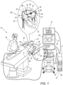
- Fig. 1 is a schematic, pictorial illustration of a probe-based ultrasound (US) imaging and electrophysiology (EP) mapping and ablation system 10, in accordance with an example of the present disclosure.
- US probe-based ultrasound
- EP electrophysiology
- System 10 includes multiple probes (e.g., catheters), which are percutaneously inserted by physician 24 through the patient's vascular system into a chamber of interest or vascular structure of a heart 12.
- probes e.g., catheters
- the physician may initially or anytime during the procedure, insert into heart 12 a 4D US probe 60 comprising a 2D ultrasound array 65 and an integral position sensor 63. Thereafter, a location-tracked EP mapping and ablation catheter 14 is inserted via a delivery sheath 23 into a desired location in heart 12.
- the 2D ultrasound array 65 produces a 3D sector-shaped ultrasound beam 85 occupying a defined solid angle, such a beam referred to herein as a "wedge 85."
- a wedge 85 beam the 2D ultrasound array can image a substantial volume of an organ, such as an entire cardiac chamber or area sector (like the valve or the LAA) of interest (e.g., the entire left atrium 45).
- US probe 60 images left atrium 45 while multi-arm effector 40 is placed therein.
- multi-arm catheter 14 includes one or more sensing electrodes 26 optionally distributed over a plurality of arms 16 at expandable distal end effector 40 and configured to sense intracardiac electrogram (IEGM) signals.
- Catheter 14 additionally includes a proximal position sensor (shown in Fig. 2 as a sensor 29) embedded in or near multi-arm effector 40 to track the position of a distal end of shaft 22.
- this position sensor is a magnetic-based position sensor including magnetic coils for sensing three-dimensional (3D) position.
- Catheter 14 itself, or another catheter to be inserted, may be used to perform ablation.
- physician 24 brings a multi-arm assembly 40 (also called hereinafter “expandable distal end assembly 40” or “distal end effector 40") fitted on a shaft 22 of catheter 14 into contact with a left atrium 45 wall 47 for sensing a target site in heart 12.
- physician 24 may use the same catheter 14 or, similarly, bring a distal end of an ablation catheter to a target site.
- the real-time 2D ultrasound-array produced images therefore include part of the sensing electrodes 26 in contact with tissue.
- Integral position sensor 63 of US probe 21 is preregistered with array 65 of the US probe. Because of the integral location sensor, the spatial coordinates of every voxel in the imaged cardiac chamber are known. Specifically, sensor 63 is configured to output first signals indicative of the location and orientation of the ultrasound transducer array 65 inside heart 12.
- console 24 receives location and orientation signals from position sensors 63 and 29 in response to magnetic fields from external field generators 32.
- Magnetic field generators 32 are placed at known positions on a location pad 25 external to patient 23. These location and orientation signals are indicative of the location and orientation of ultrasound array 65 in a coordinate system of the position tracking system. Using signals from sensor 29, real-time location and orientation of the distal end of catheter 14 of shaft 22 are also calculated.
- System 10 includes one or more electrode patches 38 positioned for skin contact on patient 23 to establish a location reference for location pad 25 as well as impedance-based tracking of electrodes 26.
- impedance-based tracking electrical current is directed toward electrodes 26 and sensed at electrode skin patches 38, such that the location of each electrode can be triangulated via electrode patches 38. Details of the impedance-based location tracking technology are described in US Patent Nos. 7,536,218 ; 7,756,576 ; 7,848,787 ; 7,869,865 ; and 8,456,182 .
- a recorder 11 displays electrograms 21 captured with body surface ECG electrodes 18 and intracardiac electrograms (IEGM) captured with electrodes 26 of catheter 14.
- Recorder 11 may include pacing capability for pacing the heart rhythm and/or may be electrically connected to a standalone pacer.
- System 10 may include an ablation energy generator 50 that is adapted to conduct ablative energy to one or more electrodes at a distal tip of a catheter configured for ablating.
- Energy produced by ablation energy generator 50 may include, but is not limited to, radiofrequency (RF) energy or pulsed-field ablation (PFA) energy, including monopolar or bipolar high-voltage DC pulses that may be used to effect irreversible electroporation (IRE), or combinations thereof.
- RF radiofrequency
- PFA pulsed-field ablation
- IRE irreversible electroporation
- Patient interface unit (PIU) 30 is configured to establish electrical communication between catheters, electrophysiological equipment, power supply and a workstation 55 for controlling operation of system 10.
- Electrophysiological equipment of system 10 may include, for example, multiple catheters, location pad 25, body surface ECG electrodes 18, electrode patches 38, ablation energy generator 50, and recorder 11.
- PIU 30 additionally includes processing capability for implementing real-time computations of catheter locations and for performing ECG calculations.
- Workstation 55 includes memory 57, processor unit 56 with memory or storage with appropriate operating software loaded therein, and user interface capability. Workstation 55 may provide multiple functions, optionally including (i) modeling endocardial anatomy in three dimensions (3D) and rendering the model or anatomical map 20 for display on a display device 27, (ii) displaying on display device 27 activation sequences (or other data) compiled from recorded electrograms 21 in representative visual indicia or imagery superimposed on the rendered anatomical map 20, (iii) displaying real-time location and orientation of multiple catheters within the heart chamber, and (iv) displaying on display device 27 sites of interest such as places where ablation energy has been applied.
- One commercial product embodying elements of the system 10 is available as the CARTO TM 3 System, available from Biosense Webster, Inc., 31A Technology Drive, Irvine, CA 92618.
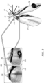
- Fig. 2 is a US biplane view 204 of arms 236 and 246 of distal end effector 40 of the EP mapping catheter 14 of Fig. 1 as derived from two US slices 213 and 214 selected from a volume 85 acquired by the 4D US probe 60 of Fig. 1 , in accordance with examples of the present disclosure. Image color is inverted for clarity.
- the physician can indicate which spline(s)/arm(s) to display (i.e., indicated arms 216 and 226), and the system can display an icon (rather than the arm itself) of the specific spline/arm of interest on the biplane view 204 or a triplane view.
- biplane 204 images 206 and 208 are derived (i.e., image processed) respectively from slices 213 and 214.
- the processor calculates planes where the processor finds target arms (i.e., indicated arms 216 and 226) in which distal end effector 40 is embedded.
- Slices 213 and 214 are then calculated as the intersection of the respective planes and image volume 85. In practice, any selected slice is taken with a given thickness (e.g., 2 mm).
- Location information enables the processor to search in an efficient manner (e.g., in a sub-volume of imaged volume 85) for arms 216 and 226.
- the processor applies an image-processing software to the sub-volume that, for example, uses thresholding of voxel values to identify an electrode 26 in contact with wall 47 tissue. With this information the processor can then calculate the desired planes.
- the location information on effector 40 is acquired using a magnetic position sensor 29 fitted on a distal end of shaft 22, and/or sensing electrodes 26 fitted on the arms of distal end effector 40.
- the location information on US probe 60 is acquired using magnetic position sensor 63, seen in Fig. 1 .
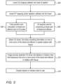
- Fig. 3 is a flow chart that schematically illustrates a method to visualize a distal end effector 40 in biplane or triplane US views (e.g., biplane view 204 images 206 and 208) based on selecting and image processing US slices (e.g., slices 213 and 214) acquired by the 4D US probe 60 of Fig. 1 , in accordance with an example of the present disclosure. While the method described is to a specific cardiac scenario that involves US imaging of a diagnostic and/or treatment cardiac catheter, the principles of the method hold for generating biplane or triplane views of other invasive devices caried by a utility probe.
- the algorithm carries out a process that begins with physician 24 inserting intracardiac echography (ICE) 4D US probe 60 inside a cardiac chamber of heart 12, at an US probe insertion step 302.
- ICE intracardiac echography
- a distal end effector e.g., effector 40
- processor 56 tracks the position and orientation of US probe 60 and of distal end effector 40.
- processor 56 receives an acquired volume wedge 85 from 4D US probe 60 that comprises at least part of the distal end effector 40 in a spatial relation with heart tissue.
- the processor selects US slices for further image processing (based on the tracking and by running an image processing application) where the slices include information on a spatial relation between one or more arms (or splines) of the distal end effector in relation to heart tissue.
- processor 56 image processes the selected slices to generate a biplane view (e.g., view 204) or a triplane view of the one or more arms (or splines) of the distal end effector in relation to heart tissue.
- a biplane view e.g., view 204
- a triplane view of the one or more arms (or splines) of the distal end effector in relation to heart tissue.
- processor 56 displays the biplane or triplane view to a user (e.g., on display 27).
- a basket catheter or another invasive device (e.g., a catheter delivering an artificial valve).
- another invasive device e.g., a catheter delivering an artificial valve
- a medical system (10) includes a utility probe (14), an ultrasound probe (60), and a processor (56).
- the utility probe is configured for insertion into a cavity of an organ, the utility probe comprising (a) a distal end effector (40) fitted at a distal end of the utility probe (14), and (b) a first sensor (29) configured to output first signals indicative of first positions of the distal end effector (14) inside the cavity.
- the ultrasound probe (60) is configured for insertion into an organ of a body, the ultrasound probe comprising (A) an ultrasound transducer array (65) configured to image a volume (85) of the organ, the volume comprising at least portion of the distal end effector (40), and (B) a second sensor (63) configured to output second signals indicative of second positions of the ultrasound transducer array (65) inside the cavity.
- the processor (56) is configured to, using the imaged volume (85), the first positions, and the second positions, (i) select one or more slices (213, 214) of the imaged volume (85) that comprise at least part of the distal end effector (40) in spatial relation with the organ, (ii) generate from the one or more selected slices at least one of a biplane view (204) and triplane view of the part of the distal end effector, and (iii) present the at least one of the biplane view (204) and triplane view to user.
- ultrasound probe (60) is an intracardiac echography (ICE) 4D ultrasound (US) catheter.
- ICE intracardiac echography
- US 4D ultrasound
- first and second sensors (29, 63) are magnetic position sensors configured to generate the first and second signals in response to a magnetic field applied by a position tracking system.
- the first sensor is an electrode (26) of the distal end effector configured to generate the first signals as part of an electrical position tracking system.
- the system (10) according to any of examples 1 through 7, wherein the processor is configured to generate from the one or more selected slices (213, 214) at least one of a biplane view (204) and triplane view using an image processing algorithm.
- a medical method includes inserting a utility probe (14) into a cavity of an organ, the utility probe comprising (a) a distal end effector (40) fitted at a distal end of the utility probe (14), and a first sensor (29) configured to output first signals indicative of first positions of the distal end effector (40) inside the cavity.
- An ultrasound probe (60 is inserted into an organ of a body, the ultrasound probe (60) comprising an ultrasound transducer array (65) configured to image a volume (85) of the organ, the volume comprising at least portion of the distal end effector (40), and a second sensor (63) configured to output second signals indicative of second positions of the ultrasound transducer array (65) inside the cavity.
- one or more slices (213, 214) of the imaged volume (85) are selected, that comprise at least part of the distal end effector (40) in spatial relation with the organ.
- At least one of a biplane view (204) and triplane view of the part of the distal end effector (40) are generated from the one or more selected slices (213, 214).
- the at least one of the biplane view (204) and triplane view are presented to a user.
- the ultrasound probe is an intracardiac echography (ICE) 4D ultrasound (US) catheter.
- ICE intracardiac echography
- US 4D ultrasound
- first and second sensors are magnetic position sensors configured to generate the first and second signals in response to a magnetic field applied by a position tracking system.
- the first sensor is an electrode of the distal end effector configured to generate the first signals as part of an electrical position tracking system.
- selecting one or more slices comprises using an image processing algorithm.
- generating from the one or more selected slices at least one of a biplane view and triplane view comprises using an image processing algorithm.
Landscapes
- Health & Medical Sciences (AREA)
- Life Sciences & Earth Sciences (AREA)
- Engineering & Computer Science (AREA)
- General Health & Medical Sciences (AREA)
- Medical Informatics (AREA)
- Public Health (AREA)
- Physics & Mathematics (AREA)
- Surgery (AREA)
- Animal Behavior & Ethology (AREA)
- Biomedical Technology (AREA)
- Heart & Thoracic Surgery (AREA)
- Veterinary Medicine (AREA)
- Molecular Biology (AREA)
- Pathology (AREA)
- Biophysics (AREA)
- Nuclear Medicine, Radiotherapy & Molecular Imaging (AREA)
- Radiology & Medical Imaging (AREA)
- Computer Vision & Pattern Recognition (AREA)
- Cardiology (AREA)
- Human Computer Interaction (AREA)
- Physiology (AREA)
- Quality & Reliability (AREA)
- Epidemiology (AREA)
- Primary Health Care (AREA)
- General Physics & Mathematics (AREA)
- Theoretical Computer Science (AREA)
- Gynecology & Obstetrics (AREA)
- Otolaryngology (AREA)
- Plasma & Fusion (AREA)
- Ultra Sonic Daignosis Equipment (AREA)
- Surgical Instruments (AREA)
Applications Claiming Priority (1)
| Application Number | Priority Date | Filing Date | Title |
|---|---|---|---|
| US18/223,890 US20250025129A1 (en) | 2023-07-19 | 2023-07-19 | Visualization of distal end effector on biplane or triplane views using intracardiac echography (ice) |
Publications (1)
| Publication Number | Publication Date |
|---|---|
| EP4494570A1 true EP4494570A1 (de) | 2025-01-22 |
Family
ID=91959318
Family Applications (1)
| Application Number | Title | Priority Date | Filing Date |
|---|---|---|---|
| EP24189319.7A Pending EP4494570A1 (de) | 2023-07-19 | 2024-07-18 | Visualisierung eines distalen endeffektors auf zwei- oder dreiebenenansichten mittels intrakardialer echographie (ice) |
Country Status (5)
| Country | Link |
|---|---|
| US (1) | US20250025129A1 (de) |
| EP (1) | EP4494570A1 (de) |
| JP (1) | JP2025015491A (de) |
| CN (1) | CN119326440A (de) |
| IL (1) | IL314353A (de) |
Citations (20)
| Publication number | Priority date | Publication date | Assignee | Title |
|---|---|---|---|---|
| US5443489A (en) | 1993-07-20 | 1995-08-22 | Biosense, Inc. | Apparatus and method for ablation |
| US5539199A (en) | 1994-02-19 | 1996-07-23 | Leuze Electronic Gmbh + Co. | Method for detecting objects in a monitored area |
| US5558091A (en) | 1993-10-06 | 1996-09-24 | Biosense, Inc. | Magnetic determination of position and orientation |
| US6172499B1 (en) | 1999-10-29 | 2001-01-09 | Ascension Technology Corporation | Eddy current error-reduced AC magnetic position measurement system |
| US6239724B1 (en) | 1997-12-30 | 2001-05-29 | Remon Medical Technologies, Ltd. | System and method for telemetrically providing intrabody spatial position |
| US6332089B1 (en) | 1996-02-15 | 2001-12-18 | Biosense, Inc. | Medical procedures and apparatus using intrabody probes |
| US6484118B1 (en) | 2000-07-20 | 2002-11-19 | Biosense, Inc. | Electromagnetic position single axis system |
| US6618612B1 (en) | 1996-02-15 | 2003-09-09 | Biosense, Inc. | Independently positionable transducers for location system |
| US6690963B2 (en) | 1995-01-24 | 2004-02-10 | Biosense, Inc. | System for determining the location and orientation of an invasive medical instrument |
| US6892091B1 (en) | 2000-02-18 | 2005-05-10 | Biosense, Inc. | Catheter, method and apparatus for generating an electrical map of a chamber of the heart |
| US20070167801A1 (en) * | 2005-12-02 | 2007-07-19 | Webler William E | Methods and apparatuses for image guided medical procedures |
| US7536218B2 (en) | 2005-07-15 | 2009-05-19 | Biosense Webster, Inc. | Hybrid magnetic-based and impedance-based position sensing |
| US7604601B2 (en) | 2005-04-26 | 2009-10-20 | Biosense Webster, Inc. | Display of catheter tip with beam direction for ultrasound system |
| US7756576B2 (en) | 2005-08-26 | 2010-07-13 | Biosense Webster, Inc. | Position sensing and detection of skin impedance |
| US7848787B2 (en) | 2005-07-08 | 2010-12-07 | Biosense Webster, Inc. | Relative impedance measurement |
| US7869865B2 (en) | 2005-01-07 | 2011-01-11 | Biosense Webster, Inc. | Current-based position sensing |
| US8456182B2 (en) | 2008-09-30 | 2013-06-04 | Biosense Webster, Inc. | Current localization tracker |
| US20150320439A1 (en) * | 2012-06-28 | 2015-11-12 | Koninklijke Philips N.V. | Ultrasonically guided biopsies in three dimensions |
| US20220133284A1 (en) * | 2019-03-13 | 2022-05-05 | University Of Florida Research Foundation, Inc. | Guidance and tracking system for templated and targeted biopsy and treatment |
| US20230034665A1 (en) * | 2014-07-09 | 2023-02-02 | Neil Glossop | Systems, methods, and devices for assisting or performing guided interventional procedures using custom templates |
Family Cites Families (4)
| Publication number | Priority date | Publication date | Assignee | Title |
|---|---|---|---|---|
| JP4972648B2 (ja) * | 2005-10-11 | 2012-07-11 | カーネギー−メロン ユニバーシティ | センサに案内されるカテーテル誘導システム |
| CN105592778B (zh) * | 2013-10-14 | 2019-07-23 | 波士顿科学医学有限公司 | 高分辨率心脏标测电极阵列导管 |
| US20190060003A1 (en) * | 2017-08-28 | 2019-02-28 | Edwards Lifesciences Corporation | Cardiac mapping and navigation for transcatheter procedures |
| US20240173016A1 (en) * | 2022-11-29 | 2024-05-30 | Biosense Webster (Israel) Ltd. | Assessment of tissue ablation using intracardiac ultrasound catheter |
-
2023
- 2023-07-19 US US18/223,890 patent/US20250025129A1/en active Pending
-
2024
- 2024-07-16 IL IL314353A patent/IL314353A/en unknown
- 2024-07-18 EP EP24189319.7A patent/EP4494570A1/de active Pending
- 2024-07-18 JP JP2024114597A patent/JP2025015491A/ja active Pending
- 2024-07-19 CN CN202410972779.7A patent/CN119326440A/zh active Pending
Patent Citations (21)
| Publication number | Priority date | Publication date | Assignee | Title |
|---|---|---|---|---|
| US5443489A (en) | 1993-07-20 | 1995-08-22 | Biosense, Inc. | Apparatus and method for ablation |
| US5558091A (en) | 1993-10-06 | 1996-09-24 | Biosense, Inc. | Magnetic determination of position and orientation |
| US5539199A (en) | 1994-02-19 | 1996-07-23 | Leuze Electronic Gmbh + Co. | Method for detecting objects in a monitored area |
| US6690963B2 (en) | 1995-01-24 | 2004-02-10 | Biosense, Inc. | System for determining the location and orientation of an invasive medical instrument |
| US6332089B1 (en) | 1996-02-15 | 2001-12-18 | Biosense, Inc. | Medical procedures and apparatus using intrabody probes |
| US6618612B1 (en) | 1996-02-15 | 2003-09-09 | Biosense, Inc. | Independently positionable transducers for location system |
| US6788967B2 (en) | 1997-05-14 | 2004-09-07 | Biosense, Inc. | Medical diagnosis, treatment and imaging systems |
| US6239724B1 (en) | 1997-12-30 | 2001-05-29 | Remon Medical Technologies, Ltd. | System and method for telemetrically providing intrabody spatial position |
| US6172499B1 (en) | 1999-10-29 | 2001-01-09 | Ascension Technology Corporation | Eddy current error-reduced AC magnetic position measurement system |
| US6892091B1 (en) | 2000-02-18 | 2005-05-10 | Biosense, Inc. | Catheter, method and apparatus for generating an electrical map of a chamber of the heart |
| US6484118B1 (en) | 2000-07-20 | 2002-11-19 | Biosense, Inc. | Electromagnetic position single axis system |
| US7869865B2 (en) | 2005-01-07 | 2011-01-11 | Biosense Webster, Inc. | Current-based position sensing |
| US7604601B2 (en) | 2005-04-26 | 2009-10-20 | Biosense Webster, Inc. | Display of catheter tip with beam direction for ultrasound system |
| US7848787B2 (en) | 2005-07-08 | 2010-12-07 | Biosense Webster, Inc. | Relative impedance measurement |
| US7536218B2 (en) | 2005-07-15 | 2009-05-19 | Biosense Webster, Inc. | Hybrid magnetic-based and impedance-based position sensing |
| US7756576B2 (en) | 2005-08-26 | 2010-07-13 | Biosense Webster, Inc. | Position sensing and detection of skin impedance |
| US20070167801A1 (en) * | 2005-12-02 | 2007-07-19 | Webler William E | Methods and apparatuses for image guided medical procedures |
| US8456182B2 (en) | 2008-09-30 | 2013-06-04 | Biosense Webster, Inc. | Current localization tracker |
| US20150320439A1 (en) * | 2012-06-28 | 2015-11-12 | Koninklijke Philips N.V. | Ultrasonically guided biopsies in three dimensions |
| US20230034665A1 (en) * | 2014-07-09 | 2023-02-02 | Neil Glossop | Systems, methods, and devices for assisting or performing guided interventional procedures using custom templates |
| US20220133284A1 (en) * | 2019-03-13 | 2022-05-05 | University Of Florida Research Foundation, Inc. | Guidance and tracking system for templated and targeted biopsy and treatment |
Also Published As
| Publication number | Publication date |
|---|---|
| JP2025015491A (ja) | 2025-01-30 |
| CN119326440A (zh) | 2025-01-21 |
| IL314353A (en) | 2025-02-01 |
| US20250025129A1 (en) | 2025-01-23 |
Similar Documents
| Publication | Publication Date | Title |
|---|---|---|
| EP3453328B1 (de) | Algorithmus zur mesh-einpassung | |
| JP2020507395A (ja) | 心臓現象(Cardiac phenomena)の有病率を決定するための方法およびシステム | |
| EP4122413A1 (de) | Genaue gewebenähe | |
| EP4101375A1 (de) | Automatische anatomische merkmalsidentifizierung und abbildungssegmentierung | |
| US20240173016A1 (en) | Assessment of tissue ablation using intracardiac ultrasound catheter | |
| EP4494570A1 (de) | Visualisierung eines distalen endeffektors auf zwei- oder dreiebenenansichten mittels intrakardialer echographie (ice) | |
| US20240164686A1 (en) | Three-dimensional display of a multi-electrode catheter and signals acquired over time | |
| US20240212157A1 (en) | Cropping volumetric image of region of interest from three-dimensional ultrasound image | |
| US12324669B2 (en) | Detecting local activation source in atrial fibrillation | |
| EP4393389B1 (de) | Systeme zur abbildung von gewebekontakt mittels triangulation | |
| US20250186108A1 (en) | Estimation of catheter proximity to tissue using contact force sensing | |
| EP4529836A1 (de) | Schätzung der kontaktkraft einer expandierbaren katheteranordnung | |
| EP4574031A1 (de) | Bereitstellung eines blutpool-richtungsvektors auf der basis gemessener impedanzen | |
| WO2023118995A1 (en) | Visualization of change in anatomical slope using 4d ultrasound catheter | |
| CN120225119A (zh) | 使用有向图的病灶心律失常源查找器 | |
| IL320045A (en) | Mapping of space |
Legal Events
| Date | Code | Title | Description |
|---|---|---|---|
| PUAI | Public reference made under article 153(3) epc to a published international application that has entered the european phase |
Free format text: ORIGINAL CODE: 0009012 |
|
| STAA | Information on the status of an ep patent application or granted ep patent |
Free format text: STATUS: THE APPLICATION HAS BEEN PUBLISHED |
|
| AK | Designated contracting states |
Kind code of ref document: A1 Designated state(s): AL AT BE BG CH CY CZ DE DK EE ES FI FR GB GR HR HU IE IS IT LI LT LU LV MC ME MK MT NL NO PL PT RO RS SE SI SK SM TR |
|
| STAA | Information on the status of an ep patent application or granted ep patent |
Free format text: STATUS: REQUEST FOR EXAMINATION WAS MADE |
|
| 17P | Request for examination filed |
Effective date: 20250627 |