EP4501206A1 - Bildverarbeitungsvorrichtung, bildverarbeitungsverfahren und speichermedium - Google Patents
Bildverarbeitungsvorrichtung, bildverarbeitungsverfahren und speichermedium Download PDFInfo
- Publication number
- EP4501206A1 EP4501206A1 EP22933492.5A EP22933492A EP4501206A1 EP 4501206 A1 EP4501206 A1 EP 4501206A1 EP 22933492 A EP22933492 A EP 22933492A EP 4501206 A1 EP4501206 A1 EP 4501206A1
- Authority
- EP
- European Patent Office
- Prior art keywords
- model
- image processing
- lesion
- endoscopic image
- processing device
- Prior art date
- Legal status (The legal status is an assumption and is not a legal conclusion. Google has not performed a legal analysis and makes no representation as to the accuracy of the status listed.)
- Pending
Links
Images




Classifications
-
- G—PHYSICS
- G06—COMPUTING OR CALCULATING; COUNTING
- G06V—IMAGE OR VIDEO RECOGNITION OR UNDERSTANDING
- G06V10/00—Arrangements for image or video recognition or understanding
- G06V10/20—Image preprocessing
- G06V10/25—Determination of region of interest [ROI] or a volume of interest [VOI]
-
- A—HUMAN NECESSITIES
- A61—MEDICAL OR VETERINARY SCIENCE; HYGIENE
- A61B—DIAGNOSIS; SURGERY; IDENTIFICATION
- A61B1/00—Instruments for performing medical examinations of the interior of cavities or tubes of the body by visual or photographical inspection, e.g. endoscopes; Illuminating arrangements therefor
- A61B1/00002—Operational features of endoscopes
- A61B1/00043—Operational features of endoscopes provided with output arrangements
- A61B1/00045—Display arrangement
- A61B1/0005—Display arrangement combining images e.g. side-by-side, superimposed or tiled
-
- A—HUMAN NECESSITIES
- A61—MEDICAL OR VETERINARY SCIENCE; HYGIENE
- A61B—DIAGNOSIS; SURGERY; IDENTIFICATION
- A61B1/00—Instruments for performing medical examinations of the interior of cavities or tubes of the body by visual or photographical inspection, e.g. endoscopes; Illuminating arrangements therefor
- A61B1/00002—Operational features of endoscopes
- A61B1/00004—Operational features of endoscopes characterised by electronic signal processing
- A61B1/00009—Operational features of endoscopes characterised by electronic signal processing of image signals during a use of endoscope
- A61B1/000094—Operational features of endoscopes characterised by electronic signal processing of image signals during a use of endoscope extracting biological structures
-
- A—HUMAN NECESSITIES
- A61—MEDICAL OR VETERINARY SCIENCE; HYGIENE
- A61B—DIAGNOSIS; SURGERY; IDENTIFICATION
- A61B1/00—Instruments for performing medical examinations of the interior of cavities or tubes of the body by visual or photographical inspection, e.g. endoscopes; Illuminating arrangements therefor
- A61B1/00002—Operational features of endoscopes
- A61B1/00004—Operational features of endoscopes characterised by electronic signal processing
- A61B1/00009—Operational features of endoscopes characterised by electronic signal processing of image signals during a use of endoscope
- A61B1/000096—Operational features of endoscopes characterised by electronic signal processing of image signals during a use of endoscope using artificial intelligence
-
- A—HUMAN NECESSITIES
- A61—MEDICAL OR VETERINARY SCIENCE; HYGIENE
- A61B—DIAGNOSIS; SURGERY; IDENTIFICATION
- A61B1/00—Instruments for performing medical examinations of the interior of cavities or tubes of the body by visual or photographical inspection, e.g. endoscopes; Illuminating arrangements therefor
- A61B1/00002—Operational features of endoscopes
- A61B1/00043—Operational features of endoscopes provided with output arrangements
- A61B1/00055—Operational features of endoscopes provided with output arrangements for alerting the user
-
- G—PHYSICS
- G06—COMPUTING OR CALCULATING; COUNTING
- G06T—IMAGE DATA PROCESSING OR GENERATION, IN GENERAL
- G06T7/00—Image analysis
- G06T7/0002—Inspection of images, e.g. flaw detection
- G06T7/0012—Biomedical image inspection
-
- G—PHYSICS
- G06—COMPUTING OR CALCULATING; COUNTING
- G06T—IMAGE DATA PROCESSING OR GENERATION, IN GENERAL
- G06T7/00—Image analysis
- G06T7/90—Determination of colour characteristics
-
- G—PHYSICS
- G06—COMPUTING OR CALCULATING; COUNTING
- G06V—IMAGE OR VIDEO RECOGNITION OR UNDERSTANDING
- G06V10/00—Arrangements for image or video recognition or understanding
- G06V10/70—Arrangements for image or video recognition or understanding using pattern recognition or machine learning
- G06V10/82—Arrangements for image or video recognition or understanding using pattern recognition or machine learning using neural networks
-
- G—PHYSICS
- G06—COMPUTING OR CALCULATING; COUNTING
- G06V—IMAGE OR VIDEO RECOGNITION OR UNDERSTANDING
- G06V10/00—Arrangements for image or video recognition or understanding
- G06V10/70—Arrangements for image or video recognition or understanding using pattern recognition or machine learning
- G06V10/87—Arrangements for image or video recognition or understanding using pattern recognition or machine learning using selection of the recognition techniques, e.g. of a classifier in a multiple classifier system
-
- G—PHYSICS
- G06—COMPUTING OR CALCULATING; COUNTING
- G06T—IMAGE DATA PROCESSING OR GENERATION, IN GENERAL
- G06T2207/00—Indexing scheme for image analysis or image enhancement
- G06T2207/10—Image acquisition modality
- G06T2207/10024—Color image
-
- G—PHYSICS
- G06—COMPUTING OR CALCULATING; COUNTING
- G06T—IMAGE DATA PROCESSING OR GENERATION, IN GENERAL
- G06T2207/00—Indexing scheme for image analysis or image enhancement
- G06T2207/10—Image acquisition modality
- G06T2207/10068—Endoscopic image
-
- G—PHYSICS
- G06—COMPUTING OR CALCULATING; COUNTING
- G06T—IMAGE DATA PROCESSING OR GENERATION, IN GENERAL
- G06T2207/00—Indexing scheme for image analysis or image enhancement
- G06T2207/20—Special algorithmic details
- G06T2207/20084—Artificial neural networks [ANN]
-
- G—PHYSICS
- G06—COMPUTING OR CALCULATING; COUNTING
- G06T—IMAGE DATA PROCESSING OR GENERATION, IN GENERAL
- G06T2207/00—Indexing scheme for image analysis or image enhancement
- G06T2207/30—Subject of image; Context of image processing
- G06T2207/30004—Biomedical image processing
- G06T2207/30096—Tumor; Lesion
-
- G—PHYSICS
- G06—COMPUTING OR CALCULATING; COUNTING
- G06V—IMAGE OR VIDEO RECOGNITION OR UNDERSTANDING
- G06V2201/00—Indexing scheme relating to image or video recognition or understanding
- G06V2201/03—Recognition of patterns in medical or anatomical images
-
- G—PHYSICS
- G06—COMPUTING OR CALCULATING; COUNTING
- G06V—IMAGE OR VIDEO RECOGNITION OR UNDERSTANDING
- G06V2201/00—Indexing scheme relating to image or video recognition or understanding
- G06V2201/03—Recognition of patterns in medical or anatomical images
- G06V2201/032—Recognition of patterns in medical or anatomical images of protuberances, polyps nodules, etc.
Definitions
- the present disclosure relates to a technical field of an image processing device, an image processing method, and a storage medium for processing an image to be acquired in endoscopic examination.
- Patent Literature 1 discloses an endoscopic examination system that detects a region of interest based on an endoscopic image and a target region detection threshold value and that determines whether the region of interest is either a flat lesion or a raised lesion.
- Patent Literature 2 discloses an endoscopic examination system configured to perform a three-dimensional reconstruction of a target organ of examination by the SfM (Structure from Motion) using time-series endoscopic images and identify a captured region through matching (image registration) between the three-dimensionally reconstructed data and a standard model of the target organ thereby to display the identified captured region.
- SfM Structure from Motion
- an example object of the present disclosure to provide an image processing device, an image processing method, and a storage medium capable of accurately detecting a lesion in an endoscope examination.
- One mode of the image processing device is an image processing device including:
- One mode of the image processing method is an image processing method executed by a computer, the image processing method including:
- One mode of the storage medium is a storage medium storing a program executed by a computer, the program causing the computer to:
- An example advantage according to the present invention is to accurately detect a lesion in accordance with a condition of the examination target or a photographed portion of the examination target.
- FIG. 1 shows a schematic configuration of an endoscopic examination system 100.
- an endoscopic examination system 100 is a system configured to inform an examiner, such as a doctor who performs examination or treatment using an endoscope, of a part (also referred to as "lesion part") of examination target which is suspected of a lesion, and mainly includes an image processing device 1, a display device 2, and an endoscope 3 connected to the image processing device 1.
- the lesion part is an example of the "part of interest”.
- the image processing device 1 acquires an image (also referred to as "endoscopic image Ia") captured by the endoscope 3 in time series from the endoscope 3 and displays a screen image based on the endoscopic image Ia on the display device 2.
- the endoscopic image Ia is an image captured at predetermined time intervals in at least one of the insertion process of the endoscope 3 to the subject and/or the ejection process of the endoscope 3 from the subject.
- the image processing device 1 analyzes the endoscopic image Ia to detect the lesion part in the endoscopic image Ia, and displays the information on the detection result on the display device 2.
- the display device 2 is a display or the like for display information based on the display signal supplied from the image processing device 1.
- the endoscope 3 mainly includes an operation unit 36 for examiner to perform a predetermined input, a shaft 37 which has flexibility and which is inserted into the organ to be photographed of the subject, a tip unit 38 having a built-in photographing unit such as an ultrasmall image pickup device, and a connecting unit 39 for connecting to the image processing device 1.
- the configuration of the endoscopic examination system 100 shown in FIG.1 is an example, and various change may be applied thereto.
- the image processing device 1 may be configured integrally with the display device 2.
- the image processing device 1 may be configured by a plurality of devices.
- the target of the endoscopic examination in the present disclosure is not limited to a large bowel, it may be any organ subject to endoscopic examination such as esophagus, stomach, pancreas.
- the target of the endoscopic examination in the present disclosure include a laryngendoscope, a bronchoscope, an upper digestive tube endoscope, a duodenum endoscope, a small bowel endoscope, a large bowel endoscope, a capsule endoscope, a thoracoscope, a laparoscope, a cystoscope, a cholangioscope, an arthroscope, a spinal endoscope, a blood vessel endoscope, and an epidural endoscope.
- target conditions referred to as "target conditions" of the lesion part to be detected in endoscopic examination are exemplified as (a) to (f) below.
- FIG. 2 shows the hardware configuration of the image processing device 1.
- the image processing device 1 mainly includes a processor 11, a memory 12, an interface 13, an input unit 14, a light source unit 15, and an audio output unit 16. Each of these elements is connected via a data bus 19.
- the processor 11 executes a predetermined process by executing a program or the like stored in the memory 12.
- the processor 11 is one or more processors such as a CPU (Central Processing Unit), a GPU (Graphics Processing Unit), and a TPU (Tensor Processing Unit).
- the processor 11 is an example of a computer.
- the processor 11 may be configured by a plurality of processors.
- the processor 11 is an example of a computer.
- the memory 12 is configured by a variety of volatile memories which is used as working memories, and nonvolatile memories which stores information necessary for the process to be executed by the image processing device 1, such as a RAM (Random Access Memory) and a ROM (Read Only Memory).
- the memory 12 may include an external storage device such as a hard disk connected to or built in to the image processing device 1, or may include a storage medium such as a removable flash memory.
- the memory 12 stores a program for the image processing device 1 to execute each process in the present example embodiment.
- the memory 12 stores first model information D1 relating to a first model used to make a determination as to the condition (also referred to as "background condition") other than the target condition to be detected in the endoscopic examination, and second model information D2 relating to a second model used to detect the target condition.
- the background condition include inflammation, bleeding, Barrett's esophagus if the examination target is an esophagus, and any other conditions other than the target condition.
- the first model information D1 and the second model information D2 will be described later.
- the interface 13 performs an interface operation between the image processing device 1 and an external device. For example, the interface 13 supplies the display information "Ib" generated by the processor 11 to the display device 2. Further, the interface 13 supplies the light generated by the light source unit 15 to the endoscope 3. The interface 13 also provides an electrical signal to the processor 11 indicative of the endoscopic image Ia supplied from the endoscope 3.
- the interface 13 may be a communication interface, such as a network adapter, for wired or wireless communication with the external device, or a hardware interface compliant with a USB (Universal Serial Bus), a SATA (Serial AT Attachment), or the like.
- the input unit 14 generates an input signal based on the operation by the examiner. Examples of the input unit 14 include a button, a touch panel, a remote controller, and a voice input device.
- the light source unit 15 generates light for supplying to the tip unit 38 of the endoscope 3.
- the light source unit 15 may also incorporate a pump or the like for delivering water and air to be supplied to the endoscope 3.
- the audio output unit 16 outputs a sound under the control of the processor 11.
- the first model information D1 is information relating to the first model configured to output information relating to the background condition when an endoscopic image is inputted to the first model.
- the first model data D1 contains the parameters required to build the first model.
- the first model is a classification model configured to output a classification result regarding the presence or absence (and the degree) of the background condition when an endoscopic image is inputted to the model.
- the classification result outputted by the first model may indicate the presence or absence (and the degree) of a particular type of the background condition, or may indicate the detected type (and the degree) of the background condition.
- the first model may be any machine learning model (including any statistical model, hereinafter the same) such as a neural network and a support vector machine.
- the first model information D1 includes various parameters such as a layer structure, a neuron structure of each layer, the number of filters and the size of filters in each layer, and the weight for each element of each filter.
- the first model is not limited to being a machine-learning model, but may be a model configured to determine whether or not there is a particular background condition based on the RGB ratio of the inputted endoscopic image.
- the first model may be a model configured to determine that there is a background condition (e.g., irritation), upon determining that the ratio (e.g., the average ratio of all pixels) of R among the RGB of the inputted endoscopic image is equal to or greater than a predetermined threshold value.
- a background condition e.g., irritation
- the ratio e.g., the average ratio of all pixels
- the second model information D2 is information relating to the second model and includes parameters required for building the second model, wherein the second model is configured to output information regarding the target condition to be detected in the endoscopic examination when an endoscopic image is inputted to the second model.
- the second model may be any machine learning model (including any statistical model, hereinafter the same) such as a neural network and a support vector machine.
- the second model information D2 includes various parameters such as a layer structure, a neuron structure of each layer, the number of filters and filter sizes in each layer, and a weight for each element of each filter.
- the second model is trained to output, when an endoscopic image is inputted thereto, at least a score (also referred to as "lesion confidence score S") indicating the confidence level of the presence of a lesion part subjected to the target condition in the inputted endoscopic image.
- a score also referred to as "lesion confidence score S”
- the second model may output, as the lesion confidence score S, the confidence level outputted when a neural network is adopted as the architecture of the classification model.
- the second model may be a model configured to output not only the lesion confidence score S but also information (also referred to as "lesion region information") indicating the position or region (area) of the lesion part in the inputted endoscopic image.
- the second model may be a model configured to output a heat map indicating the lesion confidence score S for each unit region (e.g., a region of one or more pixels) in the inputted endoscopic image.
- the image processing device 1 may make a determination as to whether or not the each unit region corresponds to a lesion part subjected to the target condition.
- the second model corresponds to a model which has learned the relation between an endoscopic image inputted to the second model and the presence or absence of a lesion part in the inputted endoscopic image.
- the first model may be provided for each background condition.
- the parameters for building the first model for each background condition are included in the first model data D1.
- the second model may be provided for each target condition. In this situation, the parameters for building the second model for each target condition are included in the second model data D2.
- the first model and the second model are learning models
- the first model and the second model are trained in advance based on sets of an input image, which conforms to the input format of each model, and the corresponding correct answer data indicating a correct answer to be outputted when the input image is inputted to each model.
- the parameters of the models obtained by training are stored as the first model information D1 and the second model information D2 in the memory 12, respectively.
- the image processing device 1 sets a threshold value (also referred to as "lesion threshold value") for comparing with the lesion confidence score S to determine the presence or absence of the lesion part of the target condition. As a result, the image processing device 1 accurately sets the lesion threshold value and improves the lesion detection accuracy.
- a threshold value also referred to as "lesion threshold value”
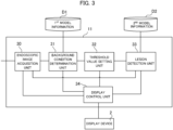
- FIG. 3 is a functional block diagram of the image processing device 1 related to the lesion detection process.
- the processor 11 of the image processing device 1 functionally includes an endoscopic image acquisition unit 30, a background condition determination unit 31, a threshold value setting unit 32, a lesion detection unit 33, and a display control unit 34.
- any blocks to exchange data with each other are connected by a solid line, but the combination of the blocks to exchange data with each other is not limited thereto. The same applies to the drawings of other functional blocks described below.
- the endoscopic image acquisition unit 30 acquires the endoscopic image Ia taken by the endoscope 3 through the interface 13 at predetermined intervals. Then, the endoscopic image acquisition unit 30 supplies the acquired endoscopic image Ia to the background condition determination unit 31, the lesion detection unit 33, and the display control unit 34, respectively.
- the background condition determination unit 31 Based on the endoscopic image Ia supplied from the endoscopic image acquisition unit 30, the background condition determination unit 31 makes a determination regarding the background condition and supplies information (also referred to as "background condition information") indicating the determination result of the background condition to the threshold value setting unit 32.
- the background condition determination unit 31 inputs the endoscopic image Ia supplied from the endoscopic image acquisition unit 30 to the first model configured by referring to the first model information D1, and makes the determination regarding the background condition based on the information outputted by the first model in response to the input of the endoscopic image Ia to the first model.
- the first model outputs a classification result regarding the presence or absence of each of various background conditions assumed in the target organ of examination, and the background condition determination unit 31 generates background condition information indicating the type of the detected background condition, based on the classification result.
- the classification result and background condition information described above may include information regarding the degree of the background condition when a background condition is detected.
- the background condition determination unit 31 may supply the background condition information to the display control unit 34.
- the threshold value setting unit 32 sets the lesion threshold value based on the background condition information supplied from the background condition determination unit 31, and supplies the set lesion threshold value to the lesion detection unit 33.
- the memory 12 stores relation information (e.g., look-up table or formula) which indicates a relation between the type of background condition and the lesion threshold value to be set, and the threshold value setting unit 32 sets, by referring to the relation information, the lesion threshold value corresponding to the background condition indicated by the background condition information.
- the lesion detection tests based on the second model and the lesion threshold value by use of test data are conducted beforehand and then the above-mentioned relation information is generated such that the F value (harmonic mean of the precision ratio and the recall ratio) is maximized for each type of the background condition in the tests.
- the training data in this case is, for example, sets of an endoscopic image prepared for each type of background condition and the corresponding correct answer data indicating the presence or absence of a lesion part in the endoscopic image.
- the above-described relation information may indicate the relation between the type and degree of the background condition and the lesion threshold value to be set.
- the threshold value setting unit 32 may supply the display control unit 34 with the set lesion threshold value.
- the lesion detection unit 33 On the basis of the endoscopic image Ia supplied from the endoscopic image acquisition unit 30 and the lesion threshold value supplied from the threshold value setting unit 32, the lesion detection unit 33 detects a lesion part, and supplies information (also referred to as "lesion detection information") indicating the detection result to the display control unit 34. In this case, the lesion detection unit 33 generates the lesion detection information by comparing the lesion confidence score S, which is outputted from the second model when the endoscopic image Ia is inputted into the second model configured by referring to the second model information D2, with the lesion threshold value. If the second model further outputs the lesion region information indicating the region of the lesion part in the endoscopic image Ia, the lesion detection unit 33 may include the lesion region information in the lesion detection information.
- the display control unit 34 On the basis of the latest endoscopic image Ia supplied from the endoscopic image acquisition unit 30 and the lesion detection information supplied from the lesion detection unit 33, the display control unit 34 generates display information Ib and supplies the display device 2 with the generated display information Ib to thereby cause the display device 2 to display the latest endoscopic image Ia and the lesion detection result.
- the display control unit 34 may further display information on the background condition information supplied from the background condition determination unit 31 or/and the lesion threshold value supplied from the threshold value setting unit 32 on the display device 2. The display example of the display device 2 by the display control unit 34 will be described later.
- the display control unit 34 may perform the audio output control of the sound output unit 16 to output a warning sound or voice guidance or the like for notifying the user that the lesion part is detected.
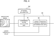
- FIG. 4 shows an example of a functional block of the lesion detection unit 33.
- the condition detection unit 33 functionally includes a confidence score calculation unit 41, and a target condition presence / absence determination unit 42.
- the confidence score calculation unit 41 inputs the endoscopic image Ia to the second model configured by referring to the second model information D2. Then, the confidence score calculation unit 41 supplies the lesion confidence score S outputted by the second model based on the inputted endoscopic image Ia to the target condition presence / absence determination unit 42. In the case where the second model is prepared for each type of the target condition, the lesion confidence score S is calculated for each type of the target condition.
- the target condition presence / absence determination unit 42 determines whether or not there is a lesion part subjected to the target condition in the endoscopic image Ia. Specifically, if the lesion confidence score S is equal to or larger than the lesion threshold value, the target condition presence / absence determination unit 42 determines that there is a lesion part subjected to the target condition in the endoscopic image Ia. In contrast, if the lesion confidence score S is smaller than the lesion threshold value, the target condition presence / absence determination unit 42 determines that there is no lesion part subjected to the target condition in the endoscopic image Ia.
- the target condition presence / absence determination unit 42 determines, for each type of the target condition, whether or not there is a lesion part. Then, the target condition presence / absence determination unit 42 outputs the lesion detection information based on the determination result to the display control unit 34.
- the lesion detection information may include not only information on the presence or absence of the lesion part subjected to the target condition but also the lesion confidence score S and the lesion region information outputted by the second model.
- the functional configuration as shown in FIGS. 3 and 4 enables the image processing device 1 to accurately detect the target condition in consideration of the background condition of the target examination.
- the degree of difficulty in detecting the target condition varies depending on the background condition. For example, since the color of the surface of a large bowel having a history of an inflammatory condition is different in the color from a standard large bowel, the degree of difficulty in detecting the target condition, such as a tumor, of such a large bowel increases. Therefore, in this case, it is desired that the lesion threshold value for the lesion confidence score S be a low value.
- the image processing device 1 adaptively sets a lesion threshold value in accordance with a background condition.
- Each component of the endoscope image acquisition unit 30, the background condition determination unit 31, the threshold value setting unit 32, the lesion detection unit 33, and the display control unit 34 can be realized, for example, by the processor 11 which executes a program.
- the necessary program may be recorded in any non-volatile storage medium and installed as necessary to realize the respective components.
- at least a part of these components is not limited to being realized by a software program and may be realized by any combination of hardware, firmware, and software.
- At least some of these components may also be implemented using user-programmable integrated circuitry, such as FPGA (Field-Programmable Gate Array) and microcontrollers. In this case, the integrated circuit may be used to realize a program for configuring each of the above-described components.
- each component may be configured by a ASSP (Application Specific Standard Produce), ASIC (Application Specific Integrated Circuit) and/or a quantum processor (quantum computer control chip).
- ASSP Application Specific Standard Produce
- ASIC Application Specific Integrated Circuit
- quantum processor quantum computer control chip
- FIG. 5 shows a first display example of a display screen image displayed by the display device 2 in the endoscopic examination.
- the display control unit 34 of the image processing device 1 transmits the display information Ib generated on the basis of the information supplied from the other processing units 30 to 33 in the processor 11 to the display device 2, so that the display screen image shown in FIG. 5 is displayed on the display device 2.
- the display control unit 34 of the image processing device 1 displays on the display screen image the latest endoscopic image 70 that is a moving image based on endoscopic images Ia acquired by the endoscopic image acquisition unit 30, the first display field 71 relating to the background condition, and the second display field 72 relating to the target condition.
- the display control unit 34 herein displays the content based on the background condition information in the first display field 71.
- the display control unit 34 herein displays, in the first display field 71, information to the effect that the inflammation at the level 3 on a scale of the level 0 to level 3 has occurred.
- the display control unit 34 displays, in the second display field 72, the detection result of the lesion part subjected to the target condition, the lesion confidence score S, and the lesion threshold value.
- the display control unit 34 displays indication of the possible presence of a lesion part in the second display field 72 while displaying the frame 73 surrounding the region of the lesion part on the latest endoscope image 70.
- the display control unit 34 displays on the second display field 72 information to the effect that a relatively low lesion threshold value is set.
- information e.g., disease name
- the display control unit 34 may display information indicating the type of the target condition in the second display field 72.
- the display control unit 34 may display the type and degree of the background condition, the lesion threshold value, the lesion confidence score S or the like in a visual way using color coding or gauge or the like.
- the display control unit 34 may highlight the latest endoscopic image 70 or/and the second display field 72 by edging effect or the like or may cause the audio output unit 16 to output warning sound or voice guidance or the like for notifying the user that a lesion part has been detected.
- the display control unit 34 can suitably notify the examiner of the detection result and the related information of the lesion part subjected to the target condition. Further, by displaying information regarding the lesion threshold and/or information regarding the background condition, the display control unit 34 can suitably let the examiner grasp whether or not now is in a difficult situation to detect a lesion.
- FIG. 6 is an example of a flowchart illustrating an outline of a process that is executed by the image processing device 1 during the endoscopic examination in the first example embodiment.
- the image processing device 1 acquires the endoscopic image Ia (step S11).
- the endoscopic image acquisition unit 30 of the image processing device 1 receives the endoscopic image Ia from the endoscope 3 through the interface 13.
- the image processing device 1 determines the background condition based on the endoscopic image Ia acquired at step S11 (step S12). In this case, the image processing device 1 determines the background condition based on the information outputted by the first model when the endoscopic image Ia is inputted into the first model, which is configured based on the first model information D1.
- the image processing device 1 sets the lesion threshold value based on the background condition determined at step S12 (step S13). Before or after step S13, the image processing device 1 acquires the lesion confidence score S outputted by the second model when the endoscopic image Ia is inputted into the second model, which is configured on the basis of the second model information D2.
- the image processing device 1 determines whether or not the lesion confidence score S is equal to or larger than the lesion threshold value (step S14). Then, upon determining that the lesion confidence score S is equal to or greater than the lesion threshold value (step S14; Yes), the image processing device 1 determines that there is a lesion part subjected to the target condition, and displays the endoscopic image Ia acquired at step S11 and the information related to the detected lesion on the display device 2 (step S15). In this case, the image processing device 1 may display, as information related to the detected lesion, for example, information to the effect that a lesion part is detected, the region of the detected lesion part, the lesion confidence score S, and/or the established lesion threshold value.
- the image processing device 1 determines that there is no lesion part subjected to the target condition and displays the endoscopic image Ia acquired at step S11 on the display device 2 (step S16). Also in this instance, the image processing device 1 may display the calculated lesion confidence score S, the set lesion threshold, and the like in the same manner as in the process at step S15.
- the image processing device 1 determines whether or not the endoscopic examination has been completed, after the process at step S15 or step S16 (step S17). For example, upon detecting a predetermined input or the like to the input unit 14 or the operation unit 36, the image processing device 1 determines that the endoscopic examination has been completed. Upon determining that the endoscopic examination has been completed (step S17; Yes), the image processing device 1 ends the process of the flowchart. On the other hand, upon determining that the endoscopic examination has not been completed (step S17; No), the image processing device 1 proceeds back to the process at step S11. Then, the image processing device 1 proceeds with the processes at step S11 to step S16 using the endoscopic image Ia newly generated by the endoscope 3.
- the image processing device 1 may set the lesion threshold value based on a part (also referred to as "photographed part") currently photographed by the endoscope 3.
- FIG. 7 is an example of functional blocks of the processor 11 of the image processing device 1 according to the first modification.
- the processor 11 according to the first modification functionally includes an endoscopic image acquisition unit 30, a part determination unit 31A, a threshold value setting unit 32A, a lesion detection unit 33, and a display control unit 34.
- the endoscope image acquisition unit 30, the lesion detection unit 33, and the display control unit 34 are identical to the endoscope image acquisition unit 30, the lesion detection unit 33, and the display control unit 34, which are shown in FIG. 3 , and therefore the detailed description thereof will be omitted.
- the part determination unit 31A determines a photographed part of the examination target serving as a subject in the endoscopic image Ia. For example, when the examination target is the large bowel, the portion determination unit 31A classifies the photographed part into any of the following parts: a rect (a lower rect, an upper rect, and a rectal sigmoid portion), a sigmoid colon, a descending colon, a transverse colum, an ascending colon, a caecum, a cyclone, an ecliptic valve, and an appendix.
- the classes for the classification need not be divided according to the name, and the classes may be flexibly set according to the difficulty in detecting the lesion.
- the part determination unit 31A may determine the photographed part.
- the first model is, for example, a classification model configured to output a classification result regarding a photographed part indicated by the inputted endoscopic image when an endoscopic image is inputted to the model, and the first model information D1 includes parameters required for configuring the first model.
- the part determination unit 31A receives information regarding the insertion state of the endoscope 3 (e.g., the length of the shaft 37 inserted into the examination target) from the endoscope 3 or a sensor thereof (not shown) or the input unit 14 and determines the photographed part in accordance with the insertion state.
- information regarding the insertion state of the endoscope 3 e.g., the length of the shaft 37 inserted into the examination target
- the input unit 14 determines the photographed part in accordance with the insertion state.
- information indicating the relation between the insertion state of the endoscope 3 and the photographed part is stored in advance.
- the threshold value setting unit 32A sets a lesion threshold value based on the determined photographed part.
- relation information e.g., a look-up table or expression
- the threshold value setting unit 32 sets the lesion threshold value corresponding to the determined photographed part with reference to the relation information.
- the above-described relation information indicates that the lesion threshold value increases (becomes strict) with increasing degree of difficulty in detecting a lesion in the photographed part.
- FIG. 8 shows a second display example of a display screen image displayed on the display device 2 in the endoscopic examination according to the first modification.
- the display control unit 34 of the image processing device 1 transmits the display information Ib generated on the basis of the information supplied from the other processing units in the processor 11 to the display device 2, so that the display screen image according to the second display example is displayed on the display device 2.
- the display control unit 34 of the image processing device 1 displays the latest endoscopic image 70, the first display field 71A relating to the photographed part, and the second display field 72 relating to the target condition on the display screen image.
- the latest endoscopic image 70 similarly to the first display example, a moving image based on the latest endoscopic image Ia acquired by the endoscopic image acquisition unit 30 is displayed.
- the display control unit 34 herein displays information on the photographed part determined by the part determination unit 31A on the first display field 71A.
- the display control unit 34 displays a three-dimensional model 94 representing the whole large bowel that is the target organ of examination, and displays the name (rectum, herein) and the position (see the broken line frame 95) of the photographed part on the first display field 71A in association with the three dimensional model 94.
- the display control unit 34 performs three-dimensional reconstruction of the examination target by a SfM or the like using a time-series endoscopic images and identifies the photographed part through image registration (matching) between the reconstruction and the standard model of the examination target stored in advance in the memory 12.
- the display control unit 34 displays a model representing the entire examination target in which the photographed part is clearly indicated, which enables the examiner to suitably grasp the position of the photographed part.
- the display control unit 34 displays, in the second display field 72, information indicating that there is no lesion part subjected to the target condition, the lesion confidence score S, and the lesion threshold value.
- the image processing device 1 according to the first modification can set a lesion threshold value in consideration of the difficulty in detecting the lesion part according to the photographed part and accurately perform the detection of the lesion part.
- the image processing device 1 may set the lesion threshold value based on the presence or absence of a residue or the color of the examination target. For example, in a case of setting the lesion threshold value based on the presence or absence of the residue, the image processing device 1 sets the lesion threshold value in accordance with the amount of the residue. For example, the image processing device 1 may decrease the lesion threshold value with an increase in the amount of residue. In some embodiments, based on the endoscopic image Ia and the learning model trained in advance, the image processing device 1 may determine whether or not there is a residue and the amount of residue when the residue has been detected.
- the memory 12 stores, in advance, the parameters of the model trained to output the presence or absence of a residue (and the amount of the residue when a residue has been detected) in the examination target indicated by the inputted endoscope image Ia when an endoscope image Ia is inputted to the model.
- the image processing device 1 sets the lesion threshold value, for example, in accordance with the RGB ratio of the endoscopic image Ia.
- the memory 12 stores, in advance, information indicating the relation between the RGB ratio and the lesion threshold value to be set.
- the image processing device 1 can accurately perform detection of the lesion part even when the lesion threshold value is set on the basis of the state (residue, color, and the like) of the examination target other than the background condition.
- the image processing device 1 may detect the lesion subjected to the target condition based on the state (including background condition, residue, and/or color) or the photographed part, without setting the lesion threshold value.
- FIG. 9 is an example of a functional block diagram of the processor 11 according to the first example of the second modification.
- the processor 11 according to the first example of the second modification functionally includes an endoscopic image acquisition unit 30, a determination unit 31B, a lesion detection unit 33B, and a display control unit 34. Since the endoscope image acquisition unit 30 and the display control unit 34 are identical to the endoscope image acquisition unit 30 and the display control unit 34 shown in FIG. 3 , the detailed description thereof will be omitted.
- the determination unit 31B corresponds to the background condition determination unit 31 in FIG. 3 or the part determination unit 31A according to the first modification, and determines the state (including the background condition, the residue, and/or the color) of the examination target or the photographed part of the examination target. Then, the determination unit 31B supplies the determination result to the lesion detection unit 33B.
- the lesion detection unit 33B detects the lesion part subjected to the target condition, based on the determination result from the determination unit 31B, the endoscopic image Ia supplied from the endoscopic image acquisition unit 30, and the second model configured by the second model information D2.
- the lesion detection unit 33B converts the determination result from the determination unit 31B and the endoscopic image Ia into data in a predetermined tensor format, and detects the lesion part subjected to the target condition, based on the information outputted by the second model when the data after the conversion is inputted into the second model.
- the second model is preliminarily trained to output, when data in the predetermined tensor format is inputted thereto, at least the lesion confidence score S as to the presence of the lesion part subjected to the target condition.
- the second model information D2 including the learned parameters is stored in advance in the memory 12.
- the data in the predetermined tensor format may be, for example, data in which data indicating the determination result from the determination unit 31B is superimposed on the endoscopic image Ia in the channel direction.
- the second model in the first example of the second modification corresponds to a model which learned a relation between input data and the presence or absence of a lesion part in an endoscopic image included in the input data.
- the image processing device 1 includes information regarding the state of the examination target, such as the background condition, and/or the photographed part of the examination target in the input data of the second model. This also allows the image processing device 1 to perform accurate lesion detection in consideration of the state of the examination target, such as a background condition, and the photographed part of the examination target.
- FIG. 10 is a functional block diagram of the processor 11 according to the second example of the second modification.
- the processor 11 according to the second example of the second modification functionally includes an endoscopic image acquisition unit 30, a determination unit 31C, a model selection unit 33., a lesion detection unit 33Cb, and a display control unit 34. Since the endoscope image acquisition unit 30 and the display control unit 34 herein are identical to the endoscope image acquisition unit 30 and the display control unit 34 shown in FIG. 3 , the detailed description thereof will be omitted.
- the determination unit 31C corresponds to the determination unit 31B according to the first example of the second modification, and determines the state of the examination target (including background condition, residue, and/or color) or the photographed part of the examination target.
- the determination unit 31C supplies the determination result to the model selection unit 33..
- the model selection unit 33 selects, based on the determination result from the determination unit 31C, the second model to be used by the lesion detection unit 33Cb. Then, the model selection unit 33. extracts the parameters of the selected second model from the second model information D2, and supplies the parameters to the lesion detection unit 33Cb. In this instance, parameters of a plurality of candidates (also referred to as "second model candidates") for the second model which are possibly used by the lesion detection unit 33Cb are stored in the second model information D2. The second model candidates are linked with classes classified according to the state of the examination target or the photographed part determined by the determination unit 31C, respectively.
- the second model candidates are trained in advance based on the training data including endoscopic images according to the linked classes and the correct answer data indicating the presence or absence of a lesion in each of the endoscopic images.
- the model selection unit 33 extracts the parameters of the second model candidate linked with the class corresponding to the determination result generated by the determination unit 31C from the second model information D2, and supplies the extracted parameters to the lesion detection unit 33Cb as the parameters of the second model to be used.
- the lesion detection unit 33Cb inputs the endoscopic image Ia to the second model configured based on the parameters supplied from the model selection unit 33. Then, the lesion detection unit 33Cb detects the presence or absence of the lesion part based on the information outputted by the second model in response to the input of the endoscopic image Ia, and supplies the lesion detection information indicating the detection result to the display control unit 34.
- the image processing device 1 switches the second model to be used to perform the lesion detection according to the state of the examination target such as the background condition or the photographed part. This also allows the image processing device 1 to perform accurate lesion detection in consideration of the state of the examination target such as the background condition and the photographed part thereof.
- the image processing device 1 may process the moving image data configured by the endoscopic images Ia generated during the endoscopic examination.
- the image processing device 1 sequentially performs the process according to the flowchart in FIG. 6 with respect to each endoscopic image Ia in the time series constituting the moving image data when the moving image data to be processed is specified based on the user input or the like through the input unit 14 at an arbitrary timing after the examination.
- the image processing device 1 Upon determining at step S17 that the process of the target moving image data has ended, the image processing device 1 terminates the processing of the flowchart.
- the image processing device 1 upon determining that the process of the target moving image data has not ended, the image processing device 1 returns the process to step S11 and performs process according to the flowchart for the subsequently captured endoscopic image Ia.
- the first model information D1 and the second model information D2 may be stored in a storage device separate from the image processing device 1.
- FIG. 11 is a schematic configuration diagram illustrating the endoscopic examination system 100A according to the fourth modification.
- the endoscopic examination 100A includes a server device 4 that stores the first model information D1 and the second model information D2. Further, the endoscopic examination system 100A includes a plurality of image processing devices 1 (1A, 1B, ...) capable of data communication with the server device 4 via a network.
- the image-processing devices 1 refer to the first model information D1 and the second model information D2 through the network.
- the interface 13 of each image processing device 1 includes a communication interface such as a network adapter for data communication.
- each image processing device 1 can suitably execute the processing related to the lesion detection by referring to the first model information D1 and the second model information D2 in the same way as in the above-described example embodiment.
- the target to be detected by the second model in the endoscopic examination system 100 is not limited to a lesion part, but may be any part (point) of interest which needs to be noticed by the examiner.
- a part of interest include a lesion part, an inflammation part, a part with an operating mark or other cuts, a part with a fold or a protrusion, a part on the wall surface of the lumen where the tip unit 38 of the endoscope 3 tends to get contact (caught).
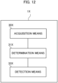
- FIG. 12 is a block diagram of an image processing device 1X according to the second example embodiment.
- the image processing device 1X includes an acquisition means 30X, a determination means 31X, and a detection means 33X.
- the image processing device 1X may be configured by a plurality of devices.
- the acquisition means 30X is configured to acquire an endoscopic image obtained by photographing an examination target by a photographing unit provided in an endoscope.
- Examples of the acquisition means 30X include the endoscopic image acquisition unit 30 in the first example embodiment (including modifications, hereinafter the same).
- the acquisition means 30X may be configured to immediately acquire the endoscopic image generated by the photographing unit, or may be configured to acquire, at a predetermined timing, the endoscopic image stored in the storage device generated by the photographing unit in advance.
- the determination means 31X is configured to determine, based on the endoscopic image, a state of the examination target or a photographed part of the examination target.
- Examples of the determination means 31X include the background condition determination unit 31, the part determination unit 31A, and the determination units 31B and 31C in the first example embodiment.
- the detection means 33X is configured to detect, based on the state or the photographed part and the endoscopic image, a part of interest to be noticed in the examination target.
- detection herein only requires to make a determination as to presence or absence of a part of interest in the examination target and does not necessarily require to identify the region of the part of interest.
- Examples of the detection means 33X include the lesion detection unit 33, the lesion detection unit 33B, and the lesion detection unit 33Cb in the first example embodiment.
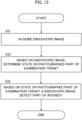
- FIG. 13 is an example of a flowchart showing a processing procedure in the second example embodiment.
- the acquisition means 30X acquires an endoscopic image obtained by photographing an examination target by a photographing unit provided in an endoscope (step S21).
- the determination means 31X determines, based on the endoscopic image, a state of the examination target or a photographed part of the examination target (step S22).
- the detection means 33X detects, based on the state or the photographed part and the endoscopic image, a part of interest to be noticed in the examination target (step S23).
- the image processing device 1X can accurately detect a region of interest from an endoscopic image obtained by photographing the examination target.
- the program is stored by any type of a non-transitory computer-readable medium (non-transitory computer readable medium) and can be supplied to a control unit or the like that is a computer.
- the non-transitory computer-readable medium include any type of a tangible storage medium.
- non-transitory computer readable medium examples include a magnetic storage medium (e.g., a flexible disk, a magnetic tape, a hard disk drive), a magnetic-optical storage medium (e.g., a magnetic optical disk), CD-ROM (Read Only Memory), CD-R, CD-R/W, a solid-state memory (e.g., a mask ROM, a PROM (Programmable ROM), an EPROM (Erasable PROM), a flash ROM, a RAM (Random Access Memory)).
- the program may also be provided to the computer by any type of a transitory computer readable medium. Examples of the transitory computer readable medium include an electrical signal, an optical signal, and an electromagnetic wave.
- the transitory computer readable medium can provide the program to the computer through a wired channel such as wires and optical fibers or a wireless channel.
- An image processing device comprising:
- the image processing device according to Supplementary Note 6, wherein the display control means is configured to cause the display device to display, as the information regarding the photographed part, a model representing a whole of the examination target with a clear indication of the photographed part.
- An image processing method executed by a computer comprising:
- a storage medium storing a program executed by a computer, the program causing the computer to:
Landscapes
- Engineering & Computer Science (AREA)
- Health & Medical Sciences (AREA)
- Life Sciences & Earth Sciences (AREA)
- Physics & Mathematics (AREA)
- Surgery (AREA)
- Medical Informatics (AREA)
- Theoretical Computer Science (AREA)
- General Health & Medical Sciences (AREA)
- General Physics & Mathematics (AREA)
- Nuclear Medicine, Radiotherapy & Molecular Imaging (AREA)
- Radiology & Medical Imaging (AREA)
- Computer Vision & Pattern Recognition (AREA)
- Pathology (AREA)
- Animal Behavior & Ethology (AREA)
- Veterinary Medicine (AREA)
- Public Health (AREA)
- Molecular Biology (AREA)
- Evolutionary Computation (AREA)
- Biophysics (AREA)
- Optics & Photonics (AREA)
- Heart & Thoracic Surgery (AREA)
- Biomedical Technology (AREA)
- Multimedia (AREA)
- Artificial Intelligence (AREA)
- Computing Systems (AREA)
- Databases & Information Systems (AREA)
- Software Systems (AREA)
- Signal Processing (AREA)
- Quality & Reliability (AREA)
- Endoscopes (AREA)
Applications Claiming Priority (1)
| Application Number | Priority Date | Filing Date | Title |
|---|---|---|---|
| PCT/JP2022/014404 WO2023181353A1 (ja) | 2022-03-25 | 2022-03-25 | 画像処理装置、画像処理方法及び記憶媒体 |
Publications (2)
| Publication Number | Publication Date |
|---|---|
| EP4501206A1 true EP4501206A1 (de) | 2025-02-05 |
| EP4501206A4 EP4501206A4 (de) | 2025-05-07 |
Family
ID=88100799
Family Applications (1)
| Application Number | Title | Priority Date | Filing Date |
|---|---|---|---|
| EP22933492.5A Pending EP4501206A4 (de) | 2022-03-25 | 2022-03-25 | Bildverarbeitungsvorrichtung, bildverarbeitungsverfahren und speichermedium |
Country Status (3)
| Country | Link |
|---|---|
| US (1) | US20250209776A1 (de) |
| EP (1) | EP4501206A4 (de) |
| WO (1) | WO2023181353A1 (de) |
Families Citing this family (1)
| Publication number | Priority date | Publication date | Assignee | Title |
|---|---|---|---|---|
| JP2024150245A (ja) * | 2023-04-10 | 2024-10-23 | 富士フイルム株式会社 | 医療支援装置、内視鏡システム、医療支援方法、及びプログラム |
Family Cites Families (5)
| Publication number | Priority date | Publication date | Assignee | Title |
|---|---|---|---|---|
| JP2010187756A (ja) * | 2009-02-16 | 2010-09-02 | Olympus Corp | 画像処理装置、画像処理方法および画像処理プログラム |
| WO2019138773A1 (ja) * | 2018-01-10 | 2019-07-18 | 富士フイルム株式会社 | 医療画像処理装置、内視鏡システム、医療画像処理方法及びプログラム |
| WO2019146077A1 (ja) | 2018-01-26 | 2019-08-01 | オリンパス株式会社 | 内視鏡画像処理装置、内視鏡画像処理方法及び内視鏡画像処理プログラム |
| WO2019155628A1 (ja) * | 2018-02-09 | 2019-08-15 | 日本電気株式会社 | 画像処理装置、画像処理方法および記録媒体 |
| WO2021234907A1 (ja) | 2020-05-21 | 2021-11-25 | 日本電気株式会社 | 画像処理装置、制御方法及び記憶媒体 |
-
2022
- 2022-03-25 EP EP22933492.5A patent/EP4501206A4/de active Pending
- 2022-03-25 WO PCT/JP2022/014404 patent/WO2023181353A1/ja not_active Ceased
- 2022-03-25 US US18/846,777 patent/US20250209776A1/en active Pending
Also Published As
| Publication number | Publication date |
|---|---|
| EP4501206A4 (de) | 2025-05-07 |
| WO2023181353A1 (ja) | 2023-09-28 |
| JPWO2023181353A1 (de) | 2023-09-28 |
| US20250209776A1 (en) | 2025-06-26 |
Similar Documents
| Publication | Publication Date | Title |
|---|---|---|
| US12591975B2 (en) | Image processing device, image processing method, and storage medium | |
| EP4434435A1 (de) | Informationsverarbeitungsvorrichtung, informationsverarbeitungsverfahren und aufzeichnungsmedium | |
| EP4501206A1 (de) | Bildverarbeitungsvorrichtung, bildverarbeitungsverfahren und speichermedium | |
| US12602779B2 (en) | Image processing device, image processing method and storage medium | |
| US20240378840A1 (en) | Image processing device, image processing method, and storage medium | |
| EP4501207A1 (de) | Bildverarbeitungsvorrichtung, bildverarbeitungsverfahren und speichermedium | |
| EP4533998A1 (de) | Bildverarbeitungsvorrichtung, bildverarbeitungsverfahren und speichermedium | |
| EP4327721A1 (de) | Bildverarbeitungsvorrichtung, bildverarbeitungsverfahren und speichermedium | |
| US20250232434A1 (en) | Image processing device, image processing method, and storage medium | |
| US20240161283A1 (en) | Image processing device, image processing method, and storage medium | |
| US12602782B2 (en) | Image processing device, image processing method, and storage medium | |
| US20250228427A1 (en) | Image processing device, image processing method, and storage medium | |
| US20250372264A1 (en) | Image processing device, image processing method, and storage medium | |
| US12599284B2 (en) | Endoscopic examination support apparatus, endoscopic examination support method, and recording medium | |
| US20260120286A1 (en) | Image processing device, image processing method, and storage medium | |
| US20240233121A1 (en) | Endoscopic examination support apparatus, endoscopic examination support method, and recording medium | |
| EP4487754A1 (de) | Bildverarbeitungsvorrichtung, bildverarbeitungsverfahren und speichermedium | |
| WO2026078862A1 (ja) | 学習装置、画像処理装置、学習方法、画像処理方法及び記憶媒体 | |
| WO2025083856A1 (ja) | 画像処理装置、画像処理方法及び記憶媒体 | |
| WO2024180593A1 (ja) | 画像処理装置、画像処理方法及び記憶媒体 |
Legal Events
| Date | Code | Title | Description |
|---|---|---|---|
| STAA | Information on the status of an ep patent application or granted ep patent |
Free format text: STATUS: THE INTERNATIONAL PUBLICATION HAS BEEN MADE |
|
| PUAI | Public reference made under article 153(3) epc to a published international application that has entered the european phase |
Free format text: ORIGINAL CODE: 0009012 |
|
| STAA | Information on the status of an ep patent application or granted ep patent |
Free format text: STATUS: REQUEST FOR EXAMINATION WAS MADE |
|
| 17P | Request for examination filed |
Effective date: 20240923 |
|
| AK | Designated contracting states |
Kind code of ref document: A1 Designated state(s): AL AT BE BG CH CY CZ DE DK EE ES FI FR GB GR HR HU IE IS IT LI LT LU LV MC MK MT NL NO PL PT RO RS SE SI SK SM TR |
|
| REG | Reference to a national code |
Ref country code: DE Ref legal event code: R079 Free format text: PREVIOUS MAIN CLASS: A61B0001045000 Ipc: G06V0010700000 |
|
| A4 | Supplementary search report drawn up and despatched |
Effective date: 20250404 |
|
| RIC1 | Information provided on ipc code assigned before grant |
Ipc: G06V 10/82 20220101ALI20250331BHEP Ipc: G06T 7/00 20170101ALI20250331BHEP Ipc: A61B 1/00 20060101ALI20250331BHEP Ipc: A61B 1/045 20060101ALI20250331BHEP Ipc: G06V 10/70 20220101AFI20250331BHEP |
|
| DAV | Request for validation of the european patent (deleted) | ||
| DAX | Request for extension of the european patent (deleted) |