EP4501948A1 - Ensemble d'afficorps et de protéine photoréceptrice - Google Patents
Ensemble d'afficorps et de protéine photoréceptrice Download PDFInfo
- Publication number
- EP4501948A1 EP4501948A1 EP23775097.1A EP23775097A EP4501948A1 EP 4501948 A1 EP4501948 A1 EP 4501948A1 EP 23775097 A EP23775097 A EP 23775097A EP 4501948 A1 EP4501948 A1 EP 4501948A1
- Authority
- EP
- European Patent Office
- Prior art keywords
- amino acid
- acid sequence
- seq
- affibody
- protein
- Prior art date
- Legal status (The legal status is an assumption and is not a legal conclusion. Google has not performed a legal analysis and makes no representation as to the accuracy of the status listed.)
- Pending
Links
Images

















Classifications
-
- C—CHEMISTRY; METALLURGY
- C07—ORGANIC CHEMISTRY
- C07K—PEPTIDES
- C07K19/00—Hybrid peptides, i.e. peptides covalently bound to nucleic acids, or non-covalently bound protein-protein complexes
-
- C—CHEMISTRY; METALLURGY
- C07—ORGANIC CHEMISTRY
- C07K—PEPTIDES
- C07K14/00—Peptides having more than 20 amino acids; Gastrins; Somatostatins; Melanotropins; Derivatives thereof
- C07K14/195—Peptides having more than 20 amino acids; Gastrins; Somatostatins; Melanotropins; Derivatives thereof from bacteria
-
- C—CHEMISTRY; METALLURGY
- C07—ORGANIC CHEMISTRY
- C07K—PEPTIDES
- C07K14/00—Peptides having more than 20 amino acids; Gastrins; Somatostatins; Melanotropins; Derivatives thereof
- C07K14/195—Peptides having more than 20 amino acids; Gastrins; Somatostatins; Melanotropins; Derivatives thereof from bacteria
- C07K14/305—Peptides having more than 20 amino acids; Gastrins; Somatostatins; Melanotropins; Derivatives thereof from bacteria from Micrococcaceae (F)
- C07K14/31—Peptides having more than 20 amino acids; Gastrins; Somatostatins; Melanotropins; Derivatives thereof from bacteria from Micrococcaceae (F) from Staphylococcus (G)
-
- C—CHEMISTRY; METALLURGY
- C07—ORGANIC CHEMISTRY
- C07K—PEPTIDES
- C07K16/00—Immunoglobulins [IG], e.g. monoclonal or polyclonal antibodies
- C07K16/12—Immunoglobulins [IG], e.g. monoclonal or polyclonal antibodies against material from bacteria
-
- C—CHEMISTRY; METALLURGY
- C07—ORGANIC CHEMISTRY
- C07K—PEPTIDES
- C07K16/00—Immunoglobulins [IG], e.g. monoclonal or polyclonal antibodies
- C07K16/12—Immunoglobulins [IG], e.g. monoclonal or polyclonal antibodies against material from bacteria
- C07K16/1203—Gram-negative bacteria
-
- C—CHEMISTRY; METALLURGY
- C07—ORGANIC CHEMISTRY
- C07K—PEPTIDES
- C07K16/00—Immunoglobulins [IG], e.g. monoclonal or polyclonal antibodies
- C07K16/12—Immunoglobulins [IG], e.g. monoclonal or polyclonal antibodies against material from bacteria
- C07K16/1267—Gram-positive bacteria
-
- C—CHEMISTRY; METALLURGY
- C07—ORGANIC CHEMISTRY
- C07K—PEPTIDES
- C07K2318/00—Antibody mimetics or scaffolds
- C07K2318/20—Antigen-binding scaffold molecules wherein the scaffold is not an immunoglobulin variable region or antibody mimetics
-
- G—PHYSICS
- G01—MEASURING; TESTING
- G01N—INVESTIGATING OR ANALYSING MATERIALS BY DETERMINING THEIR CHEMICAL OR PHYSICAL PROPERTIES
- G01N33/00—Investigating or analysing materials by specific methods not covered by groups G01N1/00 - G01N31/00
- G01N33/48—Biological material, e.g. blood, urine; Haemocytometers
- G01N33/50—Chemical analysis of biological material, e.g. blood, urine; Testing involving biospecific ligand binding methods; Immunological testing
- G01N33/53—Immunoassay; Biospecific binding assay; Materials therefor
Definitions
- the present invention relates to a set of Affibody and photoreceptor protein.
- the present invention also relates to a genetic modification technique using the set of Affibody and photoreceptor protein.
- a molecular control approach that exploits protein photoactivation has emerged and is called optogenetics.
- Light is easily controlled in terms of space or time as compared with chemicals.
- a light-dependent protein-protein interaction system is known which exploits photosensitive protein that forms a dimer in a light-dependent manner or a photoreceptor and its binding partner.
- Patent Literature 1 discloses a set of proteins useful as a tool of optogenetics, the set being capable of controlling dimer formation and dissociation by light irradiation, as an optical manipulation technique using blue light.
- Non Patent Literatures 1 to 3 have also been reported as manipulation techniques using red light.
- Patent Literature 1 Japanese Patent Laid-Open No. 2015-165776
- An object of the present invention is to develop a photoswitching protein that can optically manipulate the functions of various proteins with precision and high efficiency by light at two different long wavelengths without the use of blue light.
- the present inventors have conducted diligent studies to attain the object and consequently completed the present invention by finding that a set of Affibody and photoreceptor protein is optically controllable by red light.
- the present invention is as follows:
- the present invention can provide a photoswitching protein that can optically manipulate the functions of various proteins with precision and high efficiency by light at two different long wavelengths without the use of blue light.
- the set of Affibody and photoreceptor protein of the present invention is a set wherein the Affibody and the photoreceptor protein form a complex in a manner dependent on light at a wavelength of 600 to 750 nm.
- the photoreceptor protein used in the present invention is a prokaryotic photoreceptor protein and is known as bacteriophytochrome.
- the photoreceptor protein is a protein also abbreviated to BphP.
- the photoreceptor protein is known to vary in structure depending on light in different long wavelength regions.
- the photoreceptor protein is not particularly limited as long as the Affibody binds thereto by red light and dissociates therefrom by near-infrared light.
- photoreceptor protein examples include, but are not particularly limited to, DrBphP known as bacterium ( Deinococcus radiodurans ) -derived red sensor protein.
- DrBphP has an N-terminal photosensory module (PSM). DrBphP shifts its state from Pr (dark state) to Pfr (photo state) upon irradiation with red light. DrBphP in the photo state binds to the Affibody.
- PSM photosensory module
- the photoreceptor protein will be described by taking DrBphP as an example.
- DrBphP binds with an endogenous dye as a cofactor and DrBphP does not require further addition of the cofactor for forming a complex with the Affibody because its intracellular concentration satisfies a requirement for the concentration of the cofactor.
- the complex formation using the set of Affibody and photoreceptor protein of the present invention has the advantage that specific binding occurs in the presence of red light while the complex is not formed in the dark.
- the set of Affibody and photoreceptor protein of the present invention is capable of controlling the association of a split protein by ON/OFF of red light.
- the photoreceptor protein as DrBphP preferably
- the phrase "having 80% or higher homology to the amino acid sequence represented by SEQ ID NO” means that a mutation is acceptable for the particular SEQ ID NO.
- the "80% or higher homology” means that amino acids overlapping with the amino acid sequence represented by the particular SEQ ID NO account for at least 80% of the total number of amino acids in the amino acid sequence represented by this SEQ ID NO.
- the homology is preferably 85% or higher, 90% or higher, 95% or higher, 98% or higher, or 100%.
- sequence having this homology may be the "amino acid sequence represented by SEQ ID NO: 2" unless otherwise specified.
- the term “several” can be 10 or less, 9 or less, 8 or less, 7 or less, 6 or less, 5 or less, 4 or less, 3 or less, or 2 or less.
- 1, 1 to 2, 1 to 3, 1 to 4, 1 to 5, 1 to 6, 1 to 7, 1 to 8, 1 to 9, or 1 to 10 amino acids may be added, substituted, or deleted in the amino acid sequence represented by SEQ ID NO.
- the upper limit of the number of amino acids that may be added, substituted, or deleted in the amino acid sequence represented by SEQ ID NO can be determined within the range of the homology as long as the homology is 80% or higher, 85% or higher, 90% or higher, 95% or higher, or 98% or higher.
- addition, substitution, or deletion in a certain amino acid sequence means that the addition (insertion), the substitution, or the deletion occurs at an end or a site other than the end in the sequence.
- the number of amino acids to be added, substituted, or deleted is not particularly limited as long as the resulting polypeptide exerts the effects according to the present invention.
- the number of sites at which addition, substitution, or deletion occurs may be one or may be two or more.
- the homology to the amino acid sequence represented by SEQ ID NO can be calculated by aligning the sequences by a method known to those skilled in the art so as to attain the highest match of amino acids therebetween, and determining the ratio of the amino acids overlapping with the amino acid sequence represented by SEQ ID NO.
- homology may be interchanged with identity.
- cysteine at position 24 counted from the N terminus in SEQ ID NO: 1 not be mutated because this amino acid serves as a covalent binding site for a cofactor biliverdin and therefore binds to biliverdin.
- DrBphP is known to be composed mainly of four domains (PAS domain, GAF domain, PHY domain, and HK domain).
- the PAS domain, the GAF domain, and the PHY domain have a high commonality in proteins generically named as "bacteriophytochrome".
- the HK domain also called effector domain, is known to often differ depending on the type of the bacteriophytochrome. A mutation may be provided in each domain on the basis of such information.
- the Affibody used in the present invention means a category of proteins derived from a protein consisting of approximately 58 amino acids designed on the basis of the Z domain of Staphylococcus aureus protein A.
- the Affibody has a variable region regarded as a binding site for the photoreceptor protein, and a constant region used for maintaining the structure of the Affibody.
- the Affibody used in the present invention typically, can be any material.
- amino acids at positions 9 to 11, 13, 14, 17, 18, 24, 25, 27, 28, 32, and 35 reside on the Affibody surface in the helix bundle of the Affibody and are involved in binding to the photoreceptor protein. Specifically, in SEQ ID NO: 1, the amino acids at positions 9 to 11, 13, 14, 17, 18, 24, 25, 27, 28, 32, and 35 constitute the variable region of the Affibody.
- amino acids of the Affibody involved in binding to the photoreceptor protein are amino acids considered to be involved in binding to the photoreceptor protein in a state capable of binding to the Affibody by light excitation, and are amino acids at positions 9 to 11, 13, 14, 17, 18, 24, 25, 27, 28, 32, and 35 in the amino acid sequence represented by SEQ ID NO: 1.
- amino acids are preferably conserved in SEQ ID NO: 1. However, even Affibody in which one or some of the amino acids are substituted by other amino acids has the ability to form a complex with the photoreceptor protein.
- amino acids at positions 9 to 11, 13, 14, 17, 18, 24, 25, 27, 28, 32, and 35 in the amino acid sequence represented by SEQ ID NO: 1 may be mutated to other amino acids as long as the resulting protein has the ability to form a complex with the photoreceptor protein.
- Amino acids other than the amino acids at positions 9 to 11, 13, 14, 17, 18, 24, 25, 27, 28, 32, and 35 in the amino acid sequence represented by SEQ ID NO: 1, i.e., amino acids at positions 1 to 8, 12, 15, 16, 19 to 23, 26, 29 to 31, 33, 34, and 36 to 58, are regarded as amino acids of the constant region in the Affibody.
- amino acids other than the amino acids at positions 9 to 11, 13, 14, 17, 18, 24, 25, 27, 28, 32, and 35 are amino acids that may be considered amino acids of the constant region, and are amino acids used for maintaining the structure of the Affibody. Accordingly, the amino acids at positions other than the positions 9 to 11, 13, 14, 17, 18, 24, 25, 27, 28, 32, and 35 in SEQ ID NO: 1 may be substituted or deleted, or other amino acids may be added thereto, as long as the amino acids at positions 9 to 11, 13, 14, 17, 18, 24, 25, 27, 28, 32, and 35 are a combination of amino acids having the ability to bind to the photoreceptor protein and as long as the structure of the Affibody is maintained.
- Examples of the deletion in the Affibody include deletion of an N-terminal or a C-terminal amino acid of the Affibody.
- the deletion of an N-terminal or a C-terminal amino acid may be deletion of 1, 2, 3, 4, 5, 6, 7, 8, 9, or 10 consecutive or nonconsecutive amino acids.
- N-terminal or C-terminal amino acids may be deleted, 1 to 4 N-terminal or C-terminal amino acids may be deleted, 1 to 3 N-terminal or C-terminal amino acids may be deleted, 1 to 2 N-terminal or C-terminal amino acids may be deleted, or 1 N-terminal or C-terminal amino acid may be deleted.
- the deletion of these amino acids can be deletion of consecutive amino acids from each terminal amino acid.
- amino acids at positions other than the positions 9 to 11, 13, 14, 17, 18, 24, 25, 27, 28, 32, and 35 in SEQ ID NO: 1, 1, 2, 3, 4, 5, 6, 7, 8, 9, or 10 amino acids may be substituted.
- 1, 2, 3, 4, 5, 6, 7, 8, 9, or 10 amino acids may be added thereto as long as the amino acids at positions 9 to 11, 13, 14, 17, 18, 24, 25, 27, 28, 32, and 35 in SEQ ID NO: 1 have the ability to bind to the photoreceptor protein.
- the number of amino acids to be substituted, added, or deleted in the amino acid sequence represented by SEQ ID NO: 1 in the Affibody can be 1 to 10, 1 to 9, 1 to 8, 1 to 7, or 1 to 6, and the upper limit may be 5 or less, 4 or less, 3 or less, or 2 or less or may be 1.
- any amino acid at positions 12, 15, 16, 19 to 23, 26, 29 to 31, 33, and 34 among the amino acids located at positions 9 to 35 may be deleted as long as the amino acids at positions 9 to 11, 13, 14, 17, 18, 24, 25, 27, 28, 32, and 35 are capable of assuming a structure having the ability to bind to the photoreceptor protein.
- the Affibody used in the present invention may have
- the Affibody used in the present invention may have (9) an amino acid sequence differing from the amino acid sequence represented by SEQ ID NO: 1 by having any amino acid selected from phenylalanine (F), tryptophan (W), histidine (H), and methionine (M) at position 18 and lacking one to five N-terminal or C-terminal amino acids of SEQ ID NO: 1.
- amino acid sequence may lack one to five N-terminal and C-terminal amino acids of SEQ ID NO: 1.
- the set of photoreceptor protein and Affibody according to the present invention can accurately control complex formation and dissociation thereof through a simple process of light irradiation and shielding.
- the wavelength of the light for use when the Affibody and the photoreceptor protein form a complex in a light-dependent manner is in the range of 600 to 750 nm.
- a suitable wavelength is set from the states of existence, i.e., the state in which the photoreceptor protein binds to the Affibody in response to light and the state in which the photoreceptor protein does not bind to the Affibody.
- the lower limit value in the range of the wavelength can be 620 nm or more, 630 nm or more, 640 nm or more, 650 nm or more, 660 nm or more, 670 nm or more, 680 nm or more, 690 nm or more, or 695 nm or more
- the upper limit value can be 740 nm or less, 730 nm or less, 720 nm or less, 710 nm or less, or 705 nm or less.
- the wavelength is in the range of 640 to 720 nm.
- the Affibody and the photoreceptor protein form a complex in a light-dependent manner, while the complex formation may be reversibly canceled in a light-dependent manner.
- a suitable wavelength is set from the states of existence, i.e., the state in which the photoreceptor protein binds to the Affibody in response to light and the state in which the photoreceptor protein does not bind to the Affibody.
- states of existence i.e., the state in which the photoreceptor protein binds to the Affibody in response to light and the state in which the photoreceptor protein does not bind to the Affibody.
- light irradiation may be performed at a near-infrared light wavelength of 750 nm or more, or the set of photoreceptor protein and Affibody may be placed in the dark.
- the lower limit value in the range of the wavelength can be 760 nm or more, 770 nm or more, 780 nm or more, or 790 nm or more, and the upper limit value can be 820 nm or less or 810 nm or less.
- the set of photoreceptor protein and Affibody according to the present invention forms a complex or dissociates depending on a particular wavelength.
- the set of photoreceptor protein and Affibody according to the present invention provides a technique that permits precise control and manipulation by red light because the Affibody and the photoreceptor protein form a complex in a red light-dependent manner.
- the set of photoreceptor protein and Affibody of the present invention which can be applied to optical manipulation of various proteins is referred to as "MagRed".
- MagRed is a technique of freely controlling proteins related to vital phenomena or the treatment of diseases by light.
- MagRed is capable of providing a technique that permits optical control without adding a cofactor in mammalian cells by using red light in the optical control.
- MagneticRed as the set of Affibody and photoreceptor protein.
- MagRed has photoreceptor protein and Affibody. A domain is bound to each of them so that ON/OFF of an effect based on the domain can be accurately controlled by light irradiation and shielding.
- the domain to be bound to each of the photoreceptor protein and the Affibody is not particularly limited and can be a domain that exerts its effect by optical control.
- the domain that exerts its effect by optical control may be bound to both or one of the photoreceptor protein and the Affibody.
- the photoreceptor protein and the Affibody form a complex by optical control so that the domain bound to the photoreceptor protein and the domain bound to the Affibody can exert their effects in proximity.
- the domain bound to the photoreceptor protein and the domain bound to the Affibody may be separate domains having different effects, or the domain bound to the photoreceptor protein and the domain bound to the Affibody may exert one effect when located adjacently.
- domain bound to the photoreceptor protein and the domain bound to the Affibody are separate domains having different effects, these two domains may exert the effects when located adjacently. Examples thereof include heterodimers.
- the domain bound to the photoreceptor protein and the domain bound to the Affibody may form a homodimer.
- a portion of domains that exert an effect by forming a multimer may be used as the domain bound to the photoreceptor protein, and the remaining portion of the domains may be used as the domain bound to the Affibody.
- Examples of the case where the domain bound to the photoreceptor protein and the domain bound to the Affibody exert one effect when located adjacently include one domain split into two.
- a protein having a certain effect is split into two.
- the respective domains bound to the photoreceptor protein and the Affibody form a conjugate by optical control upon which the domain split into two is capable of exerting a function similar to that of the original domain before split.
- a recognition domain that orients a conjugate to the desired location may be bound to the other (photoreceptor protein or Affibody) to which the domain that exerts its effect by optical control is not bound.
- the recognition domain bound to one of the photoreceptor protein and the Affibody orients the photoreceptor protein or the Affibody bound by the recognition domain to the desired location.
- the photoreceptor protein or the Affibody oriented to the desired location binds to the other (photoreceptor protein or Affibody) by optical control so that the domain bound to the other may exert its effect.
- the recognition domain that orients a conjugate to the desired location is not particularly limited, and a domain that recognizes a nucleic acid is suitably used.
- MagRed is oriented to a predetermined position through the domain that recognizes a nucleic acid.
- the domain that exerts its effect by optical control is also oriented to the predetermined location and exerts its effect.
- a domain other than the domain to be bound to each of the photoreceptor protein and the Affibody may be used for allowing the domain bound to each of the photoreceptor protein and the Affibody to exert its effect.
- the domains bound to both of the photoreceptor protein and the Affibody may reside in proximity by optical control and exert their effects through an additional domain, or the domain bound to one of the photoreceptor protein and the Affibody may exert its effect through an additional domain bound to neither the photoreceptor protein nor the Affibody.
- the binding of the photoreceptor protein and the Affibody to the additional domain is not particularly limited and can be covalent binding.
- the binding of the photoreceptor protein and the Affibody to the additional domain may be binding via a linker.
- a plurality of domains may be bound to the photoreceptor protein or the Affibody, and a plurality of domains may be linked to each other and bound to the photoreceptor protein or the Affibody.
- the binding between the domains may also be binding via a linker.
- a linker known in the art may be used as the linker.
- a linker having an amino acid sequence rich in glycine and serine is suitably used as the linker.
- a linker composed mainly of glycine can be used, and a linker containing a hydrophilic amino acid serine or threonine is suitable.
- the linker is not particularly limited and may be constituted by 2 to 16 amino acids.
- a nuclear localization signal sequence and a 2A peptide sequence or an IRES sequence may be bound as other domains.
- Examples of the domain that exerts its function by optical control include, but are not particularly limited to, nuclease, recombinase, polymerase, deaminase, protease, kinase, phosphatase, antibodies and antibody-like molecules, and neurotoxin.
- the set of the present invention may be a set of photoreceptor protein and Affibody, wherein
- the present invention can provide a method for suppressing or activating the expression of a target gene, the method comprising the step of incubating the target gene and the set of photoreceptor protein and Affibody according to the present invention by light irradiation.
- the present invention can also provide a method for suppressing or activating the expression of a target gene, the method comprising the step of incubating the target gene, the set of photoreceptor protein and Affibody according to the present invention, and CRISPR-related protein by light irradiation.
- a domain that recognizes a nucleic acid near the target gene is bound to one member of the set of photoreceptor protein and Affibody according to the present invention so that the set of photoreceptor protein and Affibody according to the present invention forms a conjugate by light irradiation, through which the expression of the target gene can be suppressed or activated by the action of a domain related to the transcriptional activity of a gene, which is bound to the other member of the set of photoreceptor protein and Affibody according to the present invention.
- a domain that recognizes CRISPR-related protein preferably guide RNA containing MS2 aptamer in stem loop 2 of tracrRNA, is used in one member of the set of photoreceptor protein and Affibody according to the present invention.
- the MS2 aptamer-bound member of the set of photoreceptor protein and Affibody according to the present invention is located near the target gene recognized by the CRISPR-related protein.
- the set of photoreceptor protein and Affibody according to the present invention forms a conjugate by light irradiation, through which the expression of the target gene can be suppressed or activated by the action of a domain related to the transcriptional activity of a gene, which is bound to the other member of the set of photoreceptor protein and Affibody according to the present invention.
- a conventional protein known in the art for use in gene editing is used as the CRISPR-related protein, and other components such as guide RNA may be included together with the CRISPR-related protein.
- other components such as guide RNA may be included together with the CRISPR-related protein.
- a protein lacking nuclease activity and nickase activity is preferably used as the CRISPR-related protein.
- the guide RNA is allowed to have a sequence complementary to a gene near the target gene.
- the set of photoreceptor protein and Affibody according to the present invention is thereby located near the target gene so that the expression of the target gene can be suppressed or activated.
- a functional domain as the domain related to the transcriptional activity of a gene is a transcriptional activation domain or a transcriptional suppression domain, the expression of the target gene is activated or suppressed.
- the term "expression of a gene” is used to conceptually include both of transcription to synthesize RNA with DNA as a template and translation to synthesize a polypeptide based on the RNA sequence.
- VP64, MS2, p65, and HSF1 may be used as functional domains, and a transcriptional activation domain and aptamer binding protein known in the art may be used as functional domains equivalent thereto.
- a transcriptional activation domain and aptamer binding protein as disclosed in Nature (2015) 517, 583-588 and Nature protocols (2012) 7 (10), 1797-1807 can be used.
- the set of the present invention may be a set wherein
- the protein split into two is not particularly limited as long as the protein exerts a function by the binding of the photoreceptor protein and the Affibody.
- Nonlimiting examples of the protein to be split into two include Cre protein.
- a conventional protein known in the art may be used as the Cre protein split into two. Examples thereof include proteins as described in International Publication No. WO 2016/167300 .
- the Cre protein split into two is not particularly limited as long as two fragments thereof are capable of exerting a function by the binding of the photoreceptor protein and the Affibody.
- the fragments are constituted by an N-terminal fragment and a C-terminal fragment of the Cre protein.
- the split position of the Cre protein is capable of appropriately adopting a conventional split position known in the art.
- the N-terminal fragment and the C-terminal fragment of the Cre protein bind to the photoreceptor protein and the Affibody, respectively.
- the photoreceptor protein and the Affibody form a conjugate by light irradiation so that the N-terminal fragment of the Cre protein and the C-terminal fragment of the Cre protein bound thereto are reconstituted as the Cre protein to restore the recombinase activity of the Cre protein. Accordingly, the N-terminal fragment of the Cre protein and the C-terminal fragment of the Cre protein can be used as the set of MagRed bound to the Cre protein in light-dependent gene recombination.
- gene recombination can be performed in a light-dependent manner by using MagRed bound to the N-terminal fragment of the Cre protein and the C-terminal fragment of the Cre protein in combination with a loxP sequence or a loxP mutant sequence.
- a nucleic acid having the sequence of any member selected from lox2722, lox66, lox71, JT15, and JTZ17 can be used as the loxP mutant.
- the present invention also provides an animal having MagRed.
- MagRed is controllable by red light (up to 660 nm) and near-infrared light (up to 800 nm) having high tissue penetration and is therefore highly advantageous.
- the animal having MagRed may have a domain bound to the MagRed and may have a domain bound to the MagRed as well as an additional domain that functions together with the domain.
- the animal may have MagRed as a combination of the MagRed, as a nucleic acid encoding at least the MagRed, or as at least one expression vector containing the MagRed.
- the animal having MagRed is optically controllable by red light and therefore has gene recombination activity even in vivo. Gene recombination can be efficiently controlled by the ex vivo light irradiation of the animal in which MagRed optically controllable by red light is incorporated in the body.
- MagRed can accurately control gene recombination not only in vitro but in vivo by ON/OFF of light irradiation.
- the animal in which MagRed is incorporated is not particularly limited as long as the animal is a mammal.
- a rodent as a laboratory animal is preferred.
- the function of a gene can be knocked out by light stimulation; a mutated gene can be rather restored to normal by light stimulation and evaluated for its influence; or a reporter gene such as a fluorescent protein can be allowed to function by light stimulation so as to label tissues or cells constituting the tissues, though the approach is not particularly limited thereto.
- MagRed can be expressed in an animal or cells by selecting respective nucleic acids corresponding to the photoreceptor protein and the Affibody, selecting nucleic acids corresponding to domains, etc. to be bound to the photoreceptor protein and the Affibody, linking the respective nucleic acids such that the photoreceptor protein with the desired domain and the Affibody with the desired domain are expressible, and introducing the respective nucleic acids to an expression vector such as a plasmid, for example.
- the present invention also provides a set of nucleic acids designed such that the photoreceptor protein with the desired domain and the Affibody with the desired domain are expressible.
- nucleic acid designed such that the photoreceptor protein with the desired domain is expressible and the nucleic acid designed such that the Affibody with the desired domain is expressible may reside on one expression vector.
- the respective nucleic acids may reside on the expression vector such that the nucleic acids are linked, or may reside on the expression vector such that their expressions are separately controlled.
- nucleic acid designed such that the photoreceptor protein with the desired domain is expressible and the nucleic acid designed such that the Affibody with the desired domain is expressible may reside on separate expression vectors.
- Such a nucleic acid may be designed such that the photoreceptor protein or the Affibody and the desired domain are connected via a linker.
- each nucleic acid can be inserted directly, after digestion with restriction enzymes, or after addition of a linker to downstream of a promoter of an expression vector.
- Examples of the expression vector include, but are not particularly limited to, E. coli-derived plasmids (pBR322, pBR325, pUC12, pUC13, pUC18, pUC19, pUC118, pBluescript II, etc.), Bacillus subtilis-derived plasmids (pUB110, pTP5, pC1912, pTP4, pE194, pC194, etc.), yeast-derived plasmids (pSH19, pSH15, YEp, YRp, YIp, YAC, etc.), bacteriophages ( ⁇ phage, M13 phage, etc.), viruses (retrovirus, vaccinia virus, adenovirus, adeno-associated virus (AAV), cauliflower mosaic virus, tobacco mosaic virus, baculovirus, etc.), and cosmids.
- E. coli-derived plasmids pBR322, pBR325, pUC12,
- the promoter can be appropriately selected depending on the type of a host.
- SV40 simian virus 40
- CMV cytomegalovirus
- E. coli for example, trp promoter, T7 promoter, or lac promoter can be used.
- a DNA replication origin (ori), a selective marker (antibiotic resistance, auxotrophy, etc.), an enhancer, a splicing signal, a poly-A addition signal, a nucleic acid encoding a tag (FLAG, HA, GST, GFP, etc.), or the like may be incorporated in the expression vector.
- a transformant can be obtained by transforming appropriate host cells with the expression vector.
- the host can be appropriately selected in relation to the vector.
- E. coli, Bacillus subtilis, a bacterium of the genus Bacillus, a yeast, an insect or insect cells, or animal cells can be used.
- HEK293T cells, CHO cells, COS cells, myeloma cells, HeLa cells, or Vero cells may be used as the animal cells.
- the transformation can be performed in accordance with a method known in the art such as lipofection, calcium phosphate method, electroporation, microinjection, or particle gun method depending on the type of the host.
- the transformant is cultured in accordance with a routine method and thereby expresses the polypeptide of interest.
- the cultured cells are recovered, suspended in an appropriate buffer solution, and disrupted by a method such as sonication or freezing-thawing, and crude extracts are obtained by centrifugation or filtration.
- a supernatant is recovered.
- Purification from the crude extracts or the culture supernatant can also be performed by a method known in the art or a method equivalent thereto (e.g., salting-out, dialysis, ultrafiltration, gel filtration, SDS-PAGE, ion exchange chromatography, affinity chromatography, or reverse-phase high-performance liquid chromatography).
- a method known in the art or a method equivalent thereto e.g., salting-out, dialysis, ultrafiltration, gel filtration, SDS-PAGE, ion exchange chromatography, affinity chromatography, or reverse-phase high-performance liquid chromatography.
- RedPA-Cre and Red-CPTS were cloned into pcDNA3.1 or pcDNA3.1/V5-His vectors. All constructs related to a TetR-tetO system were cloned into pSV40 vectors. All constructs encoding sgRNA were cloned into pSPgRNA vectors having U6 promoter (Addgene plasmid #47108). Codon-optimized cDNA encoding Streptococcus pyogenes Cas9 was amplified from pX330-U6-Chimeric_BB-CBh-hSpCas9 (Addgene plasmid #42230).
- cDNA encoding a MS2(N55K) mutant and p65-HSF1 was amplified from MS2-P65-HSF1_GFP (Addgene plasmid #61423).
- EGFP-Cre gene was amplified from pENN.AAV.hSyn.HI.eGFP-Cre.WPRE.SV40 (Addgene plasmid #105540).
- cDNA encoding Cre recombinase was synthesized at Invitrogen Corp.
- cDNA encoding DrBphP, RpBphP1, or PpsR2 was synthesized at GenScript.
- cDNA (nanoReD2) encoding Zdk1 and LDB-14 was synthesized at Eurofins Genomics.
- cDNA encoding VP16-NLS and tetR was amplified from tetR-VP16-NLS/pSV40.
- Synthetic genes encoding an Affibody library were constructed by PCR reaction at three stages followed by ligation with sequence elements necessary for ribosome display.
- overlap extension PCR was performed using primers O3 and O4 so that the third helix was added with the initial reaction product as a template.
- the resulting dsDNA encoding the Affibody library was amplified by PCR using primers O5 and O6 and using T7 promoter.
- a nucleotide sequence containing a SecM stalling sequence (AKFSTPVWISQAQGIRAGPQRLT) was amplified with pDgIIISecM-PURF1 as a template by PCR using primers O7 and O8.
- AKFSTPVWISQAQGIRAGPQRLT a nucleotide sequence containing a SecM stalling sequence
- pDgIIISecM-PURF1 as a template by PCR using primers O7 and O8.
- KOD -Multi&Epi-DNA polymerase (Toyobo Co., Ltd.) was used to set the number of cycles in each PCR step to 5 or less cycles.
- Each reaction product and ligation product were separated by gel electrophoresis and purified using PureLinkPCR Purification Kit (Invitrogen Corp.), followed by subsequent reaction.
- DrBphP-PSM A sequence encoding DrBphP-PSM was cloned into pColdI vector (Takara Bio Inc.) containing Avi tag following N-terminal His6 tag.
- E. coli C41 (DE3) strain (Lucigen, Wisconsin, USA) carrying pKT270 was transformed with pColdI-Avi-DrBphP-PSM.
- IPTG isopropyl- ⁇ -D-thiogalactoside
- BV Frontier Scientific Inc.
- the cultured cells were recovered by centrifugation and lysed at room temperature for 10 minutes using 0.2 mg of lysozyme (FUJIFILM Wako Pure Chemical Corp.), 10 units of DNase I (Thermo Fisher Scientific Inc.), and 2 ml of B-PER reagent (Thermo Fisher Scientific Inc.) containing protease inhibitor cocktail (Sigma-Aldrich Co., LLC). Crude soluble extracts were purified using HisTrap FF column (GE Healthcare). DrBphP-PSM was biotinylated using BirA enzyme (BirA500, Avidity LLC) in accordance with manufacturer's protocol.
- the ribosome-protein fusion library of the Affibody was produced using a PURE system.
- a standard PURE translation mixture was prepared as mentioned above.
- In vitro transcription and translation reaction was performed at 37°C for 60 minutes. Then, the reaction was terminated with 100 ⁇ L of TBS-Mg 2+ buffer solution (50 mM Tris-HCl, 50 mM MgCl2, and 150 mM NaCl, pH 7.5). Meanwhile, a 30 ⁇ L volume of streptavidin magnetic beads (Dynabeads M-280 Streptavidin, Invitrogen Corp.) was washed once with 200 ⁇ L of TBS-Mg 2+ buffer solution.
- the biotinylated DrBphP-PSM protein was added to the washed beads, which were then incubated at 4°C for 30 minutes while mildly rotated. Then, the incubated beads were washed once with 200 ⁇ L of TBS-Mg 2+ buffer solution. Subsequently, the reaction mixture obtained through in vitro transcription and translation reaction was diluted and added thereto. Then, the obtained mixed solution was placed under a light condition of 660 nm or 760 nm so that DrBphP-PSM assumed a photo-state (Pfr) form or a dark-state (Pr) form.
- the beads were incubated at room temperature for 45 minutes while mildly rotated, and then washed twice with 200 ⁇ L of TBS-Mg 2+ buffer solution.
- 200 ⁇ L of an elution buffer solution (TBS buffer solution + 50 mM EDTA) was added to the beads, which were then incubated at room temperature for 30 minutes while mildly rotated.
- TBS buffer solution + 50 mM EDTA an elution buffer solution
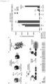
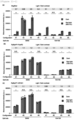
- the purified RNA was reverse-transcribed using SuperScript III (Thermo Fisher Scientific Inc.) and a primer O8, and the concentrated cDNA library was regenerated by PCR using KOD -Multi&Epi- DNA polymerase and primers O5 and O8. After six repetitive selections, the concentrated DNA library was sequenced (150 cycles, 130 bp and 45 bp, paired ends) in Illumina MiSeq machine using Miseq Reagent Kit v. 3. The results are shown in Figure 6 .
- Table 1 shows the amino acid sequences (SEQ ID NOs: 41 to 50) of typical Affibody molecules obtained by six selections.
- the amino acid sequence of No. 6 is identical to the amino acid sequence represented by SEQ ID NO: 1.
- HEK 293T cells and Neuro2a cells were cultured at 37°C in 5% CO 2 using Dulbecco's modified Eagle medium (DMEM, Sigma-Aldrich Co., LLC) supplemented with 10% fetal bovine serum (FBS, HyClone), 100 units/mL penicillin, and 100 ⁇ g/mL streptomycin (Gibco).
- DMEM Dulbecco's modified Eagle medium
- FBS fetal bovine serum
- streptomycin Gibco
- HeLa cells were cultured at 37°C in 5% CO 2 using minimal essential medium (Sigma-Aldrich Co., LLC) supplemented with 10% FBS, 100 U/mL penicillin, and 100 ⁇ g/mL streptomycin.
- HeLa cells were plated at 1.0 ⁇ 10 4 cells per well to a 96-well multiwell plate (Greiner Bio-One International GmbH) in the presence of 25 ⁇ M BV and cultured at 37°C in 5% CO 2 for 24 hours. Then, the cells were transfected using Lipofectamine 3000 (Invitrogen Corp.). Plasmids encoding VP16-NLS, tetR, and luciferase reporter were used at a ratio of 1:1:1 in transfection. The total amount of DNA was 0.1 ⁇ g per well. Aff6 of Aff6-VP16-NLS was replaced with irrelevant Affibody (Zdk1) as a control condition (Aff6-VP16-NLS (-)).
- a luminescence plate reader Centro XS3 LB 960, Berthold Technologies GmbH & Co. KG
- SEAP reporter gene assay Roche Diagnostics
- HEK 293T cells were inoculated at a cell density of 4.0 ⁇ 10 5 cells per well to 35 mm/ Tissue Culture Dish (IWAKI) in the presence of 25 ⁇ M biliverdin (BV) added thereto, and cultured at 37°C for 24 hours under a condition of 5% CO 2 .
- the cells were transfected with a plasmid encoding Nfluc-DrBphP and a plasmid encoding 3xFLAG-Aff6_V18F ⁇ N-Cfluc at a ratio of 1:1.
- the total amount of DNA was 2.5 ⁇ g per dish.
- the amount of DNA was 0.025 ⁇ g per dish in order to justify a dynamic range of bioluminescence intensity.
- the culture solution was replaced with 2 ml of HBSS supplemented with 200 ⁇ M D-luciferin.
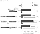
- DrBphP was optically converted from the photo-state (Pfr) form to the dark-state (Pr) form by irradiation with light (50 W/m 2 ) at 800 nm for 5 minutes using an LED array.
- bioluminescence was measured at room temperature using GloMax 20/20 luminometer (Promega Corp.).
- GloMax 20/20 luminometer Promega Corp.
- the bioluminescence measurement was discontinued, and the sample dish was removed from the luminometer.
- the cells were irradiated with light (10 W/m 2 ) at 660 nm for 1 minute using an LED array of 660 nm.
- the sample dish was immediately brought back to the luminometer to resume bioluminescence measurement.
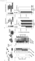
- the procedures of irradiating the cells with light at 800 nm were the same as the procedures mentioned above. The results are shown in Figure 2 .
- HEK 293T cells and Neuro-2a cells at 2.0 ⁇ 10 4 cells and HeLa cells at 1.0 ⁇ 10 4 cells were plated to each well of a 96-well multiwell plate.
- the cells were cultured at 37°C in 5% CO 2 for 24 hours.
- the cells were transfected with plasmids encoding NLS-dCas9-NLS, MS2, p65-HSF1, sgRNA, and luciferase reporter at a ratio of 1:1:1:1:1.
- the total amount of DNA was 0.1 ⁇ g per well. Procedures of light irradiation and bioluminescence detection of MagRed were as described above.
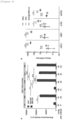
- a red light sample was incubated under a condition of pulsed irradiation of 10 W/m 2 at 760 nm (light irradiation for 1 minute and lights-out for 4 minutes). Other procedures were as described above. Under a condition supplemented with biliverdin (BV) (BV(+)), cells were inoculated in the presence of 25 ⁇ M BV and transfected as described above. 24 hours after transfection, 100 ⁇ L of a medium supplemented with 25 ⁇ M BV was added to the sample. Remaining procedures were as described above. The results are shown in Figure 9 .
- SEQ ID NOs: 51 and 52 The amino acid sequence and nucleotide sequence of NLS-dCas9-NLS are shown in SEQ ID NOs: 51 and 52.
- sgRNA is shown in SEQ ID NOs: 53 to 57.
- SEQ ID NO: 58 represents the MS2 aptamer sequence of sgRNA.
- a nucleotide sequence described on the 5'-terminal side from GTTTTA is a target sequence.
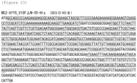
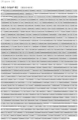
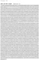
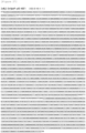
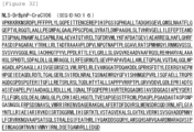
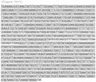
- amino acid sequence (SEQ ID NO: 51) of NLS-dCas9-NLS is as described below.
- the underlined sequences respectively represent the sequences of NLS and dCas9.
- the nucleotide acid sequence (SEQ ID NO: 52) of NLS-dCas9-NLS is as described below.
- the underlined sequences respectively represent nucleotide sequences encoding NLS and dCas9.
- sequences (SEQ ID NOs: 53 to 57) of sgRNA are as described below.
- the underlined sequences respectively represent a target sequence and a MS2 aptamer sequence.
- RNA extraction and reverse-transcription PCR were performed using CellAmp Direct RNA Prep Kit (Takara Bio Inc.) and PrimeScript Real-Time Master Mix (Takara Bio Inc.). Quantitative PCR was performed using StepOne Plus system (Thermo Fisher Scientific Inc.) and TaqMan Gene Expression Master Mix (Thermo Fisher Scientific Inc.).
- the expression levels of the target genes were quantified using TaqMan primers and probes, and GAPDH gene was measured as an endogenous control (Life Technologies Corp.; TaqMan Gene Expression Assay IDs were as follows: ASCL1: Hs04187546_g1, IL1R2: Hs01030384_m1, HBG1: Hs00361131_g1, MYOD1: Hs02330075_g1, GAPDH: Hs99999905_m1).
- the ⁇ Ct method was utilized in order to show each relative mRNA level to a negative control transfected with an empty vector.
- HEK 293T cells were inoculated at 2.0 ⁇ 10 4 cells per well to a 96-well multiwell plate in the presence of 25 ⁇ M BV and cultured at 37°C in 5% CO 2 for 24 hours.
- the cells were transfected with plasmids encoding a CreN-containing fusion protein, a CreC-containing fusion protein, and luciferase reporter at a ratio of 1:1:8.
- the total amount of DNA was 0.1 ⁇ g per well. 6 hours after transfection, light irradiation was performed at 37°C under a condition of 5% CO 2 .
- a red light sample was incubated under a condition of pulsed irradiation of 1 W/m 2 at 660 nm (light irradiation for 1 minute and lights-out for 4 minutes).
- a red light sample was incubated under a condition of pulsed irradiation of 10 W/m 2 at 760 nm (light irradiation for 1 minute and lights-out for 4 minutes).
- the multiwell plate containing the cultured HEK 293T cells was covered with aluminum foil and incubated at 37°C in 5% CO 2 .
- HEK 293T cells were plated at 0.8 ⁇ 10 5 cells/well to a 24-well plate and cultured at 37°C in 5% CO 2 for 24 hours.
- the cells were transfected with plasmids encoding NLS-CreN104-Aff6_V18F ⁇ N-Venus, NLS-DrBphP(FL)-CreC106, and fluorescent reporter at a ratio of 1:1:8.
- the total amount of DNA was 0.5 ⁇ g per well. 6 hours after transfection, the sample was irradiated with light at 37°C under a condition of 5% CO 2 .
- a red light sample was incubated under a condition of pulsed irradiation of 1 W/m 2 at 660 nm (light irradiation for 1 minute and lights-out for 4 minutes). Under a dark condition, the multiwell plate containing the cultured HEK 293T cells was covered with aluminum foil and incubated at 37°C in 5% CO 2 . After culture for 24 hours, the cells were fixed in PBS containing 4% paraformaldehyde for 15 minutes and then incubated at room temperature for 1 hour in a blocking solution (PBS containing 5% BSA and 0.3% Triton-X100) to perform blocking while enhancing cell membrane permeability.
- a blocking solution PBS containing 5% BSA and 0.3% Triton-X100
- the cells were immunostained while mildly shaken at room temperature for 1 hour in the blocking solution using a mouse anti-mCherry antibody (mCherry Monoclonal Antibody conjugated with Alexa Fluor 594, Invitrogen Corp., Cat# M11240, 1:1,000 dilution).
- the cells were washed three times with PBS and then stained with 300 nM DAPI (Invitrogen Corp.) at room temperature for 15 minutes.
- Fluorescence images obtained in experiments were analyzed using FIJI imageJ and CellProfiler. The background of the images was subtracted using Subtract background tool included in ImageJ. The nuclei were identified using the threshold of an object size at a 5-to 20-pixel unit in a DAPI channel. In order to determine populations of GFP-positive cells (GFP+) and mCherry-positive cells (mCh+), average fluorescence intensity of Venus and mCherry was measured in respective cell nuclei. The threshold of fluorescence intensity was determined such that mCh+ cells were not detected under a control condition. The number of mCh+ cells was divided by the number of GFP+ cells and multiplied by 100 to determine the ratio of mCh+/GFP+ cells.
- mice were temporarily brought back to home cages every 8 hours and fed for 1 hour.
- the mice in the dark group were placed in home cages shielded from light.
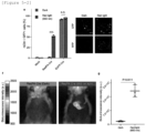
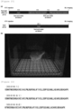
- bioluminescence was imaged. 200 mL of 100 mM D-luciferin was intraperitoneally injected to each mouse before bioluminescence imaging. Then, the mouse was anesthetized with isoflurane (FUJIFILM Wako Pure Chemical Corp.). 5 minutes after D-luciferin injection, bioluminescence images of the mice were obtained using a bioluminescence imager (Lumazone, Nippon Roper K.K.) equipped with an EMCD camera (Evolve 512, Teledyne Photometrics). The results are shown in Figures 5 and 15 .
Landscapes
- Chemical & Material Sciences (AREA)
- Organic Chemistry (AREA)
- Health & Medical Sciences (AREA)
- Life Sciences & Earth Sciences (AREA)
- Biochemistry (AREA)
- General Health & Medical Sciences (AREA)
- Biophysics (AREA)
- Genetics & Genomics (AREA)
- Medicinal Chemistry (AREA)
- Molecular Biology (AREA)
- Proteomics, Peptides & Aminoacids (AREA)
- Immunology (AREA)
- Gastroenterology & Hepatology (AREA)
- Peptides Or Proteins (AREA)
- Solid State Image Pick-Up Elements (AREA)
Applications Claiming Priority (2)
| Application Number | Priority Date | Filing Date | Title |
|---|---|---|---|
| JP2022048984A JP2023142218A (ja) | 2022-03-24 | 2022-03-24 | アフィボディと光受容体タンパク質とのセット |
| PCT/JP2023/011866 WO2023182504A1 (fr) | 2022-03-24 | 2023-03-24 | Ensemble d'afficorps et de protéine photoréceptrice |
Publications (2)
| Publication Number | Publication Date |
|---|---|
| EP4501948A1 true EP4501948A1 (fr) | 2025-02-05 |
| EP4501948A4 EP4501948A4 (fr) | 2026-03-04 |
Family
ID=88101721
Family Applications (1)
| Application Number | Title | Priority Date | Filing Date |
|---|---|---|---|
| EP23775097.1A Pending EP4501948A4 (fr) | 2022-03-24 | 2023-03-24 | Ensemble d'afficorps et de protéine photoréceptrice |
Country Status (6)
| Country | Link |
|---|---|
| US (1) | US20250197529A1 (fr) |
| EP (1) | EP4501948A4 (fr) |
| JP (1) | JP2023142218A (fr) |
| CN (1) | CN118974075A (fr) |
| CA (1) | CA3254885A1 (fr) |
| WO (1) | WO2023182504A1 (fr) |
Family Cites Families (2)
| Publication number | Priority date | Publication date | Assignee | Title |
|---|---|---|---|---|
| JP6555677B2 (ja) * | 2014-03-03 | 2019-08-07 | 国立大学法人 東京大学 | 光依存的に二量体を形成するタンパク質のセット |
| JP6892642B2 (ja) | 2015-04-13 | 2021-06-23 | 国立大学法人 東京大学 | 光依存的に又は薬物存在下でヌクレアーゼ活性若しくはニッカーゼ活性を示す、又は標的遺伝子の発現を抑制若しくは活性化するポリペプチドのセット |
-
2022
- 2022-03-24 JP JP2022048984A patent/JP2023142218A/ja active Pending
-
2023
- 2023-03-24 CA CA3254885A patent/CA3254885A1/fr active Pending
- 2023-03-24 US US18/849,071 patent/US20250197529A1/en active Pending
- 2023-03-24 WO PCT/JP2023/011866 patent/WO2023182504A1/fr not_active Ceased
- 2023-03-24 CN CN202380029188.XA patent/CN118974075A/zh active Pending
- 2023-03-24 EP EP23775097.1A patent/EP4501948A4/fr active Pending
Also Published As
| Publication number | Publication date |
|---|---|
| CA3254885A1 (fr) | 2025-03-26 |
| EP4501948A4 (fr) | 2026-03-04 |
| WO2023182504A1 (fr) | 2023-09-28 |
| CN118974075A (zh) | 2024-11-15 |
| US20250197529A1 (en) | 2025-06-19 |
| JP2023142218A (ja) | 2023-10-05 |
Similar Documents
| Publication | Publication Date | Title |
|---|---|---|
| CN111328343B (zh) | Rna靶向方法和组合物 | |
| US12497603B2 (en) | Cas effector proteins, gene editing systems, and uses thereof | |
| JP6892642B2 (ja) | 光依存的に又は薬物存在下でヌクレアーゼ活性若しくはニッカーゼ活性を示す、又は標的遺伝子の発現を抑制若しくは活性化するポリペプチドのセット | |
| CN114380922A (zh) | 在细胞内产生点突变的融合蛋白、其制备及用途 | |
| US10202656B2 (en) | Dividing of reporter proteins by DNA sequences and its application in site specific recombination | |
| CN115052986A (zh) | 包含核酸酶的组合物及其用途 | |
| EP3535394A2 (fr) | Plasmides d'adn pour la génération rapide de vecteurs de recombinaison homologues pour le développement de lignées cellulaires | |
| JP7487939B2 (ja) | 2分割されたCpf1タンパク質 | |
| Kotera et al. | A high-throughput and single-tube recombination of crude PCR products using a DNA polymerase inhibitor and type IIS restriction enzyme | |
| US20250197529A1 (en) | Set of Photoreceptor Protein and Affibody | |
| CN113652430B (zh) | 一种光控rna代谢调控系统 | |
| CN112813049B (zh) | 用于活细胞rna标记的融合蛋白及应用 | |
| US20260001920A1 (en) | Optical switch protein that functions through irradiation with long wavelength light | |
| Kuwasaki et al. | A semi-synthetic red light photoswitch for optogenetic control of protein activity | |
| US9611486B2 (en) | Constructs and method for regulating gene expression or for detecting and controlling a DNA locus in eukaryotes | |
| Pham | Detection of protein-protein interaction using bimolecular fluorescence complementation assay | |
| Shi et al. | Novel Bimolecular Fluorescence Complementation (BiFC) assay for in vivo visualization of the protein-protein interactions and cellular protein complex localizations | |
| Xie et al. | Efficient site-specific recombination by self-activating split-Dre recombinase in mammalian cells and E. coli | |
| WO2025101722A1 (fr) | Chromodomaines modifiés à haute affinité | |
| Fushimi et al. | A semi-synthetic red light photoswitch for optogenetic control of protein 3 activity 4 | |
| Renzl | Aptamer-mediated transactivation of transcription utilizing CRISPR/Cas9 and a photoreceptor | |
| O'Brien | Cloning and Expression of Hydra Innexin 2, a Gap Junction Protein Required for Coordinated Contraction of the Body Column | |
| CN122029275A (zh) | 包含异源dna结合结构域的条件dna修饰酶 | |
| Buckley | Biochemical analysis of Hel308 and HelQ helicases | |
| HK40027119A (en) | Methods for improved homologous recombination and compositions thereof |
Legal Events
| Date | Code | Title | Description |
|---|---|---|---|
| STAA | Information on the status of an ep patent application or granted ep patent |
Free format text: STATUS: THE INTERNATIONAL PUBLICATION HAS BEEN MADE |
|
| PUAI | Public reference made under article 153(3) epc to a published international application that has entered the european phase |
Free format text: ORIGINAL CODE: 0009012 |
|
| STAA | Information on the status of an ep patent application or granted ep patent |
Free format text: STATUS: REQUEST FOR EXAMINATION WAS MADE |
|
| 17P | Request for examination filed |
Effective date: 20241010 |
|
| AK | Designated contracting states |
Kind code of ref document: A1 Designated state(s): AL AT BE BG CH CY CZ DE DK EE ES FI FR GB GR HR HU IE IS IT LI LT LU LV MC ME MK MT NL NO PL PT RO RS SE SI SK SM TR |
|
| DAV | Request for validation of the european patent (deleted) | ||
| DAX | Request for extension of the european patent (deleted) | ||
| A4 | Supplementary search report drawn up and despatched |
Effective date: 20260202 |
|
| RIC1 | Information provided on ipc code assigned before grant |
Ipc: C07K 14/31 20060101AFI20260127BHEP Ipc: C07K 14/195 20060101ALI20260127BHEP |