EP4509186A2 - Anticorps monoclonal anti-pd1, composition pharmaceutique associée et utilisation associée - Google Patents
Anticorps monoclonal anti-pd1, composition pharmaceutique associée et utilisation associée Download PDFInfo
- Publication number
- EP4509186A2 EP4509186A2 EP24206755.1A EP24206755A EP4509186A2 EP 4509186 A2 EP4509186 A2 EP 4509186A2 EP 24206755 A EP24206755 A EP 24206755A EP 4509186 A2 EP4509186 A2 EP 4509186A2
- Authority
- EP
- European Patent Office
- Prior art keywords
- monoclonal antibody
- antigen
- antibody
- binding fragments
- seq
- Prior art date
- Legal status (The legal status is an assumption and is not a legal conclusion. Google has not performed a legal analysis and makes no representation as to the accuracy of the status listed.)
- Pending
Links
Images




Classifications
-
- A—HUMAN NECESSITIES
- A61—MEDICAL OR VETERINARY SCIENCE; HYGIENE
- A61K—PREPARATIONS FOR MEDICAL, DENTAL OR TOILETRY PURPOSES
- A61K39/00—Medicinal preparations containing antigens or antibodies
- A61K39/395—Antibodies; Immunoglobulins; Immune serum, e.g. antilymphocytic serum
-
- A—HUMAN NECESSITIES
- A61—MEDICAL OR VETERINARY SCIENCE; HYGIENE
- A61P—SPECIFIC THERAPEUTIC ACTIVITY OF CHEMICAL COMPOUNDS OR MEDICINAL PREPARATIONS
- A61P35/00—Antineoplastic agents
-
- A—HUMAN NECESSITIES
- A61—MEDICAL OR VETERINARY SCIENCE; HYGIENE
- A61P—SPECIFIC THERAPEUTIC ACTIVITY OF CHEMICAL COMPOUNDS OR MEDICINAL PREPARATIONS
- A61P35/00—Antineoplastic agents
- A61P35/02—Antineoplastic agents specific for leukemia
-
- A—HUMAN NECESSITIES
- A61—MEDICAL OR VETERINARY SCIENCE; HYGIENE
- A61P—SPECIFIC THERAPEUTIC ACTIVITY OF CHEMICAL COMPOUNDS OR MEDICINAL PREPARATIONS
- A61P7/00—Drugs for disorders of the blood or the extracellular fluid
- A61P7/06—Antianaemics
-
- C—CHEMISTRY; METALLURGY
- C07—ORGANIC CHEMISTRY
- C07K—PEPTIDES
- C07K16/00—Immunoglobulins [IG], e.g. monoclonal or polyclonal antibodies
- C07K16/18—Immunoglobulins [IG], e.g. monoclonal or polyclonal antibodies against material from animals or humans
- C07K16/28—Immunoglobulins [IG], e.g. monoclonal or polyclonal antibodies against material from animals or humans against receptors, cell surface antigens or cell surface determinants
-
- C—CHEMISTRY; METALLURGY
- C07—ORGANIC CHEMISTRY
- C07K—PEPTIDES
- C07K16/00—Immunoglobulins [IG], e.g. monoclonal or polyclonal antibodies
- C07K16/18—Immunoglobulins [IG], e.g. monoclonal or polyclonal antibodies against material from animals or humans
- C07K16/28—Immunoglobulins [IG], e.g. monoclonal or polyclonal antibodies against material from animals or humans against receptors, cell surface antigens or cell surface determinants
- C07K16/2803—Immunoglobulins [IG], e.g. monoclonal or polyclonal antibodies against material from animals or humans against receptors, cell surface antigens or cell surface determinants against the immunoglobulin superfamily
- C07K16/2818—Immunoglobulins [IG], e.g. monoclonal or polyclonal antibodies against material from animals or humans against receptors, cell surface antigens or cell surface determinants against the immunoglobulin superfamily against CD28 or CD152
-
- G—PHYSICS
- G01—MEASURING; TESTING
- G01N—INVESTIGATING OR ANALYSING MATERIALS BY DETERMINING THEIR CHEMICAL OR PHYSICAL PROPERTIES
- G01N33/00—Investigating or analysing materials by specific methods not covered by groups G01N1/00 - G01N31/00
- G01N33/48—Biological material, e.g. blood, urine; Haemocytometers
- G01N33/50—Chemical analysis of biological material, e.g. blood, urine; Testing involving biospecific ligand binding methods; Immunological testing
- G01N33/53—Immunoassay; Biospecific binding assay; Materials therefor
- G01N33/575—Immunoassay; Biospecific binding assay; Materials therefor for cancer
-
- G—PHYSICS
- G01—MEASURING; TESTING
- G01N—INVESTIGATING OR ANALYSING MATERIALS BY DETERMINING THEIR CHEMICAL OR PHYSICAL PROPERTIES
- G01N33/00—Investigating or analysing materials by specific methods not covered by groups G01N1/00 - G01N31/00
- G01N33/48—Biological material, e.g. blood, urine; Haemocytometers
- G01N33/50—Chemical analysis of biological material, e.g. blood, urine; Testing involving biospecific ligand binding methods; Immunological testing
- G01N33/53—Immunoassay; Biospecific binding assay; Materials therefor
- G01N33/577—Immunoassay; Biospecific binding assay; Materials therefor involving monoclonal antibodies binding reaction mechanisms characterised by the use of monoclonal antibodies
-
- G—PHYSICS
- G01—MEASURING; TESTING
- G01N—INVESTIGATING OR ANALYSING MATERIALS BY DETERMINING THEIR CHEMICAL OR PHYSICAL PROPERTIES
- G01N33/00—Investigating or analysing materials by specific methods not covered by groups G01N1/00 - G01N31/00
- G01N33/48—Biological material, e.g. blood, urine; Haemocytometers
- G01N33/50—Chemical analysis of biological material, e.g. blood, urine; Testing involving biospecific ligand binding methods; Immunological testing
- G01N33/68—Chemical analysis of biological material, e.g. blood, urine; Testing involving biospecific ligand binding methods; Immunological testing involving proteins, peptides or amino acids
-
- A—HUMAN NECESSITIES
- A61—MEDICAL OR VETERINARY SCIENCE; HYGIENE
- A61K—PREPARATIONS FOR MEDICAL, DENTAL OR TOILETRY PURPOSES
- A61K39/00—Medicinal preparations containing antigens or antibodies
- A61K2039/505—Medicinal preparations containing antigens or antibodies comprising antibodies
-
- C—CHEMISTRY; METALLURGY
- C07—ORGANIC CHEMISTRY
- C07K—PEPTIDES
- C07K2317/00—Immunoglobulins specific features
- C07K2317/20—Immunoglobulins specific features characterized by taxonomic origin
- C07K2317/24—Immunoglobulins specific features characterized by taxonomic origin containing regions, domains or residues from different species, e.g. chimeric, humanized or veneered
-
- C—CHEMISTRY; METALLURGY
- C07—ORGANIC CHEMISTRY
- C07K—PEPTIDES
- C07K2317/00—Immunoglobulins specific features
- C07K2317/50—Immunoglobulins specific features characterized by immunoglobulin fragments
- C07K2317/56—Immunoglobulins specific features characterized by immunoglobulin fragments variable (Fv) region, i.e. VH and/or VL
- C07K2317/565—Complementarity determining region [CDR]
-
- C—CHEMISTRY; METALLURGY
- C07—ORGANIC CHEMISTRY
- C07K—PEPTIDES
- C07K2317/00—Immunoglobulins specific features
- C07K2317/70—Immunoglobulins specific features characterized by effect upon binding to a cell or to an antigen
- C07K2317/76—Antagonist effect on antigen, e.g. neutralization or inhibition of binding
-
- C—CHEMISTRY; METALLURGY
- C07—ORGANIC CHEMISTRY
- C07K—PEPTIDES
- C07K2317/00—Immunoglobulins specific features
- C07K2317/90—Immunoglobulins specific features characterized by (pharmaco)kinetic aspects or by stability of the immunoglobulin
- C07K2317/92—Affinity (KD), association rate (Ka), dissociation rate (Kd) or EC50 value
-
- G—PHYSICS
- G01—MEASURING; TESTING
- G01N—INVESTIGATING OR ANALYSING MATERIALS BY DETERMINING THEIR CHEMICAL OR PHYSICAL PROPERTIES
- G01N2333/00—Assays involving biological materials from specific organisms or of a specific nature
- G01N2333/435—Assays involving biological materials from specific organisms or of a specific nature from animals; from humans
- G01N2333/705—Assays involving receptors, cell surface antigens or cell surface determinants
- G01N2333/70503—Immunoglobulin superfamily, e.g. VCAMs, PECAM, LFA-3
- G01N2333/70521—CD28, CD152
-
- G—PHYSICS
- G01—MEASURING; TESTING
- G01N—INVESTIGATING OR ANALYSING MATERIALS BY DETERMINING THEIR CHEMICAL OR PHYSICAL PROPERTIES
- G01N2800/00—Detection or diagnosis of diseases
- G01N2800/70—Mechanisms involved in disease identification
- G01N2800/7019—Ischaemia
Definitions
- the present invention belongs to the field of tumor therapy and molecular immunology, relating to an anti-PD1 antibody, the pharmaceutical composition and methods of use. Specifically, the present invention relates to an anti-PD1 monoclonal antibody.
- the transmembrane receptor PD1 (programmed cell death 1, also known as PD-1) is a member of the CD28 gene family, expressed in activated T cells, B cells and myeloid cells. Both ligands of PD1 (i.e. PDL1 and PDL2) belong to the B7 superfamily; wherein PDL1 is broadly expressed in a variety of cells including T cells, B cells, endothelial cells and epithelial cells, while PDL2 is only expressed in antigen presenting cells such as dendritic cells and macrophages.
- T cells play a very important role in the elimination of viral infections, and T cell antiviral response is usually associated with immunopathogenesis.
- PD1 plays a vital role in the negative regulation of T cell activation.
- PD1-mediated negative regulation on T cells can reduce tissue damage caused by infection, blocking or inhibiting the negative regulatory effect of PD1 may lead to autoimmune diseases, for example, pancreatic virus infection can be more effectively eliminated in PD1 gene knockout mice, but may lead to more severe liver damage ( Isai et al., 2003, J.Exp.Med. 198:39-50 ).
- tumors with highly expressed PD1 often develop into cancers that are difficult to detect ( Hamanishi et al., 2007, Proc.Natl.Acad. Sci.USA 104:3360-5 ).
- An established method to regulate PD1 expression is through injection of antibodies into the body.
- the inventors obtained hybridoma cells via fusion of mouse splenocytes and myeloma cells. By screening a large number of samples, the inventors obtained the hybridoma cell line LT003 (CCTCC Deposit Accession No.: C2015105).
- the hybridoma cell line LT003 is capable of secreting a specific monoclonal antibody (named 14C12) binding specifically to PD1, and this monoclonal antibody can effectively block the association of PD1 to PDL1.
- the inventors generated a humanized anti-PD1 antibody (named 14C12H1L1) in a creative way.
- the inventors found that the antibodies 14C12 and 14C12H1L1 herein can effectively bind to human T cells, activate T cells and induce the secretion of IFN- ⁇ and IL-2 from human lymphocytes.
- the antibodies 14C12 and 14C12H1L1 herein have the potential to become drugs for preventing and treating malignancies including lung cancer, melanoma, renal cancer, ovarian cancer and leukemia, as well as anemia.
- the said monoclonal antibody or its antigen-binding fragments thereof wherein,
- the said monoclonal antibody or its antigen-binding fragments thereof, wherein, the monoclonal antibody comprises:
- variable regions in heavy and light chains of an antibody govern binding activity.
- Each chain contains three hypervariable regions, namely, the complementary determining region (CDR) (HCDR1, HCDR2 and HCDR3 in heavy (H) chain, and LCDR1, LCDR2 and LCDR3 in light (L) chain), which are defined by Kabat, et al. (Sequences of Proteins of Immunological Interest, Fifth Edition (1991), volume 1-3, NIH Publication 91-3242, Bethesda MD ).
- CDR complementary determining region
- the antibodies 14C12 and 14C12H1L1 herein comprises the same CDR:
- the said monoclonal antibody or its antigen-binding fragment thereof wherein the said monoclonal antibody or its antigen-binding fragment are selected from Fab, Fab', F(ab')2, Fd, Fv, dAb, CDRs, single chain antibodies (e.g. scFv), humanized antibodies, chimeric antibodies, or bispecific antibodies.
- the said EC 50 is determined by an indirect ELISA.
- the said monoclonal antibody or its antigen-binding fragment thereof, wherein the said monoclonal antibody contains non-CDR regions from species other than mouse, for example, from human.
- CTCC China Center for Type Culture Collection
- the present invention relates to an isolated nucleic acid molecule comprising a nucleotide sequence capable of encoding V H of the antibody, wherein,
- the present invention relates to an isolated nucleic acid molecule comprising a nucleotide sequence capable of encoding V L of the antibody, wherein,
- the present invention relates to a vector comprising the isolated nucleic acid molecule described in the present invention.
- the present invention relates to a host cell comprising the isolated nucleic acid molecule described in the present invention, or vector described in the present invention.
- the present invention relates to a method for preparing the monoclonal antibody or its antigen-binding fragments thereof described in the present invention, by culturing the host cell in the present invention under appropriate conditions, and recovering the said monoclonal antibody or its antigen-binding fragments thereof from the cell culture.
- the present invention relates to the hybridoma cell line LT003 that is preserved in the China Center for Typical Culture Collection (CCTCC) with the CCTCC Deposit Accession NO: C2015105.
- CTCC China Center for Typical Culture Collection
- the present invention relates to a conjugate that consist of monoclonal antibody or its antigen binding fragments and conjugating part, wherein, the said monoclonal antibody is monoclonal antibody or its antigen binding fragments described in the present invention, and the said conjugating part as a detectable marker.
- the said conjugating part are radioactive isotopes, fluorescein, luminescent materials, colorful substances, or enzymes.
- the present invention relates to reagent kits, consisting of the monoclonal antibody or its antigen binding fragments, or the conjugates thereof described in the invention;
- the reagent kits may contain a secondary antibody, which specifically recognizes the said monoclonal antibody or its antigen binding fragments; optionally, the said secondary antibody may contain detectable markers, such as radioactive isotopes, fluorescein, luminescent materials, colorful substances, or enzymes.
- detectable markers such as radioactive isotopes, fluorescein, luminescent materials, colorful substances, or enzymes.
- the present invention relates to use of the said monoclonal antibody or its antigen binding fragments, or the conjugates thereof described in the invention in preparation of reagent kits, the said reagent kits are used in detection of the existence or the level of PD1 in samples.
- the present invention relates to a pharmaceutical composition
- a pharmaceutical composition comprising the said monoclonal antibody or its antigen binding fragments, or the conjugates thereof described in the invention.
- it may also comprise a pharmaceutically acceptable carrier or excipient.
- the present invention relates to use of the said monoclonal antibody or its antigen binding fragments or the conjugates thereof described in the invention in preparing drugs for prevention and/or treatment and/or adjuvant treatment and/or diagnosis of tumors or anemia; specifically, the said tumors may be melanoma, renal cancer, prostate cancer, bladder cancer, colorectal cancer, gastrointestinal cancer, liver cancer, non-small cell lung cancer, ovarian cancer and leukemia.
- 14C12H1L1 can effectively inhibit the growth of MC38 tumor cells inoculated at right side subcutaneously in PD-1 HuGEMM mice, which the antibody drug 14C12H1L1 can significantly inhibit the tumor growth in PD-1 HuGEMM tumor-bearing mice, having an efficacy equivalent to the marketed monoclonal antibody drug Nivolumab that is approved drug targeting the same target.
- the present invention relates to use of the monoclonal antibody or its antigen-binding fragments or the conjugates thereof described in the present invention for preparation drugs with the following purposes:
- the said PD1 ligand is PDL1 or PDL2, preferably PDL1.
- the present invention relates to an in vivo or in vitro method to apply to cells or subjects in need with an effective dose of the monoclonal antibody or its antigen-binding fragments or the conjugates thereof described in the present invention, and the said method is selected from the following:
- the said PD1 ligand is PDL1 or PDL2, preferably PDL1.
- the said in vitro method is intended for non-therapeutic or -diagnostic purposes.
- Interferon ⁇ is mainly naturally produced by natural killer (NK) cells and natural killer T (NKT) cells, or produced by effector T cells consisting of CD4 Th1 cells and CD8 cytotoxic T lymphocytes after being stimulated by specific antigens.
- NK natural killer
- NKT natural killer T
- IFN ⁇ plays an import role in antagonizing or inhibiting viral, some bacterial and protozoon infections.
- IFN ⁇ can activate macrophages and induce the expression of type 2 major histocompatibility complex (MHC) to activate immune responses to control the progression of tumors ( Schoenborn JR, Wilson CB. Regulation of Interferon- ⁇ During Innate and Adaptive Immune Responses. Advances in Immunology 2007; 96:41-101 ).
- MHC major histocompatibility complex
- the anti-PD1 antibody can induce the secretion of IFN ⁇ to activate immune responses.
- Interleukin 2 (IL-2) produced by T cells is a growth factor regulating T cell subsets and a crucial factor regulating immune responses, promoting activated B cells proliferation, and participating in antibody responses, hematopoiesis and oncological surveillance.
- Recombinant human IL-2 has been approved by the U. S. FDA for the treatment of malignant tumors (including melanoma, renal tumor, etc.) while undergoing clinical studies for the treatment of chronic viral infections ( Chavez, A.R., et al., Pharmacologic administration of interleukin-2. Ann N Y Acad Sci, 2009.1182:p.14-27 ).
- the anti-PD1 antibody of the present invention can specifically relieve the immunosuppression of PD1, activate T cells and induce IL-2 production, displaying promising prospects of extensive applications in gene therapies for neoplastic and parasitic diseases.
- the monoclonal antibody or its antigen-binding fragments or the conjugates thereof described in the present invention is used for the prevention and/or treatment and/or adjuvant treatment and/or diagnosis of tumors or anemia; specifically, the said tumors may be melanoma, renal cancer, prostate cancer, bladder cancer, colorectal cancer, gastrointestinal cancer, liver cancer, non-small cell lung cancer, ovarian cancer or leukemia.
- the monoclonal antibody or its antigen-binding fragments or the conjugates thereof described in the present invention is used to:
- the said PD1 ligand is PDL1 or PDL2, preferably PDL1.
- the monoclonal antibody or its antigen-binding fragments or the conjugates thereof described in the present invention only blocks the binding of PD1 to PDL1.
- the present invention relates to a method for the prevention and/or treatment and/or adjuvant treatment and/or diagnosis of tumors or anemia, including the procedure to apply to subjects with an effective dose of the monoclonal antibody or its antigen-binding fragments or the conjugates thereof described in the present invention; specifically, the said tumors may be melanoma, renal cancer, prostate cancer, bladder cancer, colorectal cancer, gastrointestinal cancer, liver cancer, non-small cell lung cancer, ovarian cancer or leukemia.
- amino acid sequence of PD1 protein (Programmed cell death protein 1, NCBI GenBank: NP_005009.2)
- PD1ECD extracellular segment of PD1
- PD1ECD-containing fragments and also comprises the fusion protein of PD1ECD, for example, fused with the Fc protein fragments of mouse or human IgG (mFc or hFc).
- the amino acid sequence of PD1 protein can have naturally or artificial mutations (including but not limited to substitutions, deletions, and/or additions), not affecting its biological functions. Therefore, in the present invention, the term "PD1 protein" should include all such sequences and their natural or artificial variants. Furthermore, when describing the sequence fragments of PD1 protein, the said sequence fragments comprise both the sequence fragments and the corresponding sequence fragments in its natural or artificial variants.
- amino acid sequence of PDL1 protein (Programmed death-ligand 1, NCBI Genebank ID: NP_054862.1)” comprises the full length PDL1 protein, or PDL1ECD (extracellular segment of PDL1) or PDL1ECD-containing fragments; and also comprises the fusion protein of PDL1ECD, for example, fused with the Fc protein fragments of mouse or human IgG (mFc or hFc).
- the amino acid sequence of PDL1 protein can have naturally or artificial mutations (including but not limited to substitutions, deletions, and/or additions), not affecting its biological functions.
- the term "PDL1 protein” should include all such sequences and their natural or artificial variants. Furthermore, when describing the sequence fragments of PDL1 protein, the said sequence fragments comprise both the sequence fragments and the corresponding sequence fragments in its natural or artificial variants.
- EC 50 refers to the concentration for 50% of maximal effect.
- the term "antibody” refers to immunoglobulin proteins, which typically composed of two pairs of polypeptide chains (each pair has a "light” (L) chain and a “heavy” (H) chain).
- the light chains are classified as ⁇ and ⁇ light chains.
- the heavy chains are classified as ⁇ , ⁇ , ⁇ , ⁇ , or ⁇ , and respectively, define isotype antibodies as IgM, IgD, IgG, IgA and IgE.
- variable regions and constant regions are connected by a "J" region consisting of about 12 or more amino acids.
- the heavy chain also contains a "D" region with about 3 or more amino acids.
- Each heavy chain contains a variable region (V H ) and a constant region (C H ), which consists of 3 domains (C H 1, C H 2, and C H 3).
- Each light chain contains a variable region (V L ) and a constant region (C L ), which consists of one domain C L .
- the constant region can mediate the binding of immune globulin to host tissues or factors, including various cells in the immune system (e.g., effector cells) and the complement component 1q (C1q) of the classical complement system.
- V H and V L can also be subdivided into regions with high variability (called complementarity determining region (CDR)), which are separated by relatively conservative regions called framework regions (FR).
- CDR complementarity determining region
- each V H and V L is composed of 3 CDRs and 4 FRs, in the order of FR1, CDR1, FR2, CDR2, FR3, CDR3, and FR4.
- the variable regions (V H and V L ) of the heavy chain and light chain form the antibody binding site. Distribution of amino acids to the regions or domains follow the definitions by Kabat in Sequences of Proteins of Immunological Interest (National Institutes of Health Bethesda, MD (1987 and 1991 )), or Chothia & Lesk (1987) Mol. Biol., 196:901-917 ; or Chothia et al. (1989) Nature, 342:878-883 .
- antibody is not restricted by any particular method of producing them. For example, it includes, in particular, recombinant antibodies, monoclonal antibodies, and polyclonal antibodies. Antibodies can be different isotypes, for example, IgG (such as IgG1, IgG2, IgG3, or IgG4 subtype), IgA1, IgA2, IgD, IgE, or IgM antibodies.
- antigen binding fragments refers to a polypeptide containing fragments of a full-length antibody, maintaining the ability to bind specifically to the same antigen, and/or to compete with the full length antibody to bind to the antigen, which is also called “the antigen binding portion”. See Fundamental Immunology, Ch. 7 (Paul, W., ed. 2, Raven Press, N. Y. (1989 )), including the entire article and references in this invention for all purposes. Antigen binding fragments can be generated by recombinant DNA techniques or by cleaving intact antibodies with proteolytic enzymes or chemicals.
- the antigen binding fragments include Fab, Fab', F(ab') 2 , Fd, Fv, dAb, and CDR fragments, single chain antibodies (e.g., scFv), chimeric antibodies, diabody, and the polypeptides that at least contains an antibody portion which is sufficient to confer a specific antigen binding capacity to the polypeptides.
- the antigen binding fragment is a diabody, namely, a dimeric antibody fragment, whose V H and V L domains are expressed on a single polypeptide chain, however, because of using a too short linker the to allow pairing between the two domains of the same chain, the domains are forced to pair with complementary domains on another chain to generate two antigen binding sites (see, for example, Holliger P. et al., Proc. Natl. Acad. Sci. USA 90: 6444-6448 (1993 ), and Poljak R. J. et al., Structure 2: 1121-1123 (1994 )).
- an antigen binding fragment (such as the above described antibody fragments) may be obtained from a given antibody (e.g. monoclonal antibodies 14C12, 14C12H1L1 provided herein in the invention), and screened for specificity in the same manner as for the full antibody.
- a given antibody e.g. monoclonal antibodies 14C12, 14C12H1L1 provided herein in the invention
- antibody refers to not only the intact antibody, but also the antigen binding fragments of the antibody.
- mAb and “monoclonal antibodies” refers to an antibody or a fragment of an antibody that is derived from a group of highly homologous antibodies, i.e. from a group of identical antibody molecules, except for mutations that may arise spontaneously.
- Monoclonal antibody has high specificity against a single epitope on the antigen.
- Polyclonal antibodies are different from monoclonal antibodies, containing at least 2 or more different antibodies, which usually recognize different epitopes on the antigen.
- Monoclonal antibodies can be obtained with hybridoma technology reported originally by Kohler et al., (Nature, 256: 495, (1975 )), as well as recombinant DNA Technology (see U.S. Patent 4,816,567 ).
- humanized antibody refers to an antibody or its fragments, derived from a human immunoglobulin (receptor antibody), whose CDRs or part of CDRs are replaced by the CDR regions of a non-human antibody (donor antibody), where the donor antibody may be a non-human antibody (for example, mice, rats, or rabbits) with predictive specificity, binding affinity, or reactivity.
- donor antibody may be a non-human antibody (for example, mice, rats, or rabbits) with predictive specificity, binding affinity, or reactivity.
- some amino acid residues of the receptor antibody framework region (FR) can also be replaced by the corresponding amino acid residues of the non-human source, or replaced by the amino acid residues of other antibodies to further improve or optimize the performance of the antibody.
- the term “isolate” or “isolated” means obtained by artificial means in the natural state. If there is a certain kind of “isolated” matter or component in nature, it may be due to the change in its natural environment, or isolated from the natural environment, or both. For example, polynucleotide or polypeptide in a natural existence in a living animal will be called “isolated” if it was separated with high purity in the same natural state.
- isolated does not exclude existence of artificial or synthetic material, or other impurities that does not affect the activity.
- vector refers to a nucleic acid delivery vehicle that can be inserted with polynucleotide.
- the vector that can have the protein that is encoded by the inserted polynucleotide expressed is called an expression vector.
- Vectors can be inserted into the host cell by transformation, transduction, or transfection, so the genetic substances carried by the vector can be expressed in the host cell.
- Vectors are well known to the technical personnel in the field, including but not limited to: plasmid; phasmid; cosmid; artificial chromosome such as yeast artificial chromosome (YAC), bacterial artificial chromosome (BAC), or P1 derived artificial chromosome (PAC); phage such as ⁇ phage or M13 phage and animal viruses etc.
- Animal viruses may include but not limited to, reverse transcriptase virus (including lentivirus), adenovirus, adeno-associated virus, herpes virus (e. g. herpes simplex virus), chicken pox virus, baculovirus, papilloma virus, and papova virus (such as SV40).
- a vector can contain multiple components that control expression, including but not limited to, promoter, transcription initiation factor, enhancer, selection element, and reporter gene. In addition, the vector may also contain replication initiation site.
- the term "host cell” refers to cells that can import vectors, including but not limited to, prokaryotic cells such as E. coli and Bacillus subtilis, fungal cells such as yeast and Aspergillus, insect cells such as S2 drosophila cells and Sf9, or animal cells such as fibroblast cells, CHO cells, COS cells, NSO cells, HeLa cells, BHK cells, HEK293 cells or human cells.
- prokaryotic cells such as E. coli and Bacillus subtilis
- fungal cells such as yeast and Aspergillus
- insect cells such as S2 drosophila cells and Sf9
- animal cells such as fibroblast cells, CHO cells, COS cells, NSO cells, HeLa cells, BHK cells, HEK293 cells or human cells.
- a specific binding of an antibody to an antigen means an affinity (K D ), for example less than about 10 -5 M, in particular, less than 10 -6 M, 10 -7 M, 10 -8 M, 10 -9 M, 10 -10 M, or less.
- K D refers to the dissociation equilibrium constant of specific interaction between antibody and antigen, to describe the binding affinity between antibodies and antigens.
- antibodies e.g., monoclonal antibodies 14C12, 14C12H1L1 in the present invention
- antigens e.g., PD1 protein
- K D bind to antigens (e.g., PD1 protein) with a K D less than approximately 10 -5 M, for example, less than 10 -6 M, 10 -7 M, 10 -8 M, 10 -9 M or 10 -10 M or even less.
- K D can be measured by any method well known to the technical personnel in the field, for example, using Fortebio Octet System
- the terms “monoclonal antibody” and “mAb” have the same meaning and are used interchangeably
- the terms “polyclonal antibody” and “PcAb” have the same meaning and are used interchangeably
- the terms “polypeptide” and “protein” have the same meaning and are used interchangeably.
- amino acids are usually represented by single letter or three letter abbreviations known to this field. For example, Alanine can be represented as A or Ala.
- hybridoma and “hybridoma cell line” can be used interchangeably, and when mentioned herein, include the subclone and progeny cells of hybridoma.
- hybridoma cell line LT003 also includes the subclone and progeny cells of LT003.
- the term "pharmaceutically acceptable carrier or excipient” refers to a carrier and/or an excipient pharmaceutically and/or physiologically compatible with subjects and active ingredients, and widely recognized in the field herein ( Remington's Pharmaceutical Sciences. Edited by Gennaro AR, 19th ed. Pennsylvania: Mack Publishing Company, 1995 ), including but not limited to: pH adjustors, surfactants, adjuvants, and ionic strength enhancers.
- pH adjustors include but not limited to phosphate buffer solution
- surfactants include but not limited to cationic, anionic or nonionic surfactants such as Tween-80
- ionic strength enhancers include but not limited to sodium chloride.
- the term "effective dose” is defined as an amount of a therapeutic sufficient to achieve or at least partially achieve the desired effect.
- effective prevention dose e.g. for cancer
- effective treatment dose is the amount to cure, or at least partially stop, the disease and its complications in patients with the disease. Determination of such an effective dose is entirely within the scope of the capabilities of the technical personnel in the field.
- the effective treatment dose will depend on the severity of the disease, the overall state of the patient's immune system, the general background of patients such as age, weight and gender, administration method of the said drug, and other concomitant treatments, etc.
- the monoclonal antibodies in the present invention is capable of binding to PD1 specifically, effectively blocking the binding of PD1 to PDL1, and relieving the immunosuppression of PD1 to activate T lymphocytes.
- the PD1 antibody 14C12H1L1 can induce the secretions of IFN- ⁇ and IL-2 much better than the control antibody 5C4 (5C4: PD1 antibody from Medarex Inc.: Alan J.Korman, et al., Human monoclonal antibodies to programmed death 1 (PD1) and methods for treating cancer using anti-PD1 antibodies alone or in combination with other immunotherapeutics, United States Patent, Patent No. US 8008449 B2 ).
- the monoclonal antibodies of the present invention have an antitumor effect equivalent to the approved drug Nivolumab for the same target.
- the antibodies of the present invention have the potential to become or to be prepared into drugs for the prevention and/or treatment of non-small cell lung cancer, renal cell carcinoma, ovarian cancer, melanoma, leukemia or anemia.
- Hybridoma cell line LT003 has been preserved in the China Center for Typical Culture Collection (CCTCC) in Wuhan University, Wuhan, China 430072 on June 16, 2015 with CCTCC Deposit Accession NO: C2015105
- T cells used were from Akeso Biopharma, Inc.; the BALB/C mice were purchased from Guangdong Medical Laboratory Animal Center.
- the PD-1 HuGEMM mice used were from Nanjing Galaxy Biopharma Co., Ltd.; MC38 cells were from Shanghai Fudan IBS Cell Center; the marketed drug for the same target, Nivolumab (Opdivo ® ) used was from Bristol-Myers Squibb Company.
- PD1-mFc PD1: Programmed cell death protein 1, NCBI GenBank ID:NP_005009.2 fusion protein as the antigen, and fused the splenocytes of immunized BALB/C mice (purchased from Guangdong Medical Laboratory Animal Center) and mouse myeloma cells into hybridoma cells by currently established method (for example, Stewart, S.J., "Monoclonal Antibody Production", in Basic Methods in antibody Production and Characterization, Eds.G.C. Howard and D.R. Bethell, Boca Raton: CRC Press, 2000 ).
- Hybridoma cell line LT003 (PD1-14C12), has been preserved in the China Center for Typical Culture Collection (CCTCC) in Wuhan University, Wuhan, China 430072 on June 16, 2015 with CCTCC Deposit Accession NO: C2015105.
- LT003 cell line in the present invention was cultured using IMDM medium containing 10% low IgG fetal bovine serum for 7 days, and then the cell culture supernatant was harvested and purified to get the antibody 14C12.
- variable region sequence is SEQ ID NO:1
- the variable region sequence is SEQ ID NO:3
- 14C12 Re
- the cell culture supernatant was centrifuged by high-speed centrifugation and filtered by vacuum filtration with microporous membrane and loaded onto the HiTrap MabSelectSuRe column, and then the antibody was eluted with Elution Buffer in one step, and then went through HiTrap Desalting column, and recovered into PBS buffer,and then the recombinant antibody 14C12 (Re) was obtained after further purification .
- the recombinant antibody 14C12 (Re) had a binding activity equivalent to the antibody 14C12, and hence can be further used in the subsequent antibody humanization design.
- variable region sequence is SEQ ID NO:5
- heavy chain constant region is Ig gamma-1 chain C region, ACCESSION:P0185
- light-chain cDNA the variable region sequence is SEQ ID NO:7
- light chain constant region is Ig kappa chain C region, ACCESSION:P01834
- 14C12H1L1 into the pUC57simple (provided by GenScript Biotech Corp.) vector (enzymatic cleavage sites: XbaI & BamHI) to obtain pUC57simple-14C12H1 and pUC57simple-14C12L1 plasmids, respectively.
- the cell culture was centrifuged by high-speed centrifugation and filtered via vacuum filtration with microporous membrane and loaded onto the HiTrap MabSelectSuRe column, and then the antibody was eluted with Elution Buffer in one step, went through HiTrap Desalting column and recovered into PBS buffer, and purified further to obtain the recombinant antibody 14C12H1L1, which was analyzed by SDS-PAGE electrophoresis.
- the binding kinetics of the antibody 14C12 and humanized antibody 14C12H1L1 to the antigen PD1 was measured by Fortebio Octet System.
- the binding activities of antibodies 14C12H1L1 and 5C4 to PD1 were measured separately by indirect ELISA as follows: After incubated with PD1-mFc at 4°C overnight, the microplate was blocked with 1% BSA at 37°C for 2h, and then the antibodies were added separately and incubated at 37°C for 30 min, and then HRP-labeled secondary antibody (goat anti-human IgG (H+L)) (Jackson, 109-035-088) was added. and then TMB (Neogen, 308177) was added to react for 5 mins and the absorbance was read at the wavelength of 450 nm in a microplate reader.
- HRP-labeled secondary antibody goat anti-human IgG (H+L)
- TMB Neogen, 308177
- Table 2 Absorbance Intensities of 14C12H1L1 and 5C4 binding to PD1 Antigen coating: PD1-mFc (0.5 ⁇ g/mL) Antibody concentration ( ⁇ g/mL) 14C12H1L1 5C4 1 2.970 2.954 2.959 2.991 0.3 2.886 2.961 2.978 3.079 0.1 2.864 2.868 2.838 2.926 0.03 2.674 2.669 2.617 2.659 0.01 2.222 2.201 1.981 2.221 0.003 1.383 1.464 1.169 1.222 0.001 0.676 0.736 0.527 0.548 0 0.062 0.062 0.065 0.073 Secondary Antibody HRP-labeled secondary antibody (goat anti-human IgG)
- Example 7 Binding Activities of Antibodies to Antigen PD1 against PDL1 by competition ELISA
- Binding activities of humanized antibody 14C12H1L1 and 5C4 to antigen PD1 against PDL1 were measured by competition ELISA as follows: After incubated with PD1-hFc at 4°C overnight, the microplate was blocked with 1% BSA for 2h, and antibodies 14C12H1L1 and 5C4 with different concentrations (see Table 3 for the dilute strengths) were mixd with PDL1-mFc for 10 min, and the mixtures were incubated at 37°C for 30 min, and then corresponding anti-human and anti-mouse enzyme-labeled second antibodies respectively and incubate at 37°C for 30 min were added. The absorbance at the wavelength of 450 nm was read in a microplate reader.
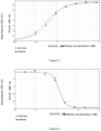
- the binding results of antibodies 14C12H1L1 and 5C4 to the antigen PD1 were shown in Figure 6 .
- the antibody 14C12H1L1 can compete against PDL1 and bind to PD1 protein effectively with dose-dependency.
- the absorbance intensities at different doses were shown in Table 3.
- EC50 of 14C12H1L1 and 5C4 binding activity were then determined to be 1.322 nM and 1.199 nM, respectively.
- Table 3 Competitive Binding of 14C12H1L1 and 5C4 to PD1 against PDL1 Antigen coating PD1-mFc 0.5 ⁇ g/mL Antibody concentration/dilution gradient 14C12H1L1 5C4 1.5 ⁇ g/ml 0.062 0.064 0.070 0.075 1:3 0.069 0.064 0.081 0.086 1:9 0.363 0.305 0.372 0.269 1:27 1.727 1.543 1.429 1.604 1:81 1.892 1.752 1.766 1.881 1:243 1.984 2.029 2.045 2.005 1:729 1.937 1.978 1.934 1.954 0 1.870 1.977 1.933 1.977 Ligand PDL1-mFc 0.3 ⁇ g/ml Secondary Antibody HRP-labeled goat anti-mouse secondary antibody
- Host cells 293T expressing PD1 antigen were constructed, and labeled with the humanized antibody 14C12H1L1 prepared in the present invention (see Example 4).
- the ability of the antibody 14C12H1L1 to bind specifically to cell surface antigen in its native conformation was analyzed and validated by flow cytometry.
- Antibody labeling and flow cytometry The 293T-PD1 obtained by the step above was digested by trypsin, and distributed into tubes each containing2 ⁇ 10 5 cells. PD1 antibodies was diluted using PBSbuffer (1% BSA) at concentrations of 50 nM, 10 nM, 5 nM, 1 nM, 0.1 nM and 0.01 nM and added into the tubes and incubated on ice with PD1-expressing 293T cells for2h. 100 ⁇ L of FITC-labeled goat anti-human secondary antibody (1:500) was added into each tube and incubated on ice for 1h. After washed with PBS 3 times, cells were re-suspended in 300 ⁇ L of PBS and fluorescence signals were measured on the flow cytometer using the FITC channel.
- PBSbuffer 1% BSA
- the binding results of the humanized antibody 14C12H1L1 to 293T-PD1 cells were shown in Figure 7 .
- the antibody 14C12H1L1 can bind target PD1 protein expressing on the surface of host cells 293T-PD1 effectively with dose-dependency.
- the fluorescence intensities at different doses were shown in Table 4. Through Curve Simulation using quantitative analyses of fluorescence intensities, EC50 of 14C12H1L1 binding with PD1 was then determined to be 1.89 nM.
- Table 4 The Fluorescence Intensities of 14C12H1L1 Binding to 293T-PD1 Surface Antigen Detected by Flow Cytometry Concentration (nM) 0.01 0.1 1 5 10 50 Fluorescence Intensity 8.32 20.31 174.62 579.41 686.49 669.54
- PBMC peripheral blood mononuclear cells
- Example 10 Mixed Lymphocyte Reaction: Secretion of Cytokines IFN- ⁇ , IL-2
- PBMC peripheral blood mononuclear cells
- T cells were isolated from PBMC and mixed with the DC cells obtained above in the ratio of 10:1 to culture together with the antibodies 14C12H1L1, 5C4 and hIgG (hIgG as an isotype control) in different ratios for 5-6 days.
- the secretions of IFN- ⁇ and IL-2 were measured with corresponding ELISA reagent kits (both purchased from Dakewe) respectively.
- the isolated PBMC (the same method as in Example 10) was stimulated with PHA (Shanghai Shenqi Biotech Co., Ltd, 50 ⁇ l/ml) for 3 days, and then mature PBMC (5 ⁇ 10 4 cells/well) mixed with Raji cells (Chinese Academy of Sciences Shanghai Branch) (5 ⁇ 10 4 cells/well) and MDA-MB-231 cells (ATCC) (1 ⁇ 10 4 cells/well) in a 96-well plate. 100 nM of 14C12H1L1 or control antibody 5C4 or hIgG (hIgG as the isotype control) were added and mixed and cultured together for three days. The secretion of IL-2 was detected with ELISA reagent kit (purchased from Dakewe) according to the instructions of the kit.
- MC38 tumor cells (1 ⁇ 10 6 cells/mouse) were inoculated subcutaneously on the right side of PD-1 HuGEMM mice (human PD-1 transgenic mice). When the mean tumor volume reached approximately 118 mm 3 , the mice were randomly divided into 4 experimental groups with 8 mice in each group. Antibodies were given through abdominal administration. The specific grouping and dosages were as follows:
- the above 4 groups were injected with antibodies twice weekly, 5 doses in total. After injection, the tumor sizes were measured twice weekly.
Landscapes
- Health & Medical Sciences (AREA)
- Life Sciences & Earth Sciences (AREA)
- Chemical & Material Sciences (AREA)
- Immunology (AREA)
- Medicinal Chemistry (AREA)
- Engineering & Computer Science (AREA)
- General Health & Medical Sciences (AREA)
- Molecular Biology (AREA)
- Organic Chemistry (AREA)
- Hematology (AREA)
- Biomedical Technology (AREA)
- Urology & Nephrology (AREA)
- Biochemistry (AREA)
- Veterinary Medicine (AREA)
- Public Health (AREA)
- Animal Behavior & Ethology (AREA)
- Pharmacology & Pharmacy (AREA)
- Chemical Kinetics & Catalysis (AREA)
- Microbiology (AREA)
- Proteomics, Peptides & Aminoacids (AREA)
- Nuclear Medicine, Radiotherapy & Molecular Imaging (AREA)
- General Chemical & Material Sciences (AREA)
- Cell Biology (AREA)
- Pathology (AREA)
- General Physics & Mathematics (AREA)
- Biotechnology (AREA)
- Analytical Chemistry (AREA)
- Physics & Mathematics (AREA)
- Food Science & Technology (AREA)
- Biophysics (AREA)
- Genetics & Genomics (AREA)
- Bioinformatics & Cheminformatics (AREA)
- Oncology (AREA)
- Mycology (AREA)
- Epidemiology (AREA)
- Diabetes (AREA)
- Peptides Or Proteins (AREA)
- Preparation Of Compounds By Using Micro-Organisms (AREA)
- Medicines Containing Antibodies Or Antigens For Use As Internal Diagnostic Agents (AREA)
- Medicines Containing Material From Animals Or Micro-Organisms (AREA)
Applications Claiming Priority (3)
| Application Number | Priority Date | Filing Date | Title |
|---|---|---|---|
| CN201610705763.5A CN106977602B (zh) | 2016-08-23 | 2016-08-23 | 一种抗pd1单克隆抗体、其药物组合物及其用途 |
| EP17842895.9A EP3505535B1 (fr) | 2016-08-23 | 2017-08-22 | Anticorps monoclonal anti-pd-1, composition pharmaceutique et utilisation de celui-ci |
| PCT/CN2017/098465 WO2018036472A1 (fr) | 2016-08-23 | 2017-08-22 | Anticorps monoclonal anti-pd-1, composition pharmaceutique et utilisation de celui-ci |
Related Parent Applications (1)
| Application Number | Title | Priority Date | Filing Date |
|---|---|---|---|
| EP17842895.9A Division EP3505535B1 (fr) | 2016-08-23 | 2017-08-22 | Anticorps monoclonal anti-pd-1, composition pharmaceutique et utilisation de celui-ci |
Publications (2)
| Publication Number | Publication Date |
|---|---|
| EP4509186A2 true EP4509186A2 (fr) | 2025-02-19 |
| EP4509186A3 EP4509186A3 (fr) | 2025-06-04 |
Family
ID=59340342
Family Applications (2)
| Application Number | Title | Priority Date | Filing Date |
|---|---|---|---|
| EP24206755.1A Pending EP4509186A3 (fr) | 2016-08-23 | 2017-08-22 | Anticorps monoclonal anti-pd1, composition pharmaceutique associée et utilisation associée |
| EP17842895.9A Active EP3505535B1 (fr) | 2016-08-23 | 2017-08-22 | Anticorps monoclonal anti-pd-1, composition pharmaceutique et utilisation de celui-ci |
Family Applications After (1)
| Application Number | Title | Priority Date | Filing Date |
|---|---|---|---|
| EP17842895.9A Active EP3505535B1 (fr) | 2016-08-23 | 2017-08-22 | Anticorps monoclonal anti-pd-1, composition pharmaceutique et utilisation de celui-ci |
Country Status (22)
| Country | Link |
|---|---|
| US (2) | US12076398B2 (fr) |
| EP (2) | EP4509186A3 (fr) |
| JP (1) | JP7082620B2 (fr) |
| KR (1) | KR102469286B1 (fr) |
| CN (1) | CN106977602B (fr) |
| AU (2) | AU2017316255C1 (fr) |
| BR (1) | BR112019003775A2 (fr) |
| EA (1) | EA201990570A1 (fr) |
| ES (1) | ES3001154T3 (fr) |
| FI (1) | FI3505535T3 (fr) |
| HU (1) | HUE069593T2 (fr) |
| IL (1) | IL264956B2 (fr) |
| LT (1) | LT3505535T (fr) |
| MX (1) | MX2019002268A (fr) |
| NZ (1) | NZ751902A (fr) |
| PL (1) | PL3505535T3 (fr) |
| PT (1) | PT3505535T (fr) |
| RS (1) | RS66372B1 (fr) |
| SG (1) | SG11201901584UA (fr) |
| SI (1) | SI3505535T1 (fr) |
| WO (1) | WO2018036472A1 (fr) |
| ZA (1) | ZA201901781B (fr) |
Families Citing this family (58)
| Publication number | Priority date | Publication date | Assignee | Title |
|---|---|---|---|---|
| CN108112254B (zh) | 2015-03-13 | 2022-01-28 | 西托姆克斯治疗公司 | 抗-pdl1抗体、可活化的抗-pdl1抗体、及其使用方法 |
| CA2991976A1 (fr) | 2015-07-13 | 2017-01-19 | Cytomx Therapeutics, Inc. | Anticorps anti-pd-1, anticorps anti-pd-1 activables, et leurs procedes d'utilisation |
| KR20250099459A (ko) | 2015-11-03 | 2025-07-01 | 얀센 바이오테크 인코포레이티드 | Pd-1과 특이적으로 결합하는 항체 및 그의 용도 |
| CN106977602B (zh) * | 2016-08-23 | 2018-09-25 | 中山康方生物医药有限公司 | 一种抗pd1单克隆抗体、其药物组合物及其用途 |
| CN106967172B (zh) | 2016-08-23 | 2019-01-08 | 康方药业有限公司 | 抗ctla4-抗pd-1 双功能抗体、其药物组合物及其用途 |
| WO2018035710A1 (fr) | 2016-08-23 | 2018-03-01 | Akeso Biopharma, Inc. | Anticorps anti-ctla4 |
| BR112019025188A2 (pt) | 2017-06-01 | 2020-06-23 | Cytomx Therapeutics, Inc. | Anticorpos anti-pdl1 ativáveis e métodos de uso dos mesmos |
| IL272159B2 (en) | 2017-08-04 | 2026-03-01 | Genmab As | PD-L1 and CD137 binding agents and their use |
| CN109425736B (zh) * | 2017-08-25 | 2021-04-09 | 北京百普赛斯生物科技股份有限公司 | 一种检测pd-1抗体血药浓度的方法及试剂盒 |
| KR20200059243A (ko) * | 2017-09-20 | 2020-05-28 | 리제너론 파마슈티칼스 인코포레이티드 | 패신저 유전자 돌연변이 부담이 높은 종양을 가진 환자에 대한 면역 치료요법 |
| US12398209B2 (en) | 2018-01-22 | 2025-08-26 | Janssen Biotech, Inc. | Methods of treating cancers with antagonistic anti-PD-1 antibodies |
| TWI841554B (zh) | 2018-03-21 | 2024-05-11 | 丹麥商珍美寶股份有限公司 | 以鉑為主之劑與抗組織因子抗體-藥物共軛物的組合治療癌症之方法 |
| EP3773916A4 (fr) * | 2018-04-04 | 2021-12-15 | TrueBinding, Inc. | Méthodes et compositions pour bloquer l'interaction entre polypeptides pd-1 non glycosylés |
| WO2019217457A1 (fr) | 2018-05-07 | 2019-11-14 | Genmab A/S | Méthodes de traitement du cancer au moyen d'une combinaison d'un conjugué de medicament anticorps anti-pd-1 et anticorps anti-facteur tissulaire |
| CA3099547A1 (fr) | 2018-05-07 | 2019-11-14 | Reshma Abdulla RANGWALA | Methodes de traitement du cancer a l'aide d'une combinaison d'un anticorps anti-pd-1 et d'un conjugue anticorps anti-facteur tissulaire-medicament |
| WO2020015703A1 (fr) | 2018-07-18 | 2020-01-23 | 正大天晴药业集团股份有限公司 | Combinaison médicamenteuse de dérivé de quinoléine et d'anticorps |
| CN109053889B (zh) * | 2018-07-25 | 2019-06-21 | 博奥信生物技术(南京)有限公司 | 一种抗人pd1单克隆抗体及用途 |
| CN109053895B (zh) * | 2018-08-30 | 2020-06-09 | 中山康方生物医药有限公司 | 抗pd-1-抗vegfa的双功能抗体、其药物组合物及其用途 |
| TWI844571B (zh) | 2018-10-30 | 2024-06-11 | 丹麥商珍美寶股份有限公司 | 使用抗血管內皮生長因子(vegf)抗體與抗組織因子(tf)抗體-藥物共軛體之組合以治療癌症之方法 |
| PL3946377T3 (pl) | 2019-04-03 | 2026-04-20 | Orega Biotech | Terapie skojarzone oparte na inhibitorach pd1 i il-17b |
| CN110373415B (zh) * | 2019-06-12 | 2023-06-09 | 安徽省昂普拓迈生物科技有限责任公司 | 特异性结合pd-l1蛋白的核酸适配体及其用途 |
| KR20220030956A (ko) | 2019-07-05 | 2022-03-11 | 오노 야꾸힝 고교 가부시키가이샤 | Pd-1/cd3 이중 특이성 단백질에 의한 혈액암 치료 |
| CN112300286A (zh) * | 2019-08-02 | 2021-02-02 | 康方药业有限公司 | 抗ctla4-抗pd-1双特异性抗体及其用途 |
| KR20220062503A (ko) | 2019-08-02 | 2022-05-17 | 씨티티큐-아케소 (상하이) 바이오메드. 테크. 컴퍼니, 리미티드 | 항-pd-1 항체 및 이의 약제학적 용도 |
| TW202120550A (zh) | 2019-08-08 | 2021-06-01 | 日商小野藥品工業股份有限公司 | 雙特異性蛋白質 |
| CN110903391B (zh) * | 2019-10-25 | 2021-05-28 | 东大生物技术(苏州)有限公司 | 一组pd-l1单克隆抗体及其医药用途 |
| CN112915202B (zh) * | 2019-12-06 | 2025-08-01 | 正大天晴药业集团股份有限公司 | 喹啉衍生物与pd-1单抗的药物组合 |
| EP4079763A4 (fr) * | 2019-12-20 | 2023-10-11 | Guangdong Feipeng Pharmaceutical Co., Ltd | Anticorps monoclonal anti-mort programmée humaine (pd-1) |
| CN113117072A (zh) * | 2020-01-14 | 2021-07-16 | 正大天晴药业集团股份有限公司 | 喹啉衍生物与pd-1单抗的药物组合 |
| US20230045494A1 (en) | 2020-02-28 | 2023-02-09 | Orega Biotech | Combination therapies based on ctla4 and il-17b inhibitors |
| KR20230004726A (ko) * | 2020-04-22 | 2023-01-06 | 아케소 바이오파마, 인크. | 항-cd73/항-pd-1 이중특이항체 및 이의 용도 |
| KR20230003178A (ko) | 2020-04-29 | 2023-01-05 | 치아타이 티안큉 파마수티컬 그룹 주식회사 | PD-1 및 TGF-β에 대한 이중 기능성 단백질 |
| CN113754767A (zh) * | 2020-06-03 | 2021-12-07 | 上海恒润达生生物科技股份有限公司 | 抗人pd-1抗体及其用途 |
| CN114761434B (zh) * | 2020-09-30 | 2023-06-27 | 苏州沙砾生物科技有限公司 | Pd-1抗体及其制备方法与应用 |
| US20240024320A1 (en) | 2020-11-17 | 2024-01-25 | Seagen Inc. | Methods of treating cancer with a combination of tucatinib and an anti-pd-1/anti-pd-l1 antibody |
| CN113336848B (zh) * | 2021-02-03 | 2022-08-19 | 上海莱馥医疗科技有限公司 | 一种抗pd-1抗体及其用途 |
| US20250114450A1 (en) * | 2021-03-12 | 2025-04-10 | Akeso Biopharma, Inc. | Pharmaceutical combination containing anti-pd-1-anti-vegfa bispecific antibody, and use thereof |
| CA3225254A1 (fr) | 2021-07-13 | 2023-01-19 | BioNTech SE | Agents de liaison multispecifiques contre cd40 et cd137 en polytherapie du cancer |
| CN115873116B (zh) | 2021-09-29 | 2026-04-28 | 中山康方生物医药有限公司 | 抗lag3抗体、药物组合物及用途 |
| IL311771A (en) | 2021-10-06 | 2024-05-01 | BioNTech SE | Multispecific binding agents against pd-l1 and cd137 in combination |
| WO2023083439A1 (fr) | 2021-11-09 | 2023-05-19 | BioNTech SE | Agoniste de tlr7 et combinaisons pour le traitement du cancer |
| CN113912710B (zh) * | 2021-11-17 | 2023-05-26 | 杭州旭科生物技术有限公司 | 抗新型冠状病毒n蛋白的单克隆抗体及其应用 |
| CN117069850B (zh) * | 2021-12-20 | 2025-02-14 | 南京诺唯赞检测技术有限公司 | 一种vce的单克隆抗体及其制备方法 |
| EP4257609A1 (fr) | 2022-04-08 | 2023-10-11 | iOmx Therapeutics AG | Polythérapies à base d'inhibiteurs de pd-1 et de sik3 |
| CN119031933B (zh) * | 2022-04-12 | 2025-08-26 | 正大天晴(广州)医药有限公司 | 蛋白酶体抑制剂与抗pd-1抗体的药物组合 |
| CA3253038A1 (fr) | 2022-05-12 | 2023-11-16 | Genmab A/S | Agents de liaison capables de se lier à cd27 en polythérapie |
| US11932693B2 (en) | 2022-05-12 | 2024-03-19 | BioNTech SE | Monoclonal antibodies directed against programmed death-1 protein and their use in medicine |
| US20250282873A1 (en) * | 2022-05-13 | 2025-09-11 | Nanjing Shunxin Pharmaceuticals Co., Ltd. Of Chiatai Tianqing Pharmaceutical Group | Drug combination of anti-tim-3 antibody and anti-pd-1 antibody |
| CN119789867A (zh) * | 2022-09-06 | 2025-04-08 | 正大天晴药业集团股份有限公司 | 结合tim-3的抗体与结合pd-1的抗体的药物组合 |
| US20260098092A1 (en) * | 2022-10-05 | 2026-04-09 | Chulalongkorn University | Monoclonal antibodies against human programmed cell death protein 1 (pd-1) |
| CN117986356B (zh) * | 2022-10-27 | 2025-07-22 | 纽迈生物科技(苏州)有限公司 | 一种制备抗体的方法 |
| CN120302979A (zh) | 2022-12-01 | 2025-07-11 | 生物技术公司 | 针对CD40和CD137的多特异性抗体与抗PD1 Ab和化学治疗的组合治疗 |
| EP4688160A1 (fr) | 2023-04-06 | 2026-02-11 | Genmab A/S | Agents de liaison multispécifiques dirigés contre pd-l1 et cd137 pour le traitement du cancer |
| US12410258B2 (en) | 2023-05-12 | 2025-09-09 | Ganmab A/S | Antibodies capable of binding to OX40, variants thereof and uses thereof |
| WO2025056014A1 (fr) * | 2023-09-13 | 2025-03-20 | 正大天晴药业集团股份有限公司 | Composition pharmaceutique comprenant un anticorps anti-pd-1 et un second anticorps |
| WO2025056011A1 (fr) * | 2023-09-13 | 2025-03-20 | 正大天晴药业集团股份有限公司 | Composition pharmaceutique comprenant un anticorps anti-pd-1 et un second anticorps |
| WO2025114541A1 (fr) | 2023-11-30 | 2025-06-05 | Genmab A/S | Anticorps pouvant se lier à ox40 en polythérapie |
| CN118652343B (zh) * | 2024-07-08 | 2025-05-09 | 吉林省医菲尼迪生物医药有限公司 | 一种临床血液免疫细胞以及在治疗疾病中的应用 |
Citations (3)
| Publication number | Priority date | Publication date | Assignee | Title |
|---|---|---|---|---|
| US4816567A (en) | 1983-04-08 | 1989-03-28 | Genentech, Inc. | Recombinant immunoglobin preparations |
| US8008449B2 (en) | 2005-05-09 | 2011-08-30 | Medarex, Inc. | Human monoclonal antibodies to programmed death 1 (PD-1) and methods for treating cancer using anti-PD-1 antibodies alone or in combination with other immunotherapeutics |
| WO2015112800A1 (fr) * | 2014-01-23 | 2015-07-30 | Regeneron Pharmaceuticals, Inc. | Anticorps humains se liant à pd-1 |
Family Cites Families (24)
| Publication number | Priority date | Publication date | Assignee | Title |
|---|---|---|---|---|
| PT1537878E (pt) | 2002-07-03 | 2010-11-18 | Ono Pharmaceutical Co | Composições de imunopotenciação |
| WO2004056875A1 (fr) * | 2002-12-23 | 2004-07-08 | Wyeth | Anticorps anti pd-1 et utilisations |
| JP5646457B2 (ja) | 2008-04-29 | 2014-12-24 | アッヴィ・インコーポレイテッド | 二重可変ドメイン免疫グロブリン及びその使用 |
| PL2342226T3 (pl) * | 2008-09-26 | 2017-01-31 | Dana-Farber Cancer Institute, Inc. | Ludzkie przeciwciała anty-PD-1, PD-L1 i PD-L2 oraz ich zastosowania |
| EP2545073B1 (fr) | 2010-03-12 | 2015-09-30 | AbbVie Biotherapeutics Inc. | Protéines ctla4 et leurs utilisations |
| GB201103955D0 (en) | 2011-03-09 | 2011-04-20 | Antitope Ltd | Antibodies |
| MX341076B (es) * | 2011-03-31 | 2016-08-04 | Merck Sharp & Dohme | Formulaciones estables de anticuerpos para el receptor humano pd-1 de meurte programada y tratamientos relacionados. |
| SI2699264T1 (en) | 2011-04-20 | 2018-08-31 | Medimmune Llc | Antibodies and other molecules that bind b7-h1 and pd-1 |
| WO2014022758A1 (fr) | 2012-08-03 | 2014-02-06 | Dana-Farber Cancer Institute, Inc. | Anticorps de liaison double à agent unique anti-pd-l1 et pd-l2 et procédés d'utilisation |
| US20160145355A1 (en) | 2013-06-24 | 2016-05-26 | Biomed Valley Discoveries, Inc. | Bispecific antibodies |
| CN104250302B (zh) * | 2013-06-26 | 2017-11-14 | 上海君实生物医药科技股份有限公司 | 抗pd‑1抗体及其应用 |
| DK3049441T3 (da) | 2013-09-27 | 2020-02-03 | Hoffmann La Roche | Anti-PDL1-antistofformuleringer |
| HUE046249T2 (hu) * | 2013-12-12 | 2020-02-28 | Shanghai hengrui pharmaceutical co ltd | PD-1 antitest, antigén-kötõ fragmense, és gyógyászati alkalmazása |
| EP3089758B1 (fr) | 2014-01-03 | 2021-01-27 | F.Hoffmann-La Roche Ag | Conjugués d'anticorps helicar-anti-helicar liés par covalence et leurs utilisations |
| CN104974253A (zh) | 2014-04-01 | 2015-10-14 | 上海中信国健药业股份有限公司 | 抗ctla-4/pd-1双特异性抗体、其制备方法及应用 |
| CN105296433B (zh) | 2014-08-01 | 2018-02-09 | 中山康方生物医药有限公司 | 一种ctla4抗体、其药物组合物及其用途 |
| CN104987421A (zh) | 2015-05-13 | 2015-10-21 | 北京比洋生物技术有限公司 | 抗ctla-4和pd-1的双重可变结构域免疫球蛋白 |
| CN105175544B (zh) | 2015-10-20 | 2021-04-09 | 安徽瀚海博兴生物技术有限公司 | 一种抗pd-1人源化单克隆抗体及其应用 |
| CN105175545B (zh) * | 2015-10-20 | 2019-01-25 | 安徽瀚海博兴生物技术有限公司 | 一种抗vegf-抗pd-1双功能抗体及其应用 |
| JP6955507B2 (ja) | 2015-12-14 | 2021-10-27 | マクロジェニクス,インコーポレーテッド | Pd‐1及びctla‐4との免疫応答性を有する二重特異性並びにその使用方法 |
| CN105754990A (zh) | 2016-01-29 | 2016-07-13 | 深圳精准医疗科技有限公司 | 一种pd-1/ctla-4双特异性抗体的制备方法及其应用 |
| CN106967172B (zh) | 2016-08-23 | 2019-01-08 | 康方药业有限公司 | 抗ctla4-抗pd-1 双功能抗体、其药物组合物及其用途 |
| CN106977602B (zh) | 2016-08-23 | 2018-09-25 | 中山康方生物医药有限公司 | 一种抗pd1单克隆抗体、其药物组合物及其用途 |
| WO2018035710A1 (fr) | 2016-08-23 | 2018-03-01 | Akeso Biopharma, Inc. | Anticorps anti-ctla4 |
-
2016
- 2016-08-23 CN CN201610705763.5A patent/CN106977602B/zh active Active
-
2017
- 2017-08-22 SI SI201731577T patent/SI3505535T1/sl unknown
- 2017-08-22 PT PT178428959T patent/PT3505535T/pt unknown
- 2017-08-22 WO PCT/CN2017/098465 patent/WO2018036472A1/fr not_active Ceased
- 2017-08-22 JP JP2019531512A patent/JP7082620B2/ja active Active
- 2017-08-22 RS RS20250001A patent/RS66372B1/sr unknown
- 2017-08-22 US US16/327,083 patent/US12076398B2/en active Active
- 2017-08-22 NZ NZ751902A patent/NZ751902A/en unknown
- 2017-08-22 LT LTEPPCT/CN2017/098465T patent/LT3505535T/lt unknown
- 2017-08-22 EP EP24206755.1A patent/EP4509186A3/fr active Pending
- 2017-08-22 ES ES17842895T patent/ES3001154T3/es active Active
- 2017-08-22 SG SG11201901584UA patent/SG11201901584UA/en unknown
- 2017-08-22 BR BR112019003775-6A patent/BR112019003775A2/pt active Search and Examination
- 2017-08-22 EP EP17842895.9A patent/EP3505535B1/fr active Active
- 2017-08-22 PL PL17842895.9T patent/PL3505535T3/pl unknown
- 2017-08-22 KR KR1020197008464A patent/KR102469286B1/ko active Active
- 2017-08-22 MX MX2019002268A patent/MX2019002268A/es unknown
- 2017-08-22 HU HUE17842895A patent/HUE069593T2/hu unknown
- 2017-08-22 FI FIEP17842895.9T patent/FI3505535T3/fi active
- 2017-08-22 EA EA201990570A patent/EA201990570A1/ru unknown
- 2017-08-22 AU AU2017316255A patent/AU2017316255C1/en active Active
-
2019
- 2019-02-21 IL IL264956A patent/IL264956B2/en unknown
- 2019-03-22 ZA ZA2019/01781A patent/ZA201901781B/en unknown
-
2024
- 2024-04-17 AU AU2024202527A patent/AU2024202527B2/en active Active
- 2024-07-22 US US18/780,086 patent/US20250041409A1/en active Pending
Patent Citations (3)
| Publication number | Priority date | Publication date | Assignee | Title |
|---|---|---|---|---|
| US4816567A (en) | 1983-04-08 | 1989-03-28 | Genentech, Inc. | Recombinant immunoglobin preparations |
| US8008449B2 (en) | 2005-05-09 | 2011-08-30 | Medarex, Inc. | Human monoclonal antibodies to programmed death 1 (PD-1) and methods for treating cancer using anti-PD-1 antibodies alone or in combination with other immunotherapeutics |
| WO2015112800A1 (fr) * | 2014-01-23 | 2015-07-30 | Regeneron Pharmaceuticals, Inc. | Anticorps humains se liant à pd-1 |
Non-Patent Citations (19)
| Title |
|---|
| "Fundamental Immunology", 1989, RAVEN PRESS |
| "NCBI", Database accession no. NP_054862.1 |
| CHAVEZ, A.R. ET AL.: "Pharmacologic administration of interleukin-2", ANN N Y ACAD SCI, vol. 1182, 2009, pages 14 - 27 |
| CHOTHIA ET AL., NATURE, vol. 342, 1989, pages 878 - 883 |
| CHOTHIALESK, MOL. BIOL., vol. 196, 1987, pages 901 - 3242 |
| CLARK, IMMUNOL. TODAY, vol. 21, 2000, pages 397 - 402 |
| HAMANISHI ET AL., PROC. NATL. ACAD. SCI. USA, vol. 104, 2007, pages 3360 - 5 |
| HELD SAHEINE A ET AL.: "Advances in immunotherapy of chronic myeloid leukemia CML", CURR CANCER DRUG TARGETS., vol. 13, no. 7, September 2013 (2013-09-01), pages 768 - 74 |
| HOLLIGER P. ET AL., PROC. NATL. ACAD. SCI. USA, vol. 90, 1993, pages 6444 - 6448 |
| HOMET M.B.PARISI G. ET AL.: "Anti-PD1 Therapy in Melanoma", SEMIN ONCOL., vol. 42, no. 3, June 2015 (2015-06-01), pages 466 - 473 |
| ISAI ET AL., J.EXP.MED., vol. 198, 2003, pages 39 - 50 |
| J SAMBROOK ET AL.: "Monoclonal Antibody Production", 2000, SCIENCE PRESS |
| JONES ET AL., NATURE, vol. 321, 1986, pages 522 - 525 |
| KOHLER ET AL., NATURE, vol. 256, 1975, pages 495 |
| POLJAK R. J. ET AL., STRUCTURE, vol. 2, 1994, pages 1121 - 1123 |
| PRESTA, CURR. OP. STRUCT. BIOL., vol. 2, 1992, pages 593 - 596 |
| REICHMANN ET AL., NATURE, vol. 332, 1988, pages 323 - 329 |
| SCHOENBORN JRWILSON CB: "Regulation of Interferon-γ During Innate and Adaptive Immune Responses", ADVANCES IN IMMUNOLOGY, vol. 96, 2007, pages 41 - 101 |
| SHINOHARA T ET AL.: "Structure and chromosomal localization of the human PD1 gene (PDCD1", GENOMICS, vol. 23, no. 3, 1995, pages 704 - 6, XP024796771, DOI: 10.1006/geno.1994.1562 |
Also Published As
Similar Documents
| Publication | Publication Date | Title |
|---|---|---|
| US20250041409A1 (en) | Anti-pd1 monoclonal antibody, pharmaceutical composition thereof and use thereof | |
| US12479919B2 (en) | Anti-CTLA4 and anti-PD-1 bifunctional antibody, pharmaceutical composition thereof and use thereof | |
| JP2021536242A (ja) | 抗pd−1/抗vegfa二官能性抗体、その医薬組成物およびその使用 | |
| EP4541821A1 (fr) | Anticorps bispécifique contenant un anticorps anti-cldn18.2, composition pharmaceutique et utilisation associées | |
| EP4165082A1 (fr) | Anticorps se liant à tnfr2 et leurs utilisations | |
| HK40121274A (en) | Anti-pd1 monoclonal antibody, pharmaceutical composition thereof and use thereof | |
| CA3034849C (fr) | Anticorps monoclonal anti-pd-1, composition pharmaceutique et utilisation de celui-ci | |
| HK40010330A (en) | Anti-pd1 monoclonal antibody, pharmaceutical composition thereof and use thereof | |
| HK40010330B (en) | Anti-pd1 monoclonal antibody, pharmaceutical composition thereof and use thereof | |
| HK40011027A (en) | Anti-ctla4 and anti-pd-1 bifunctional antibody, pharmaceutical composition thereof and use thereof | |
| EA042479B1 (ru) | Моноклональное антитело против pd1, его фармацевтическая композиция и их применение | |
| EA042365B1 (ru) | Бифункциональное антитело против ctla4 и против pd-1, его фармацевтическая композиция и их применение |
Legal Events
| Date | Code | Title | Description |
|---|---|---|---|
| PUAI | Public reference made under article 153(3) epc to a published international application that has entered the european phase |
Free format text: ORIGINAL CODE: 0009012 |
|
| STAA | Information on the status of an ep patent application or granted ep patent |
Free format text: STATUS: THE APPLICATION HAS BEEN PUBLISHED |
|
| AC | Divisional application: reference to earlier application |
Ref document number: 3505535 Country of ref document: EP Kind code of ref document: P |
|
| AK | Designated contracting states |
Kind code of ref document: A2 Designated state(s): AL AT BE BG CH CY CZ DE DK EE ES FI FR GB GR HR HU IE IS IT LI LT LU LV MC MK MT NL NO PL PT RO RS SE SI SK SM TR |
|
| REG | Reference to a national code |
Ref country code: DE Ref legal event code: R079 Free format text: PREVIOUS MAIN CLASS: A61P0035020000 Ipc: C07K0016280000 |
|
| PUAL | Search report despatched |
Free format text: ORIGINAL CODE: 0009013 |
|
| AK | Designated contracting states |
Kind code of ref document: A3 Designated state(s): AL AT BE BG CH CY CZ DE DK EE ES FI FR GB GR HR HU IE IS IT LI LT LU LV MC MK MT NL NO PL PT RO RS SE SI SK SM TR |
|
| RIC1 | Information provided on ipc code assigned before grant |
Ipc: A61P 35/02 20060101ALI20250425BHEP Ipc: A61P 35/00 20060101ALI20250425BHEP Ipc: A61K 39/395 20060101ALI20250425BHEP Ipc: G01N 33/577 20060101ALI20250425BHEP Ipc: G01N 33/68 20060101ALI20250425BHEP Ipc: C12N 5/20 20060101ALI20250425BHEP Ipc: C12N 15/13 20060101ALI20250425BHEP Ipc: C07K 16/28 20060101AFI20250425BHEP |
|
| REG | Reference to a national code |
Ref country code: HK Ref legal event code: DE Ref document number: 40121274 Country of ref document: HK |
|
| STAA | Information on the status of an ep patent application or granted ep patent |
Free format text: STATUS: REQUEST FOR EXAMINATION WAS MADE |
|
| 17P | Request for examination filed |
Effective date: 20251203 |
|
| STAA | Information on the status of an ep patent application or granted ep patent |
Free format text: STATUS: EXAMINATION IS IN PROGRESS |
|
| 17Q | First examination report despatched |
Effective date: 20260317 |