EP4509523A1 - Kir3dx1 réparé et son utilisation thérapeutique - Google Patents
Kir3dx1 réparé et son utilisation thérapeutique Download PDFInfo
- Publication number
- EP4509523A1 EP4509523A1 EP23192200.6A EP23192200A EP4509523A1 EP 4509523 A1 EP4509523 A1 EP 4509523A1 EP 23192200 A EP23192200 A EP 23192200A EP 4509523 A1 EP4509523 A1 EP 4509523A1
- Authority
- EP
- European Patent Office
- Prior art keywords
- seq
- polypeptide
- kir3dl2
- domain
- kir3dx1
- Prior art date
- Legal status (The legal status is an assumption and is not a legal conclusion. Google has not performed a legal analysis and makes no representation as to the accuracy of the status listed.)
- Withdrawn
Links
Images


Classifications
-
- C—CHEMISTRY; METALLURGY
- C07—ORGANIC CHEMISTRY
- C07K—PEPTIDES
- C07K14/00—Peptides having more than 20 amino acids; Gastrins; Somatostatins; Melanotropins; Derivatives thereof
- C07K14/435—Peptides having more than 20 amino acids; Gastrins; Somatostatins; Melanotropins; Derivatives thereof from animals; from humans
- C07K14/705—Receptors; Cell surface antigens; Cell surface determinants
- C07K14/70503—Immunoglobulin superfamily
- C07K14/7051—T-cell receptor (TcR)-CD3 complex
-
- A—HUMAN NECESSITIES
- A61—MEDICAL OR VETERINARY SCIENCE; HYGIENE
- A61K—PREPARATIONS FOR MEDICAL, DENTAL OR TOILETRY PURPOSES
- A61K40/00—Cellular immunotherapy
- A61K40/30—Cellular immunotherapy characterised by the recombinant expression of specific molecules in the cells of the immune system
- A61K40/31—Chimeric antigen receptors [CAR]
-
- A—HUMAN NECESSITIES
- A61—MEDICAL OR VETERINARY SCIENCE; HYGIENE
- A61K—PREPARATIONS FOR MEDICAL, DENTAL OR TOILETRY PURPOSES
- A61K40/00—Cellular immunotherapy
- A61K40/40—Cellular immunotherapy characterised by antigens that are targeted or presented by cells of the immune system
- A61K40/41—Vertebrate antigens
- A61K40/42—Cancer antigens
- A61K40/4202—Receptors, cell surface antigens or cell surface determinants
- A61K40/421—Immunoglobulin superfamily
-
- C—CHEMISTRY; METALLURGY
- C07—ORGANIC CHEMISTRY
- C07K—PEPTIDES
- C07K14/00—Peptides having more than 20 amino acids; Gastrins; Somatostatins; Melanotropins; Derivatives thereof
- C07K14/435—Peptides having more than 20 amino acids; Gastrins; Somatostatins; Melanotropins; Derivatives thereof from animals; from humans
- C07K14/705—Receptors; Cell surface antigens; Cell surface determinants
- C07K14/70503—Immunoglobulin superfamily
-
- C—CHEMISTRY; METALLURGY
- C07—ORGANIC CHEMISTRY
- C07K—PEPTIDES
- C07K16/00—Immunoglobulins [IG], e.g. monoclonal or polyclonal antibodies
- C07K16/18—Immunoglobulins [IG], e.g. monoclonal or polyclonal antibodies against material from animals or humans
- C07K16/28—Immunoglobulins [IG], e.g. monoclonal or polyclonal antibodies against material from animals or humans against receptors, cell surface antigens or cell surface determinants
- C07K16/2803—Immunoglobulins [IG], e.g. monoclonal or polyclonal antibodies against material from animals or humans against receptors, cell surface antigens or cell surface determinants against the immunoglobulin superfamily
-
- A—HUMAN NECESSITIES
- A61—MEDICAL OR VETERINARY SCIENCE; HYGIENE
- A61K—PREPARATIONS FOR MEDICAL, DENTAL OR TOILETRY PURPOSES
- A61K40/00—Cellular immunotherapy
- A61K40/10—Cellular immunotherapy characterised by the cell type used
- A61K40/15—Natural-killer [NK] cells; Natural-killer T [NKT] cells
-
- C—CHEMISTRY; METALLURGY
- C07—ORGANIC CHEMISTRY
- C07K—PEPTIDES
- C07K2319/00—Fusion polypeptide
- C07K2319/01—Fusion polypeptide containing a localisation/targetting motif
- C07K2319/03—Fusion polypeptide containing a localisation/targetting motif containing a transmembrane segment
-
- C—CHEMISTRY; METALLURGY
- C07—ORGANIC CHEMISTRY
- C07K—PEPTIDES
- C07K2319/00—Fusion polypeptide
- C07K2319/30—Non-immunoglobulin-derived peptide or protein having an immunoglobulin constant or Fc region, or a fragment thereof, attached thereto
Definitions
- the present invention relates to killer cell immunoglobulin-like receptors (KIRs) and to immunotherapy against tumor and autoimmune diseases associated with an increased expression of the KIR3DL2 receptor.
- KIRs killer cell immunoglobulin-like receptors
- the present invention provides polypeptides and fusion proteins comprising an amino acid sequence having at least 85% sequence identity to SEQ ID NO: 1. This corresponds to a KIR3DX1 D2 domain wherein a deletion found in humans has been repaired.
- the invention discloses engineered KIR family receptors and CARs comprising the inventive polypeptides as antigen recognition domain.
- nucleic acids encoding the polypeptides and receptors of the invention as well as cells expressing said receptors.
- polypeptides, fusion proteins and cells of the invention as well as pharmaceutical compositions comprising the same may be used for the treatment of KIR3DL2-associated conditions such as rheumatic diseases including spondylarthritides or T cell leukemias including cutaneous T-cell lymphoma (CTCL), Sézary syndrome, mycosis fungoides, adult T-cell leukemia (ATL) or myelodysplastic syndrome.
- KIR3DL2-associated conditions such as rheumatic diseases including spondylarthritides or T cell leukemias including cutaneous T-cell lymphoma (CTCL), Sézary syndrome, mycosis fungoides, adult T-cell leukemia (ATL) or myelodysplastic syndrome.
- CTCL cutaneous T-cell lymphoma
- Sézary syndrome mycosis fungoides
- ATL adult T-cell leukemia
- myelodysplastic syndrome myelodysplastic syndrome.
- the present invention also relates to
- KIRs are germline-encoded type I transmembrane glycoprotein receptors expressed on the plasma membrane of natural killer (NK) cells and subsets of T lymphocytes.
- the members of this polymorphic receptor family encompass both inhibitory and stimulatory receptors that regulate the cytotoxic activity of these lymphocytes by interacting with major histocompatibility (MHC) class I molecules and other ligands expressed on target cells.
- MHC major histocompatibility
- KIR3DL2 One representative of this family of receptors, KIR3DL2, is usually expressed by NK lymphocytes and, to a lesser percentage, also by CD4-positive helper T lymphocytes and CD8-positive cytotoxic T lymphocytes, especially after their activation.
- Human KIR3DL2 is distinguished from other KIRs by carrying two cysteine residues in the central stem region that lead to the formation of disulfide bridges and homodimerization of the KIR3DL2 receptor.
- KIR3DL2 acts an inhibitory KIR-family receptor.
- HLA histoneum-associated kinase
- HLA-A*03, HLA-A*11 and HLA-B*27 a suitable ligand such as HLA, in particular HLA-A*03, HLA-A*11 and HLA-B*27.
- HLA-A*03 and HLA-A*11 a ligand that influences NK cell development and maturation.
- HLA-A*03 and HLA-A*11 For binding to HLA-A*03 and HLA-A*11, this process appears to be dependent on the presence of a specific EBV peptide. While the exact significance of this peptide dependence remains still unclear, it is suggested that it is of regulatory function.
- Binding of a ligand on KIR3DL2 expressed on T lymphocytes acts in concert with, e.g., other T cell receptor (TCR) mediated responses.
- TCR T cell receptor
- KIR3DL2 can have an anti-apoptotic effect and trigger the production of IL-17.
- IL-17 has an antitumor effect as a pro-inflammatory cytokine.
- IL-17 has also been shown to increase carcinogenesis, metastasis and resistance to chemotherapy in various tumor types.
- KIR3DL2 was found to bind to CpG oligodeoxynucleotides (ODNs) via its D0 extracellular domain. KIR3DL2 is subsequently endocytosed and shuttles the CpG ODNs to the endosomal compartment where the CpG cargo is loaded on Toll-like receptor 9 (TLR9) (Sivori et al., 2010a). This process leads to expression of IFN-y and to elevated cytotoxicity in NK cells, thereby triggering the initiation of an inflammatory response in KIR3DL2-positive cells.
- TLR9 Toll-like receptor 9
- CpG-ODNs include DNA containing an unmethylated cytosine followed by a guanine, they mimic bacterial DNA, i.e., they are a type of pathogen-associated molecular patterns (PAMPs). Accordingly, KIR3DL2 is suspected to contribute to the immune response to microbial infections.
- PAMPs pathogen-associated molecular patterns
- KIR3DL2 plays an important role in various diseases. Indeed, the interaction between KIR3DL2 expressed on CD4 + T helper (Th) cells and the free heavy chain form of HLA-B*27, which appears to occur independent of the presence of additional peptides such as EBV peptides, leads to the differentiation of the T cells into pro-inflammatory Th17 cells that produce IL-17, which is considered to drive pathogenesis and progression of a group of rheumatic diseases known as spondylarthritides (Ridley et al., 2016). This mechanism could serve as an explanation for why the HLA-B allele HLA-B*27 is strongly associated with the occurrence of rheumatic diseases, e.g., ankylosing spondylitis.
- Th T helper
- KIR3DL2 was found to be expressed on the surface of malignant T cells in various T-cell leukemias including cutaneous T-cell lymphoma (CTCL), Sézary syndrome, mycosis fungoides, or adult T-cell leukemia (ATL) (Bagot et al., 2001; Schmitt et al., 2017, Marie-Cardine et al., 2014; Hurabielle et al., 2018).
- CTCL cutaneous T-cell lymphoma
- ATL adult T-cell leukemia
- Increased expression of KIR3DL2 has been further described in malignant disorders of hematopoiesis such as myelodysplastic syndrome.
- KIR3DL2 a biomarker for their diagnosis and monitoring as well as an interesting therapeutic target for their treatment.
- the present invention provides a polypeptide comprising an amino acid sequence having at least 85% sequence identity to SEQ ID NO: 1, wherein the polypeptide does not comprise the amino acid sequence of the chimpanzee, gorilla, orangutan, bonobo or siamang KIR3DX1 D2 domains of SEQ ID NO: 15, 16, 17, 36 or 60 respectively.
- SEQ ID NO: 1 corresponds to a repaired version of the D2 extracellular domain of human KIR3DX1.
- the killer cell immunoglobulin-like receptor ( KIR) genes in simian primates (including humans, apes, Old World monkeys, New World monkeys) and bovids (cattle, goats, antelopes) were shown to be diversified and to belong to two ancient KIR lineages: while simian primates have expanded and diversified the KIR3DL lineage and kept KIR3DX1 as a conserved single-copy gene, the opposite was found in bovid species where the KIR3DX lineage is expanded (Guethlein et al., 2007; Guethlein et al., 2015).
- KIR3DX1 was first described in humans under its original name KIR3DL0 and was found about 180 kb centromeric to the KIR3DL cluster on human chromosome 19q13.4 (Sambrook et al., 2006). Notably, the initial description of human KIR3DX1 reported a 7 bp deletion in exon 5 that encodes the extracellular immunoglobulin-like D2 domain. Sequencing of 86 unrelated individuals revealed only the deleted form and no undeleted KIR3DX1 allele (Sambrook et al., 2006), suggesting fixation of the deletion in the human genome.
- the putative amino acid sequence encoded by the human wild type KIR3DX1 gene comprises the two extracellular immunoglobin domains D0 and D1 as well as a truncated domain D2, but completely lacks the typical stem, transmembrane and cytoplasmic regions of KIR family proteins.
- KIR3DX1 protein is not a typical cell surface receptor and may be secreted. Nevertheless, prior to the present invention, the exact function of KIR3DX1 in primates and the reason for the deletion in human KIR3DX1 were completely unknown. This formed the starting point of the work that led to the present invention.
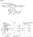
- the present inventors generated a repaired version of human KIR3DX1 by first identifying the seven deleted nucleic acids in exon 5 of the human KIR3DX1 gene. To do this, they compared currently available primate genome sequences and surprisingly discovered that the deleted sequence is highly conserved in non-human primates ( Fig. 1 ).
- KIR3DX1-repaired or simply as KIR3DX1rep the full-length human KIR3DX1 receptor expressed upon re-introduction of the 7-nucleotide deletion into the wild type KIR3DX1 gene.
- KIR3DX1rep can specifically bind to KIR3DL2.
- Fine-mapping of the interaction site and mutation studies of KIR3DL2 revealed this specific binding to depend on the D2 domain of repaired KIR3DX1, i.e. the particular domain that is affected by the 7bp deletion in wild type KIR3DX1.
- the binding interaction of repaired KIR3DX1 to KIR3DL2 involves a glycine residue at position 56 of the D0 domain of KIR3DL2 and further depends on the ability of KIR3DL2 to undergo homodimerization.
- KIR3DX1rep the D2 domain of repaired human KIR3DX1
- the inventors considered its therapeutically use as a competitive, i.e., an antagonistic inhibitor of KIR3DL2.
- polypeptide of the present invention corresponds to the D2 domain encoded by the repaired human KIR3DX1 gene, as described herein.
- the polypeptide thus preferably comprises an amino acid sequence of SEQ ID NO: 1.
- the polypeptide of the invention may also consist of an amino acid sequence of SEQ ID NO: 1.
- the amino acid sequence of the polypeptide may also comprise mutations, such as 1 to 15, e.g., 1, 2, 3, 4, 5, 6, 7, 8, 9, 10, 11, 12, 13, 14, or 15 amino acid substitutions as compared to SEQ ID NO: 1. Accordingly, the polypeptide of the invention may therefore comprise or consist of an amino acid sequence having at least 85%, at least 86%, at least 87%, at least 88%, at least 89%, at least 90%, at least 91%, at least 92%, at least 93%, at least 94%, at least 95%, at least 96%, at least 97%, at least 98% or at least 99% sequence identity to SEQ ID NO: 1, wherein, however, the amino acid sequence of the polypeptide according to the invention does not correspond to the amino acid sequence of a non-human primate KIR3DX1 D2 domain or a bovine KIR3DX1 D2 domain.
- polypeptide of the invention comprising or consisting of an amino acid having at least 85% sequence identity to SEQ ID NO: 1 does not comprise the amino acid sequence of chimpanzee, gorilla, orangutan, bonobo or siamang KIR3DX1 D2 corresponding to SEQ ID NO: 15, 16, 17, 36 and 60, respectively.
- the polypeptide comprising or consisting of an amino acid sequence having at least 85% sequence identity to SEQ ID NO: 1 as described herein is capable of binding to human KIR3DL2.
- the ability of the polypeptide to interact and bind to KIR3DL2 can be easily verified using standard in vitro protein-protein interaction assays such as e.g., co-immunoprecipitation followed by western blotting.
- an assay may comprise steps of contacting a polypeptide of the invention with purified KIR3DL2 or a cell expressing KIR3DL2 on its surface.
- the KIR3DL2 may then be isolated using a specific antibody and binding of the polypeptide of the invention can be detected by western blotting.
- the different proteins that are to be tested for their binding are modified to carry distinct protein tags such as HA-tags, V5-tags or a FLAG-tags so that conventional antibodies against these tags can be used for isolation and detection.
- the polypeptide of the invention may also be fused to the Fc portion of IgG, as described in detail below to facilitate its purification and detection.
- Alternative methods for studying protein-protein interactions are well-known to the skilled person and include, e.g., pull-down assays.
- This part of D2 consists of a stretch of 13 amino acids having the consensus sequence EWSX 1 X 2 SDPLX 3 LYX 4 (SEQ ID NO: 2), wherein X 1 , X 3 and X 4 can be any naturally occurring amino acid, and X 2 is S or P, preferably P.
- X 1 preferably is A.
- X 3 optionally is Q or H, preferably Q.
- X 4 optionally is T, A or V, preferably T.
- polypeptide according to the invention preferably comprises or consists of an amino acid sequence having at least 85% sequence identity to SEQ ID NO: 1 that does not comprise the amino acid sequence of chimpanzee, gorilla, orangutan, bonobo or siamang KIR3DX1 D2, while comprising amino acid sequence SEQ ID NO: 2.
- the polypeptide according to the invention comprises or consists of an amino acid sequence having at least 85% sequence identity to SEQ ID NO: 1 while comprising the amino acid sequence EWSAPSDPLQLYT (SEQ ID NO: 3, reconstructed human D2).
- the polypeptide according to the invention comprises or consists of an amino acid sequence having at least 85% sequence identity to SEQ ID NO: 1 while comprising the amino acid sequence EWSAPSDPLQLYA (SEQ ID NO: 4).
- SEQ ID NO: 4 is present in the chimpanzee, gorilla and bonobo D2 domain.
- polypeptide according to the invention comprises or consists of an amino acid sequence having at least 85% sequence identity to SEQ ID NO: 1 while comprising the amino acid sequence EWSAPSDPLHLYV (SEQ ID NO: 39).
- SEQ ID NO: 39 is present in the orangutan and siamang D2 domain.
- the polypeptide of the invention preferably comprises or consist of an amino acid sequence having at least 85% sequence identity to SEQ ID NO: 1 while comprising at least the consensus sequence WSX 1 X 2 S, wherein X 1 is any natural amino acid, preferably, A, and X 2 is S or P, preferably P.
- this shorter consensus sequence is suspected to form a beta bulge that may be required for mediating the interaction between the D2rep domain and the D0 domain of KIR3DL2.
- the polypeptide of the invention further comprises a first cysteine residue at amino acid position 26 and a second cysteine residue at amino acid position 77, wherein these "positions" refer to the respective location within the amino acid sequence of SEQ ID NO: 1 and are thus determined in relation to the D2 domain portion of the inventive polypeptide only.
- cysteine residues are frequently found in members of the Ig superfamily, where disulfide bridges contribute to the typical immunoglobulin-like fold.
- a polypeptide capable of binding to human KIR3DL2 consisting of an amino acid sequence having at least 85% sequence identity to SEQ ID NO: 1 and preferably comprising at least the consensus sequence WSX 1 X 2 S as described above, and which does not comprise the amino acid sequence of chimpanzee, gorilla, orangutan, bonobo or siamang KIR3DX1 D2, is referred to as D2rep.
- D2rep comprise the beta bulge motif WSAPS as present, e.g., in chimpanzee, bonobo, gorilla, orangutan, siamang as well as the reconstructed human sequence.
- D2rep also comprises SEQ ID NO: 2, e.g., SEQ ID NO: 3, SEQ ID NO: 4 or SEQ ID NO: 39, preferably, SEQ ID NO: 3.
- a polypeptide comprising D2rep was found to be sufficient to bind KIR3DL2. Accordingly, as described in more detail below, a polypeptide comprising or consisting of D2rep may be used as an inhibitor to interfere with the interaction of KIR3DL2 with its ligands. For instance, by binding to KIR3DL2 on CD4 + Th cells, the polypeptide according to the invention can interfere with the interaction of KIR3DL2 and HLA-B*27, and thus prevent the differentiation of the KIR3DL2-expressing CD4 + T cells into pro-inflammatory Th17 cells.
- the polypeptide according to the invention may also interfere with the interaction of KIR3DL2 and bacterial DNA, thereby preventing the loading of the bacterial DNA on TLR9 and consequently reducing the TLR9-mediated inflammatory response mediated by KIR3DL2-positive cells. Preventing or at least reducing CpG-mediated inflammation may be particularly useful in bacterial infection-related clinical conditions characterized by exaggerated immune responses, such as sepsis or bacterial and viral dual infections (so-called superinfections).
- the peptide according to the invention may also be used for scientific or diagnostic purposes, e.g., for labelling cells that express KIR3DL2 on their surface or in ELISA-based diagnostic assays for the detection of soluble KIR3DL2 in patient samples.
- polypeptide according to the invention may further comprise a D0 domain of KIR3DX1 consisting of an amino acid sequence having at least 70%, e.g., at least 75%, at least 80%, at least 85%, at least 90% or at least 95% sequence identity to SEQ ID NO: 5.
- the polypeptide may also comprise a KIR3DX1 D0 domain consisting of an amino acid sequence having 100% sequence identity to SEQ ID NO: 5.
- polypeptide according to the invention comprising D2rep may further comprise a D1 domain of KIR3DX1 consisting of an amino acid sequence having at least 70%, e.g., at least 75%, at least 80%, at least 85%, at least 90% or at least 95% sequence identity to SEQ ID NO: 6.
- the polypeptide may also comprise a KIR3DX1 D1 domain consisting of an amino acid sequence having 100% sequence identity to SEQ ID NO: 6.
- the polypeptide according to the invention comprises D0, D1 and D2rep domains, or the entire extracellular region of KIR3DX1rep. It may thus comprise D2rep and both the KIR3DX1 D0 domain consisting of an amino acid sequence having at least 70% of SEQ ID NO: 5 and the KIR3DX1 D1 domain consisting of an amino acid sequence having at least 70% of SEQ ID NO: 6.
- the polypeptide of the invention thus comprises all three extracellular immunoglobulin domains D0, D1 and D2 of KIR3DX1 and may therefore comprise an amino acid sequence of SEQ ID NO: 7.
- the polypeptide of the invention may also consist of SEQ ID NO: 7.
- it further comprises the stem region of KIR3DX1, e.g., an ape KIR3DX1, such as chimpanzee, bonobo, orangutan, siamang or gorilla.
- Shorter polypeptides e.g., D2rep only, may be easier and cheaper to produce than longer proteins.
- longer polypeptide versions may have a better functionality in some aspects, e.g., in fusion proteins. They may also result in better detection of KIR3DL2, e.g., the presence of the D0 and/or D1 domain in addition to D2rep may increase the binding strength of the polypeptide according to the invention to KIR3DL2.
- polypeptide of the invention comprises only D2rep or additionally also the D0 and/or the D1 domain of KIR3DX1 as described herein, in some embodiments, it may be provided in the form of a fusion protein.
- polypeptide of the invention is fused to the Fc (fragment crystallizable) region of an immunoglobulin.
- the Fc region constitutes the tail region of an immunoglobulin or antibody.
- the Fc region is composed of two identical protein fragments, derived from the second and third constant domains of the antibody's two heavy chains.
- Fc receptors located on the surface of particular immune effector cells to activate the immune system.
- effector cells such as NK cells can recognize the Fc region of cell-bound IgG via their Fcy receptor CD16, which results in the NK cell's degranulation and subsequent release of cytotoxic factors to kill the targeted cell in a process known as antibody-dependent cellular toxicity (ADCC).
- ADCC antibody-dependent cellular toxicity
- CD16 is also expressed on subsets of macrophages and monocytes, which can thus also initiate ADCC and cytokine release upon FC recognition.
- a polypeptide of the invention fused to an antibody Fc region may be used to specifically induce the killing of KIR3DL2-expressing cells via ADCC.
- the polypeptide of the invention is a fusion protein that comprises at least D2rep fused to an immunoglobulin Fc region capable of inducing ADCC when being recognized by a natural killer (NK) cell or macrophage/monocyte.
- the polypeptide of the invention comprises an IgG-Fc region, most preferably the Fc region of IgG subclass IgG1 as this subclass exhibits the highest Fcy receptor-binding affinity and thus is the most potent inducer of ADCC.
- the polypeptide of the invention is a fusion protein that comprises at least D2rep and is fused to an IgG1-Fc region, wherein the IgG1 Fc region may have an amino acid sequence of SEQ ID NO: 8 or at least 90% sequence identity thereto.
- the polypeptide of the invention may however also be fused to another immunoglobulin Fc region capable of inducing ADCC, e.g. an Fc region of IgG3.
- the polypeptide of the invention is a fusion protein comprising an Fc domain of an antibody that does not or only weakly mediates the induction of ADCC.
- IgG subclass IgG4 has weak affinities for most Fc receptors and therefore is a poor inducer of Fc-mediated effector functions (Yu et al., 2020).
- the polypeptide of the invention may, in some embodiments, also comprise the Fc region of IgG4 having an amino acid sequence of SEQ ID NO: 9 or having at least 90% sequence identity thereto.
- fusion-protein could be used for example for research and diagnostic purposes, e.g., for labeling cells expressing KIR3DL2 on their surface or in ELISA-based diagnostic assays.
- a polypeptide according to the invention comprising an Fc region that does not induce ADCC may also be used as an antagonistic inhibitor that is able to interfere with the interaction of KIR3DL2 with its ligands, as described herein.
- the human D2rep protein as described herein exhibits high sequence similarity to the wild-type D2 domains of chimpanzee, gorilla, orangutan, bonobo and siamang KIR3DX1.
- the reconstructed 13-amino acid region of D2rep required for interaction with human KIR3DL2 which is normally absent in the wild-type human D2 domain due to the deletion in exon 5 of the human KIR3DX1 gene and the ensuing frame-shift shares high sequence similarity with the corresponding region in chimpanzee, gorilla, orangutan, bonobo and siamang D2.
- the KIR3DX1 D2 domain of these five primate species is able to bind to human KIR3DL2.
- the KIR3DX1 D2 domain of less related species such as rhesus monkey does not bind to human KIR3DL.
- fusion protein comprising at least the D2 domain of chimpanzee, gorilla, orangutan, bonobo or siamang KIR3DX1 fused to an Fc region of an immunoglobin, preferably any Fc region capable of inducing ADCC.
- the fusion protein may comprise the D2 domain of chimpanzee KIR3DX1 corresponding to SEQ ID NO: 15 fused to an IgG1-Fc region, e.g., having an amino acid sequence of SEQ ID NO: 8.
- Said fusion protein is able to induce ADCC of a cell expressing human KIR3DL2.
- the fusion protein may in addition comprise the D0 and/or D1 domain of human or chimpanzee KIR3DX1, e.g.
- chimpanzee KIR3DX1 may be a polypeptide comprising the D0, D1 and D2 domain of chimpanzee KIR3DX1 according to SEQ ID NO: 18 fused to an IgG1-Fc region, e.g., having an amino acid sequence of SEQ ID NO: 8.
- the fusion protein comprising at least the D2 domain of chimpanzee KIR3DX1 may instead also comprise an Fc domain that does not significantly induce ADCC such as, e.g., an IgG4 Fc domain.
- the fusion protein may comprise the D2 domain of gorilla KIR3DX1 corresponding to SEQ ID NO: 16 fused to an IgG1-Fc region, e.g., having an amino acid sequence of SEQ ID NO: 8.
- Said fusion protein is also able to induce ADCC of a cell expressing human KIR3DL2.
- the fusion protein may in addition comprise the D0 and/or D1 domain of human or gorilla KIR3DX1, e.g. it may be a polypeptide comprising the D0, D1 and D2 domain of gorilla KIR3DX1 according to SEQ ID NO: 19 fused to an IgG1-Fc region, e.g., having an amino acid sequence of SEQ ID NO: 8.
- the fusion protein comprising at least the D2 domain of gorilla KIR3DX1 may instead also comprise an Fc domain that does not significantly induce ADCC such as, e.g., an IgG4 Fc domain.
- the fusion protein may comprise the D2 domain of orangutan KIR3DX1 corresponding to SEQ ID NO: 17 fused to an IgG1-Fc region, e.g., having an amino acid sequence of SEQ ID NO: 8. Said fusion protein is also able to induce ADCC of a cell expressing human KIR3DL2.
- the fusion protein may also comprise the D0 and/or D1 domain of human or orangutan KIR3DX1, e.g.
- polypeptide comprising the D0, D1 and D2 domain of orangutan KIR3DX1 according to SEQ ID NO: 20 fused to an IgG1-Fc region, e.g., having an amino acid sequence of SEQ ID NO: 8.
- the fusion protein comprising at least the D2 domain of orangutan KIR3DX1 may instead also comprise an Fc domain that does not significantly induce ADCC such as, e.g., an IgG4 Fc domain.
- the fusion protein may comprise the D2 domain of bonobo KIR3DX1 corresponding to SEQ ID NO: 36 fused to an IgG1-Fc region, e.g., having an amino acid sequence of SEQ ID NO: 8. Said fusion protein is able to induce ADCC of a cell expressing human KIR3DL2.
- the fusion protein may in addition comprise the D0 and/or D1 domain of human or bonobo KIR3DX1, e.g.
- the fusion protein comprising at least the D2 domain of bonobo KIR3DX1 may instead also comprise an Fc domain that does not significantly induce ADCC such as, e.g., an IgG4 Fc domain.
- the fusion protein may comprise the D2 domain of siamang KIR3DX1 corresponding to SEQ ID NO: 60 fused to an IgG1-Fc region, e.g., having an amino acid sequence of SEQ ID NO: 8.
- Said fusion protein is able to induce ADCC of a cell expressing human KIR3DL2.
- the fusion protein may in addition comprise the D0 and/or D1 domain of human or siamang KIR3DX1, e.g. it may be a polypeptide comprising the D0, D1 and D2 domain of siamang KIR3DX1 according to SEQ ID NO: 61 fused to an IgG1-Fc region, e.g., having an amino acid sequence of SEQ ID NO: 8.
- the fusion protein comprising at least the D2 domain of siamang KIR3DX1 may instead also comprise an Fc domain that does not significantly induce ADCC such as, e.g., an IgG4 Fc domain.
- fusion protein also extends to so-called multispecific engagers comprising a polypeptide of the invention, e.g., at least D2rep, or a polypeptide comprising at least the D2 domain of any of chimpanzee, gorilla, orangutan, bonobo or siamang KIR3DX1 as one of their antigen-binding domains.
- multispecific engager relates to an artificial protein that typically is constructed from the variable regions of at least two antigen-binding moieties, e.g., ligands and/or antibodies of different specificity.
- the variable region of an antibody is formed by a heavy (VH) and light (VL) chain and forms the antibody's antigen binding site. Accordingly, a multispecific engager can simultaneously bind to at least two different types of antigen or two different epitopes on the same antigen. These constructs can thus be used, e.g., to bring different cells into direct contact.
- the present invention thus further provides a fusion protein in which a polypeptide according to the invention is linked to at least one additional antigen-binding moiety to form a multispecific engager.
- the additional antigen-binding moiety may be, e.g., a variable region of an antibody or a ligand of a receptor of interest.
- the fusion protein comprises D2rep as a first antigen binding moiety linked to the variable region of an antibody capable of binding and engaging CD16 as a second antigen-binding moiety.
- bispecific engager also referred to as bispecific killer engager or BiKe
- BiKe bispecific killer engager
- the polypeptide of the invention e.g., D2rep
- an antigen-binding moiety capable of engaging with the CD3 receptor expressed, e.g., on the surface of cytotoxic T cells.
- a fusion protein constitutes a so-called bispecific T-cell engager (BiTE).
- the polypeptide of the invention may also be linked to more than one additional antigen-binding moiety, e.g., it may be linked to two, three, four, five or more antigen-binding moieties of different specificity.
- the fusion protein may be a trispecific killer engager (TriKe) comprising a polypeptide of the invention, e.g., D2rep, as a first antigen-binding moiety linked to two further antigen-binding moieties capable of engaging the activating receptors NKp46 and CD16 expressed by NK cells.
- TriKe trispecific killer engager
- such a fusion protein may additionally comprise even a fourth antigen-binding moiety (tetra-specific engager) capable of binding, e.g., another tumor-associated antigen besides KIR3DL2 or of engaging with a receptor of another immune effector cell, such as the CD3 receptor or IL-2 receptor expressed by cytotoxic T cells or CD4+ helper T cells.
- a fourth antigen-binding moiety tetra-specific engager capable of binding, e.g., another tumor-associated antigen besides KIR3DL2 or of engaging with a receptor of another immune effector cell, such as the CD3 receptor or IL-2 receptor expressed by cytotoxic T cells or CD4+ helper T cells.
- the polypeptide of the invention and the at least one additional antigen-binding moiety are linked via a suitable polypeptide linker.
- the linker allows separation of the polypeptide of the invention and the antigen-binding moieties and provides flexibility to bind the multiple epitopes on the different targeted cells.
- Suitable flexible linkers are known from the state of the art, e.g., from Felices et al., 2016.
- the multispecific engager as described herein may also comprise the D2 domain and, optionally, the D0 and/or D1 domain of chimpanzee, gorilla, orangutan, bonobo or siamang KIR3DX1 instead of a polypeptide of the invention.
- the polypeptide according to the invention may also be a functional killer cell immunoglobulin-like receptor (KIR). It thus typically does not comprise an Fc domain or additional antigen-binding moieties, but instead the stem region, the transmembrane region and the cytoplasmic region of a KIR family protein.
- KIR functional killer cell immunoglobulin-like receptor
- the KIR according to the invention may additionally also be fused to an Fc region of an antibody, e.g., an IgG1 or an IgG4 Fc region.
- the polypeptide of the invention thus comprises the D2rep domain and, preferably, also the KIR3DX1 D0 and/or D1 domains as described herein, as well as a KIR3DX1 stem region comprising an amino acid sequence having at least 70%, e.g., at least 80%, at least 90%, or 100% sequence identity to SEQ ID NO: 10, a KIR3DX1 transmembrane region comprising an amino acid sequence having at least 70%, e.g., at least 80%, at least 90%, or 100% sequence identity to SEQ ID NO: 11 and a KIR3DX1 cytoplasmic region comprising an amino acid sequence having at least 70%, e.g., at least 75%, at least 80%, at least 85%, at least 90%, at least 95% or 100% sequence identity to SEQ ID NO: 12.
- the D0, D1 and/or D2 domains are human to prevent or at least to minimize an immune response, but the stem region, the transmembrane region and/or the cytoplasmic region may be the reconstructed human sequence or an ape sequence, such as chimpanzee, bonobo, orangutan, siamang or gorilla sequence.
- such a polypeptide comprises all three extracellular domains D0, D1 and D2rep as described herein so that the full-length human KIR3DX1rep receptor is obtained.
- Such a construct may have an amino acid sequence corresponding to SEQ ID NO: 13.
- the KIR3DX1rep receptor may also be a chimeric KIR receptor.
- the receptor comprises at least the D2rep domain of human KIR3DX1 as disclosed herein, any of the D0 domain, the D1 domain, the stem region, the transmembrane region and/or the cytoplasmic region, in particular, the cytoplasmic region of the receptor may be replaced by corresponding domains/regions of one or more other members of the human KIR-family of receptors, such as, e.g., KIR2DL1, KIR2DL2, KIR2DL3, KIR2DL4, KIR2DL5A, KIR2DL5B, KIR2DS1, KIR2DS2, KIR2DS3, KIR2DS5, KIR2DP1, KIR3DP1, KIR3DL1, KIR3DS1, KIR3DL2, or KIR3DL3.
- the polypeptide according to invention may comprise the extracellular KIR3DX1 D2rep domain and the D0 domain, the D1 domain, the stem region, the transmembrane region and/or the cytoplasmic region of KIR3DL2.
- the receptor may also comprise a complete extracellular domain of human KIR3DX1rep comprising KIR3DX1 D0, D1 and D2rep, and optionally the stem region, with the transmembrane region and/or the cytoplasmic region (and, if not yet present, the stem region) from one or more other human KIR family proteins.
- one, two, three, or four of D0, D1, the stem region, the transmembrane region, and the cytoplasmic region are derived from a KIR family member other than KIR3DX1, while the remainder of the receptor is derived from the repaired human KIR3DX1rep.
- KIR3DX1 is considered to be an inhibitory KIR family receptor due to the presence of the immunoreceptor tyrosine-based inhibitory motif (ITIM) in its cytoplasmic region
- at least its transmembrane and cytoplasmic regions may be replaced by the corresponding regions of an activating KIR family member such as, e.g., KIR2DS1, KIR2DL4, and KIR3DS1.
- activating receptors do not comprise an ITIM (with the exception of KIR2L4) and contain a positively charged lysine or arginine residue in their transmembrane domain that helps to bind adapter proteins such as DAP12 or FCER1G, containing a negatively charged residue as well as immunoreceptor tyrosine-based activation motifs (ITAM).
- ITAM immunoreceptor tyrosine-based activation motifs
- the resulting chimeric KIR would thus be able to recognize KIR3DL2 via its extracellular domain comprising D2rep but would in turn activate rather than inhibit the cytotoxic activity of a cell expressing the receptor.
- An exemplary receptor comprising an extracellular domain of human KIR3DX1rep and the transmembrane as well as cytoplasmic region of the activating human KIR family receptor KIR3DS1 has an amino acid sequence of SEQ ID NO: 31.
- At least the D2rep domain of human KIR3DX1 may also be combined with any or all of the D0 domain, D1 domain, stem region, transmembrane region, and cytoplasmic region derived from a non-human KIR family protein, i.e., a non-human primate KIR family protein or a bovid KIR family protein.
- the present invention also provides a chimeric antigen receptor (CAR) comprising a polypeptide of the invention as an antigen recognition domain.
- CAR chimeric antigen receptor
- the polypeptide according to the invention comprising D2rep may form the antigen recognition domain of a CAR construct.
- chimeric antigen receptor or CAR refers to an artificial chimeric protein comprising an extracellular antigen-binding/recognition domain that in other cases typically is a single chain construct derived from an antibody, a transmembrane domain, an intracellular signalling domain and additional costimulatory domains from receptors such as CD28, OX40, and CD137. Binding of an antigen to the antigen-binding domain causes the clustering of CARs and the subsequent transmission of an activation signal. CARs are typically used to modify the antigen specificity of T cells or NK cells to target cancer cells in immunotherapy.
- the present invention provides a CAR comprising at least the KIR3DX1 D2rep domain, optionally, the KIR3DX1 D2rep domain as well as the KIR3XD1 D0 and D1 domains as described herein as an antigen-binding domain.
- the CAR comprises the entire extracellular portion of KIR3DX1rep.
- the CAR comprises the KIR3DX1 D2rep domain without the KIR3XD1 D0 and D1 domains.
- the CAR may optionally comprise a hinge or spacer domain located between the antigen recognition domain and the transmembrane domain to provide more flexibility and accessibility for the antigen-binding domain.
- the hinge region may, e.g., be an Fc region of an antibody such as IgG or an immunoglobulin-like domain or hinge domain derived from the extracellular portion of, e.g., CD8 or CD28. It may also be a stem region of a KIR, e.g., of KIR3DX1.
- transmembrane domain of the CAR anchors the CAR to the plasma membrane of a cell and ensures the stability of the receptor as a whole.
- Suitable transmembrane domains that are conventionally used in CAR constructs may be derived from, e.g., CD28, ICOS, CD4, CD8 or CD3 ⁇ .
- the CAR preferably comprises an intracellular signalling domain derived from the cytoplasmic domain of CD3 ⁇ .
- the CAR may be a first-generation CAR comprising only this CD3 ⁇ intracellular signalling domain.
- the CAR preferably is a second or third generation CAR, i.e., the intracellular domain preferably comprises at least one additional co-stimulatory domain.
- the at least one co-stimulatory domain may be derived from, e.g., CD28, 4-1 BB (also known as CD137), or OX40.
- the CAR comprises the D0, D1 and D2 domains of KIR3DX1rep as an antigen recognition domain, the stem of KI R3DX 1 rep as a hinge region, a transmembrane and cytoplasmic region of CD28 and a CD3 ⁇ intracellular signaling domain.
- the preferred CAR of the invention thus may have an amino acid sequence corresponding to SEQ ID NO: 14.
- the CAR according to the invention may also comprise an antigen recognition domain comprising at least the wild type D2 domain of chimpanzee, gorilla, orangutan, bonobo or siamang KIR3DX1.
- the herein disclosed CAR may also comprise an antigen-recognition domain comprising an amino acid sequence of SEQ ID NO: 15, 16, 17, 36 or 60.
- the CAR comprises the entire extracellular domain of chimpanzee, gorilla, orangutan, bonobo or siamang KIR3DX1 as an antigen-recognition domain, i.e.
- the antigen-recognition domain of the CAR comprises the D0, D1 and D2 domain of wild type chimpanzee, gorilla, orangutan, bonobo or siamang KIR3DX1 and thus any of the amino acid sequences of SEQ ID NO: 18, 19, 20, 37 or 61.
- it also comprises the stem region of one of these species, preferably, from the same species.
- the present invention further provides a nucleic acid encoding any of the polypeptides, fusion proteins and receptors disclosed herein.
- the nucleic acid can either be DNA or RNA. Preferably, it is DNA.
- the nucleic acid encoding the polypeptide of the invention includes an additional number of nucleotides that repairs the frameshift that occurred in the human gene compared to the other primate species. Accordingly, compared to the human wildtype sequence, it may include additional seven, four or one nucleotides, preferably, at the position where the sequence was deleted, as shown in Fig. 1 and discussed below.
- the nucleic acid preferably comprises the seven nucleotides that are deleted in the human wild type KIR3DX1 gene but are highly conserved in non-human primates.
- the exact nucleic acid sequence deleted in human KIR3DX1 depends on the precise position within exon 5 at which the start and end of the deletion is assumed to occur.
- human KIR3DX1 comprises an adenine adjacent to the deletion within exon 5 that is not found in other primate species.
- the nucleic acid encoding the polypeptides, fusion proteins and receptors disclosed herein comprises a consensus sequence that is either GAGTGGT or AGTGGTC or a nucleotide sequence which is complementary or which hybridizes to any of said consensus sequences.
- the nucleic acid sequence comprises the consensus sequence GAGTGGT.
- nucleic acid may be codon-optimized for expression in a human cell.
- the nucleic acid may encode a polypeptide of SEQ ID NO: 1 and thus, optionally, comprise a nucleic acid sequence corresponding to SEQ ID NO: 21.
- the nucleic acid encodes a polypeptide comprising both D2rep (SEQ ID NO: 1) and the KIR3DX1 D0 domain (SEQ ID NO: 5).
- Such nucleic acid may, e.g., comprise a nucleic acid sequence of SEQ ID NO: 22. It may also encode a polypeptide comprising KIR3DX1 D2rep (SEQ ID NO: 1) and D1 (SEQ ID NO: 6). Said nucleic acid may, e.g., comprise a nucleic acid sequence of SEQ ID NO: 23.
- the nucleic acid encodes the entire extracellular domain of KIR3DX1rep comprising all three of D0, D1 and D2rep (corresponding to an amino acid sequence of SEQ ID NO: 7).
- the nucleic acid may thus also comprise a nucleic acid sequence of SEQ ID NO: 24.
- the nucleic acid may also further encode any of the herein disclosed fusion proteins, i.e., a polypeptide according to the invention or a polypeptide comprising any of SEQ ID NO: 15, 16, 17, 36 or 60 fused to an immunoglobulin Fc domain, e.g., an IgG Fc domain or linked to at least one additional antigen-binding moiety capable of binding, e.g., to CD16 or CD3. It may, e.g., encode any of the above-mentioned fusion proteins comprising an IgG1 Fc domain to obtain a polypeptide capable of inducing ADCC of KIR3DL2-expressing cells.
- fusion proteins i.e., a polypeptide according to the invention or a polypeptide comprising any of SEQ ID NO: 15, 16, 17, 36 or 60 fused to an immunoglobulin Fc domain, e.g., an IgG Fc domain or linked to at least one additional antigen-binding moiety capable of binding, e.g.
- Such a nucleic acid may therefore comprise a nucleic acid sequence of, e.g., any of SEQ ID NO: 25 (D2rep-Fc), 26 (D2rep+D0-Fc), 27 (D2rep+D1-Fc) or 28 (D2rep-D0+D1-Fc).
- a nucleic acid according to the invention may also encode any of the herein disclosed KIR family receptor proteins comprising D2rep as part of their extracellular domains.
- the nucleic acid encodes a full-length KIR3DX1rep-receptor according to SEQ ID NO: 13 and may thus comprise a nucleic acid sequence of SEQ ID NO: 29. It may also encode a chimeric KIR receptor according to SEQ ID NO: 31, e.g., comprising a nucleic acid sequence of SEQ ID NO: 32.
- nucleic acid encoding a CAR construct as described herein.
- the nucleic acid may thus encode the herein described preferred CAR according to SEQ ID NO: 14, e.g., by comprising a nucleic acid sequence corresponding to SEQ ID NO: 30.
- the nucleic acid disclosed herein is preferably provided as an expression vector suitable for being expressed in a human host cell, in particular, a human lymphoid host cell such as an NK cell, an NKT cell, a T cell or an innate lymphoid cell (ILC) of the ILC1, ILC2, or ILC3 type.
- a human lymphoid host cell such as an NK cell, an NKT cell, a T cell or an innate lymphoid cell (ILC) of the ILC1, ILC2, or ILC3 type.
- the vector may, e.g., be a viral vector, or a non-viral vector, e.g., a transposon, a vector suitable for CRISPR/CAS based recombination or a plasmid suitable for in vitro RNA transcription.
- the vector consists of DNA, it preferably further comprises at least a regulatory region of a gene.
- the regulatory region may be a transcriptional regulatory region, e.g., selected from the group consisting of a promoter, an enhancer, a silencer, a locus control region, and a border element.
- Promoters or other expression control regions can be operably linked with the nucleic acid encoding the inventive polypeptide, fusion protein or receptor or to a nucleotide sequence which is complementary to or which hybridizes to said nucleotide sequence, e.g., to regulate expression of the polypeptide/fusion protein/receptor in a quantitative or in a tissue-specific manner.
- the promotor can be a native or, preferably, a heterologous promoter.
- the selection of promoters includes, e.g., strong, weak, inducible, tissue-specific and developmental-specific promoters.
- the promoter can be a non-viral promoter or a viral promoter.
- the inventive recombinant expression vectors can be designed for either transient expression, for stable expression, or for both. Also, the recombinant expression vectors can be made for constitutive expression or for inducible expression.
- the vector comprising the nucleic acid of the invention may include one or more marker genes, which allows for selection or detection (e.g. via a marker gene expressing GFP) of transduced or transfected hosts.
- the present invention further provides a cell comprising a nucleic acid according to the invention.
- the cell expresses any of the inventive receptor constructs disclosed herein, e.g., a full-length KIR3DX1rep-receptor or any of the chimeric KIR or CAR constructs disclosed herein from a nucleic acid according to the invention.
- a human cell that expresses a full-length KIR3DX1 receptor of chimpanzee (SEQ ID NO: 33), gorilla (SEQ ID NO: 34), orangutan (SEQ ID NO: 35), bonobo (SEQ ID NO: 38) or siamang (SEQ ID NO: 62).
- the cell preferably is a lymphoid cell, in particular a human lymphoid cell. It may be, e.g., a natural killer (NK) cell, a CD8 + cytotoxic T cell, a natural killer T cell (NKT cell) or a precursor thereof.
- NK cells are cells of the innate immune system that can recognize and rapidly kill target cells, in particular malignant and virus-infected cells even in the absence of antibodies and MHC class I proteins.
- NK cells are characterized by the expression of neural cell adhesion molecule (NCAM or CD56) and many express the Fc ⁇ RIII receptor CD16.
- Cytotoxic T cells normally express T cell receptors that recognize a specific antigen presented by class I MHC proteins on the surface of a target cell upon which the T cell becomes activated and kills the antigen presenting cell. Cytotoxic T cells are characterized by the expression of the glycoprotein CD8, which assists in the interaction of the TCR with MHC class I. NKT cells share properties of both NK cells and cytotoxic T cells and can both kill target cells and release different cytokines upon activation. The cell may also be a CD4+ T helper cell, which may secrete cytokines that may sustain the immune response. It may also be a B cell, e.g., a B cell secreting an Fc fusion protein of the invention.
- ILC Innate Lymphoid Cell
- ILCs Innate lymphoid cells
- CLPs common lymphoid progenitors
- ILCs In response to pathogenic tissue damage, ILCs contribute to immunity via the secretion of signalling molecules, and the regulation of both innate and adaptive immune cells (https://en.wikipedia.org/wiki/Innate_lymphoid cell).
- ILCs are divided into five distinct groups: NK cells, lymphoid tissue inducer (LTi) cells as well as ILC of groups 1-3.
- the obtained cell is a human lymphoid cell comprising a nucleic acid of the invention, and it is able to express any of the receptors disclosed herein on its surface.
- the cell is able to target KIR3DL2-presenting cells and consequentially to induce their specific killing by exerting cytotoxic activity or to release cytokines that may either promote or decrease activities of other immune cells. They may thus be utilized for immunotherapies against KIR3DL2-associated diseases as described in more detail below.
- the cell is a human cell.
- the cell is a cell isolated from a human, e.g., a human patient, in particular, a patient who is to be treated with the cell once it has been modified to express a receptor of the invention.
- the cell may be from a third-party donor who may be related or unrelated to the patient.
- Cells may be genetically engineered, e.g., in multiple ways. For example, in case the cell is a T cell, the cell may be genetically modified by knockout of the endogenous TCR and/or, in particular, if the cell is a heterologous cell, MHC class I.
- the lymphoid cells may also be generated from autologous or third-party donor stem cells, e.g., iPSC.
- the stem cells are not human embryonic stem cells.
- the cell may also be derived from a suitable cell line such as NK cell lines NK-92, NKL, YT, NK3.3, or from a T cell line such as Jurkat.
- the present invention further provides a pharmaceutical composition comprising any of the herein disclosed (fusion-)polypeptides, nucleic acids or cells as well as mixtures thereof.
- the pharmaceutical composition may comprise D2rep.
- the pharmaceutical composition may also comprise a polypeptide comprising D2rep and a D0 domain of human KIR3DX1 comprising or consisting of an amino acid sequence having at least 70% of SEQ ID NO: 5 and/or a D1 domain of KIR3DX1 comprising or consisting of an amino acid sequence having at least 70%, of SEQ ID NO: 6.
- the pharmaceutical composition may alternatively comprise a polypeptide comprising an amino acid sequence of to any of SEQ ID NO: 15, 16, 17, 36 or 60, respectively.
- the pharmaceutical composition comprises any of the aforementioned fusion proteins comprising at least D2rep or a D2 domain according to SEQ ID NO: 15, 16, 17, 36 or 60 fused to an Fc domain of an immunoglobulin, e.g., an IgG1 or an IgG4 Fc domain, or linked to at least one additional antigen-binding moiety, as described herein.
- the present invention provides a pharmaceutical composition comprising a polypeptide as disclosed herein capable of inducing ADCC in a KIR3DL2-expressing cell.
- the pharmaceutical composition may also comprise a nucleic acid that, when being expressed in a cell of a subject, gives rise to any of the herein disclosed polypeptides of the invention.
- the pharmaceutical composition may comprise an expression vector as described herein such as, e.g., a viral vector directed to a target cell that, when being expressed by said targeted cell, results in the generation of a polypeptide of the invention that may be secreted.
- the pharmaceutical composition may comprise a cell as disclosed herein, e.g., a cytotoxic lymphocyte such as an NK cell or a cytotoxic T cell expressing any of the KIR-family or CAR receptors of the present invention.
- a cytotoxic lymphocyte such as an NK cell or a cytotoxic T cell expressing any of the KIR-family or CAR receptors of the present invention.
- the pharmaceutical composition may comprise an NK cell, an NKT cell or cytotoxic T cell expressing a CAR of SEQ ID NO: 14, a full-length human KIR3DX1rep receptor corresponding to SEQ ID NO: 13 or a chimeric KIR receptor of SEQ ID NO: 31 from a corresponding nucleic acid of the present invention.
- the pharmaceutical composition may also comprise a cell, e.g., a lymphoid cell such as a natural killer (NK) cell, a CD8 + cytotoxic T cell, an NKT cell or a precursor thereof expressing a KIR3DX1 receptor of chimpanzee (SEQ ID NO: 33), gorilla (SEQ ID NO: 34), orangutan (SEQ ID NO: 35), bonobo (SEQ ID NO: 38) or siamang (SEQ ID NO: 62).
- a lymphoid cell such as a natural killer (NK) cell, a CD8 + cytotoxic T cell, an NKT cell or a precursor thereof expressing a KIR3DX1 receptor of chimpanzee (SEQ ID NO: 33), gorilla (SEQ ID NO: 34), orangutan (SEQ ID NO: 35), bonobo (SEQ ID NO: 38) or siamang (SEQ ID NO: 62).
- the pharmaceutical composition according to the invention may comprise additional ingredients such as, e.g., a pharmaceutically acceptable excipient.
- pharmaceutically acceptable refers those compounds, materials, compositions, and/or dosage forms that are, within the scope of sound medical judgement, suitable for use in contact with the tissues of the subject without causing excessive toxicity, irritation, allergic response or other problems or complications commensurate with a reasonable benefit/risk ratio.
- the pharmaceutical composition further comprises a suitable carrier, i.e., a compound, composition, structure or substance that, when combined with the polypeptides or cells described herein, aids or facilitates preparation, storage, administration, delivery, effectiveness, selectivity, or any other feature of the peptides or cells for its intended use or purpose.
- a suitable carrier i.e., a compound, composition, structure or substance that, when combined with the polypeptides or cells described herein, aids or facilitates preparation, storage, administration, delivery, effectiveness, selectivity, or any other feature of the peptides or cells for its intended use or purpose.
- a suitable carrier i.e., a compound, composition, structure or substance that, when combined with the polypeptides or cells described herein, aids or facilitates preparation, storage, administration, delivery, effectiveness, selectivity, or any other feature of the peptides or cells for its intended use or purpose.
- It may, e.g., comprise water, a saline, e.g., physiological
- the pharmaceutical composition may also comprise additional therapeutically active ingredients or agents that may be used to fight a disease of interest.
- the pharmaceutical composition may comprise, e.g., an anticancer drug such as a chemotherapeutic drug.
- an anticancer drug such as a chemotherapeutic drug.
- the present invention also covers pharmaceutical compositions in which a polypeptide, fusion protein or receptor described herein serves as a targeting moiety to target another therapeutically active agent, e.g., an anti-cancer drug, to a cell or a tissue expressing KIR3DL2.
- the other therapeutically active agent may be encapsulated as a cargo in a suitable vector, such as, e.g., a lipid vesicle, a lipid nanoparticle, a bacterial minicell or a polymeric micro- or nanoparticle, wherein the vector carries a polypeptide, fusion protein or receptor according to the invention on its surface or is linked to it.
- the polypeptide, fusion protein or receptor may then recruit the vector and thus the additional therapeutically active agent to a target cell or tissue where the agent is released to exert its therapeutic effect.
- the present invention is further directed to uses of the herein disclosed polypeptides, fusion proteins, cells and pharmaceutical compositions in methods of treating a disease or condition associated with KIR3DL2 expression.
- treating a disease or condition is to be understood to comprise a curative medical therapy of a subject with the intent to cure, ameliorate or stabilize a condition.
- condition refers to a disease, a syndrome, a disorder or a particular physiological state of an organism that is either manifested by distinct symptoms or can be diagnosed by using established markers capable of recognizing said state.
- treating a disease or condition is intended to mean that the progression of the disease or condition is to be slowed, stopped or, preferably, reversed to allow for a perceivable improvement of the subject's well-being and health.
- the subject that is to be treated preferably is a human.
- the herein disclosed polypeptides, fusion proteins, cells or pharmaceutical compositions may be used in a method of treating of a rheumatic disease in a subject, in particular a rheumatic disease associated with HLA-B*27.
- the present invention thus also provides a method of treating a rheumatic disease in a subject, which comprises a step of administering any of the polypeptides, fusion proteins, cells or pharmaceutical compositions to a subject in need thereof.
- a rheumatic disease is a condition associated with inflammation and chronic, often intermittent pain affecting the joints, tendons, muscles, ligaments and/or bones.
- Arthritis is a common form of rheumatic disease affecting the joints.
- the rheumatic disease that is to be treated using any of the herein disclosed polypeptides, cells or pharmaceutical compositions is associated with HLA-B*27. It may be a form of arthritis, most preferably a type of spondyloarthritis (SpA), such as ankylosing spondylitis (AS).
- SpA spondyloarthritis
- AS ankylosing spondylitis
- SpA spondyloarthrides
- AS also known under the names Bechterew's disease or Morbus Bechterew, is the most common form of SpA and refers to an autoimmune disease affecting the joints of the spine in particular where the spine joins the pelvis. 94% of AS patients are HLA-B*27 positive (Bowness et al., 2011).
- the molecular mechanism of AS development is thought to involve two steps in HLA-B*27-positive patients: the first step is the induced expression of KIR3DL2 on activated CD4 + T helper cells (Th cells), and the second step is the molecular interaction of KIR3DL2 with HLA-B*27, which licenses CD4 + Th cells to develop into inflammatory Th17 cells expressing increased amounts of the inflammatory cytokine IL-17 and reduced amounts of IL-2 (Chan et al., 2005; Bowness et al., 2011; Ridley et al., 2016; Wong-Baeza et al., 2013).
- the present inventors found that repaired human KIR3DX1 can bind to the D0 domain of KIR3DL2 via its D2rep domain. Accordingly, the herein disclosed polypeptides and fusion proteins comprising at least D2rep as defined herein may interfere with the interaction of HLA-B*27 and KIR3DL2 on the surface of CD4 + Th cells and thereby prevent the CD4 + Th cells from developing into Th17 cells.
- polypeptides of the invention are provided in the form of multispecific engagers capable of engaging NK cell or T cell-mediated cytotoxicity, as described herein, or as fusion proteins comprising an immunoglobulin Fc domain capable of inducing ADCC in a target cell, such as, e.g., an IgG1-Fc domain
- the use of these polypeptides can also directly trigger the killing of the CD4+ Th cells expressing KIR3DL2.
- Death of CD4+ Th cells can also be achieved by administering to the subject a cytotoxic lymphocyte expressing a KIR or CAR receptor, as disclosed herein, that recognizes KIR3DL2 on the surface of the CD4 + Th cells.
- the polypeptides, fusion proteins, cells and pharmaceutical compositions according to the invention may also be used to interfere with the interaction of KIR3DL2 and unmethylated CpG oligonucleotides derived from bacterial DNA thereby attenuating the TLR9-mediated inflammatory response of KIR3DL2-positive cells towards microbial infection.
- KIR3DL2 binds to this PAMP via its D0 domain, it is endocytosed and loads the CpG cargo on Toll-like receptor 9 (TLR9), which drives the expression of IFN-y and causes elevated cytotoxicity in the KIR3DL2-expressing cells (Sivori et al., 2010a).
- TLR9 Toll-like receptor 9
- the herein disclosed polypeptides, fusion proteins, cells or pharmaceutical compositions may also be for use in treating or even preventing the excessive TLR9-mediated inflammatory response associated with sepsis or arising in response to superinfections, secondary infections superimposed on an earlier first one, especially by a different microbial agent of exogenous or endogenous origin, that is resistant to the treatment being used against the first infection.
- a polypeptide comprising a sequence according to any of SEQ ID NO: 15, 16, 17, 36 or 60 can also be used to treat any of the herein disclosed rheumatic diseases or the excessive TLR9-mediated inflammatory response associated with sepsis or superinfections.
- another aspect of the invention is the use of the herein disclosed fusion proteins capable of inducing ADCC, cells or pharmaceutical compositions in a method of treating a cancer expressing KIR3DL2, wherein the cancer expressing KIR3DL2 may be, e.g., cutaneous T-cell lymphoma (CTCL), Sézary syndrome, mycosis fungoides, CD30 + cutaneous lymphoma or adult T-cell leukemia (ATL).
- CTCL cutaneous T-cell lymphoma
- Sézary syndrome mycosis fungoides
- ATL adult T-cell leukemia
- the present invention also discloses a method of treating a cancer expressing KIR3DL2, wherein the method comprises administering to a subject in need thereof any of the herein described fusion proteins, cells or pharmaceutical compositions.
- human lymphoid cells such as NK cells, NKT cells or CD8+ cytotoxic lymphocytes expressing the herein disclosed KIR constructs or CAR constructs may be used in immunotherapy to treat T cell lymphomas associated with KIR3DL2 expression.
- KIR3DL2-expressing cancer cells may also be killed by administering any of the herein disclosed ADCC- inducing fusion proteins or pharmaceutical compositions comprising the same.
- Cutaneous T-cell lymphoma refers to a heterogeneous group of non-Hodgkin lymphomas caused by a mutation of T cells.
- the cancerous T cells migrate to the skin, resulting in lesions that initially resemble an itchy rash before transforming into larger plaques and tumors that may ultimately spread to other tissues.
- Sézary syndrome and mycosis fungoides represent the two most common types of CTCL.
- CD30 + cutaneous lymphoma also known as primary cutaneous anaplastic large-cell lymphoma, is another type of CTCL characterized by solitary or localized skin lesions that have a tendency to ulcerate.
- CTCL Additional types of CTCL that may be treated with the herein disclosed polypeptides, cells or pharmaceutical compositions include, e.g., Pagetoid reticulosis, Granulomatous slack skin, Lymphomatoid papulosis, Pityriasis lichenoides chronica, Pityriasis lichenoides et varioliformis acuta, Secondary cutaneous CD30+ large cell lymphoma, Non-mycosis fungoides CD30- cutaneous large T-cell lymphoma, Pleomorphic T-cell lymphoma, Lennert lymphoma, Subcutaneous T-cell lymphoma, Angiocentric lymphoma or Blastic NK-cell lymphoma.
- Pagetoid reticulosis Granulomatous slack skin
- Lymphomatoid papulosis Pityriasis lichenoides chronica
- ATL is a rare yet highly aggressive form of non-Hodgkin lymphoma that is caused by infection with the human T cell leukemia/lymphotropic virus 1 (HTLV-1).
- HTLV-1 human T cell leukemia/lymphotropic virus 1
- the disease is associated with symptoms such as hypercalcemia, skin lesions, and lytic bone lesions.
- KIR3DL2 has further been described in myelodysplastic syndrome, i.e., a group of malignant disorders of hematopoiesis. Accordingly, the herein disclosed fusion proteins, cells or pharmaceutical compositions may also be used for treating myelodysplastic syndrome.
- the present invention relates to different diagnostic methods for detecting KIR3DL2 expression in a subject or a sample isolated from the subject by using a polypeptide or fusion protein as disclosed herein as a capture or detecting agent. This way it is possible to diagnose whether a subject suffers from a disease associated with elevated KIR3DL2 expression, as described herein.
- the methods preferably are in vitro methods and rely on the use of a polypeptide or fusion protein of the present invention that is able to bind and thus detect KIR3DL2 protein in a sample obtained from a subject.
- the method may comprise contacting a sample obtained from a subject with a polypeptide of fusion protein of the invention or a polypeptide comprising an amino acid sequence of any of SEQ ID NO: 15, 16, 17, 36 or 60, wherein the polypeptide or the fusion protein preferably is linked to a label. Binding can then be detected.
- the detection method comprises the step of performing an enzyme-linked immunosorbent assay (ELISA) during which a liquid sample obtained from a subject is brought into contact with a polypeptide or a fusion protein according to the present invention or a polypeptide comprising an amino acid sequence of any of SEQ ID NO: 15, 16, 17, 36 or 60 to detect soluble KIR3DL2.
- ELISA enzyme-linked immunosorbent assay
- the liquid sample is or comprises a body fluid isolated from the subject, e.g., the sample may be or may comprise any body fluid selected from the group comprising blood, lymph, saliva, urine, cerebrospinal fluid, amniotic fluid, ejaculate or vaginal fluid.
- the body fluid is blood.
- the sample may also be or comprise a mixture of different body fluids, e.g., a mixture of blood and lymph.
- the term "liquid sample" also encompasses preparations of a body fluid.
- the sample may also be or comprise a body fluid that has been processed before being contacted with a polypeptide of the invention in the ELISA, e.g., the body fluid may be diluted, concentrated, filtered, centrifuged or otherwise cleared of certain ingredients such as, e.g., cellular components. Accordingly, if the body fluid to be analyzed is blood, this also includes preparations thereof, such as plasma or serum.
- the liquid sample may further comprise additional ingredients, e.g., the body fluid contained therein may be mixed with water or a suitable buffer solution.
- ELISA refers to an analytical assay in which the presence of a ligand of interest in a liquid sample is detected by one or more detection agents such as, e.g., antibodies, wherein at least one of these detection agents is linked to an enzyme such as horseradish peroxidase that can convert an added substrate into a detectable signal.
- detection agents such as, e.g., antibodies
- an enzyme such as horseradish peroxidase that can convert an added substrate into a detectable signal.
- the ELISA may be a direct ELISA or a sandwich ELISA, preferably a sandwich ELISA.
- a direct ELISA involves steps in which the liquid sample comprising KIR3DL2, e.g., a body fluid or preparation thereof, is added into a reaction vessel such as a well of a microtiter plate where the KIR3DL2 is allowed to adhere to the surface of said reaction vessel.
- a polypeptide according to the invention comprising the amino acid sequence of D2rep or a polypeptide comprising an amino acid sequence of the chimpanzee, gorilla, orangutan or bonobo KIR3DX1 D2 domain according to any of SEQ ID NO: 15, 16, 17 or 36, respectively, is conjugated to an enzyme such as horseradish peroxidase to specifically bind to the KIR3DL2 coating the reaction vessel.
- the enzyme conjugated to the polypeptide then converts an added substrate into a detectable signal.
- the substrate may be a chromogenic substrate such as, e.g., 3,3',5,5'Tetramethylbenzidine (TMB), 3,3'-Diaminobenzidine (DAB), 2,2'-azino-bis(3-ethylbenzothiazoline-6-sulfonic acid (ABTS) or o-phenylenediamine dihydrochloride (OPD), which upon enzymatic conversion, results in a colour change in the reaction vessel.
- TMB 3,3',5,5'Tetramethylbenzidine
- DABTS 3,3'-Diaminobenzidine
- OPD o-phenylenediamine dihydrochloride
- the intensity of the colour change can optionally be quantified using a suitable spectrometer.
- the ELISA that is to be performed in the herein disclosed detection method is a sandwich-ELISA.
- a polypeptide according to the invention or a polypeptide comprising an amino acid of any of SEQ ID NO: 15, 16, 17, 36 or 60 may serve as a "capture agent" which is attached to the surface of a solid substate, typically, the reaction vessel, e.g., the wells of a 96-well microtiter plate.
- the liquid sample added to the wells comprises soluble KIR3DL2
- the KIR3DL2 will be thus captured by the attached polypeptides. Subsequent washing removes all unbound and thus unwanted antigens.
- the polypeptide of the invention or a polypeptide according to SEQ ID NO: 15, 16, 17, 36 or 60 may also be used as detection agent that is added after the KIR3DL2 has been captured by another capture agent, e.g., an antibody attached to the reaction vessel.
- the detection agent may either be directly conjugated to an enzyme such as horseradish peroxidase, or another conjugated secondary antibody is added to quantify the result.
- the herein disclosed polypeptides or fusion proteins may be used to detect and, optionally, sort cells expressing KIR3DL2 on their surface, e.g., by microscopy or with Fluorescence activated cell sorting (FACS).
- the polypeptides or fusion proteins may be labelled, e.g., with a fluorescent label. Alternatively, they may be indirectly labelled.
- the present invention also discloses an in-vitro method for detecting KIR3DL2 expression in a subject comprising steps of
- the present invention is based on the hitherto completely unexpected finding that the repaired D2 domain of human KIR3DX1, D2rep, can bind to the human KIR3DL2 receptor.
- Polypeptides comprising the D2rep domain of human KIR3DX1 can therefore be used both as a diagnostic detection agent and, in various forms, as a therapeutic agent to combat diseases and conditions associated with KIR3DL2 expression.
- Table 1 List of Sequences SEQ ID NO: 1 AA of human KIR3DX1 D2rep SEQ ID NO: 2 Consensus 13-AA sequence missing in wild type human KIR3DX1 D2 domain SEQ ID NO: 3 13-AA sequence missing in wild type human KIR3DX1 D2 domain SEQ ID NO: 4 13-AA sequence from chimpanzee and gorilla corresponding to the 13-AA sequence missing in wild type human KIR3DX1 D2 domain SEQ ID NO: 5 D0 domain of human KIR3DX1 (AA) SEQ ID NO: 6 D1 domain of human KIR3DX1 (AA) SEQ ID NO: 7 Extracellular domain of human KIR3DX1rep comprising D0, D1 and D2rep (AA) SEQ ID NO: 8 IgG1-Fc region (AA) SEQ ID NO: 9 IgG4-Fc region
- the human KIR3DX1 gene comprises a 7 bp deletion in exon 5, which results in the expression of a truncated protein that does not form a functional KIR family receptor.
- the inventors thus sought to identify the reason for the functional inactivation of KIR3DX1 specifically in humans.
- Transfection of plasmid DNA construct into cells was performed using Lipofectamine 2000 following supplier's instructions (Thermo Fisher Scientific) or with Lonza's Kit-V for Nucleofector II electroporation-based transfection system according to the manufacturer's instructions or lentiviral particles were generated through a three-plasmid transfection system (lentiviral vector plasmid containing gene of interest, packaging plasmid containing genes encoding structural proteins (Gag, Pol, and Rev) and envelope plasmid containing VSV-G) and then cells were transduced with the generated particles. Cells were either analyzed 48-55 hr post-transfection (transient transfection) or stably transfected cells were obtained by selection with the plasmid DNA specific antibiotic for at least 14 days.
- HEK-293, HUT78, Jurkat, YT and NK-92 cells were cultured in medium (DMEM + high Glucose + Stable Glutamine + 25 mM HEPES, 10% inactivated FBS and 0.1% Gentamicin), medium (IMDM + L-Glutamin + 25 mM HEPES, 10% inactivated FBS and 0.1% Gentamicin), medium (RPMI-1640 + L-Glutamin + 25 mM HEPES, 10% inactivated FBS and 0.1% Gentamicin), medium (RPMI-1640 + GlutaMAX, 20% inactivated FBS, 55 mm beta-mercaptoethanol and 0.1% Gentamicin), medium (Alpha MEM + Ribonucleosides + Deoxyribonucleosides, 12.5% Horse serum, 12.5% inactivated FBS, 2 mM L-Glutamine, 0.1% Gentamicin and 200 U/ml IL-2) at 37 °C with 5%, respectively.
- medium DMEM
- Tube containing EDTA was used to collect human peripheral blood and the PBMCs layer was separated into leucosep tube with gradient centrifugation (800 x g for 40 min) using Pancoll human (density of 1.077g/ml, Pan Biotech). PBMCs were maintained in medium (RPMI-1640 + GlutaMax, 10% inactivated FBS, 0.1% Gentamicin, with or without 500 U/ml IL-2 and 10 ng/ml IL-15).
- KIR3DL2 or mutated KIR3DL2 on the cell surface was performed by staining with either PE-conjugated mouse anti-human CD158e/k (KIR3DL1/DL2) antibody (clone 5.133, Miltenyi Biotec) or mouse anti-human KIR3DL2 monoclonal antibody (clone Q66; Pende et al. 1996 PMID: 8760804) and subsequent staining with a goat anti-mouse secondary antibody conjugated to PE.
- Recombinant human anti-FLAG IgG1 antibody conjugated to FITC (clone REA216, Miltenyi Biotec) was used to detect expression of FLAG-tagged KIR3DX1 at the cell surface.
- KIR3DL1, KIR3DS1, KIR2DL1, KIR2DL3 or KIR2DL5 expressing Jurkat cells was conducted using PE conjugated mouse anti-human CD158e/k (KIR3DL1/DL2, clone 5.133), FITC conjugated anti-human CD158e1/e2 (KIR3DL1/DS1) recombinant human IgG1 (clone REA168, Miltenyi Biotec), FITC conjugated mouse anti-human CD158a/h (KIR2DL1/DS1) antibody (clone 11PB6, Miltenyi Biotec), PerCP-conjugated mouse anti-human CD158b2 (KIR2DL3) antibody (clone 180701, R & D system) or APC conjugated mouse anti-human CD158f (KIR2DL5) antibody (clone UP-R1, Miltenyi Biotec).
- PE conjugated mouse anti-human CD158e/k KIR3DL1/DL2, clone 5.133
- IgG1-Fc-tagged proteins were first added to the target cells and subsequently stained with APC-conjugated mouse anti-human IgG-Fc recombinant antibody (clone QA19A42, BioLegend).
- NK cells were then defined as lineage-negative (-) and CD56-positive (+).
- Brilliant Violet 650 conjugated mouse anti-human CD56 (clone 5.1H11, BioLegend) was applied in combination with mouse anti-human lineage cocktail 3 (lin 3, BD Bioscience) to exclude lineage markers (CD3, CD14, CD19 and CD20).
- HEK-293 target cells were incubated with IgG1-Fc-tagged KIR3DX1 proteins and co-incubated with PBMCs. Subsequently, CD107a expression was determined on the cell surface of NK cells by flow cytometry.
- NK cells in the PBMCs were defined as lin-CD16+CD56+ cells (staining of CD16 using PerCP/Cyanine5.5 conjugated mouse anti-human CD16 antibody, clone 3G8, BioLegend) to measure the percentage of CD107a (mouse anti-human CD107a conjugated with PE, clone H4A3, BD Biosciences)-positive NK cells upon stimulation.
- NK-92 or NK-92 cells expressing FLAG-KIR3DX1rep-CD28-CD3zeta after stimulation with KIR3DL2-expressing HEK-293 cells was determined by measuring CD107a+ NK-92 cells.
- Co-IP Co-Immunoprecipitation
- HEK-293 cells stably expressing HA-tagged KIR3DL2 were lysed in ice cold NP-40 (Fluka) lysis buffer.
- the lysate sample was pre-cleared using Protein G sepharose beads (GE healthcare) and the protein concentration was determined in a Qubit 4 fluorometer using Qubit protein assay kits (Thermo Fisher Scientific).
- IgG1-Fc or IgG1-Fc-tagged KIR3DX1 proteins were added to the pre-cleared lysate and incubated overnight at 4°C with rotation. Next day, protein G beads were added to incubate another 5 hours at 4°C.
- a mouse anti-HA antibody (clone HA-7, Sigma-Aldrich) followed by a polyclonal goat anti-mouse antibody conjugated to horseradish peroxidase secondary antibody (Merckmillipore) diluted with Everyblot blocking buffer (Bio- Rad) and Immobilon Forte Western HRP Substrate (Merck Millipore) solution was applied to detect chemiluminescent signal of HA-tagged proteins.
- ECL Chemocam Imager (Intas) equipped with ChemoStar imager software for Windows was used to capture the image of the blot.
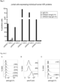
- KIR3DX1 rep was then fused to the Fc region of human IgG1 and expressed in HEK-293 cells to produce a soluble KIR3DX1rep-Fc protein, which was used to stain human PBMCs in flow cytometry. About 18% of PBMCs stained positive with KIR3DX1rep-Fc ( Figure 2A ).
- KIR3DX1rep-Fc was pulled-down analysis of human PBMCs and subsequent mass spectrometry analysis.
- KIR3DL2 was among the list of identified proteins from this pull-down.
- KIR3DX1rep-Fc The specific binding of KIR3DX1rep-Fc to KIR3DL2 was verified using KIR3DL2-transfected HEK-293 cells as well as human cutaneous T cell lymphoma cell line HUT78, which is known to endogenously express KIR3DL2.
- KIR3DX1rep to KIR3DL2 Binding of KIR3DX1rep to KIR3DL2 was verified by pull-down assay using a C-terminally HA-tagged KIR3DL2 expressed in HEK-293 cells and KIR3DX1rep-Fc ( Figure 2B ) as well as in flow cytometry analysis, where binding of KIR3DX1rep-Fc to KIR3DL2-expressing HEK-293 was detectable but not binding of the wildtype KIR3DX1 ( Figure 2C ).
- Figure 2E shows expression of KIR3DL2 on HUT78 Sézary syndrome cells and binding of KIR3DX1rep-Fc to HUT78.
- the wildtype KIR3DX1-Fc protein does not detectably bind to HUT78 cells ( Figure 2E ).
- KIR3DX1rep can bind other human KIR proteins and used Jurkat cells that were transfected to express single human KIR proteins. As shown in Figure 3 , low level of binding was detectable for KIR3DL1, but not for other KIRs including KIR3DS1.
- the D0 and D2 but not the D1 domain of KIR3DL2 were produced as Fc fusion protein, and it was found that that the D0 domain specifically bound KIR3DX1rep ( Figure 4C ).
- KIR3DX1rep As low binding of KIR3DX1rep was observed for KIR3DL1 but no binding for KIR3DS1, we compared the D0 sequences of KIR3DL1, KIR3DS1 and KIR3DL2.
- the glycine residue at position 56 is the only amino acid residue that differentiates the non-binder KIR3DS1 from the binder KIR3DL1 and KIR3DL2.
- KIR3DX1rep mutation of amino acid position 56 from serine into glycine did not change cell surface expression level of KIR3DL2, but abrogated binding of KIR3DX1rep.
- specific binding of KIR3DL2 and KIR3DX1rep involves the D0 domain of KIR3DL2 at position 56 and most probably the part of the D2 domain of KIR3DX1 that was affected by the 7 bp deletion as the wildtype KIR3DX1 protein is not able to bind KIR3DL2.
- KIR3DX1rep binds KIR3DL2 only in its homodimeric form
- Two cysteine residues at position 279 and 313 distinguish KIR3DL2 from the other human and nonhuman primate KIR proteins. These cysteines are thought to be responsible for the known homodimerization of this receptor. We mutated either one (C279Y or C313P) or both cysteine residues to amino acid residues that were invariably found in other KIR proteins at these two positions and expressed these three variants in HEK-293 cells. Successful expression of the individual KIR3DL2 mutants could be shown ( Figure 4F ). Neither the two single cysteine mutants nor the double mutant KIR3DL2 appeared to be bound by KIR3DX1rep -Fc ( Figure 4F ).
- KIR3DX1 can only bind to KIR3DL2 when both KIR3DL2-specific cysteines at positions 279 and 313 are present, and therefore, KIR3DX1rep most probably binds only the homodimerized form of KIR3DL2.
- Repaired KIR3DX1 can be used to detect and destroy KIR3DL2-expressing cells
- KIR3DL2 is a marker on cutaneous T cell lymphoma (Bagot et al., 2001; Sicard et al., 2015; Schmitt et al., 2017; Roelens et al., 2022) such as Sézary syndrome.
- a stimulatory receptor by fusing the extracellular part of FLAG-tagged KI R3DX 1 rep with the transmembrane and cytoplasmic region of human CD28 and CD3zeta ( Figure 5B ). This stimulatory KIR3DX1rep was stably transfected in the NK92 NK cell line.
- KIR3DX1rep-expressing NK92 cells showed increased degranulation as compared to untransfected parental NK92 cells upon co-incubation with HEK-293 cells expressing KIR3DL2, but no noticeable increase in degranulation was detected after co-incubation with HEK-293 cells expressing the cysteine mutants of KIR3DL2 ( Figure 5D ).
- KIR3DX1rep can bind KIR3DL2 not only in the soluble Fc-tagged form, but also in membrane-bound form as a receptor on the cell surface of killer cells.
- soluble KIR3DX1rep-Fc protein Besides cellular immunotherapy, also the usage of a soluble KIR3DX1rep-Fc protein is an option for immune therapy to target KIR3DL2-expressing cancer cells.
- ADCC antibody-dependent cellular cytotoxicity
- Repaired KIR3DX1 can be used to interfere with Th17 cell differentiation and survival
- KIR3DL2 is not only a marker on cutaneous T cell lymphoma but plays also an important role in autoimmune diseases involving the HLA-B*27 allotype (Chan et al., 2005; Bowness et all, 2011; Cauli et al., 2013; Hurabielle et al., 2017, Hurabielle et al., 2018; Khan et al., 2023).
- Ankylosing spondylitis (AS) or Morbus Bechterew is an autoimmune disease that has a strong genetic linkage with the HLA-B*27:05 allele.
- the molecular mechanism of disease development is thought to involve two steps in HLA-B*27-positive patients: the first step is the induced expression of KIR3DL2 on activated CD4+ Th cells, and the second step is the molecular interaction of KIR3DL2 with HLA-B*27, which licenses CD4+ Th cells to develop into inflammatory Th17 cells expressing increased amounts of IL-17 and reduced amounts of IL-2 (Chan et al., 2005; Bowness et al., 2011; Ridley et al., 2016; Wong-Baeza et al., 2013). Therefore, blocking interaction of KIR3DL2 with HLA-B*27 through soluble KIR3DX1rep in a form that does not lead to ADCC may help in therapy of such diseases, in particular, it may prevent an increase in severity of the disease.
- the human 721.221 B lymphoma cell line lacks HLA-A, HLA-B, and HLA-C expression and, therefore, is particularly well suited to express transfected HLA class I genes for subsequent functional studies.
- CD4+ T cells are isolated from healthy donors and from AS patients and stimulated with CD2/CD3/CD28 beads to induce KIR3DL2 expression. During this stimulation, the cells are co-incubated with 721.221-HLA-B*27:05 cells to further increase KIR3DL2 expression (Ridley et al., 2016). According to Ridley and co-workers (Ridley et al., 2016) this leads to differentiation into Th17 that increase expression of the pro-inflammatory IL-17 cytokine and decrease expression of IL-2.
- Soluble KIR3DX1rep protein (after removal of the Fc portion, or by using a Fc derived from human IgG4 that does not induce ADCC) in this assay is used to test whether the presence of KIR3DX1rep and the expected binding to KIR3DL2 interferes with interaction of KIR3DL2 with HLA-B*27 and, thereby, inhibits the differentiation into Th17 cells and lowers the expression of the pro-inflammatory IL-17 cytokine.
- soluble KIR3DX1rep may be used to treat patients with autoimmune diseases in which the interaction of KIR3DL2 with HLA-B*27 plays a role, such as AS.
- Repaired KIR3DX1 can be used to interfere with binding of CpG oligos to KIR3DL2 and subsequent activation of KIR3DL2-expressing cells
- PAMPs Pathogen-associated molecular patterns
- Such PAMPs also include DNA containing an unmethylated cytosine followed by a guanine, which is characteristic of bacterial but not for mammalian DNA.
- CpG oligos mimic such bacterial DNA and, thus, bacterial infection.
- the cellular receptor for CpG DNA (and oligos) is toll-like receptor 9 (TLR9), which upon binding of the CpG oligo stimulates the expression of pro-inflammatory cytokines such as IFN-gamma and increase activity of cytotoxic cells (Sivori et al., 2010a; Sivori et al., 2010b; Sivori et al., 2004).
- TLR9 is located in the endoplasmic reticulum and is translocated to the endosomal compartment upon exposure of the cell with CpG (Latz et al., 2004).
- KIR3DL2 is a receptor for CpG oligos, which binds this PAMP via its D0 domain.
- the KIR3DL2 is endocytosed upon binding and shuttles the CpG to the endosomal compartment where the CpG cargo is loaded on TLR9. This process leads to expression of interferon-gamma and to elevated cytotoxicity in NK cells.
- KIR3DX1rep As the D2 domain of KIR3DX1rep binds the D0 domain of KIR3DL2, we hypothesized that this binding reduces or abrogates CpG oligo binding of KIR3DL2.
- KIR3DX1rep that can be induced by the addition of tetracycline (tet-on expression system).
- This approach offers the advantage that we can analyze the same cells under normal condition (KIR3DX1rep protein not present) and under induced condition (KIR3DX1rep protein present).
- Soluble KIR3DX1rep (either without IgG1-Fc or by using IgG4-Fc) is used to study whether the binding of the soluble KIR3DX1rep interferes with KIR3DL2's ability to bind CpG oligos. If this is the case, this opens up opportunities to use soluble KIR3DX1rep for treating pathogenic immune responses such as sepsis associated with severe bacterial infections.
- soluble KIR3DX1 may be used to treat overshooting and finally pathogenic immune responses provoked by bacterial infections.
Landscapes
- Health & Medical Sciences (AREA)
- Life Sciences & Earth Sciences (AREA)
- Chemical & Material Sciences (AREA)
- Immunology (AREA)
- Organic Chemistry (AREA)
- General Health & Medical Sciences (AREA)
- Biophysics (AREA)
- Medicinal Chemistry (AREA)
- Proteomics, Peptides & Aminoacids (AREA)
- Biochemistry (AREA)
- Molecular Biology (AREA)
- Genetics & Genomics (AREA)
- Cell Biology (AREA)
- Toxicology (AREA)
- Zoology (AREA)
- Gastroenterology & Hepatology (AREA)
- Animal Behavior & Ethology (AREA)
- Public Health (AREA)
- Epidemiology (AREA)
- Veterinary Medicine (AREA)
- Peptides Or Proteins (AREA)
Priority Applications (2)
| Application Number | Priority Date | Filing Date | Title |
|---|---|---|---|
| EP23192200.6A EP4509523A1 (fr) | 2023-08-18 | 2023-08-18 | Kir3dx1 réparé et son utilisation thérapeutique |
| PCT/EP2024/073081 WO2025040580A1 (fr) | 2023-08-18 | 2024-08-16 | Kir3dx1 réparé et son utilisation thérapeutique |
Applications Claiming Priority (1)
| Application Number | Priority Date | Filing Date | Title |
|---|---|---|---|
| EP23192200.6A EP4509523A1 (fr) | 2023-08-18 | 2023-08-18 | Kir3dx1 réparé et son utilisation thérapeutique |
Publications (1)
| Publication Number | Publication Date |
|---|---|
| EP4509523A1 true EP4509523A1 (fr) | 2025-02-19 |
Family
ID=87760455
Family Applications (1)
| Application Number | Title | Priority Date | Filing Date |
|---|---|---|---|
| EP23192200.6A Withdrawn EP4509523A1 (fr) | 2023-08-18 | 2023-08-18 | Kir3dx1 réparé et son utilisation thérapeutique |
Country Status (2)
| Country | Link |
|---|---|
| EP (1) | EP4509523A1 (fr) |
| WO (1) | WO2025040580A1 (fr) |
Citations (2)
| Publication number | Priority date | Publication date | Assignee | Title |
|---|---|---|---|---|
| WO2003083039A2 (fr) * | 2001-07-05 | 2003-10-09 | Curagen Corporation | Polypeptides therapeutiques, acides nucleiques codant ces polypeptides, et leurs procedes d'utilisation |
| WO2021189053A1 (fr) * | 2020-03-20 | 2021-09-23 | Yale University | Profilage rapide d'anticorps extracellulaires (reap) pour la découverte et l'utilisation desdits anticorps |
-
2023
- 2023-08-18 EP EP23192200.6A patent/EP4509523A1/fr not_active Withdrawn
-
2024
- 2024-08-16 WO PCT/EP2024/073081 patent/WO2025040580A1/fr active Pending
Patent Citations (2)
| Publication number | Priority date | Publication date | Assignee | Title |
|---|---|---|---|---|
| WO2003083039A2 (fr) * | 2001-07-05 | 2003-10-09 | Curagen Corporation | Polypeptides therapeutiques, acides nucleiques codant ces polypeptides, et leurs procedes d'utilisation |
| WO2021189053A1 (fr) * | 2020-03-20 | 2021-09-23 | Yale University | Profilage rapide d'anticorps extracellulaires (reap) pour la découverte et l'utilisation desdits anticorps |
Non-Patent Citations (26)
| Title |
|---|
| BAGOT MMORETTA ASIVORI SBIASSONI RCANTONI CBOTTINO CBOUMSELL LBENSUSSAN A: "CD4(+) cutaneous T-cell lymphoma cells express the p140-killer cell immunoglobulin-like receptor", BLOOD, vol. 97, 2001, pages 1388 - 1391, XP002167249, DOI: 10.1182/blood.V97.5.1388 |
| BOWNESS PRIDLEY ASHAW JCHAN ATWONG-BAEZA IFLEMING MCUMMINGS FMCMI-CHAEL AKOLLNBERGER S: "Th17 cells expressing KIR3DL2+ and responsive to HLA-B27 ho-modimers are increased in ankylosing spondylitis", J IMMUNOL, vol. 186, no. 4, 15 February 2011 (2011-02-15), pages 2672 - 80, XP055373331, DOI: 10.4049/jimmunol.1002653 |
| CAULI ASHAW JGILES JHATANO HRYSNIK OPAYELI SMCHUGH KDESSOLE GPORRU GDESOGUS E ET AL.: "The arthritis-associated HLA-B*27:05 allele forms more cell surface B27 dimer and free heavy chain ligands for KIR3DL2 than HLA-B*27:09", RHEUMATOLOGY (OXFORD, ENGLAND, vol. 52, 2013, pages 1952 - 1962 |
| CHAN ATKOLLNBERGER SDWEDDERBURN LRBOWNESS P: "Expansion and enhanced survival of natural killer cells expressing the killer immunoglobulin-like receptor KIR3DL2 in spon-dylarthritis", ARTHRITIS AND RHEUMATISM, vol. 52, 2005, pages 3586 - 3595 |
| CHRISTIAN SCHMITT ET AL: "Therapeutic Antibodies to KIR3DL2 and Other Target Antigens on Cutaneous T-Cell Lymphomas", FRONTIERS IN IMMUNOLOGY, vol. 8, no. 1010, 30 August 2017 (2017-08-30), Lausanne, CH, pages 1 - 8, XP055437586, ISSN: 1664-3224, DOI: 10.3389/fimmu.2017.01010 * |
| DATABASE UniProt [online] 2 September 2008 (2008-09-02), "RecName: Full=Putative killer cell immunoglobulin-like receptor-like protein KIR3DX1; AltName: Full=Leukocyte receptor cluster member 12; Flags: Precursor;", XP002810887, retrieved from EBI accession no. UNIPROT:Q9H7L2 Database accession no. Q9H7L2 * |
| DATABASE UniProt [online] 28 March 2018 (2018-03-28), "RecName: Full=Immunoglobulin subtype domain-containing protein {ECO:0000259|SMART:SM00409};", XP002810886, retrieved from EBI accession no. UNIPROT:A0A2K5H7T9 Database accession no. A0A2K5H7T9 * |
| DATABASE UniProt [online] 28 March 2018 (2018-03-28), "SubName: Full=KIR3DX1 isoform 8 {ECO:0000313|EMBL:PNJ84591.1};", XP002810885, retrieved from EBI accession no. UNIPROT:A0A2J8XRF5 Database accession no. A0A2J8XRF5 * |
| FELICES MLENVIK TRDAVIS ZBMILLER JSVALLERA DA: "Generation of BiKEs and TriKEs to Improve NK Cell-Mediated Targeting of Tumor Cells", METHODS MOL BIOL, vol. 1441, 2016, pages 333 - 46, XP009526052, DOI: 10.1007/978-1-4939-3684-7_28 |
| GUETHLEIN LAABI-RACHED LHAMMOND JAPARHAM P: "The expanded cattle KIR genes are orthologous to the conserved single-copy KIR3DX1 gene of primates", IMMUNOGENETICS, vol. 59, 2007, pages 517 - 522, XP037853607, DOI: 10.1007/s00251-007-0214-x |
| GUETHLEIN LANORMAN PJHILTON HHG: "Parham P. Co-evolution of MHC class I and variable NK cell receptors in placental mammals", IMMUNOLOGICAL REVIEWS, vol. 267, 2015, pages 259 - 282 |
| HÉLÈNE SICARD ET AL: "A novel targeted immunotherapy for CTCL is on its way: Anti-KIR3DL2 mAb IPH4102 is potent and safe in non-clinical studies", ONCOIMMUNOLOGY, vol. 4, no. 9, 1 June 2015 (2015-06-01), pages e1022306, XP055437577, DOI: 10.1080/2162402X.2015.1022306 * |
| HURABIELLE CLEBOEUF CRAM-WOLFF CMEIGNIN VRIVET JVIGNON-PENNAMEN MDBONNAFOUS CSICARD HFITE CRAFFOUX E ET AL.: "KIR3DL2 expression in patients with adult T-cell lymphoma/leukaemia", THE BRITISH JOURNAL OF DERMATOLOGY, vol. 179, 2018, pages 197 - 199 |
| HURABIELLE CTHONNART NRAM-WOLFF CSICARD HBENSUSSAN ABAGOT MMARIE-CARDINE A: "Usefulness of KIR3DL2 to Diagnose, Follow-Up, and Manage the Treatment of Patients with Sezary Syndrome", CLINICAL CANCER RESEARCH: AN OFFICIAL JOURNAL OF THE AMERICAN ASSOCIATION FOR CANCER RESEARCH, vol. 23, 2017, pages 3619 - 3627, XP055956469, DOI: 10.1158/1078-0432.CCR-16-3185 |
| KHAN MAYONG SWEI JC: "Ankylosing spondylitis: History, epidemiology, and HLA-B27", INT JRHEUM DIS, vol. 26, 2023, pages 413 - 414 |
| KIR3DL2, BLOOD, vol. 116, 2010, pages 1637 - 1647, Retrieved from the Internet <URL:https://classic.clinicaltrials.gov/ct2/show/NCT02593045> |
| LATZ ESCHOENEMEYER AVISINTIN AFITZGERALD KAMONKS BGKNETTER CFLIEN ENILSEN NJESPEVIK TGOLENBOCK DT: "TLR9 signals after translocating from the ER to CpG DNA in the lysosome", NATURE IMMUNOLOGY, vol. 5, 2004, pages 190 - 198 |
| RIDLEY AHATANO HWONG-BAEZA ISHAW JMATTHEWS KKMOSSAWI HALADELL KPRICE DABOWNESS PKOLLNBERGER S: "Activation-Induced Killer Cell Immunoglobulin-like Receptor", ARTHRITIS & RHEUMATOLOGY (HOBOKEN, NJ, vol. 68, 2016, pages 901 - 914 |
| ROELENS MMASSON AANDRILLON ARAM-WOLFF CBIARD LBOISSON MMOURAH SBATTISTELLA MTOUBERT ABAGOT M ET AL.: "Mogamulizumab induces long term immune restoration and reshapes tumor heterogeneity in Sezary syndrome", BRIT J DERMATOL, 2022 |
| SAMBROOK JGBASHIROVA AANDERSEN HPIATAK MVERNIKOS GSCOGGILL PLIFSON JDCAR-RINGTON MBECK S: "Identification of the ancestral killer immunoglobulin-like receptor gene in primates", BMC GENOMICS, vol. 7, 2006, pages 209, XP021014585, DOI: 10.1186/1471-2164-7-209 |
| SCHMITT CMARIE-CARDINE ABENSUSSAN A: "Therapeutic Antibodies to KIR3DL2 and Other Target Antigens on Cutaneous T-Cell Lymphomas", FRONTIERS IN IMMUNOLOGY, vol. 8, 2017, pages 1010 |
| SICARD HBONNAFOUS CMOREL ABAGOT MBENSUSSAN AMARIE-CARDINE A: "A novel targeted immunotherapy for CTCL is on its way: Anti-KIR3DL2 mAb IPH4102 is potent and safe in non-clinical studies", ONCOIMMUNOLOGY, vol. 4, 2015, pages e1022306, XP055437577, DOI: 10.1080/2162402X.2015.1022306 |
| SIVORI SFALCO MCHIESA MDCARLOMAGNO SVITALE MMORETTA LMORETTA A: "CpG and double-stranded RNA trigger human NK cells by Toll-like receptors: induction of cytokine release and cytotoxicity against tumors and dendritic cells", PROCEEDINGS OF THE NATIONAL ACADEMY OF SCIENCES OF THE UNITED STATES OF AMERICA, vol. 101, 2004, pages 10116 - 10121, XP009051339, DOI: 10.1073/pnas.0403744101 |
| SIVORI SFALCO MMORETTA LMORETTA A: "Extending killer Ig-like receptor function: from HLA class I recognition to sensors of microbial products", TRENDS IN IMMUNOLOGY, vol. 31, 2010, pages 289 - 294 |
| WONG-BAEZA IRIDLEY ASHAW JHATANO HRYSNIK OMCHUGH KPIPER C ET AL.: "Brackenbridge S, Fernandes R, Chan A, et al. KIR3DL2 Binds to HLA-B27 Dimers and Free H Chains More Strongly than Other HLA Class I and Promotes the Expansion of T Cells in Ankylosing Spondylitis", JOURNAL OF IMMUNOLOGY (BALTIMORE, MD : 1950), vol. 190, 2013, pages 3216 - 3224 |
| YU, JSONG, YTIAN, W: "How to select IgG subclasses in developing anti-tumor therapeutic antibodies", J HEMATOL ONCOL, vol. 13, 2020, pages 45, Retrieved from the Internet <URL:https://doi.org/10.1186/s13045-020-00876-4> |
Also Published As
| Publication number | Publication date |
|---|---|
| WO2025040580A1 (fr) | 2025-02-27 |
Similar Documents
| Publication | Publication Date | Title |
|---|---|---|
| JP6923714B2 (ja) | Mhcクラスii拘束性mage−a3を認識するt細胞受容体 | |
| Diefenbach et al. | Selective associations with signaling proteins determine stimulatory versus costimulatory activity of NKG2D | |
| Colonna et al. | A common inhibitory receptor for major histocompatibility complex class I molecules on human lymphoid and myelomonocytic cells | |
| Cai et al. | CD160 inhibits activation of human CD4+ T cells through interaction with herpesvirus entry mediator | |
| Apps et al. | A homodimeric complex of HLA‐G on normal trophoblast cells modulates antigen‐presenting cells via LILRB1 | |
| KR101585532B1 (ko) | 항-ilt7 항체 | |
| AU2019203823A1 (en) | CS1-specific chimeric antigen receptor engineered immune effector cells | |
| JP7832111B2 (ja) | G12d突然変異rasに対するhlaクラスi拘束性t細胞受容体 | |
| CN105636985A (zh) | 用于治疗癌症和病毒感染的免疫受体调节 | |
| JP7735285B2 (ja) | G12v突然変異rasに対するhlaクラスi拘束性t細胞受容体 | |
| Germain et al. | Characterization of alternatively spliced transcript variants of CLEC2D gene | |
| US20230148144A1 (en) | Cell | |
| US12383602B2 (en) | HLA-B57 open conformers | |
| KR20240156654A (ko) | 키메라 ilt 수용체 조성물 및 방법 | |
| Kubagawa et al. | Authentic IgM Fc Receptor (FcμR) | |
| AU2002325333B2 (en) | NTB-A, a surface molecule involved in natural killer cells activity | |
| Leitner et al. | FcγR requirements and costimulatory capacity of Urelumab, Utomilumab, and Varlilumab | |
| JP2023534241A (ja) | G12d変異を有するrasに対するhlaクラスii拘束性drb t細胞受容体 | |
| CN111542547A (zh) | 对bdca2抗原具有特异性的嵌合抗原受体 | |
| US20100234294A1 (en) | Identification of new isoforms of the mhc-class i specific receptor cd160 and uses thereof | |
| EP4509523A1 (fr) | Kir3dx1 réparé et son utilisation thérapeutique | |
| JP2023542230A (ja) | Prame特異的t細胞受容体およびその使用 | |
| US20230303658A1 (en) | Human non-naturally occurring modified fc region of igg specifically binding to non-naturally occurring modified fc receptor | |
| EP4079753A1 (fr) | Récepteurs de lymphocytes t spécifiques aux épitopes mutés d'origine rac1 et rac2 | |
| JP2023535366A (ja) | G12v変異を有するrasに対するhlaクラスii拘束性drb t細胞受容体 |
Legal Events
| Date | Code | Title | Description |
|---|---|---|---|
| PUAI | Public reference made under article 153(3) epc to a published international application that has entered the european phase |
Free format text: ORIGINAL CODE: 0009012 |
|
| STAA | Information on the status of an ep patent application or granted ep patent |
Free format text: STATUS: THE APPLICATION HAS BEEN PUBLISHED |
|
| AK | Designated contracting states |
Kind code of ref document: A1 Designated state(s): AL AT BE BG CH CY CZ DE DK EE ES FI FR GB GR HR HU IE IS IT LI LT LU LV MC ME MK MT NL NO PL PT RO RS SE SI SK SM TR |
|
| STAA | Information on the status of an ep patent application or granted ep patent |
Free format text: STATUS: THE APPLICATION IS DEEMED TO BE WITHDRAWN |
|
| 18D | Application deemed to be withdrawn |
Effective date: 20250820 |