EP4509601A1 - Enzyme d'activation du facteur de coagulation x et son utilisation - Google Patents
Enzyme d'activation du facteur de coagulation x et son utilisation Download PDFInfo
- Publication number
- EP4509601A1 EP4509601A1 EP23787813.7A EP23787813A EP4509601A1 EP 4509601 A1 EP4509601 A1 EP 4509601A1 EP 23787813 A EP23787813 A EP 23787813A EP 4509601 A1 EP4509601 A1 EP 4509601A1
- Authority
- EP
- European Patent Office
- Prior art keywords
- coagulation factor
- activating enzyme
- relative content
- hour
- chromatography
- Prior art date
- Legal status (The legal status is an assumption and is not a legal conclusion. Google has not performed a legal analysis and makes no representation as to the accuracy of the status listed.)
- Pending
Links
Images


Classifications
-
- A—HUMAN NECESSITIES
- A61—MEDICAL OR VETERINARY SCIENCE; HYGIENE
- A61P—SPECIFIC THERAPEUTIC ACTIVITY OF CHEMICAL COMPOUNDS OR MEDICINAL PREPARATIONS
- A61P7/00—Drugs for disorders of the blood or the extracellular fluid
- A61P7/04—Antihaemorrhagics; Procoagulants; Haemostatic agents; Antifibrinolytic agents
-
- C—CHEMISTRY; METALLURGY
- C12—BIOCHEMISTRY; BEER; SPIRITS; WINE; VINEGAR; MICROBIOLOGY; ENZYMOLOGY; MUTATION OR GENETIC ENGINEERING
- C12N—MICROORGANISMS OR ENZYMES; COMPOSITIONS THEREOF; PROPAGATING, PRESERVING, OR MAINTAINING MICROORGANISMS; MUTATION OR GENETIC ENGINEERING; CULTURE MEDIA
- C12N9/00—Enzymes; Proenzymes; Compositions thereof; Processes for preparing, activating, inhibiting, separating or purifying enzymes
- C12N9/14—Hydrolases (3)
- C12N9/48—Hydrolases (3) acting on peptide bonds (3.4)
- C12N9/50—Proteinases, e.g. Endopeptidases (3.4.21-3.4.25)
- C12N9/64—Proteinases, e.g. Endopeptidases (3.4.21-3.4.25) derived from animal tissue
- C12N9/6402—Proteinases, e.g. Endopeptidases (3.4.21-3.4.25) derived from animal tissue from non-mammals
- C12N9/6418—Proteinases, e.g. Endopeptidases (3.4.21-3.4.25) derived from animal tissue from non-mammals from snakes
-
- C—CHEMISTRY; METALLURGY
- C07—ORGANIC CHEMISTRY
- C07K—PEPTIDES
- C07K14/00—Peptides having more than 20 amino acids; Gastrins; Somatostatins; Melanotropins; Derivatives thereof
- C07K14/435—Peptides having more than 20 amino acids; Gastrins; Somatostatins; Melanotropins; Derivatives thereof from animals; from humans
- C07K14/745—Blood coagulation or fibrinolysis factors
-
- C—CHEMISTRY; METALLURGY
- C12—BIOCHEMISTRY; BEER; SPIRITS; WINE; VINEGAR; MICROBIOLOGY; ENZYMOLOGY; MUTATION OR GENETIC ENGINEERING
- C12Y—ENZYMES
- C12Y304/00—Hydrolases acting on peptide bonds, i.e. peptidases (3.4)
- C12Y304/21—Serine endopeptidases (3.4.21)
- C12Y304/21006—Coagulation factor Xa (3.4.21.6)
-
- C—CHEMISTRY; METALLURGY
- C12—BIOCHEMISTRY; BEER; SPIRITS; WINE; VINEGAR; MICROBIOLOGY; ENZYMOLOGY; MUTATION OR GENETIC ENGINEERING
- C12Y—ENZYMES
- C12Y304/00—Hydrolases acting on peptide bonds, i.e. peptidases (3.4)
- C12Y304/24—Metalloendopeptidases (3.4.24)
- C12Y304/24058—Russellysin (3.4.24.58)
-
- A—HUMAN NECESSITIES
- A61—MEDICAL OR VETERINARY SCIENCE; HYGIENE
- A61K—PREPARATIONS FOR MEDICAL, DENTAL OR TOILETRY PURPOSES
- A61K38/00—Medicinal preparations containing peptides
Definitions
- the present application belongs to the field of biopharmaceutical technology, specifically relates to a coagulation factor X activating enzyme and its uses thereof.
- the human coagulation mechanism is a very complex and fine process, and so far, a total of 12 factors have been found to be involved in coagulation.
- the coagulation factor X is one of the key components of the coagulation system.
- Coagulation factor X (FX) is a vitamin K-dependent serine protease, its active form (FXa) is the only physiological activating enzyme of prothrombin in vivo, and plays a key role in the common coagulation pathway.
- Coagulation factor X activating enzyme can specifically activate FX, fully expose its active site to generate FXa. FXa then forms a prothrombin complex with activated platelets, FVa, and calcium ions at the injury site, thereby increasing thrombin generation.
- Thrombin activates platelets and factors V and VIII at the injury site, and forms thrombus through the conversion of fibrinogen to fibrin, in order to achieve the purpose of hemostasis in bleeding patients.
- FX activating enzyme has potentially great application value in surgical wound bleeding, medical bleeding diseases, and coagulation system diseases, such as hemophilia.
- the coagulation factor X activating enzyme of Russell's viper is a metalloprotease which is composed of a heavy chain( ⁇ chain, which has a molecular weight of 57600 D) and two light chains ( ⁇ chain, which has a molecular weight of 19400 D, and ⁇ chain, which has a molecular weight of 16400 D) through disulfide bonds, wherein the heavy chain contains a catalytic domain, while the two light chains and C-type lectin are homologous and play a regulatory role in the calcium dependent activation process of the factor X activating enzyme.
- a series of post-translational modifications, such as glycosylation modification are often performed during the synthesis of coagulation factor X activating enzyme of Russell's viper in vivo, and glycosylation modification may affect the enzyme activity.
- the present application provides a novel coagulation factor X activating enzyme of Russell's viper venom.
- the present application provides a method of isolating a factor X activating enzyme which enables the product to have high purity and high activity and significantly improves its yield for large-scale industrial production.
- the technical solution of the present application is of great importance for stopping bleeding or treatment of the bleeding disorders.
- the present application provides a novel coagulation factor X activating enzyme comprising three polypeptide chains: ⁇ , ⁇ and ⁇ , the amino acid sequence of the ⁇ chain is set forth in SEQ ID NO: 8, the amino acid sequence of the ⁇ chain is set forth in SEQ ID NO: 10, and the amino acid sequence of the ⁇ chain is set forth in SEQ ID NO: 12.
- the ⁇ , ⁇ and/or ⁇ polypeptide chain has glycosylation modifications.
- At least one amino acid residue of the amino acid sequence of the ⁇ , ⁇ and/or ⁇ polypeptide chain is covalently linked to an N-glycan.
- the amino acid residue is asparagine (Asn) residue.
- the asparagine (Asn) residue site is selected from one or more of the following:
- N-glycans at the Asn28 site in the ⁇ chain comprise:
- the relative content of N-glycans refers to the percentage of the total number of the coagulation factor X activating enzyme molecules containing the particular N-glycans at a certain site to the total number of all coagulation factor X activating enzyme molecules.
- the coagulation factor X activating enzyme described above is derived from Russell's viper (Daboia russelii siamensis). In some specific embodiments, the coagulation factor X activating enzyme is isolated from the Russell's viper venom or produced by genetic engineering.
- Another aspect of the present application provides a pharmaceutical composition comprising the coagulation factor X activating enzyme described above, and a pharmaceutically acceptable carrier.
- the pharmaceutical composition of the present application can be obtained by mixing the coagulation factor X activating enzyme having a desired purity with an optional pharmaceutically acceptable carrier, excipient or stabilizer. Further, the pharmaceutical composition of the present application can be prepared in the form of lyophilized formulation or liquid formulation.
- the pharmaceutical composition of the present invention may also contain one or more other active compounds as necessary for the particular indication being treated, preferably those with complementary activities that do not adversely affect each other.
- active compounds for example, it may be desirable to further provide other active compounds for stopping bleeding or promote coagulation in addition to the coagulation factor X activating enzyme.
- Such compounds are suitably present in combination in amounts that are effective for the purpose intended. The effective amount of such other compounds depends on the amount of coagulation factor X activating enzyme in the composition, the type of disease or condition or treatment, and other factors as described in the present application.
- Another aspect of the present invention provides a method of stopping bleeding or treatment of the bleeding disorder comprising administering to a subject in need thereof a therapeutically effective amount of the coagulation factor X activating enzyme or a pharmaceutical composition as described above.
- Another aspect of the present invention provides the use of the coagulation factor X activating enzyme or a pharmaceutical composition as described above in the manufacture of a medicament for stopping bleeding or treatment of the bleeding disorder.
- the bleeding disorder comprises surgical wound bleeding, an internal hemorrhagic disease, a hereditary hemorrhagic disease or a coagulation system disease;
- the coagulation system disease comprises hemophilia;
- the hemophilia is a hemophilia with inhibitors; and in some embodiments, the hemophilia is hemophilia A or hemophilia B.
- the coagulation factor X activating enzyme or the pharmaceutical composition of the present application can be administered to an individual (such as a human) via various routes, including, for example, intravenous, intra-arterial, intraperitoneal, intrapulmonary, oral, inhalation, intravascular, intramuscular, intra-tracheal, subcutaneous, intraocular, intrathecal, transmucosal or transdermal.
- routes including, for example, intravenous, intra-arterial, intraperitoneal, intrapulmonary, oral, inhalation, intravascular, intramuscular, intra-tracheal, subcutaneous, intraocular, intrathecal, transmucosal or transdermal.
- the coagulation factor X activating enzyme or the pharmaceutical composition to be used for in vivo administration must be sterile. This is readily accomplished by, e.g., filtration through sterile filtration membranes.
- the article of manufacture can comprise a container and a label or package insert on or associated with the container.
- Suitable containers include, for example, bottles, vials, syringes, etc.
- the containers may be formed from a variety of materials such as glass or plastic.
- the container holds a composition which is effective for treating a disease or disorder described herein, and has a sterile access port (for example, the container may be an intravenous solution bag or a vial having a stopper pierceable by a hypodermic injection needle).
- At least one of the active substances in the pharmaceutical composition is coagulation factor X activating enzyme described in the present application.
- the label or package instructions indicate that the composition may be used for stopping bleeding or treatment of the bleeding disorder.
- Package insert refers to instructions customarily included in commercial packages of therapeutic products that contain information about the indications, usage, dosage, administration, contraindications and/or warnings concerning the use of such therapeutic products.
- the article of manufacture may further comprise a second container comprising a pharmaceutically-acceptable buffer, such as bacteriostatic water for injection (BWFI), phosphate-buffered saline, Ringer's solution and dextrose solution. It may further include other materials desirable from a commercial and user standpoint, including other buffers, diluents, filters, needles, and syringes.
- Another aspect of the present application provides an isolated nucleic acid molecule encoding the coagulation factor X activating enzyme as described above.
- the vector comprises the nucleic acid molecule as described above.
- the vector is a DNA vector.
- the vector is a prokaryotic expression vector or a eukaryotic expression vector.
- the present application provides a method for inserting the nucleic acid molecule of the present application into a vector.
- the expression of the coagulation factor X activating enzyme by a natural or synthetic nucleic acid encoding the coagulation factor X activating enzyme can be achieved by inserting the nucleic acid into an appropriate expression vector, such that the nucleic acid is operably linked to 5' and 3' regulatory elements, e.g., including a promoter and a 3'untranslated region (UTR), to express the coagulation factor X activating enzyme.
- the vectors can be suitable for replication and integration in prokaryotic and/or eukaryotic host cells.
- Typical cloning and expression vectors contain transcription and translation terminators, initiation sequences, and promoters useful for regulation of the expression of the desired nucleic acid sequence.
- the vector can also comprise one or more selectable marker genes and other genetic elements.
- One example of a suitable promoter is the immediate early cytomegalovirus (CMV) promoter sequence. This promoter sequence is a strong constitutive promoter sequence capable of driving high levels of expression of any polynucleotide sequence operatively linked thereto.
- CMV immediate early cytomegalovirus
- EF-1 ⁇ Elongation Factor-1 ⁇
- constitutive promoter sequences may also be used, including, but not limited to the simian virus 40 (SV40) early promoter, mouse mammary tumor virus (MMTV), human immunodeficiency virus (HIV) long terminal repeat (LTR) promoter, MoMuLV promoter, an avian leukemia virus promoter, an Epstein-Barr virus immediate early promoter, a Rous sarcoma virus promoter, as well as human gene promoters such as, but not limited to, the actin promoter, the myosin promoter, the hemoglobin promoter, and the creatine kinase promoter. Further, the application should not be limited to the use of constitutive promoters.
- inducible promoters are also contemplated as part of the application.
- the use of an inducible promoter provides a molecular switch capable of turning on expression of the polynucleotide sequence which it is operatively linked when such expression is desired, or turning off the expression when expression is not desired.
- inducible promoters include, but are not limited to a metallothionine promoter, a glucocorticoid promoter, a progesterone promoter, and a tetracycline promoter.
- Another aspect of the present application provides an isolated host cell, wherein the host cell comprises the coagulation factor X activating enzyme, the nucleic acid molecule of the coagulation factor X activating enzyme or the vector as described above.
- the host cell is a prokaryotic or eukaryotic cell.
- the prokaryotic cell is an E. coli cell.
- Vectors can be readily introduced into host cells by any method known in the art, such as physical, chemical or biological means. Physical methods of introducing a polynucleotide into a host cell include calcium phosphate precipitation, lipofection, particle bombardment, microinjection, electroporation, and the like. Methods for producing cells comprising vectors and/or exogenous nucleic acids are well-known in the art. In some embodiments, the introduction of a polynucleotide into a host cell is carried out by calcium phosphate transfection. Biological methods for introducing a polynucleotide of interest into a host cell include the use of DNA and RNA vectors.
- Viral vectors and especially retroviral vectors, have become the most widely used method of inserting genes into mammalian, e.g., human cells.
- Other viral vectors can be derived from lentivirus, poxviruses, herpes simplex virus 1, adenoviruses and adeno-associated viruses, and the like.
- Chemical means for introducing a polynucleotide into a host cell include colloidal dispersion systems, such as macromolecule complexes, nanocapsules, microspheres, beads, and lipid-based systems including oil-in-water emulsions, micelles, mixed micelles, and liposomes.
- An exemplary colloidal system for use as a delivery vehicle in vitro and in vivo is a liposome (e.g., an artificial membrane vesicle).
- assays include, for example, “molecular biological” assays well known to those of skill in the art, such as Southern and Northern blotting, RT-PCR and PCR; “biochemical” assays, such as detecting the presence or absence of a particular peptide, e.g., by immunological means (ELISAs and Western blots) falling within the scope of the application.
- molecular biological assays well known to those of skill in the art, such as Southern and Northern blotting, RT-PCR and PCR
- biochemical assays, such as detecting the presence or absence of a particular peptide, e.g., by immunological means (ELISAs and Western blots) falling within the scope of the application.
- One aspect of the present application provides a method for the preparation of the coagulation factor activating enzyme as described above, comprising the following steps:
- Another aspect of the present application provides a method for glycosylation modification of the coagulation factor X activating enzyme as described above, and the method is to genetically engineer a host cell to produce the coagulation factor X activating enzyme having a predetermined N-glycosylation modification, and/or treating the coagulation factor X activating enzyme with glycosylation-related enzymes such that the coagulation factor X activating enzyme has a predetermined N-glycosylation modification.
- Another aspect of the present application provides a method for the preparation of the coagulation factor X activating enzyme as described above, comprising the following steps:
- One aspect of the present application provides a method for isolating a coagulation factor X activating enzyme, the method comprises the following steps in turn:
- both anion exchange chromatography and cation exchange chromatography are carried out only once.
- no further purification steps are carried out after cation exchange chromatography, or size exclusion chromatography step is further included after cation exchange chromatography and no further purification steps are carried out.
- the packed medium in the anion exchange chromatography column is agarose or polymer
- the elution buffer of the anion exchange chromatography is Tris-HCl buffer containing NaCl with a pH of 8.0 to 8.6, wherein the concentration of Tris-HCl buffer is from 10 mM to 30 mM and the concentration of NaCl is from 0.1M to 0.5M;
- the packed medium of the cation exchange chromatography column is agarose or polymer
- the elution buffer of the cation exchange chromatography is phosphate buffer containing NaCl with a pH of 6.5 to 7.5, wherein the concentration of phosphate buffer is from 10 mM to 30 mM, and the concentration of NaCl is from 0.2M to 0.6M;
- size exclusion chromatography, heparin affinity chromatography, and/or hydrophobic chromatography may further be carried out before anion exchange chromatography.
- the packed medium of the size exclusion chromatography column is selected from gel filtration media, preferably Sephacryl, Superdex or Sephadex, more preferably Sephacryl S-200 HR, Superdex75pg, Superdex200pg or SephadexG25.
- the elution buffer of the size exclusion chromatography is Tris-HCl buffer with a pH of from 8.0 to 8.6, and the concentration of Tris-HCl buffer is from 10 mM to 30 mM; preferably the pH of Tris-HCl buffer is 8.0, 8.1, 8.2, 8.3, 8.4, 8.5 or 8.6, and the concentration is 10mM, 15mM, 20mM, 25mM or 30mM.
- the elution flow rate of the size exclusion chromatography is from 30 cm/hour to 150 cm/hour, preferably form 40 cm/hour to 120 cm/hour, more preferably 60 cm/hour.
- the packed medium of the heparin affinity chromatography column is agarose medium; preferably Capto hips, Heparin Berpharose F.F., Heparin Sepharose 6 F.F., or Heparin Sepharose H.P..
- the elution buffer of the heparin affinity chromatography is Tris-HCl buffer containing NaCl with a pH of from 7.6 to 8.4, wherein the concentration of Tris-HCl buffer is from 10 mM to 30 mM and the concentration of NaCl is 0.5M to 1.5M.
- the pH of Tris-HCl buffer is 7.6, 7.8, 8.0, 8.2 or 8.4, and the concentration of Tris-HCl buffer is 10mM, 15mM, 20mM, 25mM or 30mM; and the concentration of NaCl is 0.5 M, 0.7 M, 0.8 M, 0.9 M, 1.0M, 1.1 M, 1.2 M, 1.3 M, 1.4 M or 1.5 M.
- the elution flow rate of the heparin affinity chromatography is from 30 cm/hour to 150 cm/hour, preferably from 40 cm/hour to 120 cm/hour, more preferably 60 cm/hour.
- the packed medium of the hydrophobic chromatography column is agarose medium; preferably Butyl Bestarose HP.
- the elution buffer of the hydrophobic chromatography is phosphate buffer containing NaCl with a pH of from 6.5 to 7.5, wherein the concentration of phosphate buffer is from 10 mM to 30 mM, and the concentration of NaCl is from 0.5M to 2.5M.
- the pH of the phosphate buffer is 6.5, 6.6, 6.8, 7.0, 7.2 or 7.5
- the concentration of phosphate buffer is 10mM, 15mM, 20mM, 25mM or 30mM
- the concentration of NaCl is 0.5M, 1.0M, 1.5 M, 1.7 M, 1.9 M, 2.0M, 2.1 M, 2.2 M, 2.4 M or 2.5 M.
- the elution flow rate of the hydrophobic chromatography is from 30 cm/hour to 150 cm/hour, preferably from 40 cm/hour to 120 cm/hour, more preferably 60 cm/hour.
- the packed medium of the size exclusion chromatography is gel filtration media, preferably Sephacryl, Superdex or Sephadex, more preferably Sephacryl S-200 HR, Superdex75pg, Superdex200pg or SephadexG25.
- the elution buffer of the size exclusion chromatography is phosphate buffer with a pH of from 6.5 to 7.5, and the concentration of phosphate buffer is from 40 mM to 80 mM, more preferably the pH of phosphate buffer is 6.5, 6.6, 6.8, 7.0, 7.2 or 7.5, and the concentration of phosphate buffer is 40mM, 50mM, 60mM, 70mM or 80mM;
- the elution flow rate of the size exclusion chromatography is from 5 cm/hour to 80 cm/hour, preferably from 10 cm/hour to 35 cm/hour, more preferably 20 cm/hour.
- the step of treating the stock solution of the snake venom with viral inactivation preferably the viral inactivation treatment is carried out by the S/D (solvent/detergent) method.
- the isolation methods described above can be used to isolate or prepare the coagulation factor X activating enzyme described in the present application.
- the present application also provides a method of analyzing the glycosylation of the coagulation factor X activating enzyme, and the method comprises the followed steps:
- the step of treating the coagulation factor X activating enzyme sample in step (1) comprises urea solution treatment, DTT treatment and iodoacetamide alkylation treatment.
- step (1) is: adding urea solution to the coagulation factor X activating enzyme sample, centrifuging, and sequentially performing reduction with DTT (dithiothreitol) and alkylation with iodoacetamide.
- the concentration of urea is from 4M to 10M, preferably 8M.
- the concentration of DTT is from 5mM to 40mM, preferably 10mM.
- the DTT reduction condition is from 45 °C to 65 °C, preferably 56 °C.
- the concentration of iodoacetamide is from 20mM to 50mM, preferably 30mM.
- the enzymatic step in step (2) comprises the following steps: adding ammonium bicarbonate, followed by adding trypsin for treatment, and terminating the reaction by adding formic acid after the enzymatic hydrolysis reaction.
- the step (2) is: adding ammonium bicarbonate solution, centrifugation, followed by adding trypsin for enzymatic hydrolysis, and terminating the reaction by adding formic acid solution after the enzymatic reaction, and performing ultrafiltration on the sample.
- the concentration of ammonium bicarbonate is from 30mM to 80 mM, preferably 50mM, preferably, the step of adding ammonium bicarbonate is repeated 2 times;
- the ratio of trypsin is from 1: 30 to 1: 80, preferably 1: 50, preferably, the step of adding trypsin is repeated 2 times, and the reaction is performed at 37 °C.
- the analysis is performed by liquid chromatography-mass spectrometry in step (3).
- the mobile phase of liquid chromatography contains formic acid.
- formic acid is added in a ratio of from 1:5 to 1: 30, preferably 1: 20.
- the column for liquid chromatography is a C18 column
- the glycosylation analysis described above can be used to analyze the coagulation factor X activating enzyme described in the present application.
- Glycosylation A process of modifying the protein by additional sugar chains. There are usually two types of glycosylations, O-linked glycosylation and N-linked glycosylation, respectively.
- O-linked glycosylation refers the addition of glycans to the hydroxyl groups, such as serine (Ser) and/or threonine (Thr) residue of a protein molecule.
- N-linked glycosylation refers to the addition of glycans to amide groups, such as asparagine acid (Asn) residue of a protein molecule.
- Proteins are glycosylated to form glycoproteins.
- Glycans refer to polysaccharides, oligosaccharides or monosaccharides. Glycans may be monomers or polymers of sugar residues, or may be linear or branched. Glycans may include natural sugar residues (e.g., glucose, N-acetylglucosamine, N-acetylneuraminic acid, galactose, mannose, fucose, hexose, arabinose, ribose, xylose, etc.) and/or modified sugars (e.g., 2 '-fluororibose, 2' -deoxyribose, mannose phosphate, 6 '-sulfo N-acetylglucosamine, etc.).
- natural sugar residues e.g., glucose, N-acetylglucosamine, N-acetylneuraminic acid, galactose, mannose, fucose, hexose, arabi
- Glycans may comprise homopolymers and heteropolymers of sugar residues. Glycans also encompass a glycan component of a glycoconjugate (e. g., a glycan component of a glycoprotein, glycolipid, proteoglycan). Glycans also encompass free glycans, including a glycan that that has been cleaved by a glycoconjugate or otherwise released.
- a glycoconjugate e. g., a glycan component of a glycoprotein, glycolipid, proteoglycan.
- free glycans including a glycan that that has been cleaved by a glycoconjugate or otherwise released.
- N-glycans refer to N-linked polysaccharides or oligosaccharides.
- N-linked polysaccharides or oligosaccharides refers to, for example, a polysaccharide or oligosaccharide linked to an amide group of asparagine or arginine residues in a protein by N-acetylglucosamine residues, the basic structure, composition and type of which are known in the art (see, e. g., Drickamer K, Taylor ME (2006), Introduction to Glycobiology, Vol. 2nd ed.
- N-glycans include glucose, galactose, mannose, fucose, N-acetylgalactosamine (GalNAc), N-acetylglucosamine (GlcNAc), sialic acid (SA), etc.
- the core structure of the N-glycan is known in the art, i.
- N-glycans extend from the basic pentasaccharide core. According to the branched chain components of the N-glycan, N-glycans can be classified, including high mannose type, complex type, or hybrid type. The "high mannose” N-glycans have 5 or more mannose residues. "Complex" N-glycans typically have at least one GlcNAc linked to the 1,3 mannose arm of the pentasaccharide core.
- Complex N-glycans may also have galactose or N-acetylgalactosamine residues, which may be modified with sialic acid or derivatives.
- Complex N-glycans may also have multiple antennas on the pentasaccharide core, often referred to as "multi-antenna glycans".
- the "hybrid" N-glycans have at least one GlcNAc at the end of the 1 '3 mannose arm of the pentasaccharide core and 0 or more mannose at the 1' 6 mannose arm of the pentasaccharide core.
- acetylglucosamine (GlcNAc)
- A3 represents that its sugar chain is three antennas
- B represents bisecting GlcNAc, i. e. a acetylglucosamine(GlcNAc) is attached to the mannose (Man) in the middle of the pentasaccharide core
- G represents galactose, and the number following it represents its number, G3 represents that there are 3 galactoses
- S represents sialic acid, and the number following it represents its number, S3 represents that there are 3 sialic acid.
- N-glycosylation and “N-linked glycosylation” are used interchangeably and refer to a process in which a sugar is linked to a linking group comprising an asparagine residue or an arginine residue, or comprising an N-linked glycosylation site or motif.
- Isolated biological components e.g., nucleic acid molecules, proteins, viruses, or cells
- nucleic acid molecules and proteins that have been “isolated” include those purified by standard purification methods.
- the term also includes nucleic acid molecules and proteins prepared by recombinant expression in a host cell, as well as chemically synthesized nucleic acid molecules and proteins.
- Pharmaceutically acceptable carriers include any standard pharmaceutical carrier, such as phosphate buffer aqueous solutions, emulsions (e.g., oil/water or water/oil emulsions), and various types of humectants.
- Treatment a method of obtaining beneficial or desired results, including clinical results.
- the beneficial or desired clinical results include, but are not limited to, one or more of the following: alleviating one or more symptoms resulting from the disease, diminishing the extent of the disease, stabilizing the disease (e. g., preventing or delaying the worsening of the disease), preventing or delaying the recurrence of the disease, delaying or slowing the progression of the disease, ameliorating the disease state, providing a remission (partial or total) of the disease, decreasing the dose of one or more other medications to treat the disease, delaying the progression of the disease, improving or enhancing the quality of life, and/or prolonging the life span.
- the therapeutically effective amount an amount of a particular drug or therapeutic agent (e. g., coagulation factor X activating enzyme) sufficient to achieve a desired effect in a subject or cell treated with the agent.
- a particular drug or therapeutic agent e. g., coagulation factor X activating enzyme
- the effective amount of the agent depends on a variety of factors, including, but not limited to, the subject or cell to be treated, and the mode of administration of the therapeutic composition.
- Signal peptide refers to a propeptide that exists as an N-terminal peptide in the form of a protein precursor.
- the function of the signal peptide is to promote translocation of the expressed polypeptide linked to the endoplasmic reticulum, the signal peptide is usually cleaved by the signal peptidase during this process.
- the signal peptide may be heterologous or homologous to the organism producing the polypeptide.
- the homologous conserved sequence of each chain was analyzed using the Align X function in Vector NTI 11.5 software according to the sequence of the known coagulation factor X activating enzyme three chains (i.e., the ⁇ , ⁇ and ⁇ chains). According to the analysis results, specific primers for cloning the cDNA coding sequence of three chains were designed, and primers were synthesized by Invitrogen. The sequences of primers were shown in Table 1.
- the head of Russell's viper was cut and quickly frozen with dry ice, and both sides of the venom glands were separated under freezing conditions, crushed and added to a centrifuge tube containing 1 ml Trizol (Takara,# 9108-1), the tissue was blown repeatedly until the tissue was lysis and left for 5 min at room temperature, then 200 ⁇ l chloroform was added, vortexed and mixed evenly, left for 5 min at room temperature, and then centrifuged at 12000 rpm for 10 min. The supernatant was carefully aspirated into another centrifuge tube, an equal volume of isopropanol was added, mixed well and left at room temperature for 10 min, and centrifuged at 12000 rpm for 10 min.
- the extracted total RNA was subjected to reverse transcription using a reverse transcription kit (Fermentas, # K1622), the reaction solution was prepared according to the instructions, 4 ⁇ l of total RNA was aspirated, 7 ⁇ l of DEPC water and 1 ⁇ l of Oligo T were added and mixed, incubated at 65 °C for 5 min, and immediately subjected to ice bath. 4 ⁇ l 5 ⁇ buffer, 1 ⁇ l Rnase Inhibitor, 1 ⁇ l MMLV and 2 ⁇ l dNTPs were added to the reaction, and centrifuged slightly after mixed, incubated at 42 °C for 60 min, and treated at 75 °C for 5 min. The cDNA product obtained can be used directly for PCR amplification.
- the cDNA obtained by reverse transcription was used as a template, PCR amplification was performed using three pairs of primers synthesized in Example 1, respectively.
- the reaction systems were shown in Table 2.
- Table 2 The cDNA amplification systems of the coagulation factor X activating enzyme reagent ⁇ chain ⁇ chain ⁇ chain 10 ⁇ PCR Buffer 5 ⁇ l 5 ⁇ l 5 ⁇ l dNTPs (2mM) 5 ⁇ l 5 ⁇ l 5 ⁇ l 5 ⁇ l Upstream primers (5 ⁇ M) ⁇ PF 5 ⁇ l ⁇ PF 5 ⁇ l ⁇ PF 5 ⁇ l ⁇ l Downstream primers (5 ⁇ M) ⁇ PR 5 ⁇ l ⁇ PR 5 ⁇ l ⁇ PR 5 ⁇ l cDNA template 2 ⁇ l 2 ⁇ l 2 ⁇ l Taq enzyme (5U/ ⁇ l) 0.5 ⁇ l 0.5 ⁇ l 0.5 ⁇ l H 2 O 27.5 ⁇ l 27.5 ⁇ l 27.5 ⁇ l 27.5 ⁇ l
- the PCR product was analyzed by agarose gel electrophoresis, and the amplified bands were single, clear and in the same size as expected. The bands were cut into EP tubes and stored at 4°C.
- the target bands were recovered by using a gel recovery kit (TIANGEN #DP209-02), the recovered products were ligated into T vector, transformed into E.coli competent cells, and expanded in plate. Five monoclones were selected into LB liquid medium for expansion, and the plasmid was extracted and sent to Takara Company for nucleic acid sequencing. The cDNA sequences of ⁇ chain, ⁇ chain and ⁇ chain of the coagulation factor X activating enzyme were obtained, respectively.
- Example 3 Determination of the amino acid sequence of the coagulation factor X activating enzyme.
- the open reading frame (ORF) analysis was performed by using Vector NTI based on the cDNA sequence information of the ⁇ chain obtained in Example 2. According to the analysis results, the translated protein sequences are as follows (SEQ ID NO: 7), wherein the amino acids at positions 1-20 are signal peptide sequences (shown in bold), the amino acids at positions 21-188 are propeptide sequences (shown with underlines), and the amino acids at positions 189-619 are mature peptide sequences.
- amino acid sequence of the ⁇ chain without the signal peptide and the propeptide is as follows (SEQ ID NO: 8, 431 amino acids):
- the open reading frame (ORF) analysis was performed by using Vector NTI based on the cDNA sequence information of the ⁇ chain obtained in Example 2. According to the analysis results, the translated protein sequences are as follows (SEQ ID NO: 9), wherein the amino acids at positions 1-23 are signal peptide sequences (shown in bold), and the amino acids at positions 24-158 are mature peptide sequences.
- amino acid sequence of the ⁇ chain without the signal peptide is as follows (SEQ ID NO: 10, 135 amino acids):
- the open reading frame (ORF) analysis was performed using Vector NTI based on the cDNA sequence information of the ⁇ chain obtained in Example 2. According to the analysis results, the translated protein sequences are as follows (SEQ ID NO: 11), wherein the amino acids at positions 1-23 are signal peptide sequences (shown in bold), and the amino acids at positions 24-146 are mature peptide sequences.
- amino acid sequence of the ⁇ chain without the signal peptide is as follows (SEQ ID NO: 12, 123 amino acids):
- Example 4 Structural verification of the coagulation factor X activating enzyme amino acid sequence.
- the amino acid sequences of three mature peptides (i.e., the ⁇ , ⁇ and ⁇ chains, represented by "cDNA translation") obtained in Example 3 were respectively compared with the amino acid sequence (represented by "sequence 2008") translated from the cDNA sequence ( Genbank accession number is DQ137799 , AY734997, AY734998, respectively) measured by Chen H.S in 2008 ( Chen HS et al., New insights into the functions and N-glycan structures of factor X activating enzyme from Russell's viper venom. FEBS J.
- the comparison results of the ⁇ chain cDNA translation sequence with sequence 2008 and sequence 1992 was shown in Figure 1A .
- the comparison results of the ⁇ chain cDNA translation sequence with sequence 2008 was shown in Figure 1B .
- the comparison results of the ⁇ chain cDNA translation sequence with sequence2008 and sequence 1992 was shown in Figure 1C .
- the coagulation factor X activating enzyme was further extracted, purified and obtained from the venom of Russell's viper.
- the N-terminal sequences of the ⁇ chain, ⁇ chain and ⁇ chain of the obtained coagulation factor X activating enzyme were determined by tandem mass spectrometry and Edamn degradation method by Beijing Proteome Research Center and College of Life Science of Peking University, respectively.
- the amino acid at position 24 is N (Asn, asparagine), and there is N-glycosylation modification. It is speculated that the reason for not capturing the 24th amino acid residue may be that the glycosylation modification affects the determination of this amino acid by Edman degradation method.
- N-terminal amino acid sequence of the three chains of the coagulation factor X activating enzyme described herein is:
- the N-terminal amino acid sequencing result of the coagulation factor X activating enzyme was consistent with the N-terminal sequence of the mature peptide obtained from the cDNA translation, indicating that the mature peptide obtained by cDNA translation exists in the venom of Russell's viper.
- the cDNA translation sequences of ⁇ , ⁇ and ⁇ chain of the coagulation factor X activating enzyme were analyzed to predict the possible N-glycosylation sites of these three chains by using NeNGlycl.0Server neural network software (website: http://www.cbs.dtu.dk/ services/NetNGlyc/).
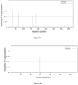
- FIG. 3A-3C The analysis results of ⁇ chain, ⁇ chain and ⁇ chain were shown in Figure 3A-3C .
- the position corresponding to the vertical line in the figure indicates that the site has the sequence feature of N-glycosylation modification, i.e. Asn-X-Thr/Ser.
- the vertical line indicates the possibility of N-glycosylation.
- the horizontal line indicates the threshold for the possibility of N-glycosylation, and the threshold is 0.5. If the score value ⁇ 0.5, this site is considered to be the predicted glycosylation site.
- N-glycosylation sites of the ⁇ chain were shown in Figure 3A .
- the possible N-glycosylation sites from the N-terminus were 28NSTA, 69NVTS, and 183NCSI, and the score values were 0.7407, 0.7516, and 0.6519, respectively.
- the score value of 163NDSS was 0.4979, close to 0.5, which is also subjected to subsequent experimental analysis as a potential site for N-glycosylation modification.
- the predicted results of N-glycosylation sites of the ⁇ -chain were shown in Figure 3B .
- the possible N-glycosylation site was 59NITE, and the score value was 0.7391.
- N-glycosylation sites of the ⁇ -chain were shown in Figure 3C .
- the possible N-glycosylation site was 24NWTD, and the score was 0.6089.
- Example 6 The novel preparation method of the coagulation factor X activating enzyme
- the virus was inactivated by S/D (solvent/detergent) method.
- S/D solvent/detergent
- the specific steps were as follows: After the collected supernatant was filtered by 0.45 ⁇ m filter membrane, Tween-80 and tributyl phosphate were added successively, so that the final volume concentration of Tween-80 was 1%, and the final volume concentration of tributyl phosphate was 0.3%. After full mixing, the mixture was bathed in water at 25°C ⁇ 2°C for 2 hours for viral inactivation.
- the venom obtained from viral inactivation was used to isolate coagulation factor X activating enzyme according to the steps listed in Table 3: Table 3 Summary of extraction methods of the coagulation factor X activating enzyme Method Step 1.1 / Anion exchange chromatography (QHP) Ultrafiltration Cation exchange chromatography (Capto SP ImpRs) / 1.2 / Cation exchange chromatography (CM Beads 6FF) Ultrafiltration Anion exchange chromatography (NenoGel 50Q) / 1.3 Size exclusion chromatograp hy Anion exchange chromatography(QHP) Ultrafiltration Cation exchange chromatography (Capto SP ImpRs) / 1.4 Size exclusion chromatograp hy Cation exchange chromatography (Capto SP ImpRs) Ultrafiltration Cation exchange chromatography (CM Beads 6FF) / 1.5 Size exclusion chromatograp hy Anion exchange chromatography(Neno Gel 50Q) Ultrafiltration Cation exchange chromatography (Capto SP ImpRs
- the Q Sepharose HP column was equilibrated with 20mMTris-HCl (pH 8.5) buffer for 3 column volumes.
- the snake venom after viral inactivation was loaded at a flow rate of 90cm/h.
- 20mM Tris-HCl (pH8.5) solution was used to wash impurities to the baseline at a flow rate of 90cm/h.
- the impurity was washed with 20mM Tris-HCl-0.2M NaCl (pH8.5) solution at a flow rate of 90cm/ h.
- the components corresponding to the target peaks collected after anion chromatography were concentrated 10 times by 30kD ultrafiltration membrane. The concentrate was then equal-volume exchanged with a 20mM phosphate buffer (pH 6.8).
- Capto SP ImpRes chromatographic column was equilibrated with 10mM (pH 6.8) phosphoric phosphate buffer for 3 column volumes, and then the components obtained after ultrafiltration were loaded at a flow rate of 60cm/h. After sample loading, 10mM phosphate buffer was used to re-equilibrate the chromatographic column to the baseline at a flow rate of 60cm/h.
- CM Beads 6FF chromatographic columns were equilibrated with 10mM (pH 6.8) phosphate buffer for 3 column volumes, and then the components after viral inactivation were loaded at a flow rate of 60cm/h. After sample loading, 10mM phosphate buffer was used to re-equilibrate the chromatographic column to the baseline at a flow rate of 60cm/h.
- the components corresponding to the target peaks collected after cationic chromatography were concentrated 10 times by 30kD ultrafiltration membrane. The concentrate was then equal-volume exchanged with 20mM Tris-HCl buffer (pH 8.5).
- NenoGel 50Q column was equilibrated with 20mMTris-HCl (pH8.5) buffer for 3 column volumes before use. The components obtained after ultrafiltration were loaded at a flow rate of 90cm/h. After sample loading, 20mMTris-HCl(pH8.5) buffer was used to re-equilibrate e to the baseline at a flow rate of 90cm/h. 20mM Tris-HCl-0.2M NaCl(pH8.5) solution was used to wash impurity at a flow rate of 90cm/ hour.
- the snake venom after viral inactivation was first subjected to size exclusion chromatography under the following conditions: the G25 chromatographic column ( ⁇ 2CV) was fully equilibrate at a flow rate of 60cm/ h with 0.02mol/L Tris-HCl (pH8.5) solution. The snake venom after viral inactivation was loaded at 20%-30% column volume each time, and washed with equilibrium solution after sampling. When the ultraviolet absorption value (UV value) rose to 1mAU, the collection of the target liquid was started, and the collection was stopped when the UV value dropped to 250 ⁇ 50 mAU.
- UV value ultraviolet absorption value
- the collected components were sequentially subjected to anion exchange chromatography, ultrafiltration and cation exchange chromatography according to steps (1) - (3) of Method 1.1, and the components corresponding to the target peak were collected.
- the snake venom after viral inactivation was subjected to size exclusion chromatography according to method 1.3.
- Capto SP ImpRes chromatographic column was equilibrated with 10mM (pH 6.8) phosphate buffer for 3 column volumes, and then the components obtained by Size exclusion Chromatography were loaded at a flow rate of 60cm/h. After sample loading, 10mM phosphate buffer was used to re-equilibrate the chromatographic column to the baseline at a flow rate of 60cm/h.
- the components corresponding to the target peaks collected after cation chromatography were concentrated 10 times by 30kD ultrafiltration membrane. The concentrate was then equal-volume exchanged with 10mM phosphate buffer (pH 6.8).
- CM Beads 6FF CM Beads 6FF chromatographic columns were equilibrated with 10mM (pH 6.8) phosphate buffer for 3 column volumes, and then the components obtained after ultrafiltration were loaded at a flow rate of 60cm/h. After sample loading, 10mM phosphate buffer was used to re-equilibrate the chromatographic column to the baseline at a flow rate of 60cm/h.
- the snake venom after viral inactivation was purified according to Method 1.3 except that the chromatography column of anion exchange chromatography was a NenoGel 50Q column.
- the snake venom after viral inactivation were sequentially subjected to anion exchange chromatography, ultrafiltration and cation exchange chromatography according to the steps of Method 1.1, and then the corresponding components were collected.
- the Sephacry1 S-200HR column was fully equilibrated with 50mM (pH6.8) phosphate buffer for 3 column volumes before use and then the sample was loaded.
- the loading volume was controlled at 5% of the size exclusion column volume.
- the chromatographic flow rate was 20cm/h, and the components corresponding to the target peak were collected.
- the snake venom after viral inactivation was purified according to Method 1.7 except that the chromatography column of anion exchange chromatography was NenoGel 50Q column.
- the snake venom after viral inactivation was purified according to Method 1.7 except that the chromatography column of cation exchange chromatography was NenoGel 50SP column.
- the snake venom after viral inactivation was purified according to Method 1.7 except that the chromatography column of cation exchange chromatography was CM Beads 6FF column.
- Size exclusion chromatography, anion exchange chromatography, ultrafiltration and cation exchange chromatography were performed sequentially according to the steps of Method 1.3, the corresponding components were collected and subjected to hydrophobic chromatography under the following conditions: The conductivity of the sample was adjusted to 140 ⁇ 5ms/cm; Butyl Bestarose HP column was equilibrated with 20mM phosphate buffer+2M NaCl (pH 7.0) for 3 column volumes before use and then the sample was loaded at a flow rate of 60cm/h. After sample loading, 20 mM phosphate buffer +2M NaCl pH7.0 solution was used to re-equilibrate the chromatographic column to the baseline at a flow rate of 60cm/h.
- the components corresponding to the target peaks collected after heparin affinity chromatography were concentrated 10 times by 30kD ultrafiltration membrane. The concentrate was then equal-volume exchanged with 20mM Tris-HCl buffer (pH 8.5).
- Heparin affinity chromatography, anion exchange chromatography, ultrafiltration and cation exchange chromatography were sequentially performed according to the steps in Method 1.14 and the corresponding components were collected. Then the collected components were subjected to size exclusion chromatography under the following conditions: The Sephacry1 S-200HR column was fully equilibrated with 50mM (pH6.8) phosphate buffer for 3 column volumes before use and then the sample was loaded. The loading volume was controlled at 5% of the size exclusion column volume. The chromatographic flow rate was 20cm/h, and the components corresponding to the target peak were collected.
- the components corresponding to the target peaks collected after hydrophobic chromatography were concentrated 10 times by 30kD ultrafiltration membrane. The concentrate was then equal-volume exchanged with 20mM Tris-HCl buffer (pH 8.5).
- the coagulation factor X activating enzyme obtained by methods 1.1-1.17 was detected according to the following methods: Detection of protein yield: The total protein concentration of snake venom during feeding was detected by BCA method, and the total protein mass was calculated according to the volume of the feeding liquid. The protein concentration of the purified coagulation factor X activating enzyme was detected by Folin phenol method (Lowry method), the second method of the determination of protein content of the third General Rule 0731 of the 2015 edition of Chinese Pharmacopoeia, and the volume was measured to obtain the total purified protein mass. The yield of the product was obtained by comparison with the total protein mass at the time of feeding.
- Detection of protein purity The purity was detected by High performance liquid chromatography (SEC-HPLC) based on size exclusion principle.
- the detection results of the coagulation factor X activating enzyme obtained by methods 1.1-1.17 were shown in Table 4.
- Table 4 The detection results of the coagulation factor X activating enzyme Method SEC-HPLC (%) Protein yield (%) 1.1 97.75 4.96 1.2 41.08 / 1.3 98.29 4.21 1.4 68.29 / 1.5 98.06 5.35 1.6 100 3.96 1.7 100 3.22 1.8 100 4.01 1.9 100 3.85 1.10 99.52 3.13 1.11 11.76 1.53 1.12 13.33 1.61 1.13 94.76 2.20 1.14 98.78 5.92 1.15 100 3.84 1.16 98.66 3.52 1.17 100 3.24
- the method of the present application for isolating coagulation factor X activating enzyme from snake venom of the Russell's viper is capable of improving the yield of the product while ensuring high purity of the final product, which can greatly reduce the cost of production and be more suitable for large-scale production.
- the coagulation factor X activating enzyme samples purified by methods 1.1, 1.6 and 1.7 of Example 6 were selected for further structural characterization and activity determination.
- Coagulation factor X activating enzyme was taken and centrifuged at 13000rpm for 10min; 400 ⁇ l of 8M urea solution was added and centrifuged at 13000rpm for 10min; DTT solution at a final concentration of 10mM was added, after reaction at 56°C for 1h, IAM (iodoacetamide) solution at a final concentration of 30 mM was added, and the reaction was carried out in the dark at room temperature for 40min, and DTT solution at a final concentration of 10mM was again added to neutralize excess IAM solution; 350 ⁇ l of 50 mM ammonium bicarbonate solution was added and centrifuged at 13000rpm for 10min, the lower layer solution was discard, and this step was repeated twice; trypsin was added at the ratio of 1:50 and reacted at 37°C for 2 hours, then trypsin was added again according to the above-mentioned enzyme dosage and reacted at 37°C overnight; formic acid solution was added at 1:20 ratio to terminate
- Analytical duration 120 min, detection mode: positive ion, parent ion scan range: 300-2000 m/z, primary mass spectral resolution: 70,000 at m/z 200, secondary mass spectral resolution: 17,500 at m/z 200.
- the identification results showed that among the three coagulation factor X activating enzyme samples, there were high glycosylation level at 6 glycosylation sites of the three peptide chains, and various N-glycan modifications were found at each site. It should be noted that although the sequence lengths of ⁇ chain and ⁇ chain are close and both have only one N-glycosylation site, in general, the glycosylation modification at the Asn24 glycosylation site of the former is more complex than that at the Asn 59 site of the latter, and the types of N-glycan are more diverse (data not shown).
Landscapes
- Health & Medical Sciences (AREA)
- Chemical & Material Sciences (AREA)
- Organic Chemistry (AREA)
- Life Sciences & Earth Sciences (AREA)
- Engineering & Computer Science (AREA)
- Bioinformatics & Cheminformatics (AREA)
- Genetics & Genomics (AREA)
- Zoology (AREA)
- Wood Science & Technology (AREA)
- General Health & Medical Sciences (AREA)
- Biochemistry (AREA)
- General Engineering & Computer Science (AREA)
- Biomedical Technology (AREA)
- Medicinal Chemistry (AREA)
- Molecular Biology (AREA)
- Microbiology (AREA)
- Biotechnology (AREA)
- Hematology (AREA)
- Diabetes (AREA)
- Toxicology (AREA)
- Proteomics, Peptides & Aminoacids (AREA)
- Gastroenterology & Hepatology (AREA)
- Biophysics (AREA)
- Chemical Kinetics & Catalysis (AREA)
- General Chemical & Material Sciences (AREA)
- Nuclear Medicine, Radiotherapy & Molecular Imaging (AREA)
- Pharmacology & Pharmacy (AREA)
- Animal Behavior & Ethology (AREA)
- Public Health (AREA)
- Veterinary Medicine (AREA)
- Medicines That Contain Protein Lipid Enzymes And Other Medicines (AREA)
Applications Claiming Priority (2)
| Application Number | Priority Date | Filing Date | Title |
|---|---|---|---|
| CN202210394772 | 2022-04-14 | ||
| PCT/CN2023/088276 WO2023198171A1 (fr) | 2022-04-14 | 2023-04-14 | Enzyme d'activation du facteur de coagulation x et son utilisation |
Publications (2)
| Publication Number | Publication Date |
|---|---|
| EP4509601A1 true EP4509601A1 (fr) | 2025-02-19 |
| EP4509601A4 EP4509601A4 (fr) | 2026-04-29 |
Family
ID=88329015
Family Applications (1)
| Application Number | Title | Priority Date | Filing Date |
|---|---|---|---|
| EP23787813.7A Pending EP4509601A4 (fr) | 2022-04-14 | 2023-04-14 | Enzyme d'activation du facteur de coagulation x et son utilisation |
Country Status (5)
| Country | Link |
|---|---|
| US (1) | US20250257347A1 (fr) |
| EP (1) | EP4509601A4 (fr) |
| CN (1) | CN119095957A (fr) |
| TW (1) | TW202346582A (fr) |
| WO (1) | WO2023198171A1 (fr) |
Families Citing this family (1)
| Publication number | Priority date | Publication date | Assignee | Title |
|---|---|---|---|---|
| CN119372186B (zh) * | 2024-12-30 | 2025-04-15 | 深圳市卫光生物制品股份有限公司 | 一种重组人凝血因子vii的纯化方法和应用 |
Family Cites Families (5)
| Publication number | Priority date | Publication date | Assignee | Title |
|---|---|---|---|---|
| US5589571A (en) * | 1994-10-28 | 1996-12-31 | Cor Therapeutics, Inc. | Process for production of inhibited forms of activated blood factors |
| CN101104847B (zh) | 2007-04-30 | 2010-06-02 | 锦州奥鸿药业有限责任公司 | 圆斑蝰蛇蛇毒凝血x因子激活酶及其提取方法与应用 |
| JP6702989B2 (ja) * | 2014-10-23 | 2020-06-03 | キュー−セラ ピーティーワイ リミテッド | 改善された凝固組成物 |
| CN109943554B (zh) | 2017-12-21 | 2021-04-16 | 舒泰神(北京)生物制药股份有限公司 | 一种从蛇毒中提取凝血因子x激活剂的方法 |
| CN109652398B (zh) | 2018-12-29 | 2021-07-20 | 上海太阳生物技术有限公司 | 凝血因子ⅹ激活剂rvv-ⅹ的制备方法及制得的rvv-ⅹ |
-
2023
- 2023-04-14 EP EP23787813.7A patent/EP4509601A4/fr active Pending
- 2023-04-14 US US18/856,472 patent/US20250257347A1/en active Pending
- 2023-04-14 CN CN202380033433.4A patent/CN119095957A/zh active Pending
- 2023-04-14 WO PCT/CN2023/088276 patent/WO2023198171A1/fr not_active Ceased
- 2023-04-14 TW TW112114121A patent/TW202346582A/zh unknown
Also Published As
| Publication number | Publication date |
|---|---|
| CN119095957A (zh) | 2024-12-06 |
| EP4509601A4 (fr) | 2026-04-29 |
| US20250257347A1 (en) | 2025-08-14 |
| WO2023198171A1 (fr) | 2023-10-19 |
| TW202346582A (zh) | 2023-12-01 |
Similar Documents
| Publication | Publication Date | Title |
|---|---|---|
| CN103446579B (zh) | 用于因子xa抑制剂的解毒剂和其使用方法 | |
| JP5650546B2 (ja) | 可溶性ヒアルロニダーゼの大規模生産 | |
| AU2018247318A1 (en) | Modified polynucleotides for the production of cytoplasmic and cytoskeletal proteins | |
| Ernst et al. | Expression in Escherichia coli, purification and characterization of heparinase I from Flavobacterium heparinum | |
| WO2014140103A2 (fr) | Molécules de facteur de coagulation x sensible à la thrombine | |
| EP4509601A1 (fr) | Enzyme d'activation du facteur de coagulation x et son utilisation | |
| US6368845B1 (en) | Polypeptides having L-asparaginase activity | |
| RU2648144C2 (ru) | КОМПОЗИЦИИ, СОДЕРЖАЩИЕ ГЕТЕРОГЕННЫЕ ПОПУЛЯЦИИ РЕКОМБИНАНТНЫХ БЕЛКОВ ЧЕЛОВЕЧЕСКОГО ФАКТОРА СВЕРТЫВАНИЯ КРОВИ Ха | |
| AU2004316095A1 (en) | Anticancer agent containing BL angiostatin | |
| JP2019530672A (ja) | 第viia因子グリコフォーム | |
| CN121752287A (zh) | 用于在治疗肺部非病毒性炎性疾病中使用的重组人α-1抗胰蛋白酶糖蛋白 | |
| EP4710929A1 (fr) | Composition de perfusion antivirale | |
| HK40052336A (en) | Antidotes for factor xa inhibitors and methods of using the same | |
| HK40001847A (en) | Modified polynucleotides for the production of biologics and proteins associated with human disease | |
| HK1206639B (en) | In vivo production of proteins | |
| JPS62282581A (ja) | ヒト正常結腸細胞由来組織プラスミノ−ゲン活性化因子 | |
| HK1151184B (en) | Recombinant fibrinogen |
Legal Events
| Date | Code | Title | Description |
|---|---|---|---|
| STAA | Information on the status of an ep patent application or granted ep patent |
Free format text: STATUS: THE INTERNATIONAL PUBLICATION HAS BEEN MADE |
|
| PUAI | Public reference made under article 153(3) epc to a published international application that has entered the european phase |
Free format text: ORIGINAL CODE: 0009012 |
|
| STAA | Information on the status of an ep patent application or granted ep patent |
Free format text: STATUS: REQUEST FOR EXAMINATION WAS MADE |
|
| 17P | Request for examination filed |
Effective date: 20241105 |
|
| AK | Designated contracting states |
Kind code of ref document: A1 Designated state(s): AL AT BE BG CH CY CZ DE DK EE ES FI FR GB GR HR HU IE IS IT LI LT LU LV MC ME MK MT NL NO PL PT RO RS SE SI SK SM TR |
|
| DAV | Request for validation of the european patent (deleted) | ||
| DAX | Request for extension of the european patent (deleted) | ||
| A4 | Supplementary search report drawn up and despatched |
Effective date: 20260327 |
|
| RIC1 | Information provided on ipc code assigned before grant |
Ipc: C12N 9/74 20060101AFI20260324BHEP Ipc: A61K 38/48 20060101ALI20260324BHEP Ipc: A61P 7/04 20060101ALI20260324BHEP Ipc: C12N 9/64 20060101ALI20260324BHEP |