EP4512890A2 - Cellules souches hermatopoietiques génétiquement modifiées et leurs utilisations - Google Patents
Cellules souches hermatopoietiques génétiquement modifiées et leurs utilisations Download PDFInfo
- Publication number
- EP4512890A2 EP4512890A2 EP24210553.4A EP24210553A EP4512890A2 EP 4512890 A2 EP4512890 A2 EP 4512890A2 EP 24210553 A EP24210553 A EP 24210553A EP 4512890 A2 EP4512890 A2 EP 4512890A2
- Authority
- EP
- European Patent Office
- Prior art keywords
- cells
- lineage
- gene
- cell
- population
- Prior art date
- Legal status (The legal status is an assumption and is not a legal conclusion. Google has not performed a legal analysis and makes no representation as to the accuracy of the status listed.)
- Pending
Links
Images

























Classifications
-
- A—HUMAN NECESSITIES
- A61—MEDICAL OR VETERINARY SCIENCE; HYGIENE
- A61K—PREPARATIONS FOR MEDICAL, DENTAL OR TOILETRY PURPOSES
- A61K35/00—Medicinal preparations containing materials or reaction products thereof with undetermined constitution
- A61K35/12—Materials from mammals; Compositions comprising non-specified tissues or cells; Compositions comprising non-embryonic stem cells; Genetically modified cells
- A61K35/14—Blood; Artificial blood
- A61K35/17—Lymphocytes; B-cells; T-cells; Natural killer cells; Interferon-activated or cytokine-activated lymphocytes
-
- A—HUMAN NECESSITIES
- A61—MEDICAL OR VETERINARY SCIENCE; HYGIENE
- A61K—PREPARATIONS FOR MEDICAL, DENTAL OR TOILETRY PURPOSES
- A61K35/00—Medicinal preparations containing materials or reaction products thereof with undetermined constitution
- A61K35/12—Materials from mammals; Compositions comprising non-specified tissues or cells; Compositions comprising non-embryonic stem cells; Genetically modified cells
- A61K35/28—Bone marrow; Haematopoietic stem cells; Mesenchymal stem cells of any origin, e.g. adipose-derived stem cells
-
- A—HUMAN NECESSITIES
- A61—MEDICAL OR VETERINARY SCIENCE; HYGIENE
- A61K—PREPARATIONS FOR MEDICAL, DENTAL OR TOILETRY PURPOSES
- A61K38/00—Medicinal preparations containing peptides
- A61K38/16—Peptides having more than 20 amino acids; Gastrins; Somatostatins; Melanotropins; Derivatives thereof
- A61K38/17—Peptides having more than 20 amino acids; Gastrins; Somatostatins; Melanotropins; Derivatives thereof from animals; from humans
- A61K38/177—Receptors; Cell surface antigens; Cell surface determinants
- A61K38/1774—Immunoglobulin superfamily (e.g. CD2, CD4, CD8, ICAM molecules, B7 molecules, Fc-receptors, MHC-molecules)
-
- A—HUMAN NECESSITIES
- A61—MEDICAL OR VETERINARY SCIENCE; HYGIENE
- A61K—PREPARATIONS FOR MEDICAL, DENTAL OR TOILETRY PURPOSES
- A61K38/00—Medicinal preparations containing peptides
- A61K38/16—Peptides having more than 20 amino acids; Gastrins; Somatostatins; Melanotropins; Derivatives thereof
- A61K38/17—Peptides having more than 20 amino acids; Gastrins; Somatostatins; Melanotropins; Derivatives thereof from animals; from humans
- A61K38/177—Receptors; Cell surface antigens; Cell surface determinants
- A61K38/178—Lectin superfamily, e.g. selectins
-
- A—HUMAN NECESSITIES
- A61—MEDICAL OR VETERINARY SCIENCE; HYGIENE
- A61K—PREPARATIONS FOR MEDICAL, DENTAL OR TOILETRY PURPOSES
- A61K39/00—Medicinal preparations containing antigens or antibodies
- A61K39/0005—Vertebrate antigens
- A61K39/0011—Cancer antigens
-
- A—HUMAN NECESSITIES
- A61—MEDICAL OR VETERINARY SCIENCE; HYGIENE
- A61K—PREPARATIONS FOR MEDICAL, DENTAL OR TOILETRY PURPOSES
- A61K39/00—Medicinal preparations containing antigens or antibodies
- A61K39/395—Antibodies; Immunoglobulins; Immune serum, e.g. antilymphocytic serum
- A61K39/39533—Antibodies; Immunoglobulins; Immune serum, e.g. antilymphocytic serum against materials from animals
- A61K39/3955—Antibodies; Immunoglobulins; Immune serum, e.g. antilymphocytic serum against materials from animals against proteinaceous materials, e.g. enzymes, hormones, lymphokines
-
- A—HUMAN NECESSITIES
- A61—MEDICAL OR VETERINARY SCIENCE; HYGIENE
- A61K—PREPARATIONS FOR MEDICAL, DENTAL OR TOILETRY PURPOSES
- A61K40/00—Cellular immunotherapy
- A61K40/10—Cellular immunotherapy characterised by the cell type used
-
- A—HUMAN NECESSITIES
- A61—MEDICAL OR VETERINARY SCIENCE; HYGIENE
- A61K—PREPARATIONS FOR MEDICAL, DENTAL OR TOILETRY PURPOSES
- A61K40/00—Cellular immunotherapy
- A61K40/10—Cellular immunotherapy characterised by the cell type used
- A61K40/11—T-cells, e.g. tumour infiltrating lymphocytes [TIL] or regulatory T [Treg] cells; Lymphokine-activated killer [LAK] cells
-
- A—HUMAN NECESSITIES
- A61—MEDICAL OR VETERINARY SCIENCE; HYGIENE
- A61K—PREPARATIONS FOR MEDICAL, DENTAL OR TOILETRY PURPOSES
- A61K40/00—Cellular immunotherapy
- A61K40/30—Cellular immunotherapy characterised by the recombinant expression of specific molecules in the cells of the immune system
- A61K40/31—Chimeric antigen receptors [CAR]
-
- A—HUMAN NECESSITIES
- A61—MEDICAL OR VETERINARY SCIENCE; HYGIENE
- A61K—PREPARATIONS FOR MEDICAL, DENTAL OR TOILETRY PURPOSES
- A61K40/00—Cellular immunotherapy
- A61K40/40—Cellular immunotherapy characterised by antigens that are targeted or presented by cells of the immune system
- A61K40/41—Vertebrate antigens
- A61K40/42—Cancer antigens
- A61K40/4202—Receptors, cell surface antigens or cell surface determinants
-
- A—HUMAN NECESSITIES
- A61—MEDICAL OR VETERINARY SCIENCE; HYGIENE
- A61K—PREPARATIONS FOR MEDICAL, DENTAL OR TOILETRY PURPOSES
- A61K40/00—Cellular immunotherapy
- A61K40/40—Cellular immunotherapy characterised by antigens that are targeted or presented by cells of the immune system
- A61K40/41—Vertebrate antigens
- A61K40/42—Cancer antigens
- A61K40/4202—Receptors, cell surface antigens or cell surface determinants
- A61K40/421—Immunoglobulin superfamily
-
- A—HUMAN NECESSITIES
- A61—MEDICAL OR VETERINARY SCIENCE; HYGIENE
- A61K—PREPARATIONS FOR MEDICAL, DENTAL OR TOILETRY PURPOSES
- A61K40/00—Cellular immunotherapy
- A61K40/40—Cellular immunotherapy characterised by antigens that are targeted or presented by cells of the immune system
- A61K40/41—Vertebrate antigens
- A61K40/42—Cancer antigens
- A61K40/4202—Receptors, cell surface antigens or cell surface determinants
- A61K40/421—Immunoglobulin superfamily
- A61K40/4211—CD19 or B4
-
- A—HUMAN NECESSITIES
- A61—MEDICAL OR VETERINARY SCIENCE; HYGIENE
- A61K—PREPARATIONS FOR MEDICAL, DENTAL OR TOILETRY PURPOSES
- A61K40/00—Cellular immunotherapy
- A61K40/40—Cellular immunotherapy characterised by antigens that are targeted or presented by cells of the immune system
- A61K40/41—Vertebrate antigens
- A61K40/42—Cancer antigens
- A61K40/4202—Receptors, cell surface antigens or cell surface determinants
- A61K40/4224—Molecules with a "CD" designation not provided for elsewhere
-
- A—HUMAN NECESSITIES
- A61—MEDICAL OR VETERINARY SCIENCE; HYGIENE
- A61K—PREPARATIONS FOR MEDICAL, DENTAL OR TOILETRY PURPOSES
- A61K45/00—Medicinal preparations containing active ingredients not provided for in groups A61K31/00 - A61K41/00
- A61K45/06—Mixtures of active ingredients without chemical characterisation, e.g. antiphlogistics and cardiaca
-
- A—HUMAN NECESSITIES
- A61—MEDICAL OR VETERINARY SCIENCE; HYGIENE
- A61P—SPECIFIC THERAPEUTIC ACTIVITY OF CHEMICAL COMPOUNDS OR MEDICINAL PREPARATIONS
- A61P35/00—Antineoplastic agents
-
- A—HUMAN NECESSITIES
- A61—MEDICAL OR VETERINARY SCIENCE; HYGIENE
- A61P—SPECIFIC THERAPEUTIC ACTIVITY OF CHEMICAL COMPOUNDS OR MEDICINAL PREPARATIONS
- A61P35/00—Antineoplastic agents
- A61P35/02—Antineoplastic agents specific for leukemia
-
- C—CHEMISTRY; METALLURGY
- C07—ORGANIC CHEMISTRY
- C07K—PEPTIDES
- C07K14/00—Peptides having more than 20 amino acids; Gastrins; Somatostatins; Melanotropins; Derivatives thereof
- C07K14/435—Peptides having more than 20 amino acids; Gastrins; Somatostatins; Melanotropins; Derivatives thereof from animals; from humans
- C07K14/705—Receptors; Cell surface antigens; Cell surface determinants
- C07K14/70503—Immunoglobulin superfamily
-
- C—CHEMISTRY; METALLURGY
- C07—ORGANIC CHEMISTRY
- C07K—PEPTIDES
- C07K14/00—Peptides having more than 20 amino acids; Gastrins; Somatostatins; Melanotropins; Derivatives thereof
- C07K14/435—Peptides having more than 20 amino acids; Gastrins; Somatostatins; Melanotropins; Derivatives thereof from animals; from humans
- C07K14/705—Receptors; Cell surface antigens; Cell surface determinants
- C07K14/70503—Immunoglobulin superfamily
- C07K14/7051—T-cell receptor (TcR)-CD3 complex
-
- C—CHEMISTRY; METALLURGY
- C07—ORGANIC CHEMISTRY
- C07K—PEPTIDES
- C07K14/00—Peptides having more than 20 amino acids; Gastrins; Somatostatins; Melanotropins; Derivatives thereof
- C07K14/435—Peptides having more than 20 amino acids; Gastrins; Somatostatins; Melanotropins; Derivatives thereof from animals; from humans
- C07K14/705—Receptors; Cell surface antigens; Cell surface determinants
- C07K14/7056—Lectin superfamily, e.g. CD23, CD72
-
- C—CHEMISTRY; METALLURGY
- C07—ORGANIC CHEMISTRY
- C07K—PEPTIDES
- C07K14/00—Peptides having more than 20 amino acids; Gastrins; Somatostatins; Melanotropins; Derivatives thereof
- C07K14/435—Peptides having more than 20 amino acids; Gastrins; Somatostatins; Melanotropins; Derivatives thereof from animals; from humans
- C07K14/705—Receptors; Cell surface antigens; Cell surface determinants
- C07K14/70596—Molecules with a "CD"-designation not provided for elsewhere
-
- C—CHEMISTRY; METALLURGY
- C07—ORGANIC CHEMISTRY
- C07K—PEPTIDES
- C07K16/00—Immunoglobulins [IG], e.g. monoclonal or polyclonal antibodies
- C07K16/18—Immunoglobulins [IG], e.g. monoclonal or polyclonal antibodies against material from animals or humans
- C07K16/28—Immunoglobulins [IG], e.g. monoclonal or polyclonal antibodies against material from animals or humans against receptors, cell surface antigens or cell surface determinants
- C07K16/2803—Immunoglobulins [IG], e.g. monoclonal or polyclonal antibodies against material from animals or humans against receptors, cell surface antigens or cell surface determinants against the immunoglobulin superfamily
-
- C—CHEMISTRY; METALLURGY
- C12—BIOCHEMISTRY; BEER; SPIRITS; WINE; VINEGAR; MICROBIOLOGY; ENZYMOLOGY; MUTATION OR GENETIC ENGINEERING
- C12N—MICROORGANISMS OR ENZYMES; COMPOSITIONS THEREOF; PROPAGATING, PRESERVING, OR MAINTAINING MICROORGANISMS; MUTATION OR GENETIC ENGINEERING; CULTURE MEDIA
- C12N15/00—Mutation or genetic engineering; DNA or RNA concerning genetic engineering, vectors, e.g. plasmids, or their isolation, preparation or purification; Use of hosts therefor
- C12N15/09—Recombinant DNA-technology
- C12N15/11—DNA or RNA fragments; Modified forms thereof; Non-coding nucleic acids having a biological activity
-
- C—CHEMISTRY; METALLURGY
- C12—BIOCHEMISTRY; BEER; SPIRITS; WINE; VINEGAR; MICROBIOLOGY; ENZYMOLOGY; MUTATION OR GENETIC ENGINEERING
- C12N—MICROORGANISMS OR ENZYMES; COMPOSITIONS THEREOF; PROPAGATING, PRESERVING, OR MAINTAINING MICROORGANISMS; MUTATION OR GENETIC ENGINEERING; CULTURE MEDIA
- C12N15/00—Mutation or genetic engineering; DNA or RNA concerning genetic engineering, vectors, e.g. plasmids, or their isolation, preparation or purification; Use of hosts therefor
- C12N15/09—Recombinant DNA-technology
- C12N15/11—DNA or RNA fragments; Modified forms thereof; Non-coding nucleic acids having a biological activity
- C12N15/113—Non-coding nucleic acids modulating the expression of genes, e.g. antisense oligonucleotides; Antisense DNA or RNA; Triplex- forming oligonucleotides; Catalytic nucleic acids, e.g. ribozymes; Nucleic acids used in co-suppression or gene silencing
- C12N15/1138—Non-coding nucleic acids modulating the expression of genes, e.g. antisense oligonucleotides; Antisense DNA or RNA; Triplex- forming oligonucleotides; Catalytic nucleic acids, e.g. ribozymes; Nucleic acids used in co-suppression or gene silencing against receptors or cell surface proteins
-
- C—CHEMISTRY; METALLURGY
- C12—BIOCHEMISTRY; BEER; SPIRITS; WINE; VINEGAR; MICROBIOLOGY; ENZYMOLOGY; MUTATION OR GENETIC ENGINEERING
- C12N—MICROORGANISMS OR ENZYMES; COMPOSITIONS THEREOF; PROPAGATING, PRESERVING, OR MAINTAINING MICROORGANISMS; MUTATION OR GENETIC ENGINEERING; CULTURE MEDIA
- C12N15/00—Mutation or genetic engineering; DNA or RNA concerning genetic engineering, vectors, e.g. plasmids, or their isolation, preparation or purification; Use of hosts therefor
- C12N15/09—Recombinant DNA-technology
- C12N15/87—Introduction of foreign genetic material using processes not otherwise provided for, e.g. co-transformation
- C12N15/90—Stable introduction of foreign DNA into chromosome
- C12N15/902—Stable introduction of foreign DNA into chromosome using homologous recombination
- C12N15/907—Stable introduction of foreign DNA into chromosome using homologous recombination in mammalian cells
-
- C—CHEMISTRY; METALLURGY
- C12—BIOCHEMISTRY; BEER; SPIRITS; WINE; VINEGAR; MICROBIOLOGY; ENZYMOLOGY; MUTATION OR GENETIC ENGINEERING
- C12N—MICROORGANISMS OR ENZYMES; COMPOSITIONS THEREOF; PROPAGATING, PRESERVING, OR MAINTAINING MICROORGANISMS; MUTATION OR GENETIC ENGINEERING; CULTURE MEDIA
- C12N5/00—Undifferentiated human, animal or plant cells, e.g. cell lines; Tissues; Cultivation or maintenance thereof; Culture media therefor
- C12N5/06—Animal cells or tissues; Human cells or tissues
- C12N5/0602—Vertebrate cells
- C12N5/0634—Cells from the blood or the immune system
- C12N5/0647—Haematopoietic stem cells; Uncommitted or multipotent progenitors
-
- C—CHEMISTRY; METALLURGY
- C12—BIOCHEMISTRY; BEER; SPIRITS; WINE; VINEGAR; MICROBIOLOGY; ENZYMOLOGY; MUTATION OR GENETIC ENGINEERING
- C12N—MICROORGANISMS OR ENZYMES; COMPOSITIONS THEREOF; PROPAGATING, PRESERVING, OR MAINTAINING MICROORGANISMS; MUTATION OR GENETIC ENGINEERING; CULTURE MEDIA
- C12N9/00—Enzymes; Proenzymes; Compositions thereof; Processes for preparing, activating, inhibiting, separating or purifying enzymes
- C12N9/14—Hydrolases (3)
- C12N9/16—Hydrolases (3) acting on ester bonds (3.1)
- C12N9/22—Ribonucleases [RNase]; Deoxyribonucleases [DNase]
-
- A—HUMAN NECESSITIES
- A61—MEDICAL OR VETERINARY SCIENCE; HYGIENE
- A61K—PREPARATIONS FOR MEDICAL, DENTAL OR TOILETRY PURPOSES
- A61K35/00—Medicinal preparations containing materials or reaction products thereof with undetermined constitution
- A61K35/12—Materials from mammals; Compositions comprising non-specified tissues or cells; Compositions comprising non-embryonic stem cells; Genetically modified cells
- A61K2035/124—Materials from mammals; Compositions comprising non-specified tissues or cells; Compositions comprising non-embryonic stem cells; Genetically modified cells the cells being hematopoietic, bone marrow derived or blood cells
-
- A—HUMAN NECESSITIES
- A61—MEDICAL OR VETERINARY SCIENCE; HYGIENE
- A61K—PREPARATIONS FOR MEDICAL, DENTAL OR TOILETRY PURPOSES
- A61K39/00—Medicinal preparations containing antigens or antibodies
- A61K2039/505—Medicinal preparations containing antigens or antibodies comprising antibodies
-
- A—HUMAN NECESSITIES
- A61—MEDICAL OR VETERINARY SCIENCE; HYGIENE
- A61K—PREPARATIONS FOR MEDICAL, DENTAL OR TOILETRY PURPOSES
- A61K39/00—Medicinal preparations containing antigens or antibodies
- A61K2039/51—Medicinal preparations containing antigens or antibodies comprising whole cells, viruses or DNA/RNA
- A61K2039/515—Animal cells
- A61K2039/5156—Animal cells expressing foreign proteins
-
- A—HUMAN NECESSITIES
- A61—MEDICAL OR VETERINARY SCIENCE; HYGIENE
- A61K—PREPARATIONS FOR MEDICAL, DENTAL OR TOILETRY PURPOSES
- A61K2239/00—Indexing codes associated with cellular immunotherapy of group A61K40/00
- A61K2239/38—Indexing codes associated with cellular immunotherapy of group A61K40/00 characterised by the dose, timing or administration schedule
-
- A—HUMAN NECESSITIES
- A61—MEDICAL OR VETERINARY SCIENCE; HYGIENE
- A61K—PREPARATIONS FOR MEDICAL, DENTAL OR TOILETRY PURPOSES
- A61K2239/00—Indexing codes associated with cellular immunotherapy of group A61K40/00
- A61K2239/46—Indexing codes associated with cellular immunotherapy of group A61K40/00 characterised by the cancer treated
-
- A—HUMAN NECESSITIES
- A61—MEDICAL OR VETERINARY SCIENCE; HYGIENE
- A61K—PREPARATIONS FOR MEDICAL, DENTAL OR TOILETRY PURPOSES
- A61K2239/00—Indexing codes associated with cellular immunotherapy of group A61K40/00
- A61K2239/46—Indexing codes associated with cellular immunotherapy of group A61K40/00 characterised by the cancer treated
- A61K2239/48—Blood cells, e.g. leukemia or lymphoma
-
- C—CHEMISTRY; METALLURGY
- C07—ORGANIC CHEMISTRY
- C07K—PEPTIDES
- C07K2317/00—Immunoglobulins specific features
- C07K2317/60—Immunoglobulins specific features characterized by non-natural combinations of immunoglobulin fragments
- C07K2317/62—Immunoglobulins specific features characterized by non-natural combinations of immunoglobulin fragments comprising only variable region components
- C07K2317/622—Single chain antibody (scFv)
-
- C—CHEMISTRY; METALLURGY
- C07—ORGANIC CHEMISTRY
- C07K—PEPTIDES
- C07K2319/00—Fusion polypeptide
- C07K2319/01—Fusion polypeptide containing a localisation/targetting motif
- C07K2319/03—Fusion polypeptide containing a localisation/targetting motif containing a transmembrane segment
-
- C—CHEMISTRY; METALLURGY
- C07—ORGANIC CHEMISTRY
- C07K—PEPTIDES
- C07K2319/00—Fusion polypeptide
- C07K2319/33—Fusion polypeptide fusions for targeting to specific cell types, e.g. tissue specific targeting, targeting of a bacterial subspecies
-
- C—CHEMISTRY; METALLURGY
- C12—BIOCHEMISTRY; BEER; SPIRITS; WINE; VINEGAR; MICROBIOLOGY; ENZYMOLOGY; MUTATION OR GENETIC ENGINEERING
- C12N—MICROORGANISMS OR ENZYMES; COMPOSITIONS THEREOF; PROPAGATING, PRESERVING, OR MAINTAINING MICROORGANISMS; MUTATION OR GENETIC ENGINEERING; CULTURE MEDIA
- C12N2310/00—Structure or type of the nucleic acid
- C12N2310/10—Type of nucleic acid
- C12N2310/20—Type of nucleic acid involving clustered regularly interspaced short palindromic repeats [CRISPR]
-
- C—CHEMISTRY; METALLURGY
- C12—BIOCHEMISTRY; BEER; SPIRITS; WINE; VINEGAR; MICROBIOLOGY; ENZYMOLOGY; MUTATION OR GENETIC ENGINEERING
- C12N—MICROORGANISMS OR ENZYMES; COMPOSITIONS THEREOF; PROPAGATING, PRESERVING, OR MAINTAINING MICROORGANISMS; MUTATION OR GENETIC ENGINEERING; CULTURE MEDIA
- C12N2510/00—Genetically modified cells
-
- C—CHEMISTRY; METALLURGY
- C12—BIOCHEMISTRY; BEER; SPIRITS; WINE; VINEGAR; MICROBIOLOGY; ENZYMOLOGY; MUTATION OR GENETIC ENGINEERING
- C12N—MICROORGANISMS OR ENZYMES; COMPOSITIONS THEREOF; PROPAGATING, PRESERVING, OR MAINTAINING MICROORGANISMS; MUTATION OR GENETIC ENGINEERING; CULTURE MEDIA
- C12N2800/00—Nucleic acids vectors
- C12N2800/80—Vectors containing sites for inducing double-stranded breaks, e.g. meganuclease restriction sites
Definitions
- a major challenge in designing targeted therapies is the successful identification of proteins that are uniquely expressed on cells that would be therapeutically relevant to eliminate (e . g ., abnormal, malignant, or other target cells) but not present on cells that one does not wish to eliminate (e.g. , normal, healthy, or other non-target cells). For example, many cancer therapeutics struggle to effectively target cancer cells while leaving normal cells unharmed.
- CD19-targeted chimeric antigen receptor T cells CAR T cells
- anti-CD20 monoclonal antibodies e . g . Rituximab
- CD19 and CD20 CD20
- targeting lineage-specific proteins of other cell populations for example, myeloid lineage cells (e.g., cancers arising from myeloid blasts, monocytes, megakaryocytes, etc ) is not feasible, as these cell populations are necessary for survival.
- compositions e.g., engineered cells, and methods that provide the ability to target one or more cells or cell populations of interest while allowing non-targeted cell populations to escape such targeting.
- genetically engineered hematopoietic cells such as hematopoietic stem cells (HSCs) having genetically modified or edited genes of one or more lineage-specific cell-surface antigens.
- HSCs hematopoietic stem cells
- the modified, e.g., edited genes are able to produce the lineage-specific cell surface proteins in modified form, which retain, at least partially, the biological activity of the lineage-specific cell-surface antigens in the HSCs or in descendant cells expressing such, but can escape targeting by cytotoxic agents that are specific to the wild-type lineage-specific cell-surface antigens.
- the modified, e.g., edited, genes do not produce the lineage-specific cell surface protein(s) or produce a truncated version of the lineage-specific cell surface protein(s) that, while able to escape targeting by cytotoxic agents that are specific to the wild-type lineage-specific cell-surface antigen(s), may not retain biological activity of the lineage-specific cell-surface antigen(s) in the HSCs or in descendant cells expressing such.
- hematopoietic stem cells having one or more modified lineage-specific cell-surface antigen.
- the one or more modified lineage-specific cell surface proteins are modified such that one or more of the lineage-specific cell surface proteins retain at least partially its biological activity of the lineage-specific cell-surface antigens in the HSCs or in descendant cells expressing such, but can escape targeting by cytotoxic agents that are specific to the corresponding wild-type lineage-specific cell-surface antigen(s).
- the second gene has been modified or edited such that expression of the second lineage-specific cell-surface antigen is reduced or eliminated in the genetically engineered hematopoietic cell (e.g., as compared with expression of the corresponding endogenous or wild-type lineage-specific cell-surface antigen).
- the second gene has been modified or edited such that the genetically engineered hematopoietic cell expresses a mutant or modified version of the second lineage-specific cell-surface antigen.
- the mutant or modified version of the second lineage-specific cell-surface antigen escapes targeting by a cytotoxic agent that targets the corresponding wild-type lineage-specific cell-surface antigen.
- Another aspect of the present disclosure features a population of genetically engineered hematopoietic cells, wherein the genetically engineered hematopoietic cells in the population comprise: (i) a first gene encoding a first lineage-specific cell-surface antigen, which gene has been modified or edited and (ii) a second gene encoding a second lineage-specific cell-surface antigen, which gene has been modified or edited.
- the first gene has been modified or edited such that expression of the first lineage-specific cell-surface antigen is reduced or eliminated in the genetically engineered hematopoietic cell (e.g., as compared with expression of the corresponding endogenous or wild-type lineage-specific cell-surface antigen).
- Another aspect of the present disclosure features a population of genetically engineered hematopoietic cells, wherein the genetically engineered hematopoietic cells of the population further comprise: (iii) a third gene encoding a third lineage-specific cell-surface antigen, which gene has been modified or edited. In some embodiments, the genetically engineered hematopoietic cells of the population further comprise: (iv) a fourth gene encoding a fourth lineage-specific cell-surface antigen, which gene has been modified or edited. In some embodiments, the genetically engineered hematopoietic cells of the population further comprise (v) a fifth gene encoding a fifth lineage-specific cell-surface antigen, which gene has been modified or edited.
- any one or more of the mutant lineage-specific cell-surface antigen(s) escapes targeting by a cytotoxic agent that targets the corresponding wild-type lineage-specific cell-surface antigen. In some embodiments, any one or more of the mutant or modified version lineage-specific cell-surface antigen(s) retains its biological activity. In some embodiments, any one or more of the mutant lineage-specific cell-surface antigen(s) escapes targeting by a cytotoxic agent that targets the corresponding wild-type lineage-specific cell-surface antigen and retains its biological activity.
- One aspect of the present disclosure features a population of genetically engineered hematopoietic cells, comprising: (i) a first group of genetically engineered hematopoietic cells, which have genetic modification or editing in a first gene encoding a first lineage-specific cell-surface antigen, wherein the first group of genetically engineered hematopoietic cells (a) have reduced or eliminated expression of the first lineage-specific cell-surface antigen or (b) express a mutant of the first lineage-specific cell-surface antigen; and (ii) a second group of genetically engineered hematopoietic cells, which have genetic modification or editing in a second gene encoding a second lineage-specific cell-surface antigen, wherein the second group of genetically engineered hematopoietic cells (a) have reduced or eliminated expression of the second lineage-specific cell-surface antigen or (b) express a mutant of the second lineage-specific cell-surface antigen.
- the present disclosure provides a genetically engineered hematopoietic cell, comprising: (i) a gene encoding a CD19, which gene has been modified or edited and (ii) a gene encoding a CD33, which gene has been modified or edited.
- the CD19 gene has been modified or edited such that expression of the CD19 antigen is reduced or eliminated in the genetically engineered hematopoietic cell (e.g, as compared with expression of the corresponding endogenous or wild-type CD19 antigen).
- the CD19 gene has been modified or edited such that the genetically engineered hematopoietic cell expresses a mutant or modified version of the CD19 antigen.
- the mutant or modified version of the CD19 antigen escapes targeting by a cytotoxic agent that targets the corresponding wild-type CD19 antigen. In some embodiments, the mutant or modified version of the CD19 antigen retains its biological activity. In some embodiments, the mutant or modified version of the CD19 antigen escapes targeting by a cytotoxic agent that targets the corresponding wild-type CD19 antigen and retains its biological activity. In some embodiments, the CD33 gene has been modified or edited such that expression of the CD33 antigen is reduced or eliminated in the genetically engineered hematopoietic cell (e.g., as compared with expression of the corresponding endogenous or wild-type CD33 antigen).
- the CD33 gene has been modified or edited such that the genetically engineered hematopoietic cell expresses a mutant or modified version of the CD33 antigen.
- the mutant or modified version of the CD33 antigen escapes targeting by a cytotoxic agent that targets the corresponding wild-type CD33 antigen.
- the mutant or modified version of the CD33 antigen retains its biological activity.
- the mutant or modified version of the CD33 antigen escapes targeting by a cytotoxic agent that targets the corresponding wild-type CD33 antigen and retains its biological activity.
- the genetically engineered hematopoietic cell comprising: (i) a modified or edited gene encoding a CD19 and (ii) a modified or edited gene encoding a CD33, the gene encoding CD19 has been modified or edited such that the entire exon 2 is deleted or a portion of exon 2 is deleted.
- the genetically engineered hematopoietic cell comprising: (i) a modified or edited gene encoding a CD19 and (ii) a modified or edited gene encoding a CD33, the gene encoding CD33 has been modified or edited such that the entire exon 2 is deleted or a portion of exon 2 is deleted.
- the genetically engineered hematopoietic cell comprising: (i) a modified or edited gene encoding a CD19 and (ii) a modified or edited gene encoding a CD33, the gene encoding CD19 has been modified or edited such that the gene is truncated, has inserted and/or deleted sequences (e.g., resulting in scrambled, frameshift, or nonsense sequence), or the entire gene is deleted (e.g., effectively a knock-out gene).
- the genetically engineered hematopoietic cell comprising: (i) a modified or edited gene encoding a CD19 and (ii) a modified or edited gene encoding a CD33, the gene encoding CD33 has been modified or edited such that the gene is truncated, has inserted and/or deleted sequences (e.g., resulting in scrambled, frameshift, or nonsense sequence), or the entire gene is deleted (e.g., effectively a knock-out gene).
- the genetically engineered hematopoietic cell comprising: (i) a modified or edited gene encoding a CD19 and (ii) a modified or edited gene encoding a CD33, the gene encoding CD19 has been modified or edited such that the entire exon 2 of CD19 is deleted or a portion of exon 2 od CD19 is deleted and the gene encoding CD33 has been modified or edited such that the CD33 gene is truncated, has inserted and/or deleted sequences (e.g., resulting in scrambled, frameshift, or nonsense sequence), or the entire CD33 gene is deleted.
- the gene encoding CD19 has been modified or edited such that the entire exon 2 of CD19 is deleted or a portion of exon 2 od CD19 is deleted
- the gene encoding CD33 has been modified or edited such that the CD33 gene is truncated, has inserted and/or deleted sequences (e.g., resulting in scrambled, frameshift, or nonsense sequence
- the genetically engineered hematopoietic cell comprising: (i) a modified or edited gene encoding a CD19 and (ii) a modified or edited gene encoding a CD33
- the gene encoding CD33 has been modified or edited such that the entire exon 2 of CD33 is deleted or a portion of exon 2 of CD33 is deleted and the gene encoding CD19 has been modified or edited such that the CD19 gene is truncated, has inserted and/or deleted sequences (e.g., resulting in scrambled, frameshift, or nonsense sequence), or the entire gene is deleted.
- the present disclosure provides a genetically engineered hematopoietic cell, comprising: (i) a gene encoding a CD19, which gene has been modified or edited such that the entire exon 2 is deleted or a portion of exon 2 is deleted and (ii) a gene encoding a CD33, which gene has been modified or edited such that the entire exon 2 is deleted or a portion of exon 2 is deleted.
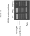
- the mutant of the CD19 antigen with exon 2 deleted or a portion of exon 2 deleted retains its biological activity.
- the mutant of the CD19 antigen with exon 2 deleted or a portion of exon 2 deleted escapes targeting by a cytotoxic agent that targets the corresponding wild-type CD19 antigen and retains its biological activity. In some embodiments, the mutant of the CD33 antigen with exon 2 deleted or a portion of exon 2 deleted retains its biological activity. In some embodiments, the mutant of the CD33 antigen with exon 2 deleted or a portion of exon 2 deleted escapes targeting by a cytotoxic agent that targets the corresponding wild-type CD33 antigen and retains its biological activity.
- the present disclosure provides a genetically engineered hematopoietic cell, comprising: (i) a gene encoding a CD19, which gene has been modified or edited such that the entire exon 2 is deleted or a portion of exon 2 is deleted and (ii) a gene encoding a CD33, which gene has been modified or edited such that the entire exon 2 is deleted or a portion of exon 2 is deleted.
- the present disclosure provides a genetically engineered hematopoietic cell, comprising a gene encoding a CD19, which gene has been modified or edited such that intron 1 and/or intron 2 in CD19 has been modified or edited.
- the genetically engineered hematopoietic cell has a sequence deletion in intron 1 and/or intron 2 of CD19 gene, e.g., either a portion of intron 1 and/or intron 2 of CD19 is deleted or the entire intron 1 and/or intron 2 of CD19 is deleted.
- the present disclosure provides a genetically engineered hematopoietic cell, comprising a gene encoding a CD33, which gene has been modified or edited such that intron 1 and/or intron 2 in CD33 has been modified or edited.
- the genetically engineered hematopoietic cell has a sequence deletion in intron 1 and/or intron 2 of CD33 gene, e.g., either a portion of intron 1 and/or intron 2 of CD33 is deleted or the entire intron 1 and/or intron 2 of CD33 is deleted.
- the present disclosure provides a genetically engineered hematopoietic cell, comprising: (i) a gene encoding a CD19, which gene has been modified or edited such that intron 1 and/or intron 2 in CD19 has been modified or edited and (ii) a gene encoding a CD33, which gene has been modified or edited such that intron 1 and/or intron 2 in CD33 has been modified or edited.
- the mutant of the CD19 antigen with intron 1 and/or intron 2 deleted or a portion of intron 1 and/or intron 2 deleted retains its biological activity. In some embodiments, the mutant of the CD19 antigen with intron 1 and/or intron 2 deleted or a portion of intron 1 and/or intron 2 deleted escapes targeting by a cytotoxic agent that targets the corresponding wild-type CD19 antigen and retains its biological activity. In some embodiments, the mutant of the CD33 antigen with intron 1 and/or intron 2 deleted or a portion of intron 1 and/or intron 2 deleted retains its biological activity.
- the mutant of the CD33 antigen with intron 1 and/or intron 2 deleted or a portion of intron 1 and/or intron 2 deleted escapes targeting by a cytotoxic agent that targets the corresponding wild-type CD33 antigen and retains its biological activity.
- any of the genetically engineered hematopoietic cells or populations of genetically engineered hematopoietic cells described herein may be hematopoietic stem cells (HSCs).
- HSCs are CD34+/CD33- cells.
- Any of the hematopoietic cells described herein can be from bone marrow cells, cord blood cells, or peripheral blood mononuclear cells (PBMCs).
- PBMCs peripheral blood mononuclear cells
- the genetically engineered hematopoietic cell is a human hematopoietic cell.
- any of the hematopoietic cells described herein are bone marrow cells, cord blood cells, or peripheral blood mononuclear cells (PBMCs) derived from a human.
- PBMCs peripheral blood mononuclear cells
- the mutant of the first lineage-specific cell-surface antigen and/or the mutant of the second lineage-specific cell-surface antigen includes a mutated or deleted non-essential epitope.
- a non-essential epitope in the first lineage-specific cell surface antigen and/or the non-essential epitope in the second lineage-specific cell surface antigen (and/or the non-essential epitope in the third, and/or fourth, and/or fifth lineage-specific cell-surface antigen has at least 3 amino acids.
- the non-essential epitope in the first lineage-specific cell surface antigen and/or the non-essential epitope in the second lineage-specific cell surface antigen (and/or the non-essential epitope in the third, and/or fourth, and/or fifth lineage-specific cell-surface antigen) is 6-10 amino acids. In some examples, the non-essential epitope in the first lineage-specific cell surface antigen and/or the non-essential epitope in the second lineage-specific cell surface antigen (and/or the non-essential epitope in the third, and/or fourth, and/or fifth lineage-specific cell-surface antigen) is 6-200 amino acids.
- the non-essential epitope in the first lineage-specific cell surface antigen and/or the non-essential epitope in the second lineage-specific cell surface antigen (and/or the non-essential epitope in the third, and/or fourth, and/or fifth lineage-specific cell-surface antigen) is 6-175 amino acids. In some examples, the non-essential epitope in the first lineage-specific cell surface antigen and/or the non-essential epitope in the second lineage-specific cell surface antigen (and/or the non-essential epitope in the third, and/or fourth, and/or fifth lineage-specific cell-surface antigen) is 6-150 amino acids.
- the non-essential epitope in the first lineage-specific cell surface antigen and/or the non-essential epitope in the second lineage-specific cell surface antigen (and/or the non-essential epitope in the third, and/or fourth, and/or fifth lineage-specific cell-surface antigen) is 6-125 amino acids. In some examples, the non-essential epitope in the first lineage-specific cell surface antigen and/or the non-essential epitope in the second lineage-specific cell surface antigen (and/or the non-essential epitope in the third, and/or fourth, and/or fifth lineage-specific cell-surface antigen) is 6-100 amino acids.
- the non-essential epitope in the first lineage-specific cell surface antigen and/or the non-essential epitope in the second lineage-specific cell surface antigen (and/or the non-essential epitope in the third, and/or fourth, and/or fifth lineage-specific cell-surface antigen) is 6-75 amino acids. In some examples, the non-essential epitope in the first lineage-specific cell surface antigen and/or the non-essential epitope in the second lineage-specific cell surface antigen (and/or the non-essential epitope in the third, and/or fourth, and/or fifth lineage-specific cell-surface antigen) is 6-50 amino acids.
- the non-essential epitope in the first lineage-specific cell surface antigen and/or the non-essential epitope in the second lineage-specific cell surface antigen is 6-25 amino acids.
- the non-essential epitope in the first lineage-specific cell surface antigen and/or the non-essential epitope in the second lineage-specific cell surface antigen is an entire exon or a portion of an exon.
- At least one of the first and second lineage-specific cell-surface antigens is associated with a hematopoietic malignancy. In some embodiments at least one of any of the modified or mutant lineage-specific cell-surface antigens is associated with a hematopoietic malignancy.
- Non-limiting examples include CD7, CD13, CD19, CD22, CD25, CD32, CD33, CD38, CD44, CD47, CD56, 96, CD117, CD123, CD135, CD174, CLL-1, folate receptor b, IL1RAP, MUC1, NKG2D/NKG2DL, TIM-3, and WT1.
- the first and second lineage-specific cell-surface antigens are selected from (a) CD19+CD33, (b) CD19+CD13, (c) CD19+CD123, (d) CD33+CD13, (e) CD33+CD123, (f) CD13+CD123.
- the modified lineage-specific cell-surface antigen is a type 1 lineage-specific cell-surface antigen. In some embodiments, at least one of the lineage-specific cell-surface antigens is a type 1 lineage-specific cell-surface antigen. In some embodiments, the lineage-specific cell-surface antigen is CD19. In some embodiments, at least one of the lineage-specific cell-surface antigens is CD19. In some embodiments, at least one of the first and second lineage-specific cell-surface antigens is a type 1 lineage-specific cell-surface antigen, for example, CD19.
- At least one of any of the modified or mutant lineage-specific cell-surface antigens is a type 1 lineage-specific cell-surface antigen, for example, CD19.
- both of the first and second lineage-specific cell-surface antigens are type 1 lineage-specific cell-surface antigens.
- the genetic modification or editing of a CD19 gene e.g., an endogenous CD19 gene
- the genetic modification or editing of a CD19 gene e.g., an endogenous CD19 gene
- the genetic modification or editing of a CD19 gene occurs in one or more introns of the CD19 gene, e.g., including modification or editing of one or more introns that result in modification(s) in exon 2 of CD19.
- the genetic modification or editing of a CD19 gene results in mutation or deletion of exon 2 of a CD19 gene.
- the genetic modification or editing of a CD19 gene results in deletion of the entire exon 2 of a CD19 gene or deletion of a portion of exon 2 of a CD19 gene.
- the mutated CD19 comprises the amino acid sequence of SEQ ID NO: 52.
- the genetic modification or editing of a CD19 gene occurs in one or more introns of the CD19 gene, e.g., including modification or editing of one or more introns that result in modification(s) in exon 4 of CD19.
- the genetic modification or editing of a CD19 gene results in mutation or deletion of exon 4 of a CD19 gene.
- the genetic modification or editing of a CD19 gene results in deletion of the entire exon 4 of a CD19 gene or deletion of a portion of exon 2 of a CD19 gene.
- the mutated CD19 comprises the amino acid sequence of SEQ ID NO: 73.
- the modified lineage-specific cell-surface antigen is a type 2 lineage-specific cell-surface antigen. In some embodiments, at least one of the lineage-specific cell-surface antigens is a type 2 lineage-specific cell-surface antigen. In some embodiments, the lineage-specific cell-surface antigen is CD33. In some embodiments, at least one of the lineage-specific cell-surface antigens is CD33. In some embodiments, at least one of the first and second lineage-specific cell-surface antigens is a type 2 lineage-specific cell-surface antigen, for example, CD33.
- At least one of any of the modified or mutant lineage-specific cell-surface antigens is a type 2 lineage-specific cell-surface antigen, for example, CD33.
- both of the first and second lineage-specific cell-surface antigens are type 2 lineage-specific cell-surface antigens.
- the genetic modification or editing of a CD33 gene e.g., an endogenous CD33 gene
- the genetic modification or editing of a CD33 gene e.g., an endogenous CD33 gene
- the genetic modification or editing of a CD33 gene occurs in one or more introns of the CD33 gene, e.g., including modification or editing of one or more introns that result in modification(s) in exon 2 of CD33.
- the genetic modification or editing of a CD33 gene occurs in intron 1 and intron 2 of the CD33 gene.
- the genetic modification or editing of a CD33 gene results in mutation or deletion of exon 2 of the CD33 gene.
- the genetic modification or editing of a CD33 gene results in deletion of the entire exon 2 of the CD33 gene or deletion of a portion of exon 2 of the CD33 gene.
- the genetic modification or editing of a CD33 gene occurs in exon 3 of the CD33 gene.
- the genetic modification or editing of a CD33 gene occurs in one or more introns of the CD33 gene, e.g., including modification or editing of one or more introns that result in modification(s) in exon 3 of CD33.
- the genetic modification or editing of a CD33 gene results in mutation or deletion of exon 3 of the CD33 gene.
- the genetic modification or editing of a CD33 gene results in deletion of the entire exon 3 of the CD33 gene or deletion of a portion of exon 3 of the CD33 gene.
- the second group of genetically engineered hematopoietic cells may contain genetic editing in exon 2 or exon 3 of a CD33 gene ( e . g ., including genetic modifications at one or more introns that result in modifications in exon 2 or exon 3).
- the CD33 gene is an endogenous CD33 gene.
- Example CD33 mutants include SEQ ID NO: 56 or SEQ ID NO: 58.
- the second lineage-specific cell surface antigen is a type 0 protein. In some embodiments, at least one of the first and second lineage-specific cell surface antigens is a type 0 protein. In some embodiments at least one of any of the modified or mutant lineage-specific cell-surface antigens is a type 0 lineage-specific cell-surface antigen. In some embodiments, both of the first and second lineage-specific cell-surface antigens are type 0 lineage-specific cell-surface antigens. In some embodiments, the genetic modification or editing of a Type 0 antigen gene occurs in an exon of the Type 0 gene. In some embodiments, the genetic modification or editing of a Type 0 antigen occurs in exon 2 of the Type 0 antigen gene.
- the genetic modification or editing of a Type 0 antigen gene occurs in one or more introns of the Type 0 antigen gene, e.g., including modification or editing of one or more introns that result in modification(s) in exon 2 of the Type 0 antigen gene. In some embodiments, the genetic modification or editing of a Type 0 antigen gene occurs in intron 1 and intron 2 of the Type 0 antigen gene.
- any of the genetically engineered hematopoietic cells described herein can be produced by genomic editing.
- the genomic editing does not involve an exogenous nuclease.
- the genomic editing involves adeno-associated virus vector mediated homologous recombination.
- the genomic editing involves an exogenous nuclease.
- Exemplary approaches include the method that involve the use of a zinc finger nuclease (ZFN), a transcription activator-like effector-based nuclease (TALEN), or a CRISPR-Cas system.
- the CRISPR-Cas system comprises a Cas endonuclease.
- the Cas endonuclease is a Cas9 endonuclease.
- provided herein are methods for producing any of the genetically engineered hematopoietic cells or populations of genetically engineered hematopoietic cells.
- a method for producing a population of genetically engineered hematopoietic cells comprising: (i) providing a population of hematopoietic cells, and (ii) genetically modifying or editing at least one lineage-specific cell surface antigen.
- the at least one lineage-specific cell surface antigen is genetically modified via CRISPR to produce the population of genetically engineered hematopoietic cells.
- a method for producing a population of genetically engineered hematopoietic cells comprising: (i) providing a population of hematopoietic cells, and (ii) genetically modifying or editing a first lineage-specific cell surface antigen, or genetically modifying or editing a second lineage-specific cell surface antigen, or genetically modifying or editing a first lineage-specific cell surface antigen and a second lineage-specific cell surface antigen in the population of hematopoietic cells via CRISPR to produce the population of genetically engineered hematopoietic cells.
- a method for producing a population of genetically engineered hematopoietic cells comprising: (i) providing a population of hematopoietic cells, and (ii) genetically modifying or editing a first lineage-specific cell surface antigen and genetically modifying or editing a second lineage-specific cell surface antigen in the population of hematopoietic cells to produce the population of genetically engineered hematopoietic cells.
- the method for producing a population of genetically engineered hematopoietic cells further comprises (iii) genetically modifying or editing one or more other lineage-specific cell surface antigen(s) in the population of hematopoietic cells to produce the population of genetically engineered hematopoietic cells.
- the genetically engineered hematopoietic cell is a human cell.
- a method for producing a genetically engineered hematopoietic cell comprising: (i) providing a hematopoietic cell, and (ii) genetically modifying or editing a first lineage-specific cell surface antigen, or genetically modifying or editing a second lineage-specific cell surface antigen, or genetically modifying or editing a first lineage-specific cell surface antigen and a second lineage-specific cell surface antigen in the hematopoietic cell via CRISPR to produce the genetically engineered hematopoietic cell.
- a method for producing a genetically engineered hematopoietic cell comprising: (i) providing a hematopoietic cell, and (ii) genetically modifying or editing a first lineage-specific cell surface antigen and genetically modifying or editing a second lineage-specific cell surface antigen to produce the genetically engineered hematopoietic cell.
- the method for producing a genetically engineered hematopoietic cell further comprises (iii) genetically modifying or editing one or more other lineage-specific cell surface antigen(s) to produce the genetically engineered hematopoietic cell.
- the genetically engineered hematopoietic cell is a human cell.
- a method for producing a population of genetically engineered hematopoietic cells comprising: (i) providing a population of hematopoietic cells, and (ii) genetically modifying or editing a CD19 gene, or genetically modifying or editing a CD33 gene, or genetically modifying or editing a CD19 gene and a CD33 gene in the population of hematopoietic cells via CRISPR to produce the genetically engineered hematopoietic cells.
- a method for producing a genetically engineered hematopoietic cell comprising: (i) providing a hematopoietic cell, and (ii) genetically modifying or editing a CD19 gene, or genetically modifying or editing a CD33 gene, or genetically modifying or editing a CD19 gene and genetically modifying or editing a CD33 gene in the hematopoietic cell via CRISPR to produce the genetically engineered hematopoietic cell.
- a method for producing a genetically engineered hematopoietic cell comprising: (i) providing a hematopoietic cell, and (ii) genetically modifying or editing a CD19 gene and genetically modifying or editing a CD33 gene to produce the genetically engineered hematopoietic cell.
- the method for producing a genetically engineered hematopoietic cell or population of genetically engineered hematopoietic cells further comprises (iii) genetically modifying or editing one or more other lineage-specific cell surface antigen(s) to produce the genetically engineered hematopoietic cell.
- the genetically engineered hematopoietic cell is a human cell.
- the genetic editing of the CD19 gene involves one or more guide nucleic acid molecules that target one or more introns of CD19.
- the genetic editing of the CD33 gene involves one or more guide nucleic acid molecules that target one or more introns of CD33.
- the genetic editing of the CD33 gene involves one or more guide nucleic acid molecules that do not target the CD33 pseudogene upstream of the CD33 gene.
- the genetic editing of the CD33 gene involves one or more guide nucleic acid molecules that (a) target one or more introns of CD33 and (b) do not target the CD33 pseudogene upstream of the CD33 gene.
- the introns of the CD19 gene comprise intron 1 and intron 2.
- the introns of the CD33 gene comprise intron 1 and intron 2.
- the genetic editing of CD19 results in exclusion of exon 2 of the CD19 gene.
- the genetic editing of CD19 results in exclusion of exon 4 of the CD19 gene.
- the genetic editing of CD33 results in exclusion of exon 2 of the CD33 gene.
- the genetic editing of the CD33 gene involves one or more guide nucleic acid molecules that target exon 3 of CD33.
- the genetic editing of the CD33 gene involves at least one guide nucleic acid molecule comprising the nucleotide sequence of SEQ ID NO: 67.
- a method for producing genetically engineered hematopoietic cells comprising: (i) providing a population of hematopoietic cells, and (ii) genetically editing a CD19 gene, a CD33 gene, or both a CD19 and a CD33 gene in the population of hematopoietic cells via CRISPR to produce the genetically engineered hematopoietic cells, wherein the genetic editing of the CD 19 gene involves at least one guide nucleic acid molecule comprising the nucleotide sequence of SEQ ID NOs: 14-26, 67, and 69-72, and/or wherein the genetic editing of the CD33 gene involves at least one guide nucleic acid molecule comprising the nucleotide sequence of SEQ ID NOs: 27-50 and 68.
- the method for producing genetically engineered hematopoietic cells comprises genetically editing a CD33 gene in the population of hematopoietic cells via CRISPR to produce the genetically engineered hematopoietic cells, wherein the genetic editing of the CD33 gene involves at least one guide nucleic acid molecule comprising the nucleotide sequence of SEQ ID NO: 67.
- step (ii) is performed by genetic editing of both a CD19 gene and a CD33 gene in the population of hematopoietic cells via CRISPR to produce the genetically engineered hematopoietic cells.
- the genetic editing of the CD19 gene involves a guide nucleic acid comprising the nucleotide sequence of SEQ ID NO: 67
- the genetic editing of the CD33 gene involves a guide nucleic acid comprising the nucleotide sequence of SEQ ID NO: 68.
- the hematopoietic cells can be HSCs, for example, CD34+/CD33- cells.
- the hematopoietic cells can be from bone marrow cells, cord blood cells, or peripheral blood mononuclear cells (PBMCs).
- PBMCs peripheral blood mononuclear cells
- the hematopoietic cells are from human bone marrow cells, human cord blood cells, or human peripheral blood mononuclear cells (PBMCs).
- PBMCs peripheral blood mononuclear cells
- Also provided herein are genetically engineered hematopoietic cells having one of more of the following features:
- genetically engineered hematopoietic cells produced by any method disclosed herein are also within the scope of the present disclosure.
- the genetically edited CD19 gene is capable of expressing a CD19 mutant comprising the amino acid sequence of SEQ ID NO: 52 or 73.
- the genetically edited CD33 gene is capable of expressing a CD33 mutant comprising the amino acid sequence of SEQ ID NO: 56 or SEQ ID NO: 58.
- a method of treating a hematopoietic malignancy comprising administering to a subject in need thereof a population of genetically engineered hematopoietic cells as disclosed herein.
- the method may further comprise administering to the subject an effective amount of a first immunotherapeutic agent.
- the first immunotherapeutic agent is a cytotoxic agent that targets cells expressing either the first lineage-specific cell-surface antigen or the second lineage-specific cell-surface antigen.
- the first immunotherapeutic agent is a cytotoxic agent that targets cells expressing the first lineage-specific cell-surface antigen
- the method further comprises administering to the subject a second immunotherapeutic agent when the hematopoietic malignancy relapses in the subject.
- the second immunotherapeutic agent may be a cytotoxic agent that targets cells expressing the second lineage-specific cell-surface antigen.
- the first immunotherapeutic agent, the second immunotherapeutic agent, or both are CAR-T cells.
- the first immunotherapeutic agent, the second immunotherapeutic agent, or both are antibody drug conjugates.
- the disclosure also provides methods of protecting hematopoietic stem cells from immunotherapy in a subject in need thereof, wherein the therapy targets one or more lineage-specific antigen(s).
- the methods comprise administering a modified hematopoietic stem cell to a subject, wherein the stem cell comprises one or more gene(s) encoding the lineage-specific antigen(s) being targeted by the immunotherapy, and wherein the gene(s) are modified, mutated or edited.
- the gene(s) are modified such that expression of the gene(s) results in modified, mutated protein(s) or complete knockout(s) of the protein or combinations thereof.
- modified, mutated protein(s) or complete knockout(s) prevent the immunotherapy from targeting the hematopoietic stem cells comprising the mutated gene(s).
- the editing results in expression of one or more lineage-specific antigen(s), which contain a partial deletion.
- the partial deletion compasses an entire exon or a portion of an exon.
- the immunotherapy administered includes one or more antibody-drug conjugate(s), which can be administered concurrently or sequentially.
- the immunotherapy administered includes cells expressing one or more chimeric antigen receptors or a pool of 2 or more cells, each expressing a different chimeric antigen receptor, which can be administered concurrently or sequentially.
- the disclosure provides a method of protecting hematopoietic stem cells from one or more chimeric antigen receptor T cell therapies targeting one or more lineage specific antigen(s) in a subject in need thereof, wherein the hematopoietic stem cells are administered, and wherein the hematopoietic stem cells are modified such that expression of the gene(s) encoding one or more lineage specific antigen(s) result in modified, mutated forms or complete knockout(s) of the lineage specific antigen(s) targeted by the CART(s).
- the mutated form(s) of the one or more lineage specific antigen(s) lack an exon, e.g. , an exon which comprises the CART or antibody-drug conjugate epitope.
- the modified hematopoietic cell can be any of the modified hematopoietic cells described here and elsewhere herein.
- Such modified hematopoietic stems cells can be generated using gene editing technologies, e.g. CRISPR, as described herein.
- CRISPR methodology can be used to delete a portion or an entire gene of interest.
- CRISPR methodology can be used to delete one or more exons comprising a targeted epitope.
- the exon sequence itself may be targeted by CRISPR, however, current conventional CRISPR therapies may lead to small insertions and deletions, which can lead to frameshift and truncated non-functional proteins.
- the intron sequences may be beneficial to target so as to more precisely edit the exon sequence of interest.
- the disclosure also provides methods for generating modified hematopoietic stem cells, comprising introducing one or more guide RNAs capable of editing one or more gene(s) encoding one or more lineage specific antigen(s) targeted by one or more chimeric antigen receptor(s).
- the editing results in expression of lineage specific antigen(s) lacking an exon.
- the editing results complete knockout of lineage specific antigen(s).
- the editing results in a combination of expression of lineage specific antigen(s) lacking an exon and complete knockout of lineage specific antigen(s) in one cell or a cell population.
- the one or more guide RNAs are selected to target one or more introns.
- targeting of adjacent introns results in excision of the gene sequence encoding the exon between the two introns.
- Targeting one intron may result in the generation of a new splice site, resulting in excision of the gene sequence encoding the adjacent exon. Exon skipping using a single guide RNA has been described (e.g., Mou et al., Genome Biology 201718:108 ). Accordingly, in some embodiments, one intron may be targeted according to the methods described herein.
- a method of protecting hematopoietic stem cells from immunotherapy may be used in a subject in need thereof, wherein the therapy targets one or more lineage-specific antigen(s).
- the methods comprise administering a modified hematopoietic stem cell to a subject, wherein the stem cell comprises one or more gene(s) encoding the lineage-specific antigen(s) CD19 and/or CD33 being targeted by the immunotherapy, and wherein the CD19 and/or CD33 gene(s) are modified, mutated or edited.
- the CD19 and/or CD33 gene(s) are modified such that expression result in modified, mutated CD19 and/or CD33 or complete knockout(s) of the CD19 and/or CD33 or combinations thereof.
- modified, mutated CD19 and/or CD33 protein(s) or complete CD19 and/or CD33 knockout(s) prevent the immunotherapy from targeting the hematopoietic stem cells comprising the mutated gene(s)
- the editing results in expression CD19 and/or CD33, which contain a partial deletion.
- the partial deletion compasses an entire exon or a portion of an exon, e.g., exon2 of CD33 and/or CD19.
- the immunotherapy administered includes one or more antibody-drug conjugate(s) directed against CD19 and/or CD33, which can be administered concurrently or sequentially.
- the immunotherapy administered includes cells expressing one or more chimeric antigen receptors directed against CD19 and/or CD33 or a pool of 2 cell populations, one expressing chimeric antigen receptor directed against CD19 and the other expressing a chimeric antigen receptor directed against CD33.
- the cells expressing chimeric antigen receptor directed against CD19 can be administered concurrently or sequentially with the cells expressing chimeric antigen receptor directed against CD33
- the disclosure provides a method of protecting hematopoietic stem cells from one or more chimeric antigen receptor T cell therapies targeting CD19 and/or CD33 in a subject in need thereof, wherein the hematopoietic stem cells are administered, and wherein the hematopoietic stem cells are modified such that expression of the gene(s) encoding CD19 and/or CD33 result in modified, mutated forms or complete knockout(s) of the CD19 and/or CD33.
- the mutated form(s) of CD19 and/or CD33 lack an exon, e . g ., an exon which comprises the CART epitope.
- the disclosure also provides methods for generating modified hematopoietic stem cells, comprising introducing one or more guide RNAs capable of editing one or more gene(s) encoding CD19 and/or CD33, wherein CD19 and/or CD33 are targeted by one or more chimeric antigen receptor(s) or antibody-drug conjugates.
- the editing results in expression of CD19 and/or CD33 lacking an exon.
- the editing results complete knockout of CD19 and/or CD33.
- the editing results in a combination of expression of lineage specific antigen(s) lacking an exon and complete knockout of CD 19 and/or CD33 in one cell or a cell population.
- the one or more guide RNAs are selected to target one or more introns.
- targeting of adjacent introns results in excision of the gene sequence encoding the exon between the two introns.
- the present disclosure provides methods, cells, compositions, and kits aimed at addressing at least the above-stated problems.
- the methods, cells, compositions, and kits described herein provide a safe and effective treatment for hematological malignancies, allowing for targeting of one or more lineage-specific cell surface proteins (e.g., type 0, type 1, or type 2 proteins) that are present not only on cancer cells but also on cells critical for the development and/or survival of the subject.
- lineage-specific cell surface proteins e.g., type 0, type 1, or type 2 proteins
- hematopoietic cells as described herein can be used (for example) in the treatment of a subject that receives two or more different therapies for cancer. Many therapies deplete the subject's endogenous, non-cancerous hematopoietic cells. Replacement or rescue hematopoietic cells described herein can replace the subject's depleted immune cells. Two particular examples of this method are described below.
- a subject receives the first therapy (e.g., against CD19), and then the cancer relapses, and then the subject receives the second therapy (e.g., against CD33).
- the present application provides, e.g., rescue cells that are resistant to both therapies.
- the rescue cells can be administered to the subject at or near the time of the first therapy, and if relapse occurs, the subject can then receive the second therapy without depleting the rescue cells.
- a subject may need to receive two therapies at once, e.g., because the cancer comprises two sub-populations of cells (e.g., one expressing CD33 and the second expressing CD123 and/or CLL-1), and each therapy only attacks one of the sub-populations.
- rescue cells resistant to both therapies can replace the subject's depleted immune cells even in the presence of both therapies.
- Examples 3 and 4 show a high frequency of multiplex editing of CD19 and CD33 HSC cells, without impairing viability.
- Example 9 shows a high frequency of editing of other pairs of cell surface antigens, e.g., CD33 and CD123, CD33 and CLL1, and CD123 and CLL1.
- Example 9 also shows that doubly edited cells show resistance to CART targeting the antigens
- the present disclosure also provides a population of rescue hematopoietic cells that comprises a first sub-population of cells that is (and/or gives rise to) cells resistant to a first therapy and a second sub-population of cells that is (and/or gives rise to) cells resistant to a second therapy.
- the population can comprise cells that are (and/or give rise to) cells resistant to both therapies; however this is not required in this embodiment).
- the cell populations can be useful, e.g., when subjects are treated with two therapies sequentially.
- the edited cell-surface antigens are antigens that are typically not expressed in normal HSCs, but become expressed in later lineages, so the transplanted HSCs are resistant to both therapies regardless of whether any HSCs are edited for both antigens.
- This population of HSCs will continue to produce differentiated cells, some of which are deficient for the first antigen, and some of which are deficient for the second antigen.
- differentiated cells deficient for the first antigen will survive, and when the subject is treated with the second therapy, differentiated cells deficient for the second antigen will survive.
- heterogeneous populations of cells can be useful as rescue cells.
- HSCs hematopoietic stem cells
- methods of producing such, for examples, via the CRISPR approach using specific guide RNAs and methods of treating a hematopoietic malignancy using the engineered hematopoietic cells, either taken alone, or in combination with one or more cytotoxic agents (e.g., CAR-T cells) that can target the wild-type lineage-specific cell-surface antigens but not those encoded by the edited genes in the engineered hematopoietic cells.
- cytotoxic agents e.g., CAR-T cells
- the present disclosure provides genetically engineered hematopoietic cells such as hematopoietic stem cells that carry genetically edited genes for reducing or eliminating expression of one or more lineage-specific cell-surface antigens, or for expressing the one or more lineage-specific cell-surface antigens in mutated form.
- the mutated antigens would retain at least partial bioactivity of the antigens but can escape targeting by cytotoxic agents such as CAR-T cells specific to the antigens.
- the lineage-specific cell-surface antigens of interest may not be expressed on hematopoietic cells such as HSCs in nature. However, such antigens may be expressed on cells differentiated from the HSCs ( e . g ., descendants thereof).
- Expressing a lineage-specific cell-surface protein” or “expressing a lineage-specific cell-surface antigen” means that at least a portion of the lineage-specific cell-surface protein, or antigen thereof, can be detected on the surface of the hematopoietic cells or descendants thereof.
- “descendants” of hematopoietic cells include any cell type or lineage of cells that arise from the hematopoietic cells.
- the descendants of the hematopoietic cells are a cell type or lineage of cells that have differentiated from the hematopoietic cells.
- the genetically engineered hematopoietic cells may be used alone for treating hematopoietic malignancies, or in combination with one or more cytotoxic agents that target the wild-type lineage-specific cell-surface antigens but not the mutant encoded by the edited genes in the genetically engineered hematopoietic cells.
- cytotoxic agents that target the wild-type lineage-specific cell-surface antigens but not the mutant encoded by the edited genes in the genetically engineered hematopoietic cells.
- Such hematopoietic cells upon differentiation, could compensate the loss of function caused by elimination of functional non-cancerous cells due to immunotherapy that targets lineage-specific cell-surface antigen(s), which may also expressed on normal cells. This approach would also broaden the choice of target proteins for immunotherapy such as CART therapy. For example, certain lineage-specific cell-surface proteins ( e .
- Type 0 antigen are essential to the development and/or survival of essential cells/tissue and thus are poor target in conventional immunotherapy. Being compensated by the genetically engineered hematopoietic stem cells described herein, such lineage-specific cell-surface proteins (e . g ., Type 0 antigen) could also be suitable targets of immunotherapy, when it is combined with the engineered HSCs.
- a population of genetically engineered hematopoietic cells such as HSCs, which collectively carry genetically edited genes of at least two lineage-specific cell-surface proteins.
- the genetically edited genes express the antigens in mutated form, e . g ., having one or more non-essential epitopes deleted or mutated so as to escape recognition ( e . g ., have a reduced binding activity) by cytotoxic agents specific to the antigens. Deletion or mutation of a non-essential epitope in a lineage-specific cell-surface protein is not expected to dramatically affect the biological activity of such an antigen.
- the hematopoietic cell population (e . g ., HSCs) described herein can be homogenous, including cells each carrying multiple genetically edited genes (e.g., 2, 3, or 4) of lineage-specific cell-surface antigens.
- the hematopoietic cell population is heterogeneous, comprising (a) cells that carry a genetically edited gene encoding a first lineage-specific cell-surface antigen, (b) cells that carry a genetically edited gene encoding a second lineage-specific cell-surface antigen (which is different from the first antigen), and/or (c) cells that carrying genetically edited genes of both the first and second lineage-specific cell-surface antigens.
- the population of cells obtained post editing comprises cells that have one or more of the target genes partially or completely deleted or both.
- the population of cells obtained post editing comprise cells which have gene(s) encoding one or more lineage-specific antigen(s) which are edited such that expression results in a lineage-specific antigen(s) having a partial sequence deletion, e.g., lacking one or more exon(s) of the lineage-specific antigen, and cells comprising edited gene(s) which result in a complete KO of the lineage-specific antigen.
- the population of cells obtained post editing comprise cells which have gene(s) encoding one or more lineage-specific antigen(s) which are edited such that expression results in a lineage-specific antigen(s) having a partial sequence deletion, e.g., lacking one or more exon(s) of the lineage-specific antigen, and also have edited gene(s) which result in a complete KO of the lineage-specific antigen.
- the population of cells obtained post editing comprise cells which have gene(s) encoding CD19 and/or CD33 which are edited such that expression results in a CD19 and/or CD33 having a partial sequence deletion, e.g., lacking one or more exon(s) of CD19 and/or CD33, and cells comprising edited CD19 and/or CD33 gene(s) which result in a complete KO of the lineage-specific antigen.
- the population of cells obtained post editing comprise cells which have gene(s) encoding CD19 and/or CD33 which are edited such that expression results in a CD19 and/or CD33 polypeptide having a partial sequence deletion, e.g., lacking one or more exon(s) of CD19 and/or CD33, and also have edited gene(s) which result in a complete KO of CD19 and/or CD33.
- protein As used herein, the terms “protein,” “peptide,” and “polypeptide” may be used interchangeably and refer to a polymer of amino acid residues linked together by peptide bonds. In general, a protein may be naturally occurring, recombinant, synthetic, or any combination of these. Also within the scope of the term are variant proteins, which comprise a mutation ( e . g ., substitution, insertion, or deletion) of one or more amino acid residues relative to the wild-type counterpart.
- lineage-specific cell-surface protein and “cell-surface lineage-specific protein” may be used interchangeably and refer to any protein that is sufficiently present on the surface of a cell and is associated with one or more populations of cell lineage(s).
- the protein may be present on one or more populations of cell lineage(s) and absent (or at reduced levels) on the cell-surface of other cell populations.
- lineage-specific cell-surface antigen and “cell-surface lineage-specific antigen” maybe used interchangeably and refer to any antigen of a lineage-specific cell-surface protein.
- lineage-specific cell-surface proteins can be classified based on a number of factors, such as whether the protein and/or the populations of cells that present the protein are required for survival and/or development of the host organism.
- a summary of exemplary types of lineage-specific proteins is provide in Table 1 below.
- Table 1 Classification of Lineage Specific Proteins Type of Lineage Specific Protein Characteristics of the Lineage Specific Protein Type 0 a) protein is required for survival of an organism, and b) cell type carrying type 0 protein is required for survival of an organism and is not unique to a tumor, or tumor-associated virus Type 1 a) protein is not required for survival of an organism, and b) cell type carrying type 1 protein is not required for survival of an organism Type 2 a) protein is not required for survival of an organism, and b) cell type carrying type 2 protein is required for the survival of an organism Type 3 a) protein is not required for the survival of an organism; b) cell type carrying protein is not required for survival of an organism; and c) The protein is unique to a tumor, or a tumor associated virus An example is the LMP-2 protein in EBV infected cells, including EBV infected tumor cells (Nasopharyngeal carcinoma and Burkitts Lymphoma)
- type 0 lineage-specific cell-surface proteins are necessary for the tissue homeostasis and survival, and cell types carrying type 0 lineage-specific cell-surface protein may be also necessary for survival of the subject.
- targeting this category of proteins may be challenging using conventional CAR T cell immunotherapies, as the inhibition or removal of such proteins and cell carrying such proteins may be detrimental to the survival of the subject.
- lineage-specific cell-surface proteins such as type 0 lineage-specific proteins
- the cell types that carry such proteins may be required for the survival, for example because it performs a vital non-redundant function in the subject, then this type of lineage specific protein may be a poor target for conventional CAR T cell based immunotherapies.
- the selection of target antigen can be expanded to essential antigens such as type 0 lineage-specific cell-surface proteins.
- the engineered hematopoietic cells e.g., HSCs
- the engineered hematopoietic cells have one or more genes of type 0 antigens edited for expression of these type 0 antigens in mutated form, which retain (at least partially) bioactivity of the type 0 antigens but can escape targeting by type 0 antigen-specific cytotoxic agents such as CAR-T cells so as to remedy the loss of normal cells expressing the type 0 antigens due to the therapy.
- type 1 cell-surface lineage-specific proteins and cells carrying type 1 cell-surface lineage-specific proteins are not required for tissue homeostasis or survival of the subject.
- Targeting type 1 cell-surface lineage-specific proteins is not likely to lead to acute toxicity and/or death of the subject.
- a type 1 protein expressed uniquely on both normal plasma cells and multiple myeloma (MM) cells would lead to elimination of both cell types.
- CD307 and other type 1 lineage specific proteins are proteins that are suitable for CAR T cell based immunotherapy.
- Lineage specific proteins of type 1 class may be expressed in a wide variety of different tissues, including, ovaries, testes, prostate, breast, endometrium, and pancreas.
- the genetically engineered hematopoietic cells have one or more genes of type 1 antigens for expression of the type 1 proteins in mutated forms, which retain (at least partially) bioactivity of the type 1 antigens but can escape targeting by type 1 antigen-specific cytotoxic agents such as CAR-T cells.
- Use of such engineered HSCs may improve the longer-term survival and quality of life of the patient. For example, targeting all plasma cells, while not expected to lead to acute toxicity and/or death, could have longer-term consequences such as reduced function of the humoral immune system leading to increased risk of infection.
- Type 2 proteins are those characterized where: (1) the protein is dispensable for the survival of an organism (i.e., is not required for the survival), and (2) the cell lineage carrying the protein is indispensable for the survival of an organism (i.e., the particular cell lineage is required for the survival).
- CD33 is a type 2 protein expressed in both normal myeloid cells as well as in Acute Myeloid Leukemia (AML) cells ( Dohner et al., NEJM 373: 1136 (2015 )).
- AML Acute Myeloid Leukemia
- the genetically engineered hematopoietic cells e . g ., HSCs
- HSCs have one or more genes of type 2 antigens for expression of the type 2 antigens in mutated form, which retain (at least partially) bioactivity of the type 2 antigens but can escape targeting by type 1 antigen-specific cytotoxic agents such as CAR-T cells.
- Use of such engineered HSCs may improve the longer-term survival and quality of life of the patient. For example, targeting all plasma cells, while not expected to lead to acute toxicity and/or death, could have longer-term consequences such as reduced function of the humoral immune system leading to increased risk of infection.
- the cell-surface lineage-specific protein is BCMA, CD19, CD20, CD30, ROR1, B7H6, B7H3, CD23, CD38, C-type lectin like molecule-1, CS1, IL-5, L1-CAM, PSCA, PSMA, CD138, CD133, CD70, CD7, CD13, NKG2D, NKG2D ligand, CLEC12A, CD11, CD123, CD56, CD34, CD14, CD33, CD66b, CD41, CD61, CD62, CD235a, CD146, CD326, LMP2, CD22, CD52, CD10, CD3/TCR, CD79/BCR, and CD26.
- the cell-surface lineage-specific protein is CD33 or CD19.
- the cell-surface lineage-specific protein may be a cancer protein, for example a cell-surface lineage-specific protein that is differentially present on cancer cells.
- the cancer protein is a protein that is specific to a tissue or cell lineage.
- the genetically engineered HSCs may have edited genes which encode at least two (e . g ., two, three, or four) lineage-specific cell-surface proteins, which can be selected from CD7, CD13, CD19, CD22, CD20, CD25, CD32, CD33, CD38, CD44, CD45, CD47, CD56, 96, CD117, CD123, CD135, CD174, CLL-1, folate receptor ⁇ , IL1RAP, MUC1, NKG2D/NKG2DL, TIM-3, and WT1.
- lineage-specific cell-surface proteins which can be selected from CD7, CD13, CD19, CD22, CD20, CD25, CD32, CD33, CD38, CD44, CD45, CD47, CD56, 96, CD117, CD123, CD135, CD174, CLL-1, folate receptor ⁇ , IL1RAP, MUC1, NKG2D/NKG2DL, TIM-3, and WT1.
- the genetically engineered HSCs may have edited genes of the following combinations: (a) CD19+CD33, (b) CD19+CD13, (c) CD19+CD123, (d) CD33+CD13, (e) CD33+CD123, (f) GD13+CD123, (g) CD19+CD33+CD13, (h) CD19+CD33+CD123, (i) CD19+CD13+CD123, (j) CD33+CD13+CD123, or (k) CD19+CD33+CD13+CD123.
- the genetically engineered HSCs may have edited genes which encode CD33 and one or more lineage-specific cell-surface proteins selected from CD7, CD13, CD19, CD22, CD20, CD25, CD32, CD33, CD38, CD44, CD45, CD47, CD56, 96, CD117, CD123, CD135, CD174, CLL-1, folate receptor ⁇ , IL1RAP, MUC1, NKG2D/NKG2DL, TIM-3, and WT1.
- one or both of the lineage-specific cell surface proteins are chosen from CD1a, CD1b, CD1c, CD1d, CD1e, CD2, CD3, CD3d, CD3e, CD3g, CD4, CD5, CD6, CD7, CD8a, CD8b, CD9, CD10, CD11a, CD11b, CD11c, CD11d, CDw12, CD13, CD14, CD15, CD16, CD16b, CD17, CD18, CD19, CD20, CD21, CD22, CD23, CD24, CD25, CD26, CD27, CD28, CD29, CD30, CD31, CD32a, CD32b, CD32c, CD33, CD34, CD35, CD36, CD37, CD38, CD39, CD40, CD41, CD42a, CD42b, CD42c, CD42d, CD43.
- one or both of the lineage-specific cell surface proteins are chosen from CD19; CD123; CD22; CD30; CD171; CS-1 (also referred to as CD2 subset 1, CRACC, SLAMF7, CD319, and 19A24); C-type lectin-like molecule-1 (CLECL1); CD33; epidermal growth factor receptor variant III (EGFRvIII); ganglioside G2 (CD2); ganglioside GD3 (aNeu5Ac(2-8)aNeu5Ac(2-3)bDGalp(1-4)bDGlep(1-1)Cer); TNF receptor family member B cell maturation (BCMA), Tn antigen ((Tn Ag) or (GalNAc.alpha-Ser/Thr)); prostate-specific membrane antigen (PSMA); Receptor tyrosine kinase-like orphan receptor 1 (ROR1); Fms-Like tyrosine Kinase 3 (FLT3); Tumor-associated glycoprotein
- one or both of the lineage-specific cell surface proteins are chosen from CD11a, CD18, CD19, CD20, CD31, CD34, CD44, CD45, CD47, CD51, CD58, CD59, CD63, CD97, CD99, CD100, CD102, CD123, CD127, CD133, CD135, CD157, CD172b, CD217, CD300a, CD305, CD317, CD321, CD33, and CLL1.
- one or both of the lineage-specific cell surface proteins are chosen from CD33, CD123, CLL1, CD38, CD135 (FLT3), CD56 (NCAM1), CD117 (c-KIT), FR ⁇ (FOLR2), CD47, CD82, TNFRSFIB (CD120B), CD191, CD96, PTPRJ (CD148), CD70, LILRB2 (CD85D), CD25 (IL2Ralpha), CD44, CD96, NKG2D Ligand, CD45, CD7, CD15, CD19, CD20, CD22, CD37, and CD82.
- one or both of the lineage-specific cell surface proteins are chosen from CD7, CD11a, CD15, CD18, CD19, CD20, CD22, CD25, CD31, CD33, CD34, CD37, CD38, CD44, CD45, CD47, CD51, CD56, CD58, CD59, CD63, CD70, CD82, CD85D, CD96, CD97, CD99, CD100, CD102, CD117, CD120B, CD123, CD127, CD133, CD135, CD148, CD157, CD172b, CD191, CD217, CD300a, CD305, CD317, CD321, CLL1, FR ⁇ (FOLR2), NKG2D Ligand.
- Table 1A lists exemplary pairs of first and second lineage-specific cell surface proteins that can be used in accordance with the compositions and methods described herein.
- the hematopoietic cells (HSCs) described herein may contain an edited gene encoding one or more lineage-specific cell-surface proteins of interest in mutated form (mutants or variants, which are used herein interchangeably), which has reduced binding or no binding to a cytotoxic agent as described herein.
- the variants may lack the epitope to which the cytotoxic agent binds.
- the mutants may carry one or more mutations of the epitope to which the cytotoxic agent binds, such that binding to the cytotoxic agent is reduced or abolished as compared to the natural or wild-type lineage-specific cell-surface protein counterpart.
- Such a variant is preferred to maintain substantially similar biological activity as the wild-type counterpart.
- the term "reduced binding” refers to binding that is reduced by at least 25%.
- the level of binding may refer to the amount of binding of the cytotoxic agent to a hematopoietic cell or descendant thereof or the amount of binding of the cytotoxic agent to die lineage-specific cell-surface protein.
- the level of binding of a hematopoietic cell or descendant thereof that has been manipulated to a cytotoxic agent may be relative to the level of binding of the cytotoxic agent to a hematopoietic cell or descendant thereof that has not been manipulated as determined by the same assay under the same conditions.
- the level of binding of a lineage-specific cell-surface protein that lacks an epitope to a cytotoxic agent may be relative to the level of binding of the cytotoxic agent to a lineage-specific cell-surface protein that contains the epitope (e . g ., a wild-type protein) as determined by the same assay under the same conditions.
- the binding is reduced by at least 25%, 30%, 40%, 50%, 55%, 60%, 65%, 70%, 75%, 80%, 81%, 82%, 83%, 84%, 85%, 86%, 87%, 88%, 89%, 90%, 91%, 92%, 93%, 94%, 95%, 96%, 97%, 98%, 99%, or 100%.
- the binding is reduced such that there is substantially no detectable binding in a conventional assay.
- no binding refers to substantially no binding, e . g ., no detectable binding or only baseline binding as determined in a conventional binding assay. In some embodiments, there is no binding between the hematopoietic cells or descendants thereof that have been manipulated and the cytotoxic agent. In some embodiments, there is no detectable binding between the hematopoietic cells or descendants thereof that have been manipulated and the cytotoxic agent. In some embodiments, no binding of the hematopoietic cells or descendant thereof to the cytotoxic agent refers to a baseline level of binding, as shown using any conventional binding assay known in the art. In some embodiments, the level of binding of the hematopoietic cells or descendants thereof that have been manipulated and the cytotoxic agent is not biologically significant. The term "no binding" is not intended to require the absolute absence of binding.
- a cell that is "negative" for a given lineage-specific cell-surface antigen has a substantially reduced expression level of the lineage-specific antigen as compared with its naturally-occurring counterpart (e.g., otherwise similar, unmodified cells), e.g., not detectable or not distinguishable from background levels, e.g., using a flow cytometry assay, e.g., an assay of Example 1.
- a cell that is negative for the lineage-specific cell-surface antigen has a level of less than 10%, 5%, 2%, or 1% of as compared with its naturally-occurring counterpart.
- the variant may share a sequence homology of at least 80% (e.g., 85%, 90%, 95%, 97%, 98%, 99%, or above) as the wild-type counterpart and, in some embodiments, may contain no other mutations in addition to those for mutating or deleting the epitope of interest.
- the "percent identity" of two amino acid sequences is determined using the algorithm of Karlin and Altschul Proc. Natl. Acad. Sci. USA 87:2264-68, 1990 , modified as in Karlin and Altschul Proc. Natl. Acad. Sci. USA 90:5873-77, 1993 . Such an algorithm is incorporated into the NBLAST and XBLAST programs (version 2.0) of Altschul, et al. J. Mol. Biol.
- the variant contains one or more amino acid residue substitutions (e.g., 2, 3, 4, 5, or more) within the epitope of interest such that the cytotoxic agent does not bind or has reduced binding to the mutated epitope.
- Such a variant may have substantially reduced binding affinity to the cytotoxic agent (e . g ., having a binding affinity that is at least 40%, 50%, 60%, 70%, 80% or 90% lower than its wild-type counterpart).
- such a variant may have abolished binding activity to the cytotoxic agent.
- the variant contains a deletion of a region that comprises the epitope of interest. Such a region may be encoded by an exon.
- the region is a domain of the lineage-specific cell-surface protein of interest that encodes the epitope.
- the variant has just the epitope deleted.
- the length of the deleted region may range from 3-60 amino acids, e.g., 5-50, 5-40, 10-30, 10-20, etc.
- the mutation(s) or deletions in a mutant of a lineage-specific cell-surface antigen may be within or surround a non-essential epitope such that the mutation(s) or deletion(s) do not substantially affect the bioactivity of the protein.
- epitope refers to an amino acid sequence (linear or conformational) of a protein, such as a lineage-specific cell-surface antigens, that is bound by the CDRs of an antibody.
- the cytotoxic agent binds to one or more (e.g., at least 2, 3, 4, 5 or more) epitopes of a lineage-specific cell-surface antigens.
- the cytotoxic agent binds to more than one epitope of the lineage-specific cell-surface antigen and the hematopoietic cells are manipulated such that each of the epitopes is absent and/or unavailable for binding by the cytotoxic agent.
- the genetically engineered HSCs described herein have one or more edited genes of lineage-specific cell-surface antigens such that the edited genes express mutated lineage-specific cell-surface antigens with mutations in one or more non-essential epitopes.
- a non-essential epitope refers to a domain within the lineage-specific protein, the mutation in which ( e . g ., deletion) is less likely to substantially affect the bioactivity of the lineage-specific protein and thus the bioactivity of the cells expressing such.
- hematopoietic cells comprising a deletion or mutation of a non-essential epitope of a lineage-specific cell-surface antigen
- such hematopoietic cells are able to proliferate and/or undergo erythropoietic differentiation to a similar level as hematopoietic cells that express a wild-type lineage-specific cell-surface antigen.
- Non-essential epitopes of a lineage-specific cell-surface antigen can be identified by the methods described herein or by conventional methods relating to protein structure-function prediction.
- a non-essential epitope of a protein can be predicted based on comparing the amino acid sequence of a protein from one species with the sequence of the protein from other species. Non-conserved domains are usually not essential to the functionality of the protein.
- non-essential epitope of a protein is predicted using an algorithm or software, such as the PROVEAN software (see, e.g., see: provean.jcvi.org; Choi et al.
- PLoS ONE (2012) 7(10): e46688 to predict potential non-essential epitopes in a lineage-specific protein of interest ("candidate non-essential epitope”). Mutations, including substitution and/or deletion, many be made in any one or more amino acid residues of a candidate non-essential epitope using convention nucleic acid modification technologies.
- the protein variants thus prepared may be introduced into a suitable type of cells, such as hematopoietic cells, and the functionality of the protein variant can be investigated to confirm that the candidate non-essential epitope is indeed a non-essential epitope.
- a non-essential epitope of a lineage-specific cell-surface antigen may be identified by introducing a mutation into a candidate region in a lineage-specific protein of interest in a suitable type of host cells (e . g ., hematopoietic cells) and examining the functionality of the mutated lineage-specific protein in the host cells. If the mutated lineage-specific protein maintains substantially the biological activity of the native counterpart, this indicates that the region where the mutation is introduced is non-essential to the function of the lineage-specific protein.
- a suitable type of host cells e . g ., hematopoietic cells
- Methods for assessing the functionality of the lineage-specific cell-surface antigen and the hematopoietic cells or descendants thereof will be known in the art and include, for example, proliferation assays, differentiation assays, colony formation, expression analysis ( e . g ., gene and/or protein), protein localization, intracellular signaling, functional assays, and in vivo humanized mouse models.
- the HSCs are obtained from a subject, such as a mammalian subject.
- the mammalian subject is a non-human primate, a rodent ( e . g ., mouse or rat), a bovine, a porcine, an equine, or a domestic animal.
- the HSCs are obtained from a human patient, such as a human patient having a hematopoietic malignancy.
- the HSCs are obtained from a healthy donor.
- HLA molecules that are recognized as foreign (non-self) can result in transplant rejection.
- the HLA loci of a donor subject may be typed to identify an individual as a HLA-matched donor for the subject.
- Methods for typing the HLA loci will be evident to one of ordinary skill in the art and include, for example, serology (serotyping), cellular typing, gene sequencing, phenotyping, and PCR methods.
- a HLA from a donor is considered "matched" with the HLA of the subject if the HLA loci of the donor and the subject are identical or sufficiently similar such that an adverse immune response is not expected.
- the HLA from the donor is not matched with the HLA of the subject.
- the subject is administered HSCs that are not HLA matched with the HLA of the subject.
- the subject is further administered one or more immunosuppressive agents to reduce or prevent rejection of the donor HSC cells.
- the HSCs do not comprise a CART.
- HSCs may be obtained from any suitable source using convention means known in the art.
- HSCs are obtained from a sample from a subject (or donor), such as bone marrow sample or from a blood sample.
- HSCs may be obtained from an umbilical cord.
- the HSCs are from bone marrow, cord blood cells, or peripheral blood mononuclear cells (PBMCs).
- PBMCs peripheral blood mononuclear cells
- bone marrow cells may be obtained from iliac crest, femora, tibiae, spine, rib or other medullary spaces of a subject (or donor). Bone marrow may be taken out of the patient and isolated through various separations and washing procedures known in the art.
- An exemplary procedure for isolation of bone marrow cells comprises the following steps: a) extraction of a bone marrow sample; b) centrifugal separation of bone marrow suspension in three fractions and collecting the intermediate fraction, or buffycoat; c) the buffycoat fraction from step (b) is centrifuged one more time in a separation fluid, commonly Ficoll TM , and an intermediate fraction which contains the bone marrow cells is collected; and d) washing of the collected fraction from step (c) for recovery of re-transfusable bone marrow cells.
- a separation fluid commonly Ficoll TM
- HSCs typically reside in the bone marrow but can be mobilized into the circulating blood by administering a mobilizing agent in order to harvest HSCs from the peripheral blood.
- a mobilizing agent such as granulocyte colony-stimulating factor (G-CSF).
- G-CSF granulocyte colony-stimulating factor
- the HSCs for use in the methods described herein may express the lineage-specific cell-surface antigen of interest. Upon any of the modifications described herein (e . g ., genetic modification or incubation with a blocking agent), the HSCs would not be targeted by the cytotoxicity agent also described herein. Alternatively, the HSCs for use in the methods described herein may not express the lineage-specific cell surface protein of interest ( e . g ., CD19); however, descendant cells differentiated from the HSCs ( e . g ., B cells) express the lineage-specific cell surface protein.
- the lineage-specific cell surface protein of interest e . g ., CD19
- descendant cells differentiated from the HSCs e . g ., B cells express the lineage-specific cell surface protein.
- an endogenous gene of the HSCs coding for the lineage-specific cell surface protein may be disrupted at a region encoding a non-essential epitope of the lineage-specific cell surface protein.
- Descendant cells differentiated from such modified HSCs e.g., in vivo ) would express a modified lineage-specific cell surface protein having the non-essential epitope mutated such that they would not be targeted by the cytotoxicity agent capable of binding the non-essential epitope.
- a sample is obtained from a subject (or donor) and is then enriched for a desired cell type (e.g. CD34 + /CD33 - cells).
- a desired cell type e.g. CD34 + /CD33 - cells.
- PBMCs and/or CD34 + hematopoietic cells can be isolated from blood as described herein.
- Cells can also be isolated from other cells, for example by isolation and/or activation with an antibody binding to an epitope on the cell surface of the desired cell type.
- Another method that can be used includes negative selection using antibodies to cell surface markers to selectively enrich for a specific cell type without activating the cell by receptor engagement.
- HSC Populations of HSC can be expanded prior to or after manipulating the HSC such that they don't bind the cytotoxic agent or have reduced binding to the cytotoxic agent.
- the cells may be cultured under conditions that comprise an expansion medium comprising one or more cytokines, such as stem cell factor (SCF), Flt-3 ligand (Flt3L), thrombopoietin (TPO), Interleukin 3 (IL-3), or Interleukin 6 (IL-6).
- SCF stem cell factor
- Flt3L Flt-3 ligand
- TPO thrombopoietin
- IL-3 Interleukin 3
- IL-6 Interleukin 6
- the cell may be expanded for about 1, 2, 3, 4, 5, 6, 7, 8, 9, 10, 11, 12, 13, 14, 15, 16, 17, 18, 19, 20, 21, 22, 23, 24, 25 days or any range necessary.
- the HSC are expanded after isolation of a desired cell population ( e .
- the HSC are expanded after genetic engineering, thereby selectively expanding cells that have undergone the genetic modification and lack the epitope ( e . g ., have a deletion or substitution of at least a portion of the epitope) of the lineage-specific cell-surface antigen to which the cytotoxic agent binds.
- a cell (“a clone") or several cells having a desired characteristic (e . g ., phenotype or genotype) following genetic modification may be selected and independently expanded.
- the HSC are expanded prior to contacting the HSC with a blocking agent that binds the epitope of the lineage-specific cell-surface antigens, thereby providing a population of HSC expressing the lineage-specific cell-surface antigens that cannot be bound by the cytotoxic agent due to blocking of the corresponding epitope by the blocking agent.
- any of the genetically engineering hematopoietic cells, such as HSCs, that carry edited genes of one or more lineage-specific cell-surface antigens can be prepared by a routine method or by a method described herein.
- the genetic engineering is performed using genome editing.
- genome editing refers to a method of modifying the genome, including any protein-coding or non-coding nucleotide sequence, of an organism to knock out the expression of a target gene.
- genome editing methods involve use of an endonuclease that is capable of cleaving the nucleic acid of the genome, for example at a targeted nucleotide sequence. Repair of the double-stranded breaks in the genome may be repaired introducing mutations and/or exogenous nucleic acid may be inserted into the targeted site.
- Genome editing methods are generally classified based on the type of endonuclease that is involved in generating double stranded breaks in the target nucleic acid. These methods include use of zinc finger nucleases (ZFN), transcription activator-like effector-based nuclease (TALEN), meganucleases, and CRISPR/Cas systems.
- ZFN zinc finger nucleases
- TALEN transcription activator-like effector-based nuclease
- meganucleases and CRISPR/Cas systems.
- the modified cells are manipulated as described herein using the TALEN technology known in the art.
- TALENs are engineered restriction enzymes that can specifically bind and cleave a desired target DNA molecule.
- a TALEN typically contains a Transcriptional Activator-Like Effector (TALE) DNA-binding domain fused to a DNA cleavage domain.
- TALE Transcriptional Activator-Like Effector
- the DNA binding domain may contain a highly conserved 33-34 amino acid sequence with a divergent 2 amino acid RVD (repeat variable dipeptide motif) at positions 12 and 13.
- RVD motif determines binding specificity to a nucleic acid sequence and can be engineered according to methods known to those of skill in the art to specifically bind a desired DNA sequence.
- the DNA cleavage domain may be derived from the FokI endonuclease.
- the FokI domain functions as a dimer, requiring two constructs with unique DNA binding domains for sites in the target genome with proper orientation and spacing.
- TALENs specific to sequences in a target gene of interest e . g ., CD19, CD33
- CD19, CD33 can be constructed using any method known in the art.
- a TALEN specific to a target gene of interest can be used inside a cell to produce a double-stranded break (DSB).
- a mutation can be introduced at the break site if the repair mechanisms improperly repair the break via non-homologous end joining. For example, improper repair may introduce a frame shift mutation.
- a foreign DNA molecule having a desired sequence can be introduced into the cell along with the TALEN. Depending on the sequence of the foreign DNA and chromosomal sequence, this process can be used to correct a defect or introduce a DNA fragment into a target gene of interest, or introduce such a defect into the endogenous gene, thus decreasing expression of the target gene.
- one or more population of hematopoietic cells is generated by genetic engineering of a lineage-specific cell-surface antigen (e . g ., those described herein) using a TALEN.
- the genetically engineered hematopoietic cells may not express the lineage-specific cell-surface antigen.
- the hematopoietic cells may be engineered to express an altered version of the lineage-specific cell-surface antigen, e . g ., having a deletion or mutation relative to the wild-type counterpart.
- Such a mutated lineage-specific cell-surface antigen may preserve a certain level of the bioactivity as the wild-type counterpart.
- a population of hematopoietic cells containing a mutated CD33 is generated by genetic engineering using a TALEN.
- exon 2 or exon 3 of CD33 is mutated using a TALEN.
- a population of hematopoietic cells containing a mutated CD19 is generated by genetic engineering using a TALEN.
- exon 2 or exon 4 of CD19 is mutated using a TALEN.
- the cells can be genetically manipulated using zinc finger (ZFN) technology known in the art.
- ZFN zinc finger
- ZFN zinc finger
- zinc finger mediated genomic editing involves use of a zinc finger nuclease, which typically comprises a DNA binding domain (i.e., zinc finger) and a cleavage domain ( i . e ., nuclease).
- the zinc finger binding domain may be engineered to recognize and bind to any target gene of interest ( e .
- Zinc finger binding domains typically comprise at least three zinc finger recognition regions ( e . g ., zinc fingers).
- Restriction endonucleases capable of sequence-specific binding to DNA (at a recognition site) and cleaving DNA at or near the site of binding are known in the art and may be used to form ZFN for use in genomic editing.
- Type IIS restriction endonucleases cleave DNA at sites removed from the recognition site and have separable binding and cleavage domains.
- the DNA cleavage domain may be derived from the FokI endonuclease.
- one or more population of hematopoietic cells is generated by genetic engineering of a lineage-specific cell-surface antigen (e.g., those described herein) using a ZFN.
- the genetically engineered hematopoietic cells may not express the lineage-specific cell-surface antigen.
- the hematopoietic cells may be engineered to express an altered version of the lineage-specific cell-surface antigen, e . g ., having a deletion or mutation relative to the wild-type counterpart.
- Such a mutated lineage-specific cell-surface antigen may preserve a certain level of the bioactivity as the wild-type counterpart.
- a population of hematopoietic cells containing a mutated CD33 is generated by genetic engineering using a ZFN.
- exon 2 or exon 3 of CD33 is mutated using a ZFN.
- a population of hematopoietic cells containing a mutated CD19 is generated by genetic engineering using a ZFN.
- exon 2 or exon 4 of CD19 is mutated using a ZFN.
- the replacement of cancer cells by a modified population of normal cells is performed using normal cells that have been manipulated such that the cells do not bind the cytotoxic agent.
- modification may include the deletion or mutation of an epitope of the lineage specific protein using a CRISPR-Cas system, where the Clustered Regularly Interspaced Short Palindromic Repeats (CRISPR)-Cas system is an engineered, non-naturally occurring CRISPR-Cas system.
- CRISPR Clustered Regularly Interspaced Short Palindromic Repeats
- the present disclosure utilizes the CRISPR/Cas system that hybridizes with a target sequence in a lineage specific protein polynucleotide, where the CRISPR/Cas system comprises a Cas endonuclease and an engineered crRNA/tracrRNA (or single guide RNA).
- the CRISPR/Cas system includes a crRNA and does not include a tracrRNA sequence.
- CRISPR/Cas complex can bind to the lineage specific protein polynucleotide and allow the cleavage of the protein polynucleotide, thereby modifying the polynucleotide.
- the CRISPR/Cas system of the present disclosure may bind to and/or cleave the region of interest within a cell-surface lineage-specific protein in a coding or non-coding region, within or adjacent to the gene, such as, for example, a leader sequence, trailer sequence or intron, or within a non-transcribed region, either upstream or downstream of the coding region.
- the guide RNAs (gRNAs) used in the present disclosure may be designed such that the gRNA directs binding of the Cas enzyme-gRNA complexes to a pre-determined cleavage sites (target site) in a genome.
- the cleavage sites may be chosen so as to release a fragment that contains a region of unknown sequence, or a region containing a SNP, nucleotide insertion, nucleotide deletion, rearrangement, etc.
- Cleavage of a gene region may comprise cleaving one or two strands at the location of the target sequence by the Cas enzyme. In one embodiment, such, cleavage can result in decreased transcription of a target gene. In another embodiment, the cleavage can further comprise repairing the cleaved target polynucleotide by homologous recombination with an exogenous template polynucleotide, wherein the repair results in an insertion, deletion, or substitution of one or more nucleotides of the target polynucleotide.
- gRNA guide RNA
- CRISPR guide sequence may be used interchangeably throughout and refer to a nucleic acid comprising a sequence that determines the specificity of a Cas DNA binding protein of a CRISPR/Cas system.
- a gRNA hybridizes to (complementary to, partially or completely) a target nucleic acid sequence in the genome of a host cell.
- the gRNA or portion thereof that hybridizes to the target nucleic acid may be between 15-25 nucleotides, 18-22 nucleotides, or 19-21 nucleotides in length.
- the gRNA sequence that hybridizes to the target nucleic acid is 15, 16, 17, 18, 19, 20, 21, 22, 23, 24, or 25 nucleotides in length.
- the gRNA sequence that hybridizes to the target nucleic acid is between 10-30, or between 15-25, nucleotides in length.
- the gRNA also comprises a scaffold sequence.
- Expression of a gRNA encoding both a sequence complementary to a target nucleic acid and scaffold sequence has the dual function of both binding (hybridizing) to the target nucleic acid and recruiting the endonuclease to the target nucleic acid, which may result in site-specific CRISPR activity.
- such a chimeric gRNA may be referred to as a single guide RNA (sgRNA).
- a "scaffold sequence,” also referred to as a tracrRNA refers to a nucleic acid sequence that recruits a Cas endonuclease to a target nucleic acid bound (hybridized) to a complementary gRNA sequence.
- Any scaffold sequence that comprises at least one stem loop structure and recruits an endonuclease may be used in the genetic elements and vectors described herein. Exemplary scaffold sequences will be evident to one of skill in the art and can be found, for example, in Jinek, et al. Science (2012) 337(6096):816-821 , Ran, et al. Nature Protocols (2013) 8:2281-2308 , PCT Application No. WO2014/093694 , and PCT Application No. WO2013/176772 .
- the CRISPR-Cas system does not include a tracrRNA sequence.
- the gRNA sequence does not comprise a scaffold sequence and a scaffold sequence is expressed as a separate transcript.
- the gRNA sequence further comprises an additional sequence that is complementary to a portion of the scaffold sequence and functions to bind (hybridize) the scaffold sequence and recruit the endonuclease to the target nucleic acid.
- the gRNA sequence is at least 50%, 55%, 60%, 65%, 70%, 75%, 80%, 85%, 90%, 95%, 96%, 97%, 98%, 99%, or at least 100% complementary to a target nucleic acid (see also U.S. Patent 8,697,359 , which is incorporated by reference for its teaching of complementarity of a gRNA sequence with a target polynucleotide sequence). It has been demonstrated that mismatches between a CRISPR guide sequence and the target nucleic acid near the 3' end of the target nucleic acid may abolish nuclease cleavage activity ( Upadhyay, et al. Genes Genome Genetics (2013) 3(12):2233-2238 ).
- the gRNA sequence is at least 50%, 55%, 60%, 65%, 70%, 75%, 80%, 85%, 90%, 95%, 96%, 97%, 98%, 99%, or at least 100% complementary to the 3' end of the target nucleic acid (e . g ., the last 5, 6, 7, 8, 9, or 10 nucleotides of the 3' end of the target nucleic acid).
- Example sgRNA sequences targeting intron 1, intron 2 or intron 4 of CD19 are provided in Table 3.
- Example sgRNA sequence targeting introns 1 and 2 of CD33 are provided in Table 4. Additional guide RNAs for editing CD19 and CD33 are provide below.
- selection of sgRNA sequences may depend on factors such as the number of predicted on-target and/or off-target binding sites. In some embodiments, the sgRNA sequence is selected to maximize potential on-target and minimize potential off-target sites.
- candidate sgRNAs may be designed by identifying a sequence within the target region that has a high predicted On-target efficiency and low Off-target efficiency based on any of the available web-based tools.
- candidate sgRNAs may be further assessed by manual inspection and/or experimental screening.
- web-based tools include, without limitation, CRISPR seek, CRISPR Design Tool, Cas-OFFinder, E-CRISP, ChopChop, CasOT, CRISPR direct, CRISPOR, BREAKING-CAS, CrispRGold, and CCTop. See, e.g., Safari, et al. Current Pharma. Biotechol. (2017) 18(13 ).
- the Cas endonuclease is a Cas9 nuclease (or variant thereof) or a Cpfl nuclease (or variant thereof).
- Cas9 endonucleases cleave double stranded DNA of a target nucleic acid resulting in blunt ends, whereas cleavage with Cpfl nucleases results in staggered ends of the nucleic acid.
- the target nucleic acid is flanked on the 3' side or 5' side by a protospacer adjacent motif (PAM) that may interact with the endonuclease and be further involved in targeting the endonuclease activity to the target nucleic acid.
- PAM protospacer adjacent motif
- the PAM sequence flanking the target nucleic acid depends on the endonuclease and the source from which the endonuclease is derived.
- the PAM sequence is NGG, although the PAM sequences NAG and NGA may be recognized with lower efficiency.
- the PAM sequence is NNGRRT.
- the PAM sequence is NNNNGATT or the degenerate PAM sequence TfNNNGHT ⁇ . See, e.g., Adli Nature Communications (2018)9:1191.
- Cas9 endonucleases derived from Streptococcus thermophilus, St1Cas9 and St3Cas9, the PAM sequences are NNAGAAW and NGGNG, respectively.
- the PAM sequence is NAAAAC.
- the PAM sequence is NNG. See, Chatterjee, et al Sci. Adv. (2016) 4: eaau0766 .
- the PAM sequence is NNNNACAC. See, e.g., Adli Nature Communications (2018)9:1191 .
- the Cas endonuclease is a Cpfl nuclease.
- Cpfl endonuclease In contrast to Cas9 endonucleases, Cpfl endonuclease generally do not require a tracrRNA sequence and recognize a PAM sequence located at the 5' end of the target nucleic acid.
- the PAM sequence is TTTN.
- the Cas endonuclease is MAD7 (also referred to as Cpfl nuclease from Eubacterium rectale ) and the PAM sequence is YTTTN.
- genetically engineering a cell also comprises introducing a Cas endonuclease into the cell.
- the Cas endonuclease and the nucleic acid encoding the gRNA are provided on the same nucleic acid ( e.g., a vector).
- the Cas endonuclease and the nucleic acid encoding the gRNA are provided on different nucleic acids ( e.g ., different vectors).
- the Cas endonuclease may be provided or introduced into the cell in protein form.
- the Cas endonuclease is a Cas9 enzyme or variant thereof.
- the Cas9 endonuclease is derived from Streptococcus pyogenes (SpCas9), Staphylococcus aureus (SaCas9), Streptococcus canis (ScCas9), Neisseria meningitidis (NmCas9), Streptococcus thermophilus, Campylobacter jujuni (CjCas9), or Treponema denticola.
- the nucleotide sequence encoding the Cas endonuclease may be codon optimized for expression in a host cell.
- the endonuclease is a Cas9 homolog or ortholog.
- the nucleotide sequence encoding the Cas9 endonuclease is further modified to alter the activity of the protein.
- the Cas9 endonuclease has been modified to inactivate one or more catalytic residues of the endonuclease.
- the Cas9 endonuclease has been modified to inactivate one of the catalytic residues of the endonuclease, referred to as a "nickase" or "Cas9n”.
- Cas9 nickase endonucleases cleave one DNA strand of the target nucleic acid. See, e.g., Dabrowska et al.
- the Cas9 nickase comprises a mutation at amino acid position D10 and/or H840. In some examples, the Cas9 nickase comprises the substitution mutation D10A and/or H840A.
- the methods described herein involve two distinct cleavage reactions, in which one Cas9 nickase is directed to cleave one DNA strand of the target nucleic acid and a Cas9 nickase is directed to cleave the second DNA strand of the target nucleic acid.
- the Cas9 endonuclease is a catalytically inactive Cas9.
- dCas9 contains mutations of catalytically active residues (D10 and H840) and does not have nuclease activity.
- the Cas9 endonuclease may be fused to another protein or portion thereof.
- dCas9 is fused to a repressor domain, such as a KRAB domain.
- such dCas9 fusion proteins are used with the constructs described herein for multiplexed gene repression (e . g ., CRISPR interference (CRISPRi)).
- dCas9 is fused to an activator domain, such as VP64 or VPR.
- such dCas9 fusion proteins are used with the constructs described herein for gene activation (e . g ., CRISPR activation (CRISPRa)).
- CRISPRa CRISPR activation
- dCas9 is fused to an epigenetic modulating domain, such as a histone demethylase domain or a histone acetyltransferase domain.
- dCas9 is fused to a LSD1 or p300, or a portion thereof.
- the dCas9 fusion is used for CRISPR-based epigenetic modulation.
- dCas9 or Cas9 is fused to a Fok1 nuclease domain (referred to as "FokI-dCas9").
- Cas9 or dCas9 fused to a Fok1 nuclease domain is used for genome editing. See, e . g ., Safari et al. Current Pharma. Biotechol. (2017):18.
- Cas9 or dCas9 is fused to a fluorescent protein ( e . g ., GFP, RFP, mCherry, etc.).
- Cas9/dCas9 proteins fused to fluorescent proteins are used for labeling and/or visualization of genomic loci or identifying cells expressing the Cas endonuclease.
- the Cas endonuclease is modified to enhance specificity of the enzyme (e . g ., reduce off-target effects, maintain robust on-target cleavage).
- the Cas endonuclease is an enhanced specificity Cas9 variant (e . g ., eSPCas9). See, e.g ., Slaymaker et al. Science (2016) 351 (6268): 84-88 .
- the Cas endonuclease is a high fidelity Cas9 variant (e.g., SpCas9-HF1). See, e.g., Kleinstiver et al. Nature (2016) 529: 490-495 .
- Cas enzymes such as Cas endonucleases, are known in the art and may be obtained from various sources and/or engineered/modified to modulate one or more activities or specificities of the enzymes.
- the Cas enzyme has been engineered/modified to recognize one or more PAM sequence.
- the Cas enzyme has been engineered/modified to recognize one or more PAM sequence that is different than the PAM sequence the Cas enzyme recognizes without engineering/modification.
- the Cas enzyme has been engineered/modified to reduce off-target activity of the enzyme.
- the nucleotide sequence encoding the Cas endonuclease is modified further to alter the specificity of the endonuclease activity (e . g ., reduce off-target cleavage, decrease the Cas endonuclease activity or lifetime in cells, increase homology-directed recombination and reduce non-homologous end joining). See, e.g., Komor et al. Cell
- the nucleotide sequence encoding the Cas endonuclease is modified to alter the PAM recognition of the endonuclease.
- the Cas endonuclease SpCas9 recognizes PAM sequence NGG
- relaxed variants of the SpCas9 comprising one or more modifications of the endonuclease e . g ., VQR SpCas9, EQR SpCas9, VRER SpCas9
- VQR SpCas9, EQR SpCas9, VRER SpCas9 may recognize the PAM sequences NGA, NGAG, NGCG.
- PAM recognition of a modified Cas endonuclease is considered "relaxed” if the Cas endonuclease recognizes more potential PAM sequences as compared to the Cas endonuclease that has not been modified.
- the Cas endonuclease SaCas9 recognizes PAM sequence NNGRRT
- a relaxed variant of the SaCas9 comprising one or more modifications of the endonuclease e . g ., KKH SaCas9
- a relaxed variant of the SaCas9 comprising one or more modifications of the endonuclease e . g ., KKH SaCas9
- the Cas endonuclease FnCas9 recognizes PAM sequence NNG, whereas a relaxed variant of the FnCas9 comprising one or more modifications of the endonuclease ( e . g ., RHA FnCas9) may recognize the PAM sequence YG.
- the Cas endonuclease is a Cpfl endonuclease comprising substitution mutations S542R and K607R and recognize the PAM sequence TYCV.
- the Cas endonuclease is a Cpfl endonuclease comprising substitution mutations S542R, K607R, and N552R and recognize the PAM sequence TATV. See, e.g., Gao et al. Nat. Biotechnol. (2017) 35(8): 789-792 .
- the methods described herein involve genetically engineering a population of hematopoietic cells using a Cas9 nuclease (or variant thereof). In some embodiments, the methods described herein involve genetically engineering a gene encoding a type 1 lineage-specific cell-surface antigen in a population of hematopoietic cells using a Cas9 nuclease (or variant thereof). In some embodiments, the methods described herein involve genetically modifying or editing a CD19 gene, or genetically modifying or editing a CD33 gene, or genetically modifying or editing a CD19 gene and a CD33 gene in the population of hematopoietic cells using a Cas9 nuclease (or variant thereof).
- the methods described herein involve genetically engineering a mutant CD19 gene in a population of hematopoietic cells using a Cas9 nuclease (or variant thereof). In some embodiments, the methods described herein involve genetically engineering a mutation in exon 2 or exon 4 of CD19 in a population of hematopoietic cells using a Cas9 nuclease (or variant thereof). In some embodiments, the methods described herein involve genetically engineering a mutant CD19 gene in a population of hematopoietic cells using a Cas9 nuclease (or variant thereof) and a guide sequence provided by any one of SEQ ID NOs: 14-26, 67, and 69-72.
- the methods described herein involve genetically engineering a mutant CD19 gene in a population of hematopoietic cells using a Cas9 nuclease (or variant thereof) and a guide sequence provided by SEQ ID NO: 67.
- the methods described herein involve genetically engineering a gene encoding a type 2 lineage-specific cell-surface antigen in a population of hematopoietic cells using a Cas9 nuclease (or variant thereof). In some embodiments, the methods described herein involve genetically engineering a mutant CD33 gene in a population of hematopoietic cells using a Cas9 nuclease (or variant thereof). In some embodiments, the methods described herein involve genetically engineering a mutation in exon 2 or exon 3 of CD33 in a population of hematopoietic cells using a Cas9 nuclease (or variant thereof).
- the methods described herein involve genetically engineering a mutant CD33 gene in a population of hematopoietic cells using a Cas9 nuclease (or variant thereof) and a guide sequence provided by any one of SEQ ID NOs: 27-50 and 68. In some embodiments, the methods described herein involve genetically engineering a mutant CD33 gene in a population of hematopoietic cells using a Cas9 nuclease (or variant thereof) and a guide sequence provided by SEQ ID NO: 68.
- the endonuclease comprises a dCas9 fused to an adenine base editor (ABE), for example an ABE evolved from the RNA adenine deaminase TadA.
- ABE adenine base editor
- the endonuclease comprises a dCas9 fused to cytodine deaminase enzyme (e.g., APOBEC deaminase, pmCDA1, activation-induced cytidine deaminase (AID)).
- a cytodine deaminase enzyme e.g., APOBEC deaminase, pmCDA1, activation-induced cytidine deaminase (AID)
- the catalytically inactive Cas endonuclease has reduced activity and is nCas9.
- the endonuclease comprises a nCas9 fused to one or more uracil glycosylase inhibitor (UGI) domains.
- the endonuclease comprises a nCas9 fused to an adenine base editor (ABE), for example an ABE evolved from the RNA adenine deaminase TadA.
- the endonuclease comprises a nCas9 fused to cytodine deaminase enzyme (e.g., APOBEC deaminase, pmCDA1, activation-induced cytidine deaminase (AID)).
- base editors include, without limitation, BE1, BE2, BE3, HF-BE3, BE4, BE4max, BE4-Gam, YE1-BE3, EE-BE3, YE2-BE3, YEE-CE3, VQR-BE3, VRER-BE3, SaBE3, SaBE4, SaBE4-Gam, Sa(KKH)-BE3, Target-AID, Target-AID-NG, xBE3, eA3A-BE3, BE-PLUS, TAM, CRISPR-X, ABE7.9, ABE7.10, ABE7.10*, xABE, ABESa, VQR-ABE, VRER-ABE, Sa(KKH)-ABE, and CRISPR-SKIP.
- the base editor has been further modified to inhibit base excision repair at the target site and induce cellular mismatch repair.
- Any of the Cas endonucleases described herein may be fused to a Gam domain (bacteriophage Mu protein) to protect the Cas endonuclease from degradation and exonuclease activity. See, e.g., Eid et al. Biochem. J. (2016) 475(11): 1955-1964 .
- the methods described herein involve genetically engineering a population of hematopoietic cells using a base editor (or variant thereof). In some embodiments, the methods described herein involve genetically engineering a gene encoding a type 1 lineage-specific cell-surface antigen in a population of hematopoietic cells using a base editor (or variant thereof). In some embodiments, the methods described herein involve genetically modifying or editing a CD19 gene, or genetically modifying or editing a CD33 gene, or genetically modifying or editing a CD19 gene and a CD33 gene in the population of hematopoietic cells using a base editor (or variant thereof).
- the methods described herein involve genetically engineering a mutant CD19 gene in a population of hematopoietic cells using a base editor (or variant thereof). In some embodiments, the methods described herein involve genetically engineering a mutation in exon 2 or exon 4 of CD19 in a population of hematopoietic cells using a base editor (or variant thereof). In some embodiments, the methods described herein involve genetically engineering a mutant CD19 gene in a population of hematopoietic cells using a base editor (or variant thereof) and a guide sequence provided by any one of SEQ ID NOs: 14-26, 67, and 69-72. In some embodiments, the methods described herein involve genetically engineering a mutant CD19 gene in a population of hematopoietic cells using a base editor (or variant thereof) and a guide sequence provided by SEQ ID NO: 67.
- the methods described herein involve genetically engineering a gene encoding a type 2 lineage-specific cell-surface antigen in a population of hematopoietic cells using a base editor (or variant thereof). In some embodiments, the methods described herein involve genetically engineering a mutant CD33 gene in a population of hematopoietic cells using a base editor (or variant thereof). In some embodiments, the methods described herein involve genetically engineering a mutation in exon 2 or exon 3 of CD33 in a population of hematopoietic cells using a base editor nuclease (or variant thereof).
- the methods described herein involve genetically engineering a mutant CD33 gene in a population of hematopoietic cells using a base editor nuclease (or variant thereof) and a guide sequence provided by any one of SEQ ID NOs: 27-50 and 68. In some embodiments, the methods described herein involve genetically engineering a mutant CD33 gene in a population of hematopoietic cells using a base editor (or variant thereof) and a guide sequence provided by SEQ ID NO: 68.
- the Cas endonuclease belongs to class 2 type V of Cas endonuclease.
- Class 2 type V Cas endonucleases can be further categorized as type V-A, type V-B, type V-C, and type V-U. See, e.g., Stella et al. Nature Structural & Molecular Biology (2017 ).
- the Cas endonuclease is a type V-A Cas endonuclease, such as a Cpf1 nuclease.
- the Cas endonuclease is a type V-B Cas endonuclease, such as a C2c1 endonuclease. See, e.g., Shmakov et al. Mol Cell (2015) 60: 385-397 .
- the Cas endonuclease is Mad7.
- the Cas endonuclease is a Cpfl nuclease or variant thereof.
- the Cas endonuclease Cpfl nuclease may also be referred to as Cas12a. See, e.g., Strohkendl et al. Mol. Cell (2016) 71: 1-9 .
- the host cell expresses a Cpfl nuclease derived from Provetella spp., Francisella spp, Acidaminococcus sp. (AsCpf1), Lachnospiraceae bacterium (LpCpf1), or Eubacterium rectale.
- the nucleotide sequence encoding the Cpfl nuclease may be codon optimized for expression in a host cell. In some embodiments, the nucleotide sequence encoding the Cpfl endonuclease is further modified to alter the activity of the protein.
- a catalytically inactive variant of Cpfl may be referred to dCas12a.
- catalytically inactive variants of Cpfl maybe fused to a function domain to form a base editor. See, e.g., Rees et al. Nature Reviews Genetics (2016) 19:770-788 .
- the catalytically inactive Cas endonuclease is dCas9.
- the endonuclease comprises a dCas12a fused to one or more uracil glycosylase inhibitor (UGI) domains.
- UFI uracil glycosylase inhibitor
- the endonuclease comprises a dCas12a fused to an adenine base editor (ABE), for example an ABE evolved from the RNA adenine deaminase TadA.
- ABE adenine base editor
- the endonuclease comprises a dCas12a fused to cytodine deaminase enzyme (e.g., APOBEC deaminase, pmCDA1, activation-induced cytidine deaminase (AID)).
- the methods described herein involve genetically engineering a population of hematopoietic cells using a Cpf1 nuclease (or variant thereof. In some embodiments, the methods described herein involve genetically engineering a gene encoding a type 1 lineage-specific cell-surface antigen in a population of hematopoietic cells using a Cpfl nuclease (or variant thereof. In some embodiments, the methods described herein involve genetically modifying or editing a CD19 gene, or genetically modifying or editing a CD33 gene, or genetically modifying or editing a CD19 gene and a CD33 gene in the population of hematopoietic cells using a Cpfl nuclease (or variant thereof.
- the methods described herein involve genetically engineering a mutant CD19 gene in a population of hematopoietic cells using a Cpfl nuclease (or variant thereof. In some embodiments, the methods described herein involve genetically engineering a mutation in exon 2 or exon 4 of CD19 in a population of hematopoietic cells using a Cpfl nuclease (or variant thereof). In some embodiments, the methods described herein involve genetically engineering a mutant CD19 gene in a population of hematopoietic cells using a Cpfl nuclease (or variant thereof) and a guide sequence provided by any one of SEQ ID NOs: 14-26, 67, and 69-72. In some embodiments, the methods described herein involve genetically engineering a mutant CD19 gene in a population of hematopoietic cells using a Cpfl nuclease (or variant thereof) and a guide sequence provided by SEQ ID NO: 67.
- the methods described herein involve genetically engineering a gene encoding a type 2 lineage-specific cell-surface antigen in a population of hematopoietic cells using a Cpfl nuclease (or variant thereof). In some embodiments, the methods described herein involve genetically engineering a mutant CD33 gene in a population of hematopoietic cells using a Cpf1 nuclease (or variant thereof). In some embodiments, the methods described herein involve genetically engineering a mutation in exon 2 or exon 3 of CD33 in a population of hematopoietic cells using a Cpfl nuclease (or variant thereof).
- the methods described herein involve genetically engineering a mutant CD33 gene in a population of hematopoietic cells using a Cpfl nuclease (or variant thereof) and a guide sequence provided by any one of SEQ ID NOs: 27-50 and 68. In some embodiments, the methods described herein involve genetically engineering a mutant CD33 gene in a population of hematopoietic cells using a Cpfl nuclease (or variant thereof) and a guide sequence provided by SEQ ID NO: 68.
- the Cas endonuclease may be a Cas14 endonuclease or variant thereof.
- Cas14 endonucleases are derived from archaea and tend to be smaller in size (e.g., 400-700 amino acids). Additionally Cas14 endonucleases do not require a PAM sequence. See, e.g., Harrington et al. Science (2018 ).
- the methods described herein involve genetically engineering a population of hematopoietic cells using a Cas14 endonuclease (or variant thereof.
- the methods described herein involve genetically engineering a gene encoding a type 1 lineage-specific cell-surface antigen in a population of hematopoietic cells using a Cas14 endonuclease (or variant thereof.
- the methods described herein involve genetically modifying or editing a CD19 gene, or genetically modifying or editing a CD33 gene, or genetically modifying or editing a CD19 gene and a CD33 gene in the population of hematopoietic cells using a Cas14 endonuclease (or variant thereof).
- the methods described herein involve genetically engineering a mutant CD19 gene in a population of hematopoietic cells using a Cas14 endonuclease (or variant thereof). In some embodiments, the methods described herein involve genetically engineering a mutation in exon 2 or exon 4 of CD19 in a population of hematopoietic cells using a Cas 14 endonuclease (or variant thereof). In some embodiments, the methods described herein involve genetically engineering a mutant CD19 gene in a population of hematopoietic cells using a Cas14 endonuclease (or variant thereof) and a guide sequence provided by any one of SEQ ID NOs: 14-26, 67, and 69-72.
- the methods described herein involve genetically engineering a mutant CD19 gene in a population of hematopoietic cells using a Cas14 endonuclease (or variant thereof) and a guide sequence provided by SEQ ID NO: 67.
- the methods described herein involve genetically engineering a gene encoding a type 2 lineage-specific cell-surface antigen in a population of hematopoietic cells using a Cas14 endonuclease (or variant thereof.
- the methods described herein involve genetically engineering a mutant CD33 gene in a population of hematopoietic cells using a Cas14 endonuclease (or variant thereof).
- the methods described herein involve genetically engineering a mutation in exon 2 or exon 3 of CD33 in a population of hematopoietic cells using a Cas14 endonuclease (or variant thereof).
- the methods described herein involve genetically engineering a mutant CD33 gene in a population of hematopoietic cells using a Cas14 endonuclease (or variant thereof) and a guide sequence provided by any one of SEQ ID NOs: 27-50 and 68. In some embodiments, the methods described herein involve genetically engineering a mutant CD33 gene in a population of hematopoietic cells using a Cas14 endonuclease (or variant thereof) and a guide sequence provided by SEQ ID NO: 68.
- any of the Cas endonucleases described herein may be modulated to regulate levels of expression and/or activity of the Cas endonuclease at a desired time. For example, it may be advantageous to increase levels of expression and/or activity of the Cas endonuclease during particular phase(s) of the cell cycle. It has been demonstrated that levels of homology-directed repair are reduced during the G1 phase of the cell cycle, therefore increasing levels of expression and/or activity of the Cas endonuclease during the S phase, G2 phase, and/or M phase may increase homology-directed repair following the Cas endonuclease editing.
- levels of expression and/or activity of the Cas endonuclease are increased during the S phase, G2 phase, and/or M phase of the cell cycle.
- the Cas endonuclease fused to a the N-terminal region of human Geminin. See, e . g ., Gutschner et al. Cell Rep. (2016) 14(6): 1555-1566 .
- levels of expression and/or activity of the Cas endonuclease are reduced during the G1 phase.
- the Cas endonuclease is modified such that it has reduced activity during the G1 phase. See, e.g., Lomova et al. Stem Cells (2018 ).
- any of the Cas endonucleases described herein may be fused to an epigenetic modifier (e.g., a chromatin-modifying enzyme, e.g., DNA methylase, histone deacetylase). See, e.g., Kungulovski et al. Trends Genet. (2016) 32(2): 101-113 .
- Cas endonucleases fused to an epigenetic modifier may be referred to as "epieffectors" and may allow for temporal and/or transient endonuclease activity.
- the Cas endonuclease is a dCas9 fused to a chromatin-modifying enzyme.
- the present disclosure provides compositions and methods for modifying or deleting a cell-surface lineage-specific protein in hematopoietic cells using a CRISPR/Cas9 system, wherein guide RNA sequence hybridizes to the nucleotide sequence encoding an epitope of the lineage-specific cell-surface antigen.
- the present disclosure provides compositions and methods for modifying or deleting two or more cell-surface lineage-specific protein in hematopoietic cells using a CRISPR/Cas9 system, wherein guide RNA sequence hybridizes to the nucleotide sequence encoding an epitope of the lineage-specific cell-surface antigen.
- the guide RNA sequence(s) hybridize to the nucleotide sequence encoding an exon of the lineage-specific cell-surface antigen.
- one or more guide RNA sequences may hybridize to one or more intron sequences, leading to skipping of an adjacent exon.
- two guide RNA sequence may be used to target regions in two nearby introns (e.g., intron 1 and intron 2 or intron 2 and intron 3), leading to skipping of the exon between the two introns.
- the cell-surface lineage-specific protein is CD33 or CD19 and the gRNA hybridizes to a portion of the nucleotide sequence that encodes an epitope of CD33 or CD19.
- the cell-surface lineage-specific protein is CD33 and the gRNA hybridizes to a portion of intron 1 or intron 2 of the nucleotide sequence encoding CD19. In some embodiments, the cell-surface lineage-specific protein is CD19 and the gRNA hybridizes to a portion of intron 1 or intron 2 of the nucleotide sequence encoding CD19.
- the cell-surface lineage-specific protein is CD33 and the gRNA hybridizes to a portion of the gene encoding exon2. In some embodiments, the cell-surface lineage-specific protein is CD18 and the gRNA hybridizes to a portion of the gene encoding exon3. In some embodiments, a first cell-surface lineage-specific protein is CD33 (wherein the gRNA hybridizes to a portion of the gene encoding exon2 and a second cell-surface lineage-specific protein is CD19 and the gRNA hybridizes to a portion of exon3. In some embodiments, the CD33 and/or CD19 gene is knocked out. In some embodiments, a portion of the CD33 and/or CD19 gene is knocked out.
- HSC hematopoietic stem cell transplantation
- GVHD graft-versus-host disease
- CD45RA and CD45RO are isoforms of CD45 (found on all hematopoietic cells except erythrocytes). In T lymphocytes, CD45RA is expressed on naive cells, while CD45RO is expressed on memory cells CD45RA T cells have a high potential for reactivity against recipient-specific proteins following HSCT, resulting in GVHD. CD45 is a type 1 lineage protein, as CD45-bearing cells are required for survival; however, the antigenic portion of CD45 may be deleted from stem cells using CRISPR to prevent and/or reduce the incidence or extent of GvHD.
- Such methods may involve providing a cell and introducing into the cell components of a CRISPR Cas system for genome editing.
- a nucleic acid that comprises a CRISPR-Cas guide RNA (gRNA) that hybridizes or is predicted to hybridize to a portion of the nucleotide sequence that encodes the lineage-specific cell-surface antigen is introduced into the cell.
- the gRNA is introduced into the cell on a vector.
- a Cas endonuclease is introduced into the cell.
- the Cas endonuclease is introduced into the cell as a nucleic acid encoding a Cas endonuclease.
- the gRNA and a nucleotide sequence encoding a Cas endonuclease are introduced into the cell on the same nucleic acid ( e . g ., the same vector).
- the Cas endonuclease is introduced into the cell in the form of a protein.
- the Cas endonuclease and the gRNA are pre-formed in vitro and are introduced to the cell in as a ribonucleoprotein complex.
- multiple gRNAs are introduced into the cell.
- the two or more guide RNAs are transfected into cells in equimolar amounts.
- the two or more guide RNAs are provided in amounts that are not equimolar.
- the two or more guide RNAs are provided in amounts that are optimized so that editing of each target occurs at equal frequency.
- the two or more guide RNAs are provided in amounts that are optimized so that editing of each target occurs at optimal frequency.
- multiple gRNAs are allowed to form gRNA-RNP complexes in the same reaction.
- two or more gRNA-RNP complexes are formed in separate reactions.
- the RNP complexes with the two or more guide RNAs can be transfected together or separately.
- Cas9- CD19_gRNA-19 RNPs and Cas9-CD33_gRNA-37 RNPs can be formed separately in two isolated incubations or together in one incubation and can be transfected together or separately, e . g ., concurrently.
- the two or more guides are transfected concurrently with each other. In some embodiments, the two or more guides are provided sequentially or consecutively, i.e., in two or more separate transfections.
- Cas9- CD19_gRNA - RNPs and Cas9-CD33 gRNA RNPs can be transfected together, e . g ., in equimolar amounts or another optimal ratio.
- RNPs comprising Cas9 and any one of the CD19 gRNAs provided by SEQ ID NOs: 14-26, 67, 69-72 are transfected with RNPs comprising Cas9 and any one of the CD33 gRNAs provided by SEQ ID NOs: 22-50 and 68.
- Cas9- CD19_gRNA -RNPs and Cas9-CD33 gRNA RNPs can be transfected sequentially, e . g ., either first Cas9- CD19_gRNA -RNPs and then Cas9-CD33 gRNA RNPs or first Cas9- CD33_gRNA -RNPs and then Cas9-CD19 gRNA RNPs.
- RNPs comprising Cas9 and any one of the CD19 gRNAs provided by SEQ ID NOs: 14-26, 67, 69-72 are transfected sequentially ( e . g ., prior to or after) RNPs comprising Cas9 and any one of the CD33 gRNAs provided by SEQ ID NOs: 22-50 and 68.
- Vectors of the present disclosure can drive the expression of one or more sequences in mammalian cells using a mammalian expression vector.
- mammalian expression vectors include pCDM8 ( Seed, Nature (1987) 329: 840 ) and pMT2PC ( Kaufman, et al., EA1BO J. (1987) 6: 187 ).
- the expression vector's control functions are typically provided by one or more regulatory elements.
- commonly used promoters are derived from polyoma, adenovirus 2, cytomegalovirus, simian virus 40, and others disclosed herein and known in the art.
- suitable expression systems for both prokaryotic and eukaryotic cells see, e .
- the vectors of the present disclosure are capable of directing expression of the nucleic acid preferentially in a particular cell type (e . g ., tissue- specific regulatory elements are used to express the nucleic acid).
- tissue-specific regulatory elements include promoters that may be tissue specific or cell specific.
- tissue-specific refers to a promoter that is capable of directing selective expression of a nucleotide sequence of interest to a specific type of tissue ( e . g ., seeds) in the relative absence of expression of the same nucleotide sequence of interest in a different type of tissue.
- cell type specific refers to a promoter that is capable of directing selective expression of a nucleotide sequence of interest in a specific type of cell in the relative absence of expression of the same nucleotide sequence of interest in a different type of cell within the same tissue.
- the term "cell type specific” when applied to a promoter also means a promoter capable of promoting selective expression of a nucleotide sequence of interest in a region within a single tissue. Cell type specificity of a promoter may be assessed using methods well known in the art, e . g ., immunohistochemical staining.
- Non-viral vector delivery systems include DNA plasmids, RNA (e.g. , a transcript of a vector described herein), naked nucleic acid, and nucleic acid complexed with a delivery vehicle.
- nucleic acids encoding CRISPR/Cas9 are introduced by transfection ( e . g ., electroporation, microinjection.
- nucleic acids encoding CRISPR/Cas9 are introduced by nanoparticle delivery, e . g ., cationic nanocarriers.
- Viral vector delivery systems include DNA and RNA viruses, which have either episomal or integrated genomes after delivery to the cell.
- Viral vectors can be administered directly to patients ( in vivo ) or they can be used to manipulate cells in vitro or ex vivo , where the modified cells may be administered to patients.
- the present disclosure utilizes viral based systems including, but not limited to retroviral, lentivirus, adenoviral, adeno-associated and herpes simplex virus vectors for gene transfer.
- the present disclosure provides vectors capable of integration in the host genome, such as retrovirus or lentivirus.
- the vector used for the expression of a CRISPR-Cas system of the present disclosure is a lentiviral vector.
- the disclosure provides for introducing one or more vectors encoding CRISPR-Cas into eukaryotic cell.
- the cell can be a cancer cell.
- the cell is a hematopoietic cell, such as a hematopoietic stem cell.
- stem cells include pluripotent, multipotent and unipotent stem cells.
- pluripotent stem cells include embryonic stem cells, embryonic germ cells, embryonic carcinoma cells and induced pluripotent stem cells (iPSCs).
- the disclosure provides introducing CRISPR-Cas9 into a hematopoietic stem cell.
- the vectors of the present disclosure are delivered to the eukaryotic cell in a subject.
- Modification of the eukaryotic cells via CRISPR/Cas9 system can takes place in a cell culture, where the method comprises isolating the eukaryotic cell from a subject prior to the modification. In some embodiments, the method further comprises returning said eukaryotic cell and/or cells derived therefrom to the subject.
- the gRNA is introduced into the cell in the form of a vector.
- the gRNA and a nucleotide sequence encoding a Cas endonuclease are introduced into the cell on the same nucleic acid ( e.g. , the same vector).
- the gRNA is introduced into the cell in the form of an RNA.
- the gRNA may comprise one or more modifications, for example, to enhance stability of the gRNA, reduce off-target activity, increase editing efficiency. Examples of modifications include, without limitation, base modifications, backbone modifications, and modifications to the length of the gRNA.
- the gRNA may comprise one or more modification chosen from phosphorothioate backbone modification, 2'-O-Me-modified sugars (e.g., at one or both of the 3' and 5' termini), 2'F-modified sugar, replacement of the ribose sugar with the bicyclic nucleotide-cEt, 3'thioPACE (MSP), or any combination thereof.
- Suitable gRNA modifications are described, e.g., in Rahdar et al. PNAS December 22, 2015 112 (51) E7110-E7117 and Hendel et al., Nat Biotechnol. 2015 Sep; 33(9): 985-989 , each of which is incorporated herein by reference in its entirety.
- a gRNA described herein comprises one or more 2'-O-methyl-3'-phosphorothioate nucleotides, e.g., at least 2, 3, 4, 5, or 6 2'-O-methyl-3'-phosphorothioate nucleotides.
- a gRNA described herein comprises modified nucleotides (e.g., 2'-O-methyl-3'-phosphorothioate nucleotides) at the three terminal positions and the 5' end and/or at the three terminal positions and the 3' end.
- the gRNA comprises one or more modified bases (e . g . 2' O-methyl nucleotides). In some embodiments, the gRNA comprises one or more modified uracil base. In some embodiments, the gRNA comprises one or more modified adenine base. In some embodiments, the gRNA comprises one or more modified guanine base. In some embodiments, the gRNA comprises one or more modified cytosine base.
- the gRNA comprises one or more modified internucleotide linkages such as, for example, phosphorothioate, phosphoramidate, and O'methyl ribose or deoxyribose residue.
- the gRNA comprises an extension of about 10 nucleotides to 100 nucleotides at the 3' end and/or 5'end of the gRNA. In some embodiments, the gRNA comprises an extension of about 10 nucleotides to 100 nucleotides, about 20 nucleotides to 90 nucleotides, about 30 nucleotides to 80 nucleotides, about 40 nucleotides to 70 nucleotides, about 40 nucleotides to 60 nucleotides, about 50 nucleotides to 60 nucleotides.
- the Cas endonuclease and the gRNA are pre-formed in vitro and are introduced to the cell in as a ribonucleoprotein complex.
- Examples of mechanisms to introduce a ribonucleoprotein complex comprising the Cas endonuclease and the gRNA include, without limitation, electroporation, cationic lipids, DNA nanoclew, and cell penetrating peptides. See, e . g ., Safari et al. Current Pharma. Biotechnol. (2017) 18(13 ); Yin et al. Nature Review Drug Discovery (2017) 16: 387-399 .
- CRISPR/Cas systems described herein may be further optimized to increase selectivity of genomic editing, for example by enhancing homologous recombination. See, e.g. , Komor et al. Cell (2017) 168: 20-36 .
- CRISPR/Cas system is optimized to inhibit nonhomologous end joining and/or promote homologous directed recombination.
- the cells are contacted with one or more small molecule to enhance Cas endonuclease genome editing.
- a subject is administered one or more small molecule to enhance Cas endonuclease genome editing.
- the cells are contacted with one or more small molecule to inhibit nonhomologous end joining and/or promote homologous directed recombination.
- small molecules that may modulate Cas endonuclease genome editing include, without limitation L755507, Brefeldin A, ligase IV inhibitor SCR7, VE-822, AZD-7762. See, e.g.
- any of the Cas endonucleases may be used with a donor single stranded DNA designed to anneal with the DNA strand initially released from the Cas endonucleases.
- the expression and/or activity of a Cas endonuclease may be regulated to induce genomic editing at a desired time.
- cells containing any of the CRISPR/Cas systems described herein may be allowed to engraft into a subject and then expression and/or activity the Cas endonuclease may be induced.
- the Cas endonuclease may be fused to an epigenetic modifier (e.g. , a chromatin-modifying enzyme, e.g. , DNA methylase, histone deacetylase).
- the genetically engineered hematopoietic cells may have edited CD19 gene, CD33 gene, or both, which are designed to express mutated CD19, CD33, or both.
- the mutated CD19 and/or CD33 include mutations or deletions in one or more non-essential epitopes so as to retain (at least partially) the bioactivity of CD19 and/or CD33.
- variants of CD19 which may comprise a deletion or mutation of a fragment of the protein that is encoded by any one of the exons of CD19, or deletion or mutation in a non-essential epitope of CD19.
- the whole sequence of the CD19 gene, containing fifteen exons, is known in the art. See, e.g. , GenBank accession no. NC_000016.
- one or more epitopes located in the region encoded by exon 2 the CD19 gene may be deleted or mutated.
- Certain modifications to the region of the CD19 gene encoding exon 2 have been shown to result in successful CD19 protein expression, membrane localization, and partial maintenance of protein function ( Sotillo et al. Cancer Discovery.
- missense or frameshift mutations in exon 2 of the CD19 gene may reduce CD19 expression in vivo.
- one or more epitopes located in the region encoded by exon 2 of the CD19 gene are mutated or deleted.
- the FMC63 epitope of CD19 which is a known target of CD19-targeted CAR therapies may be mutated or deleted ( Sotillo et al. Cancer Discovery. (2015) 5: 1282-129 ; Nicholson et al. Mol Immunol. (1997) 34:1157-1165 ; Zola et al. Immunol Cell Biol. (1991) 69:411-422 ).
- one or more epitopes located in the region encoded by exon 4 of the CD19 gene may be deleted or mutated.
- exon 2 of CD19 is mutated or deleted.
- exon 4 of CD19 is mutated or deleted.
- the amino acid sequence of an exemplary human CD19 is provided below with the fragment encoded by exon 2 underlined and the fragment encoded by exon 4 in italics (SEQ ID NO:51).
- the genetically engineered hematopoietic cells have a genetically engineered CD19 gene (e.g., a genetically engineered endogenous CD19 gene), which expresses a CD19 mutant having the fragment encoded by exon 2 deleted (CD19ex2).
- a genetically engineered CD19 gene e.g., a genetically engineered endogenous CD19 gene
- CD19ex2 a CD19 mutant having the fragment encoded by exon 2 deleted
- exon 4 of CD19 is mutated or deleted.
- the genetically engineered hematopoietic cells have a genetically engineered CD19 gene (e.g., a genetically engineered endogenous CD19 gene), which expresses a CD19 mutant having the fragment encoded by exon 4 deleted (CD19ex4).
- CD19ex4 a genetically engineered endogenous CD19 gene
- An exemplary amino acid sequence of such a CD19 mutant is provided below:
- Genetically engineered hematopoietic stem cells carrying an edited CD19 gene that expresses this CD19 mutant are also within the scope of the present disclosure.
- Such cells may be a homogenous population containing cells expressing the same CD19 mutant (e . g ., CD19ex2, CD19ex4).
- the cells may be a heterogeneous population containing cells expressing different CD19 mutants (which may due to heterogeneous editing/repairing events inside cells) or cells that do not express CD19 (CD19KO).
- the genetically engineered HSCs may be a heterogeneous population containing cells expressing CD19ex2 and cells that do not express CD19 (CD19KO).
- the genetically engineered HSCs may be a heterogeneous population containing cells expressing CD19ex4 and cells that do not express CD19 (CD19KO).
- Genetically engineered hematopoietic stem cells having edited a CD19 gene can be prepared by a suitable genome editing method, such as those known in the art or disclosed herein.
- the genetically engineered hematopoietic stem cells described herein can be generated using the CRISPR approach. See discussions herein.
- specific guide RNAs targeting a fragment of the CD19 gene can be used in the CRISPR method.
- Exemplary gRNAs for editing the CD19 gene e.g., deletion of exon 2, exon 4 are provided in Example 1, Table 3, and Example 3 below
- multiple gRNAs can be used for editing the CD19 gene via CRISPR.
- Different combinations of gRNAs e . g ., selected from those listed in Table 3, can be used in the multiplex approach.
- the pair of gRNA 6 AGCAGAGGACTCCAAAAGCT; SEQ ID NO: 18
- gRNA 14 CCATGGACAGAAGAGGTCCG; SEQ ID NO: 24
- HSCs hematopoietic cells
- RNA transcripts provided from the edited CD19 gene all encode a CD19 mutant having the fragment encoded by exon 2 deleted.
- the lineage-specific cell-surface protein is CD33.
- CD33 is encoded by seven exons, including the alternatively spliced exons 7A and 7B ( Brinkman-Van der Linden et al. Mol Cell. Biol .
- CD33 gene encodes two isoforms, one of which retains exon 2, referred to as CD33M, and one that excludes exon 2, referred to as CD33m ( FIGURE 17 ).
- regions of the protein in which mutation and/or deletion of amino acids are less likely to result in deleterious effects were predicted using PROVEAN software (see: provean.jcvi.org; Choi et al. PLoS ONE (2012) 7(10): e46688 ). Examples of the predicted regions are shown in boxes in FIGURE 2 and exemplary deletions in the predicated regions are presented in Table 2. Numbering of the amino acid residues is based on the amino acid sequence of human CD33 provided by SEQ ID NO: 1 (CD33M-7A).
- PROVEAN Score Epitope targeted by cytotoxic agent S248-E252 -5.508 SGKQE (SEQ ID NO: 8) I47-D51 -5.661 IPYYD (SEQ ID NO: 9) G249-T253 -7.078 GKQET (SEQ ID NO: 10) K250-R254 -7.184 KQETR (SEQ ID NO: 11) P48-K52 -7.239 PYYDK (SEQ ID NO: 12) Q251-A255 -7.888 QETRA (SEQ ID NO: 13)
- the nucleotide sequence encoding CD33 are genetically manipulated to delete any epitope of the protein (of the extracellular portion of CD33), or a fragment containing such, using conventional methods of nucleic acid manipulation.
- the amino acid sequences provided below are exemplary sequences of CD33 mutants that have been manipulated to lack each of the epitopes in Table 2.
- the amino acid sequence of the extracellular portion of CD33 is provided by SEQ ID NO: 1.
- the signal peptide is shown in italics and sites for manipulation are shown in underline and boldface.
- the transmembrane domain is shown in italics with underline.
- the amino acid sequence of the extracellular portion of CD33 comprising a deletion of residues S248 through E252 is provided by SEQ ID NO: 2.
- the signal peptide is shown in italics and the transmembrane domain is shown in italics with underline.
- S248_E252insdeITARND; PROVEAN score -1.916
- the amino acid sequence of the extracellular portion of CD33 comprising a deletion of residues I47 through D51 is provided by SEQ ID NO: 3.
- the signal peptide is shown in italics and the transmembrane domain is shown in italics with underline.
- I47_D5 linsdelVPFFE; PROVEAN score -1.672
- the amino acid sequence of the extracellular portion of CD33 comprising a deletion of residues G249 through T253 is provided by SEQ ID NO: 4.
- the signal peptide is shown in italics and the transmembrane domain is shown in italics with underline.
- the amino acid sequence of the extracellular portion of CD33 comprising a deletion of residues K250 through R254 is provided by SEQ ID NO: 5.
- the signal peptide is shown in italics and the transmembrane domain is shown in italics with underline.
- the amino acid sequence of the extracellular portion of CD33 comprising a deletion of residues P48 through K52 is provided by SEQ ID NO: 6.
- the signal peptide is shown in italics and the transmembrane domain is shown in italics with underline.
- the amino acid sequence of the extracellular portion of CD33 comprising a deletion of residues Q251 through A255 is provided by SEQ ID NO: 7.
- the signal peptide is shown in italics and the transmembrane domain is shown in italics with underline.
- variants of CD33 which may comprise a deletion or mutation of a fragment of the protein that is encoded by any one of the exons of CD33, or a deletion or mutation in a non-essential epitope.
- the predicted structure of CD33 includes two immunoglobulin domains, an IgV domain and an IgC2 domain.
- a portion of the immunoglobulin V domain of CD33 is deleted or mutated.
- a portion of the immunoglobulin C domain of CD33 is deleted or mutated.
- exon 2 of CD33 is deleted or mutated.
- the CD33 variant lacks amino acid residues W11 to T139 of SEQ ID NO: 1.
- the CD33 variant lacks amino acid residues G13 to T139 of SEQ ID NO: 1.
- the deleted or mutated fragment overlaps or encompasses the epitope to which the cytotoxic agent binds.
- the epitope comprises amino acids 47-51 or 248-252 of the extracellular portion of CD33 (SEQ ID NO: 1).
- the epitope comprises amino acids 248-252 (SEQ ID NO: 8), 47-51 (SEQ ID NO: 9), 249-253 (SEQ ID NO: 10), 250-254 (SEQ ID NO: 11), 48-52 (SEQ ID NO: 12), or 251-255 (SEQ ID NO: 13) of the extracellular portion of CD33 (SEQ ID NO: 1).
- the genetically engineered hematopoietic stem cells have genetic edits in a CD33 gene, wherein exon2of CD33 is mutated or deleted. In some embodiments, exon 2 of CD33 is mutated or deleted (see the amino acid sequence of the exon 2-encoded fragment above). In some embodiments, the genetically engineered hematopoietic stem cells have genetic edits in a CD33 gene resulting in expression of CD33 with deleted or mutated exon 2 of CD33. In some embodiments, genetically engineered hematopoietic stem cells express CD33, in which exon 2 of CD33 is mutated or deleted.
- the genetically engineered hematopoietic cells have a genetically engineered CD33 gene (e.g., a genetically engineered endogenous CD33 gene), wherein the engineering results in expression of a CD33 variant having the fragment encoded by exon 2 deleted (CD33ex2).
- a genetically engineered CD33 gene e.g., a genetically engineered endogenous CD33 gene
- CD33ex2 a genetically engineered endogenous CD33 gene
- the CD33 genes edited HSCs expressing CD33 variant having the fragment encoded by exon 2 deleted also comprise a partial or complete deletion in the adjacent introns (intron 1 and intron 2) in addition to the deletion of exon 2.
- the genetically engineered hematopoietic cells are different from such native cells in at least the aspect that these cells have undergone genome editing to modify a CD33 gene such as an endogenous CD33 gene.
- the parent hematopoietic stem cells for producing the genetically engineered HSCs carry a CD33 gene that produces exon 2-containing transcripts.
- genetically engineered hematopoietic stem cells carrying an edited CD33 gene that expresses this CD33 mutant are also within the scope of the present disclosure.
- Such cells may be a homogenous population containing cells expressing the same CD33 mutant (e.g., CD33ex2).
- the cells may be a heterogeneous population containing cells expressing different CD33 mutants (which may due to heterogeneous editing/repairing events inside cells) or cells that do not express CD33 (CD33KO).
- the genetically engineered HSCs may be a heterogeneous population containing cells expressing CD33ex2 and cells that do not express CD33 (CD33KO).
- Genetically engineered hematopoietic stem cells having edited a CD33 gene can be prepared by a suitable genome editing method, such as those known in the art or disclosed herein.
- the genetically engineered hematopoietic stem cells described herein can be generated using the CRISPR approach. See discussions herein.
- specific guide RNAs targeting a fragment of the CD33 gene an exon sequence or an intron sequence
- Exemplary gRNAs for editing the CD33 gene are provided in Example 2, Table 4, and Example 3 below.
- multiple gRNAs can be used for editing the CD33 gene via CRISPR.
- a CD33 pseudogene known as SIGLEC22P (Gene ID 1 14195), is located upstream of the CD33 gene and shares a certain degree of sequence homology with the CD33 gene. gRNAs that cross-target regions in the pseudogene and regions in the CD33 gene may lead to production of aberrant gene products.
- the gRNAs used in methods of editing CD33 via CRISPR preferably have low or no cross-reactivity with regions inside the pseudogene.
- the gRNAs used in methods of editing CD33 via CRISPR preferably have low or no cross-reactivity with region(s) in Exon 1. intron 1 or Exon 2 of CD33 that are homologous to the pseudogene.
- Such gRNAs can be designed by comparing the sequences of the pseudogene and the CD33 gene to choose targeting sites inside the CD33 gene that have less or no homology to regions of the pseudogene.
- the pair of gRNA 18 (TTCATGGGTACTGCAGGGCA; SEQ ID NO: 44)) and gRNA 24 (GTGAGTGGCTGTGGGGAGAG; SEQ ID NO: 50) are used for editing CD33 via CRISPR.
- methods of genetically editing CD33 in hematopoietic cells e.g. , HSCs
- the length of a gRNA sequence may be modified (increased or decreased), for example, to enhance editing specificity and/or efficiency.
- the length of gRNA 24 is 20 base pairs. In some embodiments, reducing the length of gRNA 24 may reduce the editing efficiency of gRNA 24 for CD33.
- the RNA transcripts provided from the edited CD33 gene all encode CD33 mutants having the fragment encoded by exon 2 deleted.
- a genetically engineered hematopoietic cells such as HSCs that have both the CD19 and CD33 genes edited.
- a population of genetically engineered hematopoietic HSCs in which at least 50% of the cells carry genetically edited CD19 and CD33 cells in at least on chromosome.
- the edited CD19 gene is capable of expressing a CD19 mutant having the fragment encoded by exon 2 deleted.
- the edited CD33 gene is capable of expressing a CD33 mutant having the fragment encoded by exon 3 deleted.
- the genetically engineered hematopoietic cells having both CD19 and CD33 genes edited can be prepared by conventional methods.
- such cells are prepared by CRISPR using a pair of gRNAs, one targeting CD19 and the other targeting CD33. Examples are provided in Example 3, Table 8 below.
- the pair of gRNAs can be introduced into parent HSCs simultaneously and cells having genetic edits in CD 19 and/or CD33 can be harvested for further use.
- a lineage-specific cell surface antigen can be masked, e.g., to prevent or reduce its recognition by an immunotherapeutic agent.
- masking is used on a lineage-specific cell surface antigen that is difficult to mutate, e.g., because mutation of the gene is inefficient or is deleterious to cells expressing the mutant.
- the lineage-specific cell surface protein is CD45.
- masking is performed on a cell type described herein, e.g., an HSC or HPC.
- masking is accomplished by expressing a masking protein in the cell of interest (e.g., by stably expressing DNA encoding the masking protein in the cell).
- the masking protein comprises a binding domain (e.g., an antibody or antigen-binding fragment, e.g., an scFv) that binds the lineage-specific cell surface protein, eg., in a way that reduces binding of an immunotherapeutic agent to the lineage-specific cell surface protein, e.g., by competing for binding at the same epitope.
- the binding domain binds CD45.
- the making protein further comprises one or more sequences that direct its localization to the surface of the cell.
- the masking protein comprises a transmembrane domain fused to the binding domain.
- the masking protein may comprise a linker disposed between the transmembrane domain and the binding domain.
- the masking protein may be expressed at a level that binds to a sufficient amount of the lineage-specific cells surface antigen that an immunotherapeutic agent displays reduced binding to and/or reduced killing of a cell expressing the masking protein compared to an otherwise similar cell that does not express the masking protein.
- a cell described herein has reduced binding to (and/or reduced killing by) two different immunotherapeutic agents that recognize two different lineage-specific cell surface antigens.
- the cell may have a mutation at a gene encoding a first lineage-specific cell surface antigen, and may comprise a masking protein that masks a second lineage-specific cell surface antigen.
- the cell may comprise a first masking protein that masks a first lineage-specific cell surface antigen and a second masking protein that masks a second lineage-specific cell surface antigen.
- the first and second lineage-specific cell surface antigens are antigens listed in Table 1A.
- Cells altered at one or more lineage-specific cell surface antigens are Cells altered at one or more lineage-specific cell surface antigens
- the application also discloses various cells altered with respect to a single lineage-specific cell surface antigen.
- the disclosure describes cells mutated at any one of the lineage-specific cell surface antigens described herein (e.g., mutated at one or both alleles of the lineage-specific cell surface antigen).
- the disclosure also describes cells expressing a single masking protein for a single lineage-specific cell surface antigen.
- Cytotoxic agents targeting cells e.g., cancer cells
- a lineage-specific cell-surface antigen can be co-used with the genetically engineered hematopoietic cells as described herein.
- the term "cytotoxic agent” refers to any agent that can directly or indirectly induce cytotoxicity of a target cell, which expresses the lineage-specific cell-surface antigen (e . g ., a target cancer cell).
- a cytotoxic agent may comprise a protein-binding fragment that binds and targets an epitope of the lineage-specific cell-surface antigen.
- the cytotoxic agent may comprise an antibody, which may be conjugated to a drug (e . g ., an anti-cancer drug) to form an antibody-drug conjugate (ADC).
- ADC antibody-drug conjugate
- the cytotoxic agent for use in the methods described herein may directly cause cell death of a target cell.
- the cytotoxic agent can be an immune cell (e.g. , a cytotoxic T cell) expressing a chimeric receptor.
- a signal e.g., activation signal
- the cytotoxic agent may be an ADC molecule.
- the drug moiety in the ADC would exert cytotoxic activity, leading to target cell death.
- the cytotoxic agent may indirectly induce cell death of the target cell.
- the cytotoxic agent may be an antibody, which, upon binding to the target cell, would trigger effector activities (e.g. , ADCC) and/or recruit other factors (e.g. , complements), resulting in target cell death.
- any of the cytotoxic agents described herein target a lineage-specific cell-surface antigen, e . g ., comprising a protein-binding fragment that specifically binds an epitope in the lineage-specific protein.
- more than one (e.g. , 2, 3, 4, 5 or more) cytotoxic agent is used to target more than one (e.g. , 1, 2, 3, 4, 5 or more) epitopes of a lineage-specific cell-surface antigen. In some embodiments, more than one ( e.g. , 1, 2, 3, 4, 5 or more) cytotoxic agent is used to target an epitope(s) of one or more lineage-specific cell-surface antigen(s) (e.g. , additional/alternative antigens). In some embodiments, targeting of more than one lineage-specific cell-surface antigen reduces relapse of a hematopoietic malignancy. In one embodiment, two or more cytotoxic agents are used in the methods described herein. In some embodiments, the two or more cytotoxic agents are administered concurrently. In some embodiments, the two or more cytotoxic agents are administered sequentially.
- the methods described herein involve targeting a lineage-specific cell-surface antigen and one or more additional cell-surface proteins.
- the methods described herein involved administering a cytotoxic agent targeting CD33 and at least one additional cytotoxic agent that targets an additional cell-surface protein, such as CD7, CD13, CD15, CD25 (IL-2R ⁇ ), CD30, CD32 (Fc ⁇ RIII), CD38, CD44v6, CD45, CD47, CD56, CD90 (Thy1), CD96, CD117 (c-KIT), CD123 (IL3R ⁇ ), CD135 (FLT3R), CD174 (Lewis-Y), CLL-1 (CLEC12A), folate receptor-b, IL1RAP, MUC1, NKG2D/NKG2DL, TIM-3 (HAVCR2), CD19, and WT1.
- an additional cell-surface protein such as CD7, CD13, CD15, CD25 (IL-2R ⁇ ), CD30, CD32 (Fc ⁇ RIII), CD38, CD44v6, CD45, CD47, CD56, CD90 (Thy1), CD96, CD117 (c-KIT), CD123 (IL3
- a cytotoxic agent is used to target CD33 and a second cytotoxic agent is used to target CD19. In some examples, a cytotoxic agent is used to target CD33 and a second cytotoxic agent is used to target an additional cell-surface protein. In some examples, a cytotoxic agent is used to target CD19 and a second cytotoxic agent is used to target an additional cell-surface protein. In some examples, a cytotoxic agent is used to target CD33 and a second cytotoxic agent is used to target CD13. In some examples, a cytotoxic agent is used to target CD33 and a second cytotoxic agent is used to target CD13.
- a cytotoxic agent is used to target CD33 and a second cytotoxic agent is used to target CD123.
- a cytotoxic agent is used to target CD19 and a second cytotoxic agent is used to target CD13.
- a cytotoxic agent is used to target CD19 and a second cytotoxic agent is used to target CD123.
- a cytotoxic agent is used to target CD13 and a second cytotoxic agent is used to target CD123.
- the term "antibody” refers to a glycoprotein comprising at least two heavy (H) chains and two light (L) chains inter-connected by disulfide bonds, i.e. , covalent heterotetramers comprised of two identical Ig H chains and two identical L chains that are encoded by different genes.
- Each heavy chain is comprised of a heavy chain variable region (abbreviated herein as HCVR or VH) and a heavy chain constant region.
- the heavy chain constant region is comprised of three domains, CH1, CH2 and CH3.
- Each light chain is comprised of a light chain variable region (abbreviated herein as LCVR or VL) and a light chain constant region.
- the light chain constant region is comprised of one domain, CL.
- VH and VL regions can be further subdivided into regions of hypervariability, termed complementarity determining regions (CDR), interspersed with regions that are more conserved, termed framework regions (FR).
- CDR complementarity determining regions
- FR framework regions
- Each VH and VL is composed of three CDRs and four FRs, arranged from amino-terminus to carboxy-terminus in the following order: FR1, CDR1, FR2, CDR2, FR3, CDR3, FR4.
- the variable regions of the heavy and light chains contain a binding domain that interacts with an antigen.
- the constant regions of the antibodies may mediate the binding of the immunoglobulin to host tissues or factors, including various cells of the immune system (e . g ., effector cells) and the first component (Clq) of the classical complement system. Formation of a mature functional antibody molecule can be accomplished when two proteins are expressed in stoichiometric quantities and self-assemble with the proper configuration.
- the antigen-binding fragment is a single-chain antibody fragment (scFv) that specifically binds the epitope of the lineage-specific cell-surface antigen.
- the antigen-binding fragment is a full-length antibody that specifically binds the epitope of the lineage-specific cell-surface antigen.
- the antibodies are full-length antibodies, meaning the antibodies comprise a fragment crystallizable (Fc) portion and a fragment antigen-binding (Fab) portion.
- the antibodies are of the isotype IgG, IgA, IgM, IgA, or IgD.
- a population of antibodies comprises one isotype of antibody.
- the antibodies are IgG antibodies.
- the antibodies are IgM antibodies.
- a population of antibodies comprises more than one isotype of antibody.
- a population of antibodies is comprised of a majority of one isotype of antibodies but also contains one or more other isotypes of antibodies.
- the antibodies are selected from the group consisting of IgG1, IgG2, IgG3, IgG4, IgM, IgAl, IgA2, IgAsec, IgD, IgE.
- the antibodies described herein may specifically bind to a target protein.
- specific binding refers to antibody binding to a predetermined protein, such as a cancer antigen. "Specific binding” involves more frequent, more rapid, greater duration of interaction, and/or greater affinity to a target protein relative to alternative proteins.
- a population of antibodies specifically binds to a particular epitope of a target protein, meaning the antibodies bind to the particular protein with more frequently, more rapidly, for greater duration of interaction, and/or with greater affinity to the epitope relative to alternative epitopes of the same target protein or to epitopes of another protein.
- the antibodies that specifically bind to a particular epitope of a target protein may not bind to other epitopes of the same protein.
- Antibodies may be selected based on the binding affinity of the antibody to the target protein or epitope. Alternatively or in additional, the antibodies may be mutated to introduce one or more mutations to modify (e.g., enhance or reduce) the binding affinity of the antibody to the target protein or epitope.
- the present antibodies or antigen-binding portions can specifically bind with a dissociation constant (K D ) of less than about 10 -7 M, less than about 10 -8 M, less than about 10 -9 M, less than about 10 -10 M, less than about 10 -11 M, or less than about 10 -12 M.
- K D dissociation constant
- Affinities of the antibodies according to the present disclosure can be readily determined using conventional techniques (see, e.g. , Scatchard et al., Ann. N.Y. Acad. Sci. (1949) 51:660 ; and U.S. Patent Nos. 5,283,173 , 5,468,614 , or the equivalent).
- Hybridomas can be prepared from the lymphocytes and immortalized myeloma cells using the general somatic cell hybridization technique of Kohler, B. and Milstein, C. (1975) Nature 256:495-497 or as modified by Buck, D. W., et al., In Vitro, 18:377-381 (1982 ).
- Available myeloma lines including but not limited to X63-Ag8.653 and those from the Salk Institute, Cell Distribution Center, San Diego, Calif., USA, may be used in the hybridization.
- the technique involves fusing myeloma cells and lymphoid cells using a fusogen such as polyethylene glycol, or by electrical means well known to those skilled in the art.
- an antibody of interest (e . g .. produced by a hybridoma) may be sequenced and the polynucleotide sequence may then be cloned into a vector for expression or propagation.
- the sequence encoding the antibody of interest may be maintained in vector in a host cell and the host cell can then be expanded and frozen for future use.
- the polynucleotide sequence may be used for genetic manipulation to "humanize" the antibody or to improve the affinity (affinity maturation), or other characteristics of the antibody
- the constant region may be engineered to more resemble human constant regions to avoid immune response if the antibody is used in clinical trials and treatments in humans. It may be desirable to genetically manipulate the antibody sequence to obtain greater affinity to the lineage-specific protein.
- the antibody sequence is manipulated to increase binding affinity of the antibody to the lineage-specific protein such that lower levels of the lineage-specific protein are detected by the antibody.
- antibodies that have increased binding to the lineage-specific protein may be used to reduce or prevent relapse of a hematopoietic malignancy. It will be apparent to one of skill in the art that one or more polynucleotide changes can be made to the antibody and still maintain its binding specificity to the target protein.
- Fully human antibodies can be obtained by using commercially available mice that have been engineered to express specific human immunoglobulin proteins.
- Transgenic animals that are designed to produce a more desirable (e . g ., fully human antibodies) or more robust immune response may also be used for generation of humanized or human antibodies. Examples of such technology are Xenomouse RTM from Amgen, Inc. (Fremont, Calif.) and HuMAb-Mouse RTM and TC Mouse TM from Medarex, Inc. (Princeton, N.J.).
- antibodies may be made recombinantly by phage display or yeast technology. See, for example, U.S. Pat. Nos.
- Antigen-binding fragments of an intact antibody can be prepared via routine methods.
- F(ab') 2 fragments can be produced by pepsin digestion of an antibody molecule, and Fab fragments that can be generated by reducing the disulfide bridges of F(ab')2 fragments.
- DNA encoding a monoclonal antibodies specific to a target protein can be readily isolated and sequenced using conventional procedures (e . g ., by using oligonucleotide probes that are capable of binding specifically to genes encoding the heavy and light chains of the monoclonal antibodies).
- the hybridoma cells serve as a preferred source of such DNA.
- the DNA may be placed into one or more expression vectors, which are then transfected into host cells such as E.
- coli cells simian COS cells, Chinese hamster ovary (CHO) cells, or myeloma cells that do not otherwise produce immunoglobulin protein, to obtain the synthesis of monoclonal antibodies in the recombinant host cells. See, e.g. , PCT Publication No. WO 87/04462 .
- the DNA can then be modified, for example, by substituting the coding sequence for human heavy and light chain constant domains in place of the homologous murine sequences, Morrison et al., (1984) Proc. Nat. Acad. Sci.
- genetically engineered antibodies such as “chimeric” or “hybrid” antibodies; can be prepared that have the binding specificity of a target protein.
- variable regions of VH and VL of a parent non-human antibody are subjected to three-dimensional molecular modeling analysis following methods known in the art.
- framework amino acid residues predicted to be important for the formation of the correct CDR structures are identified using the same molecular modeling analysis.
- human VH and VL chains having amino acid sequences that are homologous to those of the parent non-human antibody are identified from any antibody gene database using the parent VH and VL sequences as search queries. Human VH and VL acceptor genes are then selected.
- the CDR regions within the selected human acceptor genes can be replaced with the CDR regions from the parent non-human antibody or functional variants thereof.
- residues within the framework regions of the parent chain that are predicted to be important in interacting with the CDR regions can be used to substitute for the corresponding residues in the human acceptor genes.
- a single-chain antibody can be prepared via recombinant technology by linking a nucleotide sequence coding for a heavy chain variable region and a nucleotide sequence coding for a light chain variable region.
- a flexible linker is incorporated between the two variable regions.
- techniques described for the production of single chain antibodies U.S. Patent Nos. 4,946,778 and 4,704,692 ) can be adapted to produce a phage or yeast scFv library and scFv clones specific to a lineage-specific protein can be identified from the library following routine procedures. Positive clones can be subjected to further screening to identify those that bind lineage-specific protein.
- the cytotoxic agent for use in the methods described herein comprises an antigen-binding fragment that targets the lineage-specific protein CD33. In other examples, the cytotoxic agent for use in the methods described herein comprises an antigen-binding fragment that targets the lineage-specific protein CD19. In other example, two or more cytotoxic agents are used in the methods described herein. In some embodiments, the two or more cytotoxic agents are administered concurrently. In some embodiments, the two or more cytotoxic agents are administered sequentially. In one non-limiting example, antibodies and antigen-binding fragments targeting CD33 and CD 19 in combination are used in the methods described herein.
- antibodies and antigen-binding fragments targeting CD33 are used in combination with a cytotoxic agent (e.g ., antibodies, immune cells expressing chimeric antigen receptors, antibody-drug conjugates) that targets a second lineage-specific cell-surface antigen or an additional cell-surface protein.
- a cytotoxic agent e.g., antibodies, immune cells expressing chimeric antigen receptors, antibody-drug conjugates
- antibodies and antigen-binding fragments targeting CD19 are used in combination with a cytotoxic agent (e.g. , antibodies, immune cells expressing chimeric antigen receptors, antibody-drug conjugates) that targets a second lineage-specific cell-surface antigen or an additional cell-surface protein.
- bispecific or multi-specific antibodies may be used to target more than one epitope (e . g ., more than one epitope of a lineage-specific cell-surface antigen, epitopes of more than one lineage-specific cell-surface antigen, an epitope of lineage-specific cell-surface antigen and an epitope of an additional cell-surface antigen).
- epitope e . g ., more than one epitope of a lineage-specific cell-surface antigen, epitopes of more than one lineage-specific cell-surface antigen, an epitope of lineage-specific cell-surface antigen and an epitope of an additional cell-surface antigen.
- single-chain bispecific tandem fragment variable (scBsTaFv), bispecific T-cell engager (BiTE), bispecific single-chain Fv (bsscFv), bispecific killer-cell engager (BiKE), dual-affinity re-targeting (DART), diabody, tandem diabodies (TandAb), single-chain Fv triplebody (sctb), bispecific scFv immunofusion (Blf), Fabsc, dual-variable-domain immunoglobulin (DVD-1g), CrossMab (CH1-CL), modular bispecific antibody (IgG-scFv).
- scBsTaFv single-chain bispecific tandem fragment variable
- BiTE bispecific T-cell engager
- bsscFv bispecific single-chain Fv
- BiKE bispecific killer-cell engager
- DART dual-affinity re-targeting
- diabody tandem diabodies
- TandAb single-chain Fv triplebody
- sctb bi
- the antibody is a bispecific T-cell engager (BiTE) comprising two linked scFv molecules.
- BiTE bispecific T-cell engager
- at least of the linked scFv of the BiTE binds an epitope of a lineage-specific cell-surface protein ( e . g ., CD33 or CD19).
- the BiTE is blinatumomab. See, e.g. Slaney et al. Cancer Discovery (2016) 8(8): 924-934 .
- an antibody that targets both CD33 and CD19 may be used in the methods described herein.
- Antibodies and antigen-binding fragments targeting CD33 or CD19 or a combination thereof can be prepared by routine practice. Non-limited examples of antigen-binding fragments that target CD19 can be found in Porter DL et al. NEJM (2011) 365:725-33 and Kalos M et al. Sci Transl Med. (2011) 3:95ra73 . See also descriptions herein.
- Such CD19-targeting antigen-binding fragments can be used for making the CAR constructs described herein.
- a bispecific antibody may be used in which one molecule targets an epitope of a lineage-specific cell-surface protein on a target cell and the other molecule targets a surface antigen on an effector cell (e.g. , T cell, NK cell) such that the target cell is brought into proximity with the effector cell.
- an effector cell e.g. , T cell, NK cell
- two or more (e.g., 2, 3, 4, 5 or more) epitopes of a lineage-specific cell-surface protein have been modified, enabling two or more (e.g., 2, 3, 4, 5 or more) different cytotoxic agents (e . g ., two antibodies) to be targeted to the two or more epitopes.
- the antibodies could work synergistically to enhance efficacy.
- epitopes of two or more (e.g., 2, 3, 4, 5 or more) lineage-specific cell surface protein have been modified, enabling two or more (e.g.
- cytotoxic agents e.g., two antibodies
- one or more (e . g ., 1, 2, 3. 4, 5 or more) epitopes of a lineage-specific cell-surface protein have been modified and one or more (e.g., 1, 2, 3, 4, 5 or more) epitopes of an additional cell-surface protein have been modified, enabling two or more ( e.g.
- cytotoxic agents e.g., two antibodies
- targeting of two or more lineage-specific cell-surface protein may reduce relapse of a hematopoietic malignancy.
- the methods described herein involve administering a cytotoxic agent that targets an epitope of a lineage-specific cell-surface antigen that is mutated in the population of genetically engineered hematopoietic cells. In some embodiments, the methods described herein involve administering a cytotoxic agent that targets an epitope of a lineage-specific cell-surface antigen that is mutated in the population of genetically engineered hematopoietic cells and one or more additional cytotoxic agents that target one or more additional cell-surface proteins. In some embodiments, the antibodies work synergistically to enhance efficacy by targeting more than one cell-surface protein.
- the methods described herein involve administering to the subject a population of genetically engineered cells lacking a non-essential epitope in a lineage-specific cell-surface antigen and one or more immunotherapeutic agents (e.g., antibodies) that target cells expressing the lineage-specific cell-surface antigen.
- the methods described herein involve administering to the subject a population of genetically engineered cells lacking a non-essential epitope in a type 1 lineage-specific cell-surface antigen and one or more immunotherapeutic agents (e . g ., antibodies) that target cells expressing the lineage-specific cell-surface antigen.
- the methods described herein involve administering to the subject a population of genetically engineered cells lacking a non-essential epitope in a type 2 lineage-specific cell-surface antigen and one or more immunotherapeutic agents (e . g ., antibodies) that target cells expressing the lineage-specific cell-surface antigen.
- one or more additional immunotherapeutic agents may be further administered to the subject ( e . g ., targeting one or more additional epitopes and/or antigens), for example if the hematopoietic malignancy relapses.
- the methods described herein involve administering to the subject a population of genetically engineered cells lacking a non-essential epitope of CD33 and one or more antibodies that target cells expressing CD33. In some examples, the methods described herein involve administering to the subject a population of genetically engineered cells lacking an epitope in exon 2 or exon 3 of CD33 and one or more antibodies that target cells expressing CD33. In some examples, the methods described herein involve administering to the subject a population of genetically engineered cells expressing a mutated CD33 comprising the amino acid sequence of SEQ ID NO: 56 or SEQ ID NO: 58 and one or more antibodies that target cells expressing CD33.
- the methods described herein involve administering to the subject a population of genetically engineered cells lacking a non-essential epitope of CD19 and one or more antibodies that target cells expressing CD19. In some examples, the methods described herein involve administering to the subject a population of genetically engineered cells lacking an epitope in exon 2 or exon 4 of CD19 and one or more antibodies that target cells expressing CD19. In some examples, the methods described herein involve administering to the subject a population of genetically engineered cells expressing a mutated CD19 comprising the amino acid sequence of SEQ ID NO: 52 or SEQ ID NO: 73 and one or more antibodies that target cells expressing CD19.
- the cytotoxic agent that targets an epitope of a lineage-specific cell-surface antigen as described herein is an immune cell that expresses a chimeric receptor, which comprises an antigen-binding fragment (e . g ., a single-chain antibody) capable of binding to the epitope of the lineage-specific protein ( e . g ., CD33 or CD19).
- a target cell e.g. , a cancer cell
- the antigen-binding fragment of the chimeric receptor transduces an activation signal to the signaling domain(s) ( e .
- the immune cell expresses more than one chimeric receptor (e.g. , 2, 3, 4, 5 or more), referred to as a bispecific or multi-specific immune cell.
- the immune cell expresses more than one chimeric receptor, at least one of which targets an epitope of a lineage-specific cell-surface antigen.
- the immune cell expresses more than one chimeric receptor, each of which targets an epitope of a lineage-specific cell-surface antigen.
- the immune cell expresses more than one chimeric receptor, at least one of which targets an epitope of a lineage-specific cell-surface antigen and at least one of which targets an epitope of an additional cell-surface antigen. In some embodiments, targeting of more than one lineage-specific cell-surface protein or a lineage-specific cell-surface protein and one or more additional cell-surface protein may reduce relapse of a hematopoietic malignancy. In some embodiments, the immune cell expresses a chimeric receptor that targets more than one epitopes ( e . g ., more than one epitopes of one antigen or epitopes of more than one antigen), referred to as a bispecific chimeric receptor.
- epitopes of two or more lineage-specific cell-surface proteins are targeted by cytotoxic agents.
- two or more chimeric receptors are expressed in the same immune cell, e . g ., bispecific chimeric receptors.
- Such cells can be used in any of the methods described herein.
- cells expressing a chimeric receptor are "pooled", i.e. , two or more groups of cells express two or more different chimeric receptors.
- two or more cells expressing different chimeric antigen receptors are administered concurrently.
- two or more cells expressing different chimeric antigen receptors are administered sequentially.
- epitopes ofCD33 and CD19 are targeted by cytotoxic agents.
- the chimeric receptors targeting CD33 and CD19 are expressed in the same immune cell (i.e. , a bispecific immune cell).
- Such cells can be used in any of the methods described herein.
- cells expressing chimeric receptors targeting CD33 and CD19 "pooled", i.e. , two or more groups of cells express two or more different chimeric receptors.
- two or more groups of cells expressing chimeric receptors targeting CD33 and CD 19 are administered concurrently.
- two or more groups of cells expressing chimeric receptors targeting CD33 and CD19 are administered sequentially.
- a chimeric receptor refers to a non-naturally occurring molecule that can be expressed on the surface of a host cell and comprises binding domain that provides specificity of the chimeric receptor (e . g ., an antigen-binding fragment that binds to an epitope of a cell-surface lineage-specific protein).
- binding domain that provides specificity of the chimeric receptor (e . g ., an antigen-binding fragment that binds to an epitope of a cell-surface lineage-specific protein).
- chimeric receptors comprise at least two domains that are derived from different molecules.
- the chimeric receptor may further comprise one or more of the following: a hinge domain, a transmembrane domain, a co-stimulatory domain, a cytoplasmic signaling domain, and combinations thereof.
- the chimeric receptor comprises from N terminus to C terminus, an antigen-binding fragment that binds to a cell-surface lineage-specific protein, a hinge domain, a transmembrane domain, and a cytoplasmic signaling domain. In some embodiments, the chimeric receptor further comprises at least one co-stimulatory domain. See, e . g ., Marin-Acevedo et al. J. Hematol. Oncol.(2018)11: 8 .
- the chimeric receptor may be a switchable chimeric receptor. See , e.g. , Rodger et al. PNAS (2016) 113: 459-468 ; Cao et al. Angew. Chem. Int. Ed. (2016) 55: 7520-7524 .
- a switchable chimeric receptor comprises a binding domain that binds a soluble antigen-binding fragment, which has antigen binding specificity and may be administered concomitantly with the immune cells.
- the chimeric receptor may be a masked chimeric receptor, which is maintained in an "off" state until the immune cell expressing the chimeric receptor is localized to a desired location in the subject.
- the binding domain of the chimeric receptor e . g ., antigen-binding fragment
- the binding domain of the chimeric receptor may be blocked by an inhibitory peptide that is cleaved by a protease present at a desired location in the subject.
- the binding affinity of the binding domain may be advantageous to modulate the binding affinity of the binding domain (e . g ., antigen-binding fragment). For example, in some instances, relapse of hematopoietic malignancies results due to the reduced expression of the targeted antigen on the surface of target cells ( e . g ., antigen escape) and the lower levels of antigen any be inefficient or less efficient in stimulating cytotoxicity of the target cells. See, e.g. , Majzner et al. Cancer Discovery (2016) 8(10 ).
- the binding affinity of the binding domain e . g ., antigen-binding fragment
- Binding domains having enhanced binding affinity to an antigen may result in immune cells that response to lower levels of antigen (lower antigen density) and reduce or prevent relapse.
- the chimeric receptors described herein comprise one or more hinge domain(s).
- the hinge domain may be located between the antigen-binding fragment and a transmembrane domain.
- a hinge domain is an amino acid segment that is generally found between two domains of a protein and may allow for flexibility of the protein and movement of one or both of the domains relative to one another. Any amino acid sequence that provides such flexibility and movement of the antigen-binding fragment relative to another domain of the chimeric receptor can be used.
- the hinge domain may contain about 10-200 amino acids, e.g. , 15-150 amino acids, 20-100 amino acids, or 30-60 amino acids. In some embodiments, the hinge domain may be of about 10, 11, 12, 13, 14,15, 16, 17, 18, 19, 20, 21, 22, 23, 24, 25, 26, 27, 28, 29, 30, 35, 40, 45, 50, 55, 60, 65, 70, 75, 80, 85, 90, 95, 100, 110, 120, 130, 140, 150, 160, 170, 180, 190, or 200 amino acids in length.
- the hinge domain is a hinge domain of a naturally occurring protein. Hinge domains of any protein known in the art to comprise a hinge domain are compatible for use in the chimeric receptors described herein. In some embodiments, the hinge domain is at least a portion of a hinge domain of a naturally occurring protein and confers flexibility to the chimeric receptor. In some embodiments, the hinge domain is of CD8 ⁇ or CD28. In some embodiments, the hinge domain is a portion of the hinge domain of CD8 ⁇ , e.g., a fragment containing at least 15 ( e.g., 20, 25, 30, 35, or 40) consecutive amino acids of the hinge domain of CD8 ⁇ or CD28.
- Hinge domains of antibodies are also compatible for use in the chimeric receptors described herein.
- the hinge domain is the hinge domain that joins the constant domains CH1 and CH2 of an antibody.
- the hinge domain is of an antibody and comprises the hinge domain of the antibody and one or more constant regions of the antibody.
- the hinge domain comprises the hinge domain of an antibody and the CH3 constant region of the antibody.
- the hinge domain comprises the hinge domain of an antibody and the CH2 and CH3 constant regions of the antibody.
- the antibody is an IgG, IgA, IgM, IgE, or IgD antibody.
- the antibody is an IgG antibody. In some embodiments, the antibody is an IgG1, IgG2, IgG3, or IgG4 antibody. In some embodiments, the hinge region comprises the hinge region and the CH2 and CH3 constant regions of an IgG1 antibody. In some embodiments, the hinge region comprises the hinge region and the CH3 constant region of an IgG1 antibody.
- chimeric receptors comprising a hinge domain that is a non-naturally occurring peptide.
- the hinge domain between the C-terminus of the extracellular ligand-binding domain of an Fc receptor and the N-terminus of the transmembrane domain is a peptide linker, such as a (Gly x Ser)n linker, wherein x and n, independently can be an integer between 3 and 12, including 3, 4, 5, 6, 7, 8, 9, 10, 11, 12, or more.
- peptide linkers that may be used in a hinge domain of the chimeric receptors described herein are known in the art. See, e.g. , Wriggers et al. Current Trends in Peptide Science (2005) 80(6): 736-746 and PCT Publication WO 2012/088461 .
- the chimeric receptors described herein may comprise one or more transmembrane domain(s).
- the transmembrane domain for use in the chimeric receptors can be in any form known in the art.
- a "transmembrane domain” refers to any protein structure that is thermodynamically stable in a cell membrane, preferably a eukaryotic cell membrane.
- Transmembrane domains compatible for use in the chimeric receptors used herein may be obtained from a naturally occurring protein.
- the transmembrane domain may be a synthetic, non-naturally occurring protein segment, e . g ., a hydrophobic protein segment that is thermodynamically stable in a cell membrane.
- the transmembrane domain is obtained from a single pass transmembrane protein. In some embodiments, the transmembrane domain is of CD8 ⁇ . In some embodiments, the transmembrane domain is of CD28. In some embodiments, the transmembrane domain is of ICOS.
- the chimeric receptors described herein comprise one or more costimulatory signaling domains.
- co-stimulatory signaling domain refers to at least a portion of a protein that mediates signal transduction within a cell to induce an immune response, such as an effector function.
- the co-stimulatory signaling domain of the chimeric receptor described herein can be a cytoplasmic signaling domain from a co-stimulatory protein, which transduces a signal and modulates responses mediated by immune cells, such as T cells, NK cells, macrophages, neutrophils, or eosinophils.
- the chimeric receptor comprises more than one (at least 2, 3, 4, or more) co-stimulatory signaling domains. In some embodiments, the chimeric receptor comprises more than one co-stimulatory signaling domains obtained from different costimulatory proteins. In some embodiments, the chimeric receptor does not comprise a co-stimulatory signaling domain.
- a co-stimulatory signaling domain in a host cell may induce the cell to increase or decrease the production and secretion of cytokines, phagocytic properties, proliferation, differentiation, survival, and/or cytotoxicity.
- the co-stimulatory signaling domain of any co-stimulatory protein may be compatible for use in the chimeric receptors described herein.
- the costimulatory domain is a fusion domain comprising more than one costimulatory domain or portions of more than one costimulatory domains. In some embodiments, the costimulatory domain is a fusion of costimulatory domains from CD28 and ICOS.
- the chimeric receptors described herein comprise one or more cytoplasmic signaling domain(s). Any cytoplasmic signaling domain can be used in the chimeric receptors described herein.
- a cytoplasmic signaling domain relays a signal, such as interaction of an extracellular ligand-binding domain with its ligand, to stimulate a cellular response, such as inducing an effector function of the cell ( e.g. , cytotoxicity).
- ITAM immunoreceptor tyrosine-based activation motif
- cytoplasmic signaling domain Any ITAM-containing domain known in the art may be used to construct the chimeric receptors described herein.
- an ITAM motif may comprise two repeats of the amino acid sequence YxxL/I separated by 6-8 amino acids, wherein each x is independently any amino acid, producing the conserved motif YxxL/Ix(6-8)YxxL/I.
- the cytoplasmic signaling domain is from CD3 ⁇ .
- the chimeric receptor described herein targets a type 2 protein. In some embodiments, the chimeric receptor targets CD33. In some embodiments, the chimeric receptor described herein targets a type 1 protein. In some embodiments, the chimeric receptor targets CD19. In some embodiments, the chimeric receptor targets a type 0 protein.
- Such a chimeric receptor may comprise an antigen-binding fragment (e . g ., an scFv) comprising a heavy chain variable region and a light chain variable region that bind to CD19.
- the chimeric receptor may comprise an antigen-binding fragment (e.g. , scFv) comprising a heavy chain variable region and a light chain variable region that bind to CD33.
- the immune cells described herein express a chimeric receptor (e . g ., bispecific chimeric receptor) that targets a type 2 protein and a chimeric receptor that targets a type 1 protein. In some embodiments, the immune cells described herein express a chimeric receptor ( e.g. , bispecific chimeric receptor) that targets a type 2 protein and at least one additional cell-surface protein, such as any of those described herein. In some embodiments, the immune cells described herein express a chimeric receptor that targets CD33 and at least one additional cell-surface protein, such as any of those described herein.
- the immune cells described herein express a chimeric receptor that targets a type 1 protein and at least one additional cell-surface protein, such as any of those described herein. In some embodiments, the immune cells described herein express a chimeric receptor that targets CD19 and at least one additional cell-surface protein, such as any of those described herein. In some embodiments, the chimeric receptor described herein targets CD33 and CD19.
- the immune cells described herein express a chimeric receptor that targets a type 2 protein and at least one additional chimeric receptor that targets an additional cell-surface protein, such as any of those described herein. In some embodiments, the immune cells described herein express a chimeric receptor that targets CD33 and at least one additional chimeric receptor that targets another cell-surface protein, such as any of those described herein. In some embodiments, the immune cells described herein express a chimeric receptor that targets a type 1 protein and at least one additional chimeric receptor that targets another cell-surface protein, such as any of those described herein.
- the immune cells described herein express a chimeric receptor that targets CD 19 and at least one additional chimeric receptor that targets another cell-surface protein, such as any of those described herein. In some embodiments, the immune cells express a chimeric receptor that targets CD33 and a chimeric receptor that targets CD 19.
- a chimeric receptor construct targeting CD33 and/or CD19 may further comprise at least a hinge domain (e.g. , from CD28, CD8 ⁇ , or an antibody), a transmembrane domain (e.g. , from CD8 ⁇ , CD28 or ICOS), one or more co-stimulatory domains (from one or more of CD28, ICOS, or 4-1BB) and a cytoplasmic signaling domain (e . g ., from CD3 ⁇ ), or a combination thereof.
- a hinge domain e.g. , from CD28, CD8 ⁇ , or an antibody
- a transmembrane domain e.g. , from CD8 ⁇ , CD28 or ICOS
- co-stimulatory domains from one or more of CD28, ICOS, or 4-1BB
- cytoplasmic signaling domain e . g ., from CD3 ⁇
- the methods described herein involve administering to a subject a population of genetically engineered hematopoietic cells and an immune cell expressing a chimeric receptor that targets CD33 and/or CD19, which may further comprise at least a hinge domain (e . g ., from CD28, CD8 ⁇ , or an antibody), a transmembrane domain (e.g. , from CD8 ⁇ , CD28 or ICOS), one or more co-stimulatory domains (from one or more of CD28, ICOS, or 4-1 BB) and a cytoplasmic signaling domain ( e . g ., from CD3 ⁇ ), or a combination thereof
- a hinge domain e. g ., from CD28, CD8 ⁇ , or an antibody
- a transmembrane domain e.g. , from CD8 ⁇ , CD28 or ICOS
- co-stimulatory domains from one or more of CD28, ICOS, or 4-1 BB
- any of the chimeric receptors described herein can be prepared by routine methods, such as recombinant technology.
- Methods for preparing the chimeric receptors herein involve generation of a nucleic acid that encodes a polypeptide comprising each of the domains of the chimeric receptors, including the antigen-binding fragment and optionally, the hinge domain, the transmembrane domain, at least one co-stimulatory signaling domain, and the cytoplasmic signaling domain.
- nucleic acids encoding the components of a chimeric receptor are joined together using recombinant technology.
- Sequences of each of the components of the chimeric receptors may be obtained via routine technology, e . g ., PCR amplification from any one of a variety of sources known in the art.
- sequences of one or more of the components of the chimeric receptors are obtained from a human cell.
- the sequences of one or more components of the chimeric receptors can be synthesized.
- Sequences of each of the components e . g ., domains
- the nucleic acid encoding the chimeric receptor may be synthesized.
- the nucleic acid is DNA.
- the nucleic acid is RNA.
- Mutation of one or more residues within one or more of the components of the chimeric receptor may be performed prior to or after joining the sequences of each of the components.
- one or more mutations in a component of the chimeric receptor may be made to modulate (increase or decrease) the affinity of the component for an epitope ( e . g ., the antigen-binding fragment for the target protein) and/or modulate the activity of the component.
- the immune cells are T cells, such as primary T cells or T cell lines.
- the immune cells can be NK cells, such as established NK cell lines ( e . g ., NK-92 cells).
- the immune cells are T cells that express CD8 (CD8 + ) or CD8 and CD4 (CD8 + /CD4 + ).
- the T cells are T cells of an established T cell line, for example, 293T cells or Jurkat cells.
- expression vectors for stable or transient expression of the chimeric receptor construct may be constructed via conventional methods as described herein and introduced into immune host cells.
- nucleic acids encoding the chimeric receptors may be cloned into a suitable expression vector, such as a viral vector in operable linkage to a suitable promoter.
- the nucleic acids and the vector may be contacted, under suitable conditions, with a restriction enzyme to create complementary ends on each molecule that can pair with each other and be joined with a ligase.
- synthetic nucleic acid linkers can be ligated to the termini of the nucleic acid encoding the chimeric receptors.
- the synthetic linkers may contain nucleic acid sequences that correspond to a particular restriction site in the vector.
- the selection of expression vectors/plasmids/viral vectors would depend on the type of host cells for expression of the chimeric receptors, but should be suitable for integration and replication in eukaryotic cells.
- the chimeric receptors are expressed using a non-integrating transient expression system. In some embodiments, the chimeric receptors are integrated into the genome of the immune cell. In some embodiments, the chimeric receptors are integrated into a specific loci of the genome of immune cell using gene editing (e.g., zinc-finger nucleases, meganucleases, TALENs, CRISPR systems).
- gene editing e.g., zinc-finger nucleases, meganucleases, TALENs, CRISPR systems.
- promoters can be used for expression of the chimeric receptors described herein, including, without limitation, cytomegalovirus (CMV) intermediate early promoter, a viral LTR such as the Rous sarcoma virus LTR, HIV-LTR, HTLV-1 LTR, Maloney murine leukemia virus (MMLV) LTR, myeoloproliferative sarcoma virus (MPSV) LTR, spleen focus-forming virus (SFFV) LTR, the simian virus 40 (SV40) early promoter, herpes simplex tk virus promoter, elongation factor 1-alpha (EF1- ⁇ ) promoter with or without the EF1- ⁇ intron
- CMV cytomegalovirus
- a viral LTR such as the Rous sarcoma virus LTR, HIV-LTR, HTLV-1 LTR, Maloney murine leukemia virus (MMLV) LTR, myeoloproliferative sarcoma virus (MPSV)
- any regulatable promoter may be used, such that its expression can be modulated within an immune cell.
- the promoter regulating expression of a chimeric receptor is an inducible promoter.
- the activity of "inducible promoters" may be regulated based on the presence (or absence) of a signal, such as an endogenous signal or an exogenous signal, for example a small molecule or drug that is administered to the subject.
- the vector may contain, for example, some or all of the following: a selectable marker gene, such as the neomycin gene for selection of stable or transient transfectants in host cells; enhancer/promoter sequences from the immediate early gene of human CMV for high levels of transcription; transcription termination and RNA processing signals from SV40 for mRNA stability; 5'-and 3'-untranslated regions for mRNA stability and translation efficiency from highly-expressed genes like ⁇ -globin or ⁇ -globin; SV40 polyoma origins of replication and ColE1 for proper episomal replication; internal ribosome binding sites (IRESes), versatile multiple cloning sites; T7 and SP6 RNA promoters for in vitro transcription of sense and antisense RNA; a "suicide switch” or “suicide gene” which when triggered causes cells carrying the vector to die (e.g.
- a selectable marker gene such as the neomycin gene for selection of stable or transient transfectants in host cells
- HSV thymidine kinase HSV thymidine kinase, an inducible caspase such as iCasp9, drug-induced suicide switch, monoclonal antibody mediated suicide switch), and reporter gene for assessing expression of the chimeric receptor.
- Suitable vectors and methods for producing vectors containing transgenes are well known and available in the art. Examples of the preparation of vectors for expression of chimeric receptors can be found, for example, in US2014/0106449 , herein incorporated by reference in its entirety.
- the immune cell expressing the chimeric receptor may also express a suicide switch" or "suicide gene,” which may or may not be encoded by the same vector as the chimeric receptor, as described above.
- the immune cell expressing chimeric receptors further express an epitope tag such that upon engagement of the epitope tag, the immune cell is killed through antibody-dependent cell-mediated cytotoxicity and/or complement-mediated cytotoxicity.
- an epitope tag such that upon engagement of the epitope tag, the immune cell is killed through antibody-dependent cell-mediated cytotoxicity and/or complement-mediated cytotoxicity.
- the chimeric receptor construct or the nucleic acid encoding said chimeric receptor is a DNA molecule. In some embodiments, chimeric receptor construct or the nucleic acid encoding said chimeric receptor is a DNA vector and may be electroporated to immune cells (see, e.g. , Till, et al. Blood (2012) 119(17): 3940-3950 ). In some embodiments, the nucleic acid encoding the chimeric receptor is an RNA molecule, which may be electroporated to immune cells.
- any of the vectors comprising a nucleic acid sequence that encodes a chimeric receptor construct described herein is also within the scope of the present disclosure.
- a vector may be delivered into host cells such as host immune cells by a suitable method.
- Methods of delivering vectors to immune cells are well known in the art and may include DNA, RNA, or transposon electroporation, transfection reagents such as liposomes or nanoparticles to delivery DNA, RNA, or transposons; delivery of DNA, RNA, or transposons or protein by mechanical deformation (see, e.g. , Sharei et al. Proc. Natl. Acad. Sci. USA
- the vectors for expression of the chimeric receptors are delivered to host cells by viral transduction.
- viral methods for delivery include, but are not limited to, recombinant retroviruses (see, e.g. , PCT Publication Nos. WO 90/07936 ; WO 94/03622 ; WO 93/25698 ; WO 93/25234 ; WO 93/11230 ; WO 93/10218 ; WO 91/02805 ; U.S. Pat. Nos. 5,219,740 and 4,777,127 ; GB Patent No. 2,200,651 ; and EP Patent No.
- the vectors for expression of the chimeric receptors are retroviruses.
- the vectors for expression of the chimeric receptors are lentiviruses.
- the vectors for expression of the chimeric receptors are adeno-associated viruses.
- viral particles that are capable of infecting the immune cells and carry the vector may be produced by any method known in the art and can be found, for example in PCT Application No. WO 1991/002805A2 , WO 1998/009271 A1 , and U.S. Patent 6,194,191 .
- the viral particles are harvested from the cell culture supernatant and may be isolated and/or purified prior to contacting the viral particles with the immune cells.
- the methods described herein involve use of immune cells that express more than one chimeric receptor (e . g ., a chimeric receptors that target first epitope and chimeric receptors that target a second epitope).
- more than one chimeric receptor e . g ., a chimeric receptors that target first epitope and chimeric receptors that target a second epitope
- more than one chimeric receptor e . g ., a chimeric receptors that target first epitope and chimeric receptors that target a second epitope
- more than one chimeric receptor e . g ., a chimeric receptors that target first epitope and chimeric receptors that target a second epitope
- Such immune cells may be prepared using methods known in the art, for example by delivering a vector that encodes a first chimeric receptor simultaneously or sequentially with a second vector that encodes a second chimeric receptor.
- the resulting immune cell population is a mixed population comprising a subset of immune cells that express one chimeric receptor and a subset of immune cells that express both chimeric receptors.
- the domains of the chimeric receptor are encoded by and expressed from a single vector.
- the domains of the chimeric receptor may be encoded by and expressed from more than one vector.
- activity of immune cells expressing the chimeric receptors may be regulated by controlling assembly of the chimeric receptor.
- the domains of a chimeric receptor may be expressed as two or more non-functional segments of the chimeric receptor and induced to assemble at a desired time and/or location, for example through use of a dimerization agent (e . g ., dimerizing drug).
- the methods of preparing host cells expressing any of the chimeric receptors described herein may comprise activating and/or expanding the immune cells ex vivo.
- Activating a host cell means stimulating a host cell into an activate state in which the cell may be able to perform effector functions (e . g ., cytotoxicity). Methods of activating a host cell will depend on the type of host cell used for expression of the chimeric receptors. Expanding host cells may involve any method that results in an increase in the number of cells expressing chimeric receptors, for example, allowing the host cells to proliferate or stimulating the host cells to proliferate.
- the host cells expressing any of the chimeric receptors described herein are activated and/or expanded ex vivo prior to administration to a subject.
- immunostimulatory cytokines may enhance proliferation of immune cells expressing chimeric receptors following administration to a subject and/or promote engraftment of the immune cells. See, e . g ., Pegram et al. Leukemia (2015) 29:415-422 ; Chinnasamy et al. Clin. Cancer Res. (2012) 18: 1672-1683 ; Krenciute et al. Cancer Immunol. Res. (2017) 5: 571-581 ; Hu et al. Cell Rep. (2017) 20: 3025-3033 ; Markley et al. Blood (2010) 115: 3508-3519 ).
- any of the methods described herein may further involve administering one or more immunostimulatory cytokines concurrently with any of the immune cells expressing chimeric receptors.
- immunostimulatory cytokines include IL-12, IL-15, IL-1, IL-21, and combinations thereof.
- anti-CD19 CAR and anti-CD33 CAR molecules known in the art can be used together with the genetically engineered HSCs described herein.
- Exemplary anti-CD19 CARs include axicabtagene and tisagenlecleucel.
- Exemplary anti-CD33 CARs include those disclosed in WO2017/066760 and WO2017/079400 .
- primary human CD8 + T cells are isolated from patients' peripheral blood by immunomagnetic separation (Miltenyi Biotec). T cells are cultured and stimulated with anti-CD3 and anti-CD28 mAbs-coated beads (Invitrogen) as previously described ( Levine et al.. J. Immunol. (1997) 159(12):5921 ).
- Chimeric receptors that target a lineage-specific cell-surface proteins are generated using conventional recombinant DNA technologies and inserted into a lentiviral vector.
- the vectors containing the chimeric receptors are used to generate lentiviral particles, which are used to transduce primary CD8' T cells.
- Human recombinant IL-2 may be added every other day (50 IU/mL). T cells are cultured for -14 days after stimulation. Expression of the chimeric receptors can be confirmed using methods, such as Western blotting and flow cytometry.
- T cells expressing the chimeric receptors are selected and assessed for their ability to bind to the lineage-specific cell-surface protein such as CD19 or CD33 and to induce cytotoxicity of cells expressing the lineage-specific protein.
- Immune cells expressing the chimeric receptor are also evaluated for their ability to induce cytotoxicity of cells expressing the lineage-specific antigen in mutated form.
- immune cells expressing chimeric receptors that binds to the wild-type lineage-specific protein but not the mutated form are selected and assessed for their ability to bind to the lineage-specific cell-surface protein such as CD19 or CD33 and to induce cytotoxicity of cells expressing the lineage-specific protein.
- Immune cells expressing the chimeric receptor are also evaluated for their ability to induce cytotoxicity of cells expressing the lineage-specific antigen in mutated form.
- immune cells expressing chimeric receptors that binds to the wild-type lineage-specific protein but not the mutated form are selected and assessed for their ability to bind to the
- the cells e.g., hematopoietic stem cells
- the cells that express the mutated lineage-specific protein are also assessed for various characteristics, including proliferation, erythropoietic differentiation, and colony formation to confirm that the mutated lineage-specific protein retained the bioactivity as the wild-type counterpart.
- the immune cells expressing one or more CAR-T receptors may be further modified genetically, for example, by knock-out of the native T-cell receptor (TCR) or an MHC chain and/or by introducing a new TCR.
- the immune cells may retain the native TCR loci.
- the methods described herein involve administering to the subject a population of genetically engineered cells lacking a non-essential epitope in a lineage-specific cell-surface antigen and one or more immunotherapeutic agents (e.g. , immune cells expressing chimeric receptor(s)) that target cells expressing the lineage-specific cell-surface antigen.
- the methods described herein involve administering to the subject a population of genetically engineered cells lacking a non-essential epitope in a type 1 lineage-specific cell-surface antigen and one or more immunotherapeutic agents (e.g., immune cells expressing chimeric receptor(s)) that target cells expressing the lineage-specific cell-surface antigen.
- the methods described herein involve administering to the subject a population of genetically engineered cells lacking a non-essential epitope in a type 2 lineage-specific cell-surface antigen and one or more immunotherapeutic agents (e . g ., immune cells expressing chimeric receptor(s)) that target cells expressing the lineage-specific cell-surface antigen.
- one or more additional immunotherapeutic agents may be further administered to the subject ( e . g ., targeting one or more additional epitopes and/or antigens), for example if the hematopoietic malignancy relapses.
- the methods described herein involve administering immune cells expressing chimeric receptors that target an epitope of a lineage-specific cell-surface protein that is mutated in the population of genetically engineered hematopoietic cells. In some embodiments, the methods described herein involve administering immune cells expressing chimeric receptors that target an epitope of a lineage-specific cell-surface protein that is mutated in the population of genetically engineered hematopoietic cells and one or more additional cytotoxic agents that may target one or more additional cell-surface proteins. In some embodiments, the agents could work synergistically to enhance efficacy by targeting more than one cell-surface protein.
- the methods described herein involve administering to the subject a population of genetically engineered cells lacking a non-essential epitope of CD33 and one or more immune cells expressing chimeric receptor(s) that target cells expressing CD33. In some examples, the methods described herein involve administering to the subject a population of genetically engineered cells lacking an epitope in exon 2 or exon 3 of CD33 and one or more immune cells expressing chimeric receptor(s) that target cells expressing CD33.
- the methods described herein involve administering to the subject a population of genetically engineered cells expressing a mutated CD33 comprising the amino acid sequence of SEQ ID NO: 56 or SEQ ID NO: 58 and one or more immune cells expressing chimeric receptor(s) that target cells expressing CD33.
- the methods described herein involve administering to the subject a population of genetically engineered cells lacking a non-essential epitope of CD19 and one or more immune cells expressing chimeric receptor(s) that target cells expressing CD19. In some examples, the methods described herein involve administering to the subject a population of genetically engineered cells lacking an epitope in exon 2 or exon 4 of CD19 and one or more immune cells expressing chimeric receptor(s) that target cells expressing CD19.
- the methods described herein involve administering to the subject a population of genetically engineered cells expressing a mutated CD19 comprising the amino acid sequence of SEQ ID NO:: 52 or SEQ ID NO: 73 and one or more immune cells expressing chimeric receptor(s) that target cells expressing CD19.
- the cytotoxic agent targeting an epitope of a cell-surface lineage-specific protein is an antibody-drug conjugate (ADC).
- ADC antibody-drug conjugate
- the term "antibody-drug conjugate” can be used interchangeably with “immunotoxin” and refers to a fusion molecule comprising an antibody (or antigen-binding fragment thereof) conjugated to a toxin or drug molecule. Binding of the antibody to the corresponding epitope of the target protein allows for delivery of the toxin or drug molecule to a cell that presents the protein (and epitope thereof) on the cell surface ( e . g ., target cell), thereby resulting in death of the target cell.
- the antibody-drug conjugate (or antigen-binding fragment thereof) binds to its corresponding epitope of a lineage-specific cell-surface antigen but does not bind to a lineage-specific cell-surface antigen that lacks the epitope or in which the epitope has been mutated.
- the agent is an antibody-drug conjugate.
- the antibody-drug conjugate comprises an antigen-binding fragment and a toxin or drug that induces cytotoxicity in a target cell.
- the antibody-drug conjugate targets a type 2 protein.
- the antibody-drug conjugate targets CD33.
- the antibody-drug conjugate binds to an epitope in exon 2 or exon 3 of CD33.
- the antibody-drug conjugate targets a type 1 protein.
- the antibody-drug conjugate binds to an epitope in exon 2 or exon 4 of CD19.
- the antibody-drug conjugate targets CD19.
- the antibody-drug conjugate targets a type 0 protein.
- antibody-drug conjugates include, without limitation, brentuximab vedotin, glembatumumab vedotin/CDX-011, depatuxizumab mafodotin/ABT-414, PSMA ADC, polatuzumab vedotin/RG7596/DCDS4501A, denintuzumab mafodotin/SGN-CD19A, AGS-16C3F, CDX-014, RG7841/DLYE5953A, RG7882/DMUC406A, RG7986/DCDS0780A, SGN-LIV1A, enfortumab vedotin/ASG-22ME, AG-15ME, AGS67E, telisotuzumab vedo
- binding of the antibody-drug conjugate to the epitope of the cell-surface lineage-specific protein induces internalization of the antibody-drug conjugate, and the drug (or toxin) may be released intracellularly.
- binding of the antibody-drug conjugate to the epitope of a cell-surface lineage-specific protein induces internalization of the toxin or drug, which allows the toxin or drug to kill the cells expressing the lineage-specific protein (target cells).
- binding of the antibody-drug conjugate to the epitope of a cell-surface lineage-specific protein induces internalization of the toxin or drug, which may regulate the activity of the cell expressing the lineage-specific protein (target cells).
- the type of toxin or drug used in the antibody-drug conjugates described herein is not limited to any specific type.
- two or more ( e . g ., 2, 3, 4, 5 or more) epitopes of a lineage-specific cell-surface antigen have been modified, enabling two or more ( e . g ., 2, 3, 4, 5 or more) different cytotoxic agents ( e . g ., two ADCs) to be targeted to the two or more epitopes.
- the toxins carried by the ADCs could work synergistically to enhance efficacy ( e . g ., death of the target cells).
- epitopes of two or more ( e.g. , 2, 3, 4, 5 or more) lineage-specific cell surface protein have been modified, enabling two or more ( e .
- cytotoxic agents e . g ., two ADCs
- one or more (e . g ., 1, 2, 3, 4, 5 or more) epitopes of a lineage-specific cell-surface antigen have been modified and one or more ( e . g ., 1, 2, 3, 4, 5 or more) epitopes of an additional cell-surface protein have been modified, enabling two or more ( e . g ., 2, 3, 4, 5 or more) different cytotoxic agents ( e .
- targeting of more than one lineage-specific cell-surface antigen or a lineage-specific cell-surface antigen and one or more additional cell-surface protein/antigen may reduce relapse of a hematopoietic malignancy.
- the methods described herein involve administering ADCs that target an epitope of a lineage-specific cell-surface antigen that is mutated in the population of genetically engineered hematopoietic cells. In some embodiments, the methods described herein involve administering ADCs that target an epitope of a lineage-specific cell-surface antigen that is mutated in the population of genetically engineered hematopoietic cells and one or more additional cytotoxic agents that may target one or more additional cell-surface proteins. In some embodiments, the agents could work synergistically to enhance efficacy by targeting more than one cell-surface protein.
- An ADC described herein may be used as a follow-on treatment to subjects who have been undergone the combined therapy as described herein.
- the methods described herein involve administering to the subject a population of genetically engineered cells lacking a non-essential epitope in a lineage-specific cell-surface antigen and one or more immunotherapeutic agents (e . g ., ADCs) that target cells expressing the lineage-specific cell-surface antigen. In some embodiments, the methods described herein involve administering to the subject a population of genetically engineered cells lacking a non-essential epitope in a type 1 lineage-specific cell-surface antigen and one or more immunotherapeutic agents ( e . g ., ADCs) that target cells expressing the lineage-specific cell-surface antigen.
- immunotherapeutic agents e . g ., ADCs
- the methods described herein involve administering to the subject a population of genetically engineered cells lacking a non-essential epitope in a type 2 lineage-specific cell-surface antigen and one or more immunotherapeutic agents (e.g ., ADCs) that target cells expressing the lineage-specific cell-surface antigen.
- one or more additional immunotherapeutic agents may be further administered to the subject (e.g ., targeting one or more additional epitopes and/or antigens), for example if the hematopoietic malignancy relapses.
- the methods described herein involve administering to the subject a population of genetically engineered cells lacking a non-essential epitope of CD33 and one or more antibody-drug conjugates that target cells expressing CD33. In some examples, the methods described herein involve administering to the subject a population of genetically engineered cells lacking an epitope in exon 2 or exon 3 of CD33 and one or more antibodies antibody-drug conjugates that target cells expressing CD33. In some examples, the methods described herein involve administering to the subject a population of genetically engineered cells expressing a mutated CD33 comprising the amino acid sequence of SEQ ID NO:: 56 or SEQ ID NO: 58 and one or more antibody-drug conjugates that target cells expressing CD33.
- the methods described herein involve administering to the subject a population of genetically engineered cells lacking a non-essential epitope of CD19 and one or more antibody-drug conjugates that target cells expressing CD19. In some examples, the methods described herein involve administering to the subject a population of genetically engineered cells lacking an epitope in exon 2 or exon 4 of CD19 and one or more antibody-drug conjugates that target cells expressing CD19. In some examples, the methods described herein involve administering to the subject a population of genetically engineered cells expressing a mutated CD19 comprising the amino acid sequence of SEQ ID NO:: 52 or SEQ ID NO: 73 and one or more antibody-drug conjugates that target cells expressing CD19.
- the genetically engineered hematopoietic cells such as HSCs may be administered to a subject in need of the treatment, either taken alone or in combination of one or more cytotoxic agents that target one or more lineage-specific cell-surface antigens as described herein. Since the hematopoietic cells are genetically edited in the genes of the one or more lineage-specific cell-surface antigens, the hematopoietic cells and/or descendant cells thereof would express the one or more lineage-specific cell-surface antigens in mutated form (e.g., but functional) such that they can escape being targeted by the cytotoxic agents, for example, CAR-T cells.
- mutated form e.g., but functional
- the present disclosure provides methods for treating a hematopoietic malignancy, the method comprising administering to a subject in need thereof (i) a population of the genetically engineered hematopoietic cells described herein, and optionally (ii) a cytotoxic agent such as CAR-T cells that target a lineage-specific cell-surface antigen, the gene of which is genetically edited in the hematopoietic cells such that the cytotoxic agent does not target hematopoietic cells or descendant cells thereof.
- the administration of (i) and (ii) may be concurrently or in any order.
- the cytotoxic agents and/or the hematopoietic cells may be mixed with a pharmaceutically acceptable carrier to form a pharmaceutical composition, which is also within the scope of the present disclosure.
- the genetically engineered hematopoietic stem cells have genetic editing in genes of at least two lineage-specific cell-surface proteins/antigens A and B.
- Such hematopoietic stem cells can be administered to a subject, who has been or will be treated with a first cytotoxic agent specific to A (e.g ., anti-protein A CAR-T cells), or who is at risk of a hematopoietic malignancy that would need treatment of the cytotoxic agent.
- a first cytotoxic agent specific to A e.g ., anti-protein A CAR-T cells
- a second cytotoxic agent specific to B e.g., anti-protein B CAR-T cells
- B anti-protein B CAR-T cells
- the genetically engineered hematopoietic cells have genetic editing in both A and B genes, those cells and descendant cells thereof would also be resistant to cytotoxicity induced by the second cytotoxic agent.
- administering once the genetically engineered hematopoietic cells would be sufficient to compensate loss of normal cells expressing at least lineage-specific cell-surface proteins/antigens A and B due to multiple treatment by cytotoxic agents specific to at least proteins/antigens A and B.
- subject As used herein, "subject,” “individual,” and “patient” are used interchangeably, and refer to a vertebrate, preferably a mammal such as a human. Mammals include, but are not limited to, human primates, non-human primates or murine, bovine, equine, canine or feline species. In some embodiments, the subject is a human patient having a hematopoietic malignancy.
- an effective amount of the genetically engineered hematopoietic cells can be administered to a subject in need of the treatment.
- the hematopoietic cells can be co-used with a cytotoxic agent as described herein.
- the term "effective amount” may be used interchangeably with the term “therapeutically effective amount” and refers to that quantity of a cytotoxic agent, hematopoietic cell population, or pharmaceutical composition (e.g., a composition comprising cytotoxic agents and/or hematopoietic cells) that is sufficient to result in a desired activity upon administration to a subject in need thereof.
- the term "effective amount" refers to that quantity of a compound, cell population, or pharmaceutical composition that is sufficient to delay the manifestation, arrest the progression, relieve or alleviate at least one symptom of a disorder treated by the methods of the present disclosure. Note that when a combination of active ingredients is administered the effective amount of the combination may or may not include amounts of each ingredient that would have been effective if administered individually.
- Effective amounts vary, as recognized by those skilled in the art, depending on the particular condition being treated, the severity of the condition, the individual patient parameters including age, physical condition, size, gender and weight, the duration of the treatment, the nature of concurrent therapy (if any), the specific route of administration and like factors within the knowledge and expertise of the health practitioner.
- the effective amount alleviates, relieves, ameliorates, improves, reduces the symptoms, or delays the progression of any disease or disorder in the subject.
- the subject is a human.
- the subject is a human patient having a hematopoietic malignancy.
- the hematopoietic cells and/or immune cells expressing chimeric receptors may be autologous to the subject, i.e. , the cells are obtained from the subject in need of the treatment, manipulated such that the cells do not bind the cytotoxic agents, and then administered to the same subject.
- Administration of autologous cells to a subject may result in reduced rejection of the host cells as compared to administration of non-autologous cells.
- the host cells are allogeneic cells, i.e. , the cells are obtained from a first subject, manipulated such that the cells do not bind the cytotoxic agents, and then administered to a second subject that is different from the first subject but of the same species.
- allogeneic immune cells may be derived from a human donor and administered to a human recipient who is different from the donor.
- engineered hematopoietic cells comprising one or more genetically engineered gene(s) encoding lineage-specific cell-surface protein(s) (e.g ., CD33 or CD19 or another lineage-specific cell-surface protein described herein) and the immune cells engineered to target an epitope of the lineage-specific cell-surface protein(s) are both allogeneic to the subject.
- lineage-specific cell-surface protein(s) e.g ., CD33 or CD19 or another lineage-specific cell-surface protein described herein
- engineered hematopoietic cells comprising one or more genetically engineered gene(s) encoding lineage-specific cell-surface protein(s) (e.g., CD33 or CD19 or another lineage-specific cell-surface protein described herein) and the immune cells engineered to target an epitope of the lineage-specific cell-surface protein(s) are from the same allogeneic donor.
- lineage-specific cell-surface protein(s) e.g., CD33 or CD19 or another lineage-specific cell-surface protein described herein
- the immune cells engineered to target an epitope of the lineage-specific cell-surface protein(s) are from the same allogeneic donor.
- engineered hematopoietic cells comprising one or more genetically engineered gene(s) encoding lineage-specific cell-surface protein(s) (e.g ., CD33 or CD19 or another lineage-specific cell-surface protein described herein) and the immune cells engineered to target an epitope of the lineage-specific cell-surface protein(s) are from two different allogeneic donors.
- lineage-specific cell-surface protein(s) e.g ., CD33 or CD19 or another lineage-specific cell-surface protein described herein
- the immune cells engineered to target an epitope of the lineage-specific cell-surface protein(s) are from two different allogeneic donors.
- engineered hematopoietic cells comprising one or more genetically engineered gene(s) encoding lineage-specific cell-surface protein(s) (e.g. , CD33 or CD19 or another lineage-specific cell-surface protein described herein) and the immune cells engineered to target an epitope of the lineage-specific cell-surface protein(s) are both autologous to the subject
- engineered hematopoietic cells comprising one or more genetically engineered gene(s) encoding lineage-specific cell-surface protein(s) (e.g ., CD33 or CD19 or another lineage-specific cell-surface protein described herein) are autologous to the subject and the immune cells engineered to target an epitope of the lineage-specific cell-surface protein(s) are from an allogeneic donor.
- lineage-specific cell-surface protein(s) e.g ., CD33 or CD19 or another lineage-specific cell-surface protein described herein
- engineered hematopoietic cells comprising one or more genetically engineered gene(s) encoding lineage-specific cell-surface protein(s) (e.g., CD33 or CD19 or another lineage-specific cell-surface protein described herein) are from an allogeneic donor and the immune cells engineered to target an epitope of the lineage-specific cell-surface protein(s) are autologous to the subject.
- lineage-specific cell-surface protein(s) e.g., CD33 or CD19 or another lineage-specific cell-surface protein described herein
- the immune cells and/or hematopoietic cells are allogeneic cells and have been further genetically engineered to reduce graft-versus-host disease.
- the hematopoietic stem cells may be genetically engineered ( e.g., using genome editing) to have reduced expression of CD45RA.
- Methods for reducing graft-versus-host disease are known in the art, see, e.g., Yang et al. Curr. Opin. Hematol. (2015) 22(6): 509-515 .
- the immune cells may be genetically engineered to reduce or eliminate expression of the T cell receptor (TCR) or reduce or eliminate cell surface localization of the TCR.
- the gene encoding the TCR is knocked out or silenced ( e.g ., using gene editing methods, or shRNAs).
- the TCR is silenced using peptide inhibitors of the TCR.
- the immune cells e.g., T cells
- immune cells that naturally do not express TCRs e.g. , NK cells
- immune cells that naturally do not express TCRs may be used in any of the methods described herein.
- the immune cells and/or hematopoietic cells have been further genetically engineered to reduce host-versus-graft effects.
- immune cells and/or hematopoietic cells may be subjected to gene editing or silencing methods to reduce or eliminate expression of one or more proteins involved in inducing host immune responses, e.g ., CD52, MHC molecules, and/or MHC beta-2 microglobulin.
- the immune cells expressing any of the chimeric receptors described herein are administered to a subject in an amount effective in to reduce the number of target cells (e.g., cancer cells) by least 20%, e.g., 50%, 80%, 100%, 2-fold, 5-fold, 10-fold, 20-fold, 50-fold, 100-fold or more.
- target cells e.g., cancer cells
- a typical amount of cells, i.e. , immune cells or hematopoietic cells, administered to a mammal can be, for example, in the range of about 10 6 to 10 11 cells. In some embodiments it may be desirable to administer fewer than 10 6 cells to the subject. In some embodiments, it may be desirable to administer more than 10 11 cells to the subject.
- one or more doses of cells includes about 10 6 cells to about 10 11 cells, about 10 7 cells to about 10 10 cells, about 10 8 cells to about 10 9 cells, about 10 6 cells to about 10 8 cells, about 10 7 cells to about 10 9 cells, about 10 7 cells to about 10 10 cells, about 10 7 cells to about 10" cells, about 10 8 cells to about 10 10 cells, about 10 8 cells to about 10 11 cells, about 10 9 cells to about 10 10 cells, about 10 9 cells to about 10 11 cells, or about 10 10 cells to about 10 11 cells.
- the subject is preconditioned prior to administration of the cytotoxic agent and/or hematopoietic cells.
- the method further comprises pre-conditioning the subject.
- preconditioning a subject involves subjecting the patient to one or more therapy, such as a chemotherapy or other type of therapy, such as irradiation.
- the preconditioning may induce or enhance the patient's tolerance of one or more subsequent therapy (e.g ., a targeted therapy), as described herein.
- the pre-conditioning involves administering one or more chemotherapeutic agents to the subject.
- Non-limiting examples of chemotherapeutic agents include actinomycin, azacitidine, azathioprine, bleomycin, bortezomib, carboplatin, capecitabine, cisplatin, chlorambucil, cyclophosphamide, cytarabine, daunorubicin, docetaxel, doxifluridine, doxorubicin, epirubicin, epothilone, etoposide, fludarabine, fluorouracil, gemcitabine, hydroxyurea, idarubicin, imatinib, irinotecan, mechlorethamine, mercaptopurine, methotrexate, mitoxantrone, oxaliplatin, paclitaxel, pemetrexed, teniposide, tioguanine, topotecan, valrubicin, vinblastine, vincristine, vindesine, and vinore
- the subject is preconditioned at least one day, two days, three days, four days, 5 days, 6 days, 7 days, 8 days, 9 days, 10 days, 11 days, 12 days, 13 days, two weeks, three weeks, four weeks, five weeks, six weeks, seven weeks, eight weeks, nine weeks, ten weeks, two months, three months, four months, five months, or at least six months prior to administering the cytotoxic agent and/or hematopoietic cells.
- the chemotherapy(ies) or other therapy(ies) are administered concurrently with the cytotoxic agent and manipulated hematopoietic cells. In other embodiments, the chemotherapy(ies) or other therapy(ies) are administered after the cytotoxic agent and manipulated hematopoietic cells.
- the chimeric receptor e.g ., a nucleic acid encoding the chimeric receptor
- the subject e.g ., human patient
- One or more subsequent administrations of the cytotoxic agent e.g ., immune cells expressing the chimeric receptor
- the subject may receive more than one doses of the cytotoxic agent (e.g ., an immune cell expressing a chimeric receptor) per week, followed by a week of no administration of the agent, and finally followed by one or more additional doses of the cytotoxic agent (e.g ., more than one administration of immune cells expressing a chimeric receptor per week)
- the immune cells expressing a chimeric receptor may be administered every other day for 3 administrations per week for two, three, four, five, six, seven, eight or more weeks.
- any of the methods described herein may be for the treatment of a hematological malignancy in a subject.
- the terms “treat,” “treating,” and “treatment” mean to relieve or alleviate at least one symptom associated with the disease or disorder, or to slow or reverse the progression of the disease or disorder.
- the term “treat” also denotes to arrest, delay the onset ( i.e. , the period prior to clinical manifestation of a disease) and/or reduce the risk of developing or worsening a disease.
- the term “treat” may mean eliminate or reduce the number or replication of cancer cells, and/or prevent, delay or inhibit metastasis, etc.
- a population of genetically engineered hematopoietic cells e.g., carrying genetic edits in genes of one or more lineage-specific cell-surface proteins for expressing those proteins in mutated form
- a cytotoxic agent(s) specific to the lineage-specific cell-surface protein are co-administered to a subject via a suitable route (e.g ., infusion).
- the cytotoxic agent recognizes (binds) a target cell expressing the cell-surface lineage-specific protein for targeted killing.
- the hematopoietic cells that express the protein in mutated form, which has reduced binding activity or do not bind the cytotoxic acid ( e.g ., because of lacking binding epitope) allow for repopulation of a cell type that is targeted by the cytotoxic agent.
- the methods described herein involve administering a population of genetically engineered hematopoietic cells to a subject and administering one or more immunotherapeutic agents (e.g ., cytotoxic agents).
- immunotherapeutic agents e.g ., cytotoxic agents
- the immunotherapeutic agents may be of the same or different type (e.g ., therapeutic antibodies, populations of immune cells expressing chimeric antigen receptor(s), and/or antibody-drug conjugates).
- the methods described herein involve administering a population of genetically engineered hematopoietic cells in which CD33 is mutated to a subject and administering one or immunotherapeutic agents (e.g ., cytotoxic agents). In some embodiments, the methods described herein involve administering a population of genetically engineered hematopoietic cells in which CD33 is mutated to a subject and administering one or more therapeutic antibodies. In some embodiments, the methods described herein involve administering a population of genetically engineered hematopoietic cells in which CD33 is mutated to a subject and administering one or more populations of immune cells expressing chimeric antigen receptor(s).
- immunotherapeutic agents e.g cytotoxic agents
- the methods described herein involve administering a population of genetically engineered hematopoietic cells in which CD33 is mutated to a subject and administering one or more therapeutic antibodies.
- the methods described herein involve administering a population of genetically engineered hematopoi
- the methods described herein involve administering a population of genetically engineered hematopoietic cells in which CD33 is mutated to a subject and administering one or more antibody-drug conjugates. In some embodiments, the methods described herein involve administering a population of genetically engineered hematopoietic cells comprising a mutated CD33 set forth by SEQ ID NO: 56 or 58 to a subject and administering one or more antibody-drug conjugates.
- the methods described herein involve administering a population of genetically engineered hematopoietic cells in which CD19 is mutated to a subject and administering one or immunotherapeutic agents (e.g ., cytotoxic agents). In some embodiments, the methods described herein involve administering a population of genetically engineered hematopoietic cells in which CD19 is mutated to a subject and administering one or more therapeutic antibodies. In some embodiments, the methods described herein involve administering a population of genetically engineered hematopoietic cells in which CD19 is mutated to a subject and administering one or more populations of immune cells expressing chimeric antigen receptor(s).
- immunotherapeutic agents e.g cytotoxic agents
- the methods described herein involve administering a population of genetically engineered hematopoietic cells in which CD19 is mutated to a subject and administering one or more therapeutic antibodies.
- the methods described herein involve administering a population of genetically engineered hematopoi
- the methods described herein involve administering a population of genetically engineered hematopoietic cells in which CD19 is mutated to a subject and administering one or more antibody-drug conjugates. In some embodiments, the methods described herein involve administering a population of genetically engineered hematopoietic cells comprising a mutated CD19 set forth by SEQ ID NO: 52 or 73 to a subject and administering one or more antibody-drug conjugates.
- the treatment of the patient can involve the following steps: (1) administering a therapeutically effective amount of the cytotoxic agent to the patient and (2) infusing or reinfusing the patient with hematopoietic stem cells, either autologous or allogenic.
- the treatment of the patient can involve the following steps: (1) administering a therapeutically effective amount of an immune cell expressing a chimeric receptor to the patient, wherein the immune cell comprises a nucleic acid sequence encoding a chimeric receptor that binds an epitope of a cell-surface lineage-specific, disease-associated protein; and (2) infusing or reinfusing the patient with hematopoietic cells (e.g ., hematopoietic stem cells), either autologous or allogenic.
- the cytotoxic agent e.g ., CAR-T cells
- the genetically engineered hematopoietic cells can be administered to the subject in any order.
- the hematopoietic cells are given to the subject prior to the cytotoxic agent.
- a second cytotoxic agent can be administered to the subject after treatment with the first cytotoxic agent, e.g ., when the patient develops resistance or disease relapse.
- the hematopoietic cells given the same subject may have multiple edited genes expressing lineage-specific cell-surface proteins in mutated form such that the cytotoxic agents can target wild-type proteins but not the mutated form.
- the efficacy of the therapeutic methods using a population of genetically engineered hematopoietic cells described herein, optionally in combination with a cytotoxic agent may be assessed by any method known in the art and would be evident to a skilled medical professional.
- the efficacy of the therapy may be assessed by survival of the subject or cancer burden in the subject or tissue or sample thereof.
- the efficacy of the therapy is assessed by quantifying the number of cells belonging to a particular population or lineage of cells.
- the efficacy of the therapy is assessed by quantifying the number of cells presenting the cell-surface lineage-specific protein.
- the cytotoxic agent comprising an antigen-binding fragment that binds to the epitope of the cell-surface lineage-specific protein and the population of hematopoietic cells is administered concomitantly.
- the cytotoxic agent comprising an antigen-binding fragment that binds an epitope of a cell-surface lineage-specific protein (e.g ., immune cells expressing a chimeric receptor as described herein) is administered prior to administration of the hematopoietic cells.
- a cell-surface lineage-specific protein e.g ., immune cells expressing a chimeric receptor as described herein
- the agent comprising an antigen-binding fragment that binds an epitope of a cell-surface lineage-specific protein is administered at least about 1 day, 2 days, 3 days, 4 days, 5 days, 6 days, 1 week, 2 weeks, 3 weeks, 4 weeks, 5 weeks, 6 weeks, 7 weeks, 8 weeks, 9 weeks, 10 weeks, 11 weeks, 12 weeks, 3 months, 4 months, 5 months, 6 months or more prior to administration of the hematopoietic cells.
- the hematopoietic cells are administered prior to the cytotoxic agent comprising an antigen-binding fragment that binds an epitope of the cell-surface lineage-specific protein (e.g ., immune cells expressing a chimeric receptor as described herein).
- the cytotoxic agent comprising an antigen-binding fragment that binds an epitope of the cell-surface lineage-specific protein (e.g ., immune cells expressing a chimeric receptor as described herein).
- the population of hematopoietic cells is administered at least about 1 day, 2 days, 3 days, 4 days, 5 days, 6 days, 1 week, 2 weeks, 3 weeks, 4 weeks, 5 weeks, 6 weeks, 7 weeks, 8 weeks, 9 weeks, 10 weeks, 11 weeks, 12 weeks, 3 months, 4 months, 5 months, 6 months or more prior to administration of the cytotoxic agent comprising an antigen-binding fragment that binds to an epitope of the cell-surface lineage-specific protein.
- the cytotoxic agent targeting the cell-surface lineage-specific protein and the population of hematopoietic cells are administered at substantially the same time. In some embodiments, the cytotoxic agent targeting the cell-surface lineage-specific protein is administered and the patient is assessed for a period of time, after which the population of hematopoietic cells is administered. In some embodiments, the population of hematopoietic cells is administered and the patient is assessed for a period of time, after which the cytotoxic agent targeting the cell-surface lineage-specific protein is administered.
- cytotoxic agents and/or populations of hematopoietic cells are administered to the subject once.
- cytotoxic agents and/or populations of hematopoietic cells are administered to the subject more than once (e.g., at least 2, 3, 4, 5, or more times).
- the cytotoxic agents and/or populations of hematopoietic cells are administered to the subject at a regular interval, e.g., every six months.
- the subject is a human subject having a hematopoietic malignancy.
- a hematopoietic malignancy refers to a malignant abnormality involving hematopoietic cells (e.g ., blood cells, including progenitor and stem cells).
- hematopoietic malignancies include, without limitation, Hodgkin's lymphoma, non-Hodgkin's lymphoma, leukemia, or multiple myeloma.
- Exemplary leukemias include, without limitation, acute myeloid leukemia, acute lymphoid leukemia, chronic myelogenous leukemia, acute lymphoblastic leukemia or chronic lymphoblastic leukemia, and chronic lymphoid leukemia.
- cells involved in the hematopoietic malignancy are resistant to convention or standard therapeutics used to treat the malignancy.
- the cells e.g ., cancer cells
- the cells may be resistant to a chemotherapeutic agent and/or CAR T cells used to treat the malignancy.
- the hematopoietic malignancy is associated with CD19 + cells.
- B cell malignancies such as non-Hodgkin's lymphoma, Hodgkin's lymphoma, leukemia, multiple myeloma, acute lymphoblastic leukemia, acute lymphoid leukemia, acute lymphocytic leukemia, chronic lymphoblastic leukemia, chronic lymphoid leukemia, and chronic lymphocytic leukemia.
- the hematopoietic malignancy is a relapsing hematopoietic malignancy.
- the leukemia is acute myeloid leukemia (AML).
- AML is characterized as a heterogeneous, clonal, neoplastic disease that originates from transformed cells that have progressively acquired critical genetic changes that disrupt key differentiation and growth-regulatory pathways.
- CD33 glycoprotein is expressed on the majority of myeloid leukemia cells as well as on normal myeloid and monocytic precursors and has been considered to be an attractive target for AML therapy ( Laszlo et al., Blood Rev. (2014) 28(4):143-53 ). While clinical trials using anti-CD33 monoclonal antibody based therapy have shown improved survival in a subset of AML patients when combined with standard chemotherapy, these effects were also accompanied by safety and efficacy concerns.
- a subject may initially respond to a therapy (e.g ., for a hematopoietic malignancy) and subsequently experience relapse.
- a therapy e.g ., for a hematopoietic malignancy
- Any of the methods or populations of genetically engineered hematopoietic cells described herein may be used to reduce or prevent relapse of a hematopoietic malignancy.
- any of the methods described herein may involve administering any of the populations of genetically engineered hematopoietic cells described herein and an immunotherapeutic agent (e.g ., cytotoxic agent) that targets cells associated with the hematopoietic malignancy and further administering one or more additional immunotherapeutic agents when the hematopoietic malignancy relapses.
- an immunotherapeutic agent e.g ., cytotoxic agent
- the term "relapse” refers to the reemergence or reappearance of cells associated with a hematopoietic malignancy following a period of responsiveness to a therapy. Methods of determining whether a hematopoietic malignancy has relapsed in a subject will appreciated by one of ordinary skill in the art.
- the period of responsiveness to a therapy involves the level or quantity of cells associated with the hematopoietic malignancy the falling below a threshold, e.g.. below 20%, 15%, 10%, 5%, 4%, 3%, 2%, or 1% of the level or quantity of cells prior to administration of the therapy.
- a relapse is characterized by the level or quantity of cells associated with the hematopoietic malignancy above a threshold, e.g., above 20%, 15%, 10%, 5%, 4%, 3%, 2%, or 1% higher than the level or quantity of cells during the period of responsiveness.
- a threshold e.g., above 20%, 15%, 10%, 5%, 4%, 3%, 2%, or 1% higher than the level or quantity of cells during the period of responsiveness.
- the subject has or is susceptible to relapse of a hematopoietic malignancy (e.g ., AML) following administration of one or more previous therapies.
- a hematopoietic malignancy e.g ., AML
- the methods described herein reduce the subject's risk of relapse or the severity of relapse.
- cancers including hematopoietic malignancies
- antigen loss/antigen escape results in relapse with a phenotypically similar hematopoietic malignancy characterized by cells that lack surface expression of the antigen targeted by the previous therapy (e.g ., immunotherapeutic agent) such that the cells are no longer targeted by the previous therapy.
- a phenotypically similar hematopoietic malignancy characterized by cells that lack surface expression of the antigen targeted by the previous therapy (e.g ., immunotherapeutic agent) such that the cells are no longer targeted by the previous therapy.
- lineage switch presents as a genetically related but phenotypically different malignancy in which the cells lack surface expression of the antigen targeted by the previous therapy (e.g ., immunotherapeutic agent) such that the cells are no longer targeted by the previous therapy.
- the antigen targeted by the previous therapy e.g ., immunotherapeutic agent
- the hematopoietic malignancy has relapsed due to antigen loss/antigen escape.
- the target cells have lost the targeted epitope (e.g., of a lineage-specific cell-surface antigen) or have reduced expression of the antigen (e.g ., lineage-specific cell-surface antigen) such that the targeted epitope is not recognized by the immunotherapeutic agent or is not sufficient to induce cytotoxicity.
- the hematopoietic malignancy has relapsed due to lineage switch.
- the methods described herein reduce or avoid relapse of a hematopoietic malignancy by targeting more than one antigen (e.g ., more than one lineage-specific cell-surface antigen).
- the populations of genetically engineered hematopoietic cells express mutants of more than one lineage-specific surface antigens such that the mutated lineage-specific surface antigens are not targeted by an immunotherapeutic agent(s).
- a cancer treated with the methods herein comprises a first sub-population of cancer cells and a second sub-population of cancer cells.
- One of the sub-populations may be cancer stem cells.
- One of the sub-populations may be cancer bulk cells.
- One of the sub-populations may have one or more (e.g., at least 2, 3, 4, 5, or all) markers of differentiated hematopoietic cells.
- One of the sub-populations may have one or more (e.g., at least 2, 3, 4, 5, or all) markers of earlier lineage cells, e.g., HSCs or HPCs.
- the first sub-population of cancer cells comprises primitive AML cells and/or the second sub-population of cancer cells comprises differentiated AML cells (e.g., differentiated monocyte-like AML cells).
- a primitive AML cells expresses sternness genes (e.g., as described in van Galen et al., supra) and/or myeloid priming genes (e.g., as described in van Galen et al., supra).
- a differentiated monocyte-like AML cell expresses immunomodulatory genes (e.g., as described in van Galen et al., supra).
- one or more of the sub-populations of cancer cells are chosen from: HSC-like, progenitor-like, GMP-like, promonocyte-like, monocyte-like, or conventional dendritic cell (cDC)-like, e.g., as described in van Galen et al., supra.
- any of the immune cells expressing chimeric receptors described herein may be administered in a pharmaceutically acceptable carrier or excipient as a pharmaceutical composition.
- compositions and/or cells of the present disclosure refers to molecular entities and other ingredients of such compositions that are physiologically tolerable and do not typically produce untoward reactions when administered to a mammal ( e.g., a human).
- pharmaceutically acceptable means approved by a regulatory agency of the Federal or a state government or listed in the U.S. Pharmacopeia or other generally recognized pharmacopeia for use in mammals, and more particularly in humans.
- compositions and/or cells to be used in the present methods can comprise pharmaceutically acceptable carriers, excipients, or stabilizers in the form of lyophilized formations or aqueous solutions.
- Pharmaceutically acceptable carriers including buffers, are well known in the art, and may comprise phosphate, citrate, and other organic acids; antioxidants including ascorbic acid and methionine; preservatives; low molecular weight polypeptides; proteins, such as serum albumin, gelatin, or immunoglobulins; amino acids; hydrophobic polymers, monosaccharides; disaccharides; and other carbohydrates; metal complexes; and/or non-ionic surfactants. See, e.g. Remington: The Science and Practice of Pharmacy 20th Ed. (2000) Lippincott Williams and Wilkins, Ed. K. E. Hoover .
- kits for use in treating hematopoietic malignancy may comprise the genetically engineered hematopoietic cells such as HSCs, and optionally one or more cytotoxic agents targeting lineage-specific cell-surface antigens, the genes of which are edited in the hematopoietic cells.
- kits may include a container comprising a first pharmaceutical composition that comprises any of the genetically engineered hematopoietic cells as described herein, and optionally one or more additional containers comprising one or more cytotoxic agents (e.g ., immune cells expressing chimeric receptors described herein) targeting the lineage-specific cell-surface antigens as also described herein.
- the kit can comprise instructions for use in any of the methods described herein.
- the included instructions can comprise a description of administration of the genetically engineered hematopoietic cells and optionally descriptions of administration of the one or more cytotoxic agents to a subject to achieve the intended activity in a subject.
- the kit may further comprise a description of selecting a subject suitable for treatment based on identifying whether the subject is in need of the treatment.
- the instructions comprise a description of administering the genetically engineered hematopoietic cells and optionally the one or more cytotoxic agents to a subject who is in need of the treatment.
- the instructions relating to the use of the genetically engineered hematopoietic cells and optionally the cytotoxic agents described herein generally include information as to dosage, dosing schedule, and route of administration for the intended treatment.
- the containers may be unit doses, bulk packages ( e . g ., multi-dose packages) or sub-unit doses.
- Instructions supplied in the kits of the disclosure are typically written instructions on a label or package insert.
- the label or package insert indicates that the pharmaceutical compositions are used for treating, delaying the onset, and/or alleviating a disease or disorder in a subject.
- kits provided herein are in suitable packaging.
- suitable packaging includes, but is not limited to, vials, bottles, jars, flexible packaging, and the like.
- packages for use in combination with a specific device such as an inhaler, nasal administration device, or an infusion device.
- a kit may have a sterile access port (for example, the container may be an intravenous solution bag or a vial having a stopper pierceable by a hypodermic injection needle).
- the container may also have a sterile access port.
- At least one active agent in the pharmaceutical composition is a chimeric receptor variants as described herein.
- Kits optionally may provide additional components such as buffers and interpretive information.
- the kit comprises a container and a label or package insert(s) on or associated with the container.
- the disclosure provides articles of manufacture comprising contents of the kits described above.
- This Example reports genetic engineering of CD19 genes via CRISPR/Cas9 in cells to produce edited cells expressing mutated CD19, in which the fragment encoded by exon 2 is deleted (CD19ex2), and in vitro and in vivo characterization of such edited cells. See also above disclosures for exemplary exon-2 deleted CD19 gene products.
- All sgRNAs were designed by manual inspection for the SpCas9 PAM (5'-NGG-3') with close proximity to the target region and prioritized according to predicted specificity by minimizing potential off-target sites in the human genome with an online search algorithm (Benchling, Doench et al 2016, Hsu et at 2013). All designed synthetic sgRNAs were purchased from Synthego with chemically modified nucleotides at the three terminal positions at both the 5' and 3' ends. Modified nucleotides contained 2'-O-methyl-3'-phosphorothioate (abbreviated as "ms") and the ms-sgRNAs were HPLC-purified.
- Cas9 protein was purchased from Synthego ( FIGURES 5-8B ) and Aldervon ( FIGURES 9A, 9B , 10A-10D , 14A-14D , 17 , 18A and 18B ).
- K562 human leukemia cell lines were obtained from American Type Culture Collection (ATCC) and maintained in DMEM + 10% FBS and maintained at 37°C at 5% C42.
- K562 cells were edited by electroporation of the Cas9 ribonucleoprotein (RNP) using the Lonza Nucleofector (program SF-220) and the Human P3 Cell Nucleofection Kit (VPA-1002, Lonza).
- Raji-Fluc-GFP cells were purchased from Capital Biosciences and maintained in RPMI + 10% FBS + 1% Glutamine at 37°C at 5% CO2.
- Raji-Fluc-GFP cells were edited by electroporation of RNP using the Lonza Nucleofector (program DS-104) and SG Cell line 4D-Nucleofector X Kit S (V4XC-3032, Lonza).
- Cas9 RNP was made by incubating protein with ms-sgRNA at a molar ratio of 1:9 (20:180 pmol) at 25°C for 10 minutes immediately before electroporation. After electroporation, cells were incubated for 10 minutes in the cuvette, transferred to 1mL of the above medium, and cultured for 24-72 hrs for downstream analysis.
- Frozen CD34+ HSCs derived from mobilized peripheral blood were purchased from AllCells and thawed according to manufacturer's instructions.
- Frozen CD34+ HSCs derived from cord blood were either purchased frozen from AllCells or Stemcell and thawed and maintained according to manufacturer's instructions.
- To edit HSCs ⁇ 1e6 HSCs were thawed and cultured in StemSpan SFEM medium supplemented with StemSpan CC110 cocktail (StemCell Technologies) for 24 h before electroporation with RNP.
- StemSpan SFEM medium supplemented with StemSpan CC110 cocktail (StemCell Technologies) for 24 h before electroporation with RNP.
- To electroporate HSCs 1.5e5 were pelleted and resuspended in 20 ⁇ L Lonza P3 solution, and mixed with 10uL Cas9 RNP as described above.
- CD34+ HSCs were electroporated using the Lonza Nucle
- PCR was performed with primers flanking the CRISPR cut sites. Products were purified by PCR purification (Qiagen) and 200 ng was denatured and re-annealed in a thermocycler and digested with T7 Endonuclease I (New England Biolabs) according to manufacturer's protocol. Digested DNA were electrophoresed in a 1% agarose gel and viewed on a BioRad ChemiDoc imager.
- endpoint PCR was performed with primers flanking CRISPR cut sites that amplify an 804bp region.
- PCR products were electrophoresed in a 1% agarose gel and viewed on a BioRad ChemiDoc imager to observe the intact parental band and the expected smaller (400-600bp depending on ms-sgRNA combination) deletion product.
- Band intensities were analyzed using the Image Lab Software (Bio-Rad) and percent deletions were calculated with the formula: 100 ⁇ fraction cleaved).
- Gel bands were extracted with a gel extraction kit (Qiagen) and further purified by PCR purification (Qiagen) for Sanger sequencing (Eton Bioscience).
- Raji-fluc-GFP cells nucleofected with RNP as described above were maintained in cell culture for 48hrs.
- Live cells were stained with PE-conjugated CD19 antibody (IM1285U; Beckman Coulter) and analyzed sorted on a BD FACS Aria by expression of CD19.
- CD34+ HSCs were stained for CD33 using an anti-CD33 antibody (P67.7) and analyzed by flow cytometry on the Attune NxT flow cytometer (Life Technologies).
- CD19-directed CAR-T cells were generated by transduction of CART19-expressing lentivirus into CD4+ and CD8+ T cells from healthy human donors.
- CART19 construct contains a CD19-recognizing domain (single chain variable fragment derived from FMC63 monoclonal antibody), a costimulatory domain derived from CD28, and the CD3 zeta domain
- the cytotoxicity of CART19 was assessed by flow cytometry-based assay. Raji-fluc-GFP cells stained with CellTrace Violet dye served as target cells. T cells not transduced with CART19 construct were used as a negative control for the cytotoxicity assay.
- E and tumor target (T) cells were co-cultured at the indicated E/T ratios (10:1, 3: 1, 0: 1), with 1 ⁇ 10 4 target cells in a total volume of 200 ⁇ l per well in CTS OpTmizer-based serum free medium. After 20 hours of incubation, cells were stained for Propidium Iodide and analyzed by Attune NxT flow cytometer (Life Technologies) Live target cells were gated as Propidium Iodide-negative and CellTrace Violet-positive. Cytotoxicity was calculated as (1-(Live target cell fraction in CART19 group)/(Live target cell fraction in negative control group)) ⁇ 100%.
- cells are engrafted into NOD scid gamma mice (NSG TM mice; The Jackson Laboratory).
- NSG TM mice NOD scid gamma mice
- NSG-SGM3 mice The Jackson Laboratory
- Exon 2 of CD19 was targeted for CRISPR/Cas9-mediated genomic deletion as exemplified in FIGURE 4 .
- a pair of sgRNAs one sgRNA targeting intron 1 and one sgRNA targeting intron 2, leads to simultaneous generation of DNA double stranded breaks (DSBs) by Cas9 and excision of the region including complete loss of exon 2 of CD19.
- the ends distal to the cut site are repaired through ligation of introns 1 and 2 via non-homologous end joining (NHEJ).
- NHEJ non-homologous end joining
- a panel of sgRNAs targeting introns 1 and 2 was designed by manual inspection for the SpCas9 PAM (5'-NGG-3') with close proximity to CD19 exon 2 and prioritized according to predicted specificity by maximizing on-target and minimizing potential off-target sites in the human genome with an online search algorithm (Benchling, Doench et al (2016); Hsu et al (2013)) (Table 3).
- Exon 4 of CD19 may also be targeted for CRISPR/Cas9-mediated genomic deletion, for example using sgRNA-23 and sgRNA-24 (Table 3).
- the sequence targets CD19 and the Cas type is SpCas9.
- CD19 sgRNA panel Name sgRNA Sequence Location Strand PAM On Target (Doench et at 2016) 1 Off Target (Hsu et al 2013) 1 CD19_sgRNA -1 GAGGCTGGAAACTTGAGTTG (SEQ ID NO: 14) Intron 1 TGG 57 67 CD19_sgRNA -3 GAGGGTAAGTTACTCAGCCA (SEQ ID NO: 15) Intron 1 -1 AGG 68 60 CD19_sgRNA -4 AAATTCAGGAAAGGGTTGGA (SEQ ID NO: 16) Intron 1 1 1 AGG 53 62 CD19_sgRNA -5 AAGGGTTGGAAGGACTCTGC (SEQ ID NO: 17) Intron 1 1 CGG 60 64 CD19_sgRNA -6 AGCAGAGGACTCCAAAAGCT (SEQ ID NO: 18) Intron 1 -1 GGG 62 59 CD19_sgRNA -7 CACACCAGGTTATAGAGCAG (SEQ ID NO: 19) Intron 1 -1
- the sgRNAs were modified as described in the Materials and Methods.
- the modified sgRNAs are denoted with "ms" prefix.
- the CD19 sgRNAs targeting either intron 1 or 2 were screened in K562 cells, a human leukemic cell line and analyzed by T7E1 assay and TIDE analysis ( FIGURE 5 ).
- ms-sgRNAs 1 3-9 target intron 1
- ms-sgRNA 10 targets exon 2
- ms-sgRNA 14-16 target intron 2
- the percent INDEL for ms-sgRNA-1 was not calculated for this sample because the size-change between edited and unedited bands could not be accurately distinguished using the current set of PCR primers.
- Pairs of ms-sgRNAs were used to delete exon 2 of CD19 in K562 cells, and a PCR-based assay was used to detect CRISPR/Cas9-mediated genomic deletion of CD19 exon 2 ( FIGURE 6 ).
- the combined activity of ms-sgRNAs targeting intron 1 (ms-sgRNAs 3, 4, 5, 6, 9) were screened in combination with ms-sgRNAs targeting intron 2 (ms-sgRNAs 14, 15, 16) to generate genomic deletions.
- PCR across the genomic deletion region shows the smaller deletion PCR product (400-560 bp) compared to the larger parental band (801 bp).
- the editing efficiency was quantified as percent deletion by end-point PCR ( FIGURE 6 , panel C).
- the CD19 sgRNAs targeting either intron 1 or 2 were screened in CD34 + HSCs ( FIGURES 7 and 9 ). Pairs of ms-gRNAs were used to delete exon 2 of CD19 in CD34 + HSCs.
- the combined activity of ms-sgRNAs targeting intron 1 (ms-sgRNAs 4, 6, 9) were screened in combination with ms-sgRNAs targeting intron 2 (ms-gRNAs 14, 15, 16) to generate genomic deletions ( FIGURES 8A and 8B ).
- PCR across the genomic deletion region shows the smaller deletion PCR product compared to the larger parental band.
- the editing efficiency was quantified a percent deletion by end-point PCR.
- ms-gRNAs 1, 6, 7 Additional pairs of ms-gRNAs were used to delete exon 2 of CD19 in CD34 + HSCs.
- the combined us of ms-sgRNAs targeting intron 1 (ms-sgRNAs 1, 6, 7) in combination with ms-sgRNAs targeting intron 2 (ms-gRNAs 14, 15, 16) were found to efficiently generate genomic deletions of exon 2 ( FIGURE 10A and FIGURE 10B ).
- FIGURE 10C In vitro CFU assay shows that CD19ex2 HSCs did not affect in vitro differentiation.
- FIGURE 10D For the CFU assay, 1500 sorted CD34+ HSPCs were plated in 1.5 ml of methylcellulose (MethoCultTM H4034 Optimum, Stem Cell Technologies) on a 35 mm cell culture dish and cultured for two weeks at 37 °C in a 5% CO2 incubator. Colonies were then counted and scored based on morphological appearance
- the CD19 sgRNAs targeting either intron 1 or 2 were also screened in Raji B-cell lymphoma cells HSCs. Pairs of ms-gRNAs were used to delete exon 2 of CD19 (CD19ex2) in Raji B-cell lymphoma cells. The combined activity of ms-sgRNAs targeting intron 1 (ms-sgRNA 6) were used in combination with ms-sgRNAs targeting intron 2 (ms-gRNA 14) to generate genomic deletions.
- Figure 11A PCR across the genomic deletion region shows the smaller deletion PCR product compared to the larger parental band. The ex2 small deletion band represents a product resulting from an alternative DNA repair event after Cas9 cleavage. The ex2 small deletion sequence encodes the same CD19 mutant with deletion of the fragment encoded by exon 2. The editing efficiency was quantified a percent deletion by end-point PCR.
- Figure 11B PCR across the genomic deletion region shows the smaller deletion PCR product compared to the larger parental band.
- CD19ex2 Protein analysis of clonal lymphoma cells expression exon 2-deleted CD19 (CD19ex2) was analyzed by both Western Blot using an antibody that recognizes the C-terminus of CD19 and Flow Cytometry using the FMC63 anti-CD19 antibody (recognizing the CD19 region encoded by exon 2) or a polyclonal anti-CD19 antibody.
- exon 2-deleted CD19 (CD19ex2) in clonal lymphoma cells was analyzed by both Western Blot using an antibody that recognizes the C-terminus of CD19 (#3574 Rabbit anti-human CD19 Pab, Cell Signaling Technology) of whole cell lysates or flow cytometry using the FMC63 anti-CD19 antibody (FMC63 R-PE Mouse anti-human CD19 Mab, EMD Millipore) that recognizes the CD19 region encoded by exon 2.
- FMC63 anti-CD19 antibody FMC63 R-PE Mouse anti-human CD19 Mab, EMD Millipore
- a polyclonal anti-CD19 antibody (#3574 Rabbit anti-human CD19 Pab, Cell Signaling Technology) was used to identify intracellular CD19 by intracellular staining and flow cytometry.
- exon 2-deleted CD19 was observed in Raji B-cells with one or both chromosomes edited as detected by Western Blot. Figure 11C . Similarly, surface expression and intracellular presence of exon 2-deleted CD19 were also detected in Raji B-cells by Flow Cytometry Figure 11D .
- Raji B-cells expressing CD19exon2 were analyzed.
- Human lymphoma cell line, Raji was obtained from the American Type Culture Collection (ATCC). Raji cells were cultured in RPMI-1640 media (ATCC) supplemented with 10% heat-inactivated HyClone Fetal Bovine Serum (GE Healthcare). Cell viability and live cell number were measured by an automated cell counter, Cellmoter (Nexcelom). As shown in Figure 11E , cells expressing CD19exon2 did not show defects in proliferation (left panel) and viability (right panel).
- FIG. 12A An exemplary flowchart for generating HSCs expression CD19ex2 and characterization of the in vivo differentiation of the HSCs in a humanized mouse model (e.g. , NSG mice) is provided in Figure 12A .
- CAR-T treatment can be performed at 14w after HSPC engraftment.
- mice can be assigned the four groups shown in Table 4 below: Table 4: In vivo characterization groups Group Condition Value Endpoint Expectation 1 RNP Control + Mock CAR-T EP control Functional B Cell 2 CD19ex2 + Mock CAR-T Target Functional FL/CD 19ex2 B Cell 3 RNP control + CART19 CAR-T selectivity B-Cell Loss 4 CD19ex2 + CART 19 CAR-T selectivity Retained B-Cell
- CD34 + HSC cells expressing CD19ex2 were produced using the combination of ms-sgRNAs targeting intron 1 (ms-sgRNA 6) and ms-sgRNAs targeting intron 2 (ms-gRNA 14) to generate genomic deletions as described above. See also the Materials and Methods . Samples were split into two fractions: 2% of cells were characterized in vitro and the remaining fraction is engraftment into 6-8 week old NOD scid gamma mice (NOD. Cg-Prkdc scid Il2rg tm1Wjl /SzJ (NSG TM mice), a humanized mouse model. The Jackson Laboratory. The in vitro fraction was characterized by colony forming unit (CFU) assay and genotyping as described above.
- CFU colony forming unit
- the in vivo fraction was administered to irradiated NSG TM mice.
- Blood samples can be obtained from the mice at various time points ( e.g., 4 weeks, 8 weeks, 12 weeks) and analyzed by genotyping and to assess the percentage human CD45+ cells.
- the mice can be sacrificed and peripheral blood, bone marrow, and spleens were harvested for analysis.
- the primary endpoint is percent engraftment, which can be assessed by genotyping and flow cytometric analysis (e.g., mouse vs human CD45, CD20/CD19, CD19 deficient in exon 2, Cd34, CD33, CD3).
- a secondary endpoint can be expression of CD19 that is deficient in exon 2 by Western blotting and/or qRT-PCR.
- Percentage of hCD45+ cells in peripheral blood collected from the HSC-treated mice was determined by flow cytometry and the results are provided in FIGURE 12B .
- the edited HSCs expressing CD19ex2 showed efficient engraftment in the NSG mice at week 4, 9 and 12 at levels substantially the same as the control HSC cells.
- CD45+/CD20+ cells representing differentiated B cells
- peripheral blood collected from the NSG mice were measured at week 9 after infusion of RNP HSCs or CD19ex2 HSCs.
- the edited cells showed a significantly higher level of B cell differentiation than the unedited cells (RNP HSCs) at 9 and 12 weeks.
- Raji-fluc-GFP cells expressing endogenous CD19 deficient in exon 2 were generated ex vivo as described in the Materials and Methods.
- samples are split into two fractions: one fraction is characterized in vitro and the remaining fraction is xenografted into 6-8 week old NSG mice ( FIGURE 13 ).
- the in vitro fraction is characterized by cytotoxicity and molecular assays as described in the Materials and Methods.
- the in vivo fraction is assessed for efficacy and selectivity of CART19 in Burkett Lymphoma mouse model and assayed by the indicated assays and as described in Materials and Methods.
- the groups of mice are shown in Table 5. Briefly, one week following injection of the Raji-fluc-GFP cells expressing endogenous CD19 deficient in exon 2, the mice are infused CART19 cells. The mice are assessed at various time points (e.g., 6 days, 12 days, 18 days, 35 days) by in vivo imaging system (IVIS) to determine the abundance of Raji cells (CD19/CD19ex2).
- IVIS in vivo imaging system
- the primary endpoint of treatment efficacy is assessed, for example, by survival, tumor burden volume, and tumor burden by IVIS imaging.
- the primary endpoint of treatment selectivity is assessed, for example, by determining persistence of Raji-GFP cells.
- Secondary endpoints for CART19 therapy include pharmacokinetics and tumor infiltration, and secondary endpoints for CD19 include expression of CD19 that is deficient in exon 2.
- Raji-fluc-GFP cell lines were transfected with pairs of ms-sgRNAs and assayed for CD19 expression by fluorescence-activated cell sorting (FACS). Cells were gated into three populations based on relative CD19 expression: "hi" (high), “int” (intermediate), and “lo” (low) ( FIGURE 14A ). Parental Raji cells and Raj-fluc-GFP nucleofected with Cas9 only were included as controls. The percentage of live cells in each condition was quantified ( FIGURE 14B ). PCR was also performed across the genomic deletion region of cells in each condition showing the smaller deletion PCR product compared to the larger parental band ( FIGURE 14C ).
- the percentage CD19 exon 2 in the bulk population was also assayed by end-point PCR in each condition ( FIGURE 14D ), indicating there was a higher percentage of cells with the CD19 exon 2 deletion in the CD19 "int” and CD19 “lo” cell populations.
- CD19-directed CAR-T cells were generated as described in the Materials and Methods and incubated with Raji-fluc-GFP cells. Following 20 hours of incubation, cytotoxicity was assessed by flow cytometry.
- FIGURE 15A and FIGURE 15B shows there was reduced specific lysis of CD19 "low” Raji cells as compared to CD19 "hi" populations.
- the Raji "hi" population is genotypically mixed population of cells. Single cells may be enriched to analyze clonal populations as well as unedited parental populations.
- the control CD19-hi population is a mixed genotype (20-40% CD19exon2 delete), and enhanced killing is expected with wild-type control populations.
- FIGURE 16 outlines a comprehensive in vivo model assessing efficacy and selectivity of CART therapy paired with edited HSCs.
- HSCs deficient in exon 2 of CD19 (CD19ex2delete) are prepared.
- Groups of mice are administered either control (unedited) HSCs or HSCs deficient in exon 2 of CD19.
- the mice are administered Raji Burkitt's lymphoma cells, followed by CART19 cells one week later.
- the mice are assessed weekly by IVIS imaging, and blood samples are obtained every four weeks.
- the mice are sacrificed and peripheral blood, bone marrow, and spleens are harvested for analysis.
- This Example reports genetic engineering of CD33 genes via CRISPR/Cas9 in cells to produce edited cells expressing mutated CD33, in which the fragment encoded by exon 2 is deleted (CD33ex2), and in vitro and in vivo characterization of such edited cells using assays described in Example 1 above or known in the art. See also above disclosures for exemplary exon-2 deleted CD33 gene products.
- the Cas9 nuclease is targeted to introns 1 and 2 of CD33 by two sgRNAs.
- Simultaneous generation of DNA double stranded breaks (DSBs) by Cas9 leads to excision of the region including complete loss of exon 2.
- the ends distal to the cut site are repaired through ligation of introns 1 and 2 via non-homologous end joining (NHEJ) with the repaired junction indicated by the triangle. Transcription of the modified genome results in expression of CD33m isoform.
- a panel of ms-sgRNAs was designed by manual inspection for the SpCas9 PAM (5'-NGG-3') with close proximity to CD33 exon 2 and prioritized according to predicted specificity by minimizing potential off-target sites in the human genome with an online search algorithm (Benchling, Doench et al (2016); Hsu et al (2013)) (Table 6).
- a subset of ms-sgRNAs targeting either intron 1 or 2 was then selected based on in vitro gene editing efficiency.
- Each of the sgRNAs target human CD33 and use Cas9 type SpCas9.
- CD33 sgRNA panel Name sgRNA Sequence PAM Location On Target (Doench et al 2016) 1 Off Target (Hsu et al 2013) 1 CD33_sgRNA -1 GCTGTGGGGAGAGGGGTTGT (SEQ ID NO: 27) CGG Intron 1 39 29 CD33_sgRNA -2 CTGTGGGGAGAGGGGTTGTC (SEQ ID NO: 28) GGG Intron 1 46 35 CD33_sgRNA -3 TGGGGAAACGAGGGTCAGCT (SEQ ID NO: 29) CGG Intron 1 60 29 CD33_sgRNA -4 GGGCCCCTGTGGGGAAACGA (SEQ ID NO: 30) GGG Intron 1 65 40 CD33_sgRNA -5 AGGGCCCCTGTCGGGAAACG (SEQ ID NO: 31) AGG Intron 1 50 36 CD33_sgRNA -6 GCTGACCCTCGTTTCCCCAC (SEQ ID NO: 32) AGG Intron 1 47 31 CD33_sgRNA -1
- CD33 ms-sgRNAs targeting introns 1 or 2 were screened in primary CD34 + HSCs by TIDE assay (FIGURES 17A and 17B).
- Pairs of ms-gRNAs were used tested in CD34+ HSCs ( FIGURES 18B and 18C; FIGURE 27 ). Efficient deletion of exons 2 and 3, leading to knock-out of CD33, was observed using control sgRNAs targeting exons 2 and 3 (Sg and 811, respectively). A reduction in CD33 containing exon 2 was observed with pairs of sgRNAs targeting introns 1 and 2 ( e.g., sgRNAs 17 and 23; sgRNAs 18 and 24).
- FIGURE 19A shows results from flow cytometry showing CD34+ HSCs, either unedited (left panel, mock) or edited with guide RNAs for KO (producing a full CD33 knockout (middle panel) or edited with guide RNAs 18 and 24 resulting in the expression of mutated CD33 with exon 2-encoded fragment deleted (CD33ex2, right panel).
- FIGURE 19B shows the editing efficiency as determined from the flow cytometric analysis shown in FIGURE 19A . The percentage of CD34+ CD33ex2 events was determined using deletion PCR containing primers flanking exon 2 (shown in black).
- FIGURE 20A PCR genotyping of a number of differentiated myeloid cell clones expressing the CD33ex2 mutant show that the pair of gRNA18 and gRNA24 resulted in a mixture of exon 2 deleted CD33 alleles and CD33 knocked-out alleles.
- FIGURE 19B and FIGURE 20B The genotypes and frequencies are provided in Table 7 below. Table 7. Genotype and Frequency of Differentiated CD33ex2 Myeloid Cells Genotype Frequency KO/KO 44 of 87 ex2/ex2 16 of 87 ex2/KO 27 of 87
- FIGURE 21B illustrates the PCR primer pairs for detecting total CD33, including both full length (M) and exon 2 deletion (m) and the PCR primer pairs for detecting only CD33m.
- One clone shows partial deletion in the exon 1 region.
- the levels of full-length CD33 transcripts and CD33ex2 transcripts were analysed by an RNA analysis and the results are shown in FIGURE 21C .
- Deeper genomic characterization via PCR and sequencing analysis show reproducible repair ligation of intron 1 and intron 2.
- the repair ligation results in either transcripts resulting in CD33ex2 or full CD33 KO.
- the INDELS extend into exon 1, resulting in a CD33 KO.
- CD33ex2 Cells are Less Susceptible to Gemtuzumab Ozogamicin (GO) Cytotoxicity
- the AML cell clones expressing CD33ex2 were treated with gemtuzumab ozogamicin and the viability of the treated cells was determined.
- Human AML cell lines, THP-1 and HL-60 were obtained from the American Type Culture Collection (ATCC).
- THP-1 cells were cultured in RPMI-1640 media (ATCC) supplemented with 10% heat-inactivated HyClone Fetal Bovine Serum (GE Healthcare) and 0.05mM 2-mercaptoethanol (Sigma-Aldrich).
- HL-60 cells were cultured in Iscove's Modified Dulbecco's Medium (IMDM, Gibco) supplemented with 20% heat-inactivated HyClone Fetal Bovine Serum (GE Healthcare).
- AML cell lines THP-1 and HL-60 are more susceptible to GO cytotoxicity (IC 50 16.3 ng/ml and 35.5 ng/ml, respectively) as compared with Jurkat cells (IC 50 902.7 ng/ml). Edited AML cells expressing CD33ex2 showed lower susceptibility to GO cytotoxicity as compared with the unedited parent cells.
- FIGURE 22B shows that
- Wild-type HSC cells (CD33+), CD33 KO HSCs and CD33ex2 HSCs were treated with GO and the surviving cells were cultured in a myeloid culture medium for 30 days after withdrawal of GO treatment. The viability of the surviving cells was determined as described above. Surprisingly, CD33ex2 HSCs showed much higher viability post GO treatment, as compared with wild-type and CD33 KO HSC cells.
- FIGURE 22C .
- CD33-directed CAR-T cells were generated by transduction of CART33-expressing lentivirus into CD4 + and CD8 + T cells from healthy human donors.
- the CART33 construct contains a single chain variable fragment (scFv) that recognizes the fragment of CD33 encoded by exon 2, a hinge domain of CD8 ⁇ , a transmembrane domain of CD8, a co-stimulatory domain of 4-1BB, and a cytoplasmic signaling domain of CD3 ⁇ .
- the cytotoxicity of CART33 was assessed by flow cytometry-based assay.
- Raji (CD33 - ) cells were used as a negative control for the cytotoxicity assay.
- the effector (E) and tumor target (T) cells were co-cultured at the indicated E/T ratios (5:1 and 1:1), with 1 ⁇ 10 4 target cells in a total volume of 200 ⁇ l per well in CTS OpTmizer-based serum free medium. After 20 hours of incubation, cells were stained for Propidium Iodide and analyzed by Attune NxT flow cytometer (Life Technologies). Live target cells were gated as Propidium Iodide-negative and Cell Trace Violet-positive. Cytotoxicity was calculated as (1-(Live target cell fraction in CART19 group)/(Live target cell fraction in negative control group)) ⁇ 100%.
- FIGURE 23A A high level of cell lysis was observed in CD33* HL-60 cells ( FIGURE 23A ) and expression of CD33ex2 mutant in HL-60 cells significantly reduced the level of cell lysis ( FIGURE 23B ), similar to HL-60 cells having CD33 knocked-out ( FIGURE 23C ). The comparison results are provided in FIGURE 23D .
- the results obtained from this study show that ⁇ 70% of the HSCs include mutations in both loci of the CD19 gene and - 80% of the HSCs include mutations in both loci of the CD33 gene, indicating that at least 50% of the double-edited cells have both edited CD19 gene and edited CD33 gene on at least one chromosome. Similar levels of edited cells were observed in HL-60 cells and Nalm-6 cells.
- Steps 5-7 provided below may be performed (once or multiple times) in an exemplary treatment method as described herein:
- Steps 8-10 may be performed (once or multiple times) in an exemplary treatment method as described herein:
- the steps 8-10 result in the elimination of the patient's cancerous and normal cells expressing the targeted protein, while replenishing the normal cell population with donor cells that are resistant to the targeted therapy.
- FIGURE 30 The editing events in the HSCs were found to result in a variety of indel sequences, as exemplified in FIGURE 29 .
- CD33 knockout cells generated with CD33 sgRNA-37 and CD33ex2 del cells generated with the CD33 gRNA-18 and gRNA-24 pair were more resistant to GO treatment than cells expressing full length CD33 (mock). 50% editing observed in CD33KO cells is considered sufficient protection in dividing cells. The initial decline in the viability of CD33ex2 del cells is thought to correspond to unedited cell death.
- CD34+ HSPCs were edited with 50% of standard Cas9/gRNA ratios.
- the bulk population of cells were analyzed prior to and after GO treatment.
- CD33 modified cells were enriched at least 1.5X so that the percentage of KO cells increased to 80% and exon2delete to 50%. This data indicated that there was an enrichment of CD33 modified cells following GO-treatment.
- CD33 knockout cells generated with CD33 sgRNA-37 and CD33ex2 del cells generated with the CD33 gRNA-18 and gRNA-24 pair showed increased expression of the differentiation marker, CD14, whereas cells expressing full length CD33 (mock) did not differentiate.
- NALM6 cell lines were transfected with indicated ms-sgRNAs to modify the CD19 locus as follows:
- Transfected cell lines were clonally selected and assayed for CD19 expression by fluorescence-activated cell sorting (FACS).
- FACS fluorescence-activated cell sorting
- Samples from the cell populations were assessed by Western blot using two different antibodies - one recognizing the Ig-like C2-type domain encoded by exon 4 and the second the C-terminus of CD19.
- lysates were blotted using a polyclonal anti-CD19 antibody (#3574 Rabbit anti-human CD19 Pab, Cell Signaling Technology) that recognizes the CD 19 C-terminus.
- FIGURES 32A and 32B Western blot was performed on cells lysates with anti-CD19 antibody (OTI3B10, Origene) that recognizes the epitope encoded by exon 4 to confirm deletion of exon 4 in gRNA 23+24 nucleofected cells.
- Western blot with the C-terminus antibody confirms that CD19ex2delete and CD19ex4delete proteins are smaller than full length CD19.
- FIGURES 32A and 32B Western blot was performed on cells lysates with anti-CD19 antibody (OTI3B10, Origene) that recognizes the epitope encoded by exon 4 to confirm deletion of exon 4 in gRNA 23+24 nucleofected cells.
- Western blot with the C-terminus antibody confirms that CD19ex2delete and CD19ex4delete proteins are smaller than full length CD19.
- FIGURES 32A and 32B Western blot was performed on cells lysates with anti-CD19 antibody (OTI3
- Example 9 Generation and evaluation of cells edited for two cell surface antigens
- CD33, CD123 and CLL1 were measured in unedited MOLM-13 cells and THP-1 cells (both human AML cell lines) by flow cytometry.
- MOLM-13 cells had high levels of CD33 and CD123, and moderate-to-low levels of CLL1.
- HL-60 cells had high levels of CD33 and CLL1, and low levels of CD123 ( FIGURE 33 ).
- CD33 and CD123 were mutated in MOLM-13 cells using gRNAs and Cas9 as described herein, CD33 and CD123-modified cells were purified by flow cytometric sorting, and the cell surface levels of CD33 and CD123 were measured.
- CD33 and CD123 levels were high in wild-type MOLM-13 cells; editing of CD33 only resulted in low CD33 levels; editing of CD123 only resulted in low CD123 levels, and editing of both CD33 and CD123 resulted in low levels of both CD33 and CD123 ( FIGURE 34 ).
- the edited cells were then tested for resistance to CART effector cells using an in vitro cytotoxicity assay as described herein.
- CD33 CAR cells effectively killed wild-type and CD123 -/- cells, while CD33 -/- and CD33 -/- CD123 -/- cells showed a statistically significant resistance to CD33 CAR ( FIGURE 35 , second set of bars).
- CD123 CAR cells effectively killed wild-type and CD33 -/- cells, while CD123 -/- and CD33 -/- CD123 -/- cells showed a statistically significant resistance to CD123 CAR ( FIGURE 35 , third set of bars).
- the population of edited cells contained a high enough proportion of cells that were edited at both alleles of both antigens, and had sufficiently low cell surface levels of cell surface antigens, that a statistically significant resistance to both types of CAR cells was achieved.
- CD33 and CLL1 were mutated in HL-60 using gRNAs and Cas9 as described herein, CD33 and CLL1-modified cells were purified by flow cytometric sorting, and the cell surface levels of CD33 and CLL1 were measured.
- CD33 and CLL1 levels were high in wild-type HL-60 cells; editing of CD33 only resulted in low CD33 levels; editing of CLL1 only resulted in low CLL1 levels, and editing of both CD33 and CLL1 resulted in low levels of both CD33 and CLL1 ( FIGURE 36 ).
- the edited cells were then tested for resistance to CART effector cells using an in vitro cytotoxicity assay as described herein.
- CD33 CAR cells effectively killed wild-type and CLL1 -/- cells, while CD33 -/- and CD33 -/- CLL1 -/- cells showed a statistically significant resistance to CD33 CAR ( FIGURE 37 , second set of bars).
- CLL1 CAR cells effectively killed wild-type and CD33 -/- cells, while CLL1 -/- and CD33 -/- CLL1 -/- cells showed a statistically significant resistance to CLL1 CAR ( FIGURE 37 , third set of bars).
- a pool of CD33 CAR and CLL1 CAR cells effectively killed wild-type cells, CD33 -/- cells, and CLL1 -/- cells, while CD33 -/- CLL1 -/- cells showed a statistically significant resistance to the pool of CAR cells ( FIGURE 37 , rightmost set of bars).
- This experiment demonstrates that knockout of two antigens (CD33 and CLL1) protected the cells against CAR cells targeting both antigens.
- the population of edited cells contained a high enough proportion of cells that were edited at both alleles of both antigens, and had sufficiently low cell surface levels of cell surface antigens, that a statistically significant resistance to both types of CAR cells was achieved.
- the efficiency of gene editing in human CD34+ cells was quantified using TIDE analysis as described herein.
- editing efficiency of between about 70-90% was observed when CD33 was targeted alone or in combination with CD123 or CLL1 ( FIGURE 38 , left graph).
- editing efficiency of between about 60% was observed when CD123 was targeted alone or in combination with CD33 or CLL1 ( FIGURE 38 , center graph).
- editing efficiency of between about 40-70% was observed when CLL1 was targeted alone or in combination with CD33 or CD123 ( FIGURE 38 , right graph). This experiment illustrates that human CD34+ cells can be edited at a high frequency at two cell surface antigen loci.
- the differentiation potential of gene-edited human CD34+ cells as measured by colony formation assay as described herein.
- Cells edited for CD33, CD123, or CLL1, individually or in all pairwise combinations produced BFU-E colonies, showing that the cells retain significant differentiation potential in this assay ( FIGURE 39A ).
- the edited cells also produced CFU-G/M/GM colonies, showing that the cells retain differentiation potential in this assay that is statistically indistinguishable from the non-edited control ( FIGURE 39B ).
- the edited cells also produced detectable CFU-GEMM colonies ( FIGURE 39C ). Taken together, the differentiation assays indicate that human CD34+ cells edited at two loci retain the capacity to differentiate into variety of cell types.
- Human AML cell line HL-60 was obtained from the American Type Culture Collection (ATCC). HL-60 cells were cultured in Iscove's Modified Dulbecco's Medium (IMDM, Gibco) supplemented with 20% heat-inactivated HyClone Fetal Bovine Serum (GE Healthcare). Human AML cell line MOLM-13 was obtained from AddexBio Technologies. MOLM-13 cells were cultured in RPMI-1640 media (ATCC) supplemented with 10% heat-inactivated HyClone Fetal Bovine Serum (GE Healthcare).
- All sgRNAs were designed by manual inspection for the SpCas9 PAM (5'-NGG-3') with close proximity to the target region and prioritized according to predicted specificity by minimizing potential off-target sites in the human genome with an online search algorithm (Benchling, Doench et al 2016, Hsu et al 2013). All designed synthetic sgRNAs were purchased from Synthego with chemically modified nucleotides at the three terminal positions at both the 5' and 3' ends. Modified nucleotides contained 2'-O-methyl-3'-phosphorothioate (abbreviated as "ms") and the ms-sgRNAs were HPLC-purified. Cas9 protein was purchased from Aldervon.
- Target gene gRNA name Sequence PAM Target location CD33 CD33-gRNA37 CCCCAGGACTACTCACTCCT CGG CD33 exon 3 CD123 CD123-gRNA19 TTTCTTGAGCTGCAGCTGGG CGG CD123 exon 5 CD 123-gRNA25 AGTTCCCACATCCTGGTGCG GGG CD123 exon 6 CLL1 CLL1-gRNA4 GGTGGCTATTGTTTGCAGTG TGG CLL1 exon 4
- Cas9 protein and ms-sgRNA were mixed and incubated at room temperature for 10 minutes prior to electroporation.
- MOLM-13 and HL-60 cells were electroporated with the Cas9 ribonucleoprotein complex (RNP) using the MaxCyte ATx Electroporator System with program THP-1 and Opt-3, respectively. Cells were incubated at 37°C for 5-7 days until flow cytometric sorting.
- CD34+ cells Cryopreserved human CD34+ cells were purchased from Hemacare and thawed according to manufacturer's instructions. Human CD34+ cells were cultured for 2 days in GMP SCGM media (CellGenix) supplemented with human cytokines (Flt3, SCF, and TPO, all purchased from Peprotech). CD34+ cells were electroporated with the Cas9 RNP (Cas9 protein and ms-sgRNA at a 1:1 weight ratio) using Lonza 4D-Nucleofector and P3 Primary Cell Kit (Program CA-137). For electroporation with dual ms-sgRNAs, equal amount of each ms-sgRNA was added. Cells were cultured at 37°C until analysis.
- Cas9 RNP Cas9 protein and ms-sgRNA at a 1:1 weight ratio
- CD34+ cells were plated in 1.1 mL of methylcellulose (MethoCult H4034 Optimum, Stem Cell Technologies) on 6 well plates in duplicates and cultured for two weeks. Colonies were then counted and scored using Stem Vision (Stem Cell Technologies).
- Flurochrome-conjugated antibodies against human CD33 (P67.6), CD123 (9F5), and CLL1 (REA431) were purchased from Biolegend, BD Biosciences and Miltenyi Biotec, respectively. All antibodies were tested with their respective isotype controls. Cell surface staining was performed by incubating cells with specific antibodies for 30 min on ice in the presence of human TruStain FcX. For all stains, dead cells were excluded from analysis by DAPI (Biolegend) stain. All samples were acquired and analyzed with Attune NxT flow cytometer (ThermoFisher Scientific) and FlowJo software (TreeStar).
- Second-generation CARs were constructed to target CD33, CD123, and CLL-1, with the exception of the anti-CD33 CAR-T used in CD33/CLL-1 multiplex cytotoxicity experiment.
- Each CAR consisted of an extracellular scFv antigen-binding domain, using CD8 ⁇ signal peptide, CD8 ⁇ hinge and transmembrane regions, the 4-1BB costimulatory domain, and the CD3 ⁇ signaling domain.
- the anti-CD33 scFv sequence was obtained from clone P67.6 (Mylotarg); the anti-CD123 scFv sequence from clone 32716; and the CLL-1 scFv sequence from clone 1075.7.
- the anti-CD33 and anti-CD123 CAR constructs uses a heavy-to-light orientation of the scFv, and the anti-CLL1 CAR construct uses a light-to-heavy orientation.
- the heavy and light chains were connected by (GGGS)3 linker.
- CAR cDNA sequences for each target were sub-cloned into the multiple cloning site of the pCDH-EF1 ⁇ -MCS-T2A-GFP expression vector, and lentivirus was generated following the manufacturer's protocol (System Biosciences). Lentivirus can be generated by transient transfection of 293TN cells (System Biosciences) using Lipofectamine 3000 (ThermoFisher).
- the CAR construct was generated by cloning the light and heavy chain of anti-CD33 scFv (clone My96), to the CD8 ⁇ hinge domain, the ICOS transmembrane domain, the ICOS signaling domain, the 4-1BB signaling domain and the CD3 ⁇ signaling domain into the lentiviral plasmid pHIV-Zsgreen.
- Human primary T cells were isolated from Leuko Pak (Stem Cell Technologies) by magnetic bead separation using anti-CD4 and anti-CD8 microbeads according to the manufacturer's protocol (Stem Cell Technologies). Purified CD4+ and CD8+ T cells were mixed 1:1, and activated using anti-CD3/CD28 coupled Dynabeads (Thermo Fisher) at a 1:1 bead to cell ratio.
- T cell culture media used was CTS Optimizer T cell expansion media supplemented with immune cell serum replacement, L-Glutamine and GlutaMAX (all purchased from Thermo Fisher) and 100 IU/mL of IL-2 (Peprotech). T cell transduction was performed 24 hours post activation by spinoculation in the presence of polybrene (Sigma). CAR-T cells were cultured for 9 days prior to cryopreservation. Prior to all experiments, T cells were thawed and rested at 37°C for 4-6 hours
- the cytotoxicity of target cells was measured by comparing survival of target cells relative to the survival of negative control cells.
- CD33/CD123 multiplex cytotoxicity assays wildtype and CRISPR/Cas9 edited MOLM-13 cells were used as target cells, while wildtype and CRISPR/Cas9 edited HL60 cells were used as target cells for CD33/CLL-1 multiplex cytotoxicity assays Wildtype Raji cell lines (ATCC) were used as negative control for both experiments.
- Target cells and negative control cells were stained with CellTrace Violet (CTV) and CFSE (Thermo Fisher), respectively, according to the manufacturer's instructions. After staining, target cells and negative control cells were mixed at 1:1.
- CTV CellTrace Violet
- CFSE Thermo Fisher
- Anti-CD33, CD123, or CLL1 CAR-T cells were used as effector T cells.
- Non-transduced T cells (mock CAR-T) were used as control.
- appropriate CAR-T cells were mixed at 1:1.
- the effector T cells were co-cultured with the target cell/negative control cell mixture at a 1:1 effector to target ratio in duplicate.
- a group of target cell/negative control cell mixture alone without effector T cells was included as control.
- Cells were incubated at 37°C for 24 hours before flow cytometric analysis. Propidium iodide (ThermoFisher) was used as a viability dye.
- Specific cell lysis the fraction of live target cell to live negative control cell (termed target fraction) was used. Specific cell lysis was calculated as ((target fraction without effector cells - target fraction with effector cells)/(target fraction without effectors)) ⁇ 100%.
- inventive embodiments are presented by way of example only and that, within the scope of the appended claims and equivalents thereto, inventive embodiments may be practiced otherwise than as specifically described and claimed.
- inventive embodiments of the present disclosure are directed to each individual feature, system, article, material, kit, and/or method described herein.
- a reference to "A and/or B", when used in conjunction with open-ended language such as “comprising” can refer, in one embodiment, to A only (optionally including elements other than B); in another embodiment, to B only (optionally including elements other than A); in yet another embodiment, to both A and B (optionally including other elements); etc.
- the phrase "at least one,” in reference to a list of one or more elements, should be understood to mean at least one element selected from any one or more of the elements in the list of elements, but not necessarily including at least one of each and every element specifically listed within the list of elements and not excluding any combinations of elements in the list of elements.
- This definition also allows that elements may optionally be present other than the elements specifically identified within the list of elements to which the phrase "at least one" refers, whether related or unrelated to those elements specifically identified.
- At least one of A and B can refer, in one embodiment, to at least one, optionally including more than one, A, with no B present (and optionally including elements other than B), in another embodiment, to at least one, optionally including more than one, B, with no A present (and optionally including elements other than A); in yet another embodiment, to at least one, optionally including more than one, A, and at least one, optionally including more than one, B (and optionally including other elements); etc.
- the present disclosure also includes the following items.
Landscapes
- Health & Medical Sciences (AREA)
- Life Sciences & Earth Sciences (AREA)
- Chemical & Material Sciences (AREA)
- General Health & Medical Sciences (AREA)
- Organic Chemistry (AREA)
- Engineering & Computer Science (AREA)
- Genetics & Genomics (AREA)
- Immunology (AREA)
- Zoology (AREA)
- Biomedical Technology (AREA)
- Medicinal Chemistry (AREA)
- Bioinformatics & Cheminformatics (AREA)
- Veterinary Medicine (AREA)
- Public Health (AREA)
- Animal Behavior & Ethology (AREA)
- Cell Biology (AREA)
- Molecular Biology (AREA)
- Biotechnology (AREA)
- Wood Science & Technology (AREA)
- Epidemiology (AREA)
- Biochemistry (AREA)
- Pharmacology & Pharmacy (AREA)
- General Engineering & Computer Science (AREA)
- Biophysics (AREA)
- Hematology (AREA)
- Proteomics, Peptides & Aminoacids (AREA)
- Microbiology (AREA)
- Gastroenterology & Hepatology (AREA)
- Developmental Biology & Embryology (AREA)
- Toxicology (AREA)
- Plant Pathology (AREA)
- Physics & Mathematics (AREA)
- Mycology (AREA)
- Chemical Kinetics & Catalysis (AREA)
- General Chemical & Material Sciences (AREA)
- Nuclear Medicine, Radiotherapy & Molecular Imaging (AREA)
- Virology (AREA)
- Oncology (AREA)
- Endocrinology (AREA)
- Micro-Organisms Or Cultivation Processes Thereof (AREA)
Applications Claiming Priority (6)
| Application Number | Priority Date | Filing Date | Title |
|---|---|---|---|
| US201862723993P | 2018-08-28 | 2018-08-28 | |
| US201862728061P | 2018-09-06 | 2018-09-06 | |
| US201962789440P | 2019-01-07 | 2019-01-07 | |
| US201962809202P | 2019-02-22 | 2019-02-22 | |
| PCT/US2019/048662 WO2020047164A1 (fr) | 2018-08-28 | 2019-08-28 | Cellules souches hématopoïétiques génétiquement modifiées et utilisations associées |
| EP19766420.4A EP3844187A1 (fr) | 2018-08-28 | 2019-08-28 | Cellules souches hématopoïétiques génétiquement modifiées et utilisations associées |
Related Parent Applications (1)
| Application Number | Title | Priority Date | Filing Date |
|---|---|---|---|
| EP19766420.4A Division EP3844187A1 (fr) | 2018-08-28 | 2019-08-28 | Cellules souches hématopoïétiques génétiquement modifiées et utilisations associées |
Publications (2)
| Publication Number | Publication Date |
|---|---|
| EP4512890A2 true EP4512890A2 (fr) | 2025-02-26 |
| EP4512890A3 EP4512890A3 (fr) | 2025-04-30 |
Family
ID=67928915
Family Applications (2)
| Application Number | Title | Priority Date | Filing Date |
|---|---|---|---|
| EP24210553.4A Pending EP4512890A3 (fr) | 2018-08-28 | 2019-08-28 | Cellules souches hermatopoietiques génétiquement modifiées et leurs utilisations |
| EP19766420.4A Pending EP3844187A1 (fr) | 2018-08-28 | 2019-08-28 | Cellules souches hématopoïétiques génétiquement modifiées et utilisations associées |
Family Applications After (1)
| Application Number | Title | Priority Date | Filing Date |
|---|---|---|---|
| EP19766420.4A Pending EP3844187A1 (fr) | 2018-08-28 | 2019-08-28 | Cellules souches hématopoïétiques génétiquement modifiées et utilisations associées |
Country Status (12)
| Country | Link |
|---|---|
| US (9) | US11389485B2 (fr) |
| EP (2) | EP4512890A3 (fr) |
| JP (3) | JP2022520138A (fr) |
| KR (1) | KR20220036908A (fr) |
| CN (1) | CN113423725A (fr) |
| AU (1) | AU2019333104B2 (fr) |
| BR (1) | BR112021003670A2 (fr) |
| CA (1) | CA3110837A1 (fr) |
| IL (1) | IL281113B2 (fr) |
| MX (1) | MX2021002415A (fr) |
| SG (1) | SG11202101994XA (fr) |
| WO (1) | WO2020047164A1 (fr) |
Families Citing this family (33)
| Publication number | Priority date | Publication date | Assignee | Title |
|---|---|---|---|---|
| RU2019130504A (ru) | 2017-02-28 | 2021-03-30 | Вор Байофарма, Инк. | Композиции и способы ингибирования линиеспецифических белков |
| CA3110837A1 (fr) | 2018-08-28 | 2020-03-05 | Vor Biopharma Inc. | Cellules souches hematopoietiques genetiquement modifiees et utilisations associees |
| AU2020280103A1 (en) * | 2019-05-23 | 2021-12-23 | Vor Biopharma Inc. | Compositions and methods for CD33 modification |
| WO2021041977A1 (fr) * | 2019-08-28 | 2021-03-04 | Vor Biopharma, Inc. | Compositions et procédés pour modification de cd133 |
| AU2020338011A1 (en) * | 2019-08-28 | 2022-03-10 | Vor Biopharma Inc. | Compositions and methods for CLL1 modification |
| WO2021081244A1 (fr) * | 2019-10-22 | 2021-04-29 | Fred Hutchinson Cancer Research Center | Réduction de l'expression de cd33 mediee par des editeurs de bases pour protéger sélectivement des cellules thérapeutiques |
| CN116157135A (zh) * | 2020-06-03 | 2023-05-23 | 纽约市哥伦比亚大学理事会 | 使用基于crispr的碱基编辑器系统抑制谱系特异性抗原的组合物和方法 |
| US20240238344A1 (en) * | 2020-08-28 | 2024-07-18 | Vor Biopharma Inc. | Compositions and methods for cd123 modification |
| WO2022047168A1 (fr) * | 2020-08-28 | 2022-03-03 | Vor Biopharma Inc. | Compositions et procédés pour modification de cll1 |
| JP2023541457A (ja) * | 2020-09-14 | 2023-10-02 | ブイオーアール バイオファーマ インコーポレーテッド | Cd38修飾のための化合物および方法 |
| US20230364233A1 (en) * | 2020-09-28 | 2023-11-16 | Vor Biopharma Inc. | Compositions and methods for cd6 modification |
| WO2022072643A1 (fr) * | 2020-09-30 | 2022-04-07 | Vor Biopharma Inc. | Compositions et procédés de modification du gène cd30 |
| WO2022081828A1 (fr) * | 2020-10-16 | 2022-04-21 | The Board Of Trustees Of The Leland Stanford Junior University | Greffe de cellules souches hématopoïétiques avec une association d'agents |
| EP4243858A1 (fr) | 2020-11-13 | 2023-09-20 | Vor Biopharma Inc. | Méthodes et compositions se rapportant à des cellules génétiquement modifiées exprimant des récepteurs d'antigènes chimériques |
| US12171813B2 (en) * | 2021-02-05 | 2024-12-24 | Christiana Care Gene Editing Institute, Inc. | Methods of and compositions for reducing gene expression and/or activity |
| WO2023283585A2 (fr) | 2021-07-06 | 2023-01-12 | Vor Biopharma Inc. | Oligonucléotides d'inhibition et méthodes d'utilisation de ceux-ci |
| AU2022324093A1 (en) | 2021-08-02 | 2024-02-08 | Vor Biopharma Inc. | Compositions and methods for gene modification |
| WO2023043858A1 (fr) * | 2021-09-14 | 2023-03-23 | Vor Biopharma Inc. | Compositions et procédés d'édition de base multiplex dans des cellules hématopoïétiques |
| JP2024543369A (ja) * | 2021-11-09 | 2024-11-21 | ブイオーアール バイオファーマ インコーポレーテッド | Emr2修飾のための化合物及び方法 |
| AU2022426854A1 (en) * | 2021-12-31 | 2024-06-13 | The Trustees Of Columbia University In The City Of New York | Bispecific molecules to target the first cell |
| US20250179531A1 (en) * | 2022-02-25 | 2025-06-05 | Vor Biopharma Inc. | Compositions and methods for homology-directed repair gene modification |
| EP4488364A1 (fr) * | 2022-03-04 | 2025-01-08 | Toolgen Incorporated | Cellules souches faiblement immunogènes, cellules faiblement immunogènes différenciées ou dérivées de cellules souches, et leur procédé de production |
| AU2023251040A1 (en) * | 2022-04-04 | 2024-10-17 | Vor Biopharma Inc. | Compositions and methods for mediating epitope engineering |
| CA3248167A1 (fr) | 2022-04-11 | 2023-10-19 | Vor Biopharma Inc. | Agents de liaison et leurs méthodes d’utilisation |
| CN114743598B (zh) * | 2022-06-14 | 2022-09-02 | 湖南大学 | 一种基于信息论检测新冠病毒谱系间重组的方法 |
| WO2024015925A2 (fr) | 2022-07-13 | 2024-01-18 | Vor Biopharma Inc. | Compositions et méthodes de génération de motif de reconnaissance du proto-espaceur (pam) artificiel |
| WO2024073751A1 (fr) | 2022-09-29 | 2024-04-04 | Vor Biopharma Inc. | Procédés et compositions pour la modification et l'enrichissement de gènes |
| AU2023356353A1 (en) * | 2022-10-05 | 2025-05-08 | Garuda Therapeutics, Inc. | Modified hematopoietic stem cells and progenies thereof |
| CN118684759A (zh) * | 2023-01-17 | 2024-09-24 | 武汉大学 | 用于治疗急性髓系白血病的成分和方法 |
| WO2024168312A1 (fr) * | 2023-02-09 | 2024-08-15 | Vor Biopharma Inc. | Méthodes de traitement de malignité hématopoïétique |
| EP4688850A1 (fr) * | 2023-04-03 | 2026-02-11 | Universität Basel | Variants de protéine de surface cellulaire discernable de cd33 destinés à être utilisés en thérapie cellulaire |
| WO2024238565A1 (fr) | 2023-05-15 | 2024-11-21 | Vor Biopharma Inc. | Module de type egf contenant des agents liants l'hormone type 2 de type mucine (erm2) et procédés d'utilisation associés |
| WO2025030010A1 (fr) | 2023-08-01 | 2025-02-06 | Vor Biopharma Inc. | Compositions comprenant des cellules souches hématopoïétiques génétiquement modifiées et leurs méthodes d'utilisation |
Citations (37)
| Publication number | Priority date | Publication date | Assignee | Title |
|---|---|---|---|---|
| WO1987004462A1 (fr) | 1986-01-23 | 1987-07-30 | Celltech Limited | Sequences d'adn recombinant, vecteurs les contenant et procede d'utilisation de ces sequences |
| US4704692A (en) | 1986-09-02 | 1987-11-03 | Ladner Robert C | Computer based system and method for determining and displaying possible chemical structures for converting double- or multiple-chain polypeptides to single-chain polypeptides |
| GB2200651A (en) | 1987-02-07 | 1988-08-10 | Al Sumidaie Ayad Mohamed Khala | A method of obtaining a retrovirus-containing fraction from retrovirus-containing cells |
| US4777127A (en) | 1985-09-30 | 1988-10-11 | Labsystems Oy | Human retrovirus-related products and methods of diagnosing and treating conditions associated with said retrovirus |
| EP0345242A2 (fr) | 1988-06-03 | 1989-12-06 | Smithkline Biologicals S.A. | Expression de protéines gag de rétrovirus dans les cellules eucaryotes |
| WO1990007936A1 (fr) | 1989-01-23 | 1990-07-26 | Chiron Corporation | Therapies de recombinaison pour infections et troubles hyperproliferatifs |
| US4946778A (en) | 1987-09-21 | 1990-08-07 | Genex Corporation | Single polypeptide chain binding molecules |
| WO1991002805A2 (fr) | 1989-08-18 | 1991-03-07 | Viagene, Inc. | Retrovirus de recombinaison apportant des constructions de vecteur a des cellules cibles |
| WO1993003769A1 (fr) | 1991-08-20 | 1993-03-04 | THE UNITED STATES OF AMERICA, represented by THE SECRETARY, DEPARTEMENT OF HEALTH AND HUMAN SERVICES | Transfert induit par adenovirus de genes vers la voie gastro-intestinale |
| WO1993010218A1 (fr) | 1991-11-14 | 1993-05-27 | The United States Government As Represented By The Secretary Of The Department Of Health And Human Services | Vecteurs comprenant des genes etrangers et des marqueurs selectifs negatifs |
| WO1993011230A1 (fr) | 1991-12-02 | 1993-06-10 | Dynal As | Cellule souche modifiee de mammifere bloquant la replication virale |
| US5219740A (en) | 1987-02-13 | 1993-06-15 | Fred Hutchinson Cancer Research Center | Retroviral gene transfer into diploid fibroblasts for gene therapy |
| WO1993019191A1 (fr) | 1992-03-16 | 1993-09-30 | Centre National De La Recherche Scientifique | Adenovirus recombinants defectifs exprimant des cytokines pour traitement antitumoral |
| WO1993025698A1 (fr) | 1992-06-10 | 1993-12-23 | The United States Government As Represented By The | Particules vecteurs resistantes a l'inactivation par le serum humain |
| WO1993025234A1 (fr) | 1992-06-08 | 1993-12-23 | The Regents Of The University Of California | Procedes et compositions permettant de cibler des tissus specifiques |
| US5283173A (en) | 1990-01-24 | 1994-02-01 | The Research Foundation Of State University Of New York | System to detect protein-protein interactions |
| WO1994003622A1 (fr) | 1992-07-31 | 1994-02-17 | Imperial College Of Science, Technology & Medicine | Vecteurs retroviraux du type d, bases sur le virus du singe mason-pfizer |
| WO1994012649A2 (fr) | 1992-12-03 | 1994-06-09 | Genzyme Corporation | Therapie genique de la fibrose kystique |
| WO1994028938A1 (fr) | 1993-06-07 | 1994-12-22 | The Regents Of The University Of Michigan | Vecteurs d'adenovirus pour therapie genique |
| WO1995010655A1 (fr) | 1993-10-14 | 1995-04-20 | The Procter & Gamble Company | Utilisation de sels de polyammonium d'alcalene pour augmenter la densite cationique d'assouplissants pour textiles |
| WO1995011984A2 (fr) | 1993-10-25 | 1995-05-04 | Canji, Inc. | Vecteur recombinant d'adenovirus et procedes d'utilisation |
| US5565332A (en) | 1991-09-23 | 1996-10-15 | Medical Research Council | Production of chimeric antibodies - a combinatorial approach |
| US5580717A (en) | 1990-05-01 | 1996-12-03 | Affymax Technologies N.V. | Recombinant library screening methods |
| WO1998009271A1 (fr) | 1996-08-28 | 1998-03-05 | Burgett, Inc. | Procede et appareil pour actionner des solenoides dans un piano mecanique |
| US5733743A (en) | 1992-03-24 | 1998-03-31 | Cambridge Antibody Technology Limited | Methods for producing members of specific binding pairs |
| US6194191B1 (en) | 1996-11-20 | 2001-02-27 | Introgen Therapeutics, Inc. | Method for the production and purification of adenoviral vectors |
| US6265150B1 (en) | 1995-06-07 | 2001-07-24 | Becton Dickinson & Company | Phage antibodies |
| WO2012088461A2 (fr) | 2010-12-23 | 2012-06-28 | Biogen Idec Inc. | Peptides coupleurs et polypeptides les comportant |
| WO2013176772A1 (fr) | 2012-05-25 | 2013-11-28 | The Regents Of The University Of California | Procédés et compositions permettant la modification de l'adn cible dirigée par l'arn et la modulation de la transcription dirigée par l'arn |
| US8697359B1 (en) | 2012-12-12 | 2014-04-15 | The Broad Institute, Inc. | CRISPR-Cas systems and methods for altering expression of gene products |
| US20140106449A1 (en) | 2010-12-09 | 2014-04-17 | The Trustees Of The University Of Pennsylvania | Use of Chimeric Antigen Receptor-Modified T Cells to Treat Cancer |
| WO2014093694A1 (fr) | 2012-12-12 | 2014-06-19 | The Broad Institute, Inc. | Systèmes, procédés et compositions de crispr-nickase cas pour la manipulation de séquences dans les eucaryotes |
| WO2017066760A1 (fr) | 2015-10-16 | 2017-04-20 | The Trustees Of Columbia University In The City Of New York | Compositions et méthodes d'inhibition d'antigènes spécifiques à une lignée |
| WO2017079400A1 (fr) | 2015-11-04 | 2017-05-11 | The Trustees Of The University Of Pennsylvania | Procédés et compositions pour l'édition de gènes dans des cellules souches hématopoïétiques |
| WO2018165629A1 (fr) | 2017-03-10 | 2018-09-13 | President And Fellows Of Harvard College | Éditeur de base cytosine à guanine |
| US20180312828A1 (en) | 2017-03-23 | 2018-11-01 | President And Fellows Of Harvard College | Nucleobase editors comprising nucleic acid programmable dna binding proteins |
| US20180312825A1 (en) | 2015-10-23 | 2018-11-01 | President And Fellows Of Harvard College | Nucleobase editors and uses thereof |
Family Cites Families (73)
| Publication number | Priority date | Publication date | Assignee | Title |
|---|---|---|---|---|
| EP0705344B8 (fr) | 1993-06-24 | 2006-05-10 | Advec Inc. | Vecteurs a base d'adenovirus destines a la therapie genique |
| EP1914242A1 (fr) | 2006-10-19 | 2008-04-23 | Sanofi-Aventis | Nouveau anticorps Anti-CD38 pour le traitement du cancer |
| DK2211904T3 (en) | 2007-10-19 | 2016-10-24 | Seattle Genetics Inc | Cd19-binding agents and uses thereof |
| MX2012004522A (es) | 2009-10-19 | 2012-05-08 | Tristem Trading Cyprus Ltd | Tratamiento que usa celulas adultas maduras reprogramadas. |
| SMT202000031T1 (it) | 2010-06-09 | 2020-03-13 | Genmab As | Anticorpi contro cd38 umano |
| AU2011281062B2 (en) | 2010-07-21 | 2015-01-22 | Board Of Regents, The University Of Texas System | Methods and compositions for modification of a HLA locus |
| US20130309223A1 (en) | 2012-05-18 | 2013-11-21 | Seattle Genetics, Inc. | CD33 Antibodies And Use Of Same To Treat Cancer |
| WO2014191128A1 (fr) | 2013-05-29 | 2014-12-04 | Cellectis | Procédé de manipulation de cellules t pour l'immunothérapie au moyen d'un système de nucléase cas guidé par l'arn |
| EP3024485B1 (fr) | 2013-07-23 | 2020-11-18 | Biocon Limited | Utilisation d'un partenaire de liaison de cd6 et procédé associé |
| EP2833591A1 (fr) | 2013-07-31 | 2015-02-04 | Siemens Aktiengesellschaft | Unité d'interface pour la communication sous-marine de données |
| EP3087101B1 (fr) | 2013-12-20 | 2024-06-05 | Novartis AG | Récepteur d'antigène chimérique régulable |
| WO2015121454A1 (fr) | 2014-02-14 | 2015-08-20 | Cellectis | Cellules destinées à l'immunothérapie modifiées pour cibler un antigène présent à la fois sur les cellules immunitaires et les cellules pathologiques |
| PL3119807T3 (pl) | 2014-03-19 | 2019-09-30 | Cellectis | Specyficzne wobec cd123 chimeryczne receptory antygenowe do immunoterapii nowotworów |
| CN106795221B (zh) | 2014-04-03 | 2022-06-07 | 塞勒克提斯公司 | 用于癌症免疫治疗的cd33特异性嵌合抗原受体 |
| WO2015157070A2 (fr) | 2014-04-09 | 2015-10-15 | Editas Medicine, Inc. | Méthodes et compositions se rapportant à crispr/cas et destinées au traitement de la fibrose kystique |
| KR20200138445A (ko) | 2014-04-24 | 2020-12-09 | 보드 오브 리전츠, 더 유니버시티 오브 텍사스 시스템 | 입양 세포 요법 생성물을 생성하기 위한 유도 만능 줄기 세포의 응용 |
| WO2015195547A1 (fr) | 2014-06-16 | 2015-12-23 | University Of Washington | Procédés permettant le contrôle du potentiel des cellules souches et l'édition génique dans les cellules souches |
| US9212225B1 (en) | 2014-07-01 | 2015-12-15 | Amphivena Therapeutics, Inc. | Bispecific CD33 and CD3 binding proteins |
| TWI719942B (zh) | 2014-07-21 | 2021-03-01 | 瑞士商諾華公司 | 使用cd33嵌合抗原受體治療癌症 |
| SG10201913782UA (en) | 2014-07-21 | 2020-03-30 | Novartis Ag | Treatment of cancer using a cll-1 chimeric antigen receptor |
| DK3194570T3 (da) | 2014-09-16 | 2021-09-13 | Sangamo Therapeutics Inc | Fremgangsmåder og sammensætninger til nukleasemedieret genommodificering og -korrektion i hæmatopoietiske stamceller |
| KR101799975B1 (ko) | 2014-10-31 | 2017-11-21 | 에스케이씨하이테크앤마케팅(유) | 액정표시장치용 색순도 향상 및 휘도 보상 필름 및 이를 포함하는 액정표시장치 |
| US10201606B2 (en) | 2014-11-26 | 2019-02-12 | Miltenyi Biotec Gmbh | Combination immunotherapy of antigen-recognizing receptors and hematopoietic cells for the treatment of diseases |
| CA2969619A1 (fr) | 2014-12-03 | 2016-06-09 | Agilent Technologies, Inc. | Arn guide comportant des modifications chimiques |
| CN107406506A (zh) | 2014-12-04 | 2017-11-28 | 詹森生物科技公司 | 用于治疗急性髓系白血病的抗cd38抗体 |
| US11459390B2 (en) | 2015-01-16 | 2022-10-04 | Novartis Ag | Phosphoglycerate kinase 1 (PGK) promoters and methods of use for expressing chimeric antigen receptor |
| BR112017013981A2 (pt) | 2015-01-26 | 2018-01-02 | Cellectis | receptores antigênicos quiméricos com uma única cadeia específicos anti-cll1 (sccars) para imunoterapia de câncer |
| KR102888521B1 (ko) | 2015-04-06 | 2025-11-19 | 더 보드 어브 트러스티스 어브 더 리랜드 스탠포드 주니어 유니버시티 | Crispr/cas-매개 유전자 조절을 위한 화학적으로 변형된 가이드 rna |
| JP6961490B2 (ja) * | 2015-04-08 | 2021-11-05 | ノバルティス アーゲー | Cd20療法、cd22療法、およびcd19キメラ抗原受容体(car)発現細胞との併用療法 |
| CN108064176A (zh) | 2015-04-22 | 2018-05-22 | 库瑞瓦格股份公司 | 用于治疗肿瘤疾病的含有rna的组合物 |
| GB201507119D0 (en) | 2015-04-27 | 2015-06-10 | Ucl Business Plc | Nucleic Acid Construct |
| EP3288569A4 (fr) | 2015-04-29 | 2018-12-19 | Fred Hutchinson Cancer Research Center | Cellules souches/progénitrices hématopoïétiques et effectrices non t modifiées, et leurs utilisations |
| CA2986310A1 (fr) | 2015-05-11 | 2016-11-17 | Editas Medicine, Inc. | Systemes crispr/cas9 optimises et procedes d'edition de genes dans des cellules souches |
| CN108026526B (zh) | 2015-06-09 | 2023-05-12 | 爱迪塔斯医药公司 | 用于改善移植的crispr/cas相关方法和组合物 |
| US10501545B2 (en) | 2015-06-16 | 2019-12-10 | Genentech, Inc. | Anti-CLL-1 antibodies and methods of use |
| WO2017025323A1 (fr) | 2015-08-11 | 2017-02-16 | Cellectis | Cellules pour l'immunothérapie modifiées afin de cibler l'antigène cd38 et pour inactiver le gène cd38 |
| HK1258375A1 (zh) | 2015-11-24 | 2019-11-08 | Onk Therapeutics Limited | 人源化抗-cll-1抗体 |
| KR20230148844A (ko) | 2016-03-29 | 2023-10-25 | 유니버시티 오브 써던 캘리포니아 | 암을 표적하는 키메라 항원 수용체 |
| US11499168B2 (en) | 2016-04-25 | 2022-11-15 | Universitat Basel | Allele editing and applications thereof |
| US10767175B2 (en) | 2016-06-08 | 2020-09-08 | Agilent Technologies, Inc. | High specificity genome editing using chemically modified guide RNAs |
| CA3025667A1 (fr) | 2016-06-08 | 2017-12-14 | Intrexon Corporation | Recepteurs antigeniques chimeriques specifiques de cd33 |
| CN109562126A (zh) | 2016-06-24 | 2019-04-02 | 美商生物细胞基因治疗有限公司 | 嵌合抗原受体(car)、组合物及其使用方法 |
| AU2017291851B2 (en) | 2016-07-06 | 2022-10-13 | Cellectis | Sequential gene editing in primary immune cells |
| SI3529262T1 (sl) | 2016-10-21 | 2021-12-31 | Inserm - Institut National De La Sante Et De La Recherche Medicale | Postopki za spodbujanje odgovora T-celic |
| LT3535392T (lt) | 2016-11-02 | 2024-04-25 | Universität Basel | Imunologiškai atpažįstami ląstelių paviršiaus variantai, skirti naudoti ląstelių terapijoje |
| CA3048434A1 (fr) | 2016-12-30 | 2018-07-05 | Editas Medicine, Inc. | Molecules de guidage synthetiques, compositions et procedes associes |
| RU2019130504A (ru) * | 2017-02-28 | 2021-03-30 | Вор Байофарма, Инк. | Композиции и способы ингибирования линиеспецифических белков |
| EA201992546A1 (ru) | 2017-04-24 | 2020-03-06 | Мемориал Слоан Кеттеринг Кэнсер Сентер | Средства на основе антител к cd33 |
| CA3064000A1 (fr) | 2017-05-24 | 2018-11-29 | Effector Therapeutics, Inc. | Methodes et compositions d'immunotherapie cellulaire |
| US11718659B2 (en) | 2017-08-28 | 2023-08-08 | The Trustees Of Columbia University In The City Of New York | CD33 exon 2 deficient donor stem cells for use with CD33 targeting agents |
| EP3746554A1 (fr) | 2018-01-30 | 2020-12-09 | Editas Medicine, Inc. | Systèmes et procédés de modulation de réarrangements chromosomiques |
| CN111699254A (zh) | 2018-02-08 | 2020-09-22 | 齐默尔根公司 | 使用crispr在棒状杆菌属中进行基因组编辑 |
| TWI669677B (zh) | 2018-03-07 | 2019-08-21 | 光陽工業股份有限公司 | 充電座管理方法、用於充電座管理方法的伺服器及充電座 |
| KR102817092B1 (ko) | 2018-03-29 | 2025-06-09 | 페이트 세러퓨틱스, 인코포레이티드 | 조작된 면역 효과기 세포 및 이의 용도 |
| WO2020028533A1 (fr) | 2018-08-01 | 2020-02-06 | Yale University | Compositions et procédés d'identification de cibles de membrane pour l'amélioration de l'activité des lymphocytes t contre le cancer |
| CA3110837A1 (fr) | 2018-08-28 | 2020-03-05 | Vor Biopharma Inc. | Cellules souches hematopoietiques genetiquement modifiees et utilisations associees |
| BR112021008041A2 (pt) * | 2018-11-07 | 2021-08-10 | Crispr Therapeutics Ag | terapia contra o câncer com células imunitárias anti-cd33 |
| EP3911735A4 (fr) | 2019-01-16 | 2023-07-12 | Beam Therapeutics, Inc. | Cellules immunitaires modifiées ayant une activité anti-néoplasique et une résistance à l'immunosuppression améliorées |
| BR112021014010A2 (pt) | 2019-01-16 | 2021-09-21 | The Trustees Of Columbia University In The City Of New York | Composições e métodos para inibição de antígenos linhagem-específicos |
| CN120174005A (zh) | 2019-02-13 | 2025-06-20 | 比姆医疗股份有限公司 | 具有用于修饰靶标序列中核碱基的腺苷脱氨酶碱基编辑器的经修饰的免疫细胞 |
| AU2020280103A1 (en) * | 2019-05-23 | 2021-12-23 | Vor Biopharma Inc. | Compositions and methods for CD33 modification |
| EA202191979A1 (ru) | 2019-05-24 | 2021-11-01 | Зе Трастис Оф Коламбия Юниверсити Ин Зе Сити Оф Нью-Йорк | Композиции и способы ингибирования линиеспецифических антигенов |
| EP3987087A4 (fr) | 2019-06-21 | 2023-11-15 | Function Oncology, Inc. | Pharmacopée génétique destinée au profilage fonctionnel complet de cancers humains |
| AU2020338011A1 (en) | 2019-08-28 | 2022-03-10 | Vor Biopharma Inc. | Compositions and methods for CLL1 modification |
| WO2021041977A1 (fr) | 2019-08-28 | 2021-03-04 | Vor Biopharma, Inc. | Compositions et procédés pour modification de cd133 |
| WO2022047168A1 (fr) | 2020-08-28 | 2022-03-03 | Vor Biopharma Inc. | Compositions et procédés pour modification de cll1 |
| JP2023541457A (ja) | 2020-09-14 | 2023-10-02 | ブイオーアール バイオファーマ インコーポレーテッド | Cd38修飾のための化合物および方法 |
| US20230364233A1 (en) | 2020-09-28 | 2023-11-16 | Vor Biopharma Inc. | Compositions and methods for cd6 modification |
| JP2024502820A (ja) * | 2020-12-31 | 2024-01-23 | ブイオーアール バイオファーマ インコーポレーテッド | Cd34遺伝子改変のための組成物及び方法 |
| WO2023043858A1 (fr) * | 2021-09-14 | 2023-03-23 | Vor Biopharma Inc. | Compositions et procédés d'édition de base multiplex dans des cellules hématopoïétiques |
| EP4433512A4 (fr) | 2021-11-19 | 2025-12-31 | Univ Pennsylvania | Antigène cd45 à spécificité pan-leucocytaire génétiquement modifié pour faciliter une thérapie de lymphocytes t car |
| AU2022422572A1 (en) | 2021-12-23 | 2024-06-06 | Cimeio Therapeutics Ag | Discernible cell surface protein variants of cd45 for use in cell therapy |
| JP2025508721A (ja) | 2022-02-18 | 2025-04-10 | ダナ-ファーバー キャンサー インスティテュート, インコーポレイテッド | 細胞表面受容体のエピトープ操作 |
-
2019
- 2019-08-28 CA CA3110837A patent/CA3110837A1/fr active Pending
- 2019-08-28 BR BR112021003670-9A patent/BR112021003670A2/pt unknown
- 2019-08-28 JP JP2021511635A patent/JP2022520138A/ja active Pending
- 2019-08-28 MX MX2021002415A patent/MX2021002415A/es unknown
- 2019-08-28 KR KR1020217009137A patent/KR20220036908A/ko not_active Ceased
- 2019-08-28 AU AU2019333104A patent/AU2019333104B2/en active Active
- 2019-08-28 SG SG11202101994XA patent/SG11202101994XA/en unknown
- 2019-08-28 CN CN201980070933.9A patent/CN113423725A/zh active Pending
- 2019-08-28 WO PCT/US2019/048662 patent/WO2020047164A1/fr not_active Ceased
- 2019-08-28 IL IL281113A patent/IL281113B2/en unknown
- 2019-08-28 US US16/554,520 patent/US11389485B2/en active Active
- 2019-08-28 EP EP24210553.4A patent/EP4512890A3/fr active Pending
- 2019-08-28 EP EP19766420.4A patent/EP3844187A1/fr active Pending
- 2019-12-03 US US16/701,818 patent/US10925902B2/en active Active
-
2020
- 2020-03-10 US US16/813,958 patent/US12251403B2/en active Active
-
2021
- 2021-02-16 US US17/176,610 patent/US11903973B2/en active Active
- 2021-04-05 US US17/222,812 patent/US11571445B2/en active Active
- 2021-04-05 US US17/222,855 patent/US11382932B2/en active Active
- 2021-04-05 US US17/222,765 patent/US11464807B2/en active Active
- 2021-04-05 US US17/222,834 patent/US11559551B2/en active Active
-
2022
- 2022-05-31 US US17/828,513 patent/US20230053285A1/en active Pending
-
2024
- 2024-07-29 JP JP2024122163A patent/JP2024164023A/ja active Pending
- 2024-11-05 JP JP2024193499A patent/JP2025032083A/ja active Pending
Patent Citations (38)
| Publication number | Priority date | Publication date | Assignee | Title |
|---|---|---|---|---|
| US4777127A (en) | 1985-09-30 | 1988-10-11 | Labsystems Oy | Human retrovirus-related products and methods of diagnosing and treating conditions associated with said retrovirus |
| WO1987004462A1 (fr) | 1986-01-23 | 1987-07-30 | Celltech Limited | Sequences d'adn recombinant, vecteurs les contenant et procede d'utilisation de ces sequences |
| US4704692A (en) | 1986-09-02 | 1987-11-03 | Ladner Robert C | Computer based system and method for determining and displaying possible chemical structures for converting double- or multiple-chain polypeptides to single-chain polypeptides |
| GB2200651A (en) | 1987-02-07 | 1988-08-10 | Al Sumidaie Ayad Mohamed Khala | A method of obtaining a retrovirus-containing fraction from retrovirus-containing cells |
| US5219740A (en) | 1987-02-13 | 1993-06-15 | Fred Hutchinson Cancer Research Center | Retroviral gene transfer into diploid fibroblasts for gene therapy |
| US4946778A (en) | 1987-09-21 | 1990-08-07 | Genex Corporation | Single polypeptide chain binding molecules |
| EP0345242A2 (fr) | 1988-06-03 | 1989-12-06 | Smithkline Biologicals S.A. | Expression de protéines gag de rétrovirus dans les cellules eucaryotes |
| WO1990007936A1 (fr) | 1989-01-23 | 1990-07-26 | Chiron Corporation | Therapies de recombinaison pour infections et troubles hyperproliferatifs |
| WO1991002805A2 (fr) | 1989-08-18 | 1991-03-07 | Viagene, Inc. | Retrovirus de recombinaison apportant des constructions de vecteur a des cellules cibles |
| US5283173A (en) | 1990-01-24 | 1994-02-01 | The Research Foundation Of State University Of New York | System to detect protein-protein interactions |
| US5468614A (en) | 1990-01-24 | 1995-11-21 | The Research Foundation Of State University Of New York | System to detect protein-protein interactions |
| US5580717A (en) | 1990-05-01 | 1996-12-03 | Affymax Technologies N.V. | Recombinant library screening methods |
| WO1993003769A1 (fr) | 1991-08-20 | 1993-03-04 | THE UNITED STATES OF AMERICA, represented by THE SECRETARY, DEPARTEMENT OF HEALTH AND HUMAN SERVICES | Transfert induit par adenovirus de genes vers la voie gastro-intestinale |
| US5565332A (en) | 1991-09-23 | 1996-10-15 | Medical Research Council | Production of chimeric antibodies - a combinatorial approach |
| WO1993010218A1 (fr) | 1991-11-14 | 1993-05-27 | The United States Government As Represented By The Secretary Of The Department Of Health And Human Services | Vecteurs comprenant des genes etrangers et des marqueurs selectifs negatifs |
| WO1993011230A1 (fr) | 1991-12-02 | 1993-06-10 | Dynal As | Cellule souche modifiee de mammifere bloquant la replication virale |
| WO1993019191A1 (fr) | 1992-03-16 | 1993-09-30 | Centre National De La Recherche Scientifique | Adenovirus recombinants defectifs exprimant des cytokines pour traitement antitumoral |
| US5733743A (en) | 1992-03-24 | 1998-03-31 | Cambridge Antibody Technology Limited | Methods for producing members of specific binding pairs |
| WO1993025234A1 (fr) | 1992-06-08 | 1993-12-23 | The Regents Of The University Of California | Procedes et compositions permettant de cibler des tissus specifiques |
| WO1993025698A1 (fr) | 1992-06-10 | 1993-12-23 | The United States Government As Represented By The | Particules vecteurs resistantes a l'inactivation par le serum humain |
| WO1994003622A1 (fr) | 1992-07-31 | 1994-02-17 | Imperial College Of Science, Technology & Medicine | Vecteurs retroviraux du type d, bases sur le virus du singe mason-pfizer |
| WO1994012649A2 (fr) | 1992-12-03 | 1994-06-09 | Genzyme Corporation | Therapie genique de la fibrose kystique |
| WO1994028938A1 (fr) | 1993-06-07 | 1994-12-22 | The Regents Of The University Of Michigan | Vecteurs d'adenovirus pour therapie genique |
| WO1995010655A1 (fr) | 1993-10-14 | 1995-04-20 | The Procter & Gamble Company | Utilisation de sels de polyammonium d'alcalene pour augmenter la densite cationique d'assouplissants pour textiles |
| WO1995011984A2 (fr) | 1993-10-25 | 1995-05-04 | Canji, Inc. | Vecteur recombinant d'adenovirus et procedes d'utilisation |
| US6265150B1 (en) | 1995-06-07 | 2001-07-24 | Becton Dickinson & Company | Phage antibodies |
| WO1998009271A1 (fr) | 1996-08-28 | 1998-03-05 | Burgett, Inc. | Procede et appareil pour actionner des solenoides dans un piano mecanique |
| US6194191B1 (en) | 1996-11-20 | 2001-02-27 | Introgen Therapeutics, Inc. | Method for the production and purification of adenoviral vectors |
| US20140106449A1 (en) | 2010-12-09 | 2014-04-17 | The Trustees Of The University Of Pennsylvania | Use of Chimeric Antigen Receptor-Modified T Cells to Treat Cancer |
| WO2012088461A2 (fr) | 2010-12-23 | 2012-06-28 | Biogen Idec Inc. | Peptides coupleurs et polypeptides les comportant |
| WO2013176772A1 (fr) | 2012-05-25 | 2013-11-28 | The Regents Of The University Of California | Procédés et compositions permettant la modification de l'adn cible dirigée par l'arn et la modulation de la transcription dirigée par l'arn |
| US8697359B1 (en) | 2012-12-12 | 2014-04-15 | The Broad Institute, Inc. | CRISPR-Cas systems and methods for altering expression of gene products |
| WO2014093694A1 (fr) | 2012-12-12 | 2014-06-19 | The Broad Institute, Inc. | Systèmes, procédés et compositions de crispr-nickase cas pour la manipulation de séquences dans les eucaryotes |
| WO2017066760A1 (fr) | 2015-10-16 | 2017-04-20 | The Trustees Of Columbia University In The City Of New York | Compositions et méthodes d'inhibition d'antigènes spécifiques à une lignée |
| US20180312825A1 (en) | 2015-10-23 | 2018-11-01 | President And Fellows Of Harvard College | Nucleobase editors and uses thereof |
| WO2017079400A1 (fr) | 2015-11-04 | 2017-05-11 | The Trustees Of The University Of Pennsylvania | Procédés et compositions pour l'édition de gènes dans des cellules souches hématopoïétiques |
| WO2018165629A1 (fr) | 2017-03-10 | 2018-09-13 | President And Fellows Of Harvard College | Éditeur de base cytosine à guanine |
| US20180312828A1 (en) | 2017-03-23 | 2018-11-01 | President And Fellows Of Harvard College | Nucleobase editors comprising nucleic acid programmable dna binding proteins |
Non-Patent Citations (100)
| Title |
|---|
| "Antibodies: a practice approach", 1995, HARWOOD ACADEMIC PUBLISHERS, pages: 1988 |
| "Cell and Tissue Culture: Laboratory Procedures", 1987, ACADEMIC PRESS, INC, pages: 1993 - 8 |
| "Current Protocols in Immunology", 1991 |
| "Handbook of Experimental Immunology", 1987, HUMANA PRESS, article "Gene Transfer Vectors for Mammalian Cells" |
| "Immobilized Cells and Enzymes", 1986, LRL PRESS |
| "PCR: The Polymerase Chain Reaction", 1994 |
| "Remington: The Science and Practice of Pharmacy", 2000, LIPPINCOTT WILLIAMS AND WILKINS |
| ALTSCHUL ET AL., J. MOL. BIOL., vol. 215, 1990, pages 403 - 10 |
| ALTSCHUL ET AL., NUCLEIC ACIDS RES., vol. 25, no. 17, 1997, pages 3389 - 3402 |
| ALTSCHUL, PROC. NATL. ACAD SCI., vol. 87, 1990, pages 2264 - 68 |
| B. PERBAL: "A practical Guide To Molecular Cloning", 1984 |
| BECK ET AL., NATURE REVIEWS DRUG DISCOVERY, vol. 16, 2017, pages 315 - 337 |
| BRINKMAN-VAN DER LINDEN ET AL., MOL CELL. BIOL., vol. 23, 2003, pages 4199 - 4206 |
| BROWN ET AL., NATURE REVIEWS IMMUNOLOG, vol. 19, 2019, pages 73 - 74 |
| BUCK, D. W. ET AL., IN VITRO, vol. 18, 1982, pages 377 - 381 |
| CANCER DISCOV, vol. 8, no. 10, October 2018 (2018-10-01), pages 1219 - 1226 |
| CAO ET AL., ANGEW. CHEM. INT. ED, vol. 55, 2016, pages 7520 - 7524 |
| CHATTERJEE ET AL., SCI. ADV., vol. 4, 2018, pages 0766 |
| CHINNASAMY ET AL., CLIN. CANCER RES., vol. 18, 2012, pages 1672 - 1683 |
| CHOI ET AL., PLOS ONE, vol. 7, no. 10, 2012, pages 46688 |
| DABROWSKA ET AL., FRONTIERS IN NEUROSCIENCE, vol. 12, no. 75, 2018 |
| DOHNER ET AL., NEJM, vol. 373, 2015, pages 1136 |
| E. HARLOWD. LANE: "Using antibodies: a laboratory manual", 1999, COLD SPRING HARBOR LABORATORY PRESS |
| EID ET AL., BIOCHEM. ., vol. 475, no. 11, 2018, pages 1955 - 1964 |
| EID ET AL., BIOCHEM. J., vol. 475, no. 11, 2018, pages 1955 - 1964 |
| ELG INDI ET AL., ADVANCED DRUG DELIVERY REVIEWS, vol. 122, 2017, pages 2 - 19 |
| ELGUNDI ET AL., ADVANCED DRUG DISCOVERY REVIEWS, vol. 122, 2017, pages 2 - 19 |
| ELKINS, CANCER THER., vol. 10, 2012, pages 2222 - 32 |
| GAO ET AL., NAT. BIOTECHNOL., vol. 35, no. 8, 2017, pages 789 - 792 |
| GUTSCHNER ET AL., CELL REP., vol. 14, no. 6, 2016, pages 1555 - 1566 |
| HENDEL ET AL., NAT BIOTECHNOL., vol. 33, no. 9, September 2015 (2015-09-01), pages 985 - 989 |
| HOSEINI ET AL., BLOOD CANCER JOURNAL, vol. 7, 2017, pages 552 |
| HU ET AL., CELL CHEM. BIOL., vol. 23, 2016, pages 57 - 73 |
| HU ET AL., CELL REP., vol. 20, 2017, pages 3025 - 3033 |
| J. P. MATHERP. E. ROBERTS: "Introuction to Cell and Tissue Culture", 1998, PLENUM PRESS |
| JITIEK ET AL., SCIENCE, vol. 337, no. 6096, 2012, pages 816 - 821 |
| KALOS JVF ET AL., SCI TRANSLMED., vol. 3, 2011 |
| KARLINALTSCHUL, PROC. NATL. ACCID. SCI. USA, vol. 90, 1993, pages 5873 - 77 |
| KAUFMAN ET AL., ELVFBO J., vol. 6, 1987, pages 187 |
| KLEINSTIVER ET AL., NATURE, vol. 529, 2016, pages 490 - 495 |
| KOHLER, B.MILSTEIN, C., NATURE, vol. 256, 1975, pages 495 - 497 |
| KOMOR ET AL., CELL, vol. 168, 2017, pages 20 - 36 |
| KRENCIUTE ET AL., CANCER IMMUNOL. RES., vol. 5, 2017, pages 571 - 581 |
| KUNGULOVSKI ET AL., HENDS GENET., vol. 32, no. 2, 2016, pages 101 - 113 |
| LABANIEH ET AL., NATURE BIOMEDICAL ENGINEERING, vol. 2, 2018, pages 337 - 391 |
| LASZLO ET AL., BLOOD REV., vol. 28, no. 4, 2014, pages 143 - 5 |
| LEVINE ET AL., J. IMMUNOL., vol. 159, no. 12, 1997, pages 5921 |
| LOMOVA ET AL., STEM CELLS, 2018 |
| MAJZNER ET AL., CANCER DISCOVERY, vol. 8, no. 10, 2018, pages 924 - 934 |
| MARIN-AC.EVEDO ET AL., J. HEMATOL. OIICOT, vol. 11, 2018, pages 8 |
| MARIN-ACEVEDO ET AL., J. HEMATOL. ONCOL., vol. 11, 2018, pages 8 |
| MARKLEY ET AL., BLOOD, vol. 115, 2010, pages 3508 - 3519 |
| MARUYAMA ET AL., NAT. BIOTECHNOL., vol. 33, 2015, pages 538 - 542 |
| MCCAFFERTY ET AL., NATURE, vol. 348, 1990, pages 552 - 553 |
| MOON ET AL., NATURE COMMUNICATIONS, vol. 9, 2018, pages 1303 |
| MORRISON ET AL., PROC. NAT. ACAD. SCI., vol. 81, 1984, pages 6851 |
| MORRISON ET AL., PROC. NATL. ACAD. SCI. USA, vol. 81, 1984, pages 6851 |
| MOU ET AL., GENOME BIOLOGY, vol. 201718, pages 108 |
| NATURE REVIEWS IMMUNOLOGY, vol. 19, 2019, pages 73 - 74 |
| NEUBERGER ET AL., NATURE, vol. 314, 1984, pages 452 |
| NICHOLSON ET AL., MOL LMMUNOL., vol. 34, 1997, pages 1157 - 1165 |
| NUCLEIC ACID HYBRIDIZATION, 1985 |
| P. FINCH, ANTIBODIES, 1997 |
| PASZKIEWICZ ET AL., J. CLIN. INVEST., vol. 126, 2016, pages 4262 - 4272 |
| PEGRAM ET AL., LEUKEMIA, vol. 29, 2015, pages 415 - 422 |
| PETERS ET AL., BIOSCI. REP, vol. 35, no. 4, 2015, pages 00225 |
| PHILIP ET AL., BLOOD, vol. 124, 2014, pages 1277 - 1287 |
| PORTER DL ET AL., NKIA#, vol. 365, 2011, pages 725 - 33 |
| QUEEN ET AL., PROC. NAT . -ACAD. SCI. USA, vol. 86, 1989, pages 10029 - 10033 |
| RA.HDAR ET AL., PNAS, vol. 112, 22 December 2015 (2015-12-22), pages 7110 - 7117 |
| RAN ET AL., NATURE PROTOCOLS, vol. 8, 2013, pages 2281 - 2308 |
| REES ET AL., NATURE REVIEWS GENETICS, vol. 19, 2018, pages 770 - 788 |
| RODGER ET AL., PN48, vol. 113, 2016, pages 459 - 46 |
| SAFARI ET AL., CURRENT PHARMA. BIOTECHNOL., vol. 18, no. 13, 2017 |
| SAFARI ET AL., CURRENT PHARMA. BIOTECHOL., vol. 18, no. 13, 2017 |
| SAMBROOK ET AL.: "Molecular Cloning: A Laboratory Manual", 1989, COLD SPRING HARBOR PRESS |
| SARAI ET AL., CURRENTLY PHARMA. BIOTECHNOL., vol. 18, no. 13, 2017 |
| SCATCHARD ET AL., ANN. N.Y. ACAD. SCI., vol. 51, 1949, pages 660 |
| SEED, NATURE, vol. 329, 1987, pages 840 |
| SHAREI ET AL., PROC. NATL. ACAD. SCI., vol. 110, no. 6, 2013, pages 2082 - 2087 |
| SHMAKC ET AL., MOL CELL, vol. 60, 2015, pages 385 - 397 |
| SLAYMAKER ET AL., SCIENCE, vol. 351, no. 6268, 2016, pages 84 - 88 |
| SOTILLO ET AL., CANCER DISCOVERY, vol. 5, 2015, pages 1282 - 1295 |
| SOTILLO ET AL., CANCER DISCOVERY., vol. 5, 2015, pages 1282 - 129 |
| STELLA ET AL., NATURE STRUCTURAL & MOLECULAR BIOLOGY, 2017 |
| STROHKENDL ET AL., MOL. CELL, vol. 71, 2018, pages 1 - 9 |
| TARASEVICIUTE ET AL., HEMATOLOGY AND OIICOLOGY, vol. 31, no. 1, 2019 |
| TARASEVICIUTE ET AL., HEMATOLOGY AND ONCOLOGY, vol. 31, no. 1, 2019 |
| TASIAN, THER. ADV. HEMATOL., vol. 9, no. 6, 2018, pages 135 - 148 |
| TASIATI ET AL., BLOOD, vol. 129, 2017, pages 2395 - 2407 |
| TILL ET AL., BLOOD, vol. 119, no. 17, 2012, pages 3940 - 3950 |
| UPADHYAY ET AL., GENES GENOME GENETICS, vol. 3, no. 12, 2013, pages 2233 - 2238 |
| VAN GALEN ET AL., CELL, 7 March 2019 (2019-03-07) |
| WANG ET AL., BLOOD, vol. 118, 2011, pages 1255 - 1263 |
| WINTER ET AL., ANNU. REV. IMMUNOL., vol. 12, 1994, pages 433 - 455 |
| WRIGGERS ET AL., CURRENT TRENDS IN PEPTIDE SCIENCE, vol. 80, no. 6, 2005, pages 736 - 746 |
| YANG, CURR. OPIN. HEMATOL, vol. 22, no. 6, 2015, pages 509 - 515 |
| YIN ET AL., NATURE REVIEW DRUG DISCOVERY, vol. 16, 2017, pages 387 - 399 |
| YU ET AL., CELL STEM CELL, vol. 16, 2015, pages 142 - 147 |
| ZOLA ET AL., IMMUNOL CELL BIOL., vol. 69, 1991, pages 411 - 422 |
Also Published As
Similar Documents
| Publication | Publication Date | Title |
|---|---|---|
| US11382932B2 (en) | Genetically engineered hematopoietic stem cells and uses thereof | |
| US20240350550A1 (en) | Compositions and methods for inhibition of lineage specific proteins | |
| US20220290160A1 (en) | Compositions and methods for cll1 modification | |
| AU2020336211A1 (en) | Compositions and methods for CD123 modification | |
| KR20230029632A (ko) | Crispr-기반 염기 편집기 시스템을 이용하여 계통 특이적 항원을 저해하기 위한 조성물 및 방법 | |
| US20240110189A1 (en) | Compositions and methods for cll1 modification | |
| WO2025030010A1 (fr) | Compositions comprenant des cellules souches hématopoïétiques génétiquement modifiées et leurs méthodes d'utilisation |
Legal Events
| Date | Code | Title | Description |
|---|---|---|---|
| PUAI | Public reference made under article 153(3) epc to a published international application that has entered the european phase |
Free format text: ORIGINAL CODE: 0009012 |
|
| STAA | Information on the status of an ep patent application or granted ep patent |
Free format text: STATUS: REQUEST FOR EXAMINATION WAS MADE |
|
| 17P | Request for examination filed |
Effective date: 20241104 |
|
| AC | Divisional application: reference to earlier application |
Ref document number: 3844187 Country of ref document: EP Kind code of ref document: P |
|
| AK | Designated contracting states |
Kind code of ref document: A2 Designated state(s): AL AT BE BG CH CY CZ DE DK EE ES FI FR GB GR HR HU IE IS IT LI LT LU LV MC MK MT NL NO PL PT RO RS SE SI SK SM TR |
|
| REG | Reference to a national code |
Ref country code: DE Ref legal event code: R079 Free format text: PREVIOUS MAIN CLASS: C12N0005078900 Ipc: C07K0014725000 |
|
| PUAL | Search report despatched |
Free format text: ORIGINAL CODE: 0009013 |
|
| AK | Designated contracting states |
Kind code of ref document: A3 Designated state(s): AL AT BE BG CH CY CZ DE DK EE ES FI FR GB GR HR HU IE IS IT LI LT LU LV MC MK MT NL NO PL PT RO RS SE SI SK SM TR |
|
| RIC1 | Information provided on ipc code assigned before grant |
Ipc: C12N 5/0789 20100101ALI20250325BHEP Ipc: C07K 16/30 20060101ALI20250325BHEP Ipc: C07K 16/28 20060101ALI20250325BHEP Ipc: C07K 14/725 20060101AFI20250325BHEP |