EP4512896A1 - Procédé et kit pour identifier une interaction multifactorielle dans un échantillon biologique - Google Patents
Procédé et kit pour identifier une interaction multifactorielle dans un échantillon biologique Download PDFInfo
- Publication number
- EP4512896A1 EP4512896A1 EP23791795.0A EP23791795A EP4512896A1 EP 4512896 A1 EP4512896 A1 EP 4512896A1 EP 23791795 A EP23791795 A EP 23791795A EP 4512896 A1 EP4512896 A1 EP 4512896A1
- Authority
- EP
- European Patent Office
- Prior art keywords
- antibody
- target
- oligo
- dna
- 5biotin
- Prior art date
- Legal status (The legal status is an assumption and is not a legal conclusion. Google has not performed a legal analysis and makes no representation as to the accuracy of the status listed.)
- Pending
Links
Images






Classifications
-
- G—PHYSICS
- G01—MEASURING; TESTING
- G01N—INVESTIGATING OR ANALYSING MATERIALS BY DETERMINING THEIR CHEMICAL OR PHYSICAL PROPERTIES
- G01N33/00—Investigating or analysing materials by specific methods not covered by groups G01N1/00 - G01N31/00
- G01N33/48—Biological material, e.g. blood, urine; Haemocytometers
- G01N33/50—Chemical analysis of biological material, e.g. blood, urine; Testing involving biospecific ligand binding methods; Immunological testing
- G01N33/53—Immunoassay; Biospecific binding assay; Materials therefor
- G01N33/536—Immunoassay; Biospecific binding assay; Materials therefor with immune complex formed in liquid phase
- G01N33/542—Immunoassay; Biospecific binding assay; Materials therefor with immune complex formed in liquid phase with steric inhibition or signal modification, e.g. fluorescent quenching
-
- C—CHEMISTRY; METALLURGY
- C07—ORGANIC CHEMISTRY
- C07K—PEPTIDES
- C07K16/00—Immunoglobulins [IG], e.g. monoclonal or polyclonal antibodies
-
- C—CHEMISTRY; METALLURGY
- C07—ORGANIC CHEMISTRY
- C07K—PEPTIDES
- C07K16/00—Immunoglobulins [IG], e.g. monoclonal or polyclonal antibodies
- C07K16/12—Immunoglobulins [IG], e.g. monoclonal or polyclonal antibodies against material from bacteria
- C07K16/1203—Gram-negative bacteria
-
- C—CHEMISTRY; METALLURGY
- C07—ORGANIC CHEMISTRY
- C07K—PEPTIDES
- C07K16/00—Immunoglobulins [IG], e.g. monoclonal or polyclonal antibodies
- C07K16/18—Immunoglobulins [IG], e.g. monoclonal or polyclonal antibodies against material from animals or humans
-
- C—CHEMISTRY; METALLURGY
- C07—ORGANIC CHEMISTRY
- C07K—PEPTIDES
- C07K16/00—Immunoglobulins [IG], e.g. monoclonal or polyclonal antibodies
- C07K16/18—Immunoglobulins [IG], e.g. monoclonal or polyclonal antibodies against material from animals or humans
- C07K16/28—Immunoglobulins [IG], e.g. monoclonal or polyclonal antibodies against material from animals or humans against receptors, cell surface antigens or cell surface determinants
-
- C—CHEMISTRY; METALLURGY
- C07—ORGANIC CHEMISTRY
- C07K—PEPTIDES
- C07K16/00—Immunoglobulins [IG], e.g. monoclonal or polyclonal antibodies
- C07K16/18—Immunoglobulins [IG], e.g. monoclonal or polyclonal antibodies against material from animals or humans
- C07K16/28—Immunoglobulins [IG], e.g. monoclonal or polyclonal antibodies against material from animals or humans against receptors, cell surface antigens or cell surface determinants
- C07K16/2803—Immunoglobulins [IG], e.g. monoclonal or polyclonal antibodies against material from animals or humans against receptors, cell surface antigens or cell surface determinants against the immunoglobulin superfamily
-
- C—CHEMISTRY; METALLURGY
- C07—ORGANIC CHEMISTRY
- C07K—PEPTIDES
- C07K16/00—Immunoglobulins [IG], e.g. monoclonal or polyclonal antibodies
- C07K16/18—Immunoglobulins [IG], e.g. monoclonal or polyclonal antibodies against material from animals or humans
- C07K16/28—Immunoglobulins [IG], e.g. monoclonal or polyclonal antibodies against material from animals or humans against receptors, cell surface antigens or cell surface determinants
- C07K16/2803—Immunoglobulins [IG], e.g. monoclonal or polyclonal antibodies against material from animals or humans against receptors, cell surface antigens or cell surface determinants against the immunoglobulin superfamily
- C07K16/2809—Immunoglobulins [IG], e.g. monoclonal or polyclonal antibodies against material from animals or humans against receptors, cell surface antigens or cell surface determinants against the immunoglobulin superfamily against the T-cell receptor (TcR)-CD3 complex
-
- C—CHEMISTRY; METALLURGY
- C07—ORGANIC CHEMISTRY
- C07K—PEPTIDES
- C07K16/00—Immunoglobulins [IG], e.g. monoclonal or polyclonal antibodies
- C07K16/18—Immunoglobulins [IG], e.g. monoclonal or polyclonal antibodies against material from animals or humans
- C07K16/28—Immunoglobulins [IG], e.g. monoclonal or polyclonal antibodies against material from animals or humans against receptors, cell surface antigens or cell surface determinants
- C07K16/2803—Immunoglobulins [IG], e.g. monoclonal or polyclonal antibodies against material from animals or humans against receptors, cell surface antigens or cell surface determinants against the immunoglobulin superfamily
- C07K16/2812—Immunoglobulins [IG], e.g. monoclonal or polyclonal antibodies against material from animals or humans against receptors, cell surface antigens or cell surface determinants against the immunoglobulin superfamily against CD4
-
- C—CHEMISTRY; METALLURGY
- C07—ORGANIC CHEMISTRY
- C07K—PEPTIDES
- C07K16/00—Immunoglobulins [IG], e.g. monoclonal or polyclonal antibodies
- C07K16/18—Immunoglobulins [IG], e.g. monoclonal or polyclonal antibodies against material from animals or humans
- C07K16/28—Immunoglobulins [IG], e.g. monoclonal or polyclonal antibodies against material from animals or humans against receptors, cell surface antigens or cell surface determinants
- C07K16/2803—Immunoglobulins [IG], e.g. monoclonal or polyclonal antibodies against material from animals or humans against receptors, cell surface antigens or cell surface determinants against the immunoglobulin superfamily
- C07K16/2815—Immunoglobulins [IG], e.g. monoclonal or polyclonal antibodies against material from animals or humans against receptors, cell surface antigens or cell surface determinants against the immunoglobulin superfamily against CD8
-
- C—CHEMISTRY; METALLURGY
- C07—ORGANIC CHEMISTRY
- C07K—PEPTIDES
- C07K16/00—Immunoglobulins [IG], e.g. monoclonal or polyclonal antibodies
- C07K16/18—Immunoglobulins [IG], e.g. monoclonal or polyclonal antibodies against material from animals or humans
- C07K16/28—Immunoglobulins [IG], e.g. monoclonal or polyclonal antibodies against material from animals or humans against receptors, cell surface antigens or cell surface determinants
- C07K16/2803—Immunoglobulins [IG], e.g. monoclonal or polyclonal antibodies against material from animals or humans against receptors, cell surface antigens or cell surface determinants against the immunoglobulin superfamily
- C07K16/2818—Immunoglobulins [IG], e.g. monoclonal or polyclonal antibodies against material from animals or humans against receptors, cell surface antigens or cell surface determinants against the immunoglobulin superfamily against CD28 or CD152
-
- C—CHEMISTRY; METALLURGY
- C07—ORGANIC CHEMISTRY
- C07K—PEPTIDES
- C07K16/00—Immunoglobulins [IG], e.g. monoclonal or polyclonal antibodies
- C07K16/18—Immunoglobulins [IG], e.g. monoclonal or polyclonal antibodies against material from animals or humans
- C07K16/28—Immunoglobulins [IG], e.g. monoclonal or polyclonal antibodies against material from animals or humans against receptors, cell surface antigens or cell surface determinants
- C07K16/2803—Immunoglobulins [IG], e.g. monoclonal or polyclonal antibodies against material from animals or humans against receptors, cell surface antigens or cell surface determinants against the immunoglobulin superfamily
- C07K16/2827—Immunoglobulins [IG], e.g. monoclonal or polyclonal antibodies against material from animals or humans against receptors, cell surface antigens or cell surface determinants against the immunoglobulin superfamily against B7 molecules, e.g. CD80, CD86
-
- C—CHEMISTRY; METALLURGY
- C07—ORGANIC CHEMISTRY
- C07K—PEPTIDES
- C07K16/00—Immunoglobulins [IG], e.g. monoclonal or polyclonal antibodies
- C07K16/18—Immunoglobulins [IG], e.g. monoclonal or polyclonal antibodies against material from animals or humans
- C07K16/28—Immunoglobulins [IG], e.g. monoclonal or polyclonal antibodies against material from animals or humans against receptors, cell surface antigens or cell surface determinants
- C07K16/2803—Immunoglobulins [IG], e.g. monoclonal or polyclonal antibodies against material from animals or humans against receptors, cell surface antigens or cell surface determinants against the immunoglobulin superfamily
- C07K16/2833—Immunoglobulins [IG], e.g. monoclonal or polyclonal antibodies against material from animals or humans against receptors, cell surface antigens or cell surface determinants against the immunoglobulin superfamily against MHC-molecules, e.g. HLA-molecules
-
- C—CHEMISTRY; METALLURGY
- C07—ORGANIC CHEMISTRY
- C07K—PEPTIDES
- C07K16/00—Immunoglobulins [IG], e.g. monoclonal or polyclonal antibodies
- C07K16/18—Immunoglobulins [IG], e.g. monoclonal or polyclonal antibodies against material from animals or humans
- C07K16/28—Immunoglobulins [IG], e.g. monoclonal or polyclonal antibodies against material from animals or humans against receptors, cell surface antigens or cell surface determinants
- C07K16/2839—Immunoglobulins [IG], e.g. monoclonal or polyclonal antibodies against material from animals or humans against receptors, cell surface antigens or cell surface determinants against the integrin superfamily
- C07K16/2845—Immunoglobulins [IG], e.g. monoclonal or polyclonal antibodies against material from animals or humans against receptors, cell surface antigens or cell surface determinants against the integrin superfamily against integrin beta2-subunit-containing molecules, e.g. CD11, CD18
-
- C—CHEMISTRY; METALLURGY
- C07—ORGANIC CHEMISTRY
- C07K—PEPTIDES
- C07K16/00—Immunoglobulins [IG], e.g. monoclonal or polyclonal antibodies
- C07K16/18—Immunoglobulins [IG], e.g. monoclonal or polyclonal antibodies against material from animals or humans
- C07K16/28—Immunoglobulins [IG], e.g. monoclonal or polyclonal antibodies against material from animals or humans against receptors, cell surface antigens or cell surface determinants
- C07K16/2851—Immunoglobulins [IG], e.g. monoclonal or polyclonal antibodies against material from animals or humans against receptors, cell surface antigens or cell surface determinants against the lectin superfamily, e.g. CD23, CD72
-
- C—CHEMISTRY; METALLURGY
- C07—ORGANIC CHEMISTRY
- C07K—PEPTIDES
- C07K16/00—Immunoglobulins [IG], e.g. monoclonal or polyclonal antibodies
- C07K16/18—Immunoglobulins [IG], e.g. monoclonal or polyclonal antibodies against material from animals or humans
- C07K16/28—Immunoglobulins [IG], e.g. monoclonal or polyclonal antibodies against material from animals or humans against receptors, cell surface antigens or cell surface determinants
- C07K16/2875—Immunoglobulins [IG], e.g. monoclonal or polyclonal antibodies against material from animals or humans against receptors, cell surface antigens or cell surface determinants against the NGF/TNF superfamily, e.g. CD70, CD95L, CD153, CD154
-
- C—CHEMISTRY; METALLURGY
- C07—ORGANIC CHEMISTRY
- C07K—PEPTIDES
- C07K16/00—Immunoglobulins [IG], e.g. monoclonal or polyclonal antibodies
- C07K16/18—Immunoglobulins [IG], e.g. monoclonal or polyclonal antibodies against material from animals or humans
- C07K16/28—Immunoglobulins [IG], e.g. monoclonal or polyclonal antibodies against material from animals or humans against receptors, cell surface antigens or cell surface determinants
- C07K16/2878—Immunoglobulins [IG], e.g. monoclonal or polyclonal antibodies against material from animals or humans against receptors, cell surface antigens or cell surface determinants against the NGF-receptor/TNF-receptor superfamily, e.g. CD27, CD30, CD40, CD95
-
- C—CHEMISTRY; METALLURGY
- C07—ORGANIC CHEMISTRY
- C07K—PEPTIDES
- C07K16/00—Immunoglobulins [IG], e.g. monoclonal or polyclonal antibodies
- C07K16/18—Immunoglobulins [IG], e.g. monoclonal or polyclonal antibodies against material from animals or humans
- C07K16/28—Immunoglobulins [IG], e.g. monoclonal or polyclonal antibodies against material from animals or humans against receptors, cell surface antigens or cell surface determinants
- C07K16/2896—Immunoglobulins [IG], e.g. monoclonal or polyclonal antibodies against material from animals or humans against receptors, cell surface antigens or cell surface determinants against molecules with a "CD"-designation, not provided for elsewhere
-
- C—CHEMISTRY; METALLURGY
- C07—ORGANIC CHEMISTRY
- C07K—PEPTIDES
- C07K16/00—Immunoglobulins [IG], e.g. monoclonal or polyclonal antibodies
- C07K16/40—Immunoglobulins [IG], e.g. monoclonal or polyclonal antibodies against enzymes
-
- C—CHEMISTRY; METALLURGY
- C07—ORGANIC CHEMISTRY
- C07K—PEPTIDES
- C07K16/00—Immunoglobulins [IG], e.g. monoclonal or polyclonal antibodies
- C07K16/44—Immunoglobulins [IG], e.g. monoclonal or polyclonal antibodies against material not provided for elsewhere, e.g. haptens, metals, DNA, RNA, amino acids
-
- C—CHEMISTRY; METALLURGY
- C12—BIOCHEMISTRY; BEER; SPIRITS; WINE; VINEGAR; MICROBIOLOGY; ENZYMOLOGY; MUTATION OR GENETIC ENGINEERING
- C12Q—MEASURING OR TESTING PROCESSES INVOLVING ENZYMES, NUCLEIC ACIDS OR MICROORGANISMS; COMPOSITIONS OR TEST PAPERS THEREFOR; PROCESSES OF PREPARING SUCH COMPOSITIONS; CONDITION-RESPONSIVE CONTROL IN MICROBIOLOGICAL OR ENZYMOLOGICAL PROCESSES
- C12Q1/00—Measuring or testing processes involving enzymes, nucleic acids or microorganisms; Compositions therefor; Processes of preparing such compositions
- C12Q1/68—Measuring or testing processes involving enzymes, nucleic acids or microorganisms; Compositions therefor; Processes of preparing such compositions involving nucleic acids
- C12Q1/6804—Nucleic acid analysis using immunogens
-
- G—PHYSICS
- G01—MEASURING; TESTING
- G01N—INVESTIGATING OR ANALYSING MATERIALS BY DETERMINING THEIR CHEMICAL OR PHYSICAL PROPERTIES
- G01N33/00—Investigating or analysing materials by specific methods not covered by groups G01N1/00 - G01N31/00
- G01N33/48—Biological material, e.g. blood, urine; Haemocytometers
- G01N33/50—Chemical analysis of biological material, e.g. blood, urine; Testing involving biospecific ligand binding methods; Immunological testing
- G01N33/53—Immunoassay; Biospecific binding assay; Materials therefor
- G01N33/575—Immunoassay; Biospecific binding assay; Materials therefor for cancer
- G01N33/57525—Immunoassay; Biospecific binding assay; Materials therefor for cancer of the liver or pancreas
-
- G—PHYSICS
- G01—MEASURING; TESTING
- G01N—INVESTIGATING OR ANALYSING MATERIALS BY DETERMINING THEIR CHEMICAL OR PHYSICAL PROPERTIES
- G01N33/00—Investigating or analysing materials by specific methods not covered by groups G01N1/00 - G01N31/00
- G01N33/48—Biological material, e.g. blood, urine; Haemocytometers
- G01N33/50—Chemical analysis of biological material, e.g. blood, urine; Testing involving biospecific ligand binding methods; Immunological testing
- G01N33/53—Immunoassay; Biospecific binding assay; Materials therefor
- G01N33/575—Immunoassay; Biospecific binding assay; Materials therefor for cancer
- G01N33/5758—Immunoassay; Biospecific binding assay; Materials therefor for cancer involving compounds serving as markers for tumours, cancers or neoplasias, e.g. cellular determinants, receptors, heat shock/stress proteins, A-protein, oligosaccharides or metabolites
- G01N33/5759—Immunoassay; Biospecific binding assay; Materials therefor for cancer involving compounds serving as markers for tumours, cancers or neoplasias, e.g. cellular determinants, receptors, heat shock/stress proteins, A-protein, oligosaccharides or metabolites involving compounds localised on the membrane of tumour or cancer cells
-
- G—PHYSICS
- G01—MEASURING; TESTING
- G01N—INVESTIGATING OR ANALYSING MATERIALS BY DETERMINING THEIR CHEMICAL OR PHYSICAL PROPERTIES
- G01N33/00—Investigating or analysing materials by specific methods not covered by groups G01N1/00 - G01N31/00
- G01N33/48—Biological material, e.g. blood, urine; Haemocytometers
- G01N33/50—Chemical analysis of biological material, e.g. blood, urine; Testing involving biospecific ligand binding methods; Immunological testing
- G01N33/58—Chemical analysis of biological material, e.g. blood, urine; Testing involving biospecific ligand binding methods; Immunological testing involving labelled substances
-
- C—CHEMISTRY; METALLURGY
- C12—BIOCHEMISTRY; BEER; SPIRITS; WINE; VINEGAR; MICROBIOLOGY; ENZYMOLOGY; MUTATION OR GENETIC ENGINEERING
- C12Q—MEASURING OR TESTING PROCESSES INVOLVING ENZYMES, NUCLEIC ACIDS OR MICROORGANISMS; COMPOSITIONS OR TEST PAPERS THEREFOR; PROCESSES OF PREPARING SUCH COMPOSITIONS; CONDITION-RESPONSIVE CONTROL IN MICROBIOLOGICAL OR ENZYMOLOGICAL PROCESSES
- C12Q2563/00—Nucleic acid detection characterized by the use of physical, structural and functional properties
- C12Q2563/179—Nucleic acid detection characterized by the use of physical, structural and functional properties the label being a nucleic acid
-
- G—PHYSICS
- G01—MEASURING; TESTING
- G01N—INVESTIGATING OR ANALYSING MATERIALS BY DETERMINING THEIR CHEMICAL OR PHYSICAL PROPERTIES
- G01N2458/00—Labels used in chemical analysis of biological material
- G01N2458/10—Oligonucleotides as tagging agents for labelling antibodies
Definitions
- the present invention relates to a method and a kit for identifying multifactorial interactions in a biological sample.
- T cells In tumor microenvironments, various types of cells, including T cells, B cells, dendritic cells, macrophages, neutrophils, monocytes, fibroblasts and vascular endothelial cells, interact with each other through receptor-ligand bindings to regulate their active state. There are also needs for development of a method for detecting such interactions in these cells. Analysis of such intracellular or extracellular multifactorial interactions will contribute to establishment of a novel diagnostic method that has not been reported yet.
- CITE sequencing is a method for single cell RNA sequencing that involves simultaneously obtaining information on the amount of antibody binding (Non Patent Literature 1).
- Non Patent Literature 1 Several methods for measuring interactions between proteins in vitro by sequencing have also been reported, such as a method for quantifying proteins by joining DNA barcodes attached to the proteins (Patent literature 1), or a method for quantifying the amount of binding of a recombinant protein using DNA barcodes attached to the protein.
- Patent literature 1 a method for quantifying proteins by joining DNA barcodes attached to the proteins
- Non Patent Literature 2 to 4 a method for quantifying the amount of binding of a recombinant protein using DNA barcodes attached to the protein
- RNA molecules are amplified by PCR using primers attached to a polymer, then the PCR product is analyzed by next-generation sequencing, and an image like microphotographs is obtained.
- Patent literature 1 JP 2020-502255 A
- An object of the present invention is to provide a method for identifying multifactorial interactions in a biological sample by analyzing the nucleotide sequences of amplification products produced by a single nucleic acid amplification reaction.
- the present invention was made to solve the above problems and includes the following.
- the present invention provides a method for identifying multifactorial interactions in a biological sample by analyzing the nucleotide sequences of amplification products produced by a single nucleic acid amplification reaction.
- the method of the present invention is able to quantitatively analyze multifactorial interactions in a very small amount of tissue.
- the present invention provides a method for identifying multifactorial interactions in a biological sample (also referred to as the "method of the present invention” below).
- the method of the present invention is also called a method for detecting multifactorial interactions in a biological sample, or a method for analyzing multifactorial interactions in a biological sample.
- the method of the present invention includes the following steps (a) to (f):
- identifying multifactorial interactions when used in relation to the method of the present invention does not mean identifying an one-to-one interaction between analytical target factors, but rather means identifying simultaneous interactions of at least one analytical target factor with two or more factors. Preferably, the term means identifying multifactorial interactions among multiple selected analytical target factors through comprehensive analysis.
- the multifactorial interactions formed by the analytical target factors to be identified in the method of the present invention may be specific interactions or non-specific interactions. Such interactions may be formed by, for example, any one of electrostatic interactions, van der Waals force, hydrogen bonding, coordination bonding, charge transfer interactions, hydrophobic interactions, dipole-dipole interactions, or a combination thereof.
- the biological sample used in the method of the present invention is not limited to a particular one, and may be, for example, a portion of a living body harvested from an animal or plant, or a sample prepared from cultured cells.
- Specific examples of the biological sample include animal or plant tissues, disrupted tissue solutions, extracts, body fluid, excrement, cells, bacteria, viruses, etc.
- the biological sample used in the method of the present invention is preferably a sample containing cells.
- the biological sample may also be an unfixed or unfrozen sample, a frozen sample, or a fixed sample using a fixative such as formaldehyde.
- the biological sample may be cells cultured in any culture vessel and/or on any solid support, or may be a tissue section.
- the tissue section may be a frozen tissue section or an embedded tissue section in, for example, paraffin etc.
- the biological sample may also be a clinical specimen, and is preferably a human clinical specimen.
- Detection of the interactions in the method of the present invention is achieved by replacing the multifactorial interaction information with DNA molecule binding, and then amplifying the DNA binding, and thus the method can be performed using a small amount of a biological sample.
- the number of cells that is required to perform the method of the present invention is one or more cells, and may be 10 cells, 100 cells, 500 cells, 1000 cells, 2000 cells, 3000 cells, 4000 cells, or 5000 cells or more.
- a plurality of analytical target factors are selected.
- the analytical target factors in the method of the present invention may be any factors that are present in a biological sample, and may be endogenous factors or exogenous factors.
- the analytical target factors may be naturally occurring low molecular weight organic molecules, naturally occurring organic polymers, low molecular weight synthetic organic molecules, synthetic organic polymers, low molecular weight inorganic molecules, inorganic polymers, organometallic molecules, organic-inorganic hybrid molecules, etc.
- the analytical target factors are 3 or more types in the method of the present invention, and may be 4 types, 5 types, 10 types, 20 types, 30 types, 40 types, or 50 types or more.
- the upper limit of the number of types is not particularly limited, and may be 100 types or less, or may be 90 types or less, 80 types or less, 70 types or less, or 60 types or less.
- the analytical target factors in the method of the present invention include, but are not limited to, peptides, amino acids, etc.; lipids, such as fatty acids, simple lipids, phospholipids, etc.; saccharides, such as monosaccharides, polysaccharides, oligosaccharides, etc.; nucleic acids, such as DNA, RNA, polynucleotides, oligonucleotides, etc.; glycolipids, lipoproteins, glycoproteins, etc.; proteins, such as extracellular proteins, cell surface proteins, enzymes, receptors, major histocompatibility complexes, tumor antigens, cell adhesion factors (integrins etc.), intracellular proteins, etc.; hormones, cytokines, lymphokines, neurotransmitters, endocrine disruptors, organic compounds, inorganic compounds, toxins, bacteria, viruses; etc.
- antibody molecules against each of the analytical target factors selected in step (a) are labeled with DNA barcodes.
- the term "antibody” as used herein includes an antibody fragment that maintains an ability to specifically bind to an antigen.
- the antibodies used in the method of the present invention may be any antibodies that are capable of binding to the analytical target factors selected in step (a).
- the antibodies used in the method of the present invention may be naturally occurring antibodies, full-length immunoglobulin molecules of any isotypes formed by recombinant process of immunoglobulin gene fragments, or antibody fragments containing an antigen binding portion of an immunoglobulin molecule.
- the antibody fragments containing an antigen binding portion of an immunoglobulin molecule may be, for example, F(ab') 2 , Fab', Fab, Fv, scFv, etc.
- the antibodies used in the method of the present invention may be monoclonal antibodies or polyclonal antibodies, and are preferably monoclonal antibodies.
- the antibodies used in the method of the present invention may be chimeric antibodies or humanized antibodies.
- the antibodies used in the method of the present invention can be produced by a method well-known to a person skilled in the art.
- blood including serum, ascites fluid, egg yolk, or the like collected from an animal immunized with an antigen may be used, or such blood including serum, ascites fluid, egg yolk, or the like may be purified and then used, or the antibodies may be produced using phage display technique etc.
- the antibodies used in the method of the present invention may be the culture supernatant of hybridomas available from a known cell bank or hybridoma bank, or such culture supernatant may be purified and then used, or the antibodies may be obtained from a antibody bank, or may be commercially available antibodies.
- DNA barcode refers to a nucleic acid sequence called a tag ( JP H01-507357 A , JP 2002-518060 A ), a ZIP code ( JP 2001-519648 A ), a normalized orthogonal sequence ( JP 2002-181813 A ), a barcode probe ( Q. Xu, et al. (2009) PNAS 106(7), 2289-2294 ), etc.
- the DNA barcode used in the method of the present invention preferably contains a sequence for distinguishing different antibody types or distinguishing different antibody types and different DNA barcode types, and a sequence for distinguishing individual antibody molecules.
- the sequence for distinguishing different antibody types is a sequence that is used to distinguish different antibody types against the plurality of analytical target factors selected in step (a).
- a sequence for simultaneously distinguishing different antibody types and different DNA barcode types is used in place of the sequence for distinguishing different antibody types.
- the sequence for distinguishing individual antibody molecules is a sequence that is used to distinguish individual single antibody molecules used in the method of the present invention.
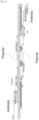
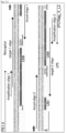
- DNA barcodes used in the method of the present invention include target 1 oligo and target 2 oligo as shown in Figs. 2-1 and 2-2 .
- target 1 oligo and target 2 oligo the sequences for distinguishing different antibody types or for distinguishing different antibody types and different DNA barcode types are the sequences in the regions indicated by "antibody barcode," and the sequences for distinguishing individual antibody molecules are the sequences in the regions indicated by "UMI.”
- the sequences for distinguishing individual antibody molecules are preferably designed not to include a contiguous sequence of a single type of nucleotides. UMI (Unique Molecular Identifier) can be designed based on, for example, a previous report ( J. A. Weinstein et al., Cell, 2019, 178:229-241 .e1-e10).
- the length (the number of nucleotide residues) of the sequences for distinguishing different antibody types or for distinguishing different antibody types and different DNA barcode types, and the length (the number of nucleotide residues) of the sequences for distinguishing individual antibody molecules are not limited to a particular one, and may be designed as appropriate.
- the DNA barcodes are preferably designed such that the nucleotide length of an amplification product containing two joined DNA barcodes is shorter than the maximal read length of the sequencer used.
- the DNA barcodes may be designed to contain adapter sequences appropriate for the sequencer used.
- the 3' regions of the DNA barcodes preferably contain a sequence to which the primers used in step (e) hybridize.
- Labeling of the antibody molecules with the DNA barcodes may be done by any method that is well-known to a person skilled in the art.
- a functional group such as an amino acid, an aldehyde group, an SH group or biotin
- a functional group that can react to the oligonucleotide such as an aldehyde group, an amino group, an SH group, or streptavidin is introduced into the antibodies, and then the functional groups are allowed to covalently interact with each other to form crosslinks, thereby labeling the antibodies with the DNA barcodes.
- the antibodies may be labeled with the DNA barcodes using a commercially available crosslinking agent (crosslinker) or a commercially available labeling kit.
- crosslinker a commercially available crosslinking agent
- labeling kit is labeling by biotin-streptavidin binding.
- labeling is performed such that the antibodies are bound to the 5' end of the DNA barcodes.
- a biological sample containing the analytical target factors is allowed to contact with the antibody molecules labeled with the DNA barcodes.
- the contact between the biological sample and the DNA barcode-labeled antibody molecules may be done by any method, including, for example, but not limited to, immersing the biological sample in an antibody solution; layering an antibody solution over the biological sample; adding the antibodies to a culture system of the biological sample; or other methods.
- the biological sample may be loaded onto a solid support and allowed to contact with the antibodies.
- the solid support may be any solid support that can be loaded with a biological molecule, and examples of such solid support include a filter, a membrane, a microwell plate, a bead, a slide, etc.
- the solid support may be made of a material such as glass, a plastic, a fluororesin, ceramics, a metal, silica, methylstyrene, latex, Sepharose, agarose, cellulose, a polymer material, etc. Preferred are a glass bead or a glass slide among them, preferably a glass slide.
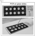
- the glass slide When a glass slide is used as a solid support, the glass slide is preferably capable of allowing culture of the biological sample and/or retaining the biological sample thereon and is then capable of using for a nucleic acid amplification reaction.
- the glass slide When the PCR method is used in step (e), the glass slide preferably has a size appropriate for a PCR device used.
- the glass slide may be provided with coating of, for example, APS, MAS or poly-L-lysine on the surface.
- the glass slide may be provided with special printing using silver stain, an ultraviolet curing ink, a fluororesin ink, etc., or provided with hydrophobic printing etc.
- the glass slide when used in the method of the present invention, may be used together with a chamber, such as a culture medium chamber or a frame-seal chamber.
- the cells may be permeated before contact with the antibodies.
- the cells are preferably permeated before contact with the antibodies.
- Permeation of the sample may be done by any method that is well-known to a person skilled in the art.
- the biological sample may be fixed in a fixative, such as formaldehyde, and then the cells may be permeated with a buffer solution containing a surfactant, such as TritonX-100.
- a reagent that is capable of simultaneously performing fixation and permeation may be added to an unfixed biological sample.
- the reagent that is capable of simultaneously performing fixation and permeation may be a cold alcohol etc.
- Blocking for preventing a non-specific antibody reaction may be performed on the biological sample before contact with the antibodies. Blocking of the sample may be done by any method that is well-known to a person skilled in the art.
- the biological sample that has been contacted with the antibodies may be post-fixed. Post-fixation may be performed in a known fixative such as formalin (aqueous formaldehyde solution). If necessary, the biological sample that has been contacted with the antibodies may be contacted with the antibodies again. For example, the biological sample contacted with the antibodies without permeation may be permeated, and then contacted with the antibodies again to allow analysis of both of factors localized on the surface of the cell membrane and factors localized within the cells as analytical targets.
- a known fixative such as formalin (aqueous formaldehyde solution).
- the DNA barcode-labeled antibody molecules may first be subjected to fluorescent labeling, enzyme labeling, etc., and then contacted with the biological sample.
- the DNA barcode-labeled antibody molecules may first be contacted with the biological sample, and then contacted with secondary antibodies, such as fluorescence-labeled antibodies or enzyme-labeled antibodies.
- secondary antibodies such as fluorescence-labeled antibodies or enzyme-labeled antibodies.
- the DNA barcode-labeled antibody molecules may first be contacted with the biological sample loaded on a glass slide, and then immunostained with fluorescence-labeled antibodies, and subjected to the following step (d).
- step (d) the biological sample is embedded in a hydrogel, and observed under a fluorescent microscope to capture an image, and then a nucleic acid amplification reaction is performed in step (e) to simultaneously obtain image information, such as the morphology of the cells or the localization of the target factors, and the nucleotide sequence information of the amplification products.
- step (d) the biological sample that has been contacted with the DNA barcode-labeled antibodies in step (c) is embedded in a hydrogel.
- the hydrogel used in the method of the present invention is not limited to a particular one.
- the hydrogel may be, for example, a swelled body formed by water-induced swelling of a three-dimensional network of crosslinked polymer chains, or may be a hydrogel formed by water-induced swelling of a structure formed by crosslinking between different molecules of polymers, such as water-soluble polysaccharides or proteins.
- the polymers are not limited to a particular one, and may be a naturally occurring polymer, a synthetic polymer, or a combination thereof.
- hydrogel examples include collagen, hyaluronic acid, chitosan, fibrin, gelatin alginate, agarose, chondroitin sulfate, polyacrylamide, polyethylene glycol (PEG), polyvinyl alcohol (PVA), polyacrylamide/polyacrylic acid (PAA), hydroxyethyl methacrylate (HEMA), poly(N-isopropylacrylamide) (NIPAM), a polyanhydride, polypropylene fumarate (PPF), etc.
- PEG polyethylene glycol
- PVA polyvinyl alcohol
- PAA polyacrylamide/polyacrylic acid
- HEMA hydroxyethyl methacrylate
- NIPAM poly(N-isopropylacrylamide)
- PPF polypropylene fumarate
- PEG polypropylene fumarate
- the hydrogel is a combination of multi-arm PEG and thiol-group modified PEG. More preferably, the hydrogel is a combination of 4-Arm PEG and 2SH PEG. Both of 4-Arm PEG and 2SH PEG are commercially available from suppliers. For example, when a combination of 4-Arm PEG and 2SH PEG is used in the method of the present invention, a 4-Arm PEG solution and a 2SH PEG solution can separately be prepared in ultrapure water each at 500 ⁇ g/ ⁇ L, and the solutions can then be combined to initiate gelation to prepare a hydrogel.
- step (c) a small number of cells may be loaded on a glass slide, and a biological sample that has been contacted with the DNA barcode-labeled antibodies may be embedded in a hydrogel. Then, after the location of the cells are identified, a single cell may be recovered together with the hydrogel, and subjected to the following step (e). In this manner, multifactorial interactions in a single cell can be identified.
- a nucleic acid amplification reaction is performed within the hydrogel by using a plurality of primers designed such that, when two antibodies come in close proximity, the primers produce an amplification product containing both DNA barcode sequences from the antibodies.
- the nucleic acid amplification reaction used in step (e) may be any nucleic acid amplification reaction that can produce such an amplification product, and may be selected from known nucleic acid amplification reactions.
- nucleic acid amplification reaction examples include PCR (Polymerase Chain Reaction) method, LAMP (Loop-Mediated Isothermal Amplification) method, WGA (Whole Genome Amplification) method, MDA (Multiple Displacement Amplification) method, HDA (Helicase-dependent amplification) method, TRC (Transcription Reverse Transcription Concerted Reaction) method, RPA (Recombinase Polymerase Amplification) method, SIBA (Strand-Invasion Based Amplification) method, ICAN (Isothermal and Chimeric primer-initiated Amplification of Nucleic acids) method, LCR (Ligase Chain Reaction) method, SDA (Strand Displacement Amplification) method, NEAR (Nicking Endonuclease Amplification Reaction) method, SMAP (Smart amplification process) method, 3SR (Self-stranded sequence replication) method, NASBA (Nucleic Acid Sequence-Based Amplification) method, TMA (Transcription-
- the nucleic acid amplification reaction is performed using primers designed such that, when two antibodies are come in close proximity, the primers produce an amplification products containing both DNA barcode sequences from the antibodies.
- the primers are designed and used as appropriate for the nucleic acid amplification reaction used.
- primers may be designed to join the DNA barcodes from two antibodies that come in close proximity based on the overlap extension PCR method, and then used for the reaction.
- Yolov et al. A.A. Yolovand Z.A.
- the primers preferably contain a primer set for joining the DNA barcode sequences of two antibodies that come in close proximity, and a primer set for amplifying the joined DNA barcode sequences.
- the primers of the primer set for joining the DNA barcode sequences of two antibodies that come in close proximity preferably contain a 5' end sequence that hybridizes to its paired primer, a 3' end sequence that hybridizes to a 3' region of one of the DNA barcode sequences, and a sequence for distinguishing individual primer molecules.
- the primers of the primer set for amplifying the joined DNA barcode sequences preferably contain a sequence that hybridizes to a 5' region of one of the DNA barcode sequences.
- the amplification primers may be designed to add adapter sequences to the ends of amplification products.
- primers used in the method of the present invention include Acryd-oligo 1 and Acryd-oligo 2 (joining primers), as well as amplification oligo 1 and amplification oligo 2 (amplification primers), as shown in Figs. 2-1 and 2-2 .
- the sequences distinguishing individual primer molecules are the sequences in the regions indicated by "UEI.”
- UEI Unique Event Identifier
- step (e) when the produced amplification product contains two joined DNA barcode sequences, the amplification product contains two UEIs each from each of the joining primers.
- the joining primers are preferably immobilized on a polymer that forms the hydrogel.
- immobilizing the joining primers to the polymer multifactorial interactions can be identified while their spatial information is maintained.
- Immobilization of the joining primers to the polymer can be done by, for example, adding the joining primers having Acridite modification at the 5' end to a polymer solution, and gelling the polymer solution to prepare a hydrogel, but the immobilization method is not limited to this, and may be done by a technique well-known to a person skilled in the art.
- the joining primers and the amplification primers are preferably designed such that the nucleotide length of an amplification product is shorter than the maximal read length of the sequencer used.
- Such an amplification product with a nucleotide length shorter than the maximal read length of a sequencer can be subjected to sequencing analysis for identification of interactions in a single read.
- the joining primers and the amplification primers are preferably designed such that the nucleotide length of an amplification product containing two DNA barcodes is 150 bp or less.
- the nucleic acid amplification reaction can be performed based on a known technique of the nucleic acid amplification reaction used.
- the reagents, the instruments, and others to be used can be commercially available products.
- the nucleic acid amplification reaction is the PCR method
- a reaction solution is added to the biological sample that is embedded in a hydrogel to initiate the reaction.
- the PCR reaction solution contains a nucleic acid synthase (DNA polymerase), nucleotides serving as substrates of the nucleic acid synthase, primers other than the primers immobilized to the hydrogel.
- the reaction solution also contains a buffering agent, a salt, etc., and if needed, further contain a protective agent for the enzyme, a modifier of Tm, a surfactant, etc.
- a specific composition of the reaction solution is preferably determined as appropriate by performing preliminary examinations on a particular combination of the biological sample, the DNA barcode-labeled antibodies, the primers, the nucleic acid synthase, etc. used.
- the PCR method when used in the method of the present invention, may be performed by the common three step method (denaturation ⁇ annealing ⁇ elongation cycles), or the two step method (denaturation ⁇ annealing/elongation cycles).
- the reaction conditions including the denaturation temperature, the annealing temperature, the elongation temperature, and the reaction time of each step are preferably determined as appropriate by performing preliminary examinations on a particular combination of the biological sample, the DNA barcode-labeled antibodies, the primers, the nucleic acid synthase, etc. used.
- the extract may be examined whether the amplification product exists.
- Detection of the amplification product may be performed by any common method that can detect a nucleic acid, and the method may be a qualitative detection method or a quantitative detection method.
- a detection method may be, for example, agarose gel electrophoresis etc.
- the amplification product may be purified.
- the purification may be done by any method appropriately selected from known purification methods.
- a commercially available nucleic acid purification kit may be used.
- adapter sequences need to be added to the amplification product.
- the adapter sequences can be added by any method, and may be added by, for example, performing PCR using the amplification product as a template and using a primer set designed to add the adapter sequences.
- the size of the amplification product with added adapter sequences is preferably within the read length of a next-generation sequencer used.
- the obtained nucleotide sequence data may be analyzed using variety of programs developed for sequence data processing. Such programs may be, for example, fastq_quality_trimmer, umi_tools, igraph, etc.
- sequence data processing may be, for example, fastq_quality_trimmer, umi_tools, igraph, etc.
- the obtained sequence data are typically subjected to processing steps, including import of the data aggregates from the sequencer, trimming of unnecessary sequence data such as the adapters and the primers, removing of low quality sequence regions, assembly of the reads, etc.
- Presence of a factor that simultaneously interacts with two or more factors in the method of the present invention can be detected by discovering an amplification product that contains two DNA barcode sequences, one of which is the same as the DNA barcode sequence for detecting the factor, and the other one is different.
- Whether the amplification product is resulted from the joining of the DNA barcodes of the antibodies that come in close proximity, i.e., whether the amplification product reflects the interaction of two factors can be determined by the presence of the sequences for distinguishing two individual antibody molecules contained in each DNA barcode (e.g., UMI sequences) and the sequences for distinguishing two individual primer molecules contained in each joining primer (e.g., UEI sequences).
- Whether the DNA barcode sequences are the same or different can be determined by determining whether the sequences for distinguishing individual antibody molecules (e.g., UMI sequences) are the same or different.
- the method of the present invention can be used to identify multifactorial interactions among factors A, B, C and D by detecting whether factor A simultaneously interacts with two or more of B, C and D, whether factor B simultaneously interacts with two or more of A, C and D, whether factor C simultaneously interacts with two or more of A, B and D, or whether factor D simultaneously interacts with two or more of A, B and C.
- the method of the present invention can be used for, for example, functional assessment of tissues, which cannot be evaluated by a conventional histological staining or genome diagnosis.
- the results obtained from the method of the present invention can be used for prediction of the therapeutic effect of various medicines, clustering of clinical cases, or the like, in clinical diagnosis.
- the present invention provides a kit for performing the method of the present invention as described above (also referred to as the "kit of the present invention” below).
- the kit of the present invention contains DNA barcode-labeled antibodies against a plurality of analytical target factors to be subjected to identification of multifactorial interactions; primers designed to produce an amplification product formed from joined DNA barcode sequences from two antibodies by a nucleic acid amplification reaction; and a hydrogel.
- the DNA barcode-labeled antibodies, the primers and the hydrogel contained in the kit of the present invention may be those described above regarding the embodiments of the method of the present invention.
- the kit of the present invention may further contain a DNA polymerase required for a nucleic acid amplification reaction, a deoxyribonucleoside triphosphate (dNTP) mixture, and a primer set for adding adapters for next-generation sequencing.
- dNTP deoxyribonucleoside triphosphate
- the kit of the present invention may further contain a buffer solution.
- the buffer solution may be, for example, but is not limited to, a buffer solution for preparation of a reagent, a buffer solution for a nucleic acid amplification reaction, a buffer solution for collecting an amplified DNA, etc.
- the kit of the present invention may further contain a reagent for purifying an amplified DNA.
- the kit of the present invention may further contain various types of tubes for preparation of a reagent, a solid support such as a glass bead or a glass slide, instructions for use, etc.
- the kit of the present invention can be used to perform the method for identifying multifactorial interactions according to the present invention in an easy and rapid manner when specific analytical target factors of interest are known.
- the kit of the present invention may be implemented as a kit of custom-made.
- the kit according to the users' purpose of use can be provided by designing the DNA barcode-labeled antibodies and the primer set as appropriate for the purpose of use.
- DNA barcode-conjugated antibodies as schematically shown in Fig. 1 , DNA barcodes having antibody barcode sequences (sequences for distinguishing different antibody types), UMI sequences (sequences for distinguishing individual antibody molecules), and adapter sequences for sequencing were designed for each of LPS and factors that are known to interact with LPS, including Caspase-11, CD14 and TLR4.
- These DNA barcodes indicated as target 1 oligo and target 2 oligo in Figs. 2-1 and 2-2 , were synthesized by a known technique, and the 5' end was biotinylated by a conventional method.
- Each of the biotinylated DNA barcodes was diluted in Tris-EDTA buffer to 100 ⁇ M, heated at 55°C for 10 minutes, and centrifuged at 18000xg at room temperature for 5 minutes. The concentration in the supernatant was checked and adjusted again, if necessary, to 100 ⁇ M.
- the sequences of the DNA barcodes including the UMI sequences and the antibody barcode sequences are shown below. The antibody barcode sequences are underlined.
- the solvent of the antibodies below was exchanged with PBS using a centrifugal filter (trade name: Amicon Ultra 0.5, Merck, Cat. No. UFC505096) following the manufacturer's protocol.
- the antibodies used were as follows: anti-LPS antibody (mouse monoclonal antibody, Abcam, Cat. No. ab35654), anti-CD14 antibody (mouse monoclonal antibody, BioLegend, Cat. No. 123302), anti-TLR4 antibody (mouse monoclonal antibody, BioLegend, Cat. No. 145401), and anti-Caspase 11 antibody (rat monoclonal antibody, SIGMA-Aldrich, Cat. No. C1354).
- the antibody concentrations were adjusted to 15 ⁇ g in 30 ⁇ L.
- the antibodies were then labeled with streptavidin using a commercially available labeling kit (trade name: LYNX Rapid Streptavidin Antibody Conjugation Kit, BIO-RAD, Cat. No. LNK163STR). The labeling was performed following the manufacturer's protocol.
- the streptavidin labeled-antibodies were allowed to bind to the corresponding biotinylated DNA barcodes to form conjugates. Briefly, 3 ⁇ L of quenching solution (included in the labeling kit above) was added to vials containing one of the streptavidin-labeled antibodies, and allowed to stand at room temperature for 30 minutes to quench the reaction. Then, 5 M NaCl and 0.1% Tween20/H 2 O, at 4 ⁇ L each, were added. The total volume was transferred to a fresh low-binding microtube, and 800 pmol of the corresponding biotinylated DNA barcode was added and reacted at room temperature overnight.
- quenching solution included in the labeling kit above
- anti-Caspase 11 antibody-target 1 oligo (5biotin)_BC28 SEQ ID NO: 2
- anti-CD14 antibody-target 1 oligo 5biotin_BC24
- anti-TLR4 antibody-target 1 oligo 5biotin_BC26
- anti-LPS antibody-target 2 oligo 5biotin_BC1
- the solvent of the solutions containing one of the DNA barcode-labeled antibodies after the reaction was exchange with PBS using a centrifugal filter (Amicon Ultra 0.5, Merck) by a conventional method, and the total volume was adjusted to 30 ⁇ L with PBS.
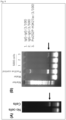
- the DNA barcode-labeled antibodies prepared as above were run by electrophoresis on a 4% TAE gel for 10 minutes. The gel was then treated with ethidium bromide for 10 minutes to visualize the bands to examine the conjugation of the DNA barcodes. As controls, 50 pmol of the DNA barcodes were run, and the relative band intensities were determined for all bands appeared on the lanes of the DNA barcode-labeled antibody samples by image analysis. The amounts of the DNA barcodes in the prepared DNA barcode-labeled antibodies were estimated. Samples for which successful conjugation of the DNA barcodes was confirmed were stored at 4°C after addition of 50 mg/mL BSA and 1% sodium azide at final concentrations of 1 ⁇ g/ ⁇ L and 0.05%, respectively.
- Purified mouse B cells were suspended in serum-free medium, and seeded in the prepared PCR tubes as described above by adjusting the density to 5000 cells/50 ⁇ L per tube.
- the cells were cultured in 5% CO 2 at 37°C for 30 minutes, and the culture supernatant was removed.
- 1 mg/mL LPS and 2.5 ⁇ g/mL IL-4 were diluted in RPMI 1640 medium to final concentrations of 50 ⁇ g/mL and 2.5 ng/mL, respectively, and added to the tubes at 50 ⁇ L each.
- the cells were cultured in 5% CO 2 at 37°C for 20 minutes to 48 hours. IL-4 was used as stimulation for B-cell activation.
- the compositions of the serum-free medium and RPMI 1640 medium are as follows:
- the cells cultured as described above were washed 3 times with PBS, and 50 ⁇ L of 4% paraformaldehyde/PBS was added to each tube to fix the cells at room temperature for 10 minutes.
- the cells after fixation were washed 3 times with PBS, and 50 ⁇ L of 0.2% Triton X-100/PBS was added to each tube and allowed to react at room temperature for 10 minutes to permeate the cells.
- the cells after permeation were washed 3 times with PBS, and blocked to prevent non-specific binding of the antibodies to Fc-receptors.
- the blocking was performed by adding, to each tube, 50 ⁇ L of 2% BSA/PBS solution containing 0.1 ⁇ L of TruStain FcX (anti-mouse CD16/32) Antibody (Clone 93 Isotype Rat IgG2a, ⁇ (trade name), BioLegend, Cat. No. 101320) and incubated on ice for 10 minutes.
- the supernatant was removed from each tube containing the cells, and 50 ⁇ L of 10% BSA/PBS was added to each tube and allowed to stand at room temperature for 1 hour to block the whole surface of the cells. Then, 25 ⁇ L of a mixture of the DNA barcode-labeled antibodies prepared in the above 1-1 or a mixture of the DNA barcodes was added to each tube, and allowed to contact with the cells at 4°C for 30 minutes. The cells were washed 3 times with PBS, and 50 ⁇ L of 4% paraformaldehyde/PBS was added to each tube and allowed to stand at room temperature for 10 minutes for post-fixation. The post-fixed cells were washed 3 times with PBS, and stored at 4°C with addition of 200 ⁇ L of PBS.
- joining PCR was performed following a previous report ( J. A. Weinstein et al., Cell, 2019, 178:229-241.e1-e10 ).
- 4arm PEG and 2SH PEG were prepared as raw materials of a hydrogel. Since these two types of PEGs, when combined together, form a gel within about 2 minutes, 4arm PEG and 2SH PEG were separately prepared in ultrapure water in a 5 mL tube to a final concentration of 500 ⁇ g/ ⁇ L.
- the required volume of each PEG was determined to be 20 ⁇ L per specimen. Specifically, at least 10 mg of powder PEG as a required amount was taken in a tube, and centrifuged at 5000 rpm at room temperature for 1 minute.
- Ultrapure water was added in an amount equivalent to the powder, and centrifuged at 5000 rpm at room temperature for 1 minute.
- the tube was mixed with inversion to allow the powder to be in contact with water, and centrifuged again. Inversion mixing and centrifugation were repeated until the solution became transparent.
- the liquid volume was measured with a pipette tip, and ultrapure water was added again so that the volume added was 2-fold of the initial water volume.
- the PEG solution was vortexed for homogeneous mixing, and centrifuged at 5000 rpm at room temperature for 1 minute.
- DNA barcodes having antibody barcode sequences, UMI sequences and adapter sequences for sequencing were designed for factors known to interact with each other after LPS stimulation (phosphorylated RNA polymerase II-CTD (Pol2S2P), Lamin B1, c-Myc, acetylated histone (H3K27ac), and p300) and IgG control, and the DNA barcodes were synthesized by a known technique in the same manner as in Example 1.
- the 5' ends of the DNA barcodes were biotinylated by a conventional method, and the DNA barcodes were diluted in Tris-EDTA buffer to 100 ⁇ M, heated at 55°C for 10 minutes, and centrifuged at 18000xg at room temperature for 5 minutes. The concentration in the supernatant was checked and adjusted again, if necessary, to 100 ⁇ M.
- the sequences of the DNA barcodes are as follows. The antibody barcode sequences are underlined.
- anti-Phospho-RNA pol II CTD (Ser2) antibody (Pol2S2P, rabbit monoclonal antibody, clone 7H23L1, Thermo-Fisher, Cat. No. 703108)
- anti-Lamin B1 antibody (mouse monoclonal antibody, clone L-5, Thermo-Fisher, Cat. No. 33-2000)
- anti-c-Myc antibody (mouse monoclonal antibody, clone 9E10, Thermo-Fisher, Cat. No.
- H3K27ac mouse monoclonal antibody, clone GT261, Thermo-Fisher, Cat. No. MA5-31555
- anti-p300 antibody mouse monoclonal antibody, clone RW128, Thermo-Fisher, Cat. No. MA1-16608
- IgG mouse, BioLegend, Cat. No. 400101
- DNA barcodes having antibody barcode sequences, UMI sequences and adapter sequences for sequencing were designed for factors known to interact with each other after LPS stimulation and IgG control, and the DNA barcodes were synthesized by a known technique in the same manner as in Example 2.
- the 5' ends of the DNA barcodes were biotinylated by a conventional method, and the DNA barcodes were diluted in Tris-EDTA buffer to 100 ⁇ M, heated at 55°C for 10 minutes, and centrifuged at 18000xg at room temperature for 5 minutes. The concentration in the supernatant was checked and adjusted again, if necessary, to 100 ⁇ M.
- the cells stimulated with LPS as described above were washed 3 times with PBS, and 50 ⁇ L of 4% paraformaldehyde/PBS was added to each well to fix the cells at room temperature for 10 minutes. Ater fixation of the cells, 30 ⁇ L of 0.2% Triton X-100/PBS was added to each well and allowed to react at room temperature for 10 minutes to permeate the cells.
- Fig. 11A shows the analytical results of the number of bindings of the anti-acetylated histone (H3K27ac) antibody molecules per UEI over time.
- the vertical axis indicates the UEI counts, the horizontal axis indicates the number of the anti-H3K27ac antibody molecules bound to a single UEI.
- H3K27ac acetylated histone
- the number of bindings of antibody molecules per UEI was also analyzed for 16 types of analytical target factors, and the results are shown in Fig. 12 . Correlation intensities are indicated by color scales, in which blue represents a correlation coefficient of zero, red represents a correlation coefficient of 1, and white represents a correlation coefficient of a mid-value (0.5).
- the dot size indicates the number of antibody bindings per UEI. These results indicate that the number of antibody bindings per UEI increases over time after LPS stimulation.
- the coefficients of positive correlations between the analytical target factors as indicated by the color scale indicate that almost all dots were blue immediately after LPS stimulation, but most of the dots turned into red 48 hours later. These results demonstrate that the correlations with other factors are enhanced in many of the analytical target factors over time after LPS stimulation.
- HLA-DR membrane protein
- HLA-A/B/C HLA-A/B/C
Landscapes
- Health & Medical Sciences (AREA)
- Chemical & Material Sciences (AREA)
- Immunology (AREA)
- Life Sciences & Earth Sciences (AREA)
- Organic Chemistry (AREA)
- Molecular Biology (AREA)
- General Health & Medical Sciences (AREA)
- Biochemistry (AREA)
- Medicinal Chemistry (AREA)
- Proteomics, Peptides & Aminoacids (AREA)
- Biophysics (AREA)
- Genetics & Genomics (AREA)
- Engineering & Computer Science (AREA)
- Urology & Nephrology (AREA)
- Hematology (AREA)
- Biomedical Technology (AREA)
- Analytical Chemistry (AREA)
- Pathology (AREA)
- Biotechnology (AREA)
- Physics & Mathematics (AREA)
- Microbiology (AREA)
- Cell Biology (AREA)
- General Physics & Mathematics (AREA)
- Food Science & Technology (AREA)
- Wood Science & Technology (AREA)
- Zoology (AREA)
- Bioinformatics & Cheminformatics (AREA)
- General Engineering & Computer Science (AREA)
- Measuring Or Testing Involving Enzymes Or Micro-Organisms (AREA)
- Oncology (AREA)
- Gastroenterology & Hepatology (AREA)
Applications Claiming Priority (2)
| Application Number | Priority Date | Filing Date | Title |
|---|---|---|---|
| JP2022068519 | 2022-04-18 | ||
| PCT/JP2023/015115 WO2023204147A1 (fr) | 2022-04-18 | 2023-04-14 | Procédé et kit pour identifier une interaction multifactorielle dans un échantillon biologique |
Publications (2)
| Publication Number | Publication Date |
|---|---|
| EP4512896A1 true EP4512896A1 (fr) | 2025-02-26 |
| EP4512896A4 EP4512896A4 (fr) | 2025-10-15 |
Family
ID=88419840
Family Applications (1)
| Application Number | Title | Priority Date | Filing Date |
|---|---|---|---|
| EP23791795.0A Pending EP4512896A4 (fr) | 2022-04-18 | 2023-04-14 | Procédé et kit pour identifier une interaction multifactorielle dans un échantillon biologique |
Country Status (4)
| Country | Link |
|---|---|
| US (1) | US20250258165A1 (fr) |
| EP (1) | EP4512896A4 (fr) |
| JP (1) | JPWO2023204147A1 (fr) |
| WO (1) | WO2023204147A1 (fr) |
Families Citing this family (1)
| Publication number | Priority date | Publication date | Assignee | Title |
|---|---|---|---|---|
| US20250095385A1 (en) * | 2023-09-20 | 2025-03-20 | Torc Robotics, Inc. | Associating detected objects and traffic lanes using computer vision |
Family Cites Families (6)
| Publication number | Priority date | Publication date | Assignee | Title |
|---|---|---|---|---|
| EP2332957B1 (fr) | 1996-02-09 | 2015-04-08 | Cornell Research Foundation, Inc. | Détection de différences entre séquences d'acide nucléique faisant appel à la réaction de détection par ligase et à des réseaux adressables |
| EP1090138A4 (fr) | 1998-06-24 | 2003-01-02 | Glaxo Group Ltd | Procede de detection de nucleotide |
| JP2002181813A (ja) | 2000-12-15 | 2002-06-26 | Olympus Optical Co Ltd | 核酸分子を用いた新規な情報解析方法およびそれを用いた核酸の解析方法 |
| GB201108678D0 (en) * | 2011-05-24 | 2011-07-06 | Olink Ab | Multiplexed proximity ligation assay |
| CN109791157B (zh) | 2016-09-26 | 2022-06-07 | 贝克顿迪金森公司 | 使用具有条形码化的寡核苷酸序列的试剂测量蛋白质表达 |
| JP7502787B2 (ja) * | 2018-04-09 | 2024-06-19 | ザ ボード オブ トラスティーズ オブ ザ レランド スタンフォード ジュニア ユニバーシティー | インサイチュ遺伝子配列決定の方法 |
-
2023
- 2023-04-14 EP EP23791795.0A patent/EP4512896A4/fr active Pending
- 2023-04-14 US US18/857,084 patent/US20250258165A1/en active Pending
- 2023-04-14 JP JP2024516235A patent/JPWO2023204147A1/ja active Pending
- 2023-04-14 WO PCT/JP2023/015115 patent/WO2023204147A1/fr not_active Ceased
Also Published As
| Publication number | Publication date |
|---|---|
| US20250258165A1 (en) | 2025-08-14 |
| JPWO2023204147A1 (fr) | 2023-10-26 |
| WO2023204147A1 (fr) | 2023-10-26 |
| EP4512896A4 (fr) | 2025-10-15 |
Similar Documents
| Publication | Publication Date | Title |
|---|---|---|
| EP4158054B1 (fr) | Trancriptomique spatiale pour les récepteurs d'antigènes | |
| Matuła et al. | Single‐cell analysis using droplet microfluidics | |
| Konig et al. | iCLIP-transcriptome-wide mapping of protein-RNA interactions with individual nucleotide resolution | |
| EP2479268B1 (fr) | Procédé de dissociation et agent de dissociation pour avidine et dérivés de biotine | |
| US12428673B2 (en) | Methods for barcoding macromolecules in individual cells | |
| KR20150132479A (ko) | 복합 유전자 세트에서의 독특하게 태그되고 재배열된 적응 면역 수용체 유전자 | |
| IL291859A (en) | A high-throughput method for screening t-cell cognate and epitope reactivity in primary human cells | |
| CN103703143A (zh) | 鉴定细胞中的多个表位的方法 | |
| US20250230498A1 (en) | Spatially identifying nucleic acids that interact with proteins | |
| KR102190922B1 (ko) | 핵산 분리 방법 | |
| JP2013545472A (ja) | 単一細胞内の生体分子の同時検出 | |
| EP4512896A1 (fr) | Procédé et kit pour identifier une interaction multifactorielle dans un échantillon biologique | |
| Devulapally et al. | Simple paired heavy-and light-chain antibody repertoire sequencing using endoplasmic reticulum microsomes | |
| WO2006049289A1 (fr) | Procede de detection de substances cibles | |
| US11280789B2 (en) | Compositions, kits, and methods for cell separation | |
| US9085773B2 (en) | Methods for identifying nucleic acid ligands | |
| CN115485392A (zh) | 用于鉴别配体阻断性抗体及用于确定抗体效价的方法 | |
| US11279971B2 (en) | In situ cell analysis in cell culture system | |
| CN104088019A (zh) | 一种基于双分子荧光互补技术的肽适配子文库构建方法 | |
| WO2021249816A1 (fr) | Procédé d'analyse unicellulaire d'échantillons multiples | |
| CN120210186A (zh) | 脂质修饰的寡核苷酸及其使用方法 | |
| US20250163406A1 (en) | Methods for generating nucleic acid encoded protein libraries and uses thereof | |
| WO2026035501A1 (fr) | Procédé de hachage spatial à base de particules | |
| EP2981616B1 (fr) | Procédé de transfection en phase solide de protéines et d'acides nucléiques | |
| CN115997125A (zh) | 鉴定配体和受体的同源对的方法 |
Legal Events
| Date | Code | Title | Description |
|---|---|---|---|
| STAA | Information on the status of an ep patent application or granted ep patent |
Free format text: STATUS: THE INTERNATIONAL PUBLICATION HAS BEEN MADE |
|
| PUAI | Public reference made under article 153(3) epc to a published international application that has entered the european phase |
Free format text: ORIGINAL CODE: 0009012 |
|
| STAA | Information on the status of an ep patent application or granted ep patent |
Free format text: STATUS: REQUEST FOR EXAMINATION WAS MADE |
|
| 17P | Request for examination filed |
Effective date: 20241115 |
|
| AK | Designated contracting states |
Kind code of ref document: A1 Designated state(s): AL AT BE BG CH CY CZ DE DK EE ES FI FR GB GR HR HU IE IS IT LI LT LU LV MC ME MK MT NL NO PL PT RO RS SE SI SK SM TR |
|
| DAV | Request for validation of the european patent (deleted) | ||
| DAX | Request for extension of the european patent (deleted) | ||
| A4 | Supplementary search report drawn up and despatched |
Effective date: 20250915 |
|
| RIC1 | Information provided on ipc code assigned before grant |
Ipc: C12N 15/09 20060101AFI20250909BHEP Ipc: C07K 16/18 20060101ALI20250909BHEP Ipc: C12M 1/00 20060101ALI20250909BHEP Ipc: C12M 1/34 20060101ALI20250909BHEP Ipc: C12Q 1/68 20180101ALI20250909BHEP Ipc: C12Q 1/6804 20180101ALI20250909BHEP Ipc: C12Q 1/6811 20180101ALI20250909BHEP Ipc: C12Q 1/6844 20180101ALI20250909BHEP Ipc: C12Q 1/686 20180101ALI20250909BHEP Ipc: C12Q 1/6869 20180101ALI20250909BHEP Ipc: C12Q 1/6876 20180101ALI20250909BHEP Ipc: G01N 33/532 20060101ALI20250909BHEP |